Page 1
8/4/2019 Chapter 7. Circulation
http://slidepdf.com/reader/full/chapter-7-circulation 1/54
Introduction
• Movement of body fluids inside the body of
animals to transport materials from the region offormation to the region of utilization or disposalis known as CIRCULATION.
• System of vessels and heart through whichblood flows in an animal is called CirculatorySystem
• William Harvey (1628) who discovered bloodcirculation including pumping activity of the
heart.
Page 2
8/4/2019 Chapter 7. Circulation
http://slidepdf.com/reader/full/chapter-7-circulation 2/54
Parts of Circulatory System
• Blood: It consisting of fluid plasma and freecells or blood corpuscles.
• Heart: Attached with muscular walls thatcontract periodically to pump the blood through
the body.• Arteries and Veins: A system of blood vessels
and capillaries through which the fluid bloodmoves.
Page 3
8/4/2019 Chapter 7. Circulation
http://slidepdf.com/reader/full/chapter-7-circulation 3/54
Medical Terms related to Circulation
• Angio-logy: Study of Blood Vascular
System including Arteries, Veins, andHeart.
• Cardio-logy: Study of heart and itsfunctioning.
• Hemato-logy: Study of formation,
composition, functions and diseases ofblood.
Page 4
8/4/2019 Chapter 7. Circulation
http://slidepdf.com/reader/full/chapter-7-circulation 4/54
Types of Circulatory System
• 1). Open Circulatory System:
– it is seen in many invertebrates (leeches, ARTHROPODS:
cockroaches, insects spiders butterflies, MOLLUSCS: jelly fish,snails, slugs and ascidians).
– Blood is pumped by the heart into a vessel which opens into
the open fluid spaces called SINUSES so that the tissues are
bathed by the blood which is known as HAEMO-LYMPH.
– From the sinus the blood is carried by the veins to the heart.
– There are no inter connecting vessels or capillaries between
the arteries and the veins, as the blood comes out of blood
vessels.
– Such system is called OPEN CIRCULATORY SYSTEM.
Page 5
8/4/2019 Chapter 7. Circulation
http://slidepdf.com/reader/full/chapter-7-circulation 5/54
Characteristics of Open Circulatory System
• 1) The blood flows at a very low velocity & at
low pressure due to the absence of smoothmuscles.
• 2) There is direct exchange of materials
between the cells & the blood because of the
direct contact between them.
• 3) The respiratory pigment, when present, is
dissolved in the plasma of the blood and
there are no red corpuscles.
Page 6
8/4/2019 Chapter 7. Circulation
http://slidepdf.com/reader/full/chapter-7-circulation 6/54
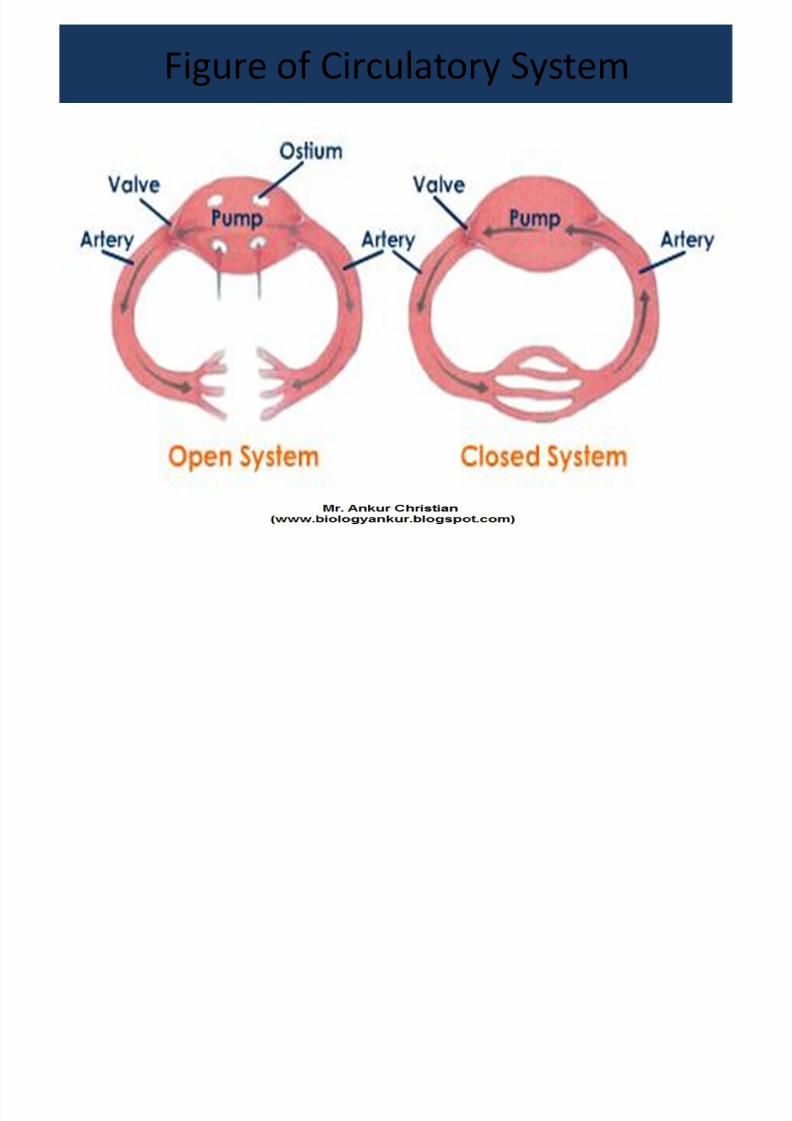
Figure of Circulatory System
Page 7
8/4/2019 Chapter 7. Circulation
http://slidepdf.com/reader/full/chapter-7-circulation 7/54
• 2). Closed Circulatory System:
– Such type of circulatory system is seen in annelids,
Cephalopods, holothurians and in all Vertebrates includingman.
– Blood is carried through a system of elastic tubes-arteries,
capillaries and veins. – The blood remains inside the blood vessels and does not
come out.
– The blood returns to the heart without actually leaving thissystem of blood vessels.
– Since blood remains in this closed system it is known asCLOSED CIRCULATORY SYSTEM.
– There are 3 types of closed circulatory system: 2 chamberedheart (fish) / 3 chambered heart (amphibians & reptiles) / 4chambered heart (man)
Page 8
8/4/2019 Chapter 7. Circulation
http://slidepdf.com/reader/full/chapter-7-circulation 8/54
Characteristics of Closed Circulatory
System• 1) The speed of circulation is more rapid due to the
presence of muscular and contractile blood
vessels.
• 2) The supply and removal of materials to and
from the tissues by the blood is enhanced, therebyincreasing the efficiency of circulation.
• 3) The volume of blood flowing through a tissue or
organ is regulated by the contraction and
relaxation of the muscles of the blood vessels.
Page 9
8/4/2019 Chapter 7. Circulation
http://slidepdf.com/reader/full/chapter-7-circulation 9/54
Functions of Blood• 1). Transportation:
– Transport O2 & CO2 between respiratory organs and tissues.
– Transport H2O & digested food from the digestive tract to other organs.
– Transport stored food from one organ / tissue to other target tissue ororgan.
– Transport metabolic wastes, excess minerals in solution and excess waterto the excretory organs.
– Transport hormones from the glands where they are produced to theplaces of use in the body.
– Transport antibodies for immunity or defense against foreign bodies, tomaintain normal health and protection from infection.
• 2). Regulation: It regulates the pH of the tissues by means of buffers.
• 3). Maintenance: It maintains the H2O balance between the tissues andexcretory system.
– It serves to maintain the temperature of the entire body within closelimits.
Page 10
8/4/2019 Chapter 7. Circulation
http://slidepdf.com/reader/full/chapter-7-circulation 10/54
Structure of Heart
• The heart is about the size of a fist & lies in the thoracic cavity.• The human heart has a mass of between 250 - 350 grams .
• It is a muscular organ covered by a tough fibrous membranethe pericardium.
• The pericardium consists of an outer, tough fibrouspericardium, which attaches to the diaphragm & also to thegreat vessels of the heart & an inner, delicate serouspericardium which is double membrane composed of an outerparietal layer & an inner visceral layer.
• Between this 2 layers is pericardial cavity filled with serousfluid or pericardial fluid which acts as lubricant & shockabsorber & protects the heart from being damage.
• The wall of the heart has 3 layers: the outer EPI-CARDIUM, themiddle MYO-CARDIUM & the inner ENDO-CARDIUM.
Page 11
8/4/2019 Chapter 7. Circulation
http://slidepdf.com/reader/full/chapter-7-circulation 11/54
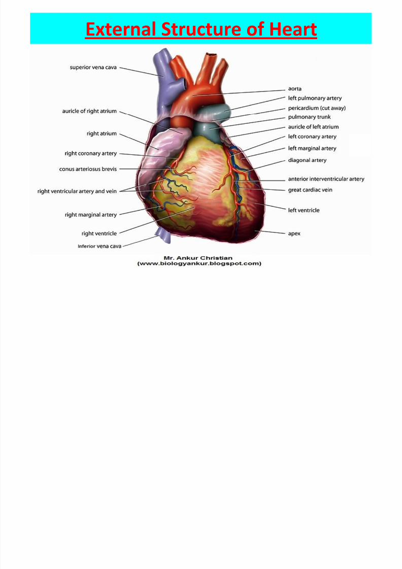
External Structure of Heart
Page 12
8/4/2019 Chapter 7. Circulation
http://slidepdf.com/reader/full/chapter-7-circulation 12/54
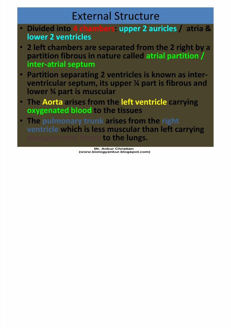
External Structure
• Divided into 4 chambers: upper 2 auricles / atria &lower 2 ventricles
• 2 left chambers are separated from the 2 right by a
partition fibrous in nature called atrial partition /inter-atrial septum
• Partition separating 2 ventricles is known as inter-
ventricular septum, its upper ¼ part is fibrous andlower ¾ part is muscular
• The Aorta arises from the left ventricle carryingoxygenated blood to the tissues
• The pulmonary trunk arises from the rightventricle which is less muscular than left carryingdeoxygenated blood to the lungs.
Page 13
8/4/2019 Chapter 7. Circulation
http://slidepdf.com/reader/full/chapter-7-circulation 13/54
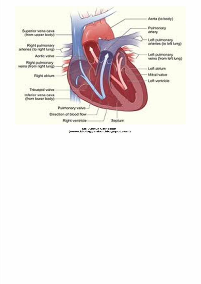
- The 3 veins (inferior + superior vena cava and coronary
sinus) open into the right atrium bringing venous blood ordeoxygenated blood from the lungs.
- The 4 chambers of the heart perform 4 different functions.
- The left ventricles pumps oxygenated blood to the tissues.
- In tissues, oxygen in the blood is used up and the blood
becomes deoxygenated.
- The deoxygenated blood is brought back to the heart
through veins which open into the right atrium.- It is then pumped into right ventricle, then into the lungs
through pulmonary trunk
- Blood inside the lungs becomes re-oxygenated and isreturned to the left atrium through the pulmonary veins
- From it then enters the left ventricle to be pumped out in
the circulation again.
Page 14
8/4/2019 Chapter 7. Circulation
http://slidepdf.com/reader/full/chapter-7-circulation 14/54
Page 15
8/4/2019 Chapter 7. Circulation
http://slidepdf.com/reader/full/chapter-7-circulation 15/54
Internal structure of Heart
Page 16
8/4/2019 Chapter 7. Circulation
http://slidepdf.com/reader/full/chapter-7-circulation 16/54
Page 17
8/4/2019 Chapter 7. Circulation
http://slidepdf.com/reader/full/chapter-7-circulation 17/54
Internal Structure
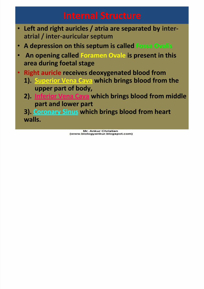
• Left and right auricles / atria are separated by inter-atrial / inter-auricular septum
• A depression on this septum is called Fossa Ovalis
• An opening called Foramen Ovale is present in thisarea during foetal stage
•Right auricle receives deoxygenated blood from1). Superior Vena Cava which brings blood from the
upper part of body,2). Inferior Vena Cava which brings blood from middle
part and lower part3). Coronary Sinus which brings blood from heartwalls.
Page 18
8/4/2019 Chapter 7. Circulation
http://slidepdf.com/reader/full/chapter-7-circulation 18/54
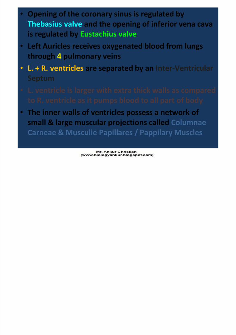
• Opening of the coronary sinus is regulated by
Thebasius valve and the opening of inferior vena cava
is regulated by Eustachius valve
• Left Auricles receives oxygenated blood from lungs
through 4 pulmonary veins• L. + R. ventricles are separated by an Inter-Ventricular
Septum
• L. ventricle is larger with extra thick walls as comparedto R. ventricle as it pumps blood to all part of body
• The inner walls of ventricles possess a network of
small & large muscular projections called Columnae
Carneae & Musculie Papillares / Pappilary Muscles
Page 19
8/4/2019 Chapter 7. Circulation
http://slidepdf.com/reader/full/chapter-7-circulation 19/54
• The wall of R. Ventricle contains a Moderator Band that extends between upper papillary muscles andinter-ventricular septum
• Auricles opens into Ventricles through Artio-Ventricular apertures controlled by valves
• The aperture between R. Auricle and R. Ventricle is
guarded by TRICUSPID valve having 3 flaps• The aperture between L. Auricle and L. Ventricle isguarded by BICUSPID valve / MITRAL valve having 2flaps
• These valve’s flaps are kept in position by inelasticChordae Tendinae connected to Papillary Muscles• L. Ventricle opens into the aorta by an opening
guarded by a ring of 3 semilunar valves.
• Similarly, R. Ventricle opens into pulmonary andthrough an opening guarded by a rising of semilunarvalves
Page 20
8/4/2019 Chapter 7. Circulation
http://slidepdf.com/reader/full/chapter-7-circulation 20/54
Miscellaneous
• Average heart beat in adult human is 72 perminute.
• SA node / SAN (Sinu-Auricular Node): it issituated on the right wall of R. Auricle abovethe opening of Superior Vena Cava. It is alsocalled the PACE-MAKER because Cardiacimpulse originates here.
• AV-node / Auriculo-Ventricular Node): It issituated between the R. Auricle and R. Ventriclenear inter-Atrial Septum. AV-node is connectedto Bundle of His / AV bundle. It is also calledPACE-SETTER.
• Purkinje Fibres: It originate from the bundle ofHis and enter into the walls of ventricle.
Page 21
8/4/2019 Chapter 7. Circulation
http://slidepdf.com/reader/full/chapter-7-circulation 21/54
Neurogenic
HeartMyogenic
Heart
Impulse for the heartbeat comes from a nervefrom a nearby ganglion.
Modified musculartissue or nodular tissuein heart muscles
initiates the electro-chemical impulse tocontrol heart beat.
e.g. Most Arthropodsand some Annelids
e.g. Molluscs andVertebrates
Page 22
8/4/2019 Chapter 7. Circulation
http://slidepdf.com/reader/full/chapter-7-circulation 22/54
Patterns of Circulation
• 2 patterns – 1). Systemic Circulation: it begins in the L. Ventricle & ends in R.Auricle
– 2). Pulmonary Circulation: It starts in the R. Ventricle & ends in
the L. Auricle• The right half of the heart is concerned with pumping
deoxygenated blood
• The left half of the heart is concerned with pumping of
oxygenated blood.• 2 sets of valves which regulate the flow of blood through the
heart which prevents the back flow of blood
– 1). Antrio-ventricular valves: It separates the cavities of Auricle &Ventricle
– 2). Semilunar valves: They are present where the PulmonaryArtery leaves the R. Ventricle and the Aorta leaves the L.Ventricle.
Page 23
8/4/2019 Chapter 7. Circulation
http://slidepdf.com/reader/full/chapter-7-circulation 23/54
Page 24
8/4/2019 Chapter 7. Circulation
http://slidepdf.com/reader/full/chapter-7-circulation 24/54
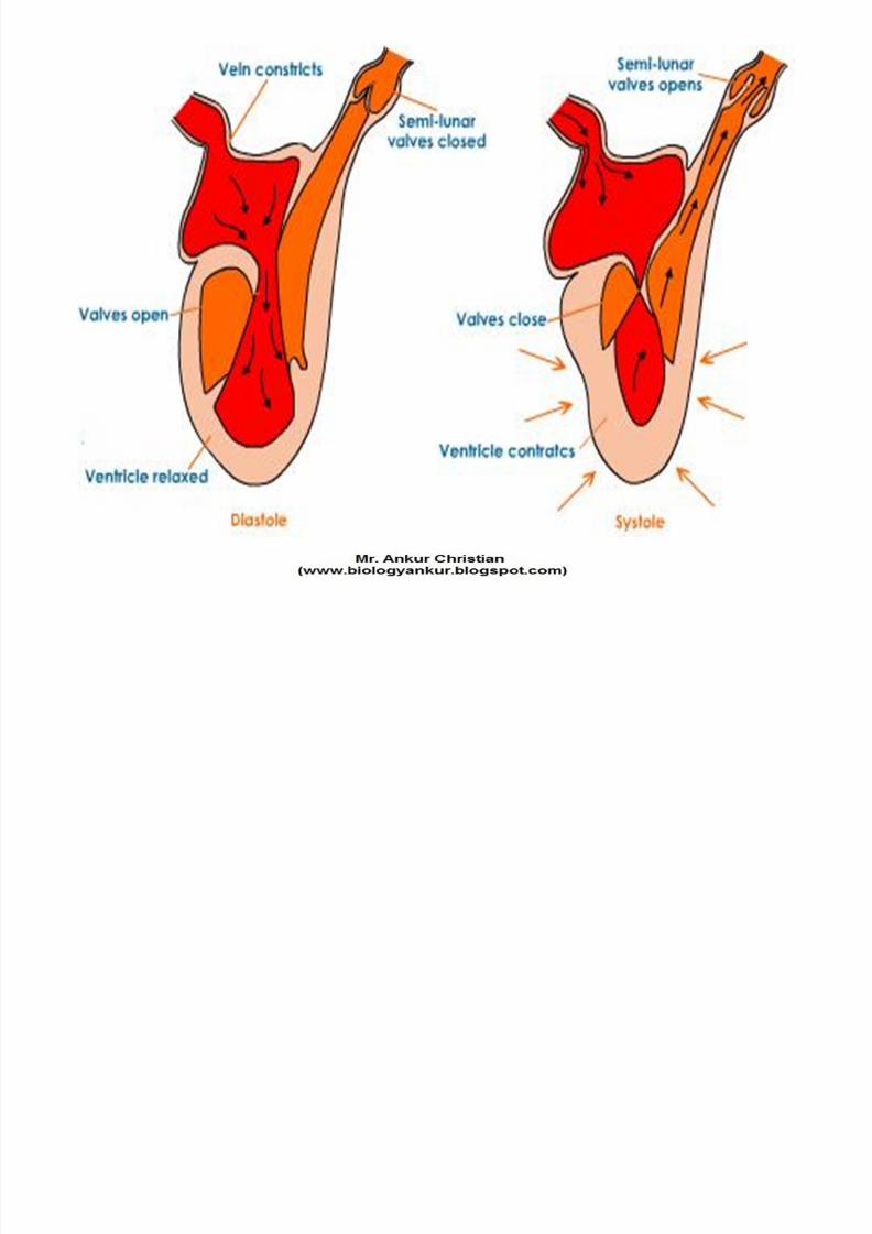
HEART BEAT:
The heart beat is the rhythmic contraction &
relaxation of all the muscles of heart• 1). Systole: the contraction of the heart is called
Systole• 2). Diastole: the relaxation of the heart is called
Diastole
• The heart rests only during the short intervalbetween contractions.
• The heart has an inbuilt capacity to contractrhythmically without any external stimulus.
Page 25
8/4/2019 Chapter 7. Circulation
http://slidepdf.com/reader/full/chapter-7-circulation 25/54
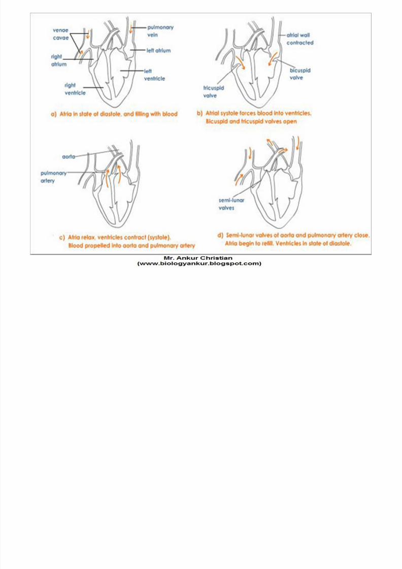
Cardiac Cycle- Event that occurs in
the heart during one
beat, is called heart
beat.
- One cardiac cycle
includes auricular
systole, ventricular
systole and complete
diastole.
Page 26
8/4/2019 Chapter 7. Circulation
http://slidepdf.com/reader/full/chapter-7-circulation 26/54
Page 27
8/4/2019 Chapter 7. Circulation
http://slidepdf.com/reader/full/chapter-7-circulation 27/54
Page 28
8/4/2019 Chapter 7. Circulation
http://slidepdf.com/reader/full/chapter-7-circulation 28/54
Page 29
8/4/2019 Chapter 7. Circulation
http://slidepdf.com/reader/full/chapter-7-circulation 29/54
Heart Sound
• Each heart beat has 2 sounds, Lubb and Dubb• The first heart sound marks the beginning of ventricular
systole (contraction), a sound due to closure of
Auriculo-Ventricular valve (AV-valve)• The second indicates the end of ventricular contraction
and is due to closure of semilunar valve.
• One heart beat is completed in about 0.8 second(onecardiac cycle)
• Tachycardia: It is a condition when there is an
abnormally rapid heart.
• Bradycardia: Slowing of heart beat than the normal iscalled Bradycardia.
Page 30
8/4/2019 Chapter 7. Circulation
http://slidepdf.com/reader/full/chapter-7-circulation 30/54
Origin of Heart Beat• In all vertebrates, heart beat is
originated by the muscles (myogenicheart) called Sinu-Auricular Node
(SA-Node) or “pace maker”
Page 31
8/4/2019 Chapter 7. Circulation
http://slidepdf.com/reader/full/chapter-7-circulation 31/54
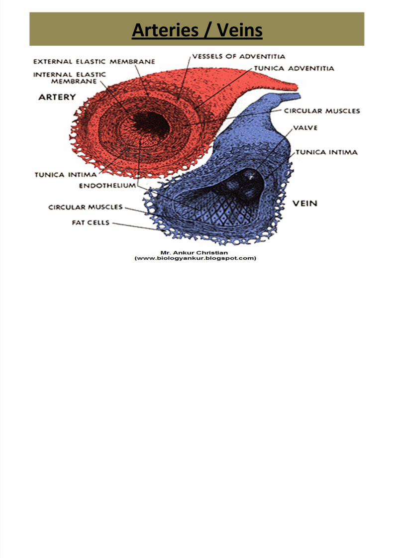
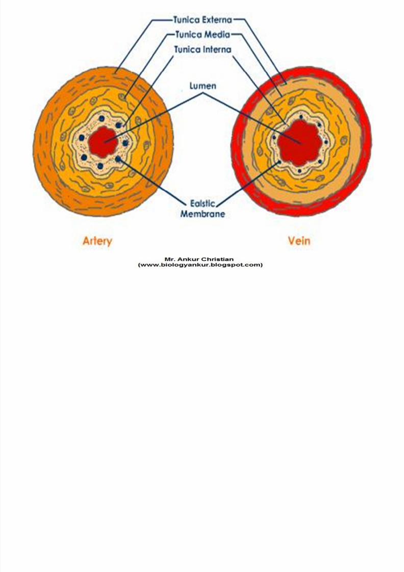
Arteries / Veins
A t i
Page 32
8/4/2019 Chapter 7. Circulation
http://slidepdf.com/reader/full/chapter-7-circulation 32/54
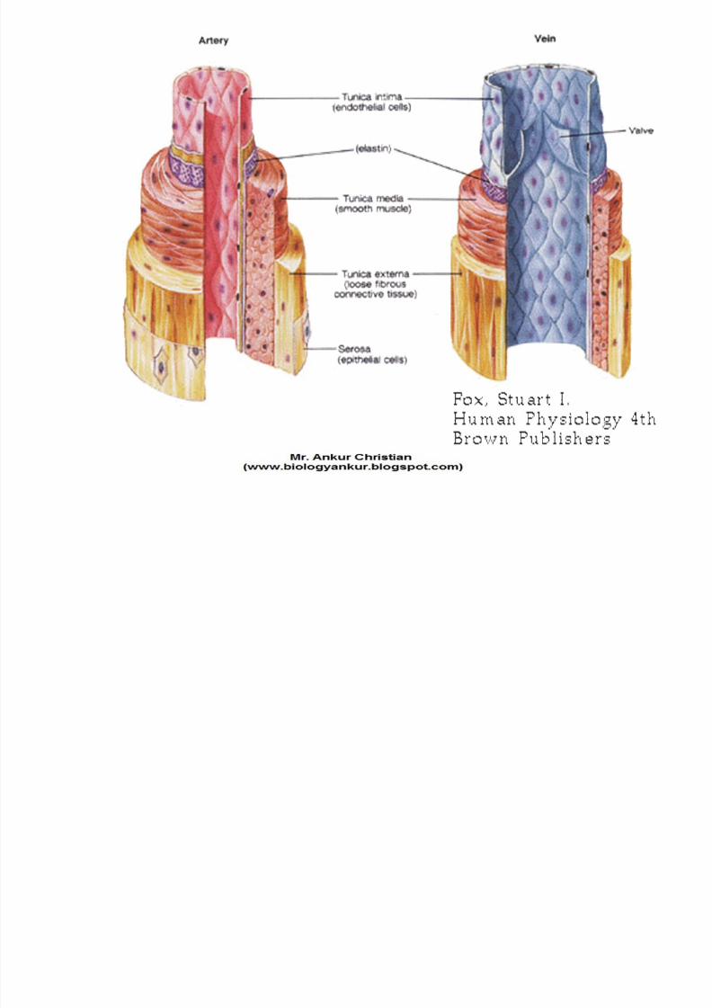
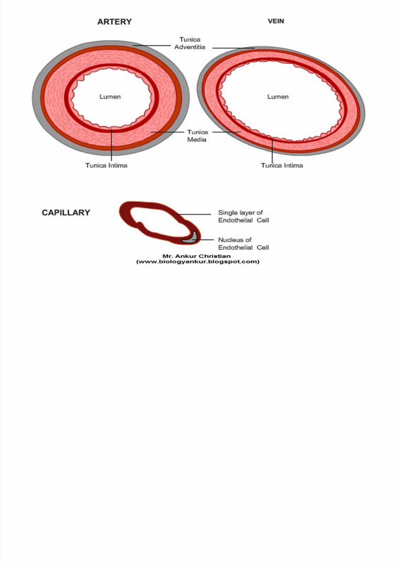
Arteries
•The artery wall consists of three layers:
• Tunica Adventitia –
– the strong outer covering
– composed of connective tissue, collagen & elastic fibres.
– These fibres allow it to stretch to prevent overexpansion due tothe pressure that is exerted on the walls by blood flow.
• Tunica Media –
– the middle layer of the walls of arteries & veins. – composed of smooth muscle & elastic fibres.
– This layer is thicker in arteries than in veins.
• Tunica Intima – – the inner layer of arteries & veins.
– composed of an elastic membrane lining & smooth endothelium(epithelial tissue) that is covered by elastic tissues.
Page 33
8/4/2019 Chapter 7. Circulation
http://slidepdf.com/reader/full/chapter-7-circulation 33/54
Page 34
8/4/2019 Chapter 7. Circulation
http://slidepdf.com/reader/full/chapter-7-circulation 34/54
Page 35
8/4/2019 Chapter 7. Circulation
http://slidepdf.com/reader/full/chapter-7-circulation 35/54
Veins
Page 36
8/4/2019 Chapter 7. Circulation
http://slidepdf.com/reader/full/chapter-7-circulation 36/54
Veins• The vein wall consists of three layers:
• Tunica Adventitia – – the strong outer covering.
– composed of connective tissue collagen & elastic fibers.
– These fibers allow it to stretch to prevent overexpansion due to thepressure that is exerted on the walls by blood flow.
• Tunica Media – – the middle layer of the walls of arteries and veins.
– It is composed of smooth muscle & elastic fibers.
– This layer is thicker in arteries than in veins.
• Tunica Intima – – the inner layer of arteries and veins.
– It is composed of an elastic membrane lining & smooth endothelium(epithelial tissue) that is covered by elastic tissues.
– Veins do not contain the elastic membrane lining that is found inarteries. In some veins the tunica intima layer also contains valves tokeep blood flowing in a single direction.
Page 37
8/4/2019 Chapter 7. Circulation
http://slidepdf.com/reader/full/chapter-7-circulation 37/54
Page 38
8/4/2019 Chapter 7. Circulation
http://slidepdf.com/reader/full/chapter-7-circulation 38/54
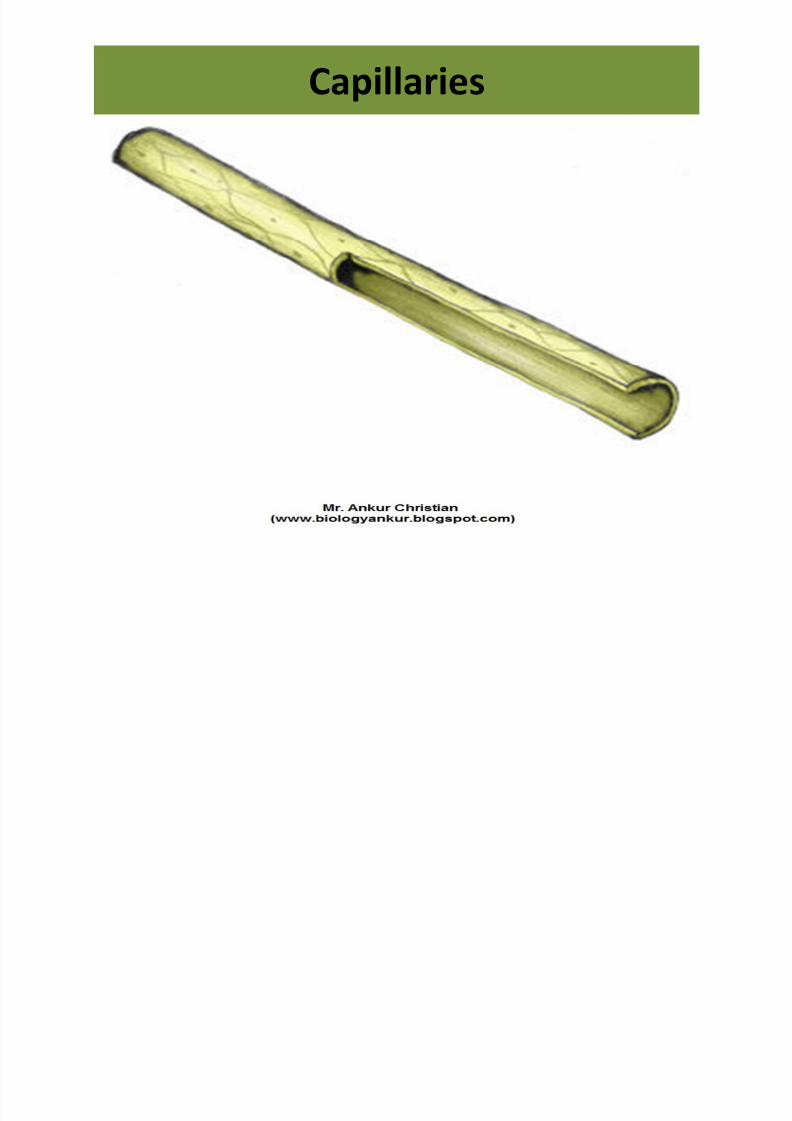
Capillaries
Page 39
8/4/2019 Chapter 7. Circulation
http://slidepdf.com/reader/full/chapter-7-circulation 39/54
• Capillaries have very thin walls comprised only of
endothelial cells, which allows substances tomove through the wall with ease.
• Capillaries are very small, measuring 5-10
micrometres in width.
• However, the cross-sectional area of capillaries
within an average size muscle would be largerthan that of the Aorta.
• This allows a fast and efficient transfer of
oxygen-carrying red blood cells to the site wherethey are needed.
Page 40
8/4/2019 Chapter 7. Circulation
http://slidepdf.com/reader/full/chapter-7-circulation 40/54
Vasa Vasorum
• They are the vessels which
supply blood to the wall of
artery and veins.
Page 41
8/4/2019 Chapter 7. Circulation
http://slidepdf.com/reader/full/chapter-7-circulation 41/54
Blood
• Blood is the fluid Connective Tissue.
•Red in colour because of the presence of
Hb (heame = iron).
• Human body contains 5-6 lits of blood.
• It is composed of 2 components Plasma
and Blood cells / corpuscles / formed
elements.
Blood Plasma
Page 42
8/4/2019 Chapter 7. Circulation
http://slidepdf.com/reader/full/chapter-7-circulation 42/54
Blood Plasma
• It is the fluid part of the blood.
• Out of 5 lits, 3.5 lits is the plasma.
• It consists of 90% of H2O, 7.3% proteins(albumin, globulin, fibrinogen and prothrombin)
and 3% is glucose, nitrogeneous wastes,
minerals, enzyme and hormones.
• Plasma transport gases and other minerals,
maintains blood pH, body immunity, body heatregulation, and regulates the osmotic pressure
of the blood.
Erythrocytes (Red Blood Corpuscles)
Page 43
8/4/2019 Chapter 7. Circulation
http://slidepdf.com/reader/full/chapter-7-circulation 43/54
Erythrocytes (Red Blood Corpuscles)• Erythros = Red, Cyton = cell
• Non-nucleated, biconcave, 7.4 um in diameter, 2 um thickness,volume is 90 um3, each RBC contains 30 ug of Hb.
• Avg normal RBC in Male = 5.0 - 5.5 millions and in Female = 4.5 –
5.0 millions, in Infant = 6.7 millions and in Feotus = 7.8 millionsper cc.
• RBC develops in Red bone marrow of large bones hence namedas Erythropoietic Organs.
• Life span of a RBC is about 120 days.• Spleen is considered the graveyard of RBC.
• Abnormal rise in the total count of RBC is called polycythemia.
• Smallest RBC is found in Traguls (Musk Deer).
• Largest RBC is found in Amphiuma.• Functions
– Transport of respiratory gases (O2 and CO2)
– Hb is an excellent acid – base buffer, which maintains the pH of blood.
Page 44
8/4/2019 Chapter 7. Circulation
http://slidepdf.com/reader/full/chapter-7-circulation 44/54
Blood platelets
• Oval to spherical in shape, 2 – 3 u in
diameter.
• Nucleus is absent, no. of platelets / cmm of
blood ranges from 200,000 – 450, 000.
• These are produced by the mega-karyocytesin the bone marrow and are destroyed in the
spleen.
• These initiate blood coagulation and repair
capillary endothelium.
Blood Groups
Page 45
8/4/2019 Chapter 7. Circulation
http://slidepdf.com/reader/full/chapter-7-circulation 45/54
Blood Groups
• Discovered by Landsteiner.
• Human Blood contains certain specific substances
called Antigens and Antibodies.• On the basis of these, human blood can be
distinguished into blood group A, B, AB and O.
• AB blood group is considered the universal
recipient and blood group O is the universal donor.
• On the basis of Rh antigen blood can bedistinguished into Rh+ and Rh- blood groups.
Principal Arteries
Page 46
8/4/2019 Chapter 7. Circulation
http://slidepdf.com/reader/full/chapter-7-circulation 46/54
Principal Arteries
• The L. Ventricle pumps oxygenated blood into a 3 cm thickvessel, called AORTA.
• The pulmonary aorta carries the blood from R. Ventricle toLungs
• Following arteries arise from the aorta: – 1). Coronary Artery to Heart wall.
– 2). Brachiocephalic to Head and Fore Arms.
– 3). Phrenic to Diaphragm. – 4). Coeliac to Alimentary Canal
• Gastric to Stomach
• Common Hepatic to Liver
• Splenic to Spleen• 5). Renal Artery to Kidney
• 6). Genital Artery to Male and Female Gonads
• 7). Common Iliac to Hind limb
Principal Veins
Page 47
8/4/2019 Chapter 7. Circulation
http://slidepdf.com/reader/full/chapter-7-circulation 47/54
Principal Veins
• The blood for anterior parts of the body is
brought into SVC (superior vena cava)
• Similarly blood from posterior part of thebody is received by PVC (posterior vena cava)
or IVC (inferior vena cava)• Renal vein receives blood from kidneys and
carries it to IVC.
Hepatic Portal System
Page 48
8/4/2019 Chapter 7. Circulation
http://slidepdf.com/reader/full/chapter-7-circulation 48/54
Hepatic Portal System
• Portal System is a part of various system in whichvein divides twice into capillaries before they join
posterior or anterior vena cava.
• Hepatic portal system collects the nutrients from
intestine by means of capillaries.
• Excess amount of nutrients are removed by liver,capillaries rejoin and form veins, which open into
inferior vena cava.
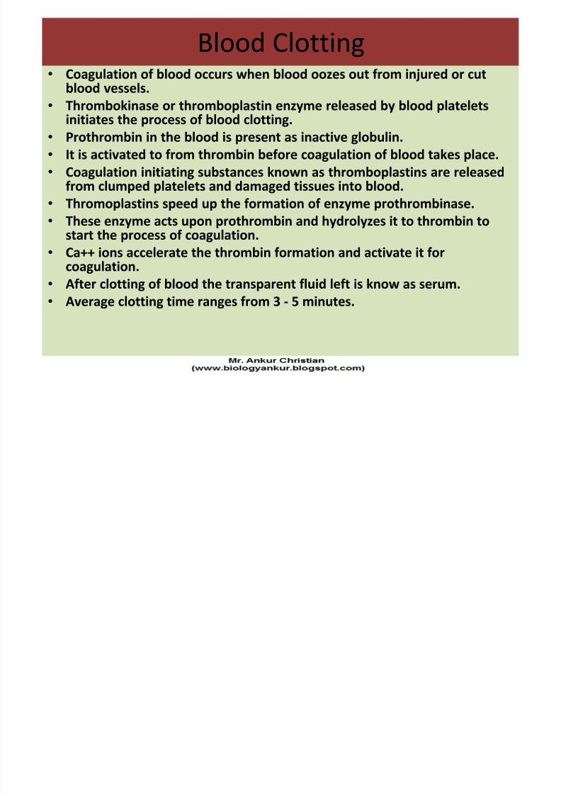
Blood Clotting
Page 49
8/4/2019 Chapter 7. Circulation
http://slidepdf.com/reader/full/chapter-7-circulation 49/54
Blood Clotting
• Coagulation of blood occurs when blood oozes out from injured or cutblood vessels.
• Thrombokinase or thromboplastin enzyme released by blood plateletsinitiates the process of blood clotting.
• Prothrombin in the blood is present as inactive globulin.
• It is activated to from thrombin before coagulation of blood takes place.
• Coagulation initiating substances known as thromboplastins are releasedfrom clumped platelets and damaged tissues into blood.
• Thromoplastins speed up the formation of enzyme prothrombinase.
• These enzyme acts upon prothrombin and hydrolyzes it to thrombin tostart the process of coagulation.
• Ca++ ions accelerate the thrombin formation and activate it forcoagulation.
• After clotting of blood the transparent fluid left is know as serum.• Average clotting time ranges from 3 - 5 minutes.
Page 50
8/4/2019 Chapter 7. Circulation
http://slidepdf.com/reader/full/chapter-7-circulation 50/54
Page 51
8/4/2019 Chapter 7. Circulation
http://slidepdf.com/reader/full/chapter-7-circulation 51/54
Page 52
8/4/2019 Chapter 7. Circulation
http://slidepdf.com/reader/full/chapter-7-circulation 52/54
Page 53
8/4/2019 Chapter 7. Circulation
http://slidepdf.com/reader/full/chapter-7-circulation 53/54
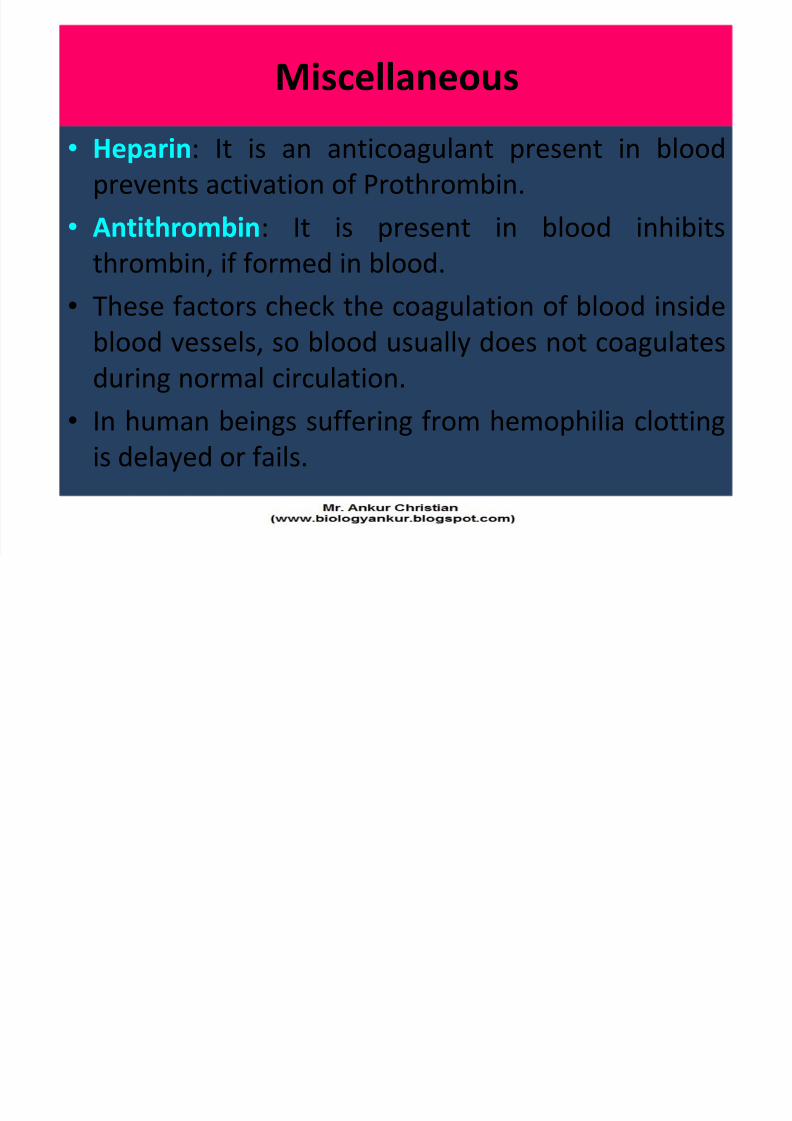
Miscellaneous
Page 54
8/4/2019 Chapter 7. Circulation
http://slidepdf.com/reader/full/chapter-7-circulation 54/54
Miscellaneous
• Heparin: It is an anticoagulant present in blood
prevents activation of Prothrombin.
• Antithrombin: It is present in blood inhibits
thrombin, if formed in blood.
• These factors check the coagulation of blood insideblood vessels, so blood usually does not coagulates
during normal circulation.
• In human beings suffering from hemophilia clotting
is delayed or fails.