156
CHAPTER II STRUCTURAL ORGANIZATION ( tissue histology )

CHAPTER II
STRUCTURAL ORGANIZATION
( tissue histology )

CHAPTER II
STRUCTURAL ORGANIZATION ( Tissue Histology )

Tissue collections of similar cells
that perform a common function.
The various types of tissues are established -
during early embryonic development.
As the embryo grows,
- organs form from specific arrangements of
tissues.

Fig. 24.2 Levels of organization within the vertebrate body

Pathology study of abnormal tissues
in diseased organs.
By knowing the normal tissues structure,
can recognize the abnormal.
Histology by a course in pathology.


Based on their structure and function,
- may be classified into four basic categories
Epithelial tissue
Connective tissue
Muscular tissue
Nervous tissue

Fig. 24.1 Vertebrate tissue types

Characteristics of Membranous Epithelial Tissues
epithelium
- is located throughout the body &
- forms such structures as
-- the outer layer of the skin,
-- the lining of
-- the covering of
the secretory part of glands.
the body cavities & vessels ,
viscera , &

Epithelium always has one free surface (the apical
surface) exposed to a body cavity, a lumen (hallow
part of a body tube or duct), or to the skin surface.
Apical surface
Apical surface


The deep surface
bound
to underlying tissue by a basement membrane

Shaped
number of layers


Simple Epitehlial Tissues
Single cell layer.
diffusion, filtration, secretion are principle function
size and shape
from thin, flattened cells to tall, columnar cells.

Some have cilia
for the movement of materials across cell surfaces.

Fig. 26.14 The small intestine
Other have microvilli
- that increase the surface area for absorption.

Simple Squamous Epithelium
flattened, irregularly shaped cells
- tightly bound together for diffusion and filtrations.
Table 24.2
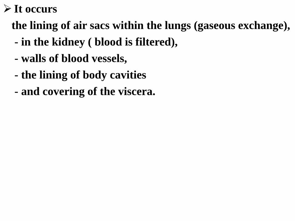
It occurs
the lining of air sacs within the lungs (gaseous exchange),
- in the kidney ( blood is filtered),
- walls of blood vessels,
- the lining of body cavities
- and covering of the viscera.


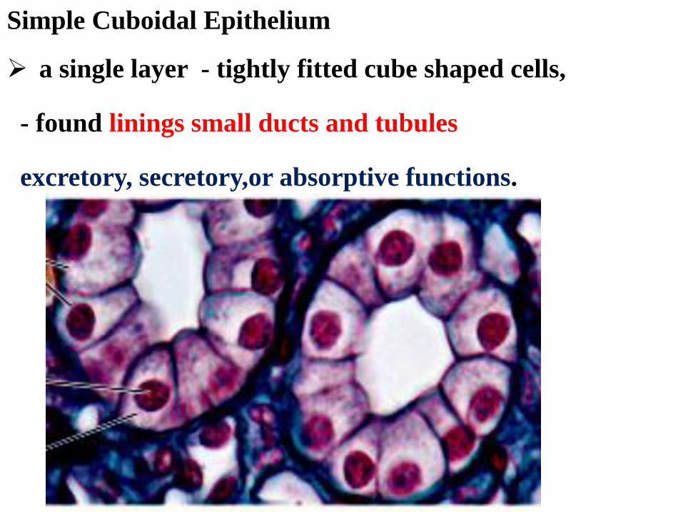
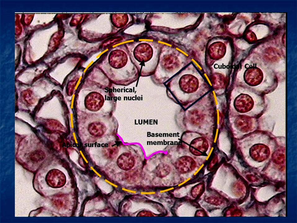
Simple Cuboidal Epithelium
a single layer - tightly fitted cube shaped cells,
- found linings small ducts and tubules
excretory, secretory,or absorptive functions.

It occurs on the surface of the ovaries, portion of the
kidneys, ducts of the salivary glands and pancreas.
Simple

Simple columnar Epithelium
Tall , narrows cells.
Specialized goblet cells
- are scattered through tissue.
Goblet cells
- secrete a lubricative & protective mucus
along surface of the tissue.

Basement
membrane
Basement
membrane
Mucus of a
goblet cell Nucleus of
simple columnar
epithelial cell Simple
columnar
epithelial cells
(c) Diagram: Simple columnar
Photomicrograph: Simple columnar
epithelium of the small intestine (575×).
- lining inside walls of the stomach & small
intestine,

(enterokinase)
CCK-PZ
Secretin
where it forms a highly absorptive surface &
also secretes certain digestive chemicals.

Simple Ciliated Columnar Epithelium
cilia along the free surfaces.
wavelike movements
that transport materials
through tubes or passageways.
in the female uterine tubes.


Pseudostratified Ciliated Columnar Epithelium
Epithelium appears to be stratified,
- because of nuclei of the cells are located at different
levels.
Pseudostratified Ciliated Columnar

Numerous globet cells and a ciliated exposed
surface are characteristic of this epithelium.
The trachea and the bronchial tubes
frequently called respiratory epithelium.
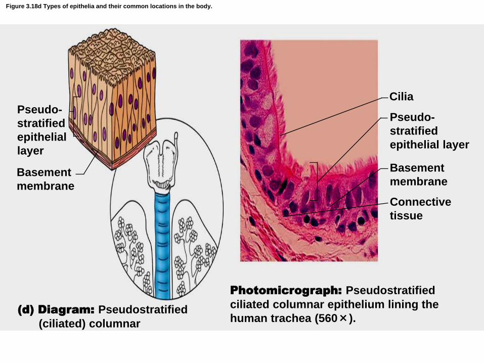
Figure 3.18d Types of epithelia and their common locations in the body.
(d) Diagram: Pseudostratified
(ciliated) columnar
Photomicrograph: Pseudostratified
ciliated columnar epithelium lining the
human trachea (560×).
Basement
membrane
Basement
membrane
Pseudo-
stratified
epithelial
layer
Pseudo-
stratified
epithelial layer
Cilia
Connective
tissue

Function is to remove dust and bacteria trapped in mucus.
Movement of cilia to pharynx

Stratified Squmaous Epithelium
- is composed of a number of cell layers
- that are flattest at the surface.

Cell divisions occur only
- within the deepest layer ( the stratum basale )
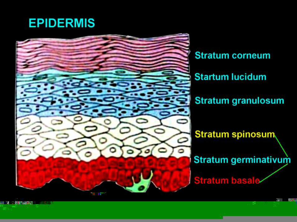

As the newly produced cells grow in size
- they are pushed toward the surface
- where they will replace the cells
- that are sloughed off.
Movement of the epithelial cells away from the
supportive basement membrane is accompanied
by the production of keratin, progressive
dehydration, and flattening.

Stratified squamous epithelial tissues:
Keratinized
Two types
nonkeratinized.


1. Keratinized stratified squamous epithelium ---
contain keratin,
- a protein that strengthens the tissue.
Keratin makes the epidermis (outer layer) of the skin -
- somewhat waterproof and
- protects it from bacterial invasion.
The outer layer of the skin are dead,
but glandular secretions keep them soft.

2. Nonkeratinized stratified squamous epithelium
lines the mouth and throat, nasal cavity, vagina
and anal canal, cornea , oesophagus ,
called mucosa
is well adapted to withstand moderate abrasion
but not fluid loss.
The cells on the exposed surface of this tissue are
alive and are always moistened.

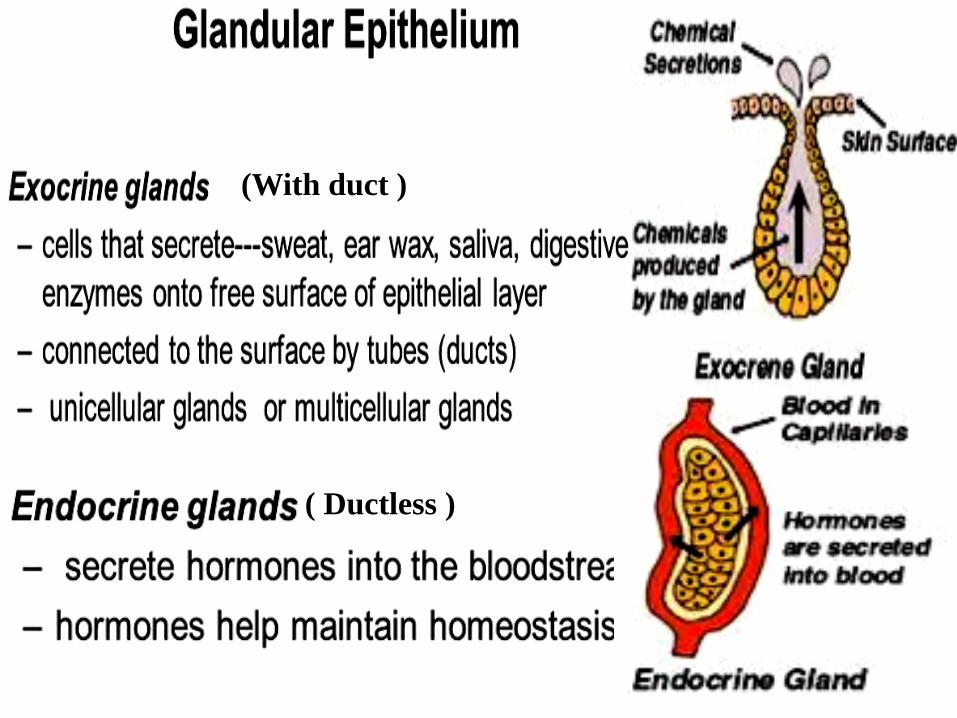
(With duct )
( Ductless )

As tissues develop in the embryo,certain epithelial cells
migrate into the underlying connective tissue, forming
secretory structures called exocrine glands
underlying
connective
tissue,


The secretions from exocrine glands
pass through ducts onto body surface
or into body cavities.
These glands should not be confused with
endocrine glands ,
- which are ductless,
- and which secrete their products (hormones)
- in to the blood or surrounding extracellular
fluid.

Exocrine glands within the skin --
oil (sebaceous) glands,
- include
mammary glands.
Exocrine glands within the digestive system
the salivary gland &
pancreatic gland.
include
sweat glands , &


Exocrine glands are classified
- according to their structure &
- how they discharge their products.
Classified according to structure,
unicellular glands
multicellular glands.
two types &

1.Unicellular glands are
single-celled glands, ( goblet cells )
-interspersed within most columnar epithelial tissues.

Goblet cells are found in the epithelial lining of
- the respiratory ,
- digestive ,
- urinary &
- reproductive system.
The mucous secretion of these cells lubricates and
protects the surface linings.


2.Multicellular glands,
- as their name implies,
- are composed of both secretory cells and cells
that form the walls of the ducts.
Multicellular glands are classified as
do not branch , do branch
simple glands or compound glands.

Multicellular glands are also classified
They are identified
.
tubular glads
acinar glands
tubulo-acinar glands
- according to the shape of their secretory portion.

as tubular glands - if the secretory portion
resembles = a tube


as acinar glands
- if the secretory portion resembles =a flask

both a tube & a flask
tubuloacinar glands


Multicellular glands are also classified
-according to the means by which they release
their product.
They are
- Merocrine glands,
- Apocrine glands,
- holocrine glands.


1.Merocrine glands
- are those that secrete a watery substance
through the cell membrane of the secretorycells.
Salivary glands , pancreatic glands , and certain
sweat glands are of this type.

2. Apocrine glands are those in which the
secretion accumulates on the surface of the
secretory cells ; then, a portion of the cell and the
secretion is pinched off and discharged.
An example of a apocrine gland is mammary gland.


3. Holocrine glands are those in which the entire
secretory cell and its product are discharged.
An example of a holocrine glands is an oil
secreting (sebaceous)glands of the skin.

Connective Tissue
is dived into subtypes –
-according to the characteristics of the matrix
- that binds the cells.
Connective Tissue provides
- structural support
- metabolic support
for other tissues and organs
of the body.

- the most abundant tissue in the body.
- more matrix than cells.
rarely touch an another at all.
Table 24.2


(Adipose
tissue) (ligament
, tendon)
Special connective tissue
1. 2
Connective Tissue
Connective Tissue
proper
loose dense
Fluid connective
tissue
Supporting
connective tissues
Blood Lymph Cartilage Bone
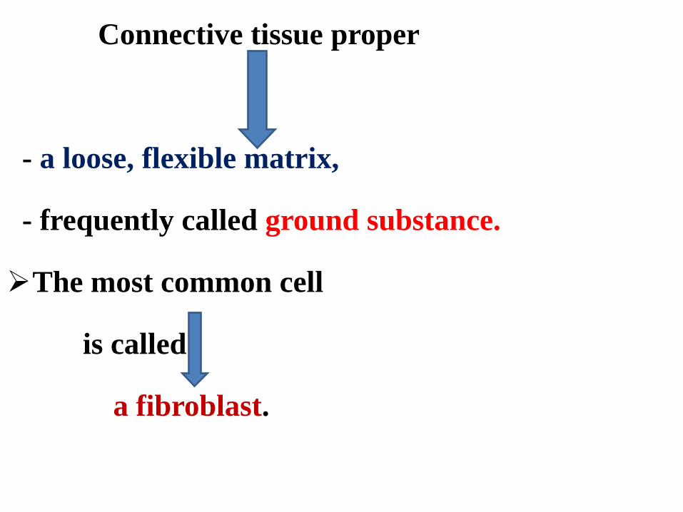
Connective tissue proper
- a loose, flexible matrix,
- frequently called ground substance.
The most common cell
is called
a fibroblast.


Collagenous fibers - collagen protein
tremendous strength
Elastic fiber - elastin protein
elasticity & extensibility

Reticular fibers - reticulin protein
form a lattice-like framework

Adipose Tissue
Contain large number of adipose cells, adipocytes.
The cell store fat within their cytoplasm, causing swell
and forcing their nuclei to one side.
Adipocyte containing fat
Nucleus
Reticular fibers
Connective tissue

Copyright © 2010 Pearson Education, Inc.
Figure 4.8b Connective tissues.
(b) Connective tissue proper: loose connective tissue, adipose
Description: Matrix as in areolar, but very sparse; closely packed adipocytes, or fat cells, have nucleus pushed to the side by large fat droplet.
Function: Provides reserve food fuel; insulates against heat loss; supports and protects organs.
Location: Under skin in the hypodermis; around kidneys and eyeballs; within abdomen; in breasts.
Photomicrograph: Adipose tissue from the subcutaneous layer under the skin (350x).
Nucleus of fat cell
Vacuole containing fat droplet
Adipose tissue
Mammary glands

Adipose tissue is found
- beneath the skin,
- around the kidneys,
- on the surface of the heart,
- surrounding joints,
- in the breast of mature females.

Functions not only as a food reserve,
- but also to support
- and protect various organs.
It helps to keep the body warm.

2. Cartilage
Structure - Cartilage cells (chondrocytes)
- Tiny spaces (lacunae)
Function - Support and protection
Three Types of cartilage
(type & amount of fibers embedded within the matrix)
a. Hyaline cartilage
b. Fibrocartilage
c. Elastic Cartilage

Hyaline cartilage
Hyaline cartilage has matrix that gives it a
glassy appearance.
Located in
- the respiratory tract,
- rib cage, and
- developing bone.


Figure 3.19b Connective tissues and their common body locations.
Chondrocyte
(cartilage cell)
Chondrocyte
in lacuna
Matrix Lacunae
Photomicrograph: Hyaline cartilage
from the trachea (400×)
(b) Diagram: Hyaline
cartilage

Fibrocartilage
- a matrix reinforced with many collagenous fibers.
-a durable tissue
-adapted to withstand tension & compression bone.


Elastic cartilage
Elastic cartilage,
-abundant elastic fibers,
-very flexible and strong
- the outer ear (Pinnae),
- portions of the larynx &
- auditory canal.
found in

Hyaline cartilage
Elastic cartilage
Fibrocartilage

Fig. 24.12 Axial and appendicular skeletons

Bone (Osseous) Tissue
Most rigid of all connective tissues,
bone has a rich blood supply.
The hardness of bone is due to the calcium
phosphate located within the matrix.
Bone tissue is classified
- compact bone
- spongy bone


Compact bone tissue constitutes hard outer
portion of a bone,
- spongy bone tissue constitutes the porous, highly
vascular inner portion.


In compact bone tissue uniform structural
arrangement can be seen.
Bone cells, osteocytes are arranged in rings
around a central (Haversian) canal,which
contains blood vessels and a nerve.

Bone cells ( osteocyte ) - in tiny space ( lacuna )
Canaliculi - Radiating
from lacuna
-Nutrients diffuse
through the canaliculi
- bone matrix
– called lamellae

Ground bone X.S.
Bone matrix
Bone cell in
lacuna
Concentric lamella
Haversian system
Haversian canal

Figure 3.19a Connective tissues and their common body locations.
Bone cells
in lacunae Central
canal
Lacunae
Lamella
(a) Diagram: Bone
Photomicrograph: Cross-sectional
view of ground bone (165×)

Compact Bone

Composition of Blood
• Blood is responsible for…..
– Transporting gases (oxygen & carbon dioxide)
– Transporting waste products
– Transporting nutrients
– Helping remove toxins from the body

RBC Structure And Function • Have no organelles or nuclei
• Hemoglobin – oxygen carrying protein
– Each RBC has about 280 million hemoglobin molecules
• Biconcave shape – 30% more surface area

Blood (Vascular) Tissue
Blood, or vascular tissue,
- is specialized fluid connective tissue
- that plays a vital role in maintaining internal
body homeostasis.


Blood tissue

1. Erythrocytes or red blood cells(RBCs), tiny
biconcave discs that lack nuclei,
Their red color is due to the protein hemoglobin.
Oxygen attaches to and is transported on the
hemoglobin molecules.
The life span of erythrocytes is between 90 and
120 days.


2.Leukocytes or white blood cells(WBCs),
- nucleated, exhibit amoeboid movement by
forming cytoplasmic extensions and serve to
protect the body against invasions by
microorganisms.

They are produced in
bone marrow & lymphatic tissue
- & have a life span ranges from 3 to 300 days.
There are five kinds of leukocytes;
- neutrophils,
- eosinophils,
- basophils ,
- lymphocytes
- monocytes.
granulocyte
agranulocyte


Neutrophils
- 40%-70% WBCs
- Nucleus multilobed - Duration of development: 6-9 days - Life Span: 6 hours to a few days - Function: phagocytize bacteria

Eosinophils
- 1%-4% WBCs
- Nucleus bilobed
- Development:6-9 days
- Life Span: 8-12 days
- Function:
1) Kill parasitic worms
2) destroy antigen-antibody complexes
3) inactivate some inflammatory chemical of allergy

Basophils
- 0.5% WBCs
- Nucleus lobed
- Development: 3-7 days
- Life Span: a few hours to a few days
- Function:
1) Release histamine and other mediators of
inflammation
2) contain heparin, an anticoagulant

Lymphocytes
- T cells and B cells
- 20%-45% WBCs
- Nucleus spherical or indented
- Development: days to weeks
- Life Span: hours to years
- Function
Mount immune response by direct cell attack (T
cells) or via antibodies (B cells)

Monocytes
- 4%-8% WBCs
- Nucleus U-shaped
- Development: 2-3 days
- Life Span: months
- Function:
Phagocytosis
develop into macrophages in tissues
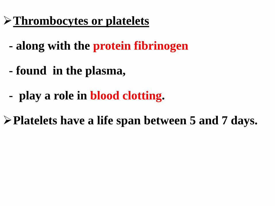
Thrombocytes or platelets
- along with the protein fibrinogen
- found in the plasma,
- play a role in blood clotting.
Platelets have a life span between 5 and 7 days.


Fig. 24.13
Muscles and How They Work


• Muscle Tissue
Muscle tissue is unique because it is able to
contract, thus making movement possible.
The muscle cells, or fibers, are long and
cylindrical.
Three types of muscle are
- smooth muscle tissue
- cardiac muscle tissue
- skeletal muscle tissue.


• Smooth muscle
Smooth muscle fibers are long, spindle –shaped cells
that contains a single nucleus.
These cells are usually grouped together in flattened
sheets, forming the muscular portion of the wall around
a lumen.

Smooth muscle tissue is common throughout the
body.
Smooth muscle is also found in
- the walls of blood vessels,
- the walls of respiratory passage, and
- in the urinary and reproductive duct.
The contraction of smooth muscle is under
involuntary (unconscious) nervous control.


• Cardiac muscle
Make up most of the wall of the heart
- characterized by
-- branching fibers,
-- a central nucleus,
-- banding patterns called striations.
The cardiac muscle fibers are joined by intercalated
discs.

Intercalated discs help to hold neighboring cells
together and spread the contract from cell to cell
- also contracts involuntarily.


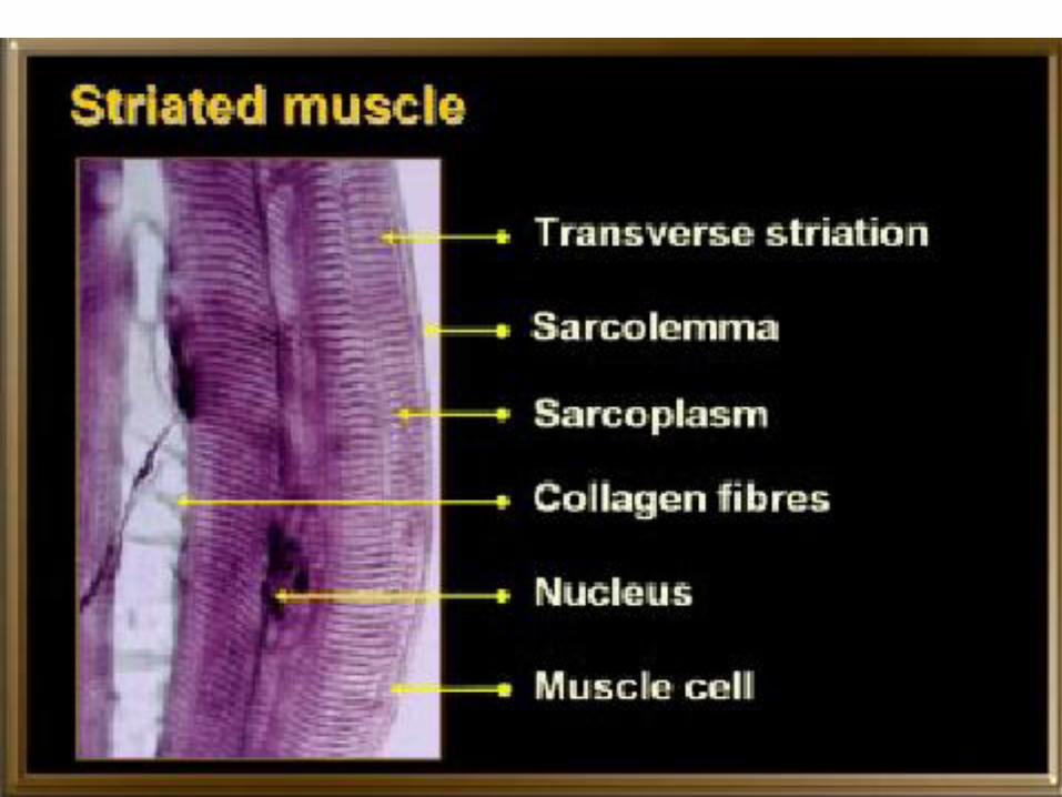
• Skeletal Muscle (Striated muscle )
Makes up the skeletal muscle that attached to the
bones of the skeleton.
Contraction of results in voluntary or
involuntary body movements.
Skeletal muscle fibers are long and multinucleate.
The striations easily seen through a microscope.


• A Simple Nerve Circuit – the Reflex Arc.
– A reflex is an autonomic response.

• Nervous Tissue
Nervous Tissue , contained within
- the brain,
- spinal cord, and
composed of two kinds of cells-
- neurons and
- neuroglial cells.

• Neurons, or nerve cells are
- the basic structural and
- functional units,
- specialized to respond to
- to generate impulses and
- conduct impulses to and from the various body
organs.
physical stmuli and
chemaical stmuli &


A neuron has three principal components.
1.The cell body contains the nucleus and
specialized organelles and microtubules.
2.The dendrites function to receive a stimulus
and conduct the impulse toward the cell body.
3.The axon is a long extension that conducts an
impulse away from the cell body.


The term nerve fiber usually refers to an axon and the myelin sheath that surrounds it.

Neuroglial cells ( glial cells )
- about five times as abundant as neurons
- do not transmit impulses
- support and bind neurons together.
- phagocytic
- assist providing nourishment for the neurons


Enjoy Your Learning
Your University Student Life
&
Good Luck


