30 Upper Airway Diseases Laryngomalacia Obstructive Sleep Apnea Subglottic Stenosis Tracheomalacia Upper Respiratory Tract Infection ( URI ) Lower Airway Diseases Asthma Bronchiolitis Cystic Fibrosis Meconium Aspiration Syndrome Respiratory diseases represent the most common systemic illnesses in children, and are likely to influence anesthetic management. This chapter reviews the most common important upper and lower airway diseases in children. UPPER AIRWAY DISEASES Laryngomalacia Laryngomalacia is the most common laryngeal disorder of the newborn. It is a congenital abnormality of the epiglottis and aryepiglottic folds that allows their inward collapse into the airway during inspiration. This inward collapse results in nearly complete upper airway obstruc- tion that is manifested by audible inspiratory stridor, usually in the few several months of life. The stridor is usually more prominent when the infant is lying supine, crying, or feeding. In most cases it is benign, and will be outgrown during the first year of life. Laryngomalacia is occasionally associated with gastroesophageal reflux and, in rare cases, causes hypoxemia or hypoventilation, and interferes with normal feeding and subsequent growth. Infants in this latter group require definitive diagnosis by direct laryngoscopy under general anesthe- sia. Rigid bronchoscopy is performed at the same time to rule out subglottic causes of airway obstruction. Severe laryngomalacia is treated by performing a supra- glottoplasty, in which a CO 2 laser is used to trim the length of the epiglottis and partially sever the aryepiglot- tic folds to prevent the epiglottis from infolding into the glottic opening during inspiration. During induction of general anesthesia, infants with laryngomalacia commonly exhibit airway obstruction that is not relieved by placement of an oral airway device. Deepening the anesthetic will often relieve the obstruc- tion because of progressive weakening of the diaphragm and decreasing the strength of inspiration. However, during upper airway obstruction, speed of inhalational induction is slowed.Positive-pressure ventilation is usually easily accomplished in these infants, especially after the onset of neuromuscular blockade. Obstructive Sleep Apnea Obstructive sleep apnea (OSA) in children is the result of adenotonsillar hypertrophy, combined with an abnor- mally small retropharyngeal space, and altered neuro- muscular control of upper airway patency during sleep. It mainly occurs in children between the ages of 2 and 4 years, and is especially prevalent in children with obe- sity and trisomy 21. The clinical manifestations include partial or complete upper airway obstruction during sleep, restless sleep, morning headaches, behavioral dis- turbances, and daytime somnolence. Severe cases of untreated longstanding OSA can result in chronic hypo- xemia, polycythemia, and cor pulmonale. Children with electrocardiographic or radiographic cardiac abnor- malities should be referred to a pediatric cardiologist for further evaluation and management. The most common therapy for pediatric OSA is adenotonsillectomy (see Chapter 29), which alleviates symptoms in most children. Some pediatric anesthe- siologists prefer to reduce the dose of the preoperative CHAPTER Respiratory Diseases RONALD S. LITMAN, D.O. 4 4206 Litman-04.qxd 27/01/2004 23:28 Eve Page 30
Transcript
30
Upper Airway Diseases
Laryngomalacia
Obstructive Sleep Apnea
Subglottic Stenosis
Tracheomalacia
Upper Respiratory Tract Infection ( URI )
Lower Airway Diseases
Asthma
Bronchiolitis
Cystic Fibrosis
Meconium Aspiration Syndrome
Respiratory diseases represent the most commonsystemic illnesses in children, and are likely to influenceanesthetic management. This chapter reviews the mostcommon important upper and lower airway diseases inchildren.
UPPER AIRWAY DISEASES
Laryngomalacia
Laryngomalacia is the most common laryngeal disorderof the newborn. It is a congenital abnormality of theepiglottis and aryepiglottic folds that allows their inwardcollapse into the airway during inspiration.This inwardcollapse results in nearly complete upper airway obstruc-tion that is manifested by audible inspiratory stridor,usually in the few several months of life. The stridor isusually more prominent when the infant is lying supine,crying, or feeding. In most cases it is benign, and will beoutgrown during the first year of life. Laryngomalacia isoccasionally associated with gastroesophageal refluxand, in rare cases, causes hypoxemia or hypoventilation,and interferes with normal feeding and subsequentgrowth. Infants in this latter group require definitivediagnosis by direct laryngoscopy under general anesthe-sia. Rigid bronchoscopy is performed at the same time
to rule out subglottic causes of airway obstruction.Severe laryngomalacia is treated by performing a supra-glottoplasty, in which a CO2 laser is used to trim thelength of the epiglottis and partially sever the aryepiglot-tic folds to prevent the epiglottis from infolding intothe glottic opening during inspiration.
During induction of general anesthesia, infants withlaryngomalacia commonly exhibit airway obstruction thatis not relieved by placement of an oral airway device.Deepening the anesthetic will often relieve the obstruc-tion because of progressive weakening of the diaphragmand decreasing the strength of inspiration. However,during upper airway obstruction, speed of inhalationalinduction is slowed.Positive-pressure ventilation is usuallyeasily accomplished in these infants, especially after theonset of neuromuscular blockade.
Obstructive Sleep Apnea
Obstructive sleep apnea (OSA) in children is the resultof adenotonsillar hypertrophy, combined with an abnor-mally small retropharyngeal space, and altered neuro-muscular control of upper airway patency during sleep.It mainly occurs in children between the ages of 2 and4 years, and is especially prevalent in children with obe-sity and trisomy 21. The clinical manifestations includepartial or complete upper airway obstruction duringsleep, restless sleep, morning headaches, behavioral dis-turbances, and daytime somnolence. Severe cases ofuntreated longstanding OSA can result in chronic hypo-xemia, polycythemia, and cor pulmonale. Children withelectrocardiographic or radiographic cardiac abnor-malities should be referred to a pediatric cardiologist forfurther evaluation and management.
The most common therapy for pediatric OSA isadenotonsillectomy (see Chapter 29), which alleviatessymptoms in most children. Some pediatric anesthe-siologists prefer to reduce the dose of the preoperative
CHAPTER Respiratory DiseasesRONALD S. LITMAN, D.O.4
4206 Litman-04.qxd 27/01/2004 23:28 Eve Page 30
Respiratory Diseases 31
sedative in children with OSA, for fear of causinglife-threatening upper airway obstruction in an unmoni-tored environment. However, it has been this author’sexperience that a routine dose of oral midazolam in chil-dren with OSA does not cause significant upper airwayobstruction.
During induction of general anesthesia, virtually allchildren with untreated OSA will exhibit partial or com-plete upper airway obstruction. Insertion of an artificialoral airway device after loss of consciousness will bypassthe obstruction and allow easy bag-mask ventilation.In the immediate postoperative period following adeno-tonsillectomy, the incidence of airway obstruction ishigher in children with OSA when compared with thosewho undergo adenotonsillectomy for recurrent infec-tions. Therefore, children with significant OSA shouldbe hospitalized overnight following the procedure.Even some time after adenotonsillectomy has been per-formed, a predisposition toward upper airway obstruc-tion during sleep or sedation may persist throughoutchildhood. Children with OSA are more likely to developadult-type OSA.
Subglottic Stenosis
Subglottic stenosis is an abnormal narrowing of theextrathoracic trachea below the level of the vocal cords.It may be present at birth (webs, strictures, etc.) or, morecommonly, is acquired secondary to chronic inflamma-tion and scarring from the presence of an endotrachealtube. Congenital subglottic stenosis is more common inchildren with multiple congenital anomalies and in chil-dren with trisomy 21.
Acquired subglottic stenosis is the most commonacquired anomaly of the larynx in children and the mostcommon abnormality requiring tracheotomy in childrenyounger than 12 months. The chronic presence of anendotracheal tube causes inflammation and scarring,par-ticularly at the level of the cricoid ring, and a restrictivescar is formed. The clinical manifestations of subglotticstenosis include inspiratory stridor during crying or atthe time of an upper respiratory tract infection, duringwhich tracheal narrowing increases secondary to edema.
Subglottic stenosis is diagnosed using rigid bron-choscopy under general anesthesia. Positive-pressureventilation via bag and mask can be difficult when thenarrowing is severe. An additional anesthetic implicationis the need for an endotracheal tube that is markedlysmaller than that predicted for age. Severe cases requiretreatment with an anterior cricoid split procedure or amore complete tracheal reconstruction (laryngotracheo-plasty),and some children require tracheostomy.Followingtracheal reconstruction, these children will remain para-lyzed and mechanically ventilated for several days whilethe tracheal tissue heals over the endotracheal tube.
Tracheomalacia
Tracheomalacia is defined as a softening of the trachealcartilage, which then becomes susceptible to collapsewhen the intraluminal tracheal pressure is less than theextraluminal tracheal pressure.Thus, airway collapse canoccur during forceful coughing or exhalation.Congenitaltracheomalacia occurs in infants with a tracheoe-sophageal fistula and some genetic disorders such astrisomy 21 and the mucopolysaccharidoses. Acquiredtracheomalacia occurs in children who required long-term mechanical ventilation during early infancy, andin children with tracheal compression lesions such as avascular ring.
Clinical manifestations of tracheomalacia includenoisy breathing,“barky”cough,wheezing,and respiratorydistress (e.g., dyspnea).These symptoms are often exac-erbated during a URI (upper respiratory infection).Many children with tracheomalacia are initially thoughtto be asthmatic until properly diagnosed.
Anesthetic implications for children with tracheomalaciaare similar to those for laryngomalacia. Positive-pressureventilation, especially after administration of a neuro-muscular blocker, will characteristically result in the abil-ity to easily open the softened trachea and establishadequate ventilation. Coughing and partial upper airwayobstruction will exacerbate tracheal collapse and rapidlylead to hypoxemia.This is particularly difficult to manageduring emergence from general anesthesia and aftertracheal extubation. Children with severe tracheo-malacia may require a “deep extubation” to avoid thesecomplications.
Upper Respiratory Tract Infection (URI)
Viral upper respiratory tract infections (URIs) arefrequent in children,especially during the winter months.Typical symptoms include rhinorrhea,congestion,cough,fever,and malaise.Subclinical manifestations may includeupper and lower airway edema,increased respiratory tractsecretions, pneumonia, and bronchial irritability.
Intraoperative airway complications during generalanesthesia seem to be more common in children with aURI. These include coughing, laryngospasm, bron-chospasm, and hypoxemia. Infants under 12 months ofage tend to have more intraoperative complications thanolder children, and use of an endotracheal tube as com-pared with a facemask or laryngeal mask airway (LMA)increases the risk of these complications. Passive expo-sure to cigarette smoke is an additional risk factor.Apneic oxygenation is less effective; thus oxyhemoglo-bin desaturation may occur when,during rapid sequenceinduction, the child is not receiving positive-pressureventilation.
4206 Litman-04.qxd 27/01/2004 23:28 Eve Page 31
Use of thiopental for induction of general anesthesiais associated with more airway complications than whenpropofol is used.
Transient postoperative hypoxemia, postintubationcroup, and postoperative pneumonia are more likely tooccur in children with a URI. Long-term complicationsand true outcomes are difficult to define and quantifyand may not differ between normal children and thosewith a current or recent URI.
With these possible complications in mind, when achild presents with a URI, it is intuitive that an electiveprocedure requiring general anesthesia should be can-celed. But, because so many children have a concurrentURI at the time of their scheduled surgery, and long-termnegative outcomes have not been demonstrated, thisdecision process is complex. How, then, should the anes-thesiologist decide when to cancel an elective proce-dure in a child with a URI? First, one should assess theseverity of the child’s illness.The child with a runny nosewithout additional findings may be suffering from vaso-motor or allergic rhinitis (Box 4-1), which is usually notassociated with perioperative airway complications.If it is clear that the illness is viral, one must thenidentify the factors that are likely to increase periopera-tive complications (Box 4-2). If any of these risk factorsare present, it may be prudent to perform the procedureat a later date when the child is in better health. Onthe other hand, there are a variety of additional factorsthat may influence the anesthesiologist’s decision toproceed with surgery or cancel the case. The mostcommon reason for proceeding with a case even thoughrisk factors are present is the continuous presence of aURI that will likely continue without surgical interven-tion.This occurs when children require adenoidectomyor myringotomy to relieve chronic middle ear fluidcollections. Nonmedical factors that might sway theanesthesiologist in favor of proceeding with the case arelogistical family concerns, such as the parents taking aday off from work, difficulty finding day care, travelinga long distance at a great inconvenience to the family,etc.Since outcomes are not proven to be worse after sur-gery in children with a URI, these factors may play a role
32 PEDIATRIC ANESTHESIA: THE REQUISITES IN ANESTHESIOLOGY
Box 4-1 Distinguishing a Viral URI from Allergic Rhinitis
Viral URI
Purulent rhinorrheaPresence of feverProductive coughOther family members illLower respiratory tract signs (e.g., wheeze, rales,
bronchospasm)
Allergic Rhinitis
Clear rhinorrheaAbsence of fever Unproductive coughNo other family members illHistory of atopy in patient or family
Box 4-2 Preoperative Factors SuggestingCancellation of an ElectiveProcedure in a Child with a URI
Coexisting medical disease (especially cardiac,pulmonary, or severe neuromuscular disease)
History of prematurityLower respiratory tract signs (e.g., wheezing, rales)High fever (>102°F)Productive coughMajor airway, abdominal, or thoracic surgeryParent is worried about proceedingSurgeon is worried about proceeding (Ha!)
in the decision of whether or not to proceed. Most chil-dren who present with a URI have neither extremelymild symptoms nor severe symptoms. In these childrenwe must use our judgment to determine the propercourse of action based on what we believe is best forthe child.
To minimize airway irritability, sevoflurane should bechosen for an inhalational induction and propofol for anintravenous induction. Neuromuscular blockade shouldbe administered as rapidly as possible to prevent laryn-gospasm.Some authors suggest administration of an anti-cholinergic agent, such as atropine or glycopyrrolate, toattenuate vagally mediated airway complications;however,this remains untested. When feasible, facemask or LMAanesthesia is preferred over endotracheal intubation.
There is no consensus when to schedule elective sur-gery following an acute URI between (and even within!)children’s hospitals. In a 1979 publication that describedthe development of lower respiratory symptoms duringgeneral anesthesia in children with a URI,McGill and col-leagues from DC Children’s Hospital wrote:“the optimalperiod of recovery from the URI that should be allowedprior to considering the patient a candidate for an elec-tive surgical procedure has not been defined.”More than20 years later, this is still true. Subclinical pathology, such
4206 Litman-04.qxd 27/01/2004 23:28 Eve Page 32
Respiratory Diseases 33
Article to Know
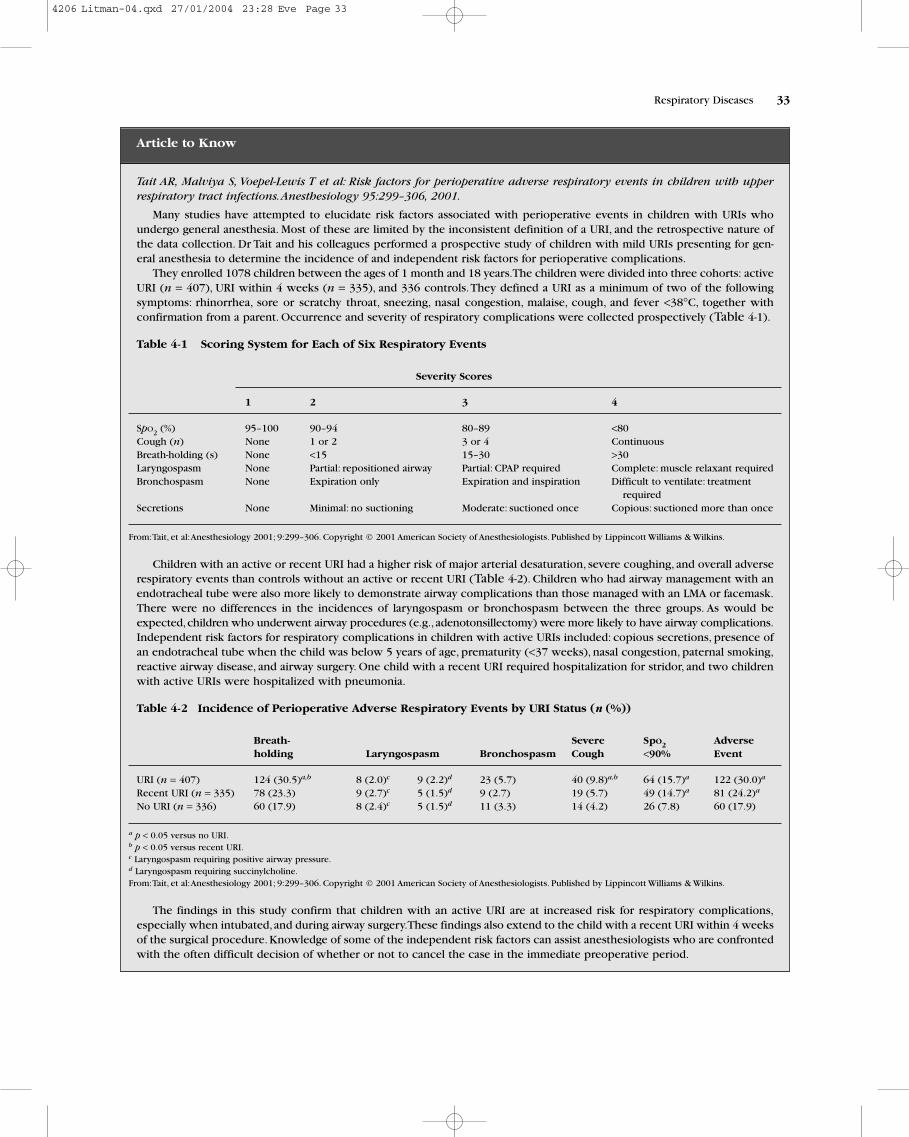
Tait AR, Malviya S, Voepel-Lewis T et al: Risk factors for perioperative adverse respiratory events in children with upperrespiratory tract infections.Anesthesiology 95:299–306, 2001.
Many studies have attempted to elucidate risk factors associated with perioperative events in children with URIs whoundergo general anesthesia. Most of these are limited by the inconsistent definition of a URI, and the retrospective nature ofthe data collection. Dr Tait and his colleagues performed a prospective study of children with mild URIs presenting for gen-eral anesthesia to determine the incidence of and independent risk factors for perioperative complications.
They enrolled 1078 children between the ages of 1 month and 18 years.The children were divided into three cohorts: activeURI (n = 407), URI within 4 weeks (n = 335), and 336 controls.They defined a URI as a minimum of two of the followingsymptoms: rhinorrhea, sore or scratchy throat, sneezing, nasal congestion, malaise, cough, and fever <38°C, together withconfirmation from a parent. Occurrence and severity of respiratory complications were collected prospectively (Table 4-1).
Table 4-1 Scoring System for Each of Six Respiratory Events
Severity Scores
1 2 3 4
SpO2 (%) 95–100 90–94 80–89 <80Cough (n) None 1 or 2 3 or 4 ContinuousBreath-holding (s) None <15 15–30 >30Laryngospasm None Partial: repositioned airway Partial: CPAP required Complete: muscle relaxant requiredBronchospasm None Expiration only Expiration and inspiration Difficult to ventilate: treatment
requiredSecretions None Minimal: no suctioning Moderate: suctioned once Copious: suctioned more than once
Children with an active or recent URI had a higher risk of major arterial desaturation, severe coughing, and overall adverserespiratory events than controls without an active or recent URI (Table 4-2). Children who had airway management with anendotracheal tube were also more likely to demonstrate airway complications than those managed with an LMA or facemask.There were no differences in the incidences of laryngospasm or bronchospasm between the three groups. As would beexpected,children who underwent airway procedures (e.g.,adenotonsillectomy) were more likely to have airway complications.Independent risk factors for respiratory complications in children with active URIs included: copious secretions, presence ofan endotracheal tube when the child was below 5 years of age, prematurity (<37 weeks), nasal congestion, paternal smoking,reactive airway disease, and airway surgery. One child with a recent URI required hospitalization for stridor, and two childrenwith active URIs were hospitalized with pneumonia.
Table 4-2 Incidence of Perioperative Adverse Respiratory Events by URI Status (n (%))
Breath- Severe SpO2 Adverse holding Laryngospasm Bronchospasm Cough <90% Event
The findings in this study confirm that children with an active URI are at increased risk for respiratory complications,especially when intubated,and during airway surgery.These findings also extend to the child with a recent URI within 4 weeksof the surgical procedure. Knowledge of some of the independent risk factors can assist anesthesiologists who are confrontedwith the often difficult decision of whether or not to cancel the case in the immediate preoperative period.
4206 Litman-04.qxd 27/01/2004 23:28 Eve Page 33
as airway edema, atelectasis, and bronchial reactivity mayremain for up to several weeks after the acute URI haveresolved.Three to four weeks seems to be a reasonablewaiting time, but for many children this merely repre-sents the period between successive illnesses.
LOWER AIRWAY DISEASES
The “lower airway” is traditionally thought of as thatportion of the respiratory system that is containedwithin the thoracic cavity. Therefore, lower airway dis-eases are those that primarily involve the lungs andbronchial system.
Asthma
Asthma is defined as a chronic disease of reversible air-way obstruction, and is characterized by bronchial hyper-reactivity, inflammation, and mucous secretion. Clinicalmanifestations of asthma include wheezing, persistentdry cough, and dyspnea on exertion. During an acuteexacerbation, marked respiratory distress occurs, whichmay include chest wall retractions and a prolonged expi-ratory phase secondary to bronchial obstruction. Recentstudies suggest that chronic airway inflammation ratherthan smooth-muscle contraction is the primary underly-ing pathophysiologic mechanism, and thus, maintenancetreatment regimens have changed accordingly.
34 PEDIATRIC ANESTHESIA: THE REQUISITES IN ANESTHESIOLOGY
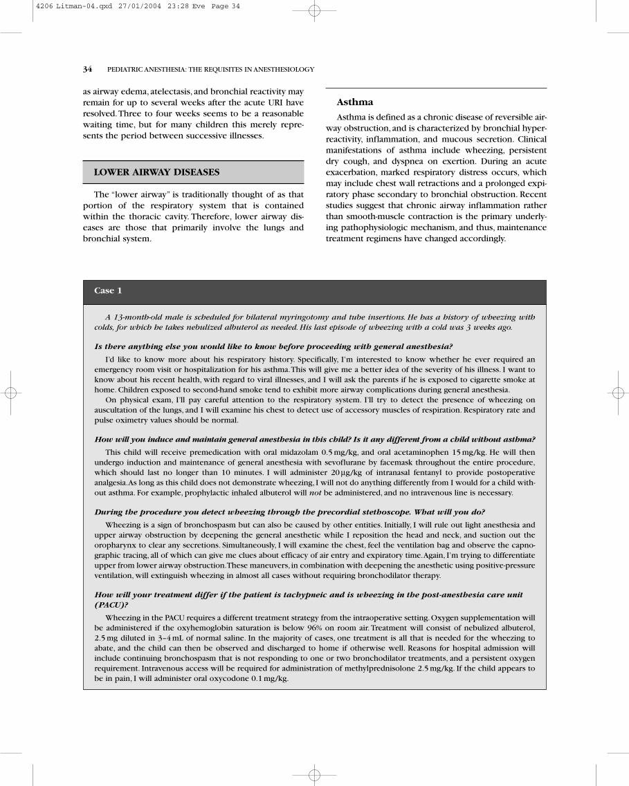
Case 1
A 13-month-old male is scheduled for bilateral myringotomy and tube insertions. He has a history of wheezing withcolds, for which he takes nebulized albuterol as needed. His last episode of wheezing with a cold was 3 weeks ago.
Is there anything else you would like to know before proceeding with general anesthesia?
I’d like to know more about his respiratory history. Specifically, I’m interested to know whether he ever required anemergency room visit or hospitalization for his asthma.This will give me a better idea of the severity of his illness. I want toknow about his recent health, with regard to viral illnesses, and I will ask the parents if he is exposed to cigarette smoke athome. Children exposed to second-hand smoke tend to exhibit more airway complications during general anesthesia.
On physical exam, I’ll pay careful attention to the respiratory system. I’ll try to detect the presence of wheezing onauscultation of the lungs, and I will examine his chest to detect use of accessory muscles of respiration. Respiratory rate andpulse oximetry values should be normal.
How will you induce and maintain general anesthesia in this child? Is it any different from a child without asthma?
This child will receive premedication with oral midazolam 0.5 mg/kg, and oral acetaminophen 15 mg/kg. He will thenundergo induction and maintenance of general anesthesia with sevoflurane by facemask throughout the entire procedure,which should last no longer than 10 minutes. I will administer 20 µg/kg of intranasal fentanyl to provide postoperativeanalgesia.As long as this child does not demonstrate wheezing, I will not do anything differently from I would for a child with-out asthma. For example, prophylactic inhaled albuterol will not be administered, and no intravenous line is necessary.
During the procedure you detect wheezing through the precordial stethoscope. What will you do?
Wheezing is a sign of bronchospasm but can also be caused by other entities. Initially, I will rule out light anesthesia andupper airway obstruction by deepening the general anesthetic while I reposition the head and neck, and suction out theoropharynx to clear any secretions. Simultaneously, I will examine the chest, feel the ventilation bag and observe the capno-graphic tracing, all of which can give me clues about efficacy of air entry and expiratory time.Again, I’m trying to differentiateupper from lower airway obstruction.These maneuvers, in combination with deepening the anesthetic using positive-pressureventilation, will extinguish wheezing in almost all cases without requiring bronchodilator therapy.
How will your treatment differ if the patient is tachypneic and is wheezing in the post-anesthesia care unit(PACU)?
Wheezing in the PACU requires a different treatment strategy from the intraoperative setting.Oxygen supplementation willbe administered if the oxyhemoglobin saturation is below 96% on room air. Treatment will consist of nebulized albuterol,2.5 mg diluted in 3–4 mL of normal saline. In the majority of cases, one treatment is all that is needed for the wheezing toabate, and the child can then be observed and discharged to home if otherwise well. Reasons for hospital admission willinclude continuing bronchospasm that is not responding to one or two bronchodilator treatments, and a persistent oxygenrequirement. Intravenous access will be required for administration of methylprednisolone 2.5 mg/kg. If the child appears tobe in pain, I will administer oral oxycodone 0.1 mg/kg.
4206 Litman-04.qxd 27/01/2004 23:28 Eve Page 34
Respiratory Diseases 35
With a prevalence of approximately 10% (and contin-ually increasing in most urban areas) asthma has becomethe most common chronic illness in children in theUnited States. Ninety percent of children with asthmapresent before the age of 6 years. An exacerbation ofasthma may be caused by allergic, environmental, infec-tious,or emotional stimuli, among others, and can last upto several hours. Some resolve spontaneously, whereasothers require aggressive medical therapy.
Anesthesiologists will most commonly encounter chil-dren with asthma prior to elective surgery.The majorityof these children will have not had a recent exacerbationof wheezing, and may be taking maintenance therapy.Preoperatively, the anesthesiologist should assess theseverity and current status of the child’s illness by focus-ing on several aspects related to the disease. Importantdetails of the medical history include the number ofemergency room visits during the previous year, numberof hospitalizations for asthma exacerbations, previousoccurrences of pneumothorax or respiratory arrest, andthe current and recent medication history. The parentsand child (if old enough) will usually be able to provide arelative estimate of the current severity of their condition.
The physical examination of the child is focused onthe respiratory system, looking for clues of ongoingbronchospasm.These include audible wheezes on expi-ration, a prolonged expiratory time, and use of accessorymuscles of respiration. Pulse oximetry measurementshould be obtained to determine the child’s baselineoxyhemoglobin saturation. A reading less than 96% inroom air is a cause for concern and further evaluation.
Based on the history and physical exam findings, theanesthesiologist should estimate whether the child isoptimized for elective surgery and whether or not toproceed with an elective anesthetic. For example, mildwheezing may be serious in a child who never wheezesbetween acute exacerbations, as opposed to the childwho continually has a baseline wheeze, who may beconsidered to be optimized at the time of surgery.
The treatment of asthma consists mainly of bronchodila-tors and inhaled corticosteroids. Nebulized β2-agonists(e.g., albuterol, levalbuterol) produce bronchodilatationvia stimulation of β2-receptors on airway smooth muscle.They are administered as daily maintenance agents or onan as-needed basis. Administration of steroids is associ-ated with decreased airway inflammation, decreasedmucus secretion, and decreased release of proinflamma-tory cytokines.Aerosolized steroids (e.g.,budesonide) arebreathed directly into the lungs and are not associatedwith systemic side-effects, but are generally not usefulduring acute exacerbations.Intravenous steroids will beginto decrease airway inflammation within several hours ofadministration and are an appropriate treatment duringan acute exacerbation.
Additional preventative therapies include orallyadministered leukotriene receptor antagonists(leukotrienes are lipid mediators generated from themetabolism of arachidonic acid, and have been shown toplay an important role in the pathogenesis of asthmaticinflammation), and inhaled or oral cromolyn, which pre-vents episodes of bronchospasm by stabilizing the mastcell membrane and preventing release of inflammatory
Case 2
A 4-year-old boy with asthma requires general endotracheal anesthesia for umbilical hernia repair. He is maintainedon inhaled steroids, inhaled cromolyn, an orally administered leukotriene antagonist, and occasionally requires nebu-lized albuterol for acute episodes of wheezing. Two weeks prior to the surgery, he required one week of oral prednisonefor an asthma exacerbation that was worse than usual.
Does the recent exacerbation and oral steroid requirement change your approach to the anesthetic management?
There are two ways the history of a recent asthma exacerbation may change my anesthetic management approach. First,I will make sure the child is now in excellent health, and without any wheezing or URI.The procedure is purely elective andshould not be performed if the child is still having symptoms of his illness. Second, if his illness has completely abated thischild may be a candidate for prophylactic oral steroid therapy for several days prior to the procedure. I will make this deter-mination by speaking with the boy’s parents several days prior to the procedure. If he has required regular hospitalizationfor his asthma, or frequent systemic steroid use in the past, it indicates that his disease is prone to flare-ups. I will ask hispediatrician to see him prior to the scheduled surgery and prescribe oral steroids for several days.This usually consists ofprednisone, up to 1 mg/kg daily.
If this child has had frequent asthma recurrences, I will alter my intraoperative management approach by avoidingendotracheal intubation. Most surgeons who perform umbilical hernia repair prefer intraoperative paralysis and there is noreason I can’t use a LMA with controlled ventilation during the procedure. I will avoid administration of medications thatare associated with histamine release, such as morphine and mivacurium, and I will remind the surgeon to administer localanesthetic into the wound, up to 1 mL/kg of 0.25% bupivacaine or 0.2% ropivacaine.
4206 Litman-04.qxd 27/01/2004 23:28 Eve Page 35
mediators such as histamine. Theophylline is no longerused as a first-line therapy because of its narrow thera-peutic range and questionable efficacy; it is reserved forchildren in status asthmaticus who fail more conventionaltherapy. Most recently, magnesium has been described aseffective treatment for asthma.The suggested mechanism
of action is smooth-muscle relaxation secondary to inhi-bition of calcium uptake. The current dose recommen-dation of intravenous magnesium for treating asthma is25–75 mg/kg over 20 minutes.
The anesthetic management of children with asthmais aimed at preventing an exacerbation of the disease.
36 PEDIATRIC ANESTHESIA: THE REQUISITES IN ANESTHESIOLOGY
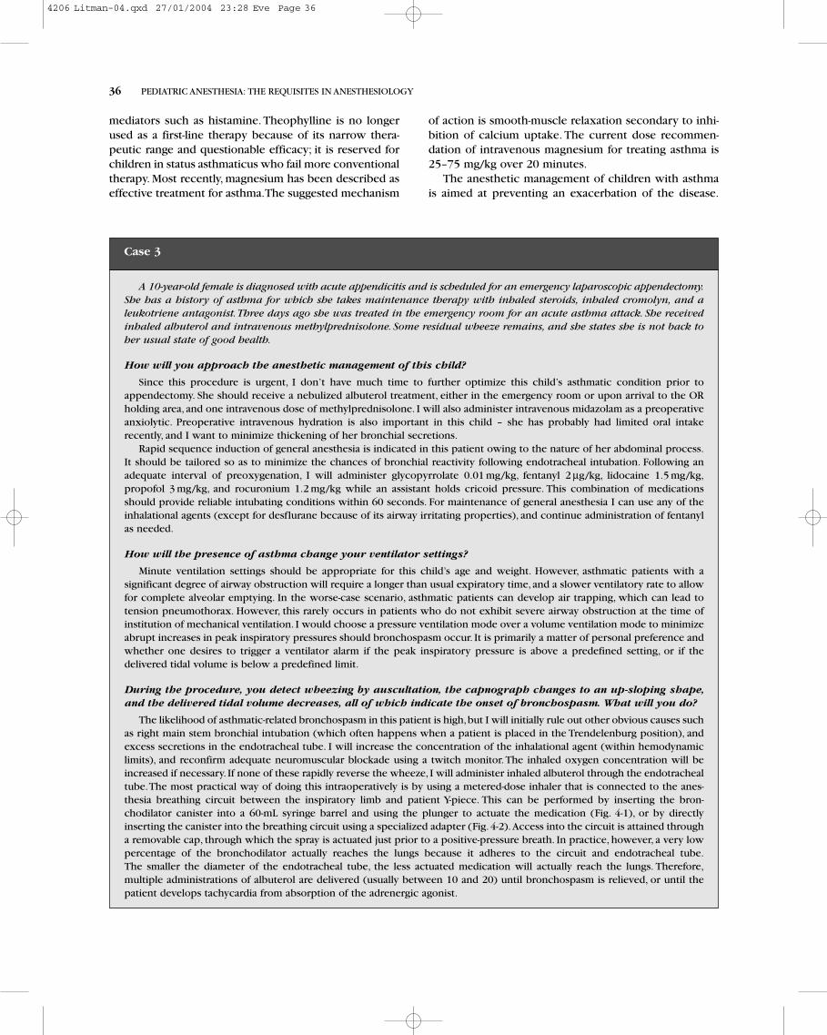
Case 3
A 10-year-old female is diagnosed with acute appendicitis and is scheduled for an emergency laparoscopic appendectomy.She has a history of asthma for which she takes maintenance therapy with inhaled steroids, inhaled cromolyn, and aleukotriene antagonist. Three days ago she was treated in the emergency room for an acute asthma attack. She receivedinhaled albuterol and intravenous methylprednisolone. Some residual wheeze remains, and she states she is not back toher usual state of good health.
How will you approach the anesthetic management of this child?
Since this procedure is urgent, I don’t have much time to further optimize this child’s asthmatic condition prior toappendectomy. She should receive a nebulized albuterol treatment, either in the emergency room or upon arrival to the ORholding area, and one intravenous dose of methylprednisolone. I will also administer intravenous midazolam as a preoperativeanxiolytic. Preoperative intravenous hydration is also important in this child – she has probably had limited oral intakerecently, and I want to minimize thickening of her bronchial secretions.
Rapid sequence induction of general anesthesia is indicated in this patient owing to the nature of her abdominal process.It should be tailored so as to minimize the chances of bronchial reactivity following endotracheal intubation. Following anadequate interval of preoxygenation, I will administer glycopyrrolate 0.01 mg/kg, fentanyl 2 µg/kg, lidocaine 1.5 mg/kg,propofol 3 mg/kg, and rocuronium 1.2 mg/kg while an assistant holds cricoid pressure. This combination of medicationsshould provide reliable intubating conditions within 60 seconds. For maintenance of general anesthesia I can use any of theinhalational agents (except for desflurane because of its airway irritating properties), and continue administration of fentanylas needed.
How will the presence of asthma change your ventilator settings?
Minute ventilation settings should be appropriate for this child’s age and weight. However, asthmatic patients with asignificant degree of airway obstruction will require a longer than usual expiratory time, and a slower ventilatory rate to allowfor complete alveolar emptying. In the worse-case scenario, asthmatic patients can develop air trapping, which can lead totension pneumothorax. However, this rarely occurs in patients who do not exhibit severe airway obstruction at the time ofinstitution of mechanical ventilation. I would choose a pressure ventilation mode over a volume ventilation mode to minimizeabrupt increases in peak inspiratory pressures should bronchospasm occur. It is primarily a matter of personal preference andwhether one desires to trigger a ventilator alarm if the peak inspiratory pressure is above a predefined setting, or if thedelivered tidal volume is below a predefined limit.
During the procedure, you detect wheezing by auscultation, the capnograph changes to an up-sloping shape,and the delivered tidal volume decreases, all of which indicate the onset of bronchospasm. What will you do?
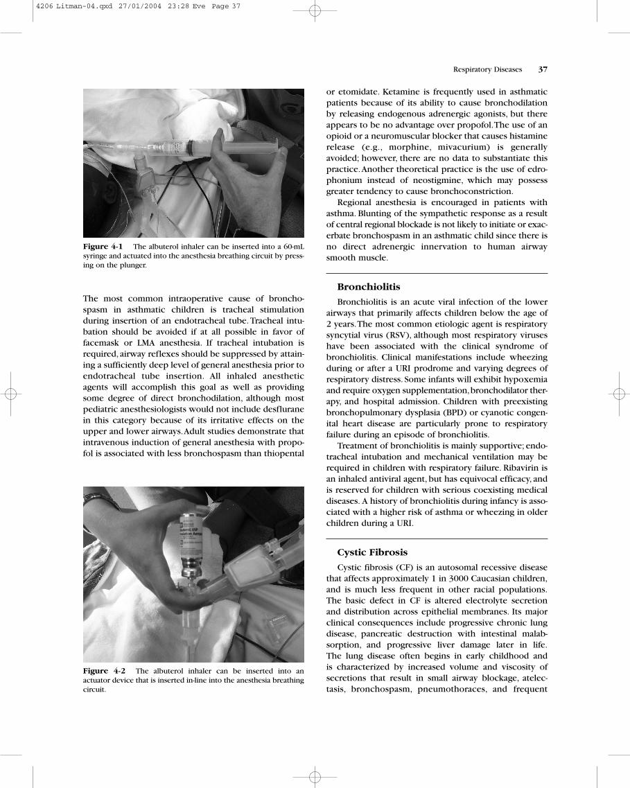
The likelihood of asthmatic-related bronchospasm in this patient is high,but I will initially rule out other obvious causes suchas right main stem bronchial intubation (which often happens when a patient is placed in the Trendelenburg position), andexcess secretions in the endotracheal tube. I will increase the concentration of the inhalational agent (within hemodynamiclimits), and reconfirm adequate neuromuscular blockade using a twitch monitor.The inhaled oxygen concentration will beincreased if necessary. If none of these rapidly reverse the wheeze, I will administer inhaled albuterol through the endotrachealtube.The most practical way of doing this intraoperatively is by using a metered-dose inhaler that is connected to the anes-thesia breathing circuit between the inspiratory limb and patient Y-piece. This can be performed by inserting the bron-chodilator canister into a 60-mL syringe barrel and using the plunger to actuate the medication (Fig. 4-1), or by directlyinserting the canister into the breathing circuit using a specialized adapter (Fig.4-2).Access into the circuit is attained througha removable cap, through which the spray is actuated just prior to a positive-pressure breath. In practice, however, a very lowpercentage of the bronchodilator actually reaches the lungs because it adheres to the circuit and endotracheal tube.The smaller the diameter of the endotracheal tube, the less actuated medication will actually reach the lungs. Therefore,multiple administrations of albuterol are delivered (usually between 10 and 20) until bronchospasm is relieved, or until thepatient develops tachycardia from absorption of the adrenergic agonist.
4206 Litman-04.qxd 27/01/2004 23:28 Eve Page 36
Respiratory Diseases 37
The most common intraoperative cause of broncho-spasm in asthmatic children is tracheal stimulationduring insertion of an endotracheal tube. Tracheal intu-bation should be avoided if at all possible in favor offacemask or LMA anesthesia. If tracheal intubation isrequired, airway reflexes should be suppressed by attain-ing a sufficiently deep level of general anesthesia prior toendotracheal tube insertion. All inhaled anestheticagents will accomplish this goal as well as providingsome degree of direct bronchodilation, although mostpediatric anesthesiologists would not include desfluranein this category because of its irritative effects on theupper and lower airways.Adult studies demonstrate thatintravenous induction of general anesthesia with propo-fol is associated with less bronchospasm than thiopental
or etomidate. Ketamine is frequently used in asthmaticpatients because of its ability to cause bronchodilationby releasing endogenous adrenergic agonists, but thereappears to be no advantage over propofol.The use of anopioid or a neuromuscular blocker that causes histaminerelease (e.g., morphine, mivacurium) is generallyavoided; however, there are no data to substantiate thispractice.Another theoretical practice is the use of edro-phonium instead of neostigmine, which may possessgreater tendency to cause bronchoconstriction.
Regional anesthesia is encouraged in patients withasthma. Blunting of the sympathetic response as a resultof central regional blockade is not likely to initiate or exac-erbate bronchospasm in an asthmatic child since there isno direct adrenergic innervation to human airwaysmooth muscle.
Bronchiolitis
Bronchiolitis is an acute viral infection of the lowerairways that primarily affects children below the age of2 years.The most common etiologic agent is respiratorysyncytial virus (RSV), although most respiratory viruseshave been associated with the clinical syndrome ofbronchiolitis. Clinical manifestations include wheezingduring or after a URI prodrome and varying degrees ofrespiratory distress. Some infants will exhibit hypoxemiaand require oxygen supplementation,bronchodilator ther-apy, and hospital admission. Children with preexistingbronchopulmonary dysplasia (BPD) or cyanotic congen-ital heart disease are particularly prone to respiratoryfailure during an episode of bronchiolitis.
Treatment of bronchiolitis is mainly supportive; endo-tracheal intubation and mechanical ventilation may berequired in children with respiratory failure. Ribavirin isan inhaled antiviral agent, but has equivocal efficacy, andis reserved for children with serious coexisting medicaldiseases. A history of bronchiolitis during infancy is asso-ciated with a higher risk of asthma or wheezing in olderchildren during a URI.
Cystic Fibrosis
Cystic fibrosis (CF) is an autosomal recessive diseasethat affects approximately 1 in 3000 Caucasian children,and is much less frequent in other racial populations.The basic defect in CF is altered electrolyte secretionand distribution across epithelial membranes. Its majorclinical consequences include progressive chronic lungdisease, pancreatic destruction with intestinal malab-sorption, and progressive liver damage later in life.The lung disease often begins in early childhood andis characterized by increased volume and viscosity ofsecretions that result in small airway blockage, atelec-tasis, bronchospasm, pneumothoraces, and frequent
Figure 4-1 The albuterol inhaler can be inserted into a 60-mLsyringe and actuated into the anesthesia breathing circuit by press-ing on the plunger.
Figure 4-2 The albuterol inhaler can be inserted into anactuator device that is inserted in-line into the anesthesia breathingcircuit.
4206 Litman-04.qxd 27/01/2004 23:28 Eve Page 37
antibiotic-resistant bacterial infections. Nasal polyp for-mation and sinus infections are common. Bronchiectasisdevelops later in life: occasional bouts of hemoptysismay lead to significant anemia.
Over the past several decades, medical managementof this disease has improved significantly, and patientsoften live well into adulthood.Treatment strategies includechest physical therapy, exercise, and frequent coughing tomobilize secretions.Bronchodilators and anti-inflammatorymedications decrease airway reactivity. Bacterial pneu-monia requires aggressive antibiotic therapy. Nebulizeddornase (Pulmozyme) can be administered to break downthick DNA complexes that are present in mucus due tocell destruction and bacterial infection. Normal growthcan often be achieved with pancreatic enzyme replace-ment, fat-soluble vitamin supplements, and high-caloriehigh-protein diets.
Common reasons that children with CF require surgeryinclude meconium ileus in the newborn period, nasalpolypectomy,and endoscopic sinus surgery.Older or moreseverely ill children may require anesthesia for place-ment of indwelling central line access, or gastrostomytube insertion. Preoperative evaluation of pulmonaryfunction is essential; possible studies include chest radi-ography, pulmonary function tests, and arterial blood gasanalysis. Optimization of infection control and physio-therapy for secretion clearance are priorities, and arecoordinated with the child’s pulmonologist.
The anesthetic technique of choice for childrenwith CF is controversial. Some advocate use of ketaminebecause of its minimal effects on ventilatory function;however, others cite ketamine’s ability to increaseairway secretions, which may worsen respiratory func-tion in patients with CF. Fluid management is alsocontroversial – some pediatric anesthesiologists prefera liberal fluid strategy to decrease viscosity ofbronchial secretions while others advocate minimiza-tion of fluids to decrease airway secretions at theexpense of increased viscosity. It seems that avoidanceof either overhydration or dehydration is the most prudentcourse of action.
In children with significant pulmonary disease andpoor nutritional status, placement of an endotracheal
38 PEDIATRIC ANESTHESIA: THE REQUISITES IN ANESTHESIOLOGY
Table 4-3 Classification of Severity of MeconiumAspiration Syndrome (MAS)
Type of MAS Therapy Required
Mild <40% oxygen therapy for <48 hoursModerate >40% oxygen therapy for >48 hoursSevere Requirement for mechanical ventilation
tube and application of mechanical ventilation oftenentails postoperative transfer to the ICU and the difficultdecision-making process concerning the timing andappropriateness of tracheal extubation. Postoperativemanagement should be proactively planned in conjunc-tion with the intensive care physicians, and with theinput of the patient and family.
Meconium Aspiration Syndrome
Fetal hypoxia triggers the passage of meconium intothe amniotic fluid, which is then swallowed into theorapharynx and aspirated into the trachea and lungsprior to or at the time of birth. Passage of thin meconiumin a vigorous, otherwise well neonate can result in mildmeconium aspiration syndrome (MAS; Table 4-3). Thepassage of thick meconium in an asphyxiated newborncan result in moderate or severe MAS. Moderate orsevere MAS occurs when aspirated meconium causesbronchial obstruction and pneumonitis, which leads toventilation/perfusion mismatch and hypoxemia. Thepresence of meconium in the amniotic fluid warrantsaggressive suctioning of the fetal mouth and pharynxprior to delivery,and attempted tracheal suctioning priorto the newborn taking its first breaths. However, when asubstantial amount of thick meconium has been aspi-rated by an asphyxiated infant, peripartum suctioningdoes not prevent severe MAS.
Hypoxemia and acidosis increase pulmonary vascularresistance and lead to persistent pulmonary hypertensionof the newborn (PPHN) (see Chapter 1). Treatmentincludes optimization of mechanical ventilation and pos-sible institution of extracorporeal membrane oxygenation(ECMO) until lung function returns to normal and PPHNis resolved.
ADDITIONAL ARTICLES TO KNOW
Cohen MM, Cameron CB: Should you cancel the operationwhen a child has an upper respiratory tract infection? AnesthAnalg 72:282–288, 1991.
DeSoto H, Patel RI, Soliman IE, Hannallah RS: Changes inoxygen saturation following general anesthesia in childrenwith upper respiratory infection signs and symptoms undergo-ing otolaryngological procedures. Anesthesiology 68:276–279,1988.
Habre W, Matsumoto I, Sly PD: Propofol or halothane anaesthe-sia for children with asthma: effects on respiratory mechanics.Br J Anaesth 77:739–743, 1996.
Kinouchi K, Tanigami H, Tashiro C et al: Duration of apnea inanesthetized infants and children required for desaturation ofhemoglobin to 95%.The influence of upper respiratory infec-tion. Anesthesiology 77:1105–1107, 1992.
McGill WA, Coveler LA, Epstein BS: Subacute upper respiratoryinfection in small children. Anesth Analg 58:331–333, 1979.
4206 Litman-04.qxd 27/01/2004 23:28 Eve Page 38
Respiratory Diseases 39
Schreiner MS, O’Hara I, Markakis DA, Politis GD: Do childrenwho experience laryngospasm have an increased risk of upperrespiratory tract infection? Anesthesiology 85:475–480, 1997.
Tait AR, Pandit UA,Voepel-Lewis T, Munro HM, Malviya S: Use ofthe laryngeal mask airway in children with upper respiratorytract infections: a comparison with endotracheal intubation.Anesth Analg 86:706–711, 1998.
Warner DO,Warner MA, Barnes RD et al: Perioperative respira-tory complications in patients with asthma. Anesthesiology85:460–467, 1997.
Willliams OA, Hills R, Goddard JM: Pulmonary collapse duringanaesthesia in children with respiratory tract symptoms.Anaesthesia 47:411–413, 1992.


![[S3Lab1] Respiratory Diseases](https://static.documents.pub/doc/80x56/577d349a1a28ab3a6b8e6d89/s3lab1-respiratory-diseases.jpg)


