80
IMMUNOLOGICAL LABORATORY Dr. Wisanu Wanasawaeng Ph.D. (Pathobiology), M.Sc. (Avian Medicine), DVM. Veterinary Laboratory Utilization (VMCL3231)
| Date post: | 07-Aug-2015 |
| Category: |
Science |
| Upload: | wisanu-wanasawaeng |
| View: | 180 times |
| Download: | 4 times |

IMMUNOLOGICAL
LABORATORY Dr. Wisanu Wanasawaeng
Ph.D. (Pathobiology), M.Sc. (Avian Medicine), DVM.
Veterinary Laboratory Utilization (VMCL3231)

Contents
Antigens &Antibodies
Precipitations
Agglutinations
Enzyme immunoassay
Immunochromatography
Test for cell-associated antigen
Fluorescent activated cell sorters
Immunoblotting
Antibody microarray

Antigens: • Antigen is a substance that stimulate the production of specific antibodies and
combine specifically with the antibodies produced.
• Most antigens are foreign to the blood and other bodily fluids.
Antibodies:
• Antibody proteins (immunoglobulins) are found in the gamma globulin class of plasma proteins.
• There are five main subclasses : IgG, IgA, IgM, IgD, and IgE.
• Most antibodies in serum are from the class IgG.
3
Antigens

Antibody Structure
Antibody consists of four
interconnected polypeptide chains
Four interconnected polypeptide chains.
Two heavy chains (H-chains) and
joined to two shorter chains (L-chains).
These four chains are arranged in the
form of a ‘Y’ ; with the stalk of the Y is
called the “crystallizable fragment”
The top of the Y is known as the
“antigen-binding fragment”. 4

antigenicity
The ability to combine specifically with the
final products of immune response e.g.
secreted antibodies
Immunogenicity
The ability to induce a humoral and/or cell-
mediated immune response
Epitope or antigenic determinant
The distinct molecular surface features of
an antigen capable to being bound by an
antibody
5
Antigenicity
Immunogenicity
Epitope or antigenic determinant

Ag-ab interaction
The bonds holding Ags and Abs
combining site are all non-
covalent in nature
Hydrogen bonds, electrostatic
bonds, Hydrophobic bonds and
Van der Walls forces
Multiple bonding ensures that
Ags will be bound tightly to Abs
6
Non-covalent bonds

Lock and key concept
The combining site of Abs is located
in the Fab portion of the molecule and
is constructed from Hypervariable
regions of the heavy and light chains
X-ray crystallography studies of Ag-
Ab interactions show the antigenic
determinant nestles in a cleft formed
by the combining site of the antibody
7
Lock and Key concept

affinity
The strength of the reaction between a single antigenic
determinant and a single Ab combining site on Abs
The sum of the attractive and repulsive forces operating
between the antigenic determinant and the combining
site of the antibody
Affinity: the equilibrium constant of the Ag-Ab reactions
Apply the law of mass action
Ka (K1/K2) = [Ag-Ab]
8
Affinity = ∑ attractive + repulsive forces
[Ag] [Ab]

Affinity constant determination
Equilibrium dialysis
9
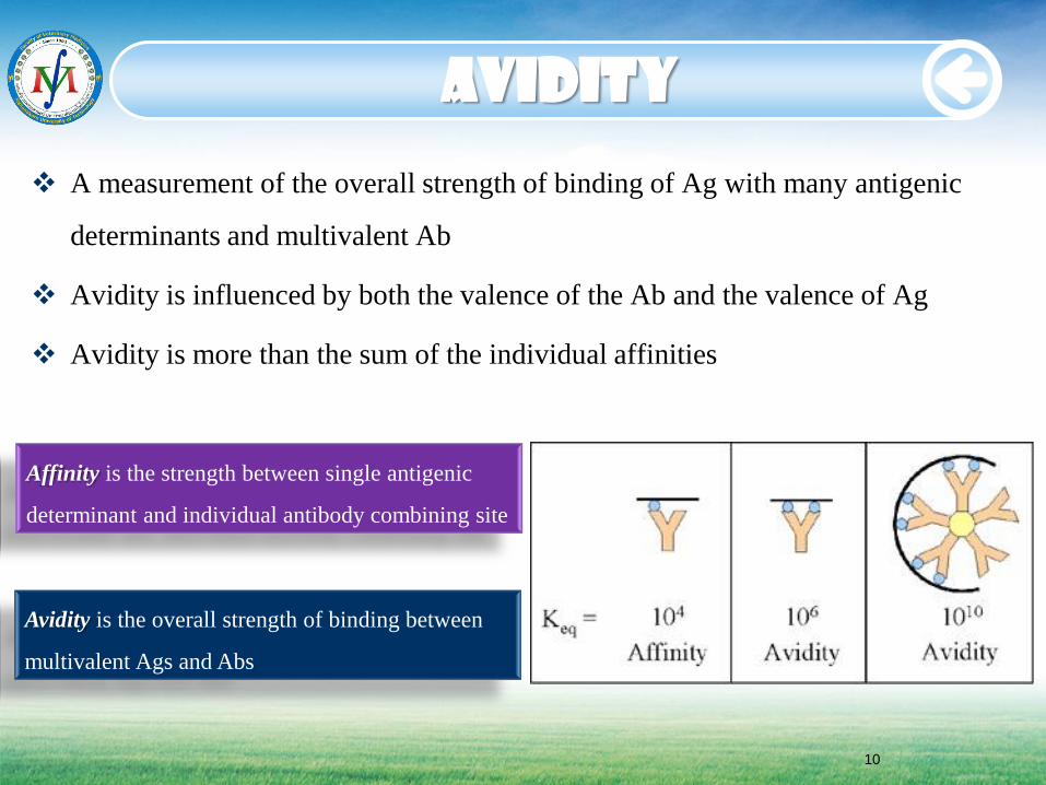
A measurement of the overall strength of binding of Ag with many antigenic
determinants and multivalent Ab
Avidity is influenced by both the valence of the Ab and the valence of Ag
Avidity is more than the sum of the individual affinities
Avidity
Affinity is the strength between single antigenic
determinant and individual antibody combining site
Avidity is the overall strength of binding between
multivalent Ags and Abs
10

Cross Reactivity
The ability of an individual antibody combining site to react with more than one
antigenic determinant.
The ability of a population of Ab molecules to react with more than one Ag.
Cross reactions arise because the cross reacting Ag shares epitope in common
with immunizing Ag.
Anti-
A Ab
Ag A
Anti-
A Ab
Ag B
Shared epitope
Anti-
A Ab
Ag C
Similar epitope
Cross reactions
11
Cross reactions

12
Classical precipitin curve
Antigen-antibody reaction:


Precipitin reactions in gels
Ab is incorporated into the agar gel &different
dilution of Ag are placed in holes punched into
the agar
Ag diffuses into gel &reacts with Ab
Equivalence point: a ring of precipitation is
formed
The positioning of Ab &Ag solution within
wells cut into an agar matrix to allow
visualization of precipitin bands formed by
reaction of components from the two solutions.
14
Radial immunodiffusion
Double immunodiffusion

Radial Immunodiffusion
Interpretation:
Diameter of ring is
proportional to the
concentration
Quantitative:
Ig levels
• Method
– Ab in gel
– Ag in a well
Ag Concentration
Dia
me
ter2
Ag Ag Ag Ag
Ab in gel Mancini method
15
16
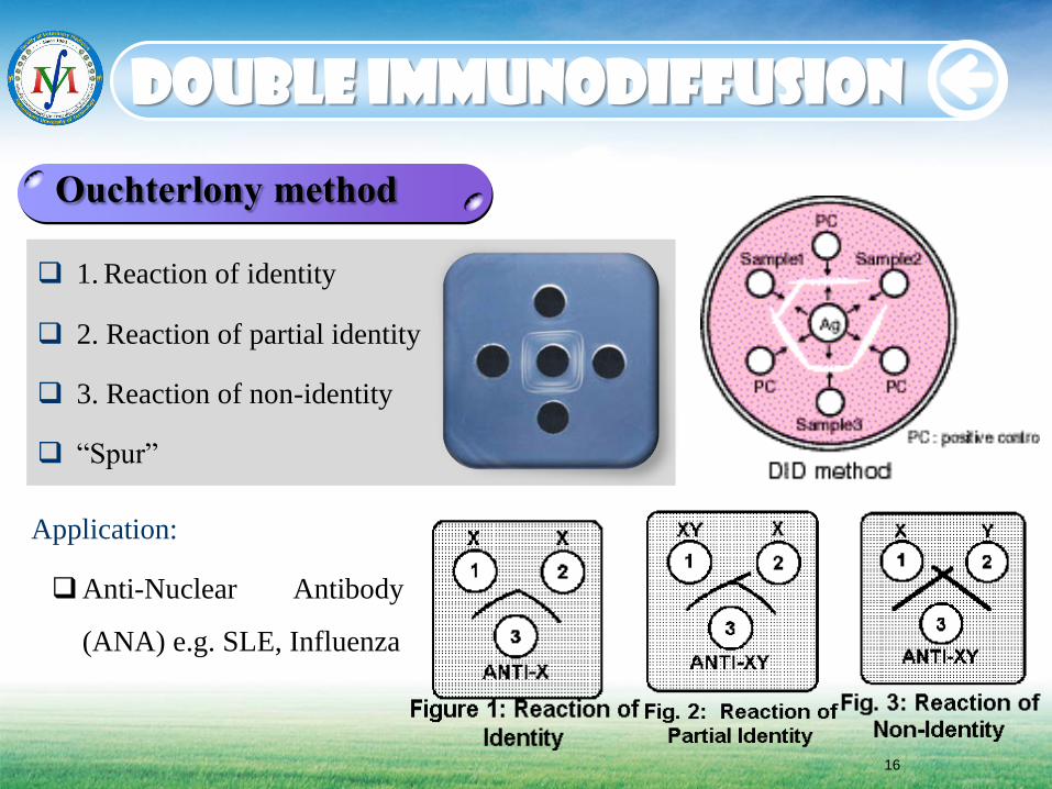
Double immunodiffusion
1. Reaction of identity
2. Reaction of partial identity
3. Reaction of non-identity
“Spur”
Ouchterlony method
Application:
Anti-Nuclear Antibody
(ANA) e.g. SLE, Influenza

Immunoelectrophoresis
Incorporation of electrophoresis with
double diffusion
1. Ag mixture is separated by charge
2. Troughs are cut to direction of electrophoresis field
3. Anti-sera is added to trough
4. Ag and Ab diffusion towards each other to produce
precipitin bands
Application:
1. Presence/Absence of specific proteins or Ig classes
2. Immunodeficiency or immunoproliferative
disorders
17

Countercurrent electrophoresis
Method
Ag and Ab migrate toward each other by electrophoresis
Used only when Ag and Ab have opposite charges
A rapid version of Ouchterlony double diffusion technique
Qualitative: rapid
Ag Ab
- +
18


Agglutination tests
When Ag is particulate, the
reaction of Ab with Ag can
be detected by agglutination
(clumping) of Ag.
Simple, inexpensive,
but sensitive
Agglutinin is used when Abs
agglutinate particulate Ag.
Haemaggluinin is used when
Ag is erythrocyte.
Several types exist:
• 1. Haemagglutination
• 2. Bacterial agglutination
• 3. Passive agglutination
• 4. Agglutination
inhibition e.g. HI
+
20

Direct agglutination
Direct agglutination:
Reactions test patient serum against large, cellular antigens to screen for the
presence of antibodies
21

Blood agglutination
Agglutination (clumping) of type A red blood cells (RBCs) by anti-A Ab.
The Abs have two combining sites and are able to attach to the A Ag on adjacent
RBCs, thus causing the RBCs to bond together.
RBC + anti-A Ab ----- Haemagglutination = Blood group A
22

23
Blood agglutination

Group O: No clumping in blood drops A or B
Group A: Clumping in blood drop A with anti-A Ab
Group B: Clumping in blood drop B with anti-B Ab
Group AB: Clumping in both blood drops A and B
24
Blood agglutination

Serogrouping of bacteria
Place 1 drop of reagent from each bottle in
agglutination site on the test
plate.
Use a conventional 10µl loop to pick some
fresh colonies from the plate
where the bacteria have been
isolated, and transfer adjacent to the test plate.
Place the loop in the drop and
return this small volume to mix with bacterial
material.
Then mix &spread with the rest of the reagent
with in agglutination site.
Using a fresh loop, repeat the
procedure above for each serotype and agglutination site on the test
plate.
Agitate the test plate gently for
1 minute.
Read agglutination pattern while
agitating.
25

indirect agglutination
Indirect agglutination
• To test patient serum for the presence of Abs against soluble Ags, serum is mixed
with latex spheres with the soluble Ags attached.
• Abs will then cause visible agglutination of the latex spheres with the soluble
antigens attached.
• Abs may be attached to the latex spheres to test for the presence of soluble antigens
in patient serum.
26

Hae
mag
glu
tin
atio
n
Agglutination reactions
using red blood cells.
Using in blood typing,
the diagnosis of certain
diseases, &identification
of viruses.
Viral HA occurs when
spikes on the virus cause
agglutination of red blood
cells - there is no antigen-
antibody interaction.
Haemagglutination test
27

28
HA
Influenza virus
Influenza/Rubella/Mump Ag
(Haemagglutinin) + RBC
Haemagglutination
Haemagglutination test
29
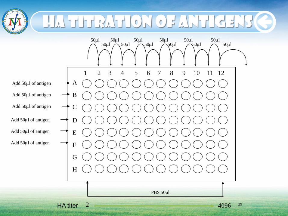
HA titration of antigens
A B
C D E F G H
1 2 3 4 5 6 7 8 9 10 11 12
HA titer 2 4096
PBS 50μl
50l 50l 50l 50l 50l 50l 50l 50l 50l 50l 50l 50l
Add 50l of antigen
Add 50l of antigen
Add 50l of antigen
Add 50l of antigen
Add 50l of antigen
Add 50l of antigen

Determination of HA titers
30

Haemagglutination Inhibition
RBC
Haemagglutination Inhibition (HI) RBC
Virus Haemagglutination (HA)
Virus
Ab - Virus
Ab Ab - Virus
31

32
Dilution of anti sera for HI test
A B
C D E F G H
1 2 3 4 5 6 7 8 9 10 11 12
Serum
titer
50 l 50 l 50 l 50 l 50 l 50 l
50l 50 l 50 l 50 l 50 l 50 l
Add 50l of serum
Add 50l of serum
Add 50l of serum
Add 50l of serum
Add 50l of serum
Add 50l of serum
Add 50l of serum
Add 50l of PBS (control)
+ 50 l
8HA antigen
to A-G wells
Then, incubate
30min.
Add 50 l
CRBC
Incubate 30min
PBS 50μl
2 4096

33
Haemagglutination Inhibition


Enzyme-linked immunosorbent
The most sensitive of tests for Ag/Ab
ELISA tests depend on enzyme conjugated to antibody reacting with a specific substrate to produce a color reaction
Allows for qualitative or quantitative testing of Ag or Ab
• Valuable tools for use in clinical labs, can measure Abs or Ags
• Inexpensive, rapid, quantitative, specific, sensitive (pg/ml)
• Expensive equipment not required (but helps!)
• Can be automated
35

Types of ELISA
36

37
Indirect ELISA
Antigen
Primary antibody
Secondary antibody HRP conjugated Species specific
anti-IgG antibody
Substrate Uncolored/Colored substrate
Positive Negative

38
Positive Negative
Antigen
Conjugate HRP conjugated Species specific
Anti-IgG Monoclonal antibody
Substrate Uncolored/Colored substrate
Capture Antibodies
Sandwich ELISA
Detector Antibodies

39
Add TMB
Competitive ELISA
Sheep anti-rabbit IgG
Rabbit anti-CAP IgG Sheep anti-rabbit IgG CAP Substrate
Rabbit anti-CAP + CAP-E + CAP
E E E E
E CAP+E
Positive Negative

40
• The most sensitive method
to measure antigen-specific T
cell function
eLISPOT

41
Blue-black colored precipitate forms at the sites of cytokine localization and
appears as spots
Each individual spot representing an individual cytokine-secreting cell.
The spots can be counted with automated ELISpot reader systems or
manually, using a stereomicroscope
eLISPOT

42
ELISPOT
New technology to detect individual activated
effector T cells
T-SPOT.TB is suitable for use with all patients at
risk of latent tuberculosis infection (LTBI) or
suspected of having TB disease, whether
immunocompetent or immunocompromised.
T-SPOT.tb


44
Immunochromatography
The need of on-site examination at the point-of-care system ex. farms, hospital
Not require any expert knowledge and complex procedure
Simple to use but quick responses

Principle
45
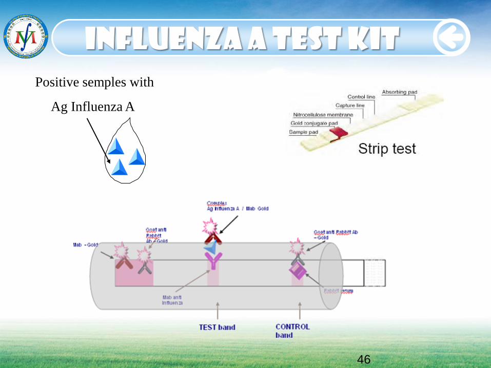
Positive semples with
Ag Influenza A
46
Influenza a test kit

Application
Pregnancy test kit: HCG pregnancy test
Drugs of abuse test kit: amphetamine, methamphetamine etc.
Steroid test kit in herbal medicine drug: dexamethazone, prednisolone etc.
Human infectious diseases test kit: influenza, HIV (1+2), HBsAb, HBsAg,
Dengue, Adeno/Rota, H. pylori etc.
Pet animal diseases test kit: CPV, FELV, FIV, LSH, RTV, Giardia, Crypto
Food borne pathogen test kit: salmonella, listeria etc.
47

Immunochromato Reader
48

Immunochromato Reader
49


Fluorescence microscope necessary for analysis of specimens
High structure resolution possible
Advanced image reconstruction (3D) and signal quantification
Multiple labeling easy
Limit shelf life of labeled specimens
Method of choice for labeling of live cells
51
Immunofluorescence

52
A.H. Coons (1941): fluorochrome-labeled antigens or antibodies
FITC (Fluorescein isothiocyanate): green
Absorption at wavelength 490-495 nm
Emission at wavelength 517 nm
TRITC (Tetramethyl rhodamine isothiocyante): red
Absorption at wavelength 550 nm
Emission at wavelength 580 nm
Fluorescence microscopy
1. Direct immunofluorescence
2. Indirect immunofluorescence
Immunofluorescence

Multiple immunofluorescence

IHC uses Abs to detect
&visualize Ags in cells
&tissues
Abs can be raised against almost any type
of Ags (protein, carbohydrate, lipid etc.)
Abs bind to Ag in a specific
manner
Bound Abs can be detected in several ways
54
Immunohistochemistry
A very important diagnostic &research tools

55
Two strategies used for the immmunohistochemical detection
of antigens in tissue
1. Direct method
2. Indirect method
Modified avidin-biotin complex (ABC) method
> Biotinylated secondary antibody
> Strept (avidin)-horseradish peroxidase
> 3, 3’-Diaminobenzidine (DAB)> brown
Immunohistochemistry

Fix
• Fix embed & section tissue
• Wash section in physiological buffer e.g. PBS
Block
• Incubate with protein solution (BSA) to reduce non-specific binding
of Abs to specimen
1 Ab
• Incubate with primary Ab + (Pos. &Neg. Control)
• Wash section in physiological buffer e.g. PBS
Detect
• Apply suitable detection system
• Mount specimens and analyze microscopically
56
Outline of procedures

Antigen expressing Cell
Primary Antibody (species X)
Biotinylated
Secondary Antibody
(anti species X)
Enzyme-conjugated
Avidin-Biotin Complex
Substrate-chromogen
solution
ABC (Avidin Biotin Complex)
Biotin (Vitamin B) binds with high affinity to Avidin-thus good linker system
Extremely high sensitivity
Endogenous biotin may be present in tissue-risk of background


59
Flow cytometry
Flow
Tip
Laser
FL
Detector
Light
Scatter
Detector
Flow cytometry
Cells in suspension labeled with fluorescent-tag
Cells analyzed on flow cytometer
FCM is a technique for
counting and examining
microscopic particles, such as
cells and chromosomes, by
suspending them in a stream of
fluid and passing them by an
electronic detection apparatus.
Simultaneous multiparametric
analysis of the physical and/or
chemical characteristics of up to
thousands of particles per second.

Data analysis
Data displayed
Gating: Histogram
Computational analysis
60

61
Fluorescent activated cell sorters
Cell sorting Monitor for fluorescence Forward angle light scatter (FALS or FSC) > cell size 90o light scatter > cytoplasmic granularity Deflection plates
Ultrasonic nozzle vibrator FACS


63
immunoblotting
• Immunoblotting or Western blotting is an extremely powerful technique for identifying a single protein (or epitope) in a complex mixture following separation based on its MW by SDS-PAGE, size and charge (non denaturing gel electrophoresis) or isoelectric point (isoelectric focusing)
• Three steps: 1. SDS-PAGE 2. Electro transfer 3. Antibody detection • Limit of Detection: 10 pg with HRP or AP labeling

A detergent that can dissolve
hydrophobic molecules with a negative
charge attached to it.
To denature all proteins to the same
linear shape.
Two important features:
1. All proteins retain only primary structure
2. All proteins have a large negative charge
and migrate toward the positive pole in
electric field
SDS-PAGE
64
SDS (Sodium dodecyl sulfate)

To put the proteins into an environment
that will allow different sized proteins to
move at different rates.
The environment of choice is
polyacrylamide, which is a polymer of
acrylamide monomers.
A polyacrylamide gel is made of a
labyrinth of tunnels through a meshwork
of fibers.
65
SDS-PAGE
PAGE (Polyacrylamide electrophoresis)

66
• Protein separation on SDS-polyacrylamide gel
• Electrophoretic transfer of protein blot into nitrocellulose membrane
SDS-PAGE

67
1. Analysis of the protein purity
2. Determination of protein molecular weight
3. Verification of protein concentration
4. Detection of proteolysis
5. Identification of immunoprecipitated proteins
6. First stage of immunoblotting
7. Detection of protein modification
8. Separation and concentration of protein antigens for antibody production
9. Separation of radioactively labeled proteins
applications

To identify specific proteins in mixtures
Proteins are separated on SDS-PAGE
Proteins are transferred to membrane
Membrane flooded with enzyme-linked
polyclonal Abs specific for protein
68
Western blotting
Western Blot

69
• Immunoblotting can be divided into two steps
• 1. Transfer of protein from the gel to the matrix
• 2. Decoration of the epitope with the specific antibody
Western Blot
Western blotting

70
• Block membrane with 3 % BSA/TBS for 1 hr • Wash membrane wit TBS • Add primary antibody at appropriate dilution in 0.5 % BSA/TBS for 1 hr • Wash membrane with TBS • Add secondary antibody at appropriate dilution in 0.5 %
BSA/TBS for 1 hr • Wash membrane with TBS • Add the adequate substrate solution: DAB
Western blotting

71
Lane 1: SF-9 cell lysate
2: Baculovirus wild type
3. H9N2 ELISA antigen
4: Molecular weight marker (kDa)
5: Blank
6: rH9N2 NP
~61kDa: Hemagglutinin
~56kDa: Nucleoprotein
~50kDa: Neuraminidase
~27kDa: Matrix Protein
Western blotting


Protein Microarray
Application of Protein arrays
Protein binding microarray, Biochip,
Protein chip
A multiplex approach to identify
protein-protein interactions, to
identify the targets of biologically
active small molecules
An important tool for proteomic
researches
Related microarray technology
including DNA microarrays, cellular
microarrays, tissue microarrays,
chemical compound microarrays and
antibody microarrays
73

Antibody (Ab) Microarray
A specific from of protein microarrays.
A collection of capture Abs are spotted and fixed on a solid surface such glass, plastic and silicon chip for the
purpose of detecting Ags.
Antibody microarray is often used for detecting protein expressions from cell lysates.
It is used for general research such as toxicity testing and drug discovery, and special markers from serum or
urine for diagnostic application
74

Procedure
Scanning of the array and the analysis of the results.
Incubation of labeled protein with the array.
Removal of unbound dye.
Labeling of extracted protein with fluorescent dyes Cy5 and Cy3.
Extraction of total cellular protein from biological samples of interest (e.g. Serum samples).
75

Biochip Array Technology
Biochip Array Technology for Multi-Analyte Testing
Simultaneous determination of multiple analystes
from a single sample.
Biochip is a solid substrate containing an array of
discrete test regions of immobilized Abs specific to
different antimicrobials
Competitive chemiluminescent immunoassay
76

Antimicrobial array 12 S
ulp
honam
ides
Oth
ers
77
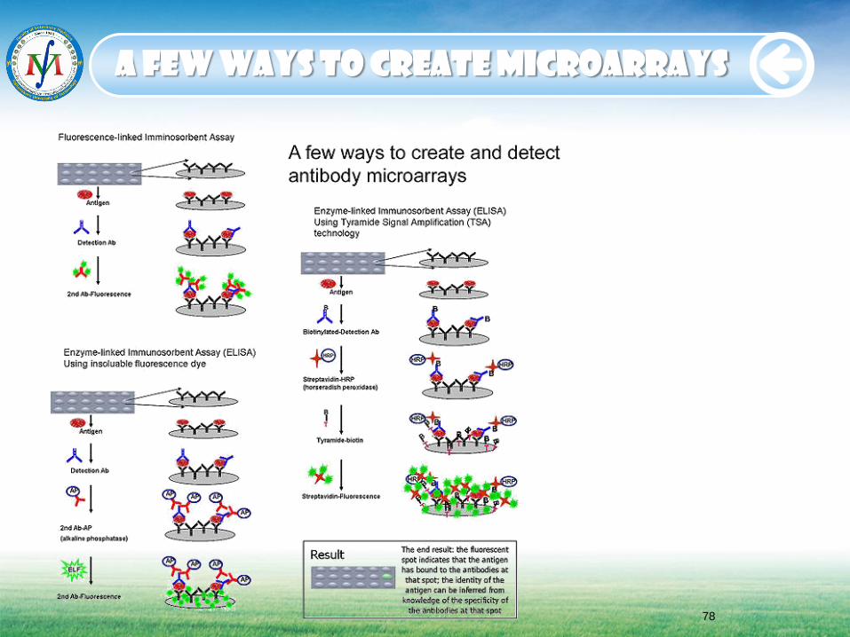
A few ways to create microarrays
78

Comparison of Immunoassay
Method Western’s
Blot ELISA ELISPOT
Immono
precipitation
Immono
cytochemistry
Immono
histochemistry Flow cytometry
Target
(Analyte)
Proteins or
polypeptide
Peptide,
proteins
Cells Protein or complex Cells Tissue section Cells
Primary
antibody
Binds
antigen
Binds
antigen
Can be linked to
organic dye or
quantum dot
Can be linked to
beads
Can be linked to
organic dye or
quantum dot
Can be linked to
organic dye or
quantum dot
Can be linked to
organic dye or
quantum dot
Secondary
antibody
Used for
signal
generation
Enzyme
linked for
generating
signal
Enzyme or dye or
dot linked
May be omitted Enzyme or dye or
dot linked
Enzyme or dye or
dot linked
Enzyme or dye or
dot linked
Data forms Protein
binding
image
Numerical
and curve
Image or
numerical
Protein binding
image
Image Image Numerical or
curve
Sensitivity ng/ml pg/ml pg/ml ng/ml ng/ml ng/ml 1/105 cells
Reproducibility Good High Good Variable Variable Variable Good
Quantification Semi Yes Semi Semi Semi Semi Yes
Applications Protein
analysis and
ident.
Antigen
analysis
Antigen analysis
Protein analysis
and ident.
Protein analysis
and ident.
Protein analysis and
ident.
Antigen analysis
Methods
79

Method Western’s
Blot ELISA ELISPOT
Immono
precipitation
Immono
cytochemistry
Immono
histochemistry Flow cytometry
Learn and
operation
Easy Easy Less easy Less easy Difficult Difficult More difficult
Assay
development
Simple Take time
(Days)
Take time
(Days)
Longer time
(Weeks)
Longer time
(Weeks)
Longer time
(Weeks)
Longer time
(Months)
Validation Simple Complicate Difficult Difficult Difficult Difficult Complicate
Standardization Straight
forward
Need
definition
Control variables Control variables More variables More variables
Need definition
High through
put
No Possible
with
automation
No No No Possible with
automation
Yes
Cost for set up Low ($1-
2K)
Low ($5-
10K)
Low ($5-10K) Low ($2-5K) Middle ($20-40K) Middle ($30-50K) High ($50-
100K)
Cost per assay Low Middle High High
High
Middle High
80
Comparison of Immunoassay
Methods


