Carlsberg Res. Commun. Vol. 45, p. 47-58, 1980 CHARACTERISTICS OF HIPROLY BARLEY I. ISOLATION AND CHARACTERISATION OF TWO WATER-SOLUBLE HIGH-LYSINE PROTEINS by IB JONASSEN Department of Biotechnology, Carlsberg Research Laboratory, Gamle Carlsberg Vej 10, DK-,2500 Copenhagen Valby and Institute of Genetics, University of Copenhagen, Oster Farimagsgade 2A, DK-1353 K. Keywords: Antibody production, antigenicity, molecular weight, isoelectric point, albumin. The water-soluble albumin of the high-lysine barley called Hiproly was fractionated by means of ion exchange chromatography and gel filtration. Three high-lysine fractions were isolated, and from the chromatographic profiles it was deduced that two of these contribute prominently to the overall high lysine content of Hiproly albumin. The two polypeptides were purified, and antisera raised against them. Immunological cross reaction was observed suggesting that the smaller polypeptide with a molecular weight of 8,100 dalton is a fragment of the larger one with a molecular weight of 9,100 dalton. The fragment contains 6 and the larger polypeptide 8 lysine residues. Both proteins lacked cysteine, but were rich in valine, glutamine/glutamic acid and asparagine/ aspartic acid. 1. INTRODUCTION In 1969 MUNCK et al. (18) reported the presence of a spontaneous high-lysine barley mutant in the World Barley Collection. The barley was of Ethiopian origin and named >>Hiproly~ because of its high lysine and high protein content. Fractionation of the crude protein of the Hiproly grain into various solubility classes revealed that the high-lysine content is partly due to an increased content of lysine rich albumin and globulin polypeptides (water- and salt-soluble proteins, respectively) and partly to an increased content of lysine in the glutelin fraction (alkali-soluble proteins). Abbreviations: BSA = bovine serum albumin. SP II A and SP I1 B -- lysine rich albumin polypeptides. 0105-1938/80/0045/0047/$ 02.40
Transcript
Carlsberg Res. Commun. Vol. 45, p. 47-58, 1980
CHARACTERISTICS OF HIPROLY BARLEY I. ISOLATION AND CHARACTERISATION
OF TWO WATER-SOLUBLE HIGH-LYSINE PROTEINS
by
IB JONASSEN
Department of Biotechnology, Carlsberg Research Laboratory, Gamle Carlsberg Vej 10, DK-,2500 Copenhagen Valby
and
Institute of Genetics, University of Copenhagen, Oster Farimagsgade 2A, DK-1353 K.
Keywords: Antibody production, antigenicity, molecular weight, isoelectric point, albumin.
The water-soluble albumin of the high-lysine barley called Hiproly was fractionated by means of ion exchange chromatography and gel filtration. Three high-lysine fractions were isolated, and from the chromatographic profiles it was deduced that two of these contribute prominently to the overall high lysine content of Hiproly albumin. The two polypeptides were purified, and antisera raised against them. Immunological cross reaction was observed suggesting that the smaller polypeptide with a molecular weight of 8,100 dalton is a fragment of the larger one with a molecular weight of 9,100 dalton. The fragment contains 6 and the larger polypeptide 8 lysine residues. Both proteins lacked cysteine, but were rich in valine, glutamine/glutamic acid and asparagine/ aspartic acid.
1. INTRODUCTION
In 1969 MUNCK et al. (18) reported the presence of a spontaneous high-lysine barley mutant in the World Barley Collection. The barley was of Ethiopian origin and named >>Hiproly~ because of its high lysine and high
protein content. Fractionation of the crude
protein of the Hiproly grain into various solubility classes revealed that the high-lysine content is partly due to an increased content of lysine rich albumin and globulin polypeptides (water- and salt-soluble proteins, respectively) and partly to an increased content of lysine in the glutelin fraction (alkali-soluble proteins).
Abbreviations: BSA = bovine serum albumin. SP II A and SP I1 B -- lysine rich albumin polypeptides.
0105-1938/80/0045/0047/$ 02.40
I. JONASSEN: Characteristics of Hiproly barley 1.
It has been shown (18) that the high-lysine character is due to a single recessive gene. MUNCK (17) reported that F4 lines from the cross Hiproly • normal lysine barley could be classified into three groups: normal lysine, elevated lysine, and high lysine. Grain hardness and cohesiveness of starch with the matrix protein (18) were also found to be characteristic for the high-lysine segregants, but there are different opinions as to whether or not these properties are due to a pleiotropic effect (16) or to another gene linked with the lys gene (10). Disc- electrophoretic analyses of the water-soluble proteins of Hiproly (17) showed that 4 bands were markedly increased in the high-lysine barley when compared to the normal-lysine barley variety Kristina. Gel filtration analysis of the water- and salt-soluble proteins of Hiproly and of the commercial barley variety Carlsberg II (12) showed an increase in a high-lysine protein fraction with an apparent molecular weight of 30,000 dalton. Disc-electrophoresis separated this fraction into four bands all of which were increased in concentration in Hip- roly.
The multiple effects of the lys gene makes it desireable to define its action at the molecular level. As a first step two high-lysine water- soluble proteins were purified and immunoche- mical assays developed for these proteins.
2. MATERIALS AND METHODS 2.1. Plant material
Hordeum vulgare variety Hiproly was a kind gift from fil.kand. A. TALLBERG, The Swedish Seed Association, Svalof, Sweden, and grown at Carlsberg Plant Breeding's farm >>Hyldagerg~.rd~< in the summer of 1978.
2.2. Chemicals and proteins
8 M- and 6 M-urea solutions were deionized on an ion-exchange column (AG-501XS, Bio-Rad Laboratories, Richmond, Calif., U.S.A.) imme- diately before use. Freund's complete and in- complete adjuvants were obtained from Difco laboratories, Detroit, Michigan, U.S.A. Sepha- dex G 25 Medium, G 50 Superfine and G 75 Superfine were from Pharmacia Fine Chemicals, Uppsala, Sweden. DEAE- and CM-cellulose
types DE-52 and CM-52 were from Whatman, Maidstone, England. Carrier ampholytes, Am- pholine pH 5-7, were obtained from LKB, Bromma, Sweden. Cytochrome C, ribonuclease A, myoglobin, a-chymotrypsinogen A, ovalbu- rain and bovine serum albumin were obtained from Sigma Chemical Co., St. Louis, MO., U.S.A. Pepsin was from Merck, Darmstadt, Germany. Unless otherwise specified, all other chemicals were of analytical grade and were used without further purification.
2.3. Equipment
Column eluates were monitored at 280 nm with an LKB Uvicord II and the absorbance at 280 nm of the individual fractions measured in a Jasco Uvidec-1 spectrophotometer. Isoelectric focusing was performed using LKB 8101 equipment.
2.4. Sodium dodecyl sulphate polyacrylamide gel electrophoresis
Two different analytical slab gel electrophore- tic systems were employed. The first was a 1 mm thick 11% gel slab using the discontinuous alkaline buffer system of NEVILLE (19). The second was a 1 mm thick 7.5-15 % gradient of polyacrylamide as described by CHug and BENNOUN (6). All samples were prepared as described in (6).
2.5. Amino acid analyses
Two methods of amino acid analysis were employed. All hydrolysates obtained were analy- sed on a Durrum D-500 amino acid analyzer.
2.5.1. Amino acid analysis of protein.fractions Protein fractions were analysed as duplicate
samples, each containing 1 mg protein. The samples were hydrolysed in 0.2 ml 6 M-HC1 containing 0.05% phenol for 24 hours at 110 ~ in evacuated ampoules. Values for the amino acid content are expressed as means of the duplicate determinations.
2.5.2. Amino acid analysis of pure proteins Hexaplicate samples containing 0.030 mg
protein were hydrolysed in 0.1 ml 6 M-HC1
48 Carlsberg Res. Commun. Vol. 45, p. 47-58, 1980
I. JONASSEN: Characteristics of Hiproly barley I.
containing 0.05 % phenol. Hydrolysis was car- ried out at 110 ~ in evacuated ampoules for 24, 48, and 72 hours. Values for valine and isoleucine are those for 72 hours hydrolysis. Cysteine was determined as cysteic acid after performic acid oxidation as described by H~RS (11). Values for threonine and serine were obtained after extrapolation to 0-hours hydroly- sis. Tryptophane was determined from the tyrosine/tryptophane ratio by the method of BENZE and ScnMtO (3). Values of all other amino acids are expressed as an average for the total of 6 values for each polypeptide.
2.6. Preparation of water-soluble protein
The water-soluble albumin fraction as defined by OSBORNE (20) was isolated as follows. Barley. (cv. Hiproly) was ground in a Cyclotec sample mill (Udy Analyzer Company, Boulder. Colo- rado, U.S.A.) using a 1 mm screen. The flour was defatted with acetone as described by hN6VERSEN and KOIE (13). All subsequent procedures were performed at 4 ~ Defatted flour ( 150 g) was extracted by stirring for 1 hour with 0.0025 M-NaEDTA, 0.0025 M-i_-ascor- bic acid, and 0.001 M-dithiothreitol (700 ml). The extract was clarified by centrifugation at 27,000 x g for 25 min and allowed to stand at 4 ~ for two days to reduce the viscosity (15). Precipitated protein was then removed by centrifugation (27 ,000xg, 25 rain) and the clarified extract was subjected to Sephadex G 25 chromatography to remove salts and polyphe- nols, as recommended by STROBs and GmBONS (22). Due to the decrease in salt concentration, contaminating globulin precipitated, and could be removed by centrifugation (27,000 x g, 25 min). The resultant extract was lyophilized; the final yield being 4 g (26 mg.g J defatted flour).
2.7. Isolation of albumin proteins
Isolation and purification of albumin proteins were carried out according to the flow-sheet in Figure 1.
2.7.1. Fractionation with DEAE-cellulose DEAE-cellulose chromatography was perfor-
med at 4 ~ using a modified version of the technique described by EL-NZGOMV et al. (8). A
D E I
CM-cellulose cromatos
CM I SP I1 A SP III A SP IV A
CM 1t SP il B SP I11 B
CM III
CM IV
CM V
CM VI
Figure t. Fractionation of the water soluble protein of Hiproly barley as outlined in section 2.7 has been summarized in the Figure.
Albumin
1 DEAE-cel lu lose ch roma tog raphy
I
DE II DE III DE LV DE V DE VI
Sephadex G 75 Superf ine chromatography
SP V A SP VI A
SP V B SP VI B
SP V C SP Vl C
2.6 x 43 cm column of DEAE-cellulose was equilibrated with elution buffer (0.001 M- dithiothreitol, 0.03 M-glycin-NaOH pH 8.1). Albumin (1.5 g) was dissolved in 200 ml elution buffer and applied to the column which was subsequently subjected to a linear gradient of NaC1 (0.0-0. t 2 M) in elution buffer at a flow rate of 1.1 ml-hour -I, and 20 ml fractions were collected. Protein fractions were pooled accor- ding to the peaks in the elution profile, dialyzed, and concentrated using an Amincon CH3 con- centrator fitted with a HIPI0 membrane. The concentrated fractions were lyophylized.
2.7.2. Fractionation with CM-cellulose Peak DE I obtained from the DEAE-cellulose
chromatography was subjected to CM-cellulose chromatography at 4 ~ 200 mg lyophilized protein was dissolved in 12 ml elution buffer (0.001 M-dithiothreitol, 0.02 M-phosphate buf- fer, pH 6.50) and applied to a 0.9 x 50 cm CM- cellulose column equilibrated with the same buffer. Elution was performed with a linear gradient of NaCI (0.0-0.14M) in the elution buffer at a flow rate of 9 ml.hour -I, 3 ml fractions were collected. Fractions were bulked according to peaks in the elution profile, dialyzed, and lyophilized. Samples of each fraction were analysed by SDS gel electrophore- sis and amino acid analysis.
2.7.3. Sephadex G 75 superfine chromatography Fractions (DE II-DE VI) from the DEAE-
Carlsberg Res. Commun. Vol. 45, p. 47-58, 1980 49
I. JONASSEN: Characteristics of Hiproly barley I.
Albumin
DEAE-cellulose chromatography !
DE II
Sephadex G 25 gel filtration
CM-cellulose chromatography I ;
SP II A SP II B
Sephadex G 25 gel f i l tration Sephadex G 25 gel filtration t
DEAE-cellulose chromatography DEAE-cellulose chromatography t
Sephadex G 25 gel fi ltration Sephadex G 25 gel fi ltration t
lyophilisation lyophilisatioh
DEAE-cellulose chromatography DEAE-cellulose chromatography in 8M-urea in 8M-urea
Sephadex G 25 gel f i l tration Sephadex G 25 gel filtration
SP II A SP I[ B
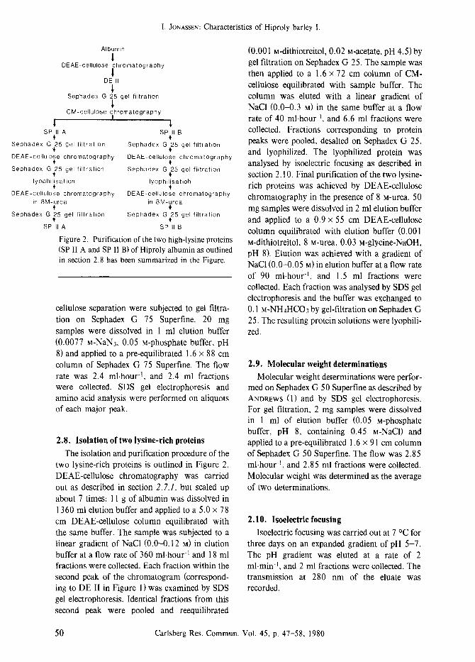
Figure 2. Purification of the two high-lysine proteins (SP II A and SP II B) of Hiproly albumin as outlined in section 2.8 has been summarized in the Figure.
cellulose separation were subjected to gel filtra- tion on Sephadex G 75 Superfine. 20 mg samples were dissolved in 1 ml elution buffer (0.0077 M-NaN3, 0.05 M-phosphate buffer, pH 8) and applied to a pre-equilibrated 1.6 x 88 cm column of Sephadex G 75 Superfine. The flow rate was 2.4 ml.hour -I, and 2.4 ml fractions were collected. SDS gel electrophoresis and amino acid analysis were performed on aliquots of each major peak.
2.8. Isolation of two lysine-rich proteins
The isolation and purification procedure of the two lysine-rich proteins is outlined in Figure 2. DEAE-cellulose chromatography was carried out as described in section 2.7.1, but scaled up about 7 times: 11 g of albumin was dissolved in 1360 ml elution buffer and applied to a 5.0 • 78 cm DEAE-cellulose column equilibrated with the same buffer. The sample was subjected to a linear gradient of NaCI (0.0-0.12 M) in elution buffer at a flow rate of 360 ml.hour 4 and 18 ml fractions were collected. Each fraction within the second peak of the chromatogram (correspond- ing to DE II in Figure 1 ) was examined by SDS gel electrophoresis. Identical fractions from this second peak were pooled and reequilibrated
(0.001 M-dithiotreitol, 0.02 M-acetate, pH 4.5)by gel filtration on Sephadex G 25. The sample was then applied to a 1.6 x 72 cm column of CM- cellulose equilibrated with sample buffer. The column was eluted with a linear gradient of NaCI (0.0-0.3 M) in the same buffer at a flow rate of 40 ml.hour -I, and 6.6 ml fractions were collected. Fractions corresponding to protein peaks were pooled, desalted on Sephadex G 25, and lyophilized. The lyophilized protein was analysed by isoelectric focusing as described in section 2.10. Final purification of the two lysine- rich proteins was achieved by DEAE-cellulose chromatography in the presence of 8 M-Urea. 50 mg samples were dissolved in 2 ml elution buffer and applied to a 0.9 x 55 cm DEAE-cellulose column equilibrated with elution buffer (0.001 M-dithiotreitol, 8 M-urea, 0.03 M-glycine-NaOH, pH 8). Elution was achieved with a gradient of NaC1 (0.0--0.05 M) in elution buffer at a flow rate of 90 ml.hour 4, and 1.5 ml fractions were collected. Each fraction was analysed by SDS gel electrophoresis and the buffer was exchanged to 0.1 M-NHaHCO3 by gel-filtration on Sephadex G 25. The resulting protein solutions were lyophili- zed.
2.9. Molecular weight determinations Molecular weight determinations were perfor-
med on Sephadex G 50 Superfine as described by ANOREWS (1) and by SDS gel electrophoresis. For gel filtration, 2 mg samples were dissolved in 1 ml of elution buffer (0.05 M-phosphate buffer, pH 8, containing 0.45 M-NaC1) and applied to a pre-equilibrated 1.6 • 91 cm column of Sephadex G 50 Superfine. The flow was 2.85 ml.hour -I, and 2.85 ml fractions were collected. Molecular weight was determined as the average of two determinations.
2.10. Isoelectric focusing Isoelectric focusing was carried out at 7 ~ for
three days on an expanded gradient of pH 5-7. The pH gradient was eluted at a rate of 2 ml.min -I, and 2 ml fractions were collected. The transmission at 280 nm of the eluate was recorded.
Figure 3. Elution profile of DEAE-cellulose chromatography of 1.5 g albumin as outlined in section 2.7.1. Appropriate fractions were pooled and designated as indicated by the horizontal bars.
2.11. Determination of carbohydrate The method of DuBois et al. (7) was used With
the following modifications: 0.8 ml samples containing 2.5 mg.ml t of protein were added 0.04 ml 80% w/v phenol, the samples were mixed, and 2 ml of concentrated H2SO4 were added. Samples were incubated at 100 ~ for 10 min. The absorbance at 490 nm was measured against a blank lacking sugar. Results were expressed as glucose equivalents.
2.12. Preparation and purification of antiserum against purified high-lysine proteins
Antibodies were raised against isolated prote- ins polymerized with glutaraldehyde as described by BOLLUM (4), and the degree of polymerization was determined by SDS gel electrophoresis. 6 rabbits were treated using BOLLUM'S immuniza- tion schedule, and 3 using a schedule devised by HARBOE and INGlt.D (9). The antisera obtained
were purified as recommended by HARBOE and INGILD (9).
2.13. Immunoelectrophoretic techniques Crossed immunoelectrophoresis and tandem
crossed immunoelectrophoresis was carried out as described by AXELSEN et al. (2).
105
09C
T ~ O75
o ~ 0.60
z < 045
0
< 0 3 0
015
CM I CMIII
car
i i i , i i i i
10 20 30 40 50 60 70 80 FRACTION NUMBER
Figure 4. Elution profile of CM-cellulose chroma- tography of 200 mg lyophilized protein obtained from fraction DE I as outlined in section 2.7.2.
Appropriate fractions were pooled and named as indicated by the horizontal bars.
11 0.16
014
012
0.10 0
67 0.08 ~_
5 0 0 6 0
4 0.04 ~
0.02
Carlsberg Res. Commun. Vol. 45, p. 47-58, 1980 51
I. JONASSEN: Characteristics of Hiproly barley I.
0.8
E 0.6
0.4
O m~ 0.2 <
0.8
E r 0.6 o
0.4 z
m
0.2
0.8
o o61 r
A SPll____B
SPI I IB
C
SP IVA
0.4
O 0.2
0.8
E 0.6
<
O mm 0.2 <
0.8
E 0.6
04 <
g m m 0.2
spy. c
D
S P V B
E SP VI_..._B
S P V I A
I I I I l 30 40 50
FRACTION NUMBER I I I I I I I ~ I I 100 80 60 40 30 20
MOLECULAR WEIGHT * 1 0 3
Figure 5. Elution profiles of Sephadex G 75 Super- fine gel filtration of the fractions DE II to DE VI obtained by DEAE-cellulose chromatography (Figure 3) as outlined in section 2.7.3.
Appropriate fractions were pooled and named as indicated by the horizontal bars. A to E correspond to fraction DE II to DE VI of Figure 3.
3. RESULTS AND DISCUSSION 3.1. Demonstration of high-lysine proteins in
barley (cv. Hiproly) albumin Hiproly albumin was separated by DEAE-
cellulose ion-exchange chromatography into six fractions designated DE I to DE VI (Figure 3). The DE I fraction separated on a CM-cellulose ion exchange column into six peaks (Figure 4). Fractions DE II to DE VI were subjected to Sephadex gel filtration and yielded in all, 11 well defined fractions designated with the letters SP. (Figure 5). Amino acid analysis of the individual fractions (Table I) revealed three fractions with a high content of lysine, namely fractions CM VI, fraction SP II A and SP I IB . Fraction CM VI contained 8 mole % lysine and consisted of a single protein with a molecular weight of 30,000 dalton as determined by SDS gel electrophoresis. The elution behaviour of this component on CM- cellulose indicated a protein with a high isoelec- tric point. The two peaks combined in fraction DE II (7.1 mole % lysine) could be resolved by gel filtration into SP II A and SP IIB. The latter contained 8.4 and 8.6 mole % lysine, respecti- vely (Figures 3 and 5A). SDS gel electrophoresis of these fractions revealed one major protein in each peak with a molecular weight below 10,000 dalton, but the major protein in SP II A appeared to be the larger of the two. The fractions from the DEAE-cellulose column were eluted with glycine buffer and dialysed. Amino acid analyses revealed that contaminating glycine buffer still was present in the fractions. This is reflected in a high content of glycine in the fractions DE I to DE VI. As that also lowered the mole percentage of lysine it explains why the lysine content of fraction DE II is increased after gel filtration to produce fraction SP II A and SP I IB . (Table I).
It can be deduced from the absorption profile of Figure 4 that fraction CM VI only constitutes a minor part of fraction DE I. On the other hand fractions SP II A and SP I I B constitute (Figure 5A) nearly all of fraction DE II. This is also reflected in the lysine content of fraction DE I with a value of 4.2 mole % and that of DE II with a value of 7.1 mole %. Fraction DE II thus contains two proteins which contribute very substantially to the overall lysine content of the Hiproly barley albumin.
52 Carlsberg Res. Commun. Vol. 45, p. 47-58, 1980
I. JONASSEN: Characteristics of Hiproly barley I.
Table I.
Mole % lysine in the albumin fractions obtained
DEAE-cellulose CM-cellulose gel filtration chromatography chromatography (Sephadex G 75 Superfine)
DE I 4,2 CM I 4.8 DE II 7.1 CM II 3.6 DE III 4.3 CM Ill 4.4 DE IV 5.3 CM IV 4.2 DE V 5.2 CM V 4.2 DE VI 4.7 CM VI 8.0
SP II A 8.4 SP 1I B 8.6 SP III A 5.0 SP Ill B 5.4 SP IV A 6.5 SP V A 5.2 SP V B 5.0 SP V C 6.3 SP VI A 5.2 SP VI B 5.0 SP VI C 6.4
3.2. Purification and isolation of two lysine- rich proteins
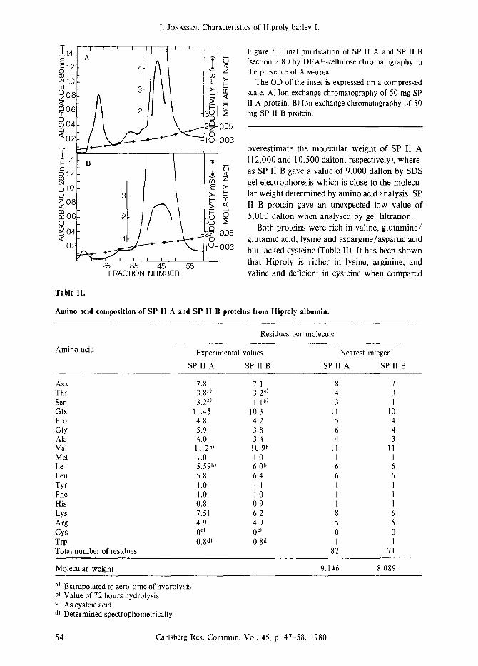
In order to produce larger amounts of purified SP II A and SP II B proteins for physical and chemical characterisation the isolation procedure described in section 2.8 and Figure 2 was employed. Change of buffers was consistently performed by gel filtration instead of cellophane membranes in order to minimize loss of material due to the low molecular weight of the two proteins. CM-cellulose chromatography was used for concentration of fraction DE II and its separation into SP II A and SP I I B (Figure 6). The final DEAE-cellulose chromatography was performed in the presence of 8 M-urea in order to avoid aggregation (Figure 7). The yield of SP II A exceeded on the average the yield of SP II B by 1.4 fold.
The protein corresponding to fraction SP I I B resulted in only one band on SDS gel electropho- resis (Figure 9B) while in the SP II A protein preparation, a small contamination of slightly lower molecular weight could be detected (Figure 9A). The effect of this contamination on immunological purity will be considered in section 3.4.
3.3. Physical and chemical characterisation of two high-lysine proteins
The molecular weight of the two proteins was calculated on the basis of their amino acid composition (Table II). SP II A consisted of 82
residues corresponding to a molecular weight of 9,146 dalton, while SP II B contained 71 residues yielding a molecular weight of 8,089 dalton.
Carbohydrate determination as described in section 2.11, indicated that the protein SP II A contained about 1% glucose or glucose equiva- lent, while the protein SP 11 B contained no carbohydrate. This in part explains the behaviour of the two proteins on gel filtration (Sephadex G 50 Superfine) and SDS gel electrophoresis. Gel filtration and SDS gel electrophoresis tend to
105 / ~ "
SPI IB
O15
10 20 30 40 50 60 70 80 FRACTION NUMBER
Figure 6. Preparative CM-cellulose chromatography of 502 mg of the DE 1! albumin fraction (section 2.8.).
Appropriate fractions were pooled and named as indicated by the horizontal bars.
0 90
~ 075
~Z 060
O45
< 030
10 9 8 7 6 5
0 2O
015 Z
010 ~
OO5
Carlsberg Res. Commun. Vo[. 45, p. 47-58, 1980 53
I. JONASSEN: Characteristics of Hiproly barley I.
E 1 ' ' .~11.4 At I ~ t ~1.2 4 ! oo o9 e4 1.0 E UA
~0.6 ~ 00.4 ,30
m ? 0 <0.2 1u-
7 F f ol .2 cJ 09 LU 1.0 E o 3 > - o,8 0 ~0.6 -2- }3~
04 )g < 1- "-
O-
25 35 45 55 FRACTION NUMBER
Figure 7. Final purification of SP II A and SP II B (section 2.8.) by DEAE-cellulose chromatography in
z the presence of 8 M-urea. The OD of the inset is expressed on a compressed
,~ scale. A) Ion of 50 SP exchange chromatography mg II A protein. B) Ion exchange chromatography of 50
2; mg SP 11 B protein.
0.05
0.03 overestimate the molecular weight of SP II A (12,000 and 10500 dalton, respectively), where- as SP II B gave a value of 9,000 dalton by SDS
~_ gel electrophoresis which is close to the molecu- ~- lar weight determined by amino acid analysis. SP �9 ~, II B protein gave an unexpected low value of O 5,000 dalton when analysed by gel filtration.
Both proteins were rich in valine, glutamine/ 0.05
glutamic acid, lysine and aspargine/aspartic acid 0.03 but lacked cysteine (Table II). It has been shown
that Hiproly is richer in lysine, arginine, and valine and deficient in cysteine when compared
Table II.
Amino acid composition of SP II A and SP II B proteins from Hiproly albumin.
Amino acid
Residues per molecule
Experimental values
SP II A SP II B
Nearest integer
SP II A SP 11 B
Asx 7.8 7.1 8 7 Thr 3.8 a) 3.2 a) 4 3 Ser 3.2 al 1. I a) 3 1 GIx 11.45 10.3 11 10 Pro 4.8 4.2 5 4 Gly 5.9 3.8 6 4 Ala 4.0 3.4 4 3 Val 11.2 b) 10.9 b) 11 11 Met 1.0 1.0 1 1 Ile 5.59 b) 6.0hi 6 6 Leu 5.8 6.4 6 6 Tyr 1.0 1.1 I 1 Phe 1.0 1.0 I I His 0.8 0.9 1 I Lys 7.51 6.2 8 6 Arg 4.9 4.9 5 5 Cys 0 c~ 0 c~ 0 0 Trp 0.8all 0.8d) 1 1 Total number of residues 82 71
Molecular weight 9.146 8.089
a) Extrapolated to zero-time of hydrolysis b) Value of 72 hours hydrolysis c) As cysteic acid dl Determined spectrophometrically
54 Carlsberg Res. Commun. Vol. 45, p. 47-58, 1980
Table III.
Rabbit immunization scheme
I. JONASSEN: Characteristics of Hiproly barley I.
Rabbit Antigen Immunization schedule reference
Precipitating antibody activity
1,2 SP II A (9) 3 SP II A, + BSA (9) 4, 5, 6, SP IIB (4) 7, 8, SP II B (4) 9 SP II B, + BSA (4)
activity activity none activity none
to other barley varieties (17, 21). It is therefore of considerable interest to quantify SP II A and SP II B albumin in Hiproly versus other barley varieties in order to elucidate whether the deviating amino acid composition of Hiproly grains can in part be explained by a high content of SP II A and SP II B. This topic will be treated in a subsequent paper (14).
Isoelectric focusing was performed as descri- bed in section 2.10 and yielded a pI of 5.94 and 7.02 for SP II A and SP II B, respectively. A pronounced aggregation was observed during isoelectric focusing, and the experiment was repeated in the presence of 6 M-urea which prevents protein aggregation but lowers the activity of H + (5). The isoelectric points measured in the presence of 6 M-urea were increased to 6.34 and 7.40 for SP II A and SP II B, respectively, which is in good agreement with the results reported by U~ (23). Aggregation of the two high-lysine proteins was also observed during fractionation (Figure 5A) where dimers were observed. This pronounced tendency for aggregation could explain why INGVERSEN and KO~E (12) identified a high-lysine fraction of molecular weight of 30,000 dalton in their gel filtration experiments with Hiproly.
3.4. Immunochemistry and immunisation
Rabbit antiserum produced against a crude salt extract of Hiproly barley (albumin and globulin) was used without success for crossed immunoelectrophoresis versus SP II A and SP II
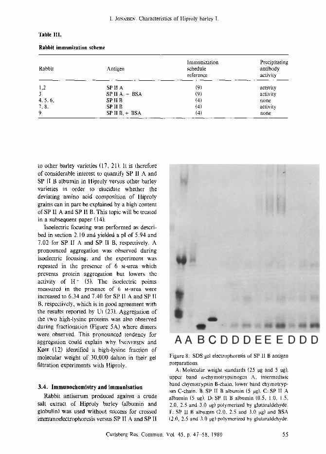
A A B C D D D E E E D D D Figure 8. SDS gel electrophoresis of SP lI B antigen preparations.
A: Molecular weight standards (25 rag and 5 rag), upper band a-chymotrypsinogen A, intermediate band chymotrypsin B-chain, lower band chymotryp- sin C-chain. B: SP II B albumin (5 rag). C: SP II A albumin (5 rag). D: SP II B albumin (0.5, 1.0, 1.5, 2.0, 2.5 and 3.0 rag) polymerized by glutaraldehyde. E: SP II B albumin (2.0, 2.5 and 3.0 rag) and BSA (2.0, 2.5 and 3.0 rag) polymerized by glutaraldehyde.
Carlsberg Res. Commun. Vol. 45, p. 47-58, 1980 55
A ~7~ B
iiit~iiiiii~ iiiiiiii~il ~
:iii[iii!iiil
iiii!iiiiiii~
i~iiiiiiiiii
~i!iiiiiiiii :iiii~iiiiiii~
?i!i!!~!!! ~
:!ilili?ilt
iiiii!i!Jiii~'
I. JONASSEN: Characteristics of Hiproly barley 1.
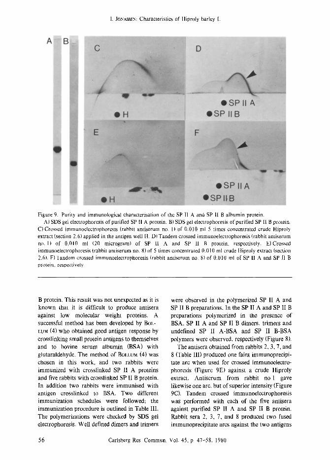
Figure 9. Purity and immunological characterisation of the SP I1 A and SP I1 B albumin protein. A) SDS gel electrophoresis of purified SP I1 A protein. B) SDS gel electrophoresis of purified SP II B protein.
C) Crossed immunoelectrophoresis (rabbit antiserum no. 1) of 0.010 ml 5 times concentrated crude Hiproly extract (section 2.6) applied in the antigen well H. D) Tandem crossed immunoelectrophoresis (rabbit antiserum no. 1) of 0.010 ml (20 microgram) of SP II A and SP 11 B protein, respectively. E)Crossed immunoelectrophoresis (rabbit antiserum no. 8) of 5 times concentrated 0.010 ml crude Hiproly extract (section 2.6). F)Tandem crossed immunoelectrophoresis (rabbit antiserum no. 8) of 0.010 mt of SP II A and SP 11 B protein, respectively.
B protein. This result was not unexpected as it is known that it is difficult to produce antisera against low molecular weight proteins. A successful method has been developed by BOL- LUM (4) who obtained good antigen response by crosslinking small protein antigens to themselves and to bovine serum albumin (BSA) with glutaraldehyde. The method of BOLLUM (4) was chosen in this work, and two rabbits were immunized with crosslinked SP II A proteins and five rabbits with crosslinked SP II B protein. In addition two rabbits were immunised with antigen crosslinked to BSA. Two different immunization schedules were followed; the immunization procedure is outlined in Table III. The polymerizations were checked by SDS gel electrophoresis. Well defined dimers and trimers
were observed in the polymerized SP II A and SP II B preparations. In the SP II A and SP II B preparations polymerized in the presence of BSA, SP II A and SP II B dimers, trimers and undefined SP II A-BSA and SP II B-BSA polymers were observed, respectively (Figure 8).
The antisera obtained from rabbits 2, 3, 7, and 8 (Table III) produced one faint immunoprecipi- tate arc when used for crossed immunoelectro- phoresis (Figure 9E) against a crude Hiproly extract. Antiserum from rabbit no l gave likewise one arc, but of superior intensity (Figure 9C). Tandem crossed immunoelectrophoresis was performed with each of the five antisera against purified SP II A and SP II B protein. Rabbit sera 2, 3, 7, and 8 produced two fused immunoprecipitate arcs against the two antigens
56 Carlsberg Res. Commun. Vol. 45, p. 47-58, 1980
I. JONASSEN: Characteristics of Hiproly barley I.
indicating immunological identity between SP II A and SP II B (Figure 9F). A similar situation
was found for antiserum no. 1, but the immu- noprecipitate developed against the SP II A was much stronger (Figure 9D arrow) than the immunoprecipitate produced by the four other antisera against SP II A (e.g. Figure 9F arrow). The small arc inside the large immunoprecipitate of Figure 9D could not be detected reproducibly and is considered an artefact. Crossed immuno- electrophoresis reveals the presence of SP II A and SP II B proteins in the crude extract of Hiproly. The asymetric shape of the arcs indicates the presence of at least two electropho- retically different, but cross-reacting proteins.
The limited resolution is probably due to the closely spaced isoelectric points and the relatively high diffusion rate of the two proteins due to their low molecular weight.
Tandem crossed immunoelectrophoresis ver- sus the two purified high-lysine proteins displa- yed immunochemical identity. However, one antiserum (no. 1 ), produced against the larger of the two proteins, reacted preferentially with this protein (SP II A), indicating that rabbit no. 1 had identified specific antigenic sites on the SP II A protein where the other rabbits had failed. Consideration of the immunological identity between SP II A and SP II B, the additional antigenic sites on SP II A, together with the amino acid analyses of these proteins leads to the suggestion that SP II B is a fragment of the SP I1 A protein: The primary structure of these two high-lysine proteins will be considered in a later paper.
ACKNOWLEDGEMENTS I wish to express my sincere gratitude to Dr.
L. MUNCK and to Dr. T. NILSSON-TILLGREN for support and encouragement and to Dr. G. GIBBONS and professor D. VON WETTSTEIN for their assistance in preparing this communication and to Lic. Tech. B. S. ENEVOLDSEN for performing the carbohydrate analyses. I am indebted to METTE HOJ for her excellent technical assistance and pleasant collaboration. I am most grateful to Dr. J. HEJGARD, Department of Biochemistry and Nutrition, Technical Univer- sity of Denmark, Lyngby, for advice and
guidance with the immunological part of the
work.
REFERENCES
1. ANDREWS, P.: Estimation of molecular size and molecular weights of biological compounds by gel filtration. Methods Biochem. Anal. 18, 1-53 (1970)
2. AXELSEN, N. H., J. KROLL & B. WEEKE (eds.): A manual of quantitative immunoelectrophoresis. Methods and applications. Universitetsforlaget. Oslo (1973)
3. BENZE, W. L. & K. SCHMID: Determination of tyrosine and tryptophane in proteins. Anal. Chem. 29, 1193-1196 (1957)
4. BOLLUM, F. J.: Antibody to terminal deoxynucle- otidyl transferase. Proc. Nat. Acad. Sci. U.S.A. 72, 4119-4122 (1975)
5. BULL, H. B., K. BREESE, G. L. FERGUSON & C. A. SWENSON: The pH of urea solutions. Arch. Biochem. Biophys. 104, 297-304 (1964)
6. CHUA, N.-H. & P. BENNOUN: Thylakoid memb- rane polypeptides of Chlamydomonas reinhardti: Wild type and mutant strains deficient in photosystem II reaction center. Proc. Nat. Acad. Sci. U.S.A. 72, 2175-2179 (1975)
7. DUBOIS, M., K. A. GILLES, J. K. HAMILTON, P. A. REBERS & F. SMrrH: Colorimetric method for determination of sugars and related substances. Anal. Chem. 28, 350-356 (1956)
8. EL-NEGOUMY, A. M., C. W. NEWMAN & B. R. Moss: Chromatographic fractionation and com- position of the components of the salt-soluble proteins from Hiproly (CI 3947) and Hiproly normal (CI 4362) barleys. Cereal Chem. 54, 333-344 (1977)
9. HARBOE, N. & A. INGILD: Immunisation, isola- tion of immunoglobulins, estimation of antibody titre. In: A manual of quantitative immunoelec- trophoresis. Methods and applications. N. H. Axelsen, J. Kroll & B. Weeke eds., Universitets- forlaget Oslo, pp 161-164 (1973)
10. HELM, J. H., R. J. METZER & W. E. KRONSTAD: Inheritance of high lysine in Hiproly barley and its association with the Hiproly endosperm gene. Crop Sci. 14, 637-640 (1974)
l l. HIRS, C. H. W.: Determination of cystine as cysteic acid. Meth. Enzymol. I 1, 59-62 (1962)
12. INGVERSEN, J. & B. KOIE: Lysine-rich proteins in high-lysine Hordeum vulgare grain. Phytoche- mistry 12, ll07-1111 (1973) INGVERSEN, J. & B. KOIE: Lysine-rich proteins in the salt-soluble protein fraction of barley. Phytochemistry 12, 73-78 ( 1973)
13.
Carlsberg Res. Commun. Vol. 45, p. 47-58, 1980 57
I. JONASSEN: Characteristics of Hiproly barley I.
14. JONASSEN, I.: Characteristics of Hiproly barley II. Quantification of two proteins contributing to its high lysine content. Carlsberg Res. Commun. 45, 59-68 (1980)
15. MIKOLA, J., M. NUMMI & T.-M. ENARI: Effects of standing on the protein extracts of barley grain. Nature 195, 808 (1962)
16. MUNCK, L.: Barley seed proteins. Symposium on seed protein synthesis properties and processing (Los Angeles, California). Amer. Chem. Soc. (Ed.) G. E. Inglett Avis Publ. Co. (1972)
17. MUNCK, L.: Improvement of nutritional value in cereals. Hereditas 72, l-128 (1972)
18. MUNCK, L., K.-E. KARl,SON, A. HAGBERG & B. O. EGGUM: Gene for improved nutritional value in barley seed protein. Science 168, 985-987 (1970)
[ 9. NEVILLE, D. M. Jr.: Molecular weight determina- tion of proteindodecyl sulfate complexes by gel
electrophoresis in a discontinuous buffer system. J. Biol. Chem. 246, 6328-6334 (1971)
20. OSaORNE, T. B.: The proteids of barley. J. Am. Chem. 17, 539-567 (1895)
21. POMERANZ, Y., D. M. WESENBERG; R. T. SMITH, G. S. ROaBINS & J. T. GILBERTSON: Amino acid composition of barley kernels from different parts of the spike. Cereal Chem. 53, 839-845 (1976)
22. STROB~K, S. & G. C. GmBONS: Ribulose-l,5- diphosphate carboxylase from barley (Hordeum vulgate). Isolation, characterization, and peptide mapping studies of the subunits. Carlsberg Res. Commun. 41, 57-72 (1976)
23. UI, N,: Isoelectric points and conformation of proteins. I. Effect of urea on the behavior of some proteins in isoelectric focusing. Biochim. Biophys Acta 299, 567-581 (1971)