Photopolymerized micelles of diacetylene amphiphile: physical characterization and cell delivery properties
Patrick Neubergab‡, Aurélia Périnoa ‡, Emmanuelle Morin-Picardatab ‡, Nicolas Antonc, Zeinab Darwichd, Denis Weltine, Yves Melyd, Andrey S. Klymchenkod, Jean-Serge Remyb*, and Alain
Wagnera*
a Laboratory of Functional Chemo Systems, and Labex Medalis, CAMB, UMR 7199 CNRS, , Faculty of Pharmacy, University of Strasbourg, 74 route du Rhin,, 67401 Illkirch (France) ; b Laboratory V-SAT, Vectors - Synthesis and Therapeutic
Applications, and Labex Medalis, CAMB, UMR7199 CNRS, Faculty of Pharmacy, University of Strasbourg, 74 route du Rhin,, 67401 Illkirch (France) ; c Laboratory of Biogalenic Pharmacy, CAMB, UMR 7199 CNRS, Faculty of Pharmacy,
University of Strasbourg, 74 route du Rhin,, 67401 Illkirch (France) ; d Laboratory of Biophotonic and Pharmacology, UMR 7213, Faculty of Pharmacy, University of Strasbourg, 74 route du Rhin, 67401 Illkirch (France) ; e Phytodia SAS, Boulevard
Materials and methodsThe 10,12-pentacosadiynoic acid, octaethylenglycol, 1-(3-dimethylaminopropyl)-3-
ethylcarbodiimide hydrochloride (EDC), hydroxy, diisopropylethylamine, mesyl chloride, sodium azide (NaN3), triphenylphosphine, methanol, acetonitrile, tetrahydrofuran and dimethylformamide were purchased from Sigma-Aldrich.
Synthesis of surfactant 1
The synthesis of N-(23-hydroxy-3,6,9,15,18,21-heptaoxatrico-1-yl)pentacosa-10,12-diynamide was achieved by a similar protocol as described in our previous paper (C. Thauvin 2011)1.
To a solution of 10,12-pentacosadiynoic acid (342 mg; 0.99 mmoles) and N-hydroxysuccinimide (136 mg; 1.19 mmoles) in dichloromethane were added 1-ethyl-3-(3-dimethylaminopropyl)carbodiimide (284 mg; 1.48 mmoles) and N,N-diisopropylethylamine (207 µl; 1.19 mmoles). The mixture was stirred overnight at room temperature. After evaporation of the solvents the crude was dissolved in ethyl acetate and extracted with water. The organic phase was dried over anhydrous sodium sulfate, filtered off and evaporated. The obtained white solid (activated ester) was dissolved in dry THF (25 ml) and 23-amino-3,6,9,12,15,18,21-heptaoxatricosanol (304 mg; 0.82 mmoles) were added along with triethylamine. The reaction mixture was stirred overnight at room temperature. After evaporation of the solvents the crude was purified by silica gel chromatography with 5% methanol / dichloromethane as an eluent to yield a white solid.
This white solid is photosensitive and turns light blue on storage, and dark blue in presence of light
Rf = 0.29 (5 % methanol in dichloromethane on silica TLC plates)
Synthesis and polymerization of the micellesThe formation of micelles was performed by solubilizing 15 mg of amphiphile 1 into 3
mL of phosphate buffer pH 7.4 (100 mM). The solution was sonicated for 30 min in a sonication bath (80 W, 25°C). For the polymerization step, quartz cuvettes containing 3 mL of micelle solution were placed into a Cross-Linker Bio-Link 254 (Fischer Bioblock). Photopolymerization was achieved at 254 nm and 48 W.
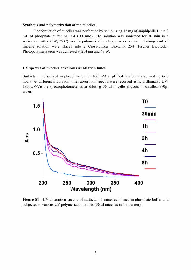
UV spectra of micelles at various irradiation times
Surfactant 1 dissolved in phosphate buffer 100 mM at pH 7.4 has been irradiated up to 8 hours. At different irradiation times absorption spectra were recorded using a Shimatzu UV-1800UV/Visible spectrophotometer after diluting 30 µl micelle aliquots in distilled 970µl water.
Figure S1 : UV absorption spectra of surfactant 1 micelles formed in phosphate buffer and subjected to various UV polymerization times (30 µl micelles in 1 ml water).
3
Transmission Electron Microscopy (TEM)
Scale bar: 50 nm.Fig. S2 TEM images of NPM (left) and PM after 30 min UV polymerization (right).
Five microliters of a diluted micellar solution (0.1 mg/mL in MQ water) was allowed to adsorb for 1 minute onto 300 mesh copper grids (Ted Pella, 822-F). Grids were wicked up from one side and placed for 30 s on a 50 mL drop of 2% uranyl acetate, wicked up again and air dried before imaging. Images were obtained using a Philips CM 120 microscope.
Size measurements by Dynamic Light Scattering (DLS) The hydrodynamic diameters of micelles were determined by the Zetasizer Nano ZS
system (Malvern Instruments) with the following specifications: sampling time = 90 s; refractive index of phosphate buffer n = 1.340; refractive index of particles (RI) = 1.43; medium viscosity = 1.0140 cP; temperature = 25°C. The values are presented as the number average size ± standard deviation of three runs. The error on the measurements is calculated as the width at mid-height of the peak divided by two.
4
Figure S3 Mean hydrodynamic diameter by number of micelles at increasing UV polymerization times.
Fluorescent spectroscopy and determination of the Critical Micellar Concentrations (CMC)
Absorption spectra were recorded on a Cary 4 spectrophotometer (Varian) and fluorescence spectra on a FluoroMax 3.0 (Jobin Yvon, Horiba) spectrofluorometer. Fluorescence emission spectra of NR12S were systematically recorded at 520 nm excitation wavelength at room temperature. All the spectra were corrected from the fluorescence of the corresponding blank (suspension of micelles in PBS without the probe).
FCS experiments were carried out on a home-built two-photon laser scanning set-up provided by a titanium-saphire laser (Tsunami, Spectra Physics) and photons were detected with Avalanche Photodiodes (APD SPCM-AQR-14-FC, Perkin Elmer) connected to a counter/timer PCI board (PCI6602, National Instrument).3
Table S4 FCS data for micelles of 1 after various photopolymerization times.Brightness is the photon count rate (kHz) per particle.
In addition to the dynamic surface tension measurements for calculating the CMC values, we used a fluorescent method based on hydrophobic inclusion of pyrene: CMC values
of micelles were determined using pyrene as an extrinsic fluorescence probe.2 Pyrene (1 µl of a 1 mM solution in dimethylsulfoxide) was added to 1 ml of an aqueous phosphate buffered micelle solution (pH 7.4) and mixed by vortexing (1µM pyrene solution). The micelle concentration was varied from 1.38.10-3 mM to 2.75 mM (0.001 mg/ml to 2 mg/ml). CMC measurements were carried out at 25 °C using a Fluorolog spectrofluorometer (Jobin Yvon, Horiba). The fluorescence emission intensities of pyrene at 374 nm and 383 nm were obtained with excitation at 339 nm and the fluorescence intensity ratio, I374/I383, was plotted against the logarithm of the polymer concentration. The CMC value was determined from the intersection of the two tangent lines. The CMC value of NPM determined using this method was 14.6 µg/ml. The values of the CMCs determined by the pyrene inclusion method are higher than the CMCs determined by dynamic tensiometry, which were measured in water. The pyrene method is less precise for low CMCs due to the detection limit of pyrene fluorescence. At the low surfactant concentrations needed for measuring their CMCs, the surfactant molecules can no longer efficiently incorporate the pyrene dye, which results in artificially high experimental CMC values.
The CMC value of polymerized micelles could also not be measured accurately by the pyrene method, probably due to the relatively high detection limit of pyrene and low insertion of the dye into polymerized micelles.
6
NMR Spectroscopy
1D-1H and 2D-DOSY experiments are carried out at 300 K on a 600 MHz Bruker Avance III NMR spectrometer with a 5 mm DOTY probe delivering up to 500 G / cm gradients and on a 500 MHz Bruker Avance III spectometer with a BBFO probe.
For DOSY experiments on the 600MHz spectrometer gradients are linearly sampled in 40 points; TD(time domain) = 40 with 64 scans (NS = 64). The gradient pulse length is δ / 2 (P30) = 1.35 ms and the Δ diffusion delay (D20) is 50 msec (adapted to the sample). The DOSY spectra are obtained by applying an Inverse Laplace Transform (ILT) along the diffusion axis, using the commercial software NMRnotebook (NMRTEC, Illkirch).
For DOSY experiments on the 500MHz spectrometer gradients are linearly sampled in 32 points; TD(time domain) = 32 with 32 scans (NS = 32). The gradient pulse length is δ/2 (P30) = 1.6 ms and the Δ diffusion delay (D20) is 100 msec (adapted to the sample). The DOSY spectra are obtained again by applying an Inverse Laplace Transform (ILT) along the diffusion axis. Viscosities of deuterated solvents are previously measured with a DM500 Anton Paar viscosimeter. Measured viscosity for deuterated methanol: 0.602 cP (= 0.00602 Pa . s) ; measured viscosity for deuterated Water D2O : 1.13 cP (= 0.00113 Pa . s)
Applying the Stokes Einstein relation for the diffusion of spherical particles through a liquid, hydrodynamic radii were calculated from the measured diffusion coefficients
𝐷 =
𝑘𝐵 𝑇6 𝜋 𝜂 𝑟(ℎ)
Measured hydrodynamic radii calculated from the diffusion coefficients were in accordance with DLS and FCS experiments. Non polymerized micelles (NPM) with a diffusion coefficient of 37.3 10-12 m2 / s have a calculated hydrodynamic radius of 5,2nm, while polymerized micelles (PM) have a hydrodynamic radius of 6.7 nm (D = 28.7 10-12 m2
/ s in deuterated water. Monomer have a diffusion coefficient of D = 530 10-12 m2 / s in deuterated methanol.
Polymerized micelles were further analyzed after lyophilization and dissociation in deuterated methanol, where non-covalent interactions come loose. The DOSY analysis of this methanolic surfactant solution revealed two distinct populations, one corresponding to monomeric surfactant the other corresponding to polymeric surfactant. The percentage of covalently bridged surfactants versus monomer molecules could be calculated from peak integration from the 2D-plot. At 4 hours UV-polymerization there is around 75 % polymerized surfactant compared to 25 % monomeric surfactant.
Table S5 DOSY NMR integration of monomer versus polymeric surfactant
Figure S6 DOSY NMR spectrum of UV irradiated micelles that have been lyophilized and dissociated in deuterated methanol. The respective integration boxes are highlighted in the plot
8
Cell Culture and fluorescence microscopyHeLa cells were cultured in Dulbecco's modified Eagle medium (DMEM, high
glucose, Gibco-Invitrogen) supplemented with 10% (v/v) fetal bovine serum (FBS, Lonza), 1% antibiotic solution (penicillin-streptomycin, Gibco-invitrogen) in a humidified incubator with 5% CO2 /95% air atmosphere at 37 °C. Cell concentration of 5-10104 cells/mL was maintained by removal of a portion of the culture and replacement with fresh medium 3 times per week.
For the microscopy studies, cells were seeded onto a chambered cover glass (IBiDi) at a density of 5×10⁴ cells/IBiDi. 0.05 mg/mL of micelles were added to cells with the probe NR12S, and incubated for different times. Finally, cells in IBiDi dishes were washed with Opti-MEM.
Fluorescence microscopy experiments were performed by using a home-built two-photon laser scanning set-up based on an Olympus IX70 inverted microscope with an Olympus 60x 1.2NA water immersion objective.3 Two-photon excitation was provided by a titanium-saphire laser (Tsunami, Spectra Physics) and photons were detected with Avalanche Photodiodes (APD SPCM-AQR-14-FC, Perkin Elmer) connected to a counter/timer PCI board (PCI6602, National Instrument). Imaging was carried out using two fast galvo mirrors in the de-scanned fluorescence collection mode. Typical acquisition time was 5 s with an excitation power around 40 mW ( = 830 nm) at the sample level. Images were recorded simultaneously using a dichroic mirror (Beamsplitter 585 DCXR), density filter 630/30 nm. The images were processed with ImageJ program.
Cytotoxicity assay
Cytotoxicity of compounds was evaluated on HaCaT keratinocyte cell line, plated 24h before exposure to products in 96 well cell culture plates. Micelles were added to cells for 48 hours. Cell viability was measured by quantifiying the mitochondrial succinate dehydrogenase with MTT assay (3-(4,5-dimethylthiazol-2-yl)-diphenyl tetrazolium bromide is reduced to a by viable cells to give a blue precipitate that is quantified after solubilization in DMSO).
9
Fig. S7 Cytotoxicity of photo-polymerized micelles in HaCaT cells.
References1. C. Thauvin, A. Perino, E. Contal, E. Morin, P. Schultz, S. Meunier and A. Wagner, J.
Phys. Chem. C, 2011, 115, 7319-7322.2. M. Wilhelm, C. L. Zhao, Y. Wang, R. Xu, M. A. Winnik, J. L. Mura, G. Riess and M.
D. Croucher, Macromolecules, 1991, 24, 1033-1040.3. J. P. Clamme, J. Azoulay and Y. Mély, Biophys. J., 2003, 84, 1960-1968.