أCHARACTERIZATION AND DIFFERENTIATION POTENTIAL OF RAT BONE MARROW MESENCHYMAL STEM CELLS INTO CARDIAC-LIKE CELLS RAMIN KHANABDALI DISSERTATION SUBMITTED IN FULFILLMENT OF THE REQUIREMENTS FOR THE DEGREE OF MASTER OF BIOTECHNOLOGY INSTITUTE OF BIOLOGICAL SCIENCES FACULTY OF SCIENCE UNIVERSITY OF MALAYA KUALA LUMPUR 2014
Transcript
أ
CHARACTERIZATION AND DIFFERENTIATION POTENTIAL OF
RAT BONE MARROW MESENCHYMAL STEM CELLS INTO
CARDIAC-LIKE CELLS
RAMIN KHANABDALI
DISSERTATION SUBMITTED IN FULFILLMENT OF
THE REQUIREMENTS FOR THE DEGREE OF
MASTER OF BIOTECHNOLOGY
INSTITUTE OF BIOLOGICAL SCIENCES
FACULTY OF SCIENCE
UNIVERSITY OF MALAYA
KUALA LUMPUR
2014
i
UNIVERSITI MALAYA
ORIGINAL LITERARY WORK DECLARATION
Name of Candidate: RAMIN KHANABDALI I/C/Passport No: P95424150
Regisration/Matric No.: SGF120015
Name of Degree: MASTER OF BIOTECHNOLOGY
Title of Project Paper/Research Report/Dissertation/Thesis (“this Work”):
“CHARACTERIZATION AND DIFFERENTIATION POTENTIAL OF RAT BONE MARROW
MESENCHYMAL STEM CELLS INTO CARDIAC-LIKE CELLS.”
Field of Study : STEM CELL
I do solemnly and sincerely declare that:
(1) I am the sole author/writer of this Work, (2) This Work is original, (3) Any use of any work in which copyright exists was done by way of fair dealing and for
permitted purposes and any excerpt or extract from, or reference to or reproduction of any copyright work has been disclosed expressly and sufficiently and the title of the Work and its authorship have been acknowledged in this Work,
(4) I do not have any actual knowledge nor do I ought reasonably to know that the making of this
work constitutes an infringement of any copyright work, (5) I hereby assign all and every rights in the copyright to this Work to the University of Malaya
(“UM”), who henceforth shall be owner of the copyright in this Work and that any reproduction
or use in any form or by any means whatsoever is prohibited without the written consent of UM having been first had and obtained,
(6) I am fully aware that if in the course of making this Work I have infringed any copyright
whether intentionally or otherwise, I may be subject to legal ac tion or any other action as may be determined by UM.
(Candidate Signature) Date:
Subscribed and solemnly declared before,
Witness’s Signature Date:
Name ASSOC. PROF. DR DURRIYYAH SHARIFAH HASSAN ADLI
Designation
Witness’s Signature Date:
Name DR SHAMSUL AZLIN AHMAD SHAMSUDDIN
Designation
ii
ABSTRACT
Heart diseases are the leading cause of death worldwide. Despite the development of a
broad array of treatment options, current therapies only delay progression of the disease
and failed to prevent myocardial scar formation and replace the lost cardiomyocytes
(cardiac muscle cells). Over the past decade the use of adult stem cells, particularly bone
marrow derived mesenchymal stem cells, to safely facilitate recovery of cardiac function
after myocardial infarction has received a lot of interest. Mesenchymal stem cells (MSCs),
which are adherent stromal cells of a non-hematopoietic origin, have great differentiation
potential and under appropriate in vitro culture conditions can trans-differentiate into
cardiomyocyte cells. This study investigated the characterization of rat bone marrow
derived-mesenchymal stem cells (BM-MSCs) and in vitro differentiation potential of them
into cardiomyocyte- like cells by two DNA-demethylating agents, 5-azacytidine and
zebularine. MSCs were isolated from Sprague Dawley’s bone marrow and cultured in
complete Dulbecco’s Modified Eagle Medium (DMEM). Morphological characteristics of
MSCs were analyzed by phase contrast microscopy. Selected surface antigens CD44,
CD117, known MSCs markers, and CD34, a hematopoietic marker (negative marker),
were analyzed by immunocytochemistry. In addition, CD45, known hematopoietic marker
(negative marker) and CD44 were analyzed by flow cytometry for the MSC cell population
count. Passage 1 (P1) cultured MSCs were then treated in separate culture flasks for 24
hours with a 3µM optimized concentration of 5-azacytidine and zebularine. After 20 days,
treated cells were analyzed for the expression of rat cardiac specific genes; namely, alpha-
myosin heavy chain (CAMHC), cardiac troponin-T (cTnT), and cardiac transcription factor
(GATA-4) by reverse transcriptase polymerase chain reaction (RT-PCR). The endogenous
housekeeping gene, glyceraldehyde-3-phosphate dehydrogenase (GAPDH) was used as an
iii
internal standard gene for normalization of mRNA. The isolation of MSCs from rat bone
marrow was successfully completed. Isolated MSCs exhibited spindle-shaped morphology
with adherence ability to the surface of flasks and proliferated in the culture medium.
Immunocytochemistry results showed that cell surface antigen expression was observed to
be positive for CD44 and CD117. However, MSCs were negative for CD34 (hematopoietic
marker); hence, confirming the absence of hematopoietic cells. Furthermore, CD44 was
found to be >85% positive, while CD45 was more than 60% negative in MSCs after flow
cytometry cell population analysis. Upon induction with 5-azacytidine and zebularine, the
morphology of the MSCs changed and the cells showed extended cytoplasmic processes
with ball- like appearance. After 20 days, they were connected with adjoining cells forming
myotube- like structures. The mRNAs of CAMHC, cTnT and GATA-4 were detected in
both treated and untreated cells. However, RT-PCR analysis for the expression of cardiac
specific genes showed that treated MSC cells expressed cTnT, CAMHC and GATA-4
significantly higher compared to untreated cells. While there were no significant
differences between 5-azacytdine and zebularine treated cells, zebularine could be a good
replacement for 5-azacytidine as it is more stable and less toxic to biological system. These
results showed that bone marrow mesenchymal stem cells (BM-MSCs) could differentiate
in vitro towards a cardiomyogenic lineage.
iv
ABSTRAK
Penyakit jantung adalah punca utama kematian di seluruh dunia. Walaupun terdapat
kemajuan dalam pelbagai opsyen rawatan, terapi semasa hanya dapat melambatkan
perkembanga penyakit dan gagal untuk menghalang pembentukan parut miokardium dan
menggantikan kardiomiosit sel-sel yang hilang. Sepanjang dekad yang lalu, penggunaan
sel stem dewasa, terutamanya sel-sel stem mesenkimal yang diperolehi dari sum-sum
tulang untuk memudahkan pemulihan fungsi jantung dengan selamat selepas infarksi
miokardium telah mendepat banyak pnumpuan. Sel-sel stem mesenkimal (Mesenchymal
Stem Cells) yang merupakan sel stromal adherent yang bukan berasal dari hematopoietik,
mempunyai potensi yang besar dalam pembezaan/diferensiasi dan di bawah keadaan in
vitro kultur yang sesuai boleh trans-diferensiasi untuk menjadi sel kardiomiosit. Kajian ini
menyiasat tentang pencirian sel-sel mesenkimal yang diperolehi dari sum-sum tulang tikus
(BM-MSCs - Bone Marrow Mesenchymal Stem Cells) dan potensi diferensiasi in vitro sel-
sel tersebut menjadi sel-sel mirip kardiomiosit dengan menggunakan dua ejen demetilasi
DNA, 5-azacytidine dan zebularine. MSCs telah diasingkan daripada sum-sum tulang
Sprague Dawley dan dikultur di dalam Dulbecco's Modified Eagle Medium (DMEM) yang
lengkap. Ciri-ciri morfologi MSCs dianalisa dengan menggunakan mikroskop fasa-
kontras. Antigen permukaan yang terpilih CD44, CD117, yang dikenali sebagai penanda
bagi MSCs, dan CD34, suatu penanda bagi hematopoietik (penanda negatif), dianalisa
dengan immunositokimia. Sebagai tambahan , CD45, iaitu penanda bagi hematopoietik
(penanda negatif) dan CD44, dianalis dengan flow cytometry untuk mendapatkan bilangan
populasi sel-sel MSC. Pasaj 1 (P1) MSC yang telah dikulturkan kemudiannya dirawat di
dalam kelalang kultur yang berasingan selama 24 jam dengan kepekatan 3μM 5-
azacytidine dan zebularine yang telah dioptimakan. Selepas 20 hari, sel-sel yang telah
v
dirawat dianalisa untuk ekspresi gen-gen spesifik jantung tikus; iaitu alpha-myosin heavy
chain (CAMHC), cardiac troponin-T (cTnT), dan cadiac transcription factor (GATA-4)
dengan menggunakan reverse transcription polymerase chain reaction (RT-PCR). BM-
MSC yang telah diasingkan mempamerkan morfologi berbentuk gelendong dengan
keupayaan melekat kepada permukaan kelalang dan telah berkembang biak dalam medium
kultur. Keputusan immunositokimia menunjukkan bahawa ekspresi antigen permukaan sel
diamati positif untuk CD44 dan CD117. Walau bagaimanapun, MSC adalah negatif untuk
CD34 (penanda bagi hematopoietik), oleh itu, mengesahkan ketiadaan sel-sel
hematopoietik. Tambahan pula, CD44 didapati > 85% positif, manakala CD45 (penanda
hematopoietik); adalah lebih daripada 60% negatif dalam MSC melalui analisis populasi
sel menggunakan flow cytometry. Setelah induksi menggunakan 5-azacytidine dan
Zebularine, morfologi MSC telah berubah dan sel-sel mempamerkan unjuran proses
sitoplasm dengan penampilan seperti bebola. Selepas 20 hari, sel-sel yang bersebelahan
telah berhubung dan membentuk struktur seperti miotiub. mRNA bagi CAMHC, cTnT and
GATA-4, dan GATA-4 telan dikesan dalam kedua-dua sel dirawat dan tidak dirawat.
Walau bagaimanapun, analisis RT- PCR untuk ekspresi spesifik gen kardiak menunjukkan
bahawa sel-sel MSC yang dirawat mempamerkan kehadiran cTnT, CAMHC dan GATA-4
yang signifikannya lebih tinggi berbanding sel-sel yang tidak dirawat. Manakala, tidak ada
perbezaan yang signifikan di antara sel-sel yang dirawat 5-azacytidine dan zebularine.
Zebularine boleh menjadi pengganti yang baik untuk 5-azacytidine kerana ia lebih stabil
dan kurang toksik kepada sistem biologi. Keputusan ini menunjukkan bahawa sel sum-sum
tulang mesenkima (BM- MSC) boleh didiferensiasi secara in vitro menjadi kumpulan
kardiomiogenik.
vi
ACKNOWLEDGMENT
Alhamdulillah and thanks to Allah for giving me enough strength, courage and
patience to complete this dissertation. There are so many people who contributed to this
dissertation both directly and indirectly. I would like to acknowledge many people for their
help and inspiration during my master work.
Firstly, I would like to thank my supervisor, Dr. Shamsul Azlin Ahmad Shamsuddin,
for giving me the opportunity to be his student and for the time, encouragement, patience
and commitment over the last two years. I would also like to express my special gratitude
to my co-supervisor, Assoc. Prof. Dr. Durriyyah Sharifah Hasan Adli, for almost four years
of unfaltering guidance, enlighten advices and support. She has finally beaten the word
“control” into my brain!! I feel very lucky to be part of the big NeuoRG lab family; a
group of intelligent and fun fellow lab-mates.
Additionally, I need to thank all the wonderful people with whom I was lucky to
interact in the Stem Cell Laboratory in International Center for Chemical and Biological
Sciences (ICCBS), University of Karachi, Pakistan. I would like to appreciate and thank
Dr. Asmat Saleem, leader of the group, Dr. Irfan Khan, Dr Nadia Naeem, Dr. Khanwal
Haneef and the rest of lab-mates for their great hospitality, guidance, support and assisting
me to conduct this research.
Last, but definitely not least, I would not have made it to this point in life without my
family and their constant prayers and support. Thank you, Mom, Shahram, Reza, Shahin,
Mehry and my little sister Rojin.
vii
TABLE OF CONTENT
Page
ABSTRACT…………………………………………………………………………...........ii
ABSTRAK………………………………………………………………………………......iv
ACKNOWLEDGEMENT………………………………………………………………....vi
TABLE OF CONTENT.......................................................................................................vii
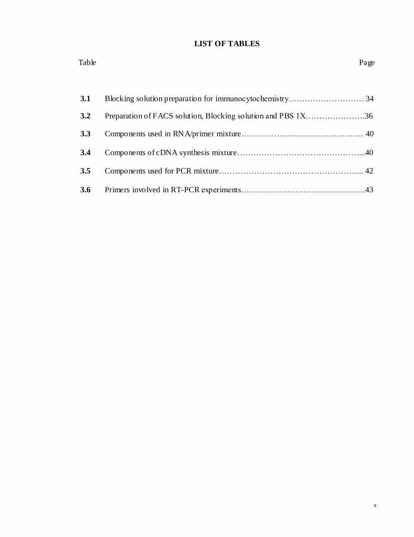
LIST OF TABLES……………………………………………………………………........x
LIST OF FIGUERS………………………………………………………………………. xi
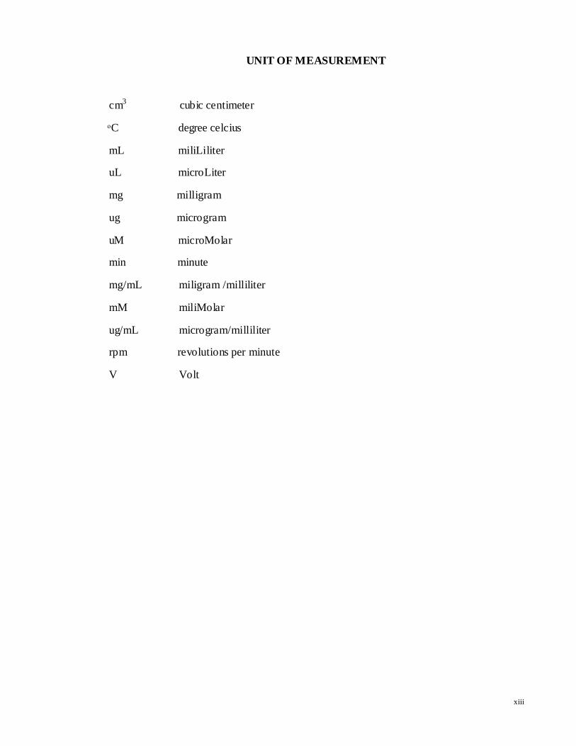
LIST OF ABBREVIATIONS………………………………………………………….... xii
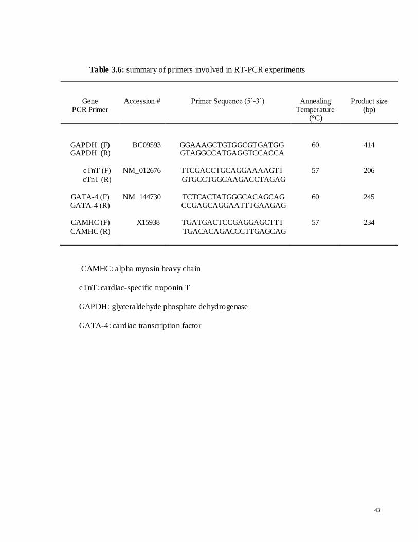
Table 3.6: summary of primers involved in RT-PCR experiments
Gene PCR Primer
Accession # Primer Sequence (5’-3’)
Annealing Temperature
(°C)
Product size (bp)
GAPDH (F) GAPDH (R)
BC09593
GGAAAGCTGTGGCGTGATGG GTAGGCCATGAGGTCCACCA
60
414
cTnT (F) cTnT (R)
NM_012676
TTCGACCTGCAGGAAAAGTT
GTGCCTGGCAAGACCTAGAG
57
206
GATA-4 (F) GATA-4 (R)
NM_144730
TCTCACTATGGGCACAGCAG CCGAGCAGGAATTTGAAGAG
60
245
CAMHC (F) CAMHC (R)
X15938
TGATGACTCCGAGGAGCTTT
TGACACAGACCCTTGAGCAG
57
234
CAMHC: alpha myosin heavy chain
cTnT: cardiac-specific troponin T
GAPDH: glyceraldehyde phosphate dehydrogenase
GATA-4: cardiac transcription factor
44
3.7.2.5 Agarose Gel Electrophoresis
The amplified products were separated using 1% (w/v) agarose gel electrophoresis. Details
regarding preparation of agarose gel and TBE buffer preparations are presented in
Appendix B. One fifth of each PCR product was electrophoretically resolved on 1%
agarose gel containing 1.5µl of 0.3µg/ml ethidium bromide.
To start, 0.5gm agarose was dissolved in 10ml 5X TBE buffers, by heating the mixture in
microwave oven for 1 minute. The solution was allowed to cool approximately to 60°C.
1.5µl ethidium bromide was added and swirled gently. The gel was poured into the
horizontal gel casting unit and allowed to polymerize for approximately 30 to 40 minutes.
The tank was filled with 5X TBE buffer before. 10 µl DNA ladder and PCR products were
loaded into the wells.
3.7.2.6 Densitometry and Statistical Analysis
Densitometry analysis was performed to measure the integrated density value (IDV) of
each gene. The IDV of each band was compared with the corresponding GAPDH band,
which was used to normalize the level of mRNA. The related information regarding
densitometry was obtained by gel documentation system (Alpha Innotech, USA). Data
obtained were presented as mean ± standard error of the mean (SEM) and calculated using
Microsoft Excel. Statistical significance (*p<0.05) was determined by using SPSS
software and data were subjected to t-test to determine significant differences in gene
expression level between differentiated and undifferentiated MSCs.
45
CHAPTER 4
RESULTS
4.1 Identification and Characterization of BM- MSCs
4.1.1 Characteristics of Isolated and in Vitro BM-MSCs
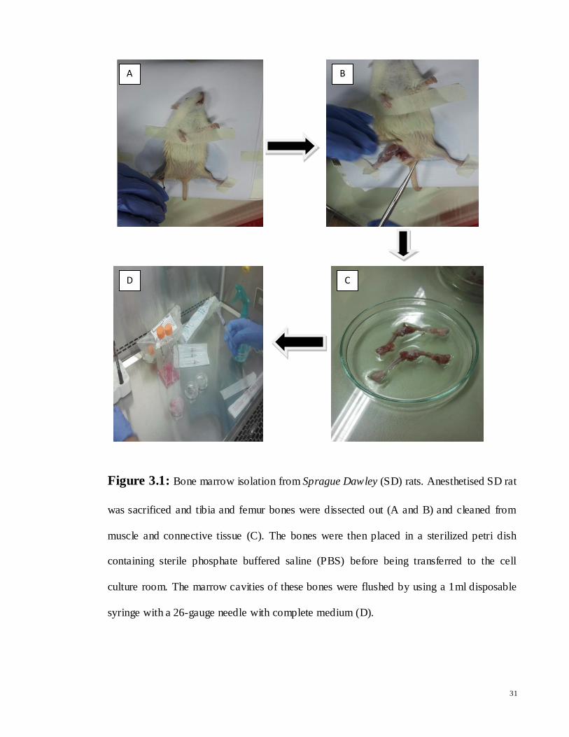
Bone marrow mesenchymal stem cells (BM-MSCs) were isolated from the tibia and femur
of adult Sprague Dawley (SD) rats and cultured according to the adhesive property of
mesenchymal stem cells (MSC) as described by Peister et al. (2004) and Soleimani and
Nadri (2009). When isolated BM-MSCs were seeded in culture flask, initially roundish or
polygonal cell types appeared in bone marrow dissociates culture, which was a mixture of
MSCs and non-adherent cell populations such as hematopoietic stem cells (HSCs). The
HSCs were removed after frequent changing the culture medium. After 2-3 days, MSCs
adhered to the wall in small quantity and scattered about showing spindle-shaped or
fibroblast- liked morphology as previously reported by (Colter et al., 2001; Peister et al.,
2004; Soleimani & Naderi, 2009) (Figure 4.1). This result showed that bone marrow
heterogeneity can be broken down in culture by prolonged growth and in a time dependent
manner, resulting in distinct morphology such as fibroblast- liked phenotypes. Along with
morphological characteristics, reverse transcriptase (RT) PCR, flow cytometry and
immunostaining were also performed to confirm BM-MSCs which will be explained in the
next sections.
46
Figure 4.1: Morphology of undifferentiated BM-MSCs. BM-MSCs exhibited fibroblast-
liked morphology with their characteristic property of attaching to plastic culture dishes.
At passage 0(P0), MSCs appeared as a mixture of small or spindle shaped cells (A and B).
As MSC cells reached 70-80% confluence within 16-20 days, homogenous population of
cells with uniform fibroblast- liked morphology was observed (C) and (D).
(D) X200
(A)X100 (B)X100
(C)X100
47
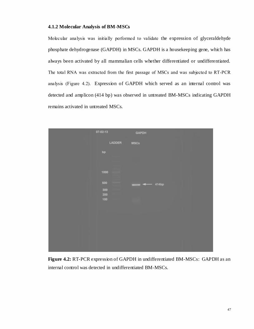
4.1.2 Molecular Analysis of BM-MSCs
Molecular analysis was initially performed to validate the expression of glyceraldehyde
phosphate dehydrogenase (GAPDH) in MSCs. GAPDH is a housekeeping gene, which has
always been activated by all mammalian cells whether differentiated or undifferentiated.
The total RNA was extracted from the first passage of MSCs and was subjected to RT-PCR
analysis (Figure 4.2). Expression of GAPDH which served as an internal control was
detected and amplicon (414 bp) was observed in untreated BM-MSCs indicating GAPDH
remains activated in untreated MSCs.
Figure 4.2: RT-PCR expression of GAPDH in undifferentiated BM-MSCs: GAPDH as an
internal control was detected in undifferentiated BM-MSCs.
48
4.1.3 Immunocytochemistry Analysis of BM-MSCs
In order to further distinguish mesenchymal stem cells from hematopoietic stem
cells, immunocytochemistry and flow cytometry were used to determine the presence of
cell specific surface markers on MSCs. MSCs were first characterized for the positive
presence of CD44, CD117 and negative presence of CD34 by immunocytochemistry. MSC
cells were also treated only with secondary antibody (negative control). DAPI indicated the
nucleus and merged indicated the positive or negative reactivity of marker proteins in
immunocytochemistry analysis. Secondary antibodies employed were Alexa fluor 546
(red) conjugated IgGs.
Immunocytochemistry analysis revealed that expression of cells surface marker
were negative when cells treated only with secondary antibodies (negative control),
indicating that in the absence of primary antibody, no reaction occurs between cells and
secondary antibodies (Figure 4.3). Cell surface antigen expression was observed to be
positive for CD44 (Figure 4.4) and CD117 (Figure 4.5) which are known as rat MSC
markers. In addition, cells were negative for hematopoietic ce ll surface marker CD34,
which validating that cells were MSCs (Figure 4.6).
.
49
Figure 4.3: Immunostaining identification of BM-MSCs on the basis of surface marker
expression (Negative Control): A) The nuclei stained with DAPI (blue), B) Secondary
antibodies employed were Alexa fluor 546 (red) conjugated IgGs, which was negative, and
C) Merged image indicated the negative reaction of the marker protein in the absence of
primary antibody ( magnifications: X200).
A
C
B
50
Figure 4.4: Immunostaining identification of a BM-MSC on the basis of CD44 positive
expression, (magnifications: X400). A) The single nuclei stained with DAPI (blue), B)
Secondary antibodies employed were Alexa fluor 546 (red) conjugated IgGs and C)
Merged image indicated the positive reactivity of marker proteins. This image showed
cells were positive for CD44, which validated that the cells were MSCs.
A B
C
51
Figure 4.5: Immunostaining identification of MSCs on the basis of CD117 positive
expression, (magnification: X400). A) The nuclei of MSCs stained by DAPI (blue), B)
Secondary antibodies employed were Alexa fluor 546 (red) conjugated IgGs and C)
Merged indicated the positive reactivity of marker proteins. Cells were positive for CD117,
which confirmed the fibroblast- liked cells were MSCs rather than hematopoietic stem
cells.
A
C
B
52
Figure 4.6: Immunostaining identification of BM-MSCs on the basis of CD34 negative
expression, (magnification: X400). A) DAPI stained the nuclei blue, B) Secondary
antibodies did not bind to primary antibodies, and C) merged image indicated the negative
reactivity of marker proteins. Secondary antibodies employed were Alexa fluor 546 (red)
conjugated IgGs. Hence, undifferentiated BM-MSCs were negative for CD34, a known
cell surface marker for hematopoietic cells. This result also validated that the fibroblast-
liked cells were MSCs rather than hematopoietic stem cells.
A B
C
53
4.1.4 FACS Analysis of BM-MSCs
Fluorescence-activated cell sorting (FACS) analysis was further performed for the cell
population count of MSCs against CD44 (known MSCs marker) and CD45 (hematopoietic
marker). To note, unlabeled cells (blank control) and cells with only secondary antibody
(negative control) were used as a reference in this study where both of them should give
about the same results. Cells were trypsinized, labeled with antibodies against the indicated
antigens and analyzed by FACS in triplicates. Green Alexa Flour 488 goat anti-mouse was
used as secondary antibody.
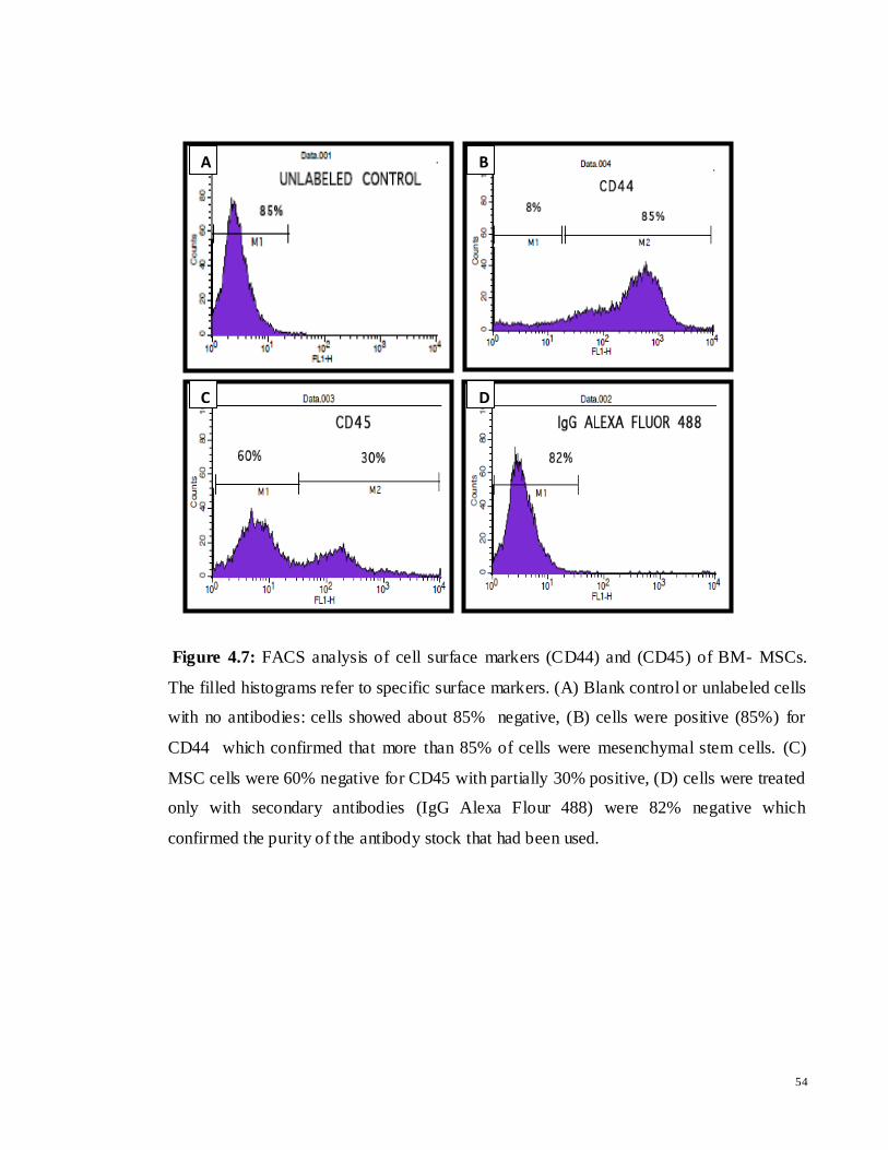
The FACS analysis revealed that untreated MSCs were more than 85% positive for CD44
(Figure 4.7 (B)). While MSCs showed to be more than 60%, 85% and 82% negative for
CD45, unlabeled cells (blank control) and negative control (only secondary antibody)
respectively. These results, along with morphological characteristics, indicated that the
cultured BM-MSCs populations were almost mesenchymal stem cells and most of the
HSCs had been removed.
54
Figure 4.7: FACS analysis of cell surface markers (CD44) and (CD45) of BM- MSCs.
The filled histograms refer to specific surface markers. (A) Blank control or unlabeled cells
with no antibodies: cells showed about 85% negative, (B) cells were positive (85%) for
CD44 which confirmed that more than 85% of cells were mesenchymal stem cells. (C)
MSC cells were 60% negative for CD45 with partially 30% positive, (D) cells were treated
only with secondary antibodies (IgG Alexa Flour 488) were 82% negative which
confirmed the purity of the antibody stock that had been used.
A B
C D
55
4.2 Differentiation of BM-MSCs into Cardiomyocytes-like Cells
4.2.1 Characteristics of Differentiated MSCs after Treatment
The induction of cardiomyocyte- like cells from MSCs was successfully performed.
In this study, to induce myocardial differentiation, MSCs at 70-80% confluence were
incubated in serum-containing medium (DMEM) supplemented with 3µM 5-azacytidine
and zebularine separately for 24 hours, and subsequent culturing in complete medium
(DMEM) up to 20 days. After exposure to 5-azacytidine and zebularine, changes in
morphology were observed. During exposure, some adherent cells died, and the surviving
cells began to proliferate and differentiate. The morphology of the MSCs changed, with the
remaining adherent cells enlarging and forming a ball- liked appearance, or lengthening in
one direction forming a stick-like morphology (Figure 4.8). Towards the end, the cells
changed into myocyte- like cells along with cluster- like aggregates.
56
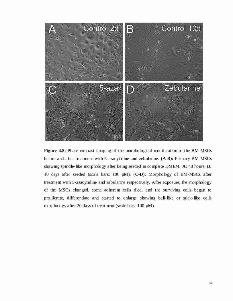
Figure 4.8: Phase contrast imaging of the morphological modification of the BM-MSCs
before and after treatment with 5-azacytidine and zebularine. (A-B): Primary BM-MSCs
showing spindle- like morphology after being seeded in complete DMEM. A: 48 hours; B:
10 days after seeded (scale bars: 100 µM). (C-D): Morphology of BM-MSCs after
treatment with 5-azacytidine and zebularine respectively. After exposure, the morphology
of the MSCs changed, some adherent cells died, and the surviving cells began to
proliferate, differentiate and started to enlarge showing ball- like or stick- like cells
morphology after 20 days of treatment (scale bars: 100 µM).
57
4.2.2 Expression of Cardiac Specific mRNA in Treated and Untreated MSCs
The mRNA expression of cardiomyogenic specific markers of untreated and treated
MSCs with 3µM of 5-azacytidne and zebularine were assessed by reverse transcriptase
polymerase chain reaction (RT-PCR) analysis (Figure 4.9). The housekeeping gene
GAPDH was employed for internal normalization of RNA. The amplicon of GAPDH was
found in both undifferentiated and differentiated MSCs indicating that GAPDH remained
activated in both treated and untreated cells. RT-PCR analysis revealed the presence of
cardiac alpha myosin heavy chain (CAMHC), cardiac troponin-T (cTnT) and cardiac
transcription factors (GATA-4) amplicon in treated MSCs. However, very low intensity
bands of selected cardiac specific genes were observed in untreated MSCs. In addition
cardiac transcription factors (GATA-4) band was not observed in untreated MSCs.
58
Figure 4.9: Expression of cardiac specific genes in treated BM-MSCs: The amplicon of
GAPDH was strongly detected in both untreated and treated MSCs. Reverse Transcriptase
(RT) PCR analysis showed the amplicon presence of CAMHC, cTnT and GATA-4 in
treated MSCs.
59
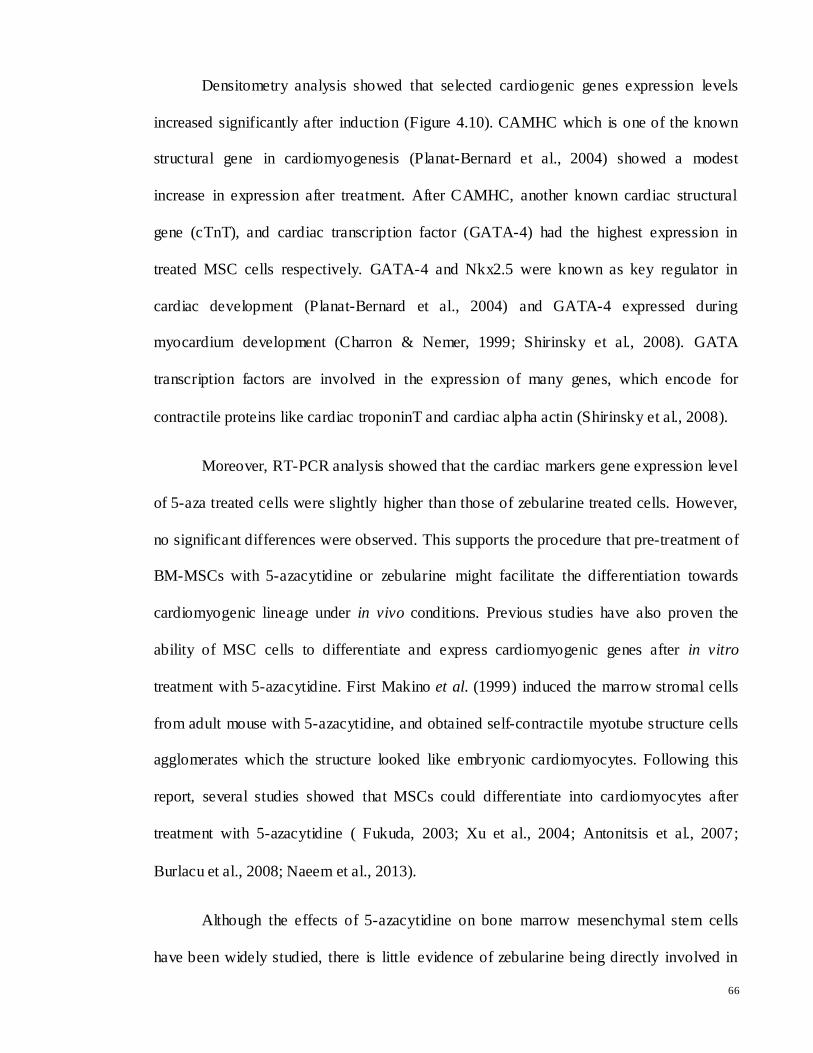
4.2.3 Densitometry Analysis
Densitometry analysis was performed to measure the integrated density value (IDV) of
each band. Relative quantification of each band was performed using gel documentation
system and IDV of each band was compared with the corresponding GAPDH band. The
gene expression level of CAMHC (Figure 4.10 (A)), cTnT (Figure 4.10 (B)) and GATA-4
(Figure 4.10 (C)) were significantly increased after treatment compared to untreated cells.
CAMHC had the highest expression, whereas GATA-4 showed the lowest expression
compared to CAMHC and cTnT after treatment with 5-azacytidine and zebularine. While
selected cardiac specific gene expression level were slightly higher in 5-azacytidine treated
cells compared to zebularine, however there were no significant differences between 5-
azacytidine and zebularine treated cells. This indicated that besides 5-azacytidine,
zebularine could be a good candidate for MSCs induction into cardiomyocytes. Data
obtained were presented as average (mean ± SD; standard deviation) and calculated using
Microsoft Excel. Statistical significance (*p<0.05) was determined using SPSS software
and subjected to t-test to determine a significant difference of the gene expression level
between differentiated and undifferentiated cells.
60
0
0.05
0.1
0.15
0.2
0.25
0.3
0.35
0.4
C CAMHC 3uM 5aza CAMHC 3uM Zeb CAMHC
rela
tive
exp
ress
ion
of
CA
MH
A/G
AP
DH
CAMHC expression in MSCs after 20 days treatment
average
*
*
0
0.05
0.1
0.15
0.2
0.25
C cTnT 3uM 5aza cTnT 3uM Zeb cTnT
rela
tive
exp
ress
ion
of
cTn
T/G
AP
DH
cTnT expression in MSCs after 20 days treatment
average
* *
61
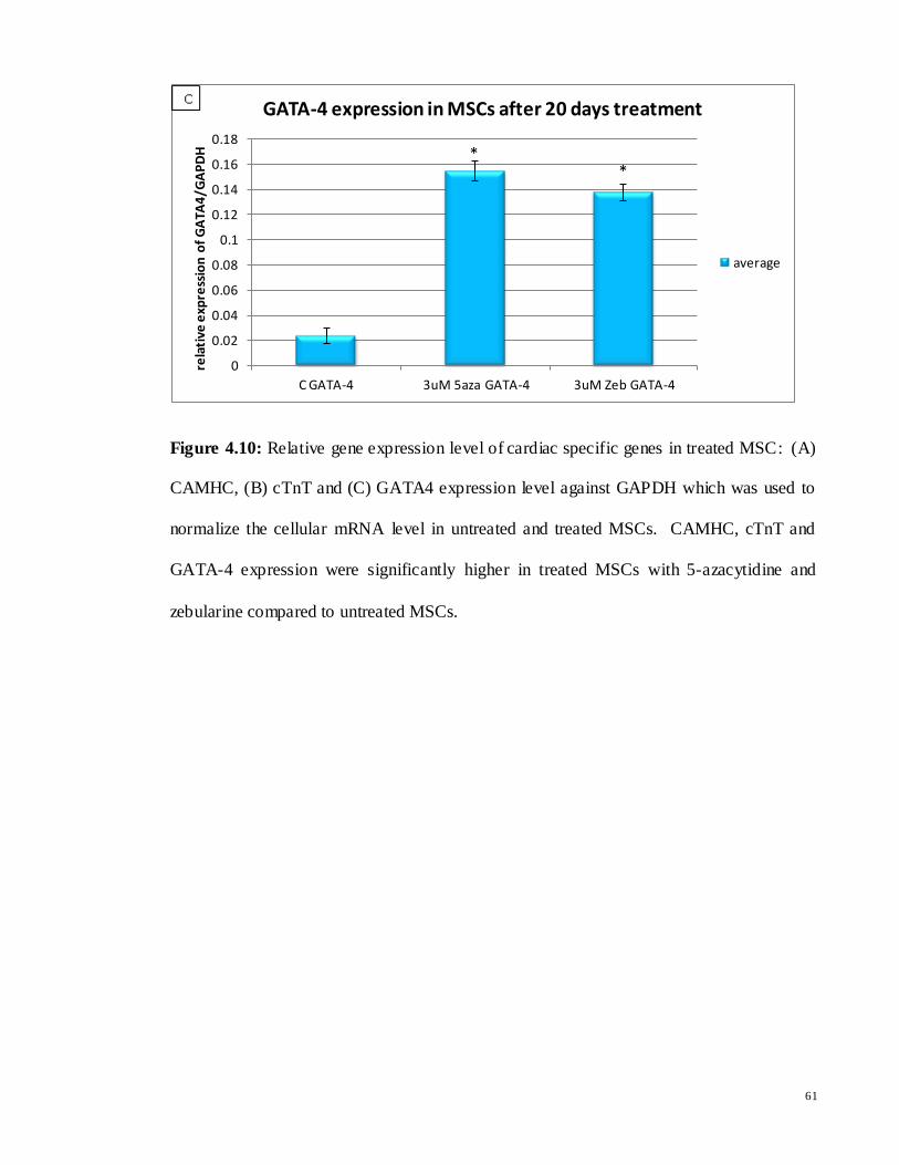
Figure 4.10: Relative gene expression level of cardiac specific genes in treated MSC: (A)
CAMHC, (B) cTnT and (C) GATA4 expression level against GAPDH which was used to
normalize the cellular mRNA level in untreated and treated MSCs. CAMHC, cTnT and
GATA-4 expression were significantly higher in treated MSCs with 5-azacytidine and
zebularine compared to untreated MSCs.
0
0.02
0.04
0.06
0.08
0.1
0.12
0.14
0.16
0.18
C GATA-4 3uM 5aza GATA-4 3uM Zeb GATA-4
rela
tive
exp
ress
ion
of
GA
TA4
/GA
PD
H
GATA-4 expression in MSCs after 20 days treatment
average
* *
62
CHAPTER 5
DISCUSSION
5.1 General Discussion
Cardiovascular diseases particularly myocardial infarction (MI) is the leading cause
of death in the world. This is because most of the cardiomyocytes become differentiated
immediately after birth; hence causing limited or no capacity of adult cardiac musc le to
regenerate damaged area following MI (Burlacu et al., 2008). The use of bone marrow
mesenchymal stem cells (BM-MSCs) and progenitor cells is actively tested as cell-based
therapeutics to restore function of post-MI. Therefore, these cell-based therapies are paving
a path to novel treatment approaches (Webster, 2005; Tendera et al., 2009). BM-MSCs are
adult multi-potential progenitor cells, which have the potential to differentiate into various
tissues under appropriate culture condition. BM-MSCs are being aggressively explored for
their potential and action in affecting the repair of cardiac tissue after myocardial infarction
and have shown the ability to trans-differentiate into cardiomyocytes, both in vitro and in
vivo (Li et al., 2007; Jackson et al., 2007). Cell-based treatment using BM-MSCs have
resulted in encouraging and promising results for the treatment of MI (Shi et al., 2011;
Kim et al., 2011; Li et al., 2012; Raynaud et al., 2013).
In this study, rat BM-MSCs were successfully isolated by a property of adherence
to plastic. After 2-3 days isolation of MSCs and frequent changing of culture medium,
BM-MSCs adhered to the surface of flask in small quantity and scattered about showing
spindle-shaped or fibroblast-like morphology (Figure 4.1), parallel to previous studies.
Naeem et al. (2013), Jianquan et al. (2012), Antonitsis et al. (2007) and Li et al. (2002)
reported the phase contrast microscopy of MSCs revealed as small, bright with fibroblast-
like morphology in primary culture.
63
While there is a wide range of surface markers that have been tested to characterize
MSCs, there is currently no single set of phenotypic markers used to identify a MSC.
Therefore, isolated MSC populations are still not entirely homogenous (Peister et al., 2004;
Rastegar et al., 2010; Williams & Hare, 2011). In this study, after morphological
identification of BM-MSCs, they were further characterized for the positive presence of
selected known MSCs markers: CD44, CD117, and absence of hematopoietic markers
CD34, CD45 using flow cytometry and immunocytochemistry.
Cytometric analysis showed that the isolated MSCs strongly expressed the known
surface markers of MSCs, such as CD44 (85%) (Figure 4.7(B)). The FACS analysis also
revealed that unlabeled cells (no antibody) and cells with only secondary antibody
(negative control) were observed to be 85% (Figure4.7 (A)) and 82% (Figure 4.7 (D))
negative respectively. Unlabeled cells (blank control) and cells with only secondary
antibody (negative control) were used as reference in this study where both of them should
give about the same result as both of them used as negative control. MSCs were also more
than 60% negative for the hematopoietic surface marker CD45 with partially positive
(30%) which could be due to contaminations of other cells for example endothelial cells
(Figure 4.7 (C)).
Furthermore, immunocytochemistry results showed cultured cells were positive for
the well defined MSCs markers, such as CD44 (Figure 4.4), CD117 (figure 4.5) and
negative for CD34 (Figure 4.6), one of the well known hematopoietic surface markers.
Cells were also treated only with secondary antibodies (Alexa Flour 546) as a negative
control (Figure 4.3). This negative control showed that in the absence of primary antibody,
no reaction occurred between cells and secondary antibodies which validated and
confirmed completeness of the experimental procedures. The surface marker expression
profile accords well with previous studies (Bruder et al., 1998; Colter et al., 2000; Javazon
64
et al., 2001; Naeem et al., 2013). The absence of CD34 and CD45, have been widely
accepted as the major differences between MSCs and hematopoietic stem cells (HSCs)
(Colter et al., 2000). Based on the expression of a panel of surface markers, the results of
this study support the identity of isolated cells as MSCs. Variable expression of cell
surface markers has also been observed due to variation in tissue source, isolation and
culture methods (Vacanti et al., 2005; Faast et al., 2006; Ock et al., 2010; Kumar et al.,
2012).
Trans-differentiation potential of MSC into cardiac cell types has been explored
extensively, with several groups reporting that these stem cells can trans-differentiate into
cardiomyocytes (Tomita et al., 1999; Orlic et al., 2001; Rastegar et al., 2010; Williams et
al., 2011; Naeem et al., 2013). However, the environmental-driven differentiation of
uncommitted stem cells is difficult to anticipate and may result in other cell types (Balsam
et al., 2004). One alternative strategy for overcoming ineffective differentiation is the
pretreatment of stem cells to turn them into progenitor cells before becoming adult cells
(Rosca & Burlacu, 2011). There have been some small molecules such as dexamethasone,
ascorbic acid, 5-azacytidine, or all-trans retinoic acid which are capable of inducing the
differentiation of stem cells into different cells types (Ding & Schultz, 2004; Rosca &
Burlacu, 2011; Naeem et al., 2012).
In the present study, after successful isolation and characterization of bone marrow
derived MSCs, the effect of two DNA demethylating agents on differentiation potential of
BM-MSCs into cardiomyocytes were investigated. BM-MSCs were treated separately with
3µM optimized concentrations of 5-azacytidine and zebularine, as described by Naeem and
coworkers (2013). 5-azacytidine is the most popular chemical for inducing stem cells into
cardiomyocytes. 5-azacytidine is analogue of cytidine and it can form covalent conjunction
65
compound with DNA. It is hypothesized that the treatment of cells by 5-azacytidine makes
the cells less responsive to other inductive factors secreted by the microenvironment that
might modulate the differentiation (Rosca & Burlacu, 2011). Zebularine, a cytidine analog
containing a 2-pyrimidinone ring, is another novel DNA methyltransferase (DNMT)
inhibitor, which have been developed as a more stable and less toxic drug compared to 5-
azacytidine (Yoo et al., 2004; Naeem et al., 2013). Zebularine could be a good candidate
for inducer as an alternative to 5-azacytidine which is less stable and more toxic.
The present study demonstrated that demethylating agents, 5-azacytidine and
zebularine could induce changes in BM-MSCs leading to their differentiation in vitro and
directing them towards the cardiomyogenic lineage. Morphological results showed that
fibroblast- liked cells gradually increased in size after exposure to 5-azacytidine and
zebularine. It was observed that a certain concentration of 5-azacytidine and zebularine
changed rat MSCs morphology and promoted the cells to form mytube structures after two
weeks of treatment (Figure 4.8).
Notably, data from reverse transcriptase (RT)-PCR revealed the presence of
cardiac-specific genes, including alpha myosin heavy chain (CAMHC), cardiac troponin-T
(cTnT) and cardiac transcription factor (GATA4) after treatment with 5-azacytidine and
zebularine. However, a very low intensity band of selected cardiac markers were observed
in untreated MSC cells. GAPDH is a housekeeping gene, which has always been activated
by all mammalian cells by undifferentiated or differentiated cells (Barber et al., 2005).
GAPDH amplicon was strongly presented in cells, both before and after differentiation,
indicating that GAPDH remains activated in both types of cells (Figure 4.9).
66
Densitometry analysis showed that selected cardiogenic genes expression levels
increased significantly after induction (Figure 4.10). CAMHC which is one of the known
structural gene in cardiomyogenesis (Planat-Bernard et al., 2004) showed a modest
increase in expression after treatment. After CAMHC, another known cardiac structural
gene (cTnT), and cardiac transcription factor (GATA-4) had the highest expression in
treated MSC cells respectively. GATA-4 and Nkx2.5 were known as key regulator in
cardiac development (Planat-Bernard et al., 2004) and GATA-4 expressed during
myocardium development (Charron & Nemer, 1999; Shirinsky et al., 2008). GATA
transcription factors are involved in the expression of many genes, which encode for
contractile proteins like cardiac troponinT and cardiac alpha actin (Shirinsky et al., 2008).
Moreover, RT-PCR analysis showed that the cardiac markers gene expression level
of 5-aza treated cells were slightly higher than those of zebularine treated cells. However,
no significant differences were observed. This supports the procedure that pre-treatment of
BM-MSCs with 5-azacytidine or zebularine might facilitate the differentiation towards
cardiomyogenic lineage under in vivo conditions. Previous studies have also proven the
ability of MSC cells to differentiate and express cardiomyogenic genes after in vitro
treatment with 5-azacytidine. First Makino et al. (1999) induced the marrow stromal cells
from adult mouse with 5-azacytidine, and obtained self-contractile myotube structure cells
agglomerates which the structure looked like embryonic cardiomyocytes. Following this
report, several studies showed that MSCs could differentiate into cardiomyocytes after
treatment with 5-azacytidine ( Fukuda, 2003; Xu et al., 2004; Antonitsis et al., 2007;
Burlacu et al., 2008; Naeem et al., 2013).
Although the effects of 5-azacytidine on bone marrow mesenchymal stem cells
have been widely studied, there is little evidence of zebularine being directly involved in
67
stem cell differentiation (Liu et al., 2003; Antonitsis et al., 2007; Burlacu et al., 2008). The
results of this study showed that there were no significant differences between gene
expression levels of 5-azacytidine and zebularine treated cells, although both increased
expression of selected cardiac markers significantly after treatment. However, further
studies need to be done to assess the mechanism and differentiation potential of zebularine
into cardiomyocytes both in vitro and in vivo. The potential of zebularine was widely
studied in microbial systems, cancer therapy, as well as mammalian cell lines (Cheng et
al., 2003). Cheng and colleagues (2003) reported that zebularine and 5-azacytidine can
induce the expression of the myogenic phenotype in mouse embryonic fibroblast cells and
inhibited the methylation of specific loci in both the mouse CII-d and human p16
promoter. This study agrees with proposed of Naeem and his colleagues (2013) on
zebularine that it could be used as a new candidate for cardiomyogeneic inducer. With
better stability and less cytotoxic potential, zebularine could be a good replacement for 5-
azacytidine for differentiation of MSCs into cardiomyocytes.
It is worth noting that even though the cardiac specific genes were up regulated and
expression increased after 5-azacytidine and zebularine treatment, they were also found to
be present in untreated cells, in accordance with previous data that suggested a promoting
rather than inductive effect of myogenic gene transcription (Burlacu et al., 2008; Rosca &
Burlacu, 2011). This is also in accordance with the hypothesis that the transcriptional
machinery of adult stem cells is operating at a low level, but is not silenced, such that these
cells express a variety of gene families that characterize differentiated progeny (Zipori,
2004; Rosca & Burlacu, 2011). Rosca and Burlacu (2011) stated that treatment of MSC
with 5-azacytidine may promote subsequent cardiac differentiation but it is dependent on
finding the adequate conditions for cardiomyogenic differentiation. Correct concentration
68
of 5-azacytidine coupled with the use of growth factors and cytokines may be able to
create an adequate conditions for cardiomyogenic differentiation. Rangappa et al. (2003)
also stated that 5- azacytidine promotes rather than induces the myogenic differentiation of
bone marrow progenitor cells as it enhanced the appearance of myogenic markers.
According to this hypothesis, adult stem cells are in a standby state, prepared to
differentiate at any moment. Taken all together, these results suggested that, 5-azacytidine
and zebularine promoted changes in phenotype by expressing markers and activating
cardiac specific genes. It can be expected that treated MSCs prior to transplantation may
increase the likelihood of successful regeneration of damaged myocardium in vivo
environment. Because the cells would be still multipotent at the time of transplantation and
also more open to the subsequent differentiation stimuli.
5.2 Limitations and Future Studies
Despite of BM-MSCs differentiation and expression of cardiac specific genes by 5-
azacytidine and zebularine, treatment by these components alone may not be sufficient to
sustain terminal differentiation of MSCs into cardiomyocytes. Pre-treatment of BM-MSCs
with 5-azacytidine and zebularine before transplantation could facilitate the differentiation
towards cardiomyogenic lineage under in vivo conditions. Following this priming step, the
cardiac differentiation process could be completed in vivo by the factors within the cardiac
environment. It would be worthwhile to investigate whether the treatment of multipotent
stem cells with demethylating agents such as 5-azacytidine and zebularine prior to
transplantation can direct them towards specific cardiomyogenic lineage in the in vivo
environment.
69
CHAPETR 6
CONCLUSION
In this study, successfully isolated and cultured cells displayed the typical fibroblast-
like morphology and surface antigen profile of bone marrow mesenchymal stem cells
(BM-MSCs). Further, distinctive morphological characteristics and the expression of genes
specific to cardiac myocytes supported their potential to differentiate in vitro into
cardiomyocyte- like cells upon exposure to 5-azacytidine and zebularine.
In vitro cardiomyogenic differentiations of rat BM-MSCs, thus, offer a suitable model
to understand their molecular and functional identities prior to transplantation. These
results suggested that, 5-azacytidine and zebularine induce changes in phenotype by
expressing markers and activating specific genes, but its treatment alone may not be
sufficient to sustain terminal differentiation of MSCs into cardiomyocytes. Besides 5-
azacytidine, which is a well known compound, zebularine as a new candidate could be a
good replacement for differentiation of mesenchymal stem cells into cardiomyocytes
because of its stability and less toxicity to biological systems. In order to assure the quality
of the final therapeutic product, it is however important to evaluate the differentiation
potential’s stability in vivo. This would enhance the rate of bone marrow stem cell
differentiation into mature cardiomyocytes in the injured heart.
70
REFERENCES
Aasen, T., Raya, A., Barrero, M. J., Garreta, E., Consiglio, A., Gonzalez, F.,... & Belmonte, J. C. I. (2008). Efficient and rapid generation of induced pluripotent stem cells from
human keratinocytes. Nature biotechnology, 26(11), 1276-1284.
Adewumi, O., Aflatoonian, B., Ahrlund-Richter, L., Amit, M., Andrews, P. W., Beighton, G., ... & Mummery, C. L. (2007). Characterization of human embryonic stem cell
lines by the International Stem Cell Initiative. Nature biotechnology, 25(7), 803-816.
Aggarwal, S. & Pittenger, M. F. (2005). Human mesenchymal stem cells modulate allogeneic immune cell responses. Blood, 105, 1815–1822.
Alfarano, C., Roubeix, C., Chaaya, R. (2012). Intraparenchymal injection of bone marrow
mesenchymal stem cells reduces kidney fibrosis after ischemia-reperfusion in cyclosporineimmunosuppressed rats. Cell Transplant, 21, 2009–2019.
Almeida-Porada, G., Flake, A.W., Glimp, H.A., Zanjani, E.D. (1999). Cotransplantation of stroma results in enhancement of engraftment and early expression of donor
hematopoietic stem cells in utero. Experimental Hematology, 27, 1569–1575.
Amoh, Y., Li, L., Campillo, R., Kawahara, K., Katsuoka, K., Penman, Hoffman, R.M. (2005). Implanted hair follicle stem cells form Schwann cells that support repair of
severed peripheral nerves. Proceeding of the National Academy of Science, 102, 17734-17738.
Anker, P. S., Scherjon, S. A, Kleijburg-van der Keur, C., Noort, W., Claas, F. H. J.,
Willemze, R., … Kanhai, H. H. H. (2003). Amniotic fluid as a novel source of mesenchymal stem cells for therapeutic transplantation. Blood, 102 (4), 1548–1549.
Antonitsis, P., Ioannidou-Papagiannaki, E., Kaidoglou, A., & Papakonstantinou, C. (2007). In vitro cardiomyogenic differentiation of adult human bone marrow mesenchymal
stem cells. The role of 5-azacytidine. Interactive Cardiovascular and Thoracic Surgery, 6 (5), 593–597.
Aránega, A. (2011). Therapeutic Approaches in Regenerative Medicine of Cardiovascular
Diseases: From Bench to Bedside. Advances in regenerative medicine, 61.
Asahara, T. (1997). Isolation of Putative Progenitor Endothelial Cells for Angiogenesis. Science, 275(5302), 964–966.
Asumda, F. Z. (2013). Age-associated changes in the ecological niche: implications for
mesenchymal stem cell aging. Stem cell research & therapy,4(3), 1-11.
71
Baksh, D., Song, L. & Tuan, R.S. (2004). Adult mesenchymal stem cells: characterization, differentiation, and application in cell and gene therapy. Journal of Cell Molecular
Medicine, 8, 301-316.
Ball, L.M., Bernardo, M.E., Locatelli, F., Egeler, R.M. (2008). Potential role of mesenchymal stromal cells in pediatric hematopoietic SCT. Bone Marrow Transplant, 42, 60–66.
Barbash, I.M., Chouraqui, P., Baron, J. (2003). Systemic delivery of bone marrow-derived
mesenchymal stem cells to the infarcted myocardium: Feasibility, cell migration, and body distribution. Circulation, 108, 863–868.
Barber, R. D., Harmer, D. W., Coleman, R. A., & Clark, B. J. (2005). GAPDH as a
housekeeping gene: analysis of GAPDH mRNA expression in a panel of 72 human tissues. Physiological genomics, 21(3), 389-395.
Barry, F. P. (2003). Biology and clinical applications of mesenchymal stem cells. Birth
Defects Research. Part C, Embryo Today : Reviews, 69(3), 250–6.
Barry, F. P., & Murphy, J. M. (2004). Mesenchymal stem cells: clinical applications and biological characterization. The International Journal of Biochemistry & Cell Biology, 36(4), 568–84.
Bjerknes, M., Cheng, H. (2002). Multipotential stem cells in adult mouse gastric
epithelium. American Journal of Physiology Gastrointestinal and Liver Physiology, 283, 767–777.
Bruder,S.P., Kurth,A.A., Shea,M., Hayes,W.C., Jaiswal,N. & Kadiyala,S. (1998). Bone
regeneration by implantation of purified, culture-expanded human mesenchymal stem cells. Journal of Orthopedic Research, 16, 155-162.
Burlacu, A., Rosca, A.-M., Maniu, H., Titorencu, I., Dragan, E., Jinga, V., & Simionescu,
M. (2008). Promoting effect of 5-azacytidine on the myogenic differentiation of bone marrow stromal cells. European Journal of Cell Biology, 87(3), 173–84.
Campagnoli,C., Robert, I. A. G., Kumar, S., Bennett, P. R., Bellantuono, I., & Fisk, N. M.(2001). Identification of mesenchymal stem/progenitor cells in human first-
trimester fetal blood, liver, and bone marrow. Blood, 98, 2396–2402.
Caplan, A.I. (1986). Molecular and cellular differentiation of muscle, cartilage, and bone in the developing limb. Progressive Clinical Biology Research. 1986, 217, 307–318.
Caplan, A. I., & Dennis, J. E. (2006). Mesenchymal stem cells as trophic mediators. Journal of Cellular Biochemistry, 98(5), 1076–1084.
Cedar, H., & Bergman, Y. (2009). Lsinking DNA methylation and histone modification:
patterns and paradigms. Nature Reviews Genetics, 10, 295–304.
Chacko, S.M., Khan, M., Kuppusamy, M.L., Pandian, R.P., Varadharaj, S. (2009) Myocardial oxygenation and functional recovery in infarct rat hearts transplanted with
mesenchymal stem cells. American Journal of Physiology and Heart Circulation Physiology, 296, 1263–1273.
Charron, F. and Nemer, M. (1999). Gata transcription factors and cardiac development. Cell and Developmental Biology, 10, 85-91.
Cheng, J.C., Matsen, C.B., Gonzales, F.A., Ye, W., Greer, S., Marquez, V.E., Jones, P.A.,
Selker, E.U. (2003). Inhibition of DNA methylation and reactivation of silenced genes by zebularine. Journal of the National Cancer Institute, 95, 399-409.
Chen, S. L., Fang, W. W., Ye, F., Liu, Y. H., Qian, J., Shan, S. J., ... & Sun, J. P. (2004).
Effect on left ventricular function of intracoronary transplantation of autologous bone marrow mesenchymal stem cell in patients with acute myocardial infarction. The
American journal of cardiology, 94(1), 92-95.
Cho, J., Zhai, P., Maejima, Y., & Sadoshima, J. (2011). Myocardial injection with GSK-3 -overexpressing bone marrow-derived mesenchymal stem cells attenuates cardiac
dysfunction after myocardial infarction. Circulation Research, 108, 478–489.
Christensen, J. (2010). Isolation, expansion and characterization of single mesenchymal stem cells (Doctoral Dissertation).
Colleoni, S., Donofrio, G., Lagutina, I., Duchi, R., Galli, C., Lazzari, G. (2005).
Establishment, differentiation, electroporation, viral transduction, and nuclear transfer of bovine and porcine mesenchymal stem cells. Cloning and Stem Cells, 7, 154–166.
Colter,D.C., Class,R., DiGirolamo,C.M. & Prockop,D.J. (2000). Rapid expansion of
recycling stem cells in cultures of plastic-adherent cells from human bone marrow. Proceeding National Academy of Science, 97(7), 3213-8.
Curley, G.F., Hayes, M., Ansari, B.,… & et al. (2012). Mesenchymal stemcells enhance recovery and repair following ventilator- induced lung injury in the rat. Thorax, 67,
496–501.
Davy, P.M.M. (2011). Evaluation of bone marrow derived stem cells in the treatment of acute myocardial infarction (University of Hawaii), (Doctoral dissertation). Retrieved
from ProQuest Dissertations & Theses database. (UMI No. 3485480)
73
Deb, A., Wang, S., Skelding, K. A., Miller, D., Simper, D., & Caplice, N. M. (2003). Bone marrow-derived cardiomyocytes are present in adult human heart: a study of gender-
mismatched bone marrow transplantation patients. Circulation, 107, 1247– 1249.
Dennis, J.E., Merriam, A., Awadallah, A., Yoo, J.U., Johnstone, B., Caplan, A.I. (1999). A quadripotential mesenchymal progenitor cell isolated from the marrow of an adult mouse. Journal of Bone Mineral Research, 14, 700–709.
Dezawa, M., Ishikawa, H., Itokazu, Y., Yoshihara, T., Hoshino, M.,Takeda, S., Ide, C., Nabeshima, Y. (2005). Bone marrow stromal cells generate muscle cells and repair muscle degeneration. Science, 309, 314-317.
Devine,S.M. (2002). Mesenchymal stem cells: will they have a role in the clinic? Journal
of Cell Biochemistry Supply, 38, 73-79.
Digirolamo, C. M., Stokes, D., Colter, D., Phinney, D. G., Class, R., & Prockop, D. J. (1999). Propagation and senescence of human marrow stromal cells in culture: a
simple colony- forming assay identifies samples with the greatest potential to propagate and differentiate. British Journal of Haematology, 107, 275–281.
Dill, T., Schachinger, V.,… & Rolf, A. (2009). Intracoronary administration of bone
marrow-derived progenitor cells improves left ventricular function in patients at risk for adverse remodeling after acute ST-segment elevation myocardial infarction: results of the Reinfusion of Enriched Progenitor cells And Infarct Remodeling in
Acute Myocardial Infarction study (REPAIR-AMI) cardiac magnetic resonance imaging substudy. American Heart Journal, 157,541–7.
Dimarakis, I., Levicar, N., Nihoyannopoulos, P., Gordon, M.Y., Habib, N.A. (2006). In
vitro stem cell differentiation into cardiomyocytes. Part 2: chemicals, extracellular matrix, physical stimuli and coculture assays. Journal Cardiothoracic-renal Research, 1, 115-121.
Di Nicola, M., Carlo-Stella, C., Magni, M., Milanesi, M., Longoni, P. D., Matteucci, P.,
Grisanti, S.,… & Gianni, A. M. (2002). Human bone marrow stromal cells suppress T- lymphocyte proliferation induced by cellular or nonspecific mitogenic stimuli.
Blood, 99, 3838–3843.
Ding, S., Schultz, P.G. (2004). A role of chemistry in stem cell biology. Nature Biotechnology, 22, 833-840
Dominici, M., Le Blanc, K., Mueller, I., Slaper-Cortenbach, I., Marini, F. C., Krause, D.
S., ... & Horwitz, E. M. (2006). Minimal criteria for defining multipotent mesenchymal stromal cells. The International Society for Cellular Therapy position statement. Cytotherapy, 8(4), 315-317.
Antitumor properties of 2(1H)-pyrimidinone riboside (zebularine) and its fluorinated analogues. Journal of Medicinal Chemistry, 34, 3280-3284.
74
Edalatmanesh, M.A., Bahrami, A.R., Hosseini, E., Hosseini, M., & Khatamsaz, S. (2011). Bone marrow derived mesenchymal stem cell transplantation in cerebellar
degeneration: a behavioral study. Behavioural Brain Research, 225, 63–70.
Eizawa, T., Ikeda, U., Murakami, Y., Matsui, K., Yoshioka, T., Takahashi, M., Muroi, K., Shimada, K.( 2004). Decrease in circulating endothelial progenitor cells in patients with stable coronary artery disease. Heart, 90, 685–686.
Elnakish, M. T., Hassan, F., Dakhlallah, D., Marsh, C. B., Alhaider, I. A, & Khan, M. (2012). Mesenchymal stem cells for cardiac regeneration: translation to bedside reality. Stem Cells International, 2012, 646038.
kinetics of well and poorly differentiated human prostate cancer cells, Biotechnology and Bioengineering, 80, 580–588.
Evans, M.J. and Kaufman, M.H. (1981) Establishment in culture of pluripotential cells
from mouse embryos. Nature, 5819, 154-156.
Faast, R., Harrison, S.J., Beebe, L.F., McIlfatrick, S.M., Ashman, R.J., Nottle, M.B. (2006). Use of adult mesenchymal stem cells isolated from bone marrow and blood for
somatic cell nuclear transfer in pigs. Cloning and Stem Cells, 8, 166–173.
Fadini, G.P., Pucci, L., Vanacore, R., Baesso, I., Penno, G., Balbarini, A., Di Stefano, R., Miccoli, R., Kreutzenberg, S., Coracina, A. (2007). Glucose tolerance is negatively associated with circulating progenitor cell levels. Diabetologia, 50, 2156–2163.
Fan,C.G., Tang, F. W.,… Zhang, Q. J. (2005). Characterization and neural differentiation
of fetal lung mesenchymal stem cell. Cell Transplantation, 14, 311–321.
Ferrari, G., Cusella-De Angelis, G., Coletta, M., Paolucci, E., Stornaiuolo, a, Cossu, G., & Mavilio, F. (1998). Muscle regeneration by bone marrow-derived myogenic
progenitors. Science, 279(5356), 1528–30.
Fouse, S.D., Shen, Y., Pollegrini, M., Cole, S., Meissner, A., Van Neste, L., Jaenisch, R., Fan, G. (2008). Promoter CpG Methylation Contributes to ES Cell Gene Regulation
in Parallel with Oct4/Nanog, PcG Complex, and Histone H3 K4/K27 Trimethylation. Cell Stem Cell, 2(2), 160-9.
Friedenstein, A. J., Piatetzky-Shapiro, I. I., & Petrakova, K. V. (1966). Osteogenesis in transplants of bone marrow cells. Journal of Embryology and Experimental
Morphology, 16 (3), 381–90.
Friedstein, A.J., Chailakhjan, R.K, & Lalykina, K.S. (1970). The development of fibroblast colonies in monolayer cultures of guinea-pig bone marrow and spleen cells. Cell
SSEA-4 identifies mesenchymal stem cells from BM. Blood, 109, 1743–1751.
Garcia,C.J., Trigueros,C., Madrenas, J., Perez, S. J.A., Rodriguez, R., Menendez, P. (2008). Mesenchymal stem cells and their use as cell replacement therapy and disease modeling tool. Journal of Cellular and Molecular Medicine, 12, 2552-2565.
Gimble, J. M., Guilak, F., Nuttall, M. E., Sathishkumar, S., Vidal, M., & Bunnell, B. a.
(2008). In vitro Differentiation Potential of Mesenchymal Stem Cells. Transfusion Medicine and Hemotherapy , 35 (3), 228–238.
Giorgetti, A., Montserrat, N., Aasen, T., Gonzalez, F., Rodriguez-Piza, I., Vassena, R.,
Raya, A., Boue, S., Barrero, M. J. & Corbella, B. A. (2009). Generation of induced pluripotent stem cells from human cord blood using OCT4 and SOX2. Cell Stem Cell,
5,353-357.
Gonzalez, F., Boue, S., Izpisua Belmonte, J.C. (2011). Methods for making induced pluripotent stem cells: reprogramming a la carte. Nature Review Genetics, 12, 231-242.
Gronthos, S., Mankani, M., Brahim, J., Robey, P. G., & Shi, S. (2000). Postnatal human
dental pulp stem cells (DPSCs) in vitro and in vivo. Proceedings of the National Academy of Sciences of the United States of America, 97(25), 13625–13630.
Haase, A., Olmer, R., Schwanke, K., Wunderlich, S., Merkert, S., Hess, C., Zweigerdt, R.,
Gruh, I., Meyer, J., Wagner, S. (2009). Generation of induced pluripotent stem cells from human cord blood. Cell Stem Cell, 5, 434-441.
Plappert, T., Kiupel, M., John-Sutton, M.G., Itescu, S., Gorman, R.C. (2009). Allogeneic mesenchymal precursor cell therapy to limit remodeling after myocardial
infarction: the effect of cell dosage. The Annal of Thoracic Surgery, 87,794–801.
Hare, J.M. (2009). Translational development of mesenchymal stem cell therapy for cardiovascular diseases. Texas Heart Institute Journal, 36,145–147.
76
Hare, J. M., Traverse, J. H., Henry, T. D., Dib, N., Strumpf, R. K., Schulman, S. P., … Sherman, W. (2009). A randomized, double-blind, placebo-controlled, dose-
escalation study of intravenous adult human mesenchymal stem cells (prochymal) after acute myocardial infarction. Journal of the American College of Cardiology, 54 (24), 2277–2286.
Harris, M. (1892). Induction of thymidine kinase in enzyme-deficient Chinese hamster
cells. Cell, 29, 483–492.
Hatzistergos, K. E., Quevedo, H., Oskouei, B.N., Hu, Q., Feigenbaum, G.S., Margitich, I.S., Mazhari, R., Boyle, A.J., Zambrano, J.P., Rodriguez, J.E., Dulce, R., Pattany,
P.M., Valdes, D., Revilla, C., Heldman, A.W., McNiece, I., Hare, J.M. (2010). Bone marrow mesenchymal stem cells stimulate cardiac stem cell proliferation and
Haynesworth, S.E., Baber, M.A., Caplan, A.I. (1992). Cell surface antigens on human marrow-derived mesenchymal cells are detected by monoclonal antibodies. Bone, 13, 69-80.
Heineke, J., Auger-Messier, M., Xu, J., Oka, T., Sargent, M. A., York, A., ... & Molkentin,
J. D. (2007). Cardiomyocyte GATA4 functions as a stress-responsive regulator of angiogenesis in the murine heart. The Journal of clinical investigation, 117(11), 3198-
Humphreys, T. (2011). Evaluation of bone marrow derived stem cells in the treatment of
acute myocardial infarction a dissertation submitted to the graduate division of the university of hawaii at mānoa in partial fulfillment of the requirements for the degree of doctor of.
function after myocardial infarction in micewithout long-termengraftment. Biochemical and Biophysical Research Communications, 354, 700–706.
Jackson, K. A., Majka, S. M., Wang, H., Pocius, J., Hartley, C. J., Majesky, M. W., Entman, M. L., Michael, L. H., Hirschi, K. K., & Goodell, M. A. (2001).
Regeneration of ischemic cardiac muscle and vascular endothelium by adult stem cells.The Journal of Clinical Investigation, 107, 1395–1402.
Jüttermann, R., Li, E., Jaenisch, R. (1994). Toxicity of 5-aza-2'-deoxycytidine to mammalian cells is mediated primarily by covalent trapping of DNA
methyltransferase rather than DNA demethylation. Proceeding of the National Academy of Science, 91, 11797-11801.
Kaplan, R. N., Rafii, S., & Lyden, D. (2006). Preparing the “soil”: the premetastatic niche. Cancer Research, 66(23), 11089–11093.
Kanazawa, H., Fujimoto, Y.,… Teratani, T. (2011). Bone marrow derived mesenchymal stem cells ameliorate hepatic ischemia reperfusion injury in a rat model. PLoS ONE, 6, 19195- 2011.
Kim, C.H., Marquez, V.E., Mao, D.T., Haines, D.R., McCormack, J.J. (1986). Synthesis of
pyrimidin-2-one nucleosides as acid-stable inhibitors of cytidine deaminase. Journal of Medicinal Chemistry, 29, 1374-1380.
Kim, D. H., Yoo, K. H., Choi, K. S., Choi, J., Choi, S.-Y., Yang, S.-E., … Koo, H. H.
(2005). Gene expression profile of cytokine and growth factor during differentiation of bone marrow-derived mesenchymal stem cell. Cytokine, 31(2), 119–126.
Kim, Y. S., & Ahn, Y. (2012). A long road for stem cells to cure sick hearts: update on
recent clinical trials. Korean Circulation Journal, 42(2), 71–79.
Khakoo, A.Y., & Finkel, T. (2005). Endothelial progenitor cells. Annual Review of Medicine, 56, 79–101.
Kolf, C. M., Cho, E., & Tuan, R. S. (2007). Mesenchymal stromal cells. Biology of adult
mesenchymal stem cells: regulation of niche, self- renewal and differentiation. Arthritis Research & Therapy, 9(1), 204.
Kraitchman, D.L., Tatsumi, M., Gilson, W.D., Ishimori, T., Kedziorek, D., Walczak, .P,
Segars, W.P., Chen, H.H., Fritzges, D., Izbudak, I., Young, R.G., Marcelino, M., Pittenger, M.F., Solaiyappan, M., Boston, R.C., Tsui, B.M., Wahl, R.L., Bulte, J.W. (2005).Dynamic imaging of allogeneic mesenchymal stem cells trafficking to
Krampera, M., Marconi, S., Pasini, A., Galie, M., Rigotti, G., Mosna, F., Tinelli, M., Lovato, L., Anghileri, E., Andreini, A., Pizzolo, G., Sbarbati, A., Bonetti, B. (2007).
78
Induction of neural- like differentiation in human mesenchymal stem cells derived from bone marrow, fat, spleen and thymus. Bone, 40, 382–390.
Kuçi, S., Kuçi, Z., Kreyenberg, H., Deak, E., Pütsch, K., Huenecke, S., … Bader, P. (2010).
CD271 antigen defines a subset of multipotent stromal cells with immunosuppressive and lymphohematopoietic engraftment-promoting properties. Haematologica, 95(4), 651–659.
Kumar, B. M., Maeng, G.-H., Lee, Y.-M., Kim, T.-H., Lee, J.-H., Jeon, B.-G., … Rho, G.-J. (2012). Neurogenic and cardiomyogenic differentiation of mesenchymal stem cells isolated from minipig bone marrow. Research in Veterinary Science, 93(2), 749–757.
Kumar, S., & Singh, N. P. (2006). Stem cells: A new paradigm. Indian Journal of Human
Genetics, 12, 4-10.
Kunter, U., Rong, S.,… Djuric, Z. (2006). Transplanted mesenchymal stem cells accelerate glomerular healing in experimental glomerulonephritis. Journal of the American
Laurie, G. (2004). Patenting Stem Cells of Human Origin. 1–17.
Lee,R.H., Seo,M.J., Reger, R.L. ( 2006). Multipotent stromal cells from human marrow
home to and promote repair of pancreatic islets and renal glomeruli in diabetic NOD/scid mice. Proceedings of the National Academy of Sciences of the United States of America, 103, 17438–17443.
Lee,K.D., Kuo,T.K., Whang-Peng,J., Chung,Y.F., Lin,C.T., Chou,S.H., Chen,J.R., Chen,Y.P. & Lee,O.K. (2004). In vitro hepatic differentiation of human mesenchymal stem cells. Hepatology, 40, 1275-1284.
Lee, K., Majumdar, M.K., Buyaner, D., Hendricks, J.K., Pittenger, M.F., Mosca, J.D.
(2001) Human mesenchymal stem cells maintain transgene expression during expansion and differentiation. Molecular Therapy, 3, 857-866.
Leri, A., Kajstura, J., Anversa, P. (2005). Cardiac stem cells and mechanisms of
Li, E. (2002). Chromatin modification and epigenetic reprogramming in mammalian development. Nature Reviews Genetics, 3, 662–673.
Li, L., and Clevers, H. (2010). Coexistence of quiescent and active adult stem cells in
mammals. Science, 327, 542-545.
79
Li, Y., Chen, J., Zhang, C.L., Wang, L., Lu, D., Katakowski, M., Gao, Q., Shen, L.H., Zhang, J., Lu, M., Chopp, M. (2005). Gliosis and brain remodeling after treatment of
stroke in rats with marrow stromal cells. Glia, 49, 407-417.
Li, W., Ma, N., Ong, L.L., Nesselmann, C., Klopsch, C., Ladilov, Y., Furlani, D., Piechaczek, C., Moebius, J.M., Lutzow, K., Lendlein, A., Stamm, C., Li, R.K., Steinhoff, G. (2007). Bcl-2 engineered MSCs inhibited apoptosis and improved heart
function. Stem Cells, 25, 2118-2127.
Li, X.H., Fu, Y.H., Lin, Q.X., Liu, Z.Y., Shan, Z.X. (2012). Induced bone marrow mesenchymal stem cells improve cardiac performance of infarcted rat hearts.
Molecular Biology Research, 39, 1333–1342.
Liu, Y., Song, J., Liu, W., Chen, X., Hu, C. (2003). Growth and differentiation of rat bone marrow stromal cells: does 5-azacytidine trigger their cardiomyogenic differentiation.
Cardiovascular Research, 58, 460-468.
Loh, Y. H., Agarwal, S., Park, I. H., Urbach, A., Huo, H., Heffner, G. C., Kim, K., Miller, J. D., Ng, K., Daley, G. Q. (2009). Generation of induced pluripotent stem cells from human blood. Blood, 113, 5476-5479.
Loh, Y.H., Hartung, O., Li, H., Guo, C., Sahalie, J. M., Manos, P. D., Urbach, A., Heffner, G. C., Grskovic, M., Vigneault, F. (2010). Reprogramming of T-cells from human peripheral blood. Cell Stem Cell, 7, 15-19.
Verhaar, M.C., Braam, B., Rabelink, T.J., Zonneveld, A.J.( 2004). Endothelial progenitor cell dysfunction: A novel concept in the pathogenesis of vascular
complications of type 1 diabetes. Diabetes, 53, 195–199.
Lu, J., Moochhala, S., Moore, X. L. (2006). Adult bone marrow cells differentiate into neural phenotypes and improve functional recovery in rats following traumatic brain injury. Neurosci Lett, 398, 12–17.
Ma, T. (2010). Mesenchymal stem cells: From bench to bedside. World Journal of Stem
Cells, 2, 13-17.
Majumdar, M.K., Thiede, M.A., Mosca, J.D., Moorman, M.,Gerson, S.L. (1998). Phenotypic and functional comparison of cultures of marrow-derived mesenchymal
stem cells (mscs) and stromal cells. Journal of Cellular Physiology, 176, 186–192.
Majumdar, M.K., Keane-Moore, M., Buyaner, D., Hardy, W.B., Moorman, M.A., McIntosh, K.R., and Mosca, J.D. (2003). Characterization and functionality of cell
surface molecules on human mesenchymal stem cells. Journal of Biomedical Science, 10, 228–241.
Makino, S., Fukuda, K., Miyoshi, S., Konishi, F., odama H.K., Pan, J., Sano, M., Takahashi, T., Hori, S., Abe, H., Hata, J., Umezawa, A., Ogawa, S. (1999).
80
Cardiomyocytes can be generated from marrow stromal cells in vitro.The Journal of Clinical Investigation, 103, 697–705.
Mareddy, S., Crawford, R., Brooke, G., Xiao, Y. (2007). Clonal isolation and
characterization of bone marrow stromal cells from patients with osteoarthritis. Tissue Engineering, 13, 819-829.
Martin-Rendon, E., Sweeney, D., Gridlestone, J., Navarrete, C., Watt, S.M. (2008). %-
azacytidine treated human mesenchymal stem/progenitor cells derived from umbilical cord, cord blood and bone marrow do not generate cardiomyocytes in vitro at high frequencies. Vox Sanguinis, 95, 137–148.
McNeer, K. W. (2007). Therapeutic application of mesenchymal stem cells. 2(5), 733–739.
Melo, L.G., Pachori, A.S., Kong, D. (2004). Molecular and cellbased therapies for
protection, rescue, and repair of ischemic myocardium: reasons for cautious optimism. Circulation, 109, 2386-2393.
Mendis, S., Puska, P., Norrving, B. (2011). Global Atlas on Cardiovascular Disease
Prevention and Control. World Health Organization, Geneva, Switzerland, 2011.
Miyoshi, K., Tsuji, D., Kudoh, K., Satomura, K., Muto, T., Itoh, K. & Noma, T. (2010).
Generation of human induced pluripotent stem cells from oral mucosa. Journal of Bioscience Bioengineering, 110, 345-50.
Moscoso, I., Centeno, A., Lopez, E., Rodriguez-Barbosa, J.I., Santamarina, I., Filgueira, P.,
Sanchez, M.J., Dominguez-Perles, R., Penuelas-Rivas, G., Domenech, N. (2005). Differentiation in vitro of primary and immortalized porcine mesenchymal stem cells into cardiomyocytes for cell transplantation. Transplantation Proceedings, 37, 481–
J.D.,Williams, D.A., Field, L.J. (2004). Haematopoietic stem cells do not transdifferentiate into cardiac myocytes in myocardial infarcts. Nature, 428, 664-668.
Naeem, N., Haneef, K., Kabir, N., Iqbal, H., Jamall, S., & Salim, A. (2013). DNA methylation inhibitors, 5-azacytidine and zebularine potentiate the transdifferentiation
of rat bone marrow mesenchymal stem cells into cardiomyocytes. Cardiovascular Therapeutics, 31(4), 201–9.
Nakagami, H., Maeda, K., Morishita, R., Iguchi, S., Nishikawa, T., Takami, Y., Kikuchi,
Y., Saito, Y., Tamai, K., Ogihara, T., Kaneda, Y. (2005). Novel autologous cell therapy in ischemic limb disease through growth factor secretion by cultured adipose
81
tissue-derived stromal cells. Arteriosclerosis, Thrombosis, and Vascular Biology, 25, 2542-2547.
Netanely, D. (2006). Gene Expression Analysis of Mesenchymal Stem Cell
Differentiation and Leukemic Over Expression of Tissue Specific Genes. (University of Weizmann Institute of Science), (Master dissertation). Retrieved from ProQuest Dissertations & Theses database.
Noort, W. A., Kruisselbrink, A. B., in’t Anker, P. S., Kruger, M., van Bezooijen, R. L., de Paus, R. A., Heemskerk, M. H., Lowik, C. W., Falkenburg, J. H., Willemze, R., & Fibbe, W. E. (2002). Mesenchymal stem cells promote engraftment of human
Ng, W. A., Grupp, I. L., Subramaniam, A., & Robbins, J. (1991). Cardiac myosin heavy
chain mRNA expression and myocardial function in the mouse heart. Circulation research, 68(6), 1742-1750.
Obradovic, S., Rusovic, S., Balint, B., Ristic´-Andelkov, A., Romanovic, R., Baskot, B., Vojvodic, D., Gligic, B. (2004). Autologous bone marrow-derived progenitor cell
transplantation for myocardial regeneration after acute infarction. Vojnosanit Pregl, 61, 519–529.
mesenchymal stem cells derived from bone marrow extract and skin tissues. Tissue Engineering, 16, 1481–1491.
Oda, Y., Yoshimura, Y., Ohnishi, H., Tadokoro, M., Katsube, Y., Sasao, M., Kubo, Y.,
Hattori, K., Saito, S., Horimoto. (2010). Induction of pluripotent stem cells from human third molar mesenchymal stromal cells . Journal of Biological Chemistry, 285, 29270-29278.
Oka, T., Xu, J., & Molkentin, J. D. (2007). Re-employment of developmental transcription
factors in adult heart disease. In Seminars in cell & developmental biology 18(1), 117-131.
Okita, K., Ichisaka, T.,… & Yamanaka, S. (2007). Generation of germline competent
Ortiz, L.A., DuTreil, M., Fattman, C., & et al. (2007). Interleukin 1 receptor antagonist mediates the antiinflammatory and antifibrotic effect of mesenchymal stem cells
during lung injury.Proceedings of the National Academy of Sciences of the United States of America, 104, 11002–11007.
Owen, M. (1988). Marrow stromal stem cells. Journal of Cell Science Supply, 10, 63–76.
Piersma, A.H., Brockbank, K.G., Ploemacher, R.E., Van, V.E., Brakel, P. K.M., Visser,
P.J.(1985). Characterization of fibroblastic stromal cells from murine bone marrow. Journal of Experimental Hematology, 13, 237–243.
Peister,A., Mellad, J. A., Larson, B. L., Hall, B. M., Gibson, L. F. & Prockop,D.J. (2004).
Adult stem cells from bone marrow (MSCs) isolated from different strains of inbred mice vary in surface epitopes, rates of proliferation, and differentiation potential. Blood, 103, 1662- 1668.
J., Vaughn, W.K., Coulter, S., Fernandes, M.R., Willerson, J.T. (2008). Comparison of intracoronary and transendocardial delivery of allogeneic mesenchymal cells in a
canine model of acute myocardial infarction. Journal of Molecular Cell Cardiology, 44, 486–495.
Pittenger, M. F., Mackay, M., Beck, S. C., Jaiswal, R. K., Douglas, R., Mosca, J. D., …
Marshak, D. R. (1999). Multilineage potential of adult human mesenchymal stem cells. Science, 284 (5411), 143–147.
Pittenger, M. F., & Martin, B. J. (2004). Mesenchymal stem cells and their potential as cardiac therapeutics. Circulation Research, 95 (1), 9–20.
Planat-Bernard, V., Menard, M., Andre, M., Puceat, A., Perez, J.M., Garcia-Verdugo, L.,
Casteilla, L. (2004). Spontaneous cardiomyocyte differentiation from adipose tissue stroma cells. Circulation Research, 94:223-9.
Prockop, D. J. (1997). Marrow stromal cells as stem cells for nonhematopoietic tissues.
Science , 276, (5309), 71-74.
Psaltis, P. J., Zannettino, A. C. W., Worthley, S. G., Gronthos, S. (2008). Concise review: mesenchymal stromal cells: potential for cardiovascular repair. Stem Cells, 26, 2201–
2210.
Qian, Q., Qian, H., Zhang, X., Zhu, W., Yan, Y., Ye, S., … Xu, W. (2012). 5-Azacytidine induces cardiac differentiation of human umbilical cord-derived mesenchymal stem cells by activating extracellular regulated kinase. Stem Cells and Development, 21(1),
J.M. (2009). Allogeneic mesenchymal stem cells restore cardiac function in chronic ischemic cardiomyopathy via trilineage differentiating capacity. Proceeding the National Academy of Science, 106, 14022–14027.
83
Rangappa, S., Fen, C., Lee, E.H., Bongso, A., Wei, E.S.K. (2003). Transformation of adult mesenchymal stem cells isolated from the fatty tissue into cardiomyocytes. The
Annals of Thoracic Surgery, 75, 775–779.
Rastegar, F., Shenaq, D., Huang, J., Zhang, W., Zhang, B.-Q., He, B.-C., … He, T.-C. (2010). Mesenchymal stem cells: Molecular characteristics and clinical applications. World Journal of Stem Cells, 2 (4), 67–80.
Raynaud, C. M., Halabi, N., Elliott, D. a, Pasquier, J., Elefanty, A. G., Stanley, E. G., & Rafii, A. (2013). Human embryonic stem cell derived mesenchymal progenitors express cardiac markers but do not form contractile cardiomyocytes. PloS One, 8(1),
e54524.
Reiser, J., Zhang, X.Y., Hemenway, C.S., Mondal, D., Pradhan, L., La Russa, V.F. (2005). Potential of mesenchymal stem cells in gene therapy approaches for inherited and
acquired diseases. Expert Opinion of Biological Therapy, 5, 1571-1584.
Reik, W. (2007). Stability and flexibility of epigenetic gene regulation in mammalian development. Nature, 447(7143), 425-432.
L., LeBlanc, K. (2007). Tissue repair using allogeneic mesenchymal stem cells for hemorrhagic cystitis, pneumomediastinum and perforated colon. Leukemia, 21, 2271-2276.
Rizvi, A. Z., Hunter, J. G. & Wong, M. H. (2005). Gut-derived stem cells. Surgery, 137,
585-590.
Robinton, D.A., & Daley, G.Q. (2012). The promise of induced pluripotent stem cells in research and therapy. Nature, 481, 295-305.
Rosca, A.-M., & Burlacu, A. (2011). Effect of 5-azacytidine: evidence for alteration of the
multipotent ability of mesenchymal stem cells. Stem Cells and Development, 20(7), 1213–1221.
Rosova, I., Dao, M., Capoccia, B., Link, D., Nolta, J.A. (2008). Hypoxic Preconditioning
Results in Increased Motility and Improved Therapeutic Potential of Human Mesenchymal Stem Cells. Stem Cells, 26, 2173–2182.
Sato, Y., Araki, H., Kato, J., Nakamura, K., Kawano, Y., Kobune, M., … Niitsu, Y. (2005). Human mesenchymal stem cells xenografted directly to rat liver are differentiated into
human hepatocytes without fusion. Blood, 106(2), 756–763.
Sasportas, L. S., Kasmieh, R., Wakimoto, H., Hingtgen, S., van de Water, J. A., Mohapatra, G., ... & Shah, K. (2009). Assessment of therapeutic efficacy and fate of engineered
human mesenchymal stem cells for cancer therapy.Proceedings of the National Academy of Sciences, 106(12), 4822-4827.
84
Seki, T., Yuasa, S., Oda, M., Egashira, T., Yae, K., Kusumoto, D., Nakata, H., Tohyama, S., Hashimoto, H., Kodaira, M. (2010). Generation of induced pluripotent stem cells
from human terminally differentiated circulating T-cells. Cell Stem Cell, 7, 11-14.
Shake, J. G., Gruber, P. J., Baumgartner, W. A., Senechal, G., Meyers, J., Redmond, J. M., Pittenger, M. F., & Martin, B. J. (2002). Mesenchymal stem cell implantation in a swine myocardial infarct model: engraftment and functional effects. The Annals of
Thoracic Surgery, 73, 1919–1925
Shiota, M., Heike, T., Haruyama, M., Baba, S., Tsuchiya, A., Fujino, H., Kobayashi, H., Kato, T., Umeda, K., Yoshimoto, M., Nakahata, T. (2007). Isolation and
characterization of bone marrow-derived mesenchymal progenitor cells with myogenic and neuronal properties. Experimental Cell Research, 313, 1008-1023.
Shi, Q., Rafii, S., Wu, M. H., Wijelath, E. S., Yu, C., Ishida, a, … Hammond, W. P. (1998).
Evidence for circulating bone marrow-derived endothelial cells. Blood, 92 (2), 362–367.
Shirinsky, V. P., Khapchaev, A. Y., & Stepanova, O. V. (2008). Molecular mechanisms of cardiomyogenesis and the prospects for cardiomyocyte regeneration in cardiac
failure. Molecular biology, 42(5), 762-772.
Si,Y., Zhao,Y., Hao, H. (2012). Infusion of mesenchymal stemcells ameliorates hyperglycemia in type 2 diabetic rats: identification of a novel role in improving
Willerson, J.T., Perin, E.C. (2005). Mesenchymal stem cells differentiate into an endothelial phenotype, enhance vascular density, and improve heart function in a canine chronic ischemia model. Circulation, 111, 150–156.
Smith, A.G. (2001). Origins and properties of mouse embryonic stem cells. Annual Review
of Cell and Developmental Biology, 17, 435–462.
Soleimani, M., & Nadri, S. (2009). A protocol for isolation and culture of mesenchymal stem cells from mouse bone marrow. Nature Protocols, 4(1), 102–6.
Stamm, C., Westphal, B., Kleine, H. D., Petzsch, M., Kittner, C., Klinge, H., Schumichen,
C., Nienaber, C. A., Freund, M., & Steinhoff, G. (2003). Autologous bone-marrow stem-cell transplantation for myocardial regeneration. Lancet, 361, 45– 46.
Studeny, M., Marini, F.C., Dembinski, J.L., Zompetta, C., Cabreira-Hansen, M., Bekele,
B.N., Champlin, R.E., Andreeff, M. (2004). Mesenchymal stem cells:Potential precursors for tumor stroma and targeted delivery vehicles for anticancer agents. Journal of Nature Cancer, 21,1593–1603.
85
Sulewska, A., Niklinska, W., Kozlowski, M., Minarowski, L., Naumnik, W., Niklinski, J., Dabrowska, K., Chyczewski, L. (2007). DNA methylation in states of cell physiology
and pathology. Folia Histochem Cytobiol, 45(3), 149-58.
Swanepoel, M. (2006). Embryonic stem cell research and cloning. (University of Pretoria), (Master dissertation). Retrieved from ProQuest Dissertations & Theses database.
Takahashi, K., Tanabe, K., Ohnuki, M., Narita, M., Ichisaka, T., Tomoda, K., & Yamanaka,
S. (2007). Induction of pluripotent stem cells from adult human fibroblasts by defined factors. Cell, 131(5), 861–872.
Tavassoli, M. & Crosby, W.H. (1970). Bone marrow histogenesis: a comparison of fatty and red marrow. Science, 169, 291–293.
Taylor, S.M., Jones, P.A. (1982). Mechanism of action of eukaryotic DNA
methyltransferase. Use of 5 azacytosine-containing DNA. Journal of Molecular Biology, 162, 679–692.
Torrente, Y., and Pollie, E. (2008). Mesenchymal stem cell transplantation for
Tang, J., Xie, Q., Pan, G., Wang, J., Wang, M. (2006). Mesenchymal stem cells participate in angiogenesis and improve heart function in rat model of myocardial ischemia with
reperfusion. European Journal of Cardio-Thoracic Surgery, 30-353-361.
Tendera, M., Wojakowski, W., Ruzyllo, W., Chojnowska, L., Kepka, C. (2009). Intracoronary infusion of bone marrow-derived selected CD34+CXCR4+ cells and nonselected mononuclear cells in patients with acute STEMI and reduced left
ventricular ejection fraction: results of randomized, multicentre Myocardial Regeneration by Intracoronary Infusion of Selected Population of Stem Cells in Acute
Myocardial Infarction (REGENT) Trial. European Heart Journal, 30, 1313-1321.
Toma, C. (2002). Human Mesenchymal Stem Cells Differentiate to a Cardiomyocyte Phenotype in the Adult Murine Heart. Circulation, 105 (1), 93–98.
Tomita, S., Li, R. K., Weisel, R. D., Mickle, D. A., Kim, E. J., Sakai, T., & Jia, Z. Q.
(1999). Autologous transplantation of bone marrow cells improves damaged heart function. Circulation, 100(suppl 2), II-247.
Thomson, J.A., Itskovitz, E. J., Shapiro, S.S., Waknitz, M.A., Swiergiel, J.J., Marshall,V.S,& Jones, J.M.(1998). Embryonic stem cell lines derived from human
blastocysts. Science, 282, 1145-1147.
Uccelli, A., Moretta, L., & Pistoia, V. (2008). Mesenchymal stem cells in health and disease. Nature Reviews. Immunology, 8(9), 726–36.
86
Vacanti,V., Kong, E., Suzuki, G., Sato, K., Canty, J.M., Lee, T. (2005). Phenotypic changes of adult porcine mesenchymal stem cells induced by prolonged passaging in
culture. Journal of Cellular Physiology, 205, 194–201.
Vesely, J., Cihak, A. (1975). Mechanism of cystostatic effect of 5-azacytidine and its usefulness in clinic. Cas Lek Cesk, 114, 607–11.
Wakitani, S., Saito, T., & Caplan, A. I. (1995). Myogenic cells derived from rat bone
translational findings, and therapeutic implications for cardiac disease. Circulation Research, 109(8), 923-940
Williams, A. R., Trachtenberg, B., Velazquez, D. L., McNiece, I., Altman, P., Rouy, D., …
Hare, J. M. (2011). Intramyocardial stem cell injection in patients with ischemic cardiomyopathy: functional recovery and reverse remodeling. Circulation Research, 108 (7), 792–796.
offspring derived from fetal and adult mammalian cells. Nature, 385 (6619), 810-813.
Woodbury, D., Schwarz, E. J., Prockop, D.J., Black, I. B. (2000). Adult rat and human bone marrow stromal cells differentiate into neurons. Journal Neuroscience Research,
61, 364–370.
Xu, W., Zhang, X., Qian, H., Zhu, W., Sun, X., Hu, J., Zhou, H., Chen, Y. (2004). Mesenchymal stem cells from adult human bone marrow differentiate into a
cardiomyocyte phenotype in vitro. Experimental Biology and Medicine, 229, 623–631.
Ye, N.S., Zhang, R.L., Zhao, Y.F., Feng, X., Wang, Y.M., Luo, G.A. (2006). Effect of 5- azacytidine on the protein expression of porcine bone marrow mesenchymal stem cells in vitro. Genomics, Proteomics & Bioinformatics, 4, 18–25.
Yoo, J., Barthel, T., Nishimura, K., Solchaga, L., Caplan, A., Goldberg, V., Johnstone, B.
(1998). The chondrogenic potential of human bone-marrow-derived mesenchymal progenitor cells. Journal of Bone Joint Surgery, 80, 1745–1757.
Yoo, C.B., Cheng, J.C., Jones, P.A. (2004). Zebularine: a new drug for epigenetic therapy.
Biochemical Society Transaction, 32, 910–210.
87
Young R. G., Butler D. L., Weber W., Caplan A. I, Gordon S. L., Fink D. J. (1998). Use of mesenchymal stem cells in a collagen matrix for Achilles tendon repair. Journal of
Orthopaedic Research, 16, 406-413
Young, H. E., & Black, A. S. A. C. (2004). Adult Stem Cells, 102, 75–102.
Zhang, D.Z., Gai, L., Liu, H., Jin, Q., Huang, J., Zhu, X. (2007). Transplantation of autologous adipose-derived stem cells ameliorates cardiac function in rabbits with
myocardial infarction. Chinese Medical Journal, 120(4), 300-307.
Zhang, Y., Chu, Y., Shen, W., Dou, Z. (2009). Effect of 5-azacytidine induction duration on differentiation of human first trimester fetal mesenchymal stem cells towards cardiomyocyte- like cells. Interactive CardioVascular and Thoracic Surgery, 9, 943–
946.
Zhao, W., Li, J.J., Cao, D.Y., & et al. (2012). Intravenous injection of mesenchymal stem cells is effective in treating liver fibrosis. World Journal of Gastroenterology, 18,
1048–1058.
Zhou, L., Cheng, X., Connolly, B., Dickman, M., Hurd, P., Hornby, D. (2002). Zebularine: a novel DNA methylation inhibitor that forms a covalent complex with DNA
methyltransferases. Journal of Molecular Biology, 321, 591.
Zipori, D. (2004). The nature of stem cells: state rather than entity. Nature Reviews Genetics, 5(11), 873-878.
Zuk, P. A., Zhu, M., Ashjian, P., Ugarte, D. A. De, Huang, J. I., Mizuno, H., … Hedrick, M. H. (2002). Human Adipose Tissue Is a Source of Multipotent Stem Cells. 13,
4279–4295.
88
Appendix A: List of Apparatus, Chemical and Material