CHARACTERIZATION OF AN EQUINE RHINITIS A VIRUS (ERAV/ON/05) AND
DEVELOPMENT OF AN EXPERIMENTAL INFECTION MODEL IN HORSES Andrés Diaz-Méndez Advisor: Dr. Laurent Viel University of Guelph, 2012 Advisor: Dr. Éva Nagy In 2005 an equine rhinitis A virus (ERAV) isolate was recovered from a
febrile horse during a respiratory outbreak in Ontario. This isolate (ERAV/ON/05)
was propagated in cell culture and used to study its genomic characteristics and
to investigate the clinical features in experimentally infected ponies. The full-
length genome of this isolate was sequenced and compared with other ERAV
available in GenBank. The isolate genome is 7839 nucleotides (nts) in length
with a variable 5’UTR and a more conserved 3’UTR. When the isolate was
compared to other reported ERAV, an insertion of 13 nts in the 5’UTR was
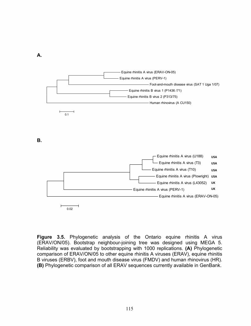
identified. Phylogenetic analysis demonstrated that ERAV/ON/05 was closely
related to the ERAV/PERV isolate, which was recovered in 1962 in the United
Kingdom. An experimental model was developed to study the clinical infection in
naïve healthy ponies (ERAV/ON/05 n=4 and placebo n=4). ERAV/ON/05
induced clinical respiratory disease compared to placebo. The clinical signs
consisted of pyrexia, nasal discharge, increased and abnormal lung sounds,
increased size of submandibular lymph nodes and persistent mucopus in the
trachea (up to 21 days post-infection). The virus was isolated from the lower and
upper airways up to day 7 post-infection, corresponding with the detection of
neutralizing ERAV antibodies. Assessment of the cytokine profile from
bronchoalveolar lavage (BAL) cells demonstrated that this infection induced
down-regulation of the mRNA expression of IL-4. One year later, four previously
infected ponies with neutralizing antibodies to ERAV were assigned to a re-
infection trial. None of the re-infected ponies developed clinical disease, and only
one animal had a four-fold increase in antibody titres to ERAV. Attempts to
recover the virus from the re-infected ponies using cell culture were negative;
however, a down-regulation of the mRNA expression of IL-4 and IFN-β was
identified in BAL cells.
In conclusion, this study shows that the genome of ERAV has not
significantly changed in the last 50 years and more importantly the virus induces
clinical respiratory disease similar to other common equine respiratory viruses.
iv
ACKNOWLEDGEMENTS
First of all, I would like to thank Dr. Laurent Viel for his unconditional
support, friendship and excellent advice during my academic years at the Ontario
Veterinary College. Dr. Viel has not only been an advisor, but also a mentor,
role model and a great friend. He has taught me great skills not only for my
career but also for life. Without him this invaluable experience would not have
been possible.
Dr. Eva Nagy has devoted her expertise to my formation as a virologist
and gave me the strength to overcome difficult moments. Her advice, guidance
and patience have given me the tools to successfully complete this work.
Dr. Joanne Hewson as a co-advisor during my MSc program and as part
of my PhD advisory committee has enormously influenced the outcome of my
academic life. I would like to thank her for her great advice and friendship.
Dr. Pat Shewen has contributed immensely to my work. Her constructive
criticism and high standards have taught me to be both rigorous and precise in
my writing, and she has shown me the power of improvement through critical
review. Muchas gracias!
I would like to acknowledge Louise, whom has always been there to
support and give me her point of view, making my work even more interesting.
She has spent valuable time proofreading my documents and giving me great
advice throughout my academic years at Guelph. Louise has put up with my
v
long days at school and my short evenings and weekends at home. Thank you
so much!
My family in Colombia has always been an important part and a
fundamental drive for my life’s accomplishments. Huge thanks to all of them for
their support, encouragement, and love; I dedicate this thesis to them. My family
in Canada, the Waterfalls, have given me the support and friendship that have
made my life here very enjoyable.
I am very grateful to Drs. Paul Doig and Rob Tremblay for their continuous
support and interest.
Finally, I am also grateful to Boehringer Ingelheim, Vetmedica, Ontario,
Canada for providing financial support during all these years.
vi
DECLARATION OF WORK PERFORMED
I, Andrés Diaz-Méndez, declare that all of the work completed during my PhD
program and stated in this thesis was performed by me, with the exception of the
following:
• Virus neutralization tests were performed by the Animal Health Laboratory
at the University of Guelph.
• Sequencing reactions were prepared and performed by the Laboratory
Services at the University of Guelph.
• Complete blood cell counts were performed by the Animal Health
Laboratory at the University of Guelph.
• Animal care including feeding and grooming was performed by personnel
at the Arkell Equine Facility and the University of Guelph Animal Isolation
Unit.
!!!!
vii
TABLE OF CONTENTS ACKNOWLEDGEMENTS iv DECLARATION OF WORK PERFORMED vi TABLE OF CONTENTS vii LIST OF ABBREVIATIONS x LIST OF TABLES xi LIST OF FIGURES xii LIST OF APPENDICES xiii Chapter 1. Literature Review ............................................................................. 1 Introduction ............................................................................................................ 1 Equine rhinitis viruses (ERV) first identification ..................................................... 3 ERBV serotypes .................................................................................................... 6 Equine rhinitis viruses seroprevalence .................................................................. 7 The virus .............................................................................................................. 11 Characteristics of picornaviruses ......................................................................... 12 Genome structure ................................................................................................ 13 Genome replication ............................................................................................. 15 Cell attachment .................................................................................................... 15 Internal ribosome entry sites (IRES) .................................................................... 18 Replication complexes and ARFs in viral replication ........................................... 19 3’ Untranslated region (3’UTR) in viral replication ............................................... 20 Respiratory viral infections and clinical association ............................................. 21 Equine rhinitis viruses and clinical infection ........................................................ 22 Equine rhinitis viruses shedding .......................................................................... 23 Long-term infection consequences ...................................................................... 24 Diagnosis of equine rhinitis virus infection ........................................................... 25 Virus isolation ...................................................................................................... 25 Serology .............................................................................................................. 27
3’UTR 3’ Untranslated region 5’UTR 5’ Untranslated region AA Amino acid AE1 Equine influenza virus 1 (H7N7) AE2 Equine influenza virus 2 (H3N8) BAL Bronchoalveolar lavage bp Base pair CBC Complete blood count cDNA Complementary DNA Cdyn Dynamic compliance CPE Cytopathic effect CXCL2 Chemokine-(C-X-C motif)-ligand 2 EHV1/4 Equine herpes virus 1 and 4 EM Electron microscopy ERAV Equine rhinitis A virus ERAV/ON/05 ERAV Canadian isolate ERBV Equine Rhinitis B virus ERBV1 Equine Rhinitis B virus 1 ERBV2 Equine Rhinitis B virus 2 ERBV3 Equine Rhinitis B virus 3 ERV Equine rhinitis viruses FBS Foetal bovine serum FMDV Foot and mouth disease virus HI Hemagglutinin inhibition test HPC Histamine provocation challenge HR Human rhinovirus IAD Inflammatory airway disease IFN-β Interferon beta IFN-γ Interferon gamma IL Interleukin IMF Immunofluorescence IRES Internal ribosome entry sites LPM Liters per minute mRNA Messenger RNA OD Outer diameter PFT Pulmonary function test PFU Plaque forming unit RACE Rapid amplification of cDNA ends RK-13 Rabbit kidney 13 cells RT-PCR Reverse transcriptase polymerase chain reaction SRH Single radial haemolysis test VN Virus neutralization test ΔPpl Difference in transpulmonary pressure
xi
LIST OF TABLES TABLE PAGE 2.1 Scoring system used to assess clinical signs following
experimental infection with equine rhinitis A virus (ERAV). 87 2.2 The number of positive infected animals for equine rhinitis A
virus (ERAV) as determined by virus isolation in clinical samples. 88
2.3 Respiratory rates of animals in control, infected, and re-
infected groups. 89 2.4 Antibody titers to equine rhinitis A virus (ERAV) and equine
rhinitis B virus (ERBV) in control, infected, and re-infected ponies. 90
3.1 Primers used to amplify regions of the Ontario equine rhinitis
A virus (ERAV/ON/05). 116 4.1 Fold increase (positive value) or decrease (negative value) of
mRNA expression for IL-4, IL-8, IFN-γ, IFN-β and CXCL2 genes in bronchoalveolar lavage (BAL) cells from ponies infected with equine rhinitis A virus (ERAV/ON/05). 137
xii
LIST OF FIGURES FIGURE PAGE 1.1 Arrangement of the structural proteins of a picornavirus. 14 1.2 Genome arrangement of the equine rhinitis A virus. 15 2.1 Endoscopic images of the mid trachea and carina of an
ERAV/ON/05 infected pony. 83 2.2 Mean and pooled standard error of body temperatures of
animals in the control, infected, and re-infected groups. 84 2.3 Mean and pooled standard error of the total clinical scores of
animals in the control, infected, and re-infected groups. 85 2.4 Equine rhinitis A virus antibody titers of control, infected, and re-
infected animals post infection with ERAV/ON/05. 86 3.1 One-step growth curve of the Ontario equine rhinitis A virus isolate
(ERAV/ON/05). 110 3.2 Plaque morphology of ERAV/ON/05 in rabbit kidney-13
cells (RK-13). 111 3.3 ClustalW alignment of the Ontario equine rhinitis A virus
(ERAV/ON/05) and Plummer’s original equine rhinitis A virus isolate from 1962 (PERV-1). 112
3.4 Equine rhinitis A virus (ERAV) sequences available in
GenBank are presented as a percentage (SimPlot Version 3.5.1). 113
3.5 Phylogenetic analysis of the Ontario equine rhinitis A virus isolate
(ERAV/ON/05). 115 4.1 Mean CT % change for mRNA expression of cytokines in
bronchoalveolar lavage cells collected from control, infected and re-infected ponies. 136
xiii
LIST OF APPENDICES
APPENDIX PAGE
1 Sampling checklist used during the infection study. 157
2 Daily clinical examination form used during the animal infection and re-infection studies. 158
3 Composition of the Virus Transport Medium (VTM). 159 4 Ontario equine rhinitis A virus (ERAV/ON/05) nucleotide
sequence. 160 5 Ontario equine rhinitis A virus (ERAV/ON/05) polyprotein
amino acid sequence. 164
1
Chapter 1
Literature Review
Introduction
Equine respiratory viral infections are commonly identified and reported
worldwide (Plummer 1962; Ditchfield et al., 1965; Guo et al., 1995; Li et al., 1996;
Carman et al., 1997; Klaey et al., 1998; Newton et al., 1999; Guthrie et al., 1999;
Daly et al., 2006; Newton et al., 2006; Dynon et al., 2007; Patterson-Kane et al.,
2008; Diaz-Mendez et al., 2010). In most cases, these infections diminish the
athletic condition of the horse and delay training and performance at an optimal
level for prolonged periods (Mumford et al., 1980).
The equine population in Ontario has been estimated at over 380,000
horses and it is suggested that the equine industry contributes over $ 675.5
million to the province’s economy (Right, 2008). Unfortunately, respiratory
disease remains one of the most costly problems in the equine industry.
Worldwide, serological data identify equine influenza virus 2 (AE2) and equine
herpes virus 1 and 4 (EHV1/4) among the most recognized equine respiratory
viruses during respiratory outbreaks. Equine rhinitis A virus (ERAV) and equine
rhinitis B virus (ERBV) have been considered of less importance and were
believed to cause only mild upper respiratory disease in the horse. However,
more recent worldwide surveillance and seroprevalence studies have
demonstrated that these viruses are highly prevalent in horses and have been
associated with respiratory disease (Willoughby et al., 1989; Li et al., 1996; Klaey
et al., 1998; Diaz-Mendez et al., 2010; Pagamjav et al., 2011).
2
Nevertheless, equine rhinitis viruses (ERV) have not been well
characterized and their role as an active component in clinical respiratory disease
is ill defined. Therefore, early studies continue to be used as references to
define ERV infection. ERAV was first identified in 1962 in the United Kingdom
(Plummer and Kerry, 1962) and was subsequently recognized globally (Ditchfield
et al., 1965; Burrows 1968; Burrows 1969; Hofer et al., 1973; Powell et al., 1974;
Sherman et al., 1977; Studdert and Gleeson, 1977; Hofer et al., 1978; Holmes et
al., 1978; Mumford and Thomson, 1978; Powell et al., 1978; Studdert and
Kobuvirus, Teschovirus, Sapelovirus, Senecavirus, Tremovirus, and
Avihepatovirus. Among these genera, some of the most important viruses
infecting not only humans, but also animals are found. Interestingly, in 2011
HRV were moved into the genus Enterovirus along with polioviruses, and the
genus Rhinovirus has been removed from the family (Knowles et al., 2011). In
fact, this new classification would have made Plummer’s “equine respiratory
enterovirus” classification simpler, at least initially.
13
In 1996, Li and co-workers and Wutz and co-workers demonstrated that
ERAV was closely related to foot and mouth disease virus (FMDV) based on
nucleotide homology, phylogenetic allocation and physicochemical
characteristics, and suggested relocation of this virus to the genus Aphthovirus
(Li et al., 1996; Wutz et al., 1996). While ERAV has been allocated to the genus
Aphthovirus, ERBVs consisting of three serotypes (ERBV1, ERBV2 and ERBV3)
have recently been included in the genus Erbovirus within the family
Picornaviridae (Knowles et al., 2011).
Genome structure
Viruses in this family are non-enveloped, with a capsid comprised of four
structural viral proteins (VP1, VP2, VP3, and VP4) forming a protomer (Hartley et
al., 2001). It has been suggested that each new virion contains at least 60 of
these protomers, giving it an icosahedral appearance (Figure 1.1). The viral
genome is made of a positive single strand of RNA, with a size that varies from
6500 to 9500 nucleotides (nt). Specifically, the ERAV genome ranges from 7400
to 7900 nt based on seven complete genome sequences available on GenBank.
The genetic material is comprised of a single polyprotein gene that encodes
structural and non-structural proteins (Hartley et al., 2001). The polyprotein gene
is divided into three regions, P1, P2, and P3 (Figure 1.2) in all members of the
family Picornaviridae. All structural proteins are coded within the P1 region (L,
VP4, VP2, VP3, and VP1), and the non-structural protein coding is distributed
along the P2 (2A, 2B, and 2C) and the P3 regions (3A, VPg, 3C, and 3D). A
14
study performed by Wutz showed that the structural proteins of the ERAV are
similar in sequence and length to those of FMDV (Wutz et al., 1996). However,
receptor binding affinity and disease characteristics are quite different.
Figure 1.1. Arrangement of the structural proteins of a picornavirus. Note the tridimensional arrangement of the VP1, VP2, VP3, and VP4 proteins. The VP4 protein is not directly exposed on the viral surface.
The 5’ untranslated region (5’UTR) of the genome represents a large
portion of the total genome ranging from 700 nt to 1200 nt, which in some cases
comprises up to 10 percent of the total genome length. This 5’UTR contains
secondary structures known as the internal ribosome entry sites (IRES), which
are directly involved in genome replication (Hinton and Crabb, 2000; Sanz et al.,
2010) and have been identified in ERVs as one of the most variable regions
throughout the entire genome (Li et al., 1996). In contrast, the 3’ untranslated
region (3’UTR) is constituted by a short nucleotide sequence that ranges from 50
VP2
VP3 VP1
VP2 VP1 VP3
VP4
15
nts to 100 nts, followed by a long poly (A) tract. An interesting feature of the
members of this family is the cap-independent translation, which is alternately
initiated by the IRES and stabilised by the poly (A) tract (Hinton and Crabb, 2000;
Pisarev et al., 2005).
Figure 1.2. Genome arrangement of the equine rhinitis A virus. Included in the diagram are the 5’ and 3’ untranslated regions (5’UTR and 3’UTR) as well as the coding sequences for structural and non-structural proteins.
Genome replication
Cell attachment
Infection of the respiratory epithelium with ERVs initiates a series of
conformational and immunological changes. Cell infection commences by viral
attachment to the specific receptor. Despite both physical and genetic similarities
across picornaviruses, a wide range of receptors used by picornaviruses have
been identified (Shafren et al., 1999; Stevenson et al., 2004; Xiao et al., 2005;
Fry et al., 2010). The ICAM-1 and sialic acid receptors localized in the
respiratory epithelium have been consistently identified as a virion target for
FMDV. Initial speculations indicated that ERVs may use both of these receptors
16
as well, but a study performed by Stevenson and coworkers confirmed that
ERAV uses the α-2,3-linked sialic acid residues as a main cell binding receptor
(Stevenson et al., 2004). In 2010, Fry and collaborators confirmed the
importance of sialic acid for ERAV attachment and cell entry by binding inhibition
assays and crystallography analysis (Fry et al., 2010). Additionally, X-ray
crystallography studies in other viruses such as poliovirus and FMDV have
shown depressions (“canyons”) in the viral surface, which have been implicated
in specific receptor binding. Size and shape differences in these canyons among
viruses in the family Picornaviridae may indicate specific attachment
characteristics associated with a wide receptor range even within the same
genus. ERVs characteristics need to be studied in more depth to confirm this
hypothesis and eliminate other possible receptors for viral attachment.
Cell attachment represents a fundamental priming step during viral
infection. Earlier studies on polioviruses (family Picornaviridae) have shown that
viral-cell attachment induces channel formation, permitting the viral RNA to enter
the host cell. However, the mode of viral entry is not well understood in
aphthoviruses (FMDV and ERAV). Specifically, two different studies looking at
ERAV cell entry and dissociation concluded that, like FMDV, these viruses
require a low pH environment using an empty capsid particle, which may protect
the viral RNA as it is incorporated into the cytoplasm of the infected cell (Tuthill et
al., 2009; Groppelli et al., 2010). However, they do not discard the possibility of
pore formation as observed during viral genome transfer in other picornaviruses.
In 2010, Groppelli and collaborators speculated that empty capsid particles
17
lacking RNA may serve as a connector between ERAV virions and endosomal
membranes to assure viable RNA transfer.
Once the RNA has been transported into the cytoplasm, the polyprotein is
cleaved into protein precursors that initiate the translation of the RNA followed by
replication (in replication complexes) and formation of new virions that are
released to infect other cells. To date, the replication process is poorly
understood and it has been controversially suggested that replication vesicles
are newly formed, or cellular transport chambers are transformed and hijacked by
the virus to replicate and complete its life cycle (Belov et al., 2012).
Unfortunately, none of these characteristics have been fully investigated for
ERVs.
Picornaviruses are unique in their translation and replication processes.
The polyprotein is cleaved early during infection by proteinases that vary from
one virus to another, such as L proteinases in aphthoviruses, or 3C and 3D in the
majority of picornaviruses (Hinton et al., 2002). Of great importance is the 3D or
RNA polymerase, which is not only known for its elongation activity, but also for
its importance generating the virion genome linked protein (VPg) or primer
protein that initiates viral translation. The VPg has been identified in virions, and
it has been demonstrated that it is removed upon cell entry by cellular enzymes
(Strauss et al., 2007). Currently, knowledge on the specific functions for each of
the non-structural proteins is limited, and it has been suggested that some of
them are essential for efficient viral replication (Hartley et al., 2001). In general,
these non-structural proteins have been found to be principally involved in
equine foetal kidney (EFK) cells, and HeLa cells (Plummer 1962; Ditchfield et al.,
1965; Powell et al., 1974; Studdert and Gleeson, 1977; Holmes et al., 1978;
McCollum and Timoney, 1992; Carman et al., 1997; Kriegshauser et al., 2005;
Diaz-Mendez et al., 2010). Today, most laboratories use RK-13 cell lines
routinely.
AE2 and EHV1/4 also have a poor isolation rate from individuals during
infectious episodes. It is well known that successful isolation is generally
optimized when samples are collected within 24-36 hours post infection. This
indicates that timing of sample collection represents an important factor for viral
recovery. Additionally, equine rhinitis viruses are not commonly isolated in cell
culture and it has been suggested that non-cytopathic ERAV strains may be
involved in clinical respiratory disease. In 1997, Li and co-workers demonstrated
that conventional virus isolation was not sensitive enough to detect these viruses
in samples collected from clinical respiratory cases (Li et al., 1997). Therefore,
immunofluorescence and RT-PCR were required to detect viral antigen and
nucleic acid in those samples. Thus, ERVs recovery rates from respiratory
outbreaks are still low, and only a few ERAV isolates have been completely
analysed by genome sequencing and nucleotide comparison (Li et al., 1996).
Even though ERBVs have been more prolifically recovered from infected horses,
27
serological evidence indicates that ERAV is more prevalent worldwide
(Willoughby et al., 1992; Carman et al., 1997; Kriegshauser et al., 2005; Gradzki
et al., 2009; Diaz-Mendez et al., 2010; Pagamjav et al., 2011).
Traditionally, cell monolayers are inoculated with a clinical sample,
incubated (37°C in the presence of CO2) and observed for 5 to 7 days to detect
a cytopathic effect (CPE). In general, CPE in cell culture is very characteristic of
ERVs and is identified by rounding of the cells, which become fragile and detach
from cell monolayers (Ditchfield et al., 1965). These cell layers are completely
destroyed by 96 hours post-infection. In some cases, up to three passages are
required to confirm positive samples and on occasion, as reported by Li in 1997,
false negative results may be confirmed positive by other methods. Even though
virus isolation is considered highly sensitive, drawbacks such as turnaround time
and possible circulation of non-cytopathic strains limit its reliability.
Serology
Serodiagnosis has been widely used either as a confirmatory tool, or
primary diagnostic method not only in equines but also in other species.
Conventionally, the VN test has been employed to determine ERV antibody
levels. Alternative methods such as complement-mediated haemolysis in
agarose gel, complement fixation, and single radial immunodiffusion have shown
poor repeatability, and low sensitivity and specificity (Knorn et al., 1980). Thus,
VN testing is considered a gold standard when determining ERVs antibody titers.
A four-fold increase in antibody titers to any viral antigen in paired samples
28
(acute and convalescent) is considered significant in the majority of the cases;
however, these changes depend on time of collection. As a general rule, serum
samples should be collected at least 10 to 14 days apart and analysed as a pair,
resulting in prolonged turnaround time, which in most cases comes too late to be
useful diagnostically. However, this information is essential when establishing
preventative disease control plans and biosecurity measures.
Molecular diagnosis
In the last decade, PCR and RT-PCR with their advantages and
disadvantages have become of great importance when assessing infectious
diseases. However, more conventional, well-established and highly specific
techniques, such as immunofluorescence, immunohistochemistry and electron
microscopy are still being used (Li et al., 1997). Real-time PCR (qPCR) and
conventional reverse transcriptase PCR (RT-PCR) methods for the detection of
ERVs in clinical samples have been developed and optimized, but at the moment
these techniques are offered only as research diagnostic tools in a small number
of laboratories (Dynon et al., 2001; Black et al., 2007; Quinlivan et al., 2010).
A study by Black and coworkers (Black et al., 2007) found that a nested
RT-PCR for ERBV was able to detect six positive samples when conventional
virus isolation failed to detect the presence of the virus in seventeen horses with
clinical respiratory disease. This, in addition to similar findings by Quinlivan in
2010 suggests that RT-PCR and qPCR may be employed as rapid diagnostic
29
methods in acute respiratory outbreaks; however the clinical significance of
positive results is still highly debatable.
ERAV in other species
During the initial ERAV study at the Wellcome Research Laboratories,
three stable workers developed a high virus neutralizing antibody titer (1:512 –
1:024) against the same virus recovered from the horses (Plummer, 1962). None
of the other workers that were in contact with the horses nor the 100 volunteers
at these facilities had detectable antibodies. Direct contact with infected horses
could represent a potential source for human infection; however, no other reports
of equine rhinoviruses infecting humans have been published up to 2005.
Preliminary studies by Plummer demonstrated that, following gastric exposure
using a stomach tube, ERAV could infect cynomolgus monkeys without inducing
clinical signs and that infection could be confirmed only by serology; however, the
same virus induced clinical disease in horses, supporting suggestions that
interspecies infection may not be likely.
In a more recent attempt to elucidate the role of these viruses, an Austrian
research group conducted a survey of 137 serum samples from veterinary
practitioners and found a very weak response (neutralizing antibodies) against
ERAV (2.7%) and ERBV1 (3.6%) (Kriegshauser et al., 2005). Thus, even in a
high-risk population such as veterinarians, ERVs may present only a very low risk
of infection.
30
Cross infection and interspecies infections have been rarely reported, but
ERAV has been identified as the cause of abortions in zebras and dromedaries
(Kimber et al., 2002; Wernery et al., 2008). The most recent event was recorded
in Dubai, United Arab Emirates, where eight pregnant dromedaries aborted and
ERAV was consistently recovered from the placenta and various foetal organs.
Experimental infection of two seven-month pregnant dromedaries confirmed a
tropism of ERAV for the reproductive tract in this species (Wernery et al., 2008).
No clinical respiratory or reproductive signs were observed in the dams prior to
abortion. This supports the notion that ERAV may be involved in equine cases
where abortions could not be associated with a specific causal agent.
Respiratory immunity
The protective immune response to ERVs has not been specifically
studied in the horse. There is evidence showing that humoral and cellular
responses are essential to overcome respiratory viral infections. The equine
respiratory tract is continuously exposed to environmental factors and common
pathogens (viruses, bacteria, parasites, and fungus). The airways comprise the
major entry port for infectious agents that must be controlled and neutralized in a
timely fashion in order to prevent disease. During infections, mechanical barriers
such as head position, mucus and mucociliary movement act as the first
response by filtering, trapping and removing undesirable organic particles and
pathogens. These mechanical barriers, as the first line of protection in
combination with a cellular and humoral immune response, play an integral and
31
essential role in the respiratory tract by providing a protective response when
challenged with a specific pathogen or antigen (e.g. viral infections or
vaccination). During infections, cells such as neutrophils and lymphocytes
become activated and respond by killing infected cells. Macrophages are
attracted to infection sites and result in phagocytosis and cell activation [e.g.
natural killer cells (NK)]. In conjunction, these cells secrete chemokines that
prime (newly exposed) or recruit (previously exposed) T and B-lymphocytes,
which eventually control and eliminate the specific pathogen.
Innate immunity (non specific)
Physical Barriers
As mentioned above, the respiratory tract is exposed to numerous
pathogens and particles that can be screened at the site of entry. The
mucociliary system in the respiratory tract plays a decisive role on mucus
clearance and systemic protection (Willoughby et al., 1992). The cilia lining the
upper airways and trachea promote upwards movement of secreted mucus.
During grazing, equines maintain their heads in a lower position contributing to
easy clearing of the mucus through gravity as well. Experimental AE2 infection
showed that damaged cilia reduce trachea clearance efficiency, increasing the
probability of upper airway infections extending into the lung (Willoughby et al.,
1992). Interestingly, Willoughby and collaborators could not demonstrate
reduced mucus clearance following experimental ERBV infection, perhaps due to
32
the presence of circulating specific ERBV antibodies, improper viral delivery or
inadequate infection dose.
Cytokines in the initial immune response
Interleukins not only play an important role in the acute innate pathogen
control phase, they also immunomodulate a specific response by recruiting and
stimulating inflammatory cells. Interferon-α (IFN-α), interferon-β (IFN-β), and
interferon-γ (IFN-γ) have been identified in the equine respiratory tract during viral
infections (Paillot et al., 2006). Specifically, IFN-α is secreted by dendritic cells
and macrophages during early viral infections and by T and B-lymphocytes in
later stages. Also, IFN-α has been recognized as a mediator in anti-inflammatory
responses, as well as a promoter of an antiviral state in non-infected cells (Moore
et al., 1996). Furthermore, IFN-α and IFN-β have been associated with up-
regulation of the class I major histocompatibility complex (MHC), increasing
recognition and destruction of infected cells by CD8+ cytotoxic T cells (Joubert et
al., 2008). In contrast, HRV infections have been shown to up-regulate gene
expression of some cytokines and more importantly to cause a down-regulation
in the expression of IFN-β, which may underlie poor immune response against
viral infections (Wark et al., 2005) and perhaps be associated with asthma
exacerbations (Papadopoulos et al., 2004; Bizzintino et al., 2011; Proud, 2011).
IFN-γ has been considered a key component of the acquired immune
response by modulating an increase in antigen presentation to CD4+ helper T
cells by MHC class II (Soboll et al., 2003; Paillot et al., 2006). Also, IFN-γ
33
secreted by NK cells has been found to promote T-lymphocyte development and
support cell differentiation (Cordeau et al., 2004). Another important modulator is
tumour necrosis factor-α (TNF-α), which is actively produced by macrophages,
T-lymphocytes and NK cells. TNF-α has been directly associated with destruction
of infected cells. Experimental challenge with fungi, hay dust and
lipopolysaccharide of RAO horses significantly modifies the expression of TNF-α,
Interleukin 1β (IL-1β), Interleukin-6 (IL-6), Interleukin-8 (IL-8), and Interleukin-10
(IL-10) (Ainsworth et al., 2003a; Ainsworth et al., 2003b). Interestingly, TNF-α,
Interleukin 1β (IL-1β), and IL-6 were found to be up regulated as well when
vaccinated and unvaccinated horses were exposed to AE2 (Quinlivan et al.,
2007), confirming that equine respiratory viruses directly affect the airway
equilibrium, as seen during human influenza and human rhinovirus infections.
Innate immunity (cellular response)
Alveolar macrophages, natural killer cells, and neutrophils cooperate in a
process to maintain and control pathogen spreading. During early infection,
neutrophils migrate to infection sites followed by macrophages. The initial
mobilization of responding cells and identification of infected cells triggers the
activation and release of pro-inflammatory cytokines that eventually activate NK
cells and T and B-lymphocytes. Natural killer cells have been associated with
active toxicity and lysis of infected or abnormal cells, and are also a major
component in the development of the adaptive immune response (Paillot et al.,
2006). Alveolar macrophages account for the majority of the cell population in
34
the equine lung and are associated with activation of the acquired immune
response. It is known that macrophage stimulation (e.g. viral infections) triggers
a dynamic reaction expressed by secretion of cytokines such as IL-1α, IL-1β,
TNF-α, IL-8, and IL-34. Similarly, neutrophils are in concordance with
macrophages and are a key component of the innate and acquired immune
response. Once neutrophils are activated, they migrate into the airway lumen, to
phagocytize infected cells. Due to its physiological characteristics, the lung
presents a unique organ for trans-endothelial neutrophil migration; in comparison
to neutrophil migration due to inflammatory processes in other organs (liver,
kidney, etc.), it seems that migration of these cells into the lung may be less
reliant on conventional L-selectin and β2-integrin cellular adhesion (Lee et al.,
2000). Additionally, neutrophils have been shown to induce B-cell activation by
production of IL-4, which is directly associated with antibody production (Lavoie
et al., 2001; Joubert et al., 2008).
Adaptive Immunity
Following the initial innate immune response, a more specific response
must be developed. This specific or adaptive immune response is mediated by
T-lymphocytes (CD4+, CD8+) and B-lymphocytes (Crouch et al., 2005; Paillot et
al., 2006). Expansion of antigen specific B-lymphocytes is accomplished during
the initial infection and antibody production is followed by transformation of
antigen-specific B cells into plasma cells.
35
Antibody production in the respiratory tract is governed by
immunoglobulins A, E, M and G (IgA, IgE, IgM and IgG) (Soboll et al., 2003;
Waller et al., 2007). Immunoglobulin A has been found as the most common
immunoglobulin in nasal secretions. Its primary role is in blocking attachment of
virus to host cells and in removal of free virus by transport of soluble antibody-
virus complexes into the mucus. Post infection, IgG is rapidly secreted into the
airway lumen to control viral colonization by blocking attachment or through the
activation of complement (Ainsworth et al., 2002; Soboll et al., 2003). In addition,
macrophages and neutrophils recognize IgG antibodies that have bound to
infected cells. They attach to the antigen-antibody complex on the cell surface
and destroy the cell. This type of recognition and destruction is known as
antibody-dependant cytotoxicity (ADCC). Antibodies bound to infected cells may
also mediate cell lysis through the activation of complement on the cell surface.
Interestingly, due to the high affinity of IgE for mast cells, the production of IgE
has been linked with increased mast cell degranulation. This has been
associated with asthma exacerbation in humans and it has been speculated that
a similar mechanism post viral infection may be involved in RAO/Heaves in the
adult horse (Joubert et al., 2008).
Infected or specialized presenting cells [e.g. dendritic cells (DC)] display
antigens with class I MHC, or both class I and class II MHC on their surface,
respectively. This is a determining factor for recognition by T cells (Siedek et al.,
1999). Class I MHC presentation is recognized by CD8+ (cytotoxic T-
lymphocytes), whereas antigens presented by the class II MHC are recognized
36
by CD4+ (helper) T-lymphocytes. During respiratory infection, macrophages and
infected epithelial cells activate DCs by releasing cellular cytokines. These
specialized presenting cells travel to draining lymph nodes and activate T-
lymphocytes by antigen presentation (Steinbach et al., 1998). Subsequently, T-
lymphocytes replicate and are mobilized to specific infection areas.
Specific lymphoid aggregations have been identified along mucosal
surfaces in different species, and the equine respiratory tract is no exception.
Collectively, these tissues are known as mucosal-associated lymphoid tissues
(MALT) (Mair et al., 1988). Strategic localization in the respiratory tract offers a
prime availability of T and B-lymphocytes (Lunn et al., 2001). The nasal cavity,
and in particular the laryngo-pharyngeal area of young horses, is functionally
layered with follicular lymphoid tissue. This follicular tissue is present and
remarkably large during the first 2 years of a horse’s life. This correlates with the
critical period of antigen exposure in young horses. Due to its localization, the
MALT may represent an important induction site for efficient immune response
during natural infection or vaccine stimulation (Chambers et al., 2001; Takada et
al., 2003; Crouch et al., 2005; Waller et al., 2007).
Conclusions
ERVs have been identified in the equine population for almost five
decades; however, they have been considered of minimum clinical importance
when compared to viruses such as AE2 and EHV1/4. Individual seroprevalence
and surveillance studies have provided important data that have been overlooked
37
when analysed alone. The combination of previous studies and current
knowledge on ERVs and other similar viruses such as HRV and FMDV, indicate
that ERAV may be implicated in conditions beyond simple “upper respiratory
infections”.
A balance between a measured immune response and controlled
inflammation is key to maintaining the integrity and functionality of the respiratory
system. However, rapid immune responses in the lung are required for effective
pathogen control and elimination, which in the long term may be associated with
excessive airway inflammation.
It is evident that respiratory infections account for a great number of the
clinical illnesses affecting the athletic horse and the majority of them are
attributable to viral infections. A number of AE2 and EHV1/4 vaccines are
available; however, there are currently no ERVs vaccines. Better understanding
of the molecular characteristics and the pathophysiology of ERVs could lead to
the development of effective vaccines and possibly disease control in the near
future.
38
Objectives
To characterize the Ontario ERAV isolate (ERAV/ON/05), by sequencing the
entire viral genome and to compare its characteristics to worldwide isolates.
To develop an infection model and to study the clinical characteristics of ERAV
infection.
To study the BAL cytokine profile in ERAV experimentally infected ponies in
order to better understand the immune response to ERAV infection.
39
References
Ainsworth DM, Appleton JA, Antczak DF, Santiago MA, Aviza G. 2002. IgG antibody responses to an inhaled antigen in horses with "heaves" (recurrent airway obstruction). Vet Immunol Immunopathol 84(3-4):169-80.
Ainsworth DM, Appleton JA, Eicker SW, Luce R, Flaminio JM, Antczak DF. 2003. The effect of strenuous exercise on mRNA concentrations of interleukin-12, interferon-gamma and interleukin-4 in equine pulmonary and peripheral blood mononuclear cells. Vet Immunol Immunopathol 91(1):61-71.
Ainsworth DM, Grunig G, Matychak MB, Young J, Wagner B, Erb HN, Antczak DF. 2003. Recurrent airway obstruction (RAO) in horses is characterized by IFN-gamma and IL-8 production in bronchoalveolar lavage cells. Vet Immunol Immunopathol 96(1-2):83-91.
Banks EM, Kyriakidou M, Little S, Hamblin AS. 1999. Epithelial lymphocyte and macrophage distribution in the adult and fetal equine lung. J Comp Pathol 120(1):1-13.
Belov GA, Nair V, Hansen BT, Hoyt FH, Fischer ER, Ehrenfeld E. 2012. Complex dynamic development of poliovirus membranous replication complexes. J Virol 86(1):302-12.
Belov GA, Altan-Bonnet N, Kovtunovych G, Jackson CL, Lippincott-Schwartz J, Ehrenfeld E. 2007. Hijacking components of the cellular secretory pathway for replication of poliovirus RNA. J Virol 81(2):558-67.
Bizzintino J, Lee WM, Laing IA, Vang F, Pappas T, Zhang G, Martin AC, Khoo SK, Cox DW, Geelhoed GC, McMinn PC, Goldblatt J, Gern JE, and Le Souëf PN. 2011. Association between human rhinovirus C and severity of acute asthma in children. Eur Respir J 37(5):1037-42.
Black WD, Hartley CA, Ficorilli NP, Studdert MJ. 2005. Sequence variation divides equine rhinitis B virus into three distinct phylogenetic groups that correlate with serotype and acid stability. J Gen Virol 86(Pt 8):2323-32.
Black WD, Hartley CA, Ficorilli NP, Studdert MJ. 2007. Reverse transcriptase-polymerase chain reaction for the detection of equine rhinitis B viruses and cell culture isolation of the virus. Arch Virol 152(1):137-49.
Black WD, Wilcox RS, Stevenson RA, Hartley CA, Ficorilli NP, Gilkerson JR, Studdert MJ. 2007. Prevalence of serum neutralising antibody to equine rhinitis A virus (ERAV), equine rhinitis B virus 1 (ERBV1) and ERBV2. Vet Microbiol 119(1):65-71.
Böhm HO. 1964. Über die isolierung und charakterisierung eines picornavirus vom pferd. Zentbl Vet Med 11(3):240-250.
40
Brown DM, Kauder SE, Cornell CT, Jang GM, Racaniello VR, Semler BL. 2004. Cell-dependent role for the poliovirus 3' noncoding region in positive-strand RNA synthesis. J Virol 78(3):1344-51.
Brown DM, Cornell CT, Tran GP, Nguyen JH, Semler BL. 2005. An authentic 3' noncoding region is necessary for efficient poliovirus replication. J Virol 79(18):11962-73.
Burrows R. 1968. Laboratory diagnosis of some virus infections of the upper respiratory tract of the horse. Equine Vet J 1:32.
Burrows R. 1969. Equine rhinoviruses. IV. equine rhinoviruses; 1969; New York: Karger, Basel/Munchen. 154 p.
Burrows R. 1979. Equine rhinovirus and adenovirus infections; Proceedings of the 24th annual convention of the American Association of Equine Practitioners, 1978. 299 p.
Burrows R, Goodridge D, Denyer M, Hutchings G, Frank CJ. 1982. Equine influenza infections in Great Britain, 1979. Vet Rec 110(21):494-7.
Carman S, Rosendal S, Huber L, Gyles C, McKee S, Willoughby RA, Dubovi E, Thorsen J, Lein D. 1997. Infectious agents in acute respiratory disease in horses in Ontario. J Vet Diagn Invest 9(1):17-23.
Chambers TM, Holland RE, Tudor LR, Townsend HG, Cook A, Bogdan J, Lunn DP, Hussey S, Whitaker-Dowling P, Youngner JS, et al. 2001. A new modified live equine influenza virus vaccine: Phenotypic stability, restricted spread and efficacy against heterologous virus challenge. Equine Vet J 33(7):630-6.
Cordeau ME, Joubert P, Dewachi O, Hamid Q, Lavoie JP. 2004. IL-4, IL-5 and IFN-gamma mRNA expression in pulmonary lymphocytes in equine heaves. Vet Immunol Immunopathol 97(1-2):87-96.
Covaleda L, Fuller FJ, Payne SL. 2010. EIAV S2 enhances pro-inflammatory cytokine and chemokine response in infected macrophages. Virology 397(1):217-23.
Crouch CF, Daly J, Henley W, Hannant D, Wilkins J, Francis MJ. 2005. The use of a systemic prime/mucosal boost strategy with an equine influenza ISCOM vaccine to induce protective immunity in horses. Vet Immunol Immunopathol 108(3-4):345-55.
Cullinane A, Weld J, Osborne M, Nelly M, Mcbride C, Walsh C. 2001. Field studies on equine influenza vaccination regimes in thoroughbred foals and yearlings. Vet J 161(2):174-85.
41
Daly JM, Yates PJ, Browse G, Swann Z, Newton JR, Jessett D, Davis-Poynter N, Mumford JA. 2003. Comparison of hamster and pony challenge models for evaluation of effect of antigenic drift on cross protection afforded by equine influenza vaccines. Equine Vet J 35(5):458-62.
Daly JM, Yates PJ, Newton JR, Park A, Henley W, Wood JL, Davis-Poynter N, Mumford JA. 2004. Evidence supporting the inclusion of strains from each of the two co-circulating lineages of H3N8 equine influenza virus in vaccines. Vaccine 22(29-30):4101-9.
Daly JM, Whitwell KE, Miller J, Dowd G, Cardwell JM, Smith KC. 2006. Investigation of equine influenza cases exhibiting neurological disease: Coincidence or association? J Comp Pathol 134(2):231-5.
Diaz-Mendez A, Viel L, Hewson J, Doig P, Carman S, Chambers T, Tiwari A, Dewey C. 2010. Surveillance of equine respiratory viruses in Ontario. Can J Vet Res 74(4):271-8.
Ditchfield J and Macpherson LW. 1965. The properties and classification of two new rhinoviruses recovered from horses in Toronto, Canada. Cornell Vet 55:181-9.
Ditchfield J, Macpherson LW, Zbitnew A. 1965. Upper respiratory disease in thoroughbred horses: Studies of its viral etiology in the Toronto area, 1960 to 1963. Can J Comp Med 29:18-22.
Dynon K, Varrasso A, Ficorilli N, Holloway S, Reubel G, Li F, Hartley C, Studdert M, Drummer H. 2001. Identification of equine herpesvirus 3 (equine coital exanthema virus), equine gammaherpesviruses 2 and 5, equine adenoviruses 1 and 2, equine arteritis virus and equine rhinitis A virus by polymerase chain reaction. Aust Vet J 79(10):695-702.
Dynon K, Black WD, Ficorilli N, Hartley CA, Studdert MJ. 2007. Detection of viruses in nasal swab samples from horses with acute, febrile, respiratory disease using virus isolation, polymerase chain reaction and serology. Aust Vet J 85(1-2):46-50.
Egger D, Teterina N, Ehrenfeld E, Bienz K. 2000. Formation of the poliovirus replication complex requires coupled viral translation, vesicle production, and viral RNA synthesis. J Virol 74(14):6570-80.
Flaminio MJ, Rush BR, Davis EG, Hennessy K, Shuman W, Wilkerson MJ. 2000. Characterization of peripheral blood and pulmonary leukocyte function in healthy foals. Vet Immunol Immunopathol 73(3-4):267-85.
Fry EE, Tuthill TJ, Harlos K, Walter TS, Rowlands DJ, Stuart DI. 2010. Crystal structure of equine rhinitis A virus in complex with its sialic acid receptor. J Gen Virol 91(Pt 8):1971-7.
42
Fukunaga Y, Kumanomido T, Imagawa H, Ando Y, Kamada M, Wada R, Akiyama Y. 1981. Isolation of picornavirus from horses associated with getah virus infection. Nihon Juigaku Zasshi 43(4):569-72.
Fukunaga Y, Kumanomido T, Kamada M, Wada R. 1983. Equine picornavirus: Isolation of virus from the oral cavity of healthy horses. Bull Equine Res Inst :103-9.
Galbraith NS. 1965. A survey of enteroviruses and adenoviruses in the faeces of normal children aged 0-4 years. A report of the public health laboratory service and the society of medical officers of health. J Hyg (Lond) 63(4):441-55.
Gradzki Z and Boguta L. 2009. Seroprevalence of equine rhinitis B viruses in Poland. Medycyna Weterynaryjna 65(2):119.
Groppelli E, Tuthill TJ, Rowlands DJ. 2010. Cell entry of the aphthovirus equine rhinitis A virus is dependent on endosome acidification. J Virol 84(12):6235-40.
Guo Y, Wang M, Zheng GS, Li WK, Kawaoka Y, Webster RG. 1995. Seroepidemiological and molecular evidence for the presence of two H3N8 equine influenza viruses in China in 1993-94. J Gen Virol 76 (Pt 8):2009-14.
Guthrie AJ, Stevens KB, Bosman PP. 1999. The circumstances surrounding the outbreak and spread of equine influenza in South Africa. Rev Sci Tech 18(1):179-85.
Hare JE and Viel L. 1998. Pulmonary eosinophilia associated with increased airway responsiveness in young racing horses. J Vet Intern Med 12(3):163-70.
Hartley CA, Ficorilli N, Dynon K, Drummer HE, Huang JA, Studdert MJ. 2001. Equine rhinitis A virus: Structural proteins and immune response. J Gen Virol 82(Pt 7):1725-8.
Hinton TM, Li F, Crabb BS. 2000. Internal ribosomal entry site-mediated translation initiation in equine rhinitis A virus: Similarities to and differences from that of foot-and-mouth disease virus. J Virol 74(24):11708-16.
Hinton TM, Ross-Smith N, Warner S, Belsham GJ, Crabb BS. 2002. Conservation of L and 3C proteinase activities across distantly related aphthoviruses. J Gen Virol 83(12):3111-21.
Hofer B, Steck F, Gerber H, Lohrer J, Nicolet J, Paccaud MF. 1973. An investigation of the etiology of viral respiratory disease in a remount depot. Equine Infectious Diseases III:527-45.
43
Hofer B, Steck F, Gerber H. 1978. Virological investigations in a horse clinic. Equine infectious diseases IV: Proceedings of the 4th international conference on equine infectious diseases; September 1976; Princeton, New Jersey, USA: Veterinary Publications, Inc. 475 p.
Hoffman A, Viel L, McDonell W. 1992. Airway hyperresponsiveness in ponies following a naturally-acquired influenza infection. Am Rev Resp Dis 145:A432.
Holmes DF, Kemen MJ, Coggins L. 1978. Equine rhinovirus infection - serologic evidence of infection in selected United States horse populations. Equine Infectious Diseases IV:315-9.
Jolly PD, Fu ZF, Robinson AJ. 1986. Viruses associated with respiratory disease of horses in New Zealand: An update. N Z Vet J 34(4):46-50.
Joubert P, Cordeau ME, Boyer A, Silversides DW, Lavoie JP. 2008. Cytokine expression by peripheral blood neutrophils from heaves-affected horses before and after allergen challenge. Vet J 178(2):227-32.
Kimber KR, Lubroth J, Dubovi EJ, Berninger ML, Demaar TW. 2002. Serologic survey of selected viral, bacterial, and protozoal agents in captive and free-ranging ungulates from central Kenya. Pilanesberg National Park, South Africa, July 22-27, 2001 Annals of the New York Academy of Sciences. 969:217-23.
Klaey M, Sanchez-Higgins M, Leadon DP, Cullinane A, Straub R, Gerber H. 1998. Field case study of equine rhinovirus 1 infection: Clinical signs and clinicopathology. Equine Vet J 30(3):267-9.
Knorn M, Finci E, Teufel P. 1980. A technique for the detection of antibodies against equine rhinovirus using complement mediated haemolysis in agarose gels. Vet Microbiol 5(2):155-59.
Knowles NJ, Hovi T, Hyypiä T, King AMQ, Lindberg M, Pallansch MA, Palmenberg AC, Simmonds P, Skern T, Stanway G, Yamashita T, Zell R. 2011. Picornaviridae. In: Virus taxonomy: Classification and nomenclature of viruses: Ninth report of the international committee on taxonomy of viruses. King AMQ, Adams MJ, Carstens EB, Lefkowitz EJ (Eds). Elsevier Inc., San Diego: pp 855-880.
Kriegshauser G, Deutz A, Kuechler E, Skern T, Lussy H, Nowotny N. 2005. Prevalence of neutralizing antibodies to equine rhinitis A and B virus in horses and man. Vet Microbiol 106(3-4):293-6.
Lavoie JP, Maghni K, Desnoyers M, Taha R, Martin JG, Hamid QA. 2001. Neutrophilic airway inflammation in horses with heaves is characterized by a Th2-type cytokine profile. Am J Respir Crit Care Med 164(8 Pt 1):1410-3.
44
Lee HY, Kehrli ME, Brogden KA Jr, Gallup JM, and Ackermann MR. Influence of β2-Integrin Adhesion Molecule Expression and Pulmonary Infection with Pasteurella haemolytica on Cytokine Gene Expression in Cattle. Infect Immun 2000 July; 68(7): 4274–4281.
Li F, Browning GF, Studdert MJ, Crabb BS. 1996. Equine rhinovirus 1 is more closely related to foot-and-mouth disease virus than to other picornaviruses. Proc Natl Acad Sci USA 93(3):990-5.
Li F, Drummer HE, Ficorilli N, Studdert MJ, Crabb BS. 1997. Identification of noncytopathic equine rhinovirus 1 as a cause of acute febrile respiratory disease in horses. J Clin Microbiol 35(4):937-43.
Lunn DP. 2001. Pharyngeal lymphoid tissue: Gatekeeper or showstopper? Equine Vet J 33(3):218-20.
Mair TS, Batten EH, Stokes CR, Bourne FJ. 1988. The distribution of mucosal lymphoid nodules in the equine respiratory tract. J Comp Pathol 99(2):159-68.
Matera MG, Calzetta L, Sanduzzi A, Page CP, Cazzola M. 2008. Effects of neuraminidase on equine isolated bronchi. Pulm Pharmacol Ther 21(4):624-9.
McCollum WH and Timoney PJ. 1992. Studies on the seroprevalence and frequency of equine rhinovirus-I and -II infection in normal horse urine; Equine Infectious Diseases VI: Proceedings of the Sixth International Conference, 7-11 July 1991, p 83.
Moore BR, Krakowka S, Cummins JM, Robertson JT. 1996. Changes in airway inflammatory cell populations in standardbred racehorses after interferon-alpha administration. Vet Immunol Immunopathol 49(4):347-58.
Mumford JA and Thomson GR. 1978. Studies on picornaviruses isolated from the respiratory tract of horses. J Equine Med Surg (1):419.
Mumford JA and Rossdale PD. 1980. Virus and its relationship to the "poor performance" syndrome. Equine Vet J 12(1):3-9.
Newton JR, Verheyen K, Wood JLN, Yates PJ, Mumford JA. 1999. Equine influenza in the United Kingdom in 1998. Vet Rec 145(16):449-52.
Newton JR, Townsend HG, Wood JL, Sinclair R, Hannant D, Mumford JA. 2000. Immunity to equine influenza: Relationship of vaccine-induced antibody in young thoroughbred racehorses to protection against field infection with influenza A/equine-2 viruses (H3N8). Equine Vet J 32(1):65-74.
Newton JR, Wood JLN, Chanter N. 2003. A case control study of factors and
45
infections associated with clinically apparent respiratory disease in UK thoroughbred racehorses. Prev Vet Med 60(1):107-32.
Newton JR, Daly JM, Spencer L, Mumford JA. 2006. Description of the outbreak of equine influenza (H3N8) in the United Kingdom in 2003, during which recently vaccinated horses in Newmarket developed respiratory disease. Vet Rec 158(6):185-192.
O.I.E. Equine respiratory diseases 2012 [Internet]; c2012. Available from: http://www.oie.int/animal-health-in-the-world/oie-listed-diseases-2012/.
Pagamjav O, Kobayashi K, Murakami H, Tabata Y, Miura Y, Boldbaatar B, Sentsui H. 2011. Serological survey of equine viral diseases in Mongolia. Microbiol Immunol 55(4):289-92.
Paillot R, Kydd JH, Sindle T, Hannant D, Edlund Toulemonde C, Audonnet JC, Minke JM, Daly JM. 2006. Antibody and IFN-gamma responses induced by a recombinant canarypox vaccine and challenge infection with equine influenza virus. Vet Immunol Immunopathol 112(3-4):225-33.
Palmenberg AC, Spiro D, Kuzmickas R, Wang S, Djikeng A, Rathe JA, Fraser-Liggett CM, Liggett SB. 2009. Sequencing and analyses of all known human rhinovirus genomes reveal structure and evolution. Science 324(5923):55-9.
Papadopoulos NG, Sanderson G, Hunter J, Johnston SL. 1999. Rhinoviruses replicate effectively at lower airway temperatures. J Med Virol 58(1):100-4.
Papadopoulos NG, Papi A, Psarras S, Johnston SL. 2004. Mechanisms of rhinovirus-induced asthma. Paediatr Respir Rev 5(3):255-60.
Patterson-Kane JC, Carrick JB, Axon JE, Wilkie I, Begg AP. 2008. The pathology of bronchointerstitial pneumonia in young foals associated with the first outbreak of equine influenza in Australia. Equine Vet J 40(3):199-203.
Perkins GA, Goodman LB, Tsujimura K, Van de Walle GR, Kim SG, Dubovi EJ, Osterrieder N. 2009. Investigation of the prevalence of neurologic equine herpes virus type 1 (EHV-1) in a 23-year retrospective analysis (1984-2007). Vet Microbiol 139(3-4):375-8.
Pisarev AV, Shirokikh NE, Hellen CU. 2005. Translation initiation by factor-independent binding of eukaryotic ribosomes to internal ribosomal entry sites. C R Biol 328(7):589-605.
46
Plateau E and Levy E. 1990. Serological prevalence of equine adenovirus and rhinovirus among horse populations in the district of Paris. Recueil De Medecine Veterinaire 164(4):413-418.
Plummer G. 1962. An equine respiratory virus with enterovirus properties. Nature (4840):519.
Plummer G and Kerry JB. 1962. Studies on an equine respiratory virus. Vet Rec 74(36):967.
Powell DG, Burrows R, Goodridge D. 1974. Respiratory viral infections among thoroughbred horses in training during 1972. Equine Vet J 6(1):19-24.
Powell DG, Burrows R, Spooner PR, Goodridge D, Thomson GR, Mumford J. 1978. A study of infectious respiratory disease among horses in Great Britain, 1971-1976. Equine Infectious Diseases IV:451-9.
Proud D. 2011. Role of rhinovirus infections in asthma. Asian Pac J Allergy Immunol 29(3):201-8.
Pusterla N, Kass PH, Mapes S, Johnson C, Barnett DC, Vaala W, Gutierrez C, McDaniel R, Whitehead B, Manning J. 2011. Surveillance programme for important equine infectious respiratory pathogens in the USA. Vet Rec 169(1):12.
Quinlivan M, Nelly M, Prendergast M, Breathnach C, Horohov D, Arkins S, Chiang YW, Chu HJ, Ng T, Cullinane A. 2007. Pro-inflammatory and antiviral cytokine expression in vaccinated and unvaccinated horses exposed to equine influenza virus. Vaccine 25(41):7056-64.
Quinlivan M, Maxwell G, Lyons P, Arkins S, Cullinane A. 2010. Real-time RT-PCR for the detection and quantitative analysis of equine rhinitis viruses. Equine Vet J 42(2):98-104.
Right B. Economic impact of the Ontario Horse Industry [Internet]. Fergus, Ontario, Canada: Ontario Ministry of Agriculture, Food and Rural Affairs; c2008. Available from: http://www.omafra.gov.on.ca/english/livestock/horses/facts/ecimpact.htm .
Sanz MA, Welnowska E, Redondo N, Carrasco L. 2010. Translation driven by picornavirus IRES is hampered from sindbis virus replicons: Rescue by poliovirus 2A protease. J Mol Biol 402(1):101-17.
Shafren DR, Gardner J, Mann VH, Antalis TM, Suhrbier A. 1999. Picornavirus receptor down-regulation by plasminogen activator inhibitor type 2. J Virol 73(9):7193-8.
causes of equine respiratory disease on Ontario standardbred racetracks. J Clin Microbiol 5(3):285-9.
Siedek EM, Whelan M, Edington N, Hamblin A. 1999. Equine herpesvirus type 1 infects dendritic cells in vitro: Stimulation of T lymphocyte proliferation and cytotoxicity by infected dendritic cells. Vet Immunol Immunopathol 67(1):17-32.
Smith KL, Allen GP, Branscum AJ, Cook RF, Vickers ML, Timoney PJ, Balasuriya UB. 2010. The increased prevalence of neuropathogenic strains of EHV-1 in equine abortions. Vet Microbiol 141(1-2):5-11.
Soboll G, Horohov DW, Aldridge BM, Olsen CW, McGregor MW, Drape RJ, Macklin MD, Swain WF, Lunn DP. 2003. Regional antibody and cellular immune responses to equine influenza virus infection, and particle mediated DNA vaccination. Vet Immunol Immunopathol 94(1):47-62.
Steck F, Hofer B, Schaeren B, Nicolet J, Gerber H. 1978. Equine rhinoviruses: New serotypes. Equine Infectious Diseases IV:321-8.
Steinbach F, Borchers K, Ricciardi-Castagnoli P, Ludwig H, Stingl G, Elbe-Burger A. 1998. Dendritic cells presenting equine herpesvirus-1 antigens induce protective anti-viral immunity. J Gen Virol 79 (Pt 12):3005-14.
Stevenson RA, Huang JA, Studdert MJ, Hartley CA. 2004. Sialic acid acts as a receptor for equine rhinitis A virus binding and infection. J Gen Virol 85(Pt 9):2535-43.
Strauss DM and Wuttke DS. 2007. Characterization of protein-protein interactions critical for poliovirus replication: Analysis of 3AB and VPg binding to the RNA-dependent RNA polymerase. J Virol 81(12):6369-78.
Studdert MJ and Gleeson LJ. 1977. Isolation of equine rhinovirus type 1. Aust Vet J 53(9):452.
Studdert MJ and Gleeson LJ. 1978. Isolation and characterization of an equine rhinovirus. Zentralblatt Fur Veterinarmedizin 25B(3):225-37.
Sugiura T, Matsumura T, Imagawa H, Fukunaga Y. 1988. A seven-year serological study of viral agents causing respiratory infection with pyrexia among racehorses in Japan; Equine Infectious Diseases V: Proceedings of the fifth international conference. 258 p.
Sutton GA, Viel L, Carman PS, Boag BL. 1997. Study of the duration and distribution of equine influenza virus subtype 2 (H3N8) antigens in experimentally infected ponies in vivo. Can J Vet Res 61(2):113-20.
Sweeney TR, Dhote V, Yu Y, Hellen CU. 2012. A distinct class of internal
48
ribosomal entry site in members of the kobuvirus and proposed salivirus and paraturdivirus genera of the picornaviridae. J Virol 86(3):1468-86.
Takada A, Matsushita S, Ninomiya A, Kawaoka Y, Kida H. 2003. Intranasal immunization with formalin-inactivated virus vaccine induces a broad spectrum of heterosubtypic immunity against influenza A virus infection in mice. Vaccine 21(23):3212-8.
Tuthill TJ, Harlos K, Walter TS, Knowles NJ, Groppelli E, Rowlands DJ, Stuart DI, Fry EE. 2009. Equine rhinitis A virus and its low pH empty particle: Clues towards an aphthovirus entry mechanism? PLoS Pathog 5(10):e1000620.
Viel L. 1983. Structural-functional correlations of the lung in horses with small airway disease. PhD thesis. University of Guelph.
Waller A, Flock M, Smith K, Robinson C, Mitchell Z, Karlstrom A, Lannergard J, Bergman R, Guss B, Flock JI. 2007. Vaccination of horses against strangles using recombinant antigens from Streptococcus equi. Vaccine 25(18):3629-35.
Wark PA, Johnston SL, Bucchieri F, Powell R, Puddicombe S, Laza-Stanca V, Holgate ST, Davies DE. 2005. Asthmatic bronchial epithelial cells have a deficient innate immune response to infection with rhinovirus. J Exp Med 201(6):937-47.
Wernery U, Knowles NJ, Hamblin C, Wernery R, Joseph S, Kinne J, Nagy P. 2008. Abortions in dromedaries (camelus dromedarius) caused by equine rhinitis A virus. J Gen Virol 89(Pt 3):660-6.
Willoughby R and Huber L. 1989. Respiratory diseases in Ontario horses: A study of the problem. Highlights of Agricultural Research in Ontario 12(3):1-3.
Willoughby R, Ecker G, McKee S, Riddolls L, Vernaillen C, Dubovi E, Lein D, Mahony JB, Chernesky M, Nagy E. 1992. The effects of equine rhinovirus, influenza virus and herpesvirus infection on tracheal clearance rate in horses. Can J Vet Res 56(2):115-21.
Wutz G, Auer H, Nowotny N, Grosse B, Skern T, Kuechler E. 1996. Equine rhinovirus serotypes 1 and 2: Relationship to each other and to aphthoviruses and cardioviruses. J Gen Virol 77:1719-30.
Xiao C, Bator-Kelly CM, Rieder E, Chipman PR, Craig A, Kuhn RJ, Wimmer E, Rossmann MG. 2005. The crystal structure of coxsackievirus A21 and its interaction with ICAM-1. Structure 13(7):1019-33.
49
Chapter 2
Experimental model for infection of horses with equine rhinitis A virus
Abstract
Objective - To investigate the clinical characteristics of experimental infection
with an equine rhinitis A virus isolate (ERAV/ON/05) recovered from a respiratory
outbreak in Southern Ontario in 2005.
Animals - Eight 8-12-month-old seronegative (ERAV) ponies from the University
of Guelph research herd.
Procedures – Ponies were randomly assigned to control (n=4) or infected (n=4)
groups. Nebulization was used to deliver either mock cell culture medium or viral
inoculum (ERAV/ON/05), as appropriate. Clinical signs were monitored daily for
21 days post-infection (p.i.), by physical examination (body temperature, lung
auscultation, sub-mandibular lymph nodes palpation, cough assessment and
endoscopic evaluation) and pulmonary function testing (PFT). Additionally, four
previously infected ponies with an intermediate or high ERAV antibody titer were
assigned to a re-infection trial a year later. Samples for virus isolation, antibody
titration and clinical data were collected according to the experimental design.
Results - ERAV/ON/05 induced clinical respiratory disease in infected ponies,
and serology demonstrated that no other respiratory viruses were present during
the trials. The disease was characterized by pyrexia, nasal discharge, increased
abnormal lung sounds, and enlarged submandibular lymph nodes. Additionally,
mucopus was endoscopically detected in the lower airways up to day 21 p.i. The
virus was isolated from the lower and upper airways up to day 7 p.i., which
50
corresponded with the appearance of detectable ERAV specific neutralizing
antibodies in serum. None of the re-infected animals developed clinical disease
and only one pony from this group had a four-fold increase in the antibody titer to
ERAV following the second infection.
Conclusions and clinical relevance – ERAV/ON/05 induced clinical respiratory
disease in these experimentally infected ponies. Previously infected ponies with
circulating ERAV antibodies did not develop clinical disease when re-exposed to
the virus. Therefore, immunization by live aerosol exposure to ERAV may be an
alternative to prevent equine respiratory disease due to this virus.
fecal and urine samples from all ponies were negative in virus isolation (equine
respiratory viruses) prior to infection. Swabs obtained from the nasopharynx
from infected and control animals after completing nebulization were cultured in
RK-13 cells and ERAV was recovered in the first passage from all infected
animals. RT-PCR using primers that targeted the VP1 gene confirmed the
positive and negative diagnoses for both groups.
72
A significant difference between infected, control and re-infected animals
was identified when comparing virus isolation over time between groups (P <
0.05). ERAV was recovered only from animals in the infected group on specific
days (1-7) and from specific areas of the respiratory tract (Table 2.2). No other
respiratory viruses were recovered from samples collected during these
experiments. All ponies in the control group were negative by the virus isolation
tests throughout the study.
Attempts at virus recovery from feces were unsuccessful in all animals.
ERAV was isolated from urine (day 1 and 7 from one pony and day 21 from a
different pony) and plasma (day 3 and 5) only on rare occasions (Table 2.2).
Virus recovery was gradually decreased from day 1 up to day 7 p.i. This last day
of viral recovery was correlated with an increase in Ab titer to ERAV and a
decrease in clinical signs (Table 2.4).
Pulmonary function testing (PFT)
Assessment of hyperreactivity of the airways was based on the changes of
transpulmonary pressure correlated to the histamine dose (histamine
bronchoprovocation test). Data from infected and control animals were plotted
and triggering histamine doses were calculated. Interestingly, ponies from both
groups (infected and control) responded on day 0 to a low triggering dose of
histamine (<6 mg of histamine). Overall, the triggering histamine doses did not
go beyond 13 mg. The clinical histamine reaction (dose-dependant) was
observed as hyperventilation associated with abdominal lift and breathing
73
difficulty. The physiological reaction was detected in the PFT by a 35% drop in
lung dynamic compliance (Cdyn) or a doubling in the transpulmonary pressure
(ΔPpl) when comparing saline and histamine administration. Ponies in the
infected group showed a mild increase in hyperreactivity (lower histamine
triggering dose) from day 0 to day 1, but this was not significantly different
between groups. However, a significant difference between infected and control
groups was detected on day 21 (P = 0.02).
BAL fluid differential cell counts
Differential cell counts were carried out on the cytospin slides prepared
from BAL fluid. A total of 200 cells per sample were counted. No significant
differences in the cell counts were found among horses in the various treatment
groups prior to the infection trial. No treatment by day effect was detected on the
macrophage, neutrophil, eosinophil and epithelial cell percentages throughout
the experiments. A significant decrease in the percentage of lymphocytes on day
7 p.i. and a significant decrease in the percentage of mast cells on day 14 p.i. in
the infected group was observed (P < 0.05). These numbers were not
significantly different in the control or re-infected animals when comparing base
line (day 0) to days 7, 14, and 21 p.i. Ciliated epithelial cells were commonly
observed on the slides from infected and control animals, however, no significant
differences were detected. In general, a non-septic suppurative inflammation
with the presence of epithelial cells and sporadic giant cells was detected in the
infected ponies.
74
Discussion
Equine respiratory viral infections are commonly recognized and identified,
but in most cases the etiology is ignored and not well investigated. Frequently,
these infections are seen as a single, transient case of disease that is overcome
with time, without regard for the long-term consequences. Equine respiratory
viruses such as influenza and herpes have been extensively investigated
(Mumford et al., 1990; Daly et al., 1996; Guthrie et al., 1999; Breathnach et al.,
2001; Perkins et al., 2009); however, other viruses such as equine rhinitis viruses
have been less explored. In an attempt to better understand the ERAV
pathophysiology, we studied the clinical aspects of an isolate recovered in 2005
in Ontario, Canada (Diaz-Mendez et al., 2010). The results obtained from this
study describe and clarify a condition that can easily be misdiagnosed as equine
influenza or herpes viral respiratory infection.
It is evident that human rhinoviruses and equine rhinitis viruses play an
important role as pathological agents during respiratory outbreaks. Neither for
humans, nor for horses is there an established animal model to study this
infectious disease. The experiments described here were developed with the
purpose of studying ERAV infection in ponies and its clinical impact as a
respiratory agent. Experimental infection models allow detailed investigation of
the disease and provide many advantages over naturally occurring disease
studies. Of great importance is the valuable information from the exact timing of
events throughout the disease course, such as, onset of clinical signs, severity
75
and duration of the disease, viral shedding, recovery time, and more importantly,
the course of the immune response.
In the present study, we have demonstrated that experimental infection of
ponies with ERAV/ON/05 induced clinical respiratory disease that lasted up to 21
days. Implementation of a course of corticosteroids to immunosuppress the
subjects prior to infection simulated the natural stressful conditions under which
young horses are exposed: weaning, mixing, traveling and, of course, training
and racing. As well, this approach provided a more consistent and reproducible
level of infection, particularly when dealing with a small sample size. The
infection was confirmed by development of clinical signs, viral recovery from
experimentally infected animals and seroconversion demonstrated by the virus
neutralization test. Respiratory clinical signs were observed only in the infected
animals and neither control nor re-infected animals developed any signs after
exposure to mock inoculum or virus, respectively.
The infection was characterized by fever, nasal discharge, and increased
abnormal lung sounds. Interestingly, increased tracheal seromucus and
mucopus were endoscopically detected for up to 21 days p.i. It may be
hypothesized that the ERAV infection may trigger a secondary mechanism
involved in persistent inflammation, epithelial damage, and possibly mucus
hypersecretion in the trachea and lower airways, as seen in humans when
infected with human rhinoviruses. Also ERAV may impair cilial function by
epithelial damage, preventing mucus movement along the mucosal surface.
Mucus accumulation has been reported and associated with secondary bacterial
76
infections in the respiratory tract of horses (Hoffman et al., 1993b). As reported
initially by Plummer (Plummer, 1962) and confirmed by the results presented
here, horses infected with ERAV may be more susceptible to mucus
accumulation in the lower airways. A study in 1992 (Willoughby et al., 1992)
evaluating tracheal clearance rates in horses experimentally infected with
influenza, herpes or rhinitis B viruses, found that pre-existing immunity to ERBV
prevented severe clinical disease. Additionally, no detectable effects on the
tracheal clearance rates were observed in the ERBV infected horses. On the
other hand, horses infected with the other two viruses (AE2, EHV) developed
respiratory clinical disease in combination with low tracheal clearance rates. It
may be reasonable to assume that ERVs could have an effect on the tracheal
clearance rate during clinical infection. However, the presence of antibodies at
the time of exposure in those experiments may have masked the effects of the
virus on the tracheal clearance rates. For these reasons, it is reasonable to
expect that immunization against ERVs would not only prevent clinical signs but
also avert major injury to the mucociliary clearance mechanism.
Results from the re-infection trial corroborate the notion that pre-existing
immunity to ERAV may protect horses and prevent detectable clinical disease
following exposure to a similar ERAV strain. It is important to emphasize that
genomic analysis of all reported ERAV strains indicates no significant changes
since the first isolation in 1962 (Chapter 3).
Interestingly, ponies that had been previously exposed to the virus and
had intermediate or high Ab titers against ERAV did not develop any detectable
77
clinical signs. However, a four-fold increase in Ab was detected in one pony
(Table 2.4) and a small change was observed in another two ponies, suggesting
anamnesis. Thus, it appears that pre-existing immunity to this viral strain
provides protection preventing clinical disease. The re-infection study was
carried out one year after the initial infection, and it is not clear if the titers in
these ponies were maintained over that period or were due to recent natural
exposure. Nevertheless, it is evident that immunization with ERAV may be
possible and perhaps prevent the development of respiratory disease due to
these viruses. Based on this, and in association with the results from the re-
infection experiment, we may infer that ponies with VN Ab titer higher than
1:1024 may be clinically protected against ERAV infection. It would be
interesting to explore if the immune response mounted against ERAV/ON/05 is
protective against other ERAV strains.
In this study, no remarkable changes in the lung function of infected and
control animals were detected by the PFT; however, all the ponies, including
controls, had a 35% drop in lung dynamic compliance (Cdyn) or a doubling in the
transpulmonary pressure (ΔPpl) at a histamine dose of < 6mg. This histamine-
triggering dose corresponded to the data by Doucet in standardbred horses
where hyperreactivity was identified between 5-8 mg (Doucet, 1994).
Contrariwise, in a study by Derksen on ponies showed a 65% drop in the Cdyn at
a histamine dose of < 1mg (Derksen al., 1985). Unfortunately these data are not
comparable due to the methodology used in that study. Histamine provocation
challenge has been used to determine the degree of reactivity of the lower
78
airways not only in humans, but also in horses (Willoughby et al., 1979; Hare and
Viel, 1998). The test is commonly used to determine small airway disease in
poor performance horses. In our case it was expected that ERAV infection would
trigger a significant hyperreaction to different doses of histamine immediately p.i.;
however, no significant differences were detected pre and post-infection between
infected and control animals until 21 days p.i. However, it should be taken into
account that all ponies were sedated for this procedure, possibly influencing the
final outcome of the test. On the other hand, the test was consistently performed
under the same circumstances. In humans, rhinovirus clinical infection has been
associated with changes (decrease) in lung function (Guilbert et al., 2011).
Based on our results, more extensive studies should be conducted to access the
lung function after ERAV infection without sedation.
Serological examination confirmed that the respiratory disease observed
in the infected animals was due to ERAV exposure. It may be argued that
bacteria played an important role in disease development here, and although
bacterial culture was not attempted, neither controls nor re-infected animals
developed respiratory disease even though they were subjected to the same
infection protocol and sample procedures. Additionally, the complete blood cell
counts (CBC) did not show marked changes among groups.
It has been demonstrated that human rhinoviruses not only replicate in the
upper airways, but also in the lower areas of the respiratory tract (Papadopoulos
et al., 2000; Mosser et al., 2002). In our studies, ERAV was recovered in the
BAL fluid of infected animals for up to 3 days p.i., confirming active replication in
79
the lower airways; however, more conclusive work is needed to confirm this in
vivo finding. Conventionally, ERVs are propagated in vitro in epithelial cells at
37°C, which may suggest that ERVs should not have a n impediment replicating
at body (lung) temperature. As expected, the virus was recovered from the upper
airways from day 1 up to day 7 p.i. when an immune response was first detected
(day 7 p.i.) in the infected group. Based on previous reports (Carman et al.,
1997), the immune response was expected to be drastically increased by day 7
p.i.; however, as seen with other equine respiratory viruses, the antibody titers to
ERAV in this study increased gradually peaking by day 14 and maintained up to
day 21 p.i (Figure 2.4) (Newton et al., 1999; Dynon et al., 2007; Diaz-Mendez et
al., 2010). Therefore, the results from this experimental infection demonstrate
that seroconversion from acutely infected animals could easily be demonstrated
on paired sera at least 2 weeks apart.
ERAV is a common equine respiratory virus capable of causing clinical
disease in the horse. Thus far, ERAV has not been recovered frequently from
clinical cases, but typically has been confirmed by serology. Generally, samples
for virus isolation are collected at a late stage of the viral infection making it
almost impossible to recover in cell culture. Results from this study
demonstrated not only that ERAV/ON/05 could be isolated from clinical samples
during the infectious phase (up to day 7 p.i.), but also that infection could be
confirmed by serology from day 7 up to day 21 p.i.
The results presented here demonstrate that currently circulating ERAV
strains are capable of causing respiratory disease in the horse and should be
80
regarded as primary agents. Furthermore, ERAV should be considered as a
potential candidate for vaccine development. It is evident that ERAV has been a
silent pathogen, which, perhaps in combination with other viruses such as
influenza, can cause a synergistic effect not only in the upper airways, but also in
the lower airways. Human rhinoviruses are known to associate with other
viruses, co-infecting the host and initiating a chain reaction that has been linked
to asthma and other lung conditions (Esper et al., 2011).
It appears that the equine virus is rapidly neutralized by antibodies making
it difficult to propagate in cell cultures. Our data show that once the immune
response is detectable in serum a decrease in clinical signs and a halt in virus
shedding is observed. Others have reported continuous viral shedding in feces
and urine (Plummer and Kerry, 1962; McCollum and Timoney, 1992); conversely
we were unsuccessful in recovering the virus from feces. It may be that the viral
strain used in our experiments behaves in a different way; however, in another
study we demonstrated that the ERAV/ON/05 had a 96% genomic homology to
other isolates reported worldwide (Chapter 3).
Clinical respiratory signs in this experimental infection resembled those
observed during clinical influenza infection and are possibly not differentiable
(Mumford et al., 1990; Sutton et al., 1997). Pyrexia, lymphadenopathy, nasal
discharge and increased abnormal lung sounds are not specific; however, the
appearance and persistence of large volumes of tracheal mucopus for an
extended period of time was particularly interesting. It was expected that once
the immune response was detected and viral shedding stopped, excess
81
secretion should have cleared. It seems that the inflammation arising from ERAV
infection requires more than 3 weeks in order to be resolved.
In humans, rhinoviruses induce long lasting upper and lower respiratory
disease that is associated with asthma and COPD exacerbations (Lemanske et
al., 1989; Denlinger et al., 2011). The mechanisms are not well understood but it
is known that HR infections induce an inflammatory immune response by various
mechanisms. Airway inflammation as a response to viral replication has been
detected by measuring pro-inflammatory cytokines such as IL-6, IL-8 and IL-16.
Up-regulation of these factors in combination with genetic predisposition and
other risk factors may be associated with asthma development. In horses, IAD
and heaves have been well characterized and it is possible that equine rhinitis
viruses alone or in combination with other respiratory viruses might play a
significant role in the development of these equine respiratory complexes.
In summary, this model for ERAV infection demonstrates that the virus is
capable of producing clinical respiratory disease in horses. Nebulization of
ERAV/ON/05 to healthy ponies resulted in clinical disease that consisted of
pyrexia, nasal discharge, increased tracheal mucopus, lymphadenopathy, and
increased abnormal lung sounds. These data suggest that ERAV should be
considered as a potential cause of disease during equine respiratory
infections/outbreaks. It is known that human rhinoviruses are associated with
asthma exacerbations and viral respiratory co-infections that may have severe
long-term consequences. Like these, the equine rhinitis viruses may be involved
in a more complex and severe syndrome that might be associated with IAD and
82
perhaps heaves. Nevertheless, ERAV has been identified in this study as an
unaccompanied etiological agent capable of causing clinical respiratory disease
in equines. ERAV infection could be described as a self-limiting upper and lower
viral respiratory infection with a clinical onset of 24 hours p.i. and clinical
persistence of 21 days.
83
Figure 2.1. Endoscopic images of the mid trachea and carina of an ERAV/ON/05 infected pony on day 0 (A and B), day 7 (C) and day 21 (D and E).
A DA
EA
CA
BA
84
Figure 2.2. Mean and pooled standard error of body temperatures of animals in the control, infected, and re-infected groups. Bars indicate the mean value of the body temperature (degrees Celsius) for each treatment group at specific times. The * indicates a significant difference when compared to the other groups within a day (P < 0.05)
37
37.2
37.4
37.6
37.8
38
38.2
38.4
38.6
38.8
0 1 2 3 4 6 8 10 14 21
Day post-infection
Control Infected Re-infected
!!*!
!!*!
!!*!
!!*!
85
Figure 2.3. Mean and pooled standard error of the total clinical scores of animals in the control, infected, and re-infected groups. Bars indicate the mean score value for each treatment group at specific times. The * indicates a significant difference when compared to the other groups within a day (P < 0.05).
0
1
2
3
4
5
6
7
8
0 1 2 3 4 6 8 10 14 21
Sco
re V
alue
Day post-infection
Control Infected Re-infected
*"
*" *"*" *"
*"
86
Figure 2.4. Equine rhinitis A virus mean antibody titers of control, infected, and re-infected animals post infection with ERAV/ON/05. Antibody titers are presented as the log10 of the reciprocal titer in the virus neutralization test (VN).
-1000
0
1000
2000
3000
4000
5000
0 7 14 21
Log1
0
Day post-infection
control infected re-infected
87
Table 2.1. Scoring system used to assess clinical signs following experimental infection with equine rhinitis A virus (ERAV). Clinical sign Degree
Score
Cough None 0 Intermittent 1 Frequent 2 Mucous membranes Pink 0 Pale 1 G.I. borborygmus Normal 0 Abnormal 1 Feces/Urine Normal 0 Abnormal 1 Lung sounds Normal 0 Slightly increased 1 Markedly increased
BAL Bronchoalveolar lavage NC Sample was not collected (as per schedule)
89
Table 2.3. Respiratory rates of animals in control, infected, and re-infected groups. Numbers indicate least squares means with lower and upper confidence intervals (95%) for each group at specified days post infection. No treatment by day interaction was observed (P = 0.1).
Day Control (Respiratory
rate)
Infected (Respiratory
rate)
Re-infected (Respiratory
rate)
0
Mean
Lower Upper
19.8 16.7 23.5
20.7 17.7 24.1
18.7 16.0 21.8
1 Mean
Lower Upper
16.9 14.5 19.7
16.9 14.5 19.7
16.6 14.0 19.6
4 Mean
Lower Upper
14.8 12.7 17.3
18.7 16.0 21.8
12.8 11.0 15.0
6 Mean
Lower Upper
16.4 14.1 19.1
16.4 14.1 19.1
12.8 11.0 15.0
21 Mean
Lower Upper
14.9 12.8 17.4
14.7 12.6 17.1
13.4 11.5 15.6
90
Table 2.4. Antibody titers to equine rhinitis A virus (ERAV) and equine rhinitis B virus (ERBV) in control, infected, and re-infected ponies. Antibody titers were determined by virus neutralization (VN) for equine rhinitis A virus (ERAV), equine rhinitis B virus (ERBV), and equine herpesvirus 1 & 4 (EHV1/4) and by single radial haemolysis (SRH) for equine influenza virus 2 (AE2). ND indicates no detectable antibody titers for the antigen tested and NT indicates sample not tested.
Control Group
Infected Group
Re-infected Group
Pony # 1 ERAV ERBV AE2 EHV 1/4 Pony # 5 ERAV ERBV AE2 EHV 1/4 Pony # 9 ERAV ERBV AE2 EHV 1/4 Day 0 <1:2 1:6 ND ND Day 0 <1:2 1:12 ND ND Day 0 1:1536 1:48 ND 1:96 Day 7 <1:2 1:4 ND NT Day 7 1:512 1:6 ND NT Day 7 1:1024 1:96 ND NT Day 14 <1:2 1:6 ND NT Day 14 1:1536 1:8 ND NT Day 14 1:1536 1:64 ND NT Day 21 <1:2 1:8 ND ND Day 21 1:1536 1:16 ND ND Day 21 1:1536 1:48 ND 1:64
Pony # 2 ERAV ERBV AE2 EHV 1/4 Pony # 6 ERAV ERBV AE2 EHV 1/4 Pony # 10 ERAV ERBV AE2 EHV 1/4 Day 0 <1:2 1:24 ND ND Day 0 <1:2 1:32 ND ND Day 0 1:6144 1:64 ND 1:256 Day 7 <1:2 1:32 ND NT Day 7 1:256 1:24 ND NT Day 7 1:12288 1:24 ND NT Day 14 <1:2 1:32 ND NT Day 14 1:2048 1:16 ND NT Day 14 1:8192 1:32 ND NT Day 21 <1:2 1:24 ND ND Day 21 1:2048 1:12 ND ND Day 21 1:32768 1:64 ND 1:64
Pony # 3 ERAV ERBV AE2 EHV 1/4 Pony # 7 ERAV ERBV AE2 EHV 1/4 Pony # 11 ERAV ERBV AE2 EHV 1/4 Day 0 <1:2 1:12 ND ND Day 0 <1:2 1:4 ND ND Day 0 1:1024 1:24 ND 1:48 Day 7 <1:2 1:16 ND NT Day 7 1:64 1:4 ND NT Day 7 1:1536 1:16 ND NT Day 14 <1:2 1:64 ND NT Day 14 1:3072 1:3 ND NT Day 14 1:2048 1:32 ND NT Day 21 <1:2 1:24 ND ND Day 21 1:1536 1:4 ND ND Day 21 1:1536 1:24 ND 1:64
Pony # 4 ERAV ERBV AE2 EHV 1/4 Pony # 8 ERAV ERBV AE2 EHV 1/4 Pony # 12 ERAV ERBV AE2 EHV 1/4 Day 0 <1:2 1:16 ND ND Day 0 <1:2 1:12 ND ND Day 0 1:3072 1:4 ND 1:384 Day 7 <1:2 1:32 ND NT Day 7 1:256 1:16 ND NT Day 7 1:2048 1:3 ND NT Day 14 <1:2 1:64 ND NT Day 14 1:2048 1:16 ND NT Day 14 1:2048 1:6 ND NT Day 21 <1:2 1:24 ND ND Day 21 1:2048 1:16 ND ND Day 21 1:4096 1:6 ND 1:768
91
References
Ainsworth DM, Wagner B, Franchini M, Grunig G, Erb HN, Tan JY. 2006. Time-dependent alterations in gene expression of interleukin-8 in the bronchial epithelium of horses with recurrent airway obstruction. Am J Vet Res 67(4):669-77.
Bizzintino J, Lee WM, Laing IA, Vang F, Pappas T, Zhang G, Martin AC, Khoo SK, Cox DW, Geelhoed GC, McMinn PC, Goldblatt J, Gern JE, and Le Souëf PN. 2011. Association between human rhinovirus C and severity of acute asthma in children. Eur Respir J 37(5):1037-42.
Borchers K, Wolfinger U, Ludwig H, Thein P, Baxi S, Field HJ, Slater JD. 1998. Virological and molecular biological investigations into equine herpes virus type 2 (EHV-2) experimental infections. Virus Res 55(1):101-6.
Breathnach CC, Yeargan MR, Sheoran AS, Allen GP. 2001. The mucosal humoral immune response of the horse to infective challenge and vaccination with equine herpesvirus-1 antigens. Equine Vet J 33(7):651-7.
Carman S, Rosendal S, Huber L, Gyles C, McKee S, Willoughby RA, Dubovi E, Thorsen J, Lein D. 1997. Infectious agents in acute respiratory disease in horses in Ontario. J Vet Diagn Invest 9(1):17-23.
Chambers TM, Holland RE, Tudor LR, Townsend HG, Cook A, Bogdan J, Lunn DP, Hussey S, Whitaker-Dowling P, Youngner JS, Sebring RW, Penner SJ, Stiegler GL. 2001. A new modified live equine influenza virus vaccine: Phenotypic stability, restricted spread and efficacy against heterologous virus challenge. Equine Vet J 33(7):630-6.
Cowled B, Ward MP, Hamilton S, Garner G. 2009. The equine influenza epidemic in Australia: Spatial and temporal descriptive analyses of a large propagating epidemic. Prev Vet Med 92(1-2):60-70.
Daly JM, Lai AC, Binns MM, Chambers TM, Barrandeguy M, Mumford JA. 1996. Antigenic and genetic evolution of equine H3N8 influenza A viruses. J Gen Virol 77 (Pt 4):661-71.
Daly JM, Whitwell KE, Miller J, Dowd G, Cardwell JM, Smith KC. 2006. Investigation of equine influenza cases exhibiting neurological disease: Coincidence or association? J Comp Pathol 134(2):231-5.
Denlinger LC, Sorkness RL, Lee WM, Evans M, Wolff M, Mathur S, Crisafi G, Gaworski K, Pappas TE, Vrtis R, Kelly EA, Gern JE, and Jarjour. 2011. Lower airway rhinovirus burden and the seasonal risk of asthma exacerbation. Am J Respir Crit Care Med.
92
Derksen F, Robinson N, Armstrong P, Stick J, Slocombe R. 1985. Airway reactivity in ponies with recurrent airway obstruction (heaves). J Appl Physiol 58(2):598-4.
Diaz-Mendez A, Viel L, Hewson J, Doig P, Carman S, Chambers T, Tiwari A, Dewey C. 2010. Surveillance of equine respiratory viruses in Ontario. Can J Vet Res 74(4):271-8.
Ditchfield J, Macpherson LW, Zbitnew A. 1965. Upper respiratory disease in thoroughbred horses: Studies of its viral etiology in the Toronto area, 1960 to 1963. Can J Comp Med Vet Sci 29:18-22.
Doucet M. 1994. Clinical findings, airway responsiveness and effect of furosemide in horses with exercise-induced pulmonary hemorrhage (EIPH). D.V.Sc. thesis. University of Guelph.
Dynon K, Black WD, Ficorilli N, Hartley CA, Studdert MJ. 2007. Detection of viruses in nasal swab samples from horses with acute, febrile, respiratory disease using virus isolation, polymerase chain reaction and serology. Aust Vet J 85(1-2):46-50.
Esper FP, Spahlinger T, Zhou L. 2011. Rate and influence of respiratory virus co-infection on pandemic (H1N1) influenza disease. J Infect 63(4):260-6.
Giguere S, Viel L, Lee E, MacKay RJ, Hernandez J, Franchini M. 2002. Cytokine induction in pulmonary airways of horses with heaves and effect of therapy with inhaled fluticasone propionate. Vet Immunol Immunopathol 85(3-4):147-58.
Guilbert TW, Singh AM, Danov Z, Evans MD, Jackson DJ, Burton R, Roberg KA, Anderson EL, Pappas TE, Gangnon R, Gern JE and Lemanske RF Jr. 2011. Decreased lung function after preschool wheezing rhinovirus illnesses in children at risk to develop asthma. J Allergy Clin Immunol 128(3):532,8.e1-10.
Guo Y, Wang M, Zheng GS, Li WK, Kawaoka Y, Webster RG. 1995. Seroepidemiological and molecular evidence for the presence of two H3N8 equine influenza viruses in China in 1993-94. J Gen Virol 76 ( Pt 8)(Pt 8):2009-14.
Guthrie AJ, Stevens KB, Bosman PP. 1999. The circumstances surrounding the outbreak and spread of equine influenza in South Africa. Rev Sci Tech 18(1):179-85.
Hare JE and Viel L. 1998. Pulmonary eosinophilia associated with increased airway responsiveness in young racing horses. J Vet Intern Med 12(3):163-70.
93
Hare JE, Viel L, Conlon PD, Marshall JS. 1999. In vitro allergen-induced degranulation of pulmonary mast cells from horses with recurrent airway obstruction (heaves). Am J Vet Res 60(7):841-7.
Hoffman AM, Viel L, Staempfli HR, Muckle CA, Yager JA. 1993a. Sensitivity and specificity of bronchoalveolar lavage and protected catheter brush methods for isolating bacteria from foals with experimentally induced pneumonia caused by Klebsiella pneumoniae. Am J Vet Res 54(11):1803-7.
Hoffman AM, Viel L, Prescott JF, Rosendal S, Thorsen J. 1993b. Association of microbiologic flora with clinical, endoscopic, and pulmonary cytologic findings in foals with distal respiratory tract infection. Am J Vet Res 54(10):1615-22.
Holmes MA, Townsend HG, Kohler AK, Hussey S, Breathnach C, Barnett C, Holland R, Lunn DP. 2006. Immune responses to commercial equine vaccines against equine herpesvirus-1, equine influenza virus, eastern equine encephalomyelitis, and tetanus. Vet Immunol Immunopathol 111(1-2):67-80.
Klaey M, Sanchez-Higgins M, Leadon DP, Cullinane A, Straub R, Gerber H. 1998. Field case study of equine rhinovirus 1 infection: Clinical signs and clinicopathology. Equine Vet J 30(3):267-9.
Lavoie JP, Maghni K, Desnoyers M, Taha R, Martin JG, Hamid QA. 2001. Neutrophilic airway inflammation in horses with heaves is characterized by a Th2-type cytokine profile. Am J Respir Crit Care Med 164(8 Pt 1):1410-3.
Lemanske RF,Jr, Dick EC, Swenson CA, Vrtis RF, Busse WW. 1989. Rhinovirus upper respiratory infection increases airway hyperreactivity and late asthmatic reactions. J Clin Invest 83(1):1-10.
Li F, Drummer HE, Ficorilli N, Studdert MJ, Crabb BS. 1997. Identification of noncytopathic equine rhinovirus 1 as a cause of acute febrile respiratory disease in horses. J Clin Microbiol 35(4):937-43.
McCollum WH and Timoney PJ. 1992. Studies on the seroprevalence and frequency of equine rhinovirus-I and -II infection in normal horse urine; Equine Infectious Diseases VI: Proceedings of the sixth international conference, 7-11 July 1991. 83 p.
WW, Gern JE. 2002. Similar frequency of rhinovirus-infectible cells in upper and lower airway epithelium. J Infect Dis 185(6):734-43.
Mumford JA, Hannant D, Jessett DM. 1990. Experimental infection of ponies with equine influenza (H3N8) viruses by intranasal inoculation or exposure to aerosols. Equine Vet J 22(2):93-8.
Newton JR, Verheyen K, Wood JLN, Yates PJ, Mumford JA. 1999. Equine influenza in the United Kingdom in 1998. Vet Rec 145(16):449-52.
Newton JR, Townsend HG, Wood JL, Sinclair R, Hannant D, Mumford JA. 2000. Immunity to equine influenza: Relationship of vaccine-induced antibody in young thoroughbred racehorses to protection against field infection with influenza A/equine-2 viruses (H3N8). Equine Vet J 32(1):65-74.
Newton JR, Daly JM, Spencer L, Mumford JA. 2006. Description of the outbreak of equine influenza (H3N8) in the United Kingdom in 2003, during which recently vaccinated horses in Newmarket developed respiratory disease. Vet Rec 158(6):185,188, 189-192.
Papadopoulos NG, Sanderson G, Hunter J, Johnston SL. 1999. Rhinoviruses replicate effectively at lower airway temperatures. J Med Virol 58(1):100-4.
Papadopoulos NG, Stanciu LA, Papi A, Holgate ST, Johnston SL. 2002. A defective type 1 response to rhinovirus in atopic asthma. Thorax 57(4):328-32.
Park AW, Wood JL, Newton JR, Daly J, Mumford JA, Grenfell BT. 2003. Optimising vaccination strategies in equine influenza. Vaccine 21(21-22):2862-70.
Patterson-Kane JC, Carrick JB, Axon JE, Wilkie I, Begg AP. 2008. The pathology of bronchointerstitial pneumonia in young foals associated with the first outbreak of equine influenza in Australia. Equine Vet J 40(3):199-203.
Perkins GA, Goodman LB, Tsujimura K, Van de Walle GR, Kim SG, Dubovi EJ, Osterrieder N. 2009. Investigation of the prevalence of neurologic equine herpes virus type 1 (EHV-1) in a 23-year retrospective analysis (1984-2007). Vet Microbiol 139(3-4):375-8.
Plummer G. 1962. An equine respiratory virus with enterovirus properties. Nature (4840):519-20.
Plummer G and Kerry JB. 1962. Studies on an equine respiratory virus. Vet Rec
95
74(36):967-70.
Sutton GA, Viel L, Carman PS, Boag BL. 1997. Study of the duration and distribution of equine influenza virus subtype 2 (H3N8) antigens in experimentally infected ponies in vivo. Can J Vet Res 61(2):113-20.
Sweeney CR, Beech J, Roby KA. 1985. Bacterial isolates from tracheobronchial aspirates of healthy horses. Am J Vet Res 46(12):2562-5.
Viel L. 1983. Structural-functional correlations of the lung in horses with small airway disease. PhD thesis. University of Guelph.
Willoughby RA and McDonell WN. 1979. Pulmonary function testing in horses. Vet Clin North Am Large Anim Pract 1(1):171-96.
Willoughby R, Ecker G, McKee S, Riddolls L, Vernaillen C, Dobovi E, Lein D, Mahony JB, Chernesky M, Nagy E and Henry Staempfli. 1992. The effects of equine rhinovirus, influenza virus and herpesvirus infection on tracheal clearance rate in horses. Can J Vet Res 56(2):115-21.
96
Chapter 3
Genomic analysis of a Canadian equine rhinitis A virus reveals low diversity among field isolates
Abstract
Equine rhinitis A virus (ERAV) is a ubiquitous virus, routinely identified in
equine respiratory infections; however, its role in disease and genetic features
are not well defined due to a lack of genomic characterization of the recovered
isolates. Therefore, we sequenced the full-length genome of a Canadian ERAV
(ERAV/ON/05) and compared it with other ERAV sequences currently available
in GenBank. The ERAV/ON/05 genome is 7839 nucleotides (nts) in length with a
highly variable 5’UTR and a more conserved 3’UTR. When ERAV/ON/05 was
compared to other reported ERAV isolates, an insertion of 13 nts in the 5’UTR
was identified. Further phylogenetic analysis demonstrated that ERAV/ON/05 is
closely related to the ERAV/PERV isolate, which was isolated in 1962 in the
United Kingdom. The polyprotein of ERAV/ON/05 had a 96% amino acid
sequence identity to reported ERAVs, and it appears that, despite the high error
rate of RNA-dependent RNA polymerase (RdRp), this isolate has retained high
sequence similarity to the strain first described by Plummer in 1962.
Key words
Equine rhinitis A virus, ERAV, equine rhinovirus 1, picornaviruses, sequencing,
variable 5’UTR, equine.
97
List of abbreviations
3’UTR 3’ untranslated region 5’UTR 5’ untranslated region BRBV Bovine rhinitis B virus bp Base pair CPE Cytopathic effect ERAV Equine rhinitis A virus ERAV/ON/05 ERAV Ontario isolate ERBV Equine rhinitis B virus ERV Equine rhinitis viruses FMDV Foot and mouth disease virus HRV Human rhinovirus IRES Internal ribosome entry sites MOI Multiplicity of infection mRNA Messenger RNA PFU Plaque forming unit RT-PCR Reverse transcriptase polymerase chain reaction
98
Introduction
It is estimated that horses are exposed to equine rhinitis A virus (ERAV) by
the time they reach their second year of age (Ditchfield et al., 1965; Willoughby et
al., 1989). As a respiratory virus, ERAV has been associated with upper
respiratory disease in horses (Li et al., 1997; Klaey et al., 1998); however, its
clinical significance as a primary agent in respiratory disease remains
controversial. Recently we have found ERAV to be highly prevalent among
horses in Ontario during respiratory outbreaks (Diaz-Mendez et al., 2010).
Generally, virus recovery from most clinical cases is unsuccessful, leaving
retrospective serology and reverse transcriptase-PCR (RT-PCR) as the only tools
for confirmation of infection. Moreover, ERAV non-cytopathic strains have been
identified in the equine population (Li et al., 1997), making its diagnosis
challenging. Thus, due to a low viral recovery rate, only a few complete genome
sequences are currently available. Human rhinovirus (HRV) is among the
leading causes of respiratory infections in humans (McErlean et al., 2007), and
more than 100 serotypes are recognized at this time (Palmenberg et al., 2009).
In contrast, in the equine species only two genera (ERAV and ERBV) with four
serotypes (ERAV, ERBV1, ERBV2, and ERBV3) are currently known (Black et
al., 2005; Knowles et al., 2011).
Equine rhinitis A virus (ERAV), previously known as Equine rhinovirus 1
(Knowles et al., 2011) was first described in the horse in 1962 (Plummer, 1962).
Sequence analysis of ERAV has demonstrated that its genetic characteristics
make it closely related to foot and mouth disease virus (FMDV) (Li et al., 1996).
99
ERAV is one of four species in the genus Aphthovirus, family Picornaviridae,
along with FMDV, bovine rhinitis A virus (BRAV) and bovine rhinitis B virus
(BRBV) (Knowles et al., 2011). Viruses in this family are not enveloped, with a
capsid comprised of four structural viral proteins (VP1, VP2, VP3, and VP4)
forming a protomer. The picornavirus genome is a positive single strand of RNA
with a size that varies from 7.0 to 8.0 kb encoding a single polyprotein that
includes the structural and non-structural proteins (Racaniello, 2007). The
polyprotein coding region is flanked by a long (up to 1200 nt) 5’ untranslated
region (5’UTR) and a short 3’ untranslated region (3’UTR) (Knowles et al., 2011).
Unfortunately, most of these characteristics have been identified only in other
members of the family Picornaviridae due to a low ERAV recovery rate.
Recently we recovered an ERAV isolate (ERAV/ON/05) from a
nasopharyngeal swab collected from an equine respiratory outbreak in Ontario,
Canada (Diaz-Mendez et al., 2010). The acute respiratory disease in these
horses was characterized by pyrexia, nasal discharge, and cough; signs, which
are not virus-specific. ERAV was first identified in North America in 1962;
however, viral recovery and genomic characterization from North America has
not been routinely attempted. Therefore, the objective of this study was to
characterize the genome of our ERAV/ON/05.
100
Materials and methods
Cells and virus
Rabbit kidney-13 (RK-13) cells were grown in Dulbecco’s modified Eagle’s
medium nutrient mixture F12 HAM (DMEM-F12) (Sigma-Aldrich Canada Ltd.
Oakville, Ontario) with 2-5% fetal bovine serum (FBS) (Sigma-Aldrich Canada
Ltd. Oakville, Ontario). The ERAV/ON/05 isolate was propagated in RK-13 cells
and aliquots were stored at -70°C for later work. RK-13 monolayers were
inoculated at 90% confluence and RNA was extracted approximately 8-12 hours
post-infection (p.i.), before cytopathic effect (CPE) was observed.
Virus titration and growth kinetics
A plaque assay was used to titrate all samples during these experiments.
Briefly, RK-13 cells were grown in 3 cm diameter Petri dishes and infected with
ERAV/ON/05 at 90% confluence. Adsorption was allowed for 45 minutes and the
inoculum was removed and replaced with a 0.7 % agarose layer.
Plates were checked every 12 hours and the plaques were counted and
classified as small and large. The viral titer was calculated from the number of
plaques at the highest dilution and recorded as a plaque forming unit (PFU). For
plaque purification, five plaques of each size (small and large) were picked into
300 µl of medium and frozen at -70°C. Viruses from each plaque were
propagated in RK-13 cells and RNA was extracted for sequence comparison of
the 5’UTR. The latter was to determine if plaque size was associated with
genomic changes in this region.
101
In order to study the growth characteristics of ERAV/ON/05, RK-13 cells
were infected with a multiplicity of infection (M.O.I.) of 5. The cells were
incubated at 37°C and samples were collected every 4 hours for a period of 28
hours. All samples were titrated by the plaque assay and the results were plotted
to construct a one-step growth curve.
RNA extraction and sequencing
RNA was extracted from ERAV/ON/05-infected RK-13 cells. Cells were
treated with 1 ml of TRIzol (Invitrogen Canada Inc. Burlington, ON) at 16-18
hours p.i. and extraction was performed according to the manufacturer’s
recommendations. RNA pellets were eluted in 30 µl of RNAse free water and
kept at -70°C. First strand cDNA was synthesized using superscript lI and
random primers (Invitrogen Canada Inc. Burlington, ON) following the
manufacturer’s recommendations. A 50 µl PCR reaction was carried out using a
set of sense and antisense primers. The genome was sequenced by the primer
walking approach, and primer design was based on seven ERAV sequences
available in GenBank. The PCR conditions were: 4 minutes at 94°C, followed by
30 cycles of 30 seconds at 94°C, 30 seconds at 55°C and 30 seconds at 72°C
with a final extension at 72°C for 10 minutes. Sequencing of the 5’ and 3’ ends
were completed with the 5’ RACE and 3’ RACE kits (Invitrogen Canada Inc.
Burlington, ON) as recommended by the manufacturer. Several nested PCRs
were required to amplify the 5’UTR end.
102
Genome analysis
Preliminary identification of the virus sequence was done by partial
sequencing of the structural protein VP1 using primers (Table 3.1) derived from
sequences available in GenBank. Sequencing reactions were set and run by the
Laboratory Services Division at the University of Guelph. All primers were
designed on Gene Runner version 3.05 (Hastings Software Inc. Hastings, NY).
The sequences were assembled and edited using EditSeq and SeqMan
DQ272578 (UK), L43052 (UK)]. Results from this initial comparison
demonstrated a maximum amino acid identity of 95% to equine rhinitis A virus
VP1 from viruses recovered in the UK and USA.
104
The complete genome of the ERAV/ON/05 isolate was 7839 nts in length
with a GC content of 47% including the poly (A) tail. Four identical 20-nt-repeats
(CTGTAGCGTCAGTAAAACGC) separated by 18, 21 and 18 nts were identified
at the 5’UTR. The ERAV/ON/05 5’UTR was composed of 940 nts with a 54% GC
content. Interestingly, various insertions (three-1nt, two-2nt, and one-13nt) and
four small deletions (two-1nt, one-2nt, and one-3nt) in the 5’UTR were identified
(Figure 3.3). mFOLD analysis of 5’UTR of the ERAV/ON/05 showed that these
insertions and deletions were located near the poly (C) tract on the first predicted
stem-loops of the internal ribosome entry site (IRES). Alignments of the 5’UTR
sequences of all available ERAV demonstrated a lower identity, ranging from
73% to 81%. No other major changes were observed throughout the entire
genome.
RK-13 cell infection with ERAV/ON/05 yielded a mixture of plaque sizes.
Sequence analysis of a 426 nt fragment at the 5’UTR of five small and five large
plaques showed no nucleotide differences in this region among plaques.
A single polyprotein ORF with 6747 nts (2248 amino acids) with 46.8% GC
content was identified. The translation initiation site was detected at nt 940 with
the AUG start codon and ending at nt 7686 with the UAA stop codon. A
subsequent AUGAUG sequence was identified 58-nt downstream from the
polyprotein start codon. Analysis with Blastx (NCBI) of the nucleotide sequence
of the polyprotein of this isolate showed a 96% nt identity with other reported
ERAVs. Amino acid sequence analysis revealed an identical protein (structural
and non-structural) arrangement and length along the entire genome. These
105
comparisons were made with PERV and PERV-1 reported genome sequences
(accession numbers NC003982 and DQ272578, respectively), which correspond
to the genomic characterization made by two different research groups using
Plummer’s isolate from 1962 (Wutz et al., 1996; Aminev and Palmenberg 2005).
The 3’UTR was composed of 110 nts with a 24.3% GC content, and a
poly-A tail. Alignments of the 3’UTR sequences of all available ERAV,
demonstrated that the identity ranged from 75% to 81%.
SimPlot analysis showed high nucleotide similarity (percentage) among
all ERAV isolates and ERAV/ON/05 (Figure 3.4A). Major disparities were
identified between nt 1250 and 1750 and between nt 4500 and 4750.
Nevertheless, this analysis showed that nucleotide similarity among all reported
isolates was between 70% and 82%. To further investigate the divergence in the
genomes, a scan (Bootscan) to identify possible recombination was performed
(Figure 3.4B). These analyses demonstrated that the Ontario isolate did not
have predicted recombination sites with other ERAV isolates. Nevertheless,
when the Ontario isolate was removed from the analysis, there was evidence
suggesting possible recombination between other ERAV isolates in the past (data
not shown).
As expected, the nucleotide sequence of ERAV/ON/05 falls within other
picornaviruses and is closely related to FMDV (Figure 3.5A). Phylogenetic
analysis showed that ERAV/ON/05 clusters with Plummer’s UK isolate from 1962
(Plummer, 1962) despite the few nucleotide changes found (GenBank accession
number: DQ272578) (Figure 3.5B).
106
Discussion
This is the first Canadian ERAV genome that has been sequenced and
analyzed. ERAV is not routinely sought and recovered from clinical cases during
equine respiratory outbreaks; therefore, isolation of ERAV has been incidental in
most cases. Previously, equine rhinitis viruses were classified within the family
Picornaviridae, but were not clearly assigned to a specific genus. In 1996 Li and
coworkers (Li et al., 1996) demonstrated that, on the basis of phylogenetic
characteristics, ERAV was closely related to FMDV. In agreement with their
findings and others (Wutz et al., 1996), we found ERAV/ON/05 to be closely
related to the reported North American and European isolates, including the first
ERAV ever recovered. This close relationship between ERAV/ON/05 and the
1962 isolate, indicates that 50 years after the first ERAV was reported, the viral
genome of ERAV/ON/05, and more specifically the polyprotein amino acid
sequence, has remained largely unchanged. This suggests that the few small
modifications detected at the nucleotide level do not compromise in vitro or in
vivo viral replication, as demonstrated in chapters 2 and 3, and might only reflect
the normal genetic evolution of this virus. However, it is not clear if these few
nucleotide changes affect virulence, and further studies should be conducted.
Unexpectedly, it appears that ERAV has been able to maintain its genomic
characteristics for over 50 years, as demonstrated by similarity between the
genome sequences of ERAV/ON/05 and Plummer’s 1962 isolate. Environmental
changes and possibly host immunomodulation have played an important role in
disease control; however, these factors have not been sufficient to induce major
107
genomic changes. In contrast, HRV has been shown to undergo genomic
mutations over time, which in combination with a high number of circulating
serotypes makes vaccine development and human disease control an
unattainable task (Palmenberg et al., 2009).
Our isolate was 7839 nts in length, including the 5’UTR, the polyprotein
gene, 3’UTR and the poly-A tail. The genome sequence of ERAV/ON/05 is one
of the few complete ERAV sequences reported to date. It is important to note
that our data showed the lowest identity scores in the 5’UTR and 3’UTR regions
and this may have been due to a lack of sequencing data from reported isolates.
The 5’UTR sequence revealed the presence of three repeats that have been
previously described in other ERAV and commonly found on the FMDV (Forss et
al., 1982; Wutz et al., 1996). It has been suggested that these repeats may be
required in the formation of the secondary structures found on the 5’UTR, the
IRES (Hinton and Crabb, 2000). The identity analysis showed that the
polyprotein of ERAV/ON/05 bears highly conserved nucleotide sequences. As an
RNA virus, ERAV is prone to constant mutations due to a lack of proofreading by
the polymerase. Even though this RNA virus has been under natural evolution
and constant replication no significant genomic changes have been introduced
since its first recovery. It is evident that the structural and non-structural proteins,
which are encoded within the polyprotein, represent the most conserved regions
in the genome. As reported elsewhere (Stevenson et al., 2003), the VP1 protein
was shown to be highly conserved, and could be considered as a diagnostic
target region as recommended by others (Hartley et al., 2001; Warner et al.,
108
2001; Stevenson et al., 2003). Additionally, there is the opportunity for an
effective vaccine to be developed since the ERAV isolates recovered and
identified are similar, irrespective of time and location.
Viral replication in cell culture was demonstrated to be rapid and efficient.
A complete viral replication cycle was detected by 4 hours, reaching a plateau in
12 hours. A short and proficient replication cycle is a common characteristic of
picornaviruses and more specifically of HRV (Todd et al., 1997), which may
reflect the viral activity in vivo. Such features might explain the severity and
speed of clinical respiratory signs in the natural host (equines).
For other picornaviruses, such as poliovirus, the presence of small
deletions and/or insertions in the 5’UTR have been associated with differences in
plaque size in cell culture, and increased virulence in vivo (Hamada et al., 1988).
Although, we found that ERAV/ON/05 generated various plaque sizes in RK-13
cells, analysis of the 5’UTR nt sequences of viruses from several plaques found
no differences between small and large plaques. This suggests that variation in
plaque size in cell culture may be due to a cell growth characteristic (e.g. stage of
the cell cycle), rather than an association between viral replication and the
nucleotide sequence in the 5’UTR of ERAV/ON/05.
In summary, our findings confirm that ERAV/ON/05 is closely related to the
equine rhinitis A viruses sequenced and reported to date. Interestingly, it
appears that the ERAV genome has not changed significantly over the past 50
years and only small nt changes were identified in the 5’UTR of the ERAV/ON/05
isolate. These changes may be incidental or reflect minor adaption to its natural
109
host, or a small degree of adaptation to other environmental changes. Further
studies to determine the evolutionary pattern of ERAV and establish a correlation
between geographical isolation and genomic characteristics are required.
Acknowledgments
This study would not have been possible without the generous support of
Boehringer Ingelheim (Canada) Ltd., Vetmedica, Burlington, Ontario, the EP
Taylor Equine Research Fund and the Equine Guelph Research Program.
110
Figure 3.1. One-step growth curve of the Ontario equine rhinitis A virus (ERAV/ON/05). Rabbit kidney-13 cells (RK-13) were infected with ERAV/ON/05 with an M.O.I. of 5 and the titers were determined by the plaque assay.
3
4
5
6
7
8
0 4 8 12 16 20 24 28Hours postinfection
Log1
0 (P
FT/m
l)Lo
g 10 (
PFU/
ml)
Hours post-infection
111
Figure 3.2. Plaque morphology of ERAV/ON/05 in rabbit kidney-13 cells (RK-13). Top left box is a photograph of control RK-13 cells. Remaining boxes are photographs of ERAV/ON/05 infected RK-13 cells, showing a variety of small and large plaques within the same plate.
Figure 3.3. ClustalW alignment of the Ontario equine rhinitis A virus (ERAV/ON/05) and Plummer’s original equine rhinitis A virus isolate from 1962 (PERV-1). The alignment shows nucleotide insertions (black) and deletions (grey) in the 5’ UTR of ERAV/ON/05.
113
A
Position (bp)
5’ UTR 3’ UTR
L VP4 VP2 VP3 VP1
2A 2B 2C 3A
VPg 3C 3Dpol
U.K. U.S.A U.K. U.K.
U.S.A U.S.A U.S.A
Sim
ilarit
y sc
ore
114
Figure 3.4. Equine rhinitis A virus (ERAV) sequences available in GenBank are presented as a percentage (SimPlot Version 3.5.1). (A) Similarity scores of complete genome sequences of ERAV are shown. The Ontario equine rhinitis A virus isolate (ERAV/ON/05) was used as a query and compared to ERAVs in GenBank under accession numbers: NC003982, DQ272578, DQ272127, L43052, DQ272128, DQ268580, and DQ272577. The horizontal bar above the plot is a cartoon of the ERAV genome. (B) Bootscan analysis of equine rhinitis A viruses available in GenBank including ERAV/ON/05.
B
Position (bp)
5’ UTR 3’ UTR
L VP4 VP2 VP3 VP1
2A 2B 2C 3A
VPg 3C 3Dpol
Pe
rmut
ed tr
ees
%
115
A. B. Figure 3.5. Phylogenetic analysis of the Ontario equine rhinitis A virus (ERAV/ON/05). Bootstrap neighbour-joining tree was designed using MEGA 5. Reliability was evaluated by bootstrapping with 1000 replications. (A) Phylogenetic comparison of ERAV/ON/05 to other equine rhinitis A viruses (ERAV), equine rhinitis B viruses (ERBV), foot and mouth disease virus (FMDV) and human rhinovirus (HR). (B) Phylogenetic comparison of all ERAV sequences currently available in GenBank.
Equine rhinitis A virus (U188)
Equine rhinitis A virus (T3)
Equine rhinitis A virus (T10)
Equine rhinitis A virus (Plowright)
Equine rhinitis A virus (L43052)
Equine rhinitis A virus (PERV-1)
Equine rhinitis A virus (ERAV-ON-05)
0.02
USA$
USA$
USA$
USA$
UK$
UK$
Equine rhinitis A virus (ERAV-ON-05)
Equine rhinitis A virus (PERV-1)
Foot-and-mouth disease virus (SAT 1 Uga 1/07)
Equine rhinitis B virus 1 (P1436 /71)
Equine rhinitis B virus 2 (P313/75)
Human rhinovirus (A CU150)
0.1
116
Table 3.1. Primers used to amplify regions of the Ontario equine rhinitis A virus (ERAV/ON/05).
Aminev A and Palmenberg A. 2005. Comparative genomics among equine rhinitis A viruses. Unpublished. Institute for Molecular Virology, University of Wisconsin-Madison, 1525 Linden Dr., Madison, WI 53706, USA: NCBI.
Black WD, Hartley CA, Ficorilli NP, Studdert MJ. 2005. Sequence variation divides equine rhinitis B virus into three distinct phylogenetic groups that correlate with serotype and acid stability. J Gen Virol 86(Pt 8):2323-32.
Diaz-Mendez A, Viel L, Hewson J, Doig P, Carman S, Chambers T, Tiwari A, Dewey C. 2010. Surveillance of equine respiratory viruses in Ontario. Can J Vet Res 74(4):271-8.
Ditchfield J, Macpherson LW, Zbitnew A. 1965. Upper respiratory disease in thoroughbred horses: Studies of its viral etiology in the Toronto area, 1960 to 1963. Can J Comp Med 29:18-22.
Forss S and Schaller H. 1982. A tandem repeat gene in a picornavirus. Nucleic Acids Res 10(20):6441-50.
Hamada N, Imamura Y, Shingu M. 1988. Correlation between plaque size and genetic variation of type 3 poliovirus from a vaccinate. J Med Virol 24(1):1-9.
Hartley CA, Ficorilli N, Dynon K, Drummer HE, Huang JA, Studdert MJ. 2001. Equine rhinitis A virus: Structural proteins and immune response. J Gen Virol 82(Pt 7):1725-8.
Hinton TM, Li F, Crabb BS. 2000. Internal ribosomal entry site-mediated translation initiation in equine rhinitis A virus: Similarities to and differences from that of foot-and-mouth disease virus. J Virol 74(24):11708-16.
Klaey M, Sanchez-Higgins M, Leadon DP, Cullinane A, Straub R, Gerber H. 1998. Field case study of equine rhinovirus 1 infection: Clinical signs and clinicopathology. Equine Vet J 30(3):267-9.
Knowles NJ, Hovi T, Hyypiä T, King AMQ, Lindberg M, Pallansch MA, Palmenberg AC, Simmonds P, Skern T, Stanway G, Yamashita T, Zell R. 2011. Picornaviridae. In: Virus taxonomy: Classification and nomenclature of viruses: Ninth report of the international committee on taxonomy of viruses. King AMQ, Adams MJ, Carstens EB, Lefkowitz EJ (Eds). Elsevier Inc., San Diego: Pp 855-880.
Larkin MA, Blackshields G, Brown NP, Chenna R, McGettigan PA, McWilliam H, Valentin F, Wallace IM, Wilm A, Lopez R, Thompson JD, Gibson TJ and Higgins DG. 2007. Clustal W and clustal X version 2.0. Bioinformatics
118
23(21):2947-8.
Li F, Browning GF, Studdert MJ, Crabb BS. 1996. Equine rhinovirus 1 is more closely related to foot-and-mouth disease virus than to other picornaviruses. Proc Natl Acad Sci USA 93(3):990-5.
Li F, Drummer HE, Ficorilli N, Studdert MJ, Crabb BS. 1997. Identification of noncytopathic equine rhinovirus 1 as a cause of acute febrile respiratory disease in horses. J Clin Microbiol 35(4):937-43.
McErlean P, Shackelton LA, Lambert SB, Nissen MD, Sloots TP, Mackay IM. 2007. Characterisation of a newly identified human rhinovirus, HRV-QPM, discovered in infants with bronchiolitis. J Clin Virol 39(2):67-75.
Palmenberg AC, Spiro D, Kuzmickas R, Wang S, Djikeng A, Rathe JA, Fraser-Liggett CM, Liggett SB. 2009. Sequencing and analyses of all known human rhinovirus genomes reveal structure and evolution. Science 324(5923):55-9.
Plummer G. 1962. An equine respiratory virus with enterovirus properties. Nature (4840):519-20.
Racaniello V. 2007. Picornaviridae. The viruses and their replication. In: Fields Virology Volume 1. 5th ed. Lippincott Williams & Wilkins, Philadelphia, USA, pp 795-838.
Stevenson RA, Hartley CA, Huang JA, Studdert MJ, Crabb BS, Warner S. 2003. Mapping epitopes in equine rhinitis A virus VP1 recognized by antibodies elicited in response to infection of the natural host. J Gen Virol 84(Pt 6):1607-12.
Tamura K, Dudley J, Nei M, Kumar S. 2007. MEGA4: Molecular evolutionary genetics analysis (MEGA) software version 4.0. Mol Biol Evol 24(8):1596-9.
Todd S, Towner JS, Semler BL. 1997. Translation and replication properties of the human rhinovirus genome in vivo and in vitro. Virology 229(1):90-7.
Warner S, Hartley CA, Stevenson RA, Ficorilli N, Varrasso A, Studdert MJ, Crabb BS. 2001. Evidence that equine rhinitis A virus VP1 is a target of neutralizing antibodies and participates directly in receptor binding. J Virol 75(19):9274-81.
Willoughby R, Huber L, Viel L. 1989. Culture and serological results in acute upper respiratory infections in horses. Proceedings from the 7th ACVIM Forum, San Diego, 604-605.
Wutz G, Auer H, Nowotny N, Grosse B, Skern T, Kuechler E. 1996. Equine rhinovirus serotypes 1 and 2: Relationship to each other and to
119
aphthoviruses and cardioviruses. J Gen Virol 77(8):1719-30.
Zuker M. 2003. Mfold web server for nucleic acid folding and hybridization prediction. Nucleic Acids Res 31(13):3406-15.
120
Chapter 4
Equine rhinitis A virus experimental infection induces changes in a bronchoalveolar lavage cytokine profile
Abstract
Equine rhinitis A virus (ERAV) is identified during respiratory outbreaks
among horses. The general clinical consensus has always been that the virus
would affect only the upper respiratory tract; however, recently we have shown
that ERAV induces clinical respiratory disease in experimentally infected horses
and the virus can be recovered from the lower airways. In addition, in humans,
the human rhinovirus (HRV) not only replicates in the upper airways, but also in
the lung, which has been associated with asthma development and viral-induced
asthma exacerbations. The objective of this study was to investigate the cell-
mediated immune response in the airways, by assessing the cytokine profile from
bronchoalveolar lavage (BAL) cells of ponies experimentally infected and re-
infected with ERAV (ERAV/ON/05). The expression of interleukin-4 (IL-4),
interleukin-8 (IL-8), interferon-gamma (IFN-γ), interferon-beta (IFN-β), and
chemokine-(C-X-C motif)-ligand 2 (CXCL2) was assessed by real-time
polymerase chain reaction (qPCR) in infected, re-infected and control ponies at
days 0, 7, 14 and 21 post-infection (p.i.). BAL cytokine analysis demonstrated
that ERAV/ON/05 infection induced down-regulation of the mRNA expression of
IL-4 in infected and re-infected ponies, and down-regulation in the mRNA
expression of IFN-β in re-infected ponies (P < 0.05). mRNA expression of IFN-γ
was not significantly different between groups, nor were IL-8 or CXCL2 mRNA
expression altered. We concluded that ERAV could induce immunological
121
changes expressed in the equine airway cytokine profile. However, IFN-γ did not
seem to play a significant role in the primary innate immune response during
(IFN-γ), interferon-beta (IFN-β), and chemokine-(C-X-C motif)-ligand 2 (CXCL2)
was assessed by real-time PCR (qPCR) (ABI 7700 Sequence Detection System,
Applied Biosystems, Carlsbad, CA). qPCR reactions and conditions were
performed as previously described and carried out in triplicate in a 96-well-plate
(Ainsworth et al., 2003b). Primers and probes for IL-4, IL-8, IFN-γ, and CXCL2
have been previously optimized and tested by Ainsworth (2003b). A set of
primers and a probe were designed for IFN-β. A series of RNA dilutions (1:10-
1:1000) to test these primers showed linear amplification of the target gene (IFN-
β; data not shown).
qPCR results were expressed as the cycle threshold (CT) average of each
sample triplicate. The averages were normalized to the β-actin gene (∆CT) on
the same sample at the specific time point. Normalization represented the CT
difference between the target gene and the housekeeping gene (β-actin). A fold
129
change for each sample was calculated using the 2-∆∆CT method (Livak et al.,
2001; Ainsworth et al., 2003b) where the average expression of each gene from
the control animals was used to calculate the fold increase or decrease at
specific time points for the infected ponies. Briefly, the 2-∆∆CT was calculated by
subtracting the average ∆CT target gene of the control ponies from the individual
(infected) ∆CT at specific time points [-∆∆CT = (Normalized gene of interest CT
infected pony - Mean normalized gene of interest CT from control ponies)]
(Ainsworth et al., 2003a and 2003b). CT% represents the number of PCR cycles
needed to detect mRNA as a percentage of 40, the maximum possible cycle.
Statistical analysis
An ANOVA test for repeated measures was used to identify significant
differences between groups. Data factors included in the model were time,
treatment and interaction between treatment and time. Since parameters were
measured over time, the AKAIKE information criterion (AIC) was used to
determine an error structure for the auto-regression. The assumptions of the
ANOVA were assessed by comprehensive residual analysis. A Shapiro-Wilk
test, a Kolmogorov-Smirnov test, a Cramer-von Mises test, and an Anderson-
Darling test were conducted to assess overall normality. Residuals were plotted
against predicted values and explanatory variables (pony, treatment and time) to
look for patterns that suggested outliers, unequal variance or other problems. If
the overall F test was significant, a Dunnett’s adjustment to compare time back to
baseline within a treatment or a multivariate t test between treatments was
130
applied. Statistical analysis was carried out using SAS 9.1.3 (SAS institute Inc.,
2004, Cary, NC). Statistical significance was set at P < 0.05.
Results
The housekeeping gene β-actin was consistently detected at around CT
18, compared to CT 40 of the negative control (distilled water) samples.
Expression of β-actin mRNA was consistent in all samples and no significant
differences between samples were detected. Expression of genes for IL-4, IL-8,
IFN-γ, IFN-β, and CXCL2 was detected in all samples prior to infection.
However, there were no significant differences between groups on day 0. An
increase or decrease in the expression of the genes assayed in this study was
reported as a cycle threshold percentage and a fold change in infected and
control groups, and as a cycle threshold percentage for the re-infected group.
Viral infection with ERAV/ON/05 induced changes in BAL cell levels of IL-
4, IL-8, IFN-γ, IFN-β, and CXCL2. However, only changes in IL-4 were
statistically different between control and infected groups (P < 0.05). Although
mRNA expression of IFN-β in the infected animals was not significantly different,
there was a trend for decreased expression of the gene over time (P = 0.07).
The cycle threshold percentages for both control and infected groups for IL-4
mRNA expression are presented in Figure 4.1. A greater CT % reflects a lower
amount of mRNA for the target gene. Table 4.1 shows the fold changes for all
cytokines in the infected group relative to the control group.
131
Significant changes in mRNA expression of IL-4 and IFN-β were identified
in the re-infected ponies (P < 0.05), and the cycle threshold percentages are
shown in Figure 4.1. Interestingly, a down-regulation pattern was observed not
only in the infected animals but also in the re-infected group for IL-4, IL-8, IFN-γ,
IFN-β, and CXCL2. Data analysis showed significant difference for IL-4 on day
21 (P = 0.02) in infected animals (Figure 4.1), and on days 14 and 21 (P = 0.01;
P = 0.03 respectively) in re-infected animals, compared to the value on Day 0
(Figure 4.1). Statistical difference for IFN-β in the re-infected ponies was
detected on day 21 (P = 0.04).
Discussion
This study presents an assessment of cytokine changes in the lung
induced by experimental ERAV infection and re-infection. To the best of our
knowledge, this is the first study looking at the immunological effects of ERAV
infection. Others have studied the immune response elicited by equine influenza
viruses, and markers such as IFN-γ, IFN-α, IL-4, IL-6, and IL-8 have been
evaluated during experimental infections (Palliot el al., 2006; Quinlivan et al.,
2007; Palliot et al.,2011). Although ERAV infections are commonly identified by
serology and rarely by virus isolation, today very little is known about the
immunomodulation caused by these viruses in the equine lung.
Picornaviruses such as human rhinovirus (HRV) have been extensively
studied and at the present time there is enough evidence to correlate these
132
infections with changes in the host (human) immune response (Message et al.,
2008; Proud et al., 2008). It is hypothesized that HRV induces cellular changes,
capable of maintaining extended periods of airway inflammation in combination
with immunomodulation impairing an antiviral response. A study in 2005 (Wark
et al., 2005) demonstrated that certain individuals, such as asthmatics, have a
deficient immune response when infected with HRV. Wark and collaborators
showed that asthmatic bronchial cells had a deficient immune response to HRV
infection compared to normal bronchial cells due to impaired IFN-β expression.
Interferons are recognized as one of the most important lines of defence of the
innate immune response, not only in humans, but also in the horse (Soboll et al.,
2003; Quinlivan et al., 2007). Interestingly, in this study ERAV infection had no
effect on the IFN-γ expression and induced down-regulation of IFN-β expression.
All ponies included here were clinically normal; however, an underlying condition
that may have contributed to decreased expression of IL-4 and IFN-β cannot be
ruled out. Perhaps viral respiratory infections in the horse may play an important
role contributing to other respiratory conditions, such as inflammatory airway
disease. Thus, more extensive studies should be conducted to clarify this idea.
In the infected and control animals, which were treated with
corticosteroids, and in the re-infected ponies, which did not receive
corticosteroids, IL-4 and IFN-β were consistently altered. Both were down-
regulated as the ERAV infection progressed. IL-4 has been previously
associated with induction of IgA and IgE responses in the respiratory tract (Soboll
et al., 2003) and it is considered a key component during viral infections and
133
allergic responses. It could be speculated that airway cell modulation and
deficient secretion of IL-4 may result in lower levels of IgA and/or IgE,
compromising the local (mucosal) immunity.
Expression of IFN-β was decreased (mRNA down-regulation) over time in
infected and re-infected animals, indicating that ERAV may impair the ability of
the bronchial epithelial cells, macrophages or T-lymphocytes to proficiently
respond to this viral infection; however, more conclusive studies must be
conducted. Similarly, HRV infections have been shown to down-regulate gene
expression of IFN-β, which may be associated with a poor immune response
against viral infections (Wark et al., 2005). Additionally, this condition in humans
has been associated with viral-induced asthma exacerbations (Papadopoulos et
al., 2004; Bizzintino et al., 2011; Proud, 2011).
IFNs initiate an antiviral state preventing viral spreading, as well as T-
lymphocyte and NK cell activation. It is not clear if these immune changes in the
horse correlate to what have been found in HRV infections and its capacity of
host cell modulation in humans. Surprisingly, in this study, the expression of type
II interferon (IFN-γ) and IL-8 were not increased as expected. It is known that
IFN-γ is secreted by activated T lymphocytes and NK cells soon after viral
infection; however, it appears that the effect of ERAV on these cells might have
been earlier than 7 days post infection (p.i.) and not detectable thereafter. The
timing of sample collection may have been too late to detect early innate immune
changes (e.g. 24 or 48 hours post-infection, Lim et al., 2005; Quinlivan et al.,
2007). Nevertheless, experimental influenza viral infection in the horse induces
134
up-regulation of IFN-γ that is detectable up to 64 days p.i. (Soboll et al., 2003;
Paillot et al., 2006). Conversely, Quinlivan and coworkers demonstrated an up-
regulation of an array of cytokines 48 hours post-viral infection (Quinlivan et al.,
2007). This indicates that our first sample collection on day 7 p.i. may have been
too late to demonstrate an innate immune response induced by ERAV. HRV has
been shown to alter normal epithelial cells by disrupting natural function without
causing direct structural damage; instead the alterations involve physiological
modifications leading to cytokine up-regulation or down-regulation, which may
explain a decreased viral immune response in ERAV infected ponies.
ERAV has been identified in the equine population for 50 years and its
long-term effects in the respiratory tract remain unknown. An objective of this
study was to characterize the lung immune response after an ERAV infection,
because HRV in humans is recognized as a major contributor to viral-induced
asthma exacerbations, as well as one of the most important factors in children
associated with asthma development in their adult life. Equine respiratory
viruses have been molecularly investigated; however, until now there has been
no evidence for ERAV’s association in lung inflammation and the immune
response behaviour in the airways. Our results demonstrated that ERAV not
only induces clinical respiratory disease (Chapter 2), but also provokes an
immune response in the airways characterized by down-regulation of mRNA
expression of IL-4 and IFN-β. The fact that IL-8, IFN-γ, and CXCL2 were not
found significantly altered may have been due to the timing of sample collection,
low statistical power or individual variability. Measuring protein expression of
135
these cytokines could improve our understanding of the effects of ERAV in terms
of respiratory immunomodulation. Furthermore, it is of interest to identify the
mechanisms by which ERAV infection and re-infection may predispose the
equine respiratory cells to a marked reduction in the mRNA expression of IL-4
and IFN-β. Hence, future studies should consider these factors to evaluate the
immune response to ERAV in the horse.
Acknowledgement
The authors thank Dr. Dorothy Ainsworth at Cornell University for qPCR
assistance and data analysis guidance. This study was funded by Boehringer
Ingelheim (Canada) Ltd., Vetmedica, and the EP Taylor Equine Research Fund.
136
Figure 4.1. Mean CT % change for mRNA expression of cytokines in bronchoalveolar lavage cells collected from control, infected and re-infected ponies. * Significantly different P < 0.05
0"
20"
40"
60"
80"
100"
120"
140"
IL*4" IL*8" IFN*γ" IFN*B" CXCL2"
CT#%#
Infected#group#(n=4)#
0"
20"
40"
60"
80"
100"
120"
140"
IL*4" IL*8" IFN*γ" IFN*B" CXCL2"
CT%#
Control#group#(n=4)#
0"
20"
40"
60"
80"
100"
120"
140"
IL*4" IL*8" IFN*γ" IFN*B" CXCL2"
CT%#
Re7infected#group#(n=4)#
Day"0" Day"7" Day"14" Day"21"
*
* * *
137
Table 4.1. Fold increase (positive value) or decrease (negative value) of mRNA expression for IL-4, IL-8, IFN-γ, IFN-β and CXCL2 genes in bronchoalveolar lavage (BAL) cells from ponies infected with equine rhinitis A virus (ERAV/ON/05). Fold value represents an increase or decrease based on the control value (uninfected) on the specified day. Statistical differences (P < 0.05) are indicated by an (*).
* Significantly different when compared to day 0 (P < 0.05) ↑ Fold increase compared to control horses on the same day ↓ Fold decrease compared to control horses on the same day
Gene target
Treatment group
Day 0 (Fold change)
Day 7 (Fold change)
Day 14 (Fold change)
Day 21
(Fold change)
IL-4
Infected
1.9 ↑
1.2 ↑
1.6 ↑
-1.1* ↓
IL-8
Infected
1.1 ↑
-1.7 ↓
-1.1 ↓
-1.1 ↓
IFN-γγγγ
Infected
1.5 ↑
1.6 ↑
1.4 ↑
1.4 ↑
IFN-β
Infected
2.4 ↑
2.3 ↑
-2.2 ↓
-1.9 ↓
CXCL2
Infected
-1.7 ↓
-1.4 ↓
1.1 ↑
-1.7 ↓
138
References
Ainsworth DM, Grunig G, Matychak MB, Young J, Wagner B, Erb HN, Antczak DF. 2003. Recurrent airway obstruction (RAO) in horses is characterized by IFN-gamma and IL-8 production in bronchoalveolar lavage cells. Vet Immunol Immunopathol 96(1-2):83-91.
Bizzintino J, Lee WM, Laing IA, Vang F, Pappas T, Zhang G, Martin AC, Khoo SK, Cox DW, Geelhoed GC, McMinn PC, Goldblatt J, Gern JE, and Le Souëf PN. 2011. Association between human rhinovirus C and severity of acute asthma in children. Eur Respir J 37(5):1037-42.
Black WD, Hartley CA, Ficorilli NP, Studdert MJ. 2005. Sequence variation divides equine rhinitis B virus into three distinct phylogenetic groups that correlate with serotype and acid stability. J Gen Virol 86(Pt 8):2323-32.
Diaz-Mendez A, Viel L, Hewson J, Doig P, Carman S, Chambers T, Tiwari A, Dewey C. 2010. Surveillance of equine respiratory viruses in Ontario. Can J Vet Res 74(4):271-8.
Gradzki Z and Boguta L. 2009. Seroprevalence of equine rhinitis B viruses in Poland. Medycyna Weterynaryjna 65(2):119.
Hartley CA, Ficorilli N, Dynon K, Drummer HE, Huang JA, Studdert MJ. 2001. Equine rhinitis A virus: Structural proteins and immune response. J Gen Virol 82(Pt 7):1725-8.
Hinton TM, Li F, Crabb BS. 2000. Internal ribosomal entry site-mediated translation initiation in equine rhinitis A virus: Similarities to and differences from that of foot-and-mouth disease virus. J Virol 74(24):11708-16.
Kriegshauser G, Deutz A, Kuechler E, Skern T, Lussy H, Nowotny N. 2005. Prevalence of neutralizing antibodies to equine rhinitis A and B virus in horses and man. Vet Microbiol 106(3-4):293-6.
Li F, Browning GF, Studdert MJ, Crabb BS. 1996. Equine rhinovirus 1 is more closely related to foot-and-mouth disease virus than to other picornaviruses. Proc Natl Acad Sci USA 93(3):990-5.
Li F, Drummer HE, Ficorilli N, Studdert MJ, Crabb BS. 1997. Identification of noncytopathic equine rhinovirus 1 as a cause of acute febrile respiratory disease in horses. J Clin Microbiol 35(4):937-43.
Lim WS, Payne SL, Edwards JF, Kim I, Ball JM. 2005. Differential effects of virulent and avirulent equine infectious anemia virus on macrophage cytokine expression. Virology 332(1):295-306.
Livak KJ and Schmittgen TD. 2001. Analysis of relative gene expression data
139
using real-time quantitative PCR and the 2(-delta delta C(T)) method. Methods 25(4):402-8.
Message SD, Laza-Stanca V, Mallia P, Parker HL, Zhu J, Kebadze T, Contoli M, Sanderson G, Kon OM, Papi A, Jeffery PK, Stanciu LA and Johnston SL. 2008. Rhinovirus-induced lower respiratory illness is increased in asthma and related to virus load and Th1/2 cytokine and IL-10 production. Proc Natl Acad Sci USA 105(36):13562-7.
Pagamjav O, Kobayashi K, Murakami H, Tabata Y, Miura Y, Boldbaatar B, Sentsui H. 2011. Serological survey of equine viral diseases in Mongolia. Microbiol Immunol 55(4):289-92.
Paillot R, Daly JM, Juillard V, Minke JM, Hannant D, Kydd JH. 2005. Equine interferon gamma synthesis in lymphocytes after in vivo infection and in vitro stimulation with EHV-1. Vaccine 23(36):4541-51.
Paillot R, Kydd JH, Sindle T, Hannant D, Edlund Toulemonde C, Audonnet JC, Minke JM, Daly JM. 2006. Antibody and IFN-gamma responses induced by a recombinant canarypox vaccine and challenge infection with equine influenza virus. Vet Immunol Immunopathol 112(3-4):225-33.
Paillot R and Prowse L. 2011. ISCOM-matrix-based equine influenza (EIV) vaccine stimulates cell-mediated immunity in the horse. Vet Immunol Immunopathol.
Papadopoulos NG, Papi A, Psarras S, Johnston SL. 2004. Mechanisms of rhinovirus-induced asthma. Paediatr Respir Rev 5(3):255-60.
Plummer G and Kerry JB. 1962. Studies on an equine respiratory virus. The Veterinary Record 74(36):967-70.
Proud D, Turner RB, Winther B, Wiehler S, Tiesman JP, Reichling TD, Juhlin KD, Fulmer AW, Ho BY, Walanski AA, Poore CL, Mizoguchi H, Jump L, Moore ML, Zukowski CK, Clymer JW. 2008. Gene expression profiles during in vivo human rhinovirus infection: Insights into the host response. Am J Respir Crit Care Med 178(9):962-8.
Proud D. 2011. Role of rhinovirus infections in asthma. Asian Pac J Allergy Immunol 29(3):201-8.
Quinlivan M, Nelly M, Prendergast M, Breathnach C, Horohov D, Arkins S, Chiang YW, Chu HJ, Ng T, Cullinane A. 2007. Pro-inflammatory and antiviral cytokine expression in vaccinated and unvaccinated horses
140
exposed to equine influenza virus. Vaccine 25(41):7056-64.
Soboll G, Horohov DW, Aldridge BM, Olsen CW, McGregor MW, Drape RJ, Macklin MD, Swain WF, Lunn DP. 2003. Regional antibody and cellular immune responses to equine influenza virus infection, and particle mediated DNA vaccination. Vet Immunol Immunopathol 94(1):47-62.
Wark PA, Johnston SL, Bucchieri F, Powell R, Puddicombe S, Laza-Stanca V, Holgate ST, Davies DE. 2005. Asthmatic bronchial epithelial cells have a deficient innate immune response to infection with rhinovirus. J Exp Med 201(6):937-47.
Wattrang E, Jessett DM, Yates P, Fuxler L, Hannant D. 2003. Experimental infection of ponies with equine influenza A2 (H3N8) virus strains of different pathogenicity elicits varying interferon and interleukin-6 responses. Viral Immunol 16(1):57-67.
141
Chapter 5
General discussion
Respiratory viral infections are common clinical conditions affecting horses
of all ages; however, they are more often identified in the young population of
athletic horses (Mumford and Rossdale, 1980). The etiological origins are
diverse but in most cases bacteria, viruses, or both are associated with
respiratory clinical signs (Wood et al., 2005). Viruses such as equine influenza
and herpes viruses are commonly suspected as the primary cause of disease
(Carman et al., 1997; Dynon et al., 2007; Diaz-Mendez et al., 2010; Pusterla et
al., 2011). However, a great number of respiratory outbreaks are poorly
investigated and tested, leading to no confirmed diagnosis. Due to their
purported clinical insignificance, equine rhinitis viruses (ERV) were generally not
included in the differential diagnosis and largely ignored, masking the true
prevalence and pathogenicity of these viruses.
In 2010, Diaz et al. confirmed the high prevalence of ERAV in the horse
population and demonstrated that in 10 out of 13 outbreaks where equine
influenza was identified, ERAV was also implicated as a concomitant factor.
This, in combination with other worldwide reports, supported the evidence that
ERAV is implicated in the equine viral respiratory complex (Willoughby et al.,
1989; Li et al., 1997; Klaey et al., 1998; Diaz-Mendez et al., 2010; Pagamjav et
al., 2011). As observed in humans, HRV, also a member of the family
Picornaviridae, is the most common virus associated with common colds and
more recently has been associated with adult viral-induced asthma exacerbations
142
(Papadopoulos et al., 2004; Bizzintino et al., 2011; Denlinger et al., 2011; Proud
2011).
Equine rhinitis viruses have been identified in the equine population for
over 50 years and may be involved in other respiratory conditions, such as
inflammatory airway disease (IAD). Their initial identification in 1962
demonstrated that infection by picornaviruses in equines was possible, in discord
with previous beliefs (Plummer 1962). However, 50 years later, the role of ERAV
as a potential cause of equine respiratory disease continues to be poorly
accepted. Nevertheless, with supporting evidence for the high prevalence of
ERAV in the Ontario horse racing population, it was clear that further
investigations of this virus as a potential contributor to equine respiratory disease
were needed. Therefore, the objectives of this thesis were to clarify the role of
equine rhinitis A virus (ERAV) as a primary agent in respiratory disease by
developing an experimental infection model and to investigate the genomic
characteristics of ERAV/ON/05 recovered from a febrile horse during a recent
respiratory outbreak.
In this study, horses were experimentally exposed to the virus by means of
nebulization using a nose fitted-mask. This procedure consistently induced
respiratory infection with clinical signs similar to equine influenza. Although
bronchoscopic inoculation and nasal spray have been previously used in equine
influenza infection studies (Mumford et al., 1990; Wattrang et al., 2003), none of
these methods truly mimic the natural aerosol infection. Inoculum liquid delivery
in the nasal passages or trachea by bottle spray or bronchoscope ensure viral
143
delivery in the airways; however, these practices provide imprecise volumes
either in the upper or lower airways, which may be reduced by the liquid
characteristics of the inoculum. One major disadvantage is the fact that most of
the administered inoculum travels by gravity into the lung where viral-cell
attachment will happen. In contrast, nebulization provides inoculum delivery that
can be adjusted according to the desired particle size and exposure time. This
guarantees breathable particles that are evenly distributed along the respiratory
tract, permitting adequate viral adhesion and penetration of the respiratory
epithelial cells. This provides a less artificial infection that mimics more closely
exposure in a natural infection.
The chamber nebulization method has also been used during
experimental influenza infections, but problems with inoculum volume
calculations and inconsistent infectious dose remain (Quinlivan et al., 2007). The
amounts of virus delivered in a closed chamber are poorly controlled, making it
more difficult to prevent the virus from escaping the infection unit. This poses a
major threat of infection to other animals or even humans. Although this
procedure was thought to be effective and mimic reality when infecting horses
with AE2, there is sufficient evidence to argue that it is not the most repeatable,
reliable and secure infection method. Individual viral dose and time of exposure
could not be accurately assessed and exposure varies with animal position within
the chamber, breathing pattern and respiratory rate. In comparison, the fitted-
mask nebulization method ensures a more controlled infectious dose while being
well tolerated by horses. Viral loads and accuracy targeting the respiratory tract
144
are some of the major advantages of this model, assuring that the infectious
agent is delivered effectively, in a way that resembles the natural viral exposure
in field conditions.
Contrary to the results presented in this thesis, several publications have
suggested that EARV infection is insignificant to the horse. Using the ERAV
infection model described in Chapter 2, we have demonstrated that ERAV
causes respiratory disease in the horse with clinical signs that are identifiable for
up to 21 days p.i. (e.g. increased abnormal lung sounds and submandibular
lymphadenopathy). These findings give supporting evidence that this virus
should be considered as a primary respiratory agent. Based on increased body
temperature and appearance of clinical signs characteristic of viral respiratory
infections, it was established that the incubation period of the virus in the infection
model was close to 24 hours. This contrasts with previously published data
suggesting that the incubation period after natural exposure was days and even
weeks (Carman et al., 1997).
The clinical disease identified by physical examination and confirmed by
serology and virus isolation in the present study showed that ERAV replicates
and causes disease not only in the upper airways but also in the trachea and the
lung. In the past, it was thought that this virus could replicate only in the upper
airways; however, the evidence that in vitro ERAV replication can be successfully
achieved at 37°C and in combination with our in vivo results, clearly
demonstrates that ERAV has the capability to efficiently survive and infect the
lower respiratory tract. Although the infective dose used in this study was likely
145
larger than that encountered by natural exposure, clinical signs were consistent
with those seen in non-artificially induced disease. This indicates that the viral
challenge dosing used here was appropriate for the purposes of our study, as the
ponies did not succumb to infection beyond what would be expected during
natural ERAV infection. To further assure our dose was appropriate, the antibody
titers detected by viral neutralization test were comparable to those observed in
surveillance studies (Diaz-Mendez et al., 2010).
Clinical signs, such as fever, nasal discharge, increased abnormal lung
sounds, and submandibular lymphadenopathy were observed in infected
animals. However, these are not specific to ERAV infection and are clinically
undistinguishable from AE2 and EHV respiratory infections, as demonstrated by
experimental infections using those pathogens (Hannant et al., 1993; Sutton et
al., 1997; Breathnach et al., 2001). Only specific diagnostic techniques such as
RT-PCR, paired serology and virus isolation provide a definitive diagnosis. In this
study, ERAV infection was confirmed by virus isolation, RT-PCR and
seroconversion to ERAV. Infected animals seroconverted by day seven p.i. and
the antibody titers to ERAV peaked by day 14 p.i. Interestingly, re-infection of
ponies one year later showed that the presence of ERAV specific antibodies was
associated with resistance to clinical respiratory disease. Here, an ERAV
neutralizing titre of 1:1024 was considered protective and prevented development
of clinical disease following exposure to an infectious dose of virus.
The original study of ERAV by Plummer in 1962 reported that the virus
was recovered from a fecal sample. Therefore, the present study included
146
attempts at viral isolation from the feces of aerosol-infected ponies. Interestingly,
our efforts to isolate ERAV from feces of the experimentally infected ponies were
unsuccessful. Therefore, it could be speculated that the first ERAV recovery
made by Plummer may have, in fact, resulted from specimen contamination by
respiratory secretions. However, Plummer’s initial discovery was not fully
described and there is insufficient information on the methodology employed at
the time (Plummer 1962) to make valid comparisons. A subsequent experiment
by Plummer and Kerry described viral infection and recovery from respiratory
secretions and blood only (Plummer and Kerry, 1962). In 1992, a large sampling
study performed in racing horses demonstrated a very high and significant
isolation rate of ERAV from urine samples (McCollum et al., 1992). Unfortunately
the study did not provide details with regards to abnormal respiratory clinical
signs or other ongoing ailments. Similarly, a recent study detected ERAV by
qPCR in urine samples of 29/100 racing horses (Quinlivan et al., 2010). In the
present ERAV infection study, the virus was recovered from urine of two of the
infected animals at different time points. This supports previous work suggesting
that equine rhinitis virus infection may include a viremic phase with excretion in
the urine and perhaps feces. As such, use of these specimens for virus isolation
deserves further investigation.
Thus far, only two genera of equine rhinitis viruses have been identified,
ERAV and ERBV with ERBV having three serotypes, ERBV1, ERBV2, and
ERBV3 (Plummer 1962; Ditchfield 1965; Li et al., 1996; Black et al., 2005; Black
et al., 2007). Based on seroprevalence studies where both ERAV and ERBV
147
have been evaluated, ERAV seems to be more prevalent in outbreaks and
suggest that it carries greater clinical significance (Plateau et al., 1990;
Kriegshauser et al., 2005; Diaz-Mendez et al., 2010; Pagamjav et al., 2011). On
the other hand, none of those studies attempted to discriminate ERBV into its
three serotypes, which are discerned by the absence of cross-reactivity in
neutralization assays (Black et al., 2005). Titers to ERBV reported by most
diagnostic laboratories may represent only a small percentage of the virus
prevalence depending on the serotypes utilized in the virus neutralization test,
especially if only one serotype is used for ERBV neutralization testing. It is
paramount that all three ERBV serotypes be part of common serological testing
to facilitate and provide worldwide serodiagnostic references.
One of the distinguishing characteristics of ERAV is the low isolation rate
in an outbreak situation compared to other equine respiratory viruses. One of the
reasons for this low isolation rate in field conditions could be a delay in case
identification by the owner or animal attendees resulting in a delayed veterinary
visit. A single report by Li and coauthors in 1997 suggested the presence of
ERAV non-cytopathic strains in the horse population, which would also contribute
to the under-diagnosis of ERAV infections (Li et al., 1997). As there are no
known isolates of these strains, further investigations cannot be undertaken at
the present time. Also, it has been suggested that ERAV may require specific
culture conditions and diagnostic identification due to the presence of these non-
cytopathic strains (Li et al., 1997). However, this is a single report and further
efforts to identify these strains are required.
148
In 2010, we reported the recovery of only one ERAV and three ERBV
isolates from 87 sampled horses (Diaz-Mendez et al., 2010). Based on our
serological analysis, we found ERAV to be highly associated with equine
influenza viruses and clinical respiratory disease. Also, those results correlated
with other authors, who have conducted surveillance studies and have attempted
unsuccessful virus isolation from clinical cases during respiratory outbreaks. In
contrast, in Chapter 2 we demonstrated that ERAV could be easily isolated from
respiratory samples for up to 7 days p.i., indicating that perhaps in most clinical
cases, samples for virus isolation are collected at a late state of infection.
Obviously, as mentioned above, if there are circulating non-cytopathic strains in
the equine population, conventional diagnostic methods, such as virus isolation
would not be sufficient for surveillance.
Analysis of the genome of one (ERAV/ON/05) of the two ERAV isolates
recovered in Ontario in the past 23 years (personal communication) is described
in Chapter 3. To date only few ERAV isolates have been recovered and fully
sequenced from around the world, and none of them are from Canada. The
genome characteristics of the Ontario isolate have changed very little over the
past 50 years compared to the first ERAV isolated by Plummer in 1962. Our data
are similar to the results reported by others, where the 5’ UTR of the genome
represents the most variable region; followed by a highly conserved single
polyprotein gene and a well conserved 3’ UTR (Wutz et al 1996). Nevertheless,
a low rate of ERAV isolation over the years in combination with a lack of genome
sequencing from the available viruses may have influenced the amino acid
149
identity results observed in this study since only selected earlier sequences are
available for full comparison. Perhaps, other circulating ERAV may hold more
significant differences than the ones observed in the ERAV/ON/05. It appears
that a few nucleotide substitutions did not influence the in vitro and in vivo virus
replication as demonstrated in Chapters 2 and 3. Surprisingly, the genomic
analysis showed that ERAV has not extensively mutated or evolved in 50 years;
therefore, it would be of great interest to investigate the genomic differences
between cytopathic and non-cytopathic ERAV isolates in the event that these
strains existed and were identifiable using an in vitro system.
Using qPCR performed on BAL-derived cells, we have demonstrated that
infection with ERAV/ON/05 induced down-regulation of IL-4 and IFN-β gene
expression, but had no effect on the IFN-γ gene expression in cells from the
lower airways. It has been well established that human rhinovirus infection of
asthmatic patients induces down-regulation of IFN-β, which is associated with a
deficient antiviral immune response (Wark et al., 2005). In contrast, IFN-γ has
been identified during equine influenza virus infection and is considered a key
component of the innate response in the respiratory tract (Wattrang et al., 2003;
Paillot et al., 2005). It may be possible that the outcome of the changes induced
by ERAV in our study would have been different if a BAL had been performed
earlier post-infection, such as 24-48 hours p.i. However, changes detected at 7
days p.i. indicate that this viral infection may contribute to a reduced viral immune
response by inducing down regulation of IFN-β gene expression, similar to that
seen in human bronchial epithelial cells. Even though further studies to confirm
150
these results are needed, the work presented in Chapter 4 is unique in that it
confirms that ERAV is more than an upper respiratory pathogen in the horse, as
the virus was associated with immunological changes in the lower airways. It
should be noted that this study did not include immunological characterization in
the upper airways of ERAV infected ponies. Assessment by nasal wash or
swabbing (Blaschke et al., 2011), or fine-needle sampling of the submandibular
lymph nodes should be considered in future studies when investigating the ERAV
pathophysiology in the upper airways.
Dexamethasone is used in infection models to mimic a physiological
stress-induced phenomenon, as well as to obtain a more homogeneous animal
response to the applied experimental infection (Borchers et al., 1998; Page et al.,
2011). Further, in a study with a small number of animals, it is critical that
variability between individual animals be minimal so as to optimize repeatability
and consistency. It could be argued that such intervention may alter the overall
immune response, but the study showed that IL-4 and IFN-β are down-regulated
in infected ponies compared to control animals which also received a course of
corticosteroids. Similar immunological changes were detected a year later in re-
infected animals, which did not receive corticosteroids prior to viral exposure.
This suggests that the course of corticosteroids did not hinder the immune
response in the infected and control ponies.
To the best of our knowledge, this is the first comprehensive study
reporting genomic characterization of a Canadian ERAV isolate and investigating
the clinical signs, endoscopic findings, cytokine profiles, virus isolation and
151
serological parameters using an ERAV infection model. The results presented
here clearly demonstrate that ERAV cannot be clinically differentiated from either
equine influenza or herpes viral respiratory infections. The infection model
described in this thesis offers an alternative to field infections to facilitate better
study and understanding of the clinical conditions induced by ERAV and its
pathophysiological mechanisms. Of special interest are the results of the
genomic analysis, demonstrating that the ERAV genome has not significantly
changed over the past 50 years, which together with the protection observed in
re-infected ponies, indicates that development of a vaccine against this virus may
be a very practical and feasible venture. This is further supported by the fact that
only two equine rhinitis virus genera (ERAV and ERBV) are known at the present
time.
Future directions
The work presented in this thesis demonstrated that ERAV can induce
clinical disease in the horse and provoke immunological changes in the airways
(local immunity); however, the ultrastructural alterations and pathogenic
mechanisms by which this virus alters the epithelial cells in the respiratory tract
continues to be a mystery. Hence, future studies should investigate: a) the
growth and replication characteristics of the ERAV in primary equine bronchial
epithelial cells (BEC); b) the local immune and inflammatory responses caused
by ERAV in an ex-vivo system using BEC; and c) the primary mucosal and
152
inflammatory impact of this virus in combination with equine influenza and/or
equine herpes virus in the airways (ex-vivo and in-vivo).
153
References
Bizzintino J, Lee WM, Laing IA, Vang F, Pappas T, Zhang G, Martin AC, Khoo SK, Cox DW, Geelhoed GC, McMinn PC, Goldblatt J, Gern JE, and Le Souëf PN. 2011. Association between human rhinovirus C and severity of acute asthma in children. Eur Respir J 37(5):1037-42.
Black WD, Hartley CA, Ficorilli NP, Studdert MJ. 2005. Sequence variation divides equine rhinitis B virus into three distinct phylogenetic groups that correlate with serotype and acid stability. J Gen Virol 86(Pt 8):2323-32.
Black WD, Wilcox RS, Stevenson RA, Hartley CA, Ficorilli NP, Gilkerson JR, Studdert MJ. 2007. Prevalence of serum neutralising antibody to equine rhinitis A virus (ERAV), equine rhinitis B virus 1 (ERBV1) and ERBV2. Vet Microbiol 119(1):65-71.
Blaschke A, Allison M, Meyers L, Rogatcheva M, Heyrend C, Mallin B, Carter M, Lafleur B, Barney T, Poritz M, Daly J, Byington C. 2011. Non-invasive sample collection for respiratory virus testing by multiplex PCR. J Clin Virol 52(3):210-4.
Borchers K, Wolfinger U, Ludwig H, Thein P, Baxi S, Field HJ, Slater JD. 1998. Virological and molecular biological investigations into equine herpes virus type 2 (EHV-2) experimental infections. Virus Res 55(1):101-6.
Breathnach CC, Yeargan MR, Sheoran AS, Allen GP. 2001. The mucosal humoral immune response of the horse to infective challenge and vaccination with equine herpesvirus-1 antigens. Equine Vet J 33(7):651-7.
Carman S, Rosendal S, Huber L, Gyles C, McKee S, Willoughby RA, Dubovi E, Thorsen J, Lein D. 1997. Infectious agents in acute respiratory disease in horses in Ontario. J Vet Diagn Invest 9(1):17-23.
Denlinger LC, Sorkness RL, Lee WM, Evans M, Wolff M, Mathur S, Crisafi G, Gaworski K, Pappas TE, Vrtis R, Kelly EA, Gern JE, and Jarjour. 2011. Lower airway rhinovirus burden and the seasonal risk of asthma exacerbation. Am J Respir Crit Care Med .
Diaz-Mendez A, Viel L, Hewson J, Doig P, Carman S, Chambers T, Tiwari A, Dewey C. 2010. Surveillance of equine respiratory viruses in Ontario. Can J Vet Res 74(4):271-8.
Ditchfield J and Macpherson LW. 1965. The properties and classification of two new rhinoviruses recovered from horses in Toronto, Canada. Cornell Vet 55:181-9.
Dynon K, Black WD, Ficorilli N, Hartley CA, Studdert MJ. 2007. Detection of viruses in nasal swab samples from horses with acute, febrile, respiratory
154
disease using virus isolation, polymerase chain reaction and serology. Aust Vet J 85(1-2):46-50.
Hannant D, Jessett DM, O'Neill T, Dolby CA, Cook RF, Mumford JA. 1993. Responses of ponies to equid herpesvirus-1 ISCOM vaccination and challenge with virus of the homologous strain. Res Vet Sci 54(3):299-305.
Klaey M, Sanchez-Higgins M, Leadon DP, Cullinane A, Straub R, Gerber H. 1998. Field case study of equine rhinovirus 1 infection: Clinical signs and clinicopathology. Equine Vet J 30(3):267-9.
Kriegshauser G, Deutz A, Kuechler E, Skern T, Lussy H, Nowotny N. 2005. Prevalence of neutralizing antibodies to equine rhinitis A and B virus in horses and man. Vet Microbiol 106(3-4):293-6.
Li F, Browning GF, Studdert MJ, Crabb BS. 1996. Equine rhinovirus 1 is more closely related to foot-and-mouth disease virus than to other picornaviruses. Proc Natl Acad Sci U S A 93(3):990-5.
Li F, Drummer HE, Ficorilli N, Studdert MJ, Crabb BS. 1997. Identification of noncytopathic equine rhinovirus 1 as a cause of acute febrile respiratory disease in horses. J Clin Microbiol 35(4):937-43.
McCollum WH and Timoney PJ. 1992. Studies on the seroprevalence and frequency of equine rhinovirus-I and -II infection in normal horse urine; Equine Infectious Diseases VI: Proceedings of the sixth international conference, 7-11 July 1991. 83 p.
Mumford JA and Rossdale PD. 1980. Virus and its relationship to the "poor performance" syndrome. Equine Vet J 12(1):3-9.
Mumford JA, Hannant D, Jessett DM. 1990. Experimental infection of ponies with equine influenza (H3N8) viruses by intranasal inoculation or exposure to aerosols. Equine Vet J 22(2):93-8.
Pagamjav O, Kobayashi K, Murakami H, Tabata Y, Miura Y, Boldbaatar B, Sentsui H. 2011. Serological survey of equine viral diseases in Mongolia. Microbiol Immunol 55(4):289-92.
Page AE, Loynachan AT, Bryant U, Stills HF,Jr, Adams AA, Gebhart CJ, Pusterla N, Horohov DW. 2011. Characterization of the interferon gamma response to Lawsonia intracellularis using an equine proliferative enteropathy challenge (EPE) model. Vet Immunol Immunopathol 143(1-2):55-65.
Paillot R, Daly JM, Juillard V, Minke JM, Hannant D, Kydd JH. 2005. Equine interferon gamma synthesis in lymphocytes after in vivo infection and in vitro stimulation with EHV-1. Vaccine 23(36):4541-51.
155
Papadopoulos NG, Papi A, Psarras S, Johnston SL. 2004. Mechanisms of rhinovirus-induced asthma. Paediatr Respir Rev 5(3):255-60.
Plummer G. 1962. An equine respiratory virus with enterovirus properties. Nature (4840):519.
Plummer G and Kerry JB. 1962. Studies on an equine respiratory virus. The Vet Rec 74(36):967.
Proud D. 2011. Role of rhinovirus infections in asthma. Asian Pac J Allergy Immunol 29(3):201-8.
Pusterla N, Kass PH, Mapes S, Johnson C, Barnett DC, Vaala W, Gutierrez C, McDaniel R, Whitehead B, Manning J. 2011. Surveillance programme for important equine infectious respiratory pathogens in the USA. Vet Rec 169(1):12.
Quinlivan M, Nelly M, Prendergast M, Breathnach C, Horohov D, Arkins S, Chiang YW, Chu HJ, Ng T, Cullinane A. 2007. Pro-inflammatory and antiviral cytokine expression in vaccinated and unvaccinated horses exposed to equine influenza virus. Vaccine 25(41):7056-64.
Quinlivan M, Maxwell G, Lyons P, Arkins S, Cullinane A. 2010. Real-time RT-PCR for the detection and quantitative analysis of equine rhinitis viruses. Equine Vet J 42(2):98-104.
Sutton GA, Viel L, Carman PS, Boag BL. 1997. Study of the duration and distribution of equine influenza virus subtype 2 (H3N8) antigens in experimentally infected ponies in vivo. Can J Vet Res 61(2):113-20.
Wark PA, Johnston SL, Bucchieri F, Powell R, Puddicombe S, Laza-Stanca V, Holgate ST, Davies DE. 2005. Asthmatic bronchial epithelial cells have a deficient innate immune response to infection with rhinovirus. J Exp Med 201(6):937-47.
Wattrang E, Jessett DM, Yates P, Fuxler L, Hannant D. 2003. Experimental infection of ponies with equine influenza A2 (H3N8) virus strains of different pathogenicity elicits varying interferon and interleukin-6 responses. Viral Immunol 16(1):57-67.
Willoughby R, Huber L, Viel L. 1989. Culture and serological results in acute upper respiratory infections in horses. Proceedings from the 7th ACVIM Forum, San Diego. pp 604-605.
Wood JLN, Newton JR, Chanter N, Mumford JA. 2005. Association between respiratory disease and bacterial and viral infections in British racehorses. J Clin Microbiol 43(1):120-6.
156
Wutz G, Auer H, Nowotny N, Grosse B, Skern T, Kuechler E. 1996. Equine rhinovirus serotypes 1 and 2: Relationship to each other and to aphthoviruses and cardioviruses. J Gen Virol 77(8):1719-30.
157
Appendices
Appendix 1. Samples check list used during the infection study
Appendix 2. Daily clinical examination form used during the animal infection and re-infection studies Emergency contacts Dr. Laurent Viel Clinical Studies 519-824-4120 Ext.54067 Dr. Andres Diaz Pathobiology 519-824-4120 Ext.52207
Cell phone 519-760-9611 Name ______________________________ ID Number____________________ Time _____________________________Date___________________________ Rectal temperature_________ Respiratory rate_________ Heart Rate________ Cough Yes / No Mucous membrane__________ Perfusion time_____________ GI motility________________________________________________________ Feces and urine in stable Yes / No Lung sounds ________________________________________________________________________________________________________________________________ Nasal discharge Yes / No Serous Mucopurulent Purulent Ocular discharge Yes / No Serous Mucopurulent Purulent Retro pharyngeal lymph nodes____________________ Size _______________ Submandibular lymph nodes______________________ Size_______________ Water and food intake ________________________________________________________________ Comments ________________________________________________________________________________________________________________________________ Samples (Check box if sample was taken) □ Nasopharyngeal swabs (2) □ Blood (3 red and 3 purple tops) □ Urine □ Feces □ BAL □ Brush biopsy □ PFT
159
Appendix 3: Composition of the Virus Transport Medium (VTM) or Antibiotic Trypsin Versene (ATV), pH 7.2 Add to 5000 ml of Milli-Q water NaCl 40 g KCL 2.9 g Dextrose 5.0 g NaHCO3 2.9 g Trypsin 250 2.5 g Versene (disodium ethylenediaminetetraacetate) 1.0 g Phenol Red Solution 0.5% 2 ml Correct pH to 7.2 Penicillin G potassium 0.315 g Streptomycin sulphate 0.5 g Filter sterilize in 100 ml bottles
160
Appendix 4. Ontario equine rhinitis A virus (ERAV/ON/05) nucleotide sequence. Highlighted sequences represent tandem repeats on the 5’ untranslated region (5’UTR). TTCTTTTTTTTTCCCTTCCCTCGATCACCGACGTTGGGGGGGGGGGGGTTGAAAAAGTTTATGCCTGTAGCGTCAGTAAAACGCGGTAAACATAGGCTTTGACTGTAGCGTCAGTAAAACGCAACAACCATACGCTGTTGTGCCTGTAGCGTCAGTAAAACGCGGCAAACGCAAGCATTAACTGTAGCGTCAGTAAAACGCAACAACCATACGCTAATGTGCCTGAGGCGTCAGTAAACGCATACAGCAAACCAGAGCTTCCCGGCTTTAAGGGTTACTGCTCGTAATGAGAGCACTTGGCAATTTGTCAGGATTTCCTGGTGGTTGTCACGGGAGAGAGGAGCCCGTTTTCGGGCACTGTTCCCAACAAACATTTGTGCGCTTCGGCGCACACCCCGCTCAGCCCCCTGTCATTGACTGGTCGAAGGCGCTCGCAATAAGACTGGTCGTCACTTGGCTTTTCTATCCGTTCAGGCTTTAGCGCGCCCTCGCGCGGCGGGTTGTCAGGCCCGTGTGCTGTACAGCACCAGGTAACCGGACAGCAGCTTGCTGGATTTTCCCGGTGCCATTGCTCTGGATGGTGTCACCAAGCTGGTGGATGAAGAGTGAACCTGATGAAGCAACACACTTGTGGTAGCGCTGCCCAAAAGGGAGCGGAATTCCCCCGCCGCGAGGCGGTCCTCTCTGGCCAAAAGCCCAGCGTTAATAGCGCCTTTTGGGATGCAGGTACCCCACCTGCCAAGTGTGAAGTGGAATCAGCGGATCTCTGATTCGGCCTGTACTGAACTACACCATCTACCGCTGTGAAGAATGTCCTGAAGGCAAGCTGGTTACAGCCCTGATCAGGAGCCCCATCCATGACTCTCGATTGGCATGGGGTCAAAAATTGTCTAAGCAGCGGCAGGGACGCGGGAGCGTTTCCTTTCCATTTTGATTTGCATGATGGCGGCGTCTAAAGTGTACAGGGTTTGCGAGCAGACTCTTCTCGCTGGCGCCGTGCGCATGATGGACAAGTTTTTGCAGAAGAGAGTTGTTTTTGTGCCACACCTAGATAAACAGGTACGCCTGACAGGTCTTCACAACTATGACAACACATGTTGGCTTAATGCCTTGACTCAGTTGACTCAGATTCTTGGAATTCGGCTTTTTGATGAACACTTTGGAAACAGAGGTTTGTTCACTCGGAAAACAATTGATTGGGTGAGTGACCAAACTGGAATAAAGGATTTAAAATCAGGAGCGCCACCCCTCGTGGTGGTTTACAAGCTCTGGCAACACGGCCATTTGGATGTCGGCACCATGGAAAAGCCCAGACCAATCACGCTTTGGTCTGGGCCCAAAGTGTGTCTGTCTGACATGTGGGCGTGTGTTTCTGCCAAGCCTGGACACGCAGTGTTCTATCTCTTGACTGATGAAGGATGGATTTGCATTGATGACAAGAAAATTTATTATGAAACACCAGAGCCCGACGATGTCTTGGTCTTCGCACCCTATGATTTTGAGTCACTCGGAAAAGATCCTCCTAGGCTGCACCAAAGATACGAAAAGGCTTTCAAAAAGTTGTCAGGAGCTGGGACCTCTACTCCGACAACCGGGAATCAGAATATGTCGGGCAATAGTGGGTCCATTGTCCAAAATTTTTACATGCAACAATACCAAAATTCAATTGATGCAGACCTGGGCGATAATGTCATCAGTCCTGAAGGCCAAGGCAGCAACACTAGTAGCTCTACCTCCTCAAGCCAGTCGTCTGGTTTAGGTGGATGGTTCTCTAGTCTGCTCAACCTAGGTACCAAGCTACTGGCTGACAAAAAGACAGAAGAAACCACAAACATTGAGGACAGGATTGAGACAACAGTTGTGGGAGTGACGATTATCAATTCACAAGGGTCAGTTGGCACAACCTATTGTTACTCCAAGCCTGACAGCAAAGCGCCCTCCACAGTGTCTGACCCGGTCACCCGGCTGGGGCCAACTCTTTCTAGACACTACACCTTCAAGGTTGGAGAGTGGCCACACTCACAATCCCATGGGCACGCCTGGATTTGCCCACTGCCCGGGGACAAACTTAAAAAGATGGGCAGTTTCCACGAGGTGGTGAAGGCA