Page 1
e-ISSN 2757-5195
Çanakkale Onsekiz Mart University
Journal of Advanced Research in Natural and Applied Sciences
Open Access
doi.org/10.28979/jarnas.936478 2021, Vol. 7, Issue 3, Pages: 423-436 dergipark.org.tr/tr/pub/jarnas
Characterization of Asbestos in Ambient Air During Refractory Material
Production from Magnesite Ore
Mehmet Ali Kucuker1,*
1Department of Environmental Engineering, Faculty of Engineering, İzmir Institute of Technology, Urla-İzmir, Turkey
Article History Abstract − Asbestos is a general term employed for crystallized silicate minerals in fibril form. It can be commonly
found in nature and from which fibre uses in the industry through processing are obtained. However, there is limited
information about the air quality in terms of asbestos for the companies that produce refractory materials from mag-
nesite ore. This deficiency in the literature can be eliminated with this study for a company with high quality and
durable refractory materials. Among the most used asbestos analysis are scanning electron microscope (SEM), trans-
mission electron microscope (TEM). Asbestos analyses have been performed in two different institutions on the
parallel samples whose preliminary processes have been completed. According to the SEM results, 23 of the observed
fibrils in a total area are bigger than 5 µmin lengths and less than 3 µm in width. Three of fibrils were observed to
have the characteristics of chrysotile fibres morphemically and elementally. In addition, airborne samples were ana-
lyzed using a TEM instrument. According to the results obtained, 13 of the observed fibrils in total were bigger than
5 µm in length and less than 3 µm in width and based on Electron Diffraction analysis and elemental composition
(EDXA) results of these fibrils, and they were not observed to have the characteristics of chrysotile fibres. Most of
the observed fibrils were noted to be other inorganic minerals (lizardite) fibrils. Additionally, the obtained results
show that the asbestos concentration in the air is below the limit specified by the provision (0.1 fiber/cm3).
Received: 12.05.2021
Accepted: 25.08.2021
Published: 20.09.2021
Research Article
Keywords – Asbestos, ambient air, magnesite ore, morphologic analysis
1. Introduction
Assessment of occupational exposure to air pollutants in workplaces has an essential place in worker health
and safety programs (Melville & Lippmann, 2001; Marioryad et al., 2011). In the last few decades, there has
been a growing interest in determining the harmful effects of inorganic fibrils in the air of the working
environment on human health. In addition, another concern is to find the source of these air pollutants and to
minimize the exposure. One of the most dangerous inorganic fibres is asbestos group fibrils in the workplaces.
Asbestos is a general term employed for crystallized silicate minerals in fibril form, commonly found in nature.
From which fibre to be used in industry through processing is obtained. Its significance for the industry is that
it is resistant against heat, friction, acidic, and alkaline. It has a high-tension resistance. It is dielectric, and
fibrous have an elastic feature. It is vital to investigate whether such inorganic fibrils are present in the air
around the working environment.
For this reason, the air quality of the working environment is constantly examined in many industrial
establishments. According to the literature, it has been noticed that asbestos and its derivatives are observed in
the working air of some industrial activities. However, there is limited information about the air quality in
terms of asbestos for the companies that produce refractory materials from magnesite ore. In order to eliminate
this deficiency in the literature, it was aimed to characterize the fibers by performing an asbestos analysis using
1 [email protected] *Corresponding Author
Null Line
Page 2
Journal of Advanced Research in Natural and Applied Sciences 2021, Vol. 7, Issue3, Pages: 423-436
424
SEM and TEM in the ambient air in the factory of a company that produces high quality and durable refractory
materials from magnesite ore for the iron and steel industry in Turkey. Additionally, this is the first record
study on the determination of the asbestos in the ambient air of the reflector material production from
magnesite ore.
1.1. Magnesite Ore and Refractory Material Production
Magnesium (Mg) is an alkaline earth element and one of the most abundant elements in the earth's crust
(Erdoğan and Yıldız, 1995). Due to the high affinity of the Mg to oxygen, it is coated with a matt-coloured
oxide layer in the ambient air. Because of this feature, MgO (Magnesia), which is formed by the element Mg
with oxygen, is used to produce refractory materials. It does not melt up to 2800 oC, and it can maintain its
solid structure at high temperatures (Erdoğan and Yıldız, 1995). The most important source of magnesia is
magnesite (MgCO3), a magnesium carbonate mineral. It is common in nature and theoretically contains 52.3%
CO2, 47.7% MgO and a limited amount of Fe2O3; its hardness is between 3.4 and 4.5. It is a mineral with a
specific gravity of 2.9-3.1 (Erdoğan and Yıldız, 1995). Sinter magnesite is produced from magnesite ore to be
used in quality and durable refractory materials, mainly for the iron and steel industry.
1.2. Asbestos Effects on Human Health
According to the World Health Organization, approximately 125 million people are exposed to asbestos at
workplaces worldwide (WHO, 2007). It can result in asbestos-related diseases such as lung
cancer, mesothelioma, or asbestosis (Park, 2018). Several factors are listed as an asbestos concentration in the
ambient air, the exposure time, size and type of the fibers, functional and anatomical conditions of exposed
employers, and the respiratory rate associated with physical exertion for the health hazard of asbestos (Rodilla
et al., 2021). The previous studies figure out the most significant risk exists with exposure to amphibole fibers,
followed by mixed fibers (amphibole and chrysotile), and finally, chrysotile (Rodilla et al., 2021). In addition,
the risk of asbestos in the air bases on the type of asbestos, the physicochemical characteristics of these fibers,
the intensity of exposure and, for some pathologies, co-exposures with other carcinogens (Rodilla et al., 2021).
Therefore, these factors should be considered while measuring the asbestos health effects. On the other hand,
the governments or unions have been lovered the exposure limit values in order to protect workers’ health year
by year.
1.3. Regulations Regarding to Asbestos
When the European Union (EU) regulations are examined, there are primarily two European Union
Directives concerning asbestos. The first regulation is the European Union Directive 83/477/EEC covering the
measures intended for work health and safety in workplaces where asbestos or asbestos-related materials are
used (EU, 1983). The existing member country legislation designed to protect workers against asbestos in
European Union countries have been prepared upon considering European Union Directive 83:477:EEC dated
19 September 1983 (EU, 1983). To strengthen the “controlled use” regime, council directives 91/383/EEC
dated 25.06.1991 (EU, 1991), 2003/18/EEC dated 27.03.2003 (EU, 2003) and 2009/148/EC dated 30.11.2009
(EU, 2009) that prescribes amendments in the said directive have been issued and adapted. Some of those
amendments have allowed the safe use of asbestos products by following specific procedures. The second one
is the European Union Directive 76/769/EEC (EU, 1976) covering the provisions regarding asbestos use and
marketing. Regulations regarding asbestos in Turkey can be listed as follows:
• “Guidelines Regarding the Measures to be Taken Inflammable, Explosive, Hazardous and Harmful
Works and Workplaces" was adopted pursuant to the article 74 of Labor Law number 1475 and was
published on the Official Gazette 14752 dated 24.12.1973 (APTSR, 2005).
• “Regulations on Controlling Harmful, Hazardous Substances and Products” adopted by
Environmental Ministry that was published on Official Gazette number 21634 dated 11.07.1993 as
well as the regulations which prescribed amendments in the laws (APTSR, 2005).
Page 3
Journal of Advanced Research in Natural and Applied Sciences 2021, Vol. 7, Issue3, Pages: 423-436
425
• “Regulations on Health and Safety Measures Intended for Workers Working with Asbestos” adopted
according to article 30 of Labor Law number 6331 and which was published on Official Gazette
number 28539 on 25.01.2013. According to article 11 of those regulations, the provision stating
“Employer shall make sure that eight hours time-weighted average (ZAOD-TWA) value of asbestos
concentration in the air to which the workers are subjected shall not exceed 0,1 fiber/cm3" is valid
(ACSGOHY, 2013).
• “Regulation on Restriction and Prohibition of Hazardous Substances and Mixtures” which is intended
for the prohibition of the manufacture and use of asbestos that is causing cancer as well as the
marketing of asbestos-containing goods and that was published in Official Gazette number 29182
dated 21.11.2014 (ZMKKYHY, 2014) has been reissued upon taking into consideration of the 17th
attachment regarding the limitations on the Registration, Evaluation, Authorization and Restriction of
Chemicals Regulations regarding the Registration, Evaluation, Permission and Limitation of
Chemicals pursuant to the European Union Parliament and Council number 1907/2006/EC (EU, 2006)
and was published on the Official Gazette number 27687 dated 29 August 2010. Thus, reissued
regulations have entered into force as of 31 December 2010. According to the third paragraph of (A)
section under the heading “1. Provisions regarding Asbestos" in the Attachment 1 of the Regulations
it is stated that "Chrysotile asbestos (white asbestos) fibers cannot be extracted, manufactured, used in
the manufacture of any other products or for any other purposes, and cannot be marketed with the
intention of sale or use” (ZMKKYHY, 2014).
2. Materials and Methods
In determining the method, the sample type to be analyzed and the analysis device used are considered
essential matters. Among the most commonly used devices in asbestos measurement are scanning electron
microscope (SEM) and transmission electron microscope (TEM). Boğaziçi University Environmental Sciences
Institute performed sampling from ambient air through using ISO 14966 "Ambient air — Determination of
numerical concentration of inorganic fibrous particles — Scanning electron microscopy (SEM) method" and
asbestos analysis has been conducted with SEM instrument consistent with the said method (ISO 14966, 2002).
Asbestos analysis in the samples obtained from ambient air has been performed in accordance with ISO 13794
“Ambient air — Determination of asbestos fibres — Indirect-transfer transmission electron microscopy (TEM)
method” by RJ Lee Group in the USA through using a TEM instrument as well (ISO 13794, 1999).
2.1. Sampling
Sampling from ambient air for asbestos analysis was performed to simulate the exposure of workers to the
dust caused by asbestos or asbestos-containing materials. The opinions of the workers and representatives were
taken while obtaining samples, and the sampling points were determined in line with such opinions. Four
airborne sampling points were chosen: i) pre-treatment and size reduction plant; ii) despatch area; iii)
production plant; and iv) storage yard. Air samples for determining the asbestos rate in ambient air were taken
using three-piece disposable special filters (capillary-pore polycarbonate filter) in 25 mm diameter and having
0.8 µm pore size upon taking into consideration the 8 hours work shift. Sample taking filters have been placed
on filter fixers adjustable to 1.5 meters from the ground (Figure 1), and sampling has been performed for 8
hours at the flow rate of 8 L/min by the flow rate adjustable vacuum pump. Two parallel samples and a total
of 8 samples were taken for minimizing test shortcomings.
Page 4
Journal of Advanced Research in Natural and Applied Sciences 2021, Vol. 7, Issue3, Pages: 423-436
426
Figure 1. Sampling from ambient air in the stockyard
2.2. Preparation of Samples for Analysis
Obtained samples from ambient air have been sent to the US accredited RJ Lee laboratories, where they
have been taken to a preliminary process. The samples were kept at 480 oC temperature for 6 hours through
plasma asher by RJ Lee and thus, water and inflammable substances were eliminated in the filter. After ashing,
the filter was rid of acid and soluble substances by being cleaned with 30% diluted hydrochloric acid (HCI).
Sample preparation includes ashing and dispersion of the collected particles. Thanks to these processes, all
asbestos that had entered the material particles or particle agglomerates were measured. The pre-treated
ambient air sample was divided into two equal parts in RJ Lee laboratories. The first part was analyzed at
Boğaziçi University - Advanced Technologies Central Laboratories, the second part was analyzed in RJ Lee
Group Laboratories. Parallel samples have been used in each analysis.
2.3. Asbestos Analysis Using SEM Instrument
Determination of the type of inorganic fibrils in the ambient air and their numerical concentration were
performed at Boğaziçi University laboratory according to ISO 14966 method and by using SEM. In this
method, fibers whose length is more than 5 microns and width is less than 3 microns, and with a size 3 times
more than the width have been taken into consideration. During the analysis accelerating voltage of the
scanning electron microscope (SEM) was adjusted as 20kV. The scanning process of the sample was
performed at the magnification in the rate of 2000 X and 20000 X depending on the observed fibril sizes. In
the analysis performed according to the ISO 14966 method with SEM, the detection limit was determined as
300 fiber/m3 on the scanning area of 1 mm2 filter (ISO 14966, 2002). Approximately an area of 1 mm2 has
been scanned on the filter, and the fibril concentration of counted fibrils has been determined by using the
formula (Equation 2.1) given below (ISO 14966, 2002). Since the pre-treated sample was divided into three
parts, the result was multiplied by the verification factor. Verification factor was found through the
multiplication of the rate remaining from the sample, which was subject to preliminary process and the parts
of samples allocated for laboratories and then dividing the same by the dilution factor. Chemical compositions
Page 5
Journal of Advanced Research in Natural and Applied Sciences 2021, Vol. 7, Issue3, Pages: 423-436
427
(elemental analysis) of the observed fibrils through energy dispersive X-rays analysis (EDXA) were
determined and classified as asbestos fibrils, calcium sulphate fibrils, and other inorganic fibrils.
Fibril Concentration (f/ml) = 𝑁𝑢𝑚𝑏𝑒𝑟 𝑜𝑓 𝐶𝑜𝑢𝑛𝑡𝑒𝑑 𝐹𝑖𝑏𝑟𝑖𝑙𝑠
𝐴𝑛𝑎𝑙𝑦𝑧𝑒𝑑 𝐴𝑟𝑒𝑎 (𝑚𝑚2)𝑥
𝐹𝑖𝑙𝑡𝑒𝑟 𝐴𝑟𝑒𝑎 (𝑚𝑚2)
𝑉𝑎𝑐𝑢𝑚𝑒𝑑 𝐴𝑖𝑟 𝐴𝑚𝑜𝑢𝑛𝑡 (𝑚𝑙)𝑥 Verification Factor (2.1)
2.4. Asbestos Analysis using TEM Instrument
Determination of types and numerical concentration of inorganic fibrils present in the ambient air according
to ISO 13794 method by using a transmission electron microscope (TEM) were performed by RJ Lee Group
laboratories in the USA. TEM sample grids were examined in low and high magnification powers and
quantitative fibril count were performed in the randomly selected grid openings. The lowest detection limit for
the scanned area of TEM samples is checked by total suspended particles concentration after ashing and watery
phase distribution processes. This depends on the chemical properties of the dispersed particles (ISO 13794,
1999). The atmosphere whose total suspended particle concentration is approximately 10 µg/m3 is equivalent
to a clear countryside atmosphere. If it is assumed that 4000 liters of the air have been filtered, an analytical
accuracy of 0.5 structure/L is reached. And the examined area of 0.195 mm2 in TEM samples is equivalent to
the detection limit of 1.8 structure/L (ISO 13794, 1999). The fibril concentration of the counted fibrils has
been determined by using the equation 2.1. Since the obtained sample were divided into three parts, the result
was multiplied by verification factor. Verification factor is found through multiplication of the rate remaining
from the sample which was subject to preliminary process and the parts of samples allocated for laboratories
and then dividing the same by the dilution factor.
In TEM analysis, electron diffraction (ED) was used to examine the crystal property of fiber, and energy-
dispersive X-ray analysis (EDXA) was used to determine the elemental composition. Fiber classification
process, order and morphological examination of fiber are based on electron diffraction pattern in a specific
area and the qualitative and quantitative energy dispersive X-ray analysis. Identification and verification of
chrysolite were only performed by quantitative ED, whereas the identification and validation of amphibole
were performed by quantitative EDXA and quantitative zone axis ED (Atabey, 2009). Fiber classification was
also performed.
2.5. Classification of the Observed Fibers
Asbestos analyses are being performed by using different instruments. In this study, the same samples have
been analyzed using appropriate standard methods in other analysis devices, and the differences have been
presented.
2.5.1. Morphological Classification
The observed fibers are separated into two classes: tube-shaped and non tube-shaped in terms of their
morphological structures (Deer et al., 2009). Electron Diffraction (ED) is an electron microscope technique
where the crystal feature of the sample is examined. Each fiber is further analysed by using energy dispersive
X-ray analysis (EDXA) techniques. The following methods should be used when fibers are examined through
ED and EDXA techniques (ISO 13794, 1999; Rees et al., 2001; Deer et al., 2009). Crystal properties of some
mineral fiber such as chrysotile are easily damaged due to the high current density required for EDXA
examinations. For this reason, ED examination should be completed before the EDXA spectrums of such fibers
are taken. In the examination of more stable fibers such as amphibole ED or EDXA may be used in the same
priority (Deer et al., 2009).
ED technique may be qualitative or quantitative. TEM device and ED technique are used for asbestos analyses.
Qualitative ED involves visual inspection of the general characteristics of ED design achieved from a randomly
directed fiber on a TEM monitor screen without detailed measurements (Deer et al., 2009). ED patterns
Page 6
Journal of Advanced Research in Natural and Applied Sciences 2021, Vol. 7, Issue3, Pages: 423-436
428
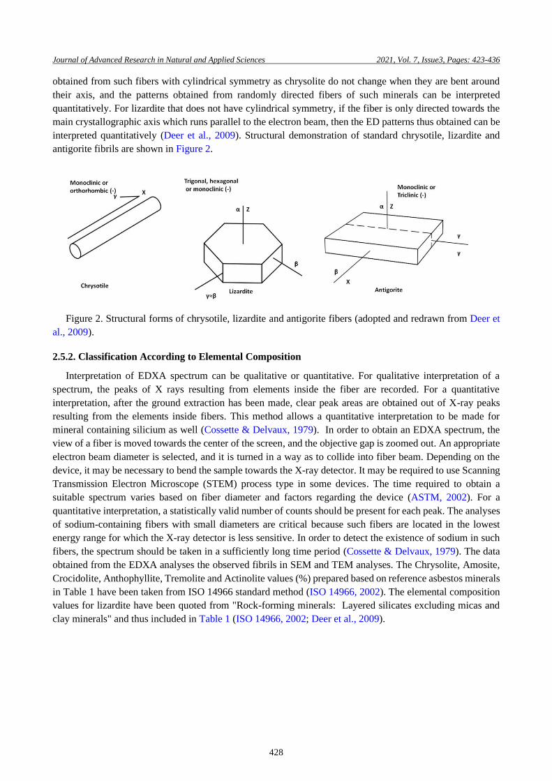
obtained from such fibers with cylindrical symmetry as chrysolite do not change when they are bent around
their axis, and the patterns obtained from randomly directed fibers of such minerals can be interpreted
quantitatively. For lizardite that does not have cylindrical symmetry, if the fiber is only directed towards the
main crystallographic axis which runs parallel to the electron beam, then the ED patterns thus obtained can be
interpreted quantitatively (Deer et al., 2009). Structural demonstration of standard chrysotile, lizardite and
antigorite fibrils are shown in Figure 2.
Figure 2. Structural forms of chrysotile, lizardite and antigorite fibers (adopted and redrawn from Deer et
al., 2009).
2.5.2. Classification According to Elemental Composition
Interpretation of EDXA spectrum can be qualitative or quantitative. For qualitative interpretation of a
spectrum, the peaks of X rays resulting from elements inside the fiber are recorded. For a quantitative
interpretation, after the ground extraction has been made, clear peak areas are obtained out of X-ray peaks
resulting from the elements inside fibers. This method allows a quantitative interpretation to be made for
mineral containing silicium as well (Cossette & Delvaux, 1979). In order to obtain an EDXA spectrum, the
view of a fiber is moved towards the center of the screen, and the objective gap is zoomed out. An appropriate
electron beam diameter is selected, and it is turned in a way as to collide into fiber beam. Depending on the
device, it may be necessary to bend the sample towards the X-ray detector. It may be required to use Scanning
Transmission Electron Microscope (STEM) process type in some devices. The time required to obtain a
suitable spectrum varies based on fiber diameter and factors regarding the device (ASTM, 2002). For a
quantitative interpretation, a statistically valid number of counts should be present for each peak. The analyses
of sodium-containing fibers with small diameters are critical because such fibers are located in the lowest
energy range for which the X-ray detector is less sensitive. In order to detect the existence of sodium in such
fibers, the spectrum should be taken in a sufficiently long time period (Cossette & Delvaux, 1979). The data
obtained from the EDXA analyses the observed fibrils in SEM and TEM analyses. The Chrysolite, Amosite,
Crocidolite, Anthophyllite, Tremolite and Actinolite values (%) prepared based on reference asbestos minerals
in Table 1 have been taken from ISO 14966 standard method (ISO 14966, 2002). The elemental composition
values for lizardite have been quoted from "Rock-forming minerals: Layered silicates excluding micas and
clay minerals" and thus included in Table 1 (ISO 14966, 2002; Deer et al., 2009).
Page 7
Journal of Advanced Research in Natural and Applied Sciences 2021, Vol. 7, Issue3, Pages: 423-436
429
Table 1
Elemental data of asbestos fibrils (ISO 14966, 2002; Deer et al., 2009). Chrysotile Amosite Crocidolite Antophylite Tremolite Actinolite Lizardite
SiO2 (%) 36 to 44 49 to 53 49 to 56 53 to 60 55 to 60 51 to 56 39 to 42
MgO (%) 38 to 42 1 to 7 0 to 3 17 to 34 20 to 26 12 to 20 39 to 43
FeO (%) 0 to 3 34 to 44 13 to 21 0 to 20 0 to 5 5 to 15 0 to 2
Fe2O3 (%) 0 to 5 0 to 5 13 to 20 0 to 5 0 to 5 0 to 5 0 to 3
Al2O3 (%) 0 to 2 0 to 1 0 to 1 0 to 3 0 to 3 0 to 3 0 to 1
CaO (%) 0 to 2 0 to 2 0 to 3 0 to 3 10 to 15 10 to 13 0 to 1
K2O (%) 0 to 1 0 to 1 0 to 1 0 to 1 0 to 1 0 to 1 0 to 1
Na2O (%) 0 to 1 0 to 1 4 to 9 0 to 1 0 to 2 0 to 2 0 to 1
H2O (%) 12 to 14 2 to 5 2 to 5 1 to 6 1 to 3 1 to 3 12 to 13
In the interpretation of fibril analysis, chrysolite's morphological structure is characteristic and can be easily
understood through experience. However, a few minerals with similar appearance and morphological
observation are insufficient for most samples (ISO 13794, 1999). If the determined characteristics of the pattern
correspond to those obtained from reference chrysolite, then the ED pattern obtained from chrysolite is highly
characteristic of that mineral. However, the crystal property of fiber could be damaged due to various factors,
and it may not provide an ED pattern. In that case, elemental composition (EDXA spectrum) could be the only
relevant data added to the morphological observation (ISO 13794, 1999).
3. Results and Discussion
3.1. Analysis of Ambient Air Samples Using SEM
The samples collected from ambient air were analyzed using a scanning electron microscope (SEM) at the
Advanced Technologies R&D Central Laboratory of Boğaziçi University. The methods obtained were
evaluated considering the chemical and structural (morphological) characteristics of the asbestos fibers as
contained in methods and reference books. The analyses conducted with an SEM instrument on 2 parallel
samples (Airborne 1 and Airborne 2) collected from ambient air are summarized in Table 2. Figure 3 shows
the 3 chrysotile fibrils observed by the analyses. Chrysotile fibrils of the samples collected from ambient air
are shown together with EDXA results. Figure 4 shows example images of another inorganic mineral (lizardite)
fibrils observed by the analysis of ambient air.
Table 2
SEM analysis results of the samples collected from ambient air
Sample
Code Verification
Analyzed
Area, mm2
Number of Fibrils
Observed Asbestos Concentration *
Chrysotile Other s/mm2 s/ml
Airborne 1 0.7666 1 1 80 (13) <0.76 <0.00001
Airborne 2 0.7965 1 2 75 (10) <1.6 <0.00004
* Concentration calculation was made according to the conditions of the sample received
“<” indicates that the concentration is lower than the determination limit
“()” indicates the number of the fibrils bigger than 5 μm
In light of the data obtained, the observed fibrils were analyzed both morphologically and elementally. The
tubular shape, which is the morphologic structure of the chrysotile, was determined in the fibrils observed as
a result of the analysis.
Page 8
Journal of Advanced Research in Natural and Applied Sciences 2021, Vol. 7, Issue3, Pages: 423-436
430
In the elemental compositions obtained as a result of the EDXA analyses of the chrysotile fibers, the percentage
of SiO2 is observed to be between 40 and 44, and the ratio of MgO is observed to be between 38 and 42. These
values match the elemental data of the chrysotile fibers as given in Table 1.
Other inorganic mineral (lizardite) fibers were found among the fibrils observed by the analyses conducted
with an SEM instrument on the samples collected from ambient air. The lizardite fibrils observed as a result
of the investigation were determined to have a morphologically planar (flat) and stringy structure. In the
elemental compositions obtained due to the EDXA analyses of other inorganic mineral (lizardite) fibers, the
percentage of SiO2 is observed to be between 39 and 42, and the percentage of MgO is observed to be between
39 and 43. These values match the elemental data of the lizardite fibers as given in Table 1.
According to the Labor Law number 6331 and which was published on Official Gazette number 28539 on
25.01.2013 and article 11 of that regulations, the provision stating “Employer shall make sure that eight hours
time-weighted average (ZAOD-TWA) value of asbestos concentration in the air to which the workers are
subjected shall not exceed 0,1 fiber/cm3" is valid (ACSGOHY, 2013). The obtained results (0.00004 fiber/cm3)
show that the asbestos concentration in the air is below the limit specified by the provision (0.1 fiber/cm3).
Since the equipment required to examine the crystal structure of the observed fibrils is not available in the
SEM device, the crystal structures of the fibers could not be examined. For this reason, it is not sufficient to
use the SEM device alone in such analyses.
Page 9
Journal of Advanced Research in Natural and Applied Sciences 2021, Vol. 7, Issue3, Pages: 423-436
431
Figure 3. Chrysotile fibrils were observed in ambient air and their elemental analysis results.
Page 10
Journal of Advanced Research in Natural and Applied Sciences 2021, Vol. 7, Issue3, Pages: 423-436
432
Figure 4. Other inorganic minerals (lizardite) fibrils were observed in ambient air and their elemental
analysis results.
Page 11
Journal of Advanced Research in Natural and Applied Sciences 2021, Vol. 7, Issue3, Pages: 423-436
433
3.2. Analysis of Ambient Air Samples Using TEM
The samples collected from ambient air were analyzed with a transmission electron microscope (TEM) at
RJ Lee Group Laboratory in the USA. The methods obtained were evaluated considering the chemical and
structural (morphological) characteristics of the asbestos fibers as contained in methods and reference books.
According to the TEM analysis results obtained from the samples collected from ambient air (Airborne 1 and
Airborne 2), Chrysotile fibril was not observed as a result of the analysis. The detected fibrils were indicated
to be other mineral (lizardite) fibrils. Results of the samples collected from ambient air analyzed using TEM
are summarized in Table 3.
Table 3
TEM analysis results of the samples collected from ambient air
Sample Code Verification
Coefficient
Analyzed
Area, mm2
Number of Fibrils
Observed Asbestos Concentration *
Chrysotile Other s/mm2 s/ml
Airborne 1 4.089 0.09243 0 53 (2) <44.2 <0.0044
Airborne 2 3.426 0.18504 0 102 (11) <18.5 <0.0019
* Concentration calculation was made according to the conditions of the sample received
“<” indicates that the concentration is lower than the determination limit
“()” indicates the number of the fibrils bigger than 5 μm.
According to EDXA analyses of the obtained fibrils, they were observed to be consistent with the spectrums
of chrysotile and antigorite among the serpentine group minerals. In addition, the fibers are understood to have
a magnesium silicate structure. Examples of the observed serpentine structures on Airborne 1 and Airborne 2
are shown in Figure 5. According to the results obtained, 13 of the fibrils observed as a result of the analysis
in total were bigger than 5 µm in length and less than 3 µm in width and based on ED analysis and elemental
composition (EDXA) results of these fibrils, and they were observed not to have the characteristics of chryso-
tile fibers. The majority of the observed fibrils were noted to be other inorganic minerals (lizardite) fibrils.
Figure 5. Typical serpentine structures were observed on samples.
Analyses with the TEM device give more reliable results in determining the types of fibrils observed. The most
striking result of this study is that it is not correct to decide on the determination of asbestos types based on a
single method or analysis device. The fibers should be determined by using appropriate methods and analysis
devices with advanced image properties according to the sample type in such studies. On the other hand, since
the detection limits of the SEM and TEM devices are different, it can be thought that the difference between
the concentrations is related to this.
Page 12
Journal of Advanced Research in Natural and Applied Sciences 2021, Vol. 7, Issue3, Pages: 423-436
434
4. Conclusion
Asbestos analysis was conducted on the samples obtained from ambient air using the SEM instrument at
both Boğaziçi University Central Laboratory and RJ KLee Group. According to the results of the analyses
conducted by Boğaziçi University, 23 of the fibrils observed in total are consistent with the phrase “bigger
than 5 µmin lengths and less than 3 µm in width”, as set out in the “Regulation on Health and Safety Measures
for Those Working with Asbestos”. 3 of these fibrils were observed to have the characteristics of chrysotile
fibers morphemically and elementally. Airborne samples were analyzed RJ Lee Group using TEM instrument
as well. According to the results obtained, 13 of the observed fibrils in total were bigger than 5 µm in length
and less than 3 µm in width and based on ED analysis and elemental composition (EDXA) results of these
fibrils; they were observed not to have the characteristics of chrysotile fibers. Majority of the observed fibrils
were noted to be other inorganic mineral (lizardite) fibrils. As a result of the asbestos analysis carried out on
ambient air samples by SEM device according to the method no ISO 14966 by Boğaziçi University, asbestos
concentration was found as 0.00004 fibre/cm3. RJ Lee Group carried out concurrent analyses according to ISO
13974 method by TEM. According to the analysis results obtained from the TEM device, asbestos concentra-
tion was 0.0004 fibre/cm3. All of these values are below the detection limit of the relevant analysis devices.
Furthermore, they are below the limit specified by the provision “The employer shall ensure that 8-hour
weighted average value (TWA) of the asbestos concentration in the air the workers are exposed to will not
exceed 0.1 fibre/cm3” according to article 11 of The Regulation on Health and Safety Precautions in Those
Working with Asbestos published in the Official Gazette dated 25.01.2013 and 28539 in force. In addition, the
most significant result of this study is that it is not appropriate to make a decision on the determination of
asbestos types based on a single method or analysis device.
Acknowledgement
The author wishes to thank Prof. Dr. Turgut T. Onay for the support.
Author Contributions
Mehmet Ali Kucuker: Conceptualization, Methodology, Validation, Investigation, Resources, Writing,
Visualization.
Conflicts of Interest
The author declares no conflict of interest.
References
ACSGOHY, (2013). Regulations on Health and Safety Measures Intended for Workers Working with Asbestos,
Official Gazette Official Journal number 28539 on 25.01.2013, Ankara. Retrieved from:
https://www.mevzuat.gov.tr/mevzuat?MevzuatNo=17050&MevzuatTur=7&MevzuatTertip=5
APTSR, (2005). Asbest veya Asbestli Malzeme ile Üretim Yapılan İşyerlerinde İş Sağlığı ve Güvenliği Proje
Denetimi Sonuç Raporu, T.C. Çalışma ve Sosyal Güvenlik Bakanlığı İş Teftiş Kurulu Başkanlığı,
Ankara. Retrieved from: https://ailevecalisma.gov.tr/medias/5974/2005_01.pdf
ASTM, (2002). “Standard Test Method for Microvacuum Sampling and Indirect Analysis of Dust by
Transmission Electron Microscopy for Asbestos Mass Concentration”, ASTM D5756, American
Society for Testing and Materials, Conshohocken, PA.
Atabey E. (2009). Türkiye de asbest, eriyonit, kuvars ve diğer mineral tozları ve etkileri. Maden Tetkik ve
Arama Müdürlüğü, pp. 188.
Cossette, M., & Delvaux, P. (1979). “Technical Evaluation of Chrysotile Asbestos Ore Bodies”, Short Course
in Mineralogical Techniques of Asbestos Determination. R. L. Ledoux, ed., Mineralogical Association
of Canada, pp. 79–110.
Deer, W. A., Howie, R. A. & Zussman J. (2009). Serpentines, Rock-forming minerals: Layered silicates
Page 13
Journal of Advanced Research in Natural and Applied Sciences 2021, Vol. 7, Issue3, Pages: 423-436
435
excluding micas and clay minerals, Vol. 3B, John Wiley & Sons, New York, pp. 157–211. Retrieved
from: https://pubs.geoscienceworld.org/canmin/article-abstract/48/5/1319/127220/
Erdoğan, N., & Yıldız, R., (1995). Magnezit ve Bazik Refrakter Malzeme Teknolojisi, Retrieved from:
https://www.researchgate.net/publication/279537853
EU, (1976). Official Journal of the European Union. Council Directive 76/769/EEC of 27 July 1976 on the
approximation of the laws, regulations and administrative provisions of the Member States relating to
restrictions on the marketing and use of certain dangerous substances and preparations [Accessed 2021
July 21]. Available at: https://eur-lex.europa.eu/legal-content/en/ALL/?uri=CELEX%3A31976L0769
EU, (1983). Official Journal of the European Union. Council Directive 83/477/EEC of 19 September 1983 on
the protection of workers from the risks related to exposure to asbestos at work (second individual
Directive within the meaning of Article 8 of Directive 80/1107/EEC) [Accessed 2021 July 21].
Available at:https://eur-lex.europa.eu/legal-content/EN/TXT/?uri=celex%3A31983L0477
EU, (1991). Official Journal of the European Union. Council Directive 91/383/EEC of 25 June 1991
supplementing the measures to encourage improvements in the safety and health at work of workers
with a fixed- duration employment relationship or a temporary employment relationship) [Accessed
2021 July 21]. Available at: https://eur-lex.europa.eu/legal-
content/EN/TXT/?uri=celex%3A31991L0383
EU, (2003). Official Journal of the European Union. Council Directive 83/477/EEC on the protection of
workers from the risks related to exposure to asbestos at work [Accessed 2021 July 21]. Available at:
https://eur-lex.europa.eu/legal-content/EN/TXT/?uri=celex%3A32003L0018
EU, (2006). Official Journal of the European Union Regulation (EC) No 1907/2006 of the European Parliament
and of the Council of 18 December 2006 concerning the Registration, Evaluation, Authorisation and
Restriction of Chemicals (REACH), establishing a European Chemicals Agency, [Accessed 2021 July
21]. Available at: https://eur-lex.europa.eu/legal-content/EN/TXT/?uri=CELEX%3A02006R1907-
20140410
EU, (2009). Official Journal of the European Union. Council Directive 2009/148/EC of the European
Parliament and of the Council of 30 November 2009 on the protection of workers from the risks related
to exposure to asbestos at work [Accessed 2021 July 21]. Available at: https://eur-lex.europa.eu/legal-
content/EN/TXT/?uri=celex%3A32009L0148
ISO 13794, (1999). Ambient Air—Determination of Asbestos Fibres — Indirect-Transfer Transmission
Electron Microscopy Method, International Organization for Standardization, Geneva, Switzerland.
ISO 14966, (2002). Ambient Air—Determination of Numerical Concentration of Inorganic Fibrous
Particles—Scanning Electron Microscopy Method, International Organization for Standardization,
Geneva, Switzerland.
Marioryad, H., Kakooei, H., Shahtaheri, S. J., Yunesian, M., & Azam, K. (2011). Assessment of airborne
asbestos exposure at an asbestos cement sheet and pipe factory in Iran. Regulatory Toxicology and
Pharmacology, 60(2), 200-205. DOI: https://doi.org/10.1016/j.yrtph.2011.03.005
Melville, R., & Lippmann, M. (2001). Influence of data elements in OSHA air sampling database on
occupational exposure levels. Applied Occupational and Environmental Hygiene, 16(9), 884-899. DOI:
https://doi.org/10.1080/10473220117850
Park, S. H. (2018). Types and health hazards of fibrous materials used as asbestos substitutes. Safety and
Health at Work, 9(3), 360-364. DOI: https://doi.org/10.1016/j.shaw.2018.05.001
Rees, D., Phillips, J. I., Garton, E. & Pooley, F.D. (2001). Asbestos lung fibre concentrations in south African
chrysotile mine workers. The Annals of Occupational Hygiene, 45(6), 473-477, DOI:
https://doi.org/10.1016/S0003-4878(00)00088-0
Rodilla, J. M. R., Cerrada, B. C., Pujadas, C. S., Delclos, G. L., & Benavides, F. G. (2021). Fiber burden and
asbestos-related diseases: an umbrella review. Gaceta Sanitaria. In Press, DOI:
https://doi.org/10.1016/j.gaceta.2021.04.001
Page 14
Journal of Advanced Research in Natural and Applied Sciences 2021, Vol. 7, Issue3, Pages: 423-436
436
WHO, (2007). World Health Organization, Outline for the development of national programmes for
elimination of asbestos-related diseases (No. WHO/SDE/PHE/07.02). World Health Organization.
ZMKKYHY, (2014). Regulation on Restriction and Prohibition of Hazardous Substances and Mixtures,
Official GazetteOfficial Journal number 27277 on 03.07.2009, Ankara. Retrieved from:
https://www.resmigazete.gov.tr/eskiler/2014/11/20141121-6.htm