Characterization of the Cellulose-Binding Domain of theClostridium cellulovorans Cellulose-Binding Protein A
MARC A. GOLDSTEIN, MASAHIRO TAKAGI, SEIICHI HASHIDA, ODED SHOSEYOV, ROY H. DOI,*AND IRWIN H. SEGEL
Department ofBiochemistry and Biophysics, University of California Davis, Davis, California 95616
Received 17 May 1993/Accepted 7 July 1993
Cellulose-binding protein A (CbpA), a component of the cellulase complex of Clostridium celulovorans,contains a unique sequence which has been demonstrated to be a cellulose-binding domain (CBD). The DNAcoding for this putative CBD was subcloned into pET-8c, an Escherichia coli expression vector. The proteinproduced under the direction of the recombinant plasmid, pET-CBD, had a high affinity for crystallinecellulose. Affinity-purified CBD protein was used in equilibrium binding experiments to characterize theinteraction of the protein with various polysaccharides. It was found that the binding capacity of highlycrystalline cellulose samples (e.g., cotton) was greater than that of samples of low crystallinity (e.g., fibrouscellulose). At saturating CBD concentration, about 6.4 ,umol of protein was bound by 1 g of cotton. Under thesame conditions, fibrous cellulose bound only 0.2 gmol of CBD per g. The measured dissociation constant wasin the 1 FM range for all cellulose samples. The results suggest that the CBD binds specifically to crystallinecellulose. Chitin, which has a crystal structure similar to that of cellulose, also was bound by the CBD. Thepresence of high levels of cellobiose or carboxymethyl cellulose in the assay mixture had no effect on the bindingofCBD protein to crystalline cellulose. This result suggests that the CBD recognition site is larger than a simplecellobiose unit or more complex than a repeating celiobiose moiety. This CBD is of particular interest becauseit is the first CBD from a completely sequenced nonenzymatic protein shown to be an independently functionaldomain.
Crystalline cellulose is resistant to the action of many0-1,4-glucanases. The closely packed cellulose chains arestabilized by hydrogen bonding to form a tight, regular arraywhich shields many of the glycosidic bonds from enzymaticattack (27). Nevertheless, some Clostridium species candegrade crystalline cellulose into simple sugars. Several ofthese strains produce a cellulase enzyme complex (cellulo-some) containing a variety of ,B-1,4-glucanases together withproteins with no known enzymatic activity (15, 16, 20). Aprevious study (20) has shown that the association of en-zyme subunits and nonenzymatic subunits was essential forthe degradation of crystalline cellulose, whereas the disso-ciated enzyme subunits could degrade only noncrystallinesubstrates.We have isolated the cellulosome from Clostridium cellu-
lovorans, an organism shown to have cellulolytic activity.This cellulase complex has a rather high specific activitycompared with those of other cellulases that have beencharacterized (20). The purified nonenzymatic subunit pro-tein had a very strong affinity for crystalline cellulose.Therefore, this protein was designated as the cellulose-binding protein A (CbpA). Recently, we cloned the struc-tural gene encoding this protein (cbpA) and determined itscomplete nucleotide sequence (21). The amino acid se-quence of CbpA deduced from the nucleotide sequence(1,848 amino acid residues, 189 kDa) revealed the followinginteresting features: (i) a putative cellulose-binding domain(CBD) (amino acids 27 to 189) which was homologous toseveral known cellulose-binding sequences (10, 21), (ii)hydrophilic regions repeated four times, and (iii) hydropho-bic regions repeated eight times. It has been shown that theability to degrade crystalline substrates is correlated with the
* Corresponding author.
binding ability of the cellulase (14). Presumably the CBDmediates the interaction of the nonenzymatic protein, CbpA,with cellulose. The cellulose is then degraded by the cellu-losomal enzymatic components, particularly endogluca-nases, which are bound to CbpA. In this way CbpA directsthe enzymatic components to the crystalline cellulose sur-face.
This paper describes (i) the subcloning of the regionencoding the putative CBD from CbpA and (ii) the interac-tion between crystalline cellulose and CbpA. A previousstudy (9) had suggested that the surface of the cellulosecrystal presented a regular, repeating set of overlappingpotential binding sites and that adsorption of a CBD to thissurface could not be analyzed by a classical equilibriumpartition analysis. However, we have established experi-mental conditions which minimize nonspecific binding to theassay tubes and to the cellulose, thereby allowing us toanalyze the interaction in terms of a reversible, two-compo-nent equilibrium binding system (19). Therefore we wereable to determine both the dissociation constant of thecellulose-CBD complex and the binding capacity of thecellulose sample by means of double-reciprocal plots. Thesubstrate specificity of the CBD and the effects of varied pHand soluble carbohydrates on CBD binding to cellulose werealso investigated.
MATERIALS AND METHODSBacterial strains and plasmids. Escherichia coli XL1-Blue
was obtained from StrataGene, La Jolla, Calif., and wasused for all cloning experiments. E. coli BL21 (DE3) andpET-8c were as described previously (23).
Materials. PC buffer (pH 7) contained 50 mM KH2PO4, 10mM sodium citrate, and 1 mM NaN3. TEDG buffer (4)contained 10 mM Tris (pH 7), 0.1 mM EDTA, 0.1 mM
dithiothreitol, and 5% (vol/vol) glycerol. Although Tris has alow buffer capacity at pH 7, the buffer was suitable becausehydrogen ions were neither produced nor used. Restrictionendonucleases were from Bethesda Research Laboratories,Bethesda, Md. All other chemicals used were of the highestpurity commercially available. Avicel PH101 (lot 1117) wasfrom FMC Corp., Philadelphia, Pa. Absorbent cotton wasfrom the Seamless Rubber Co., New Haven, Conn. Cellulonfiber was from Weyerhaeuser, Tacoma, Wash. Granularchitin from crab shells was a kind gift of DemosthenesPappagianis. All other binding substrates were purchasedfrom Sigma Chemical Co., St. Louis, Mo. Each of thepolysaccharides was washed twice with PC buffer beforeuse. Nigeran was recrystallized by dissolving the solid in hotwater, filtering, and cooling on ice. The fiber sizes of thecellulon and cotton were reduced by processing with aGifford-Wood minimill for 5 min.
Cloning of putative CBD. DNA primers complementary tothe regions of cbpA flanking the putative CBD (CbpAresidues 28 to 189) were synthesized by a Gene AssemblerPlus (Pharmacia). The forward primer contained an NcoIrestriction site (recognition sequence, CCATGG) with theATG in frame with the gene fragment to act as a translationalstart codon when cloned into the pET-8c vector cloning site.The reverse primer contained a stop codon and a BamHIsite. Polymerase chain reaction (PCR) amplification wasperformed with 20 pmol of each primer, 200 ,uM eachdeoxynucleoside triphosphate, and 1 ng of cbpA DNA(cloned into vector pGEMEX-1 [Promega] as in reference21) as a template in a total volume of 100 ,ul. Taq polymerasewas obtained from Amersham and was used under bufferconditions recommended by the manufacturer. PCR wascarried out for 40 cycles as described previously (11). ThePCR product was purified by phenol-chloroform extractionfollowed by ethanol precipitation and a wash with 70%ethanol; it was then dried under vacuum and resuspended in27 ,ul of distilled water. The DNA was then cleaved withNcoI and BamHI and run on a 2.5% low-melting-pointagarose (Nuseieve GTG; FMC Corp.) gel in TBE buffer (18).DNA bands stained with ethidium bromide were visualizedunder long-wave UV light and cut from the gel. The vector,plasmid pET-8c, was prepared by cleaving 1 ,ug of pET-8cDNA with NcoI-BamHI and cutting the linearized DNAband from the gel. Vector and insert DNAs were ligated byusing 100 ng of vector DNA and 300 ng of insert with aTakara Ligation kit. The ligated plasmids were used totransform competent E. coli XL1-Blue cells, which werethen plated on Luria-Bertani (LB) plates (18) containing 100,ug of ampicillin per ml and 12.5 ,ug of tetracycline per ml.After overnight incubation at 37°C, colonies were selectedand grown in liquid LB medium containing ampicillin andtetracycline. Plasmid DNA from each culture was rescued asdescribed previously (23) and cleaved with restriction en-zymes to verify the insertion of the gene fragment. The insertsequence was confirmed by DNA sequencing, using thesame procedures as reported previously (21).
Preparation and purification ofCBD protein. Plasmid DNAcontaining the insert was used to transform E. coli BL21(DE3). Plasmid-containing cultures were grown at 37°C withshaking in NZCYM (18) medium containing ampicillin (100,ug/ml) to a Klett reading of 160 (green filter). At this point,isopropyl-j3-D-thiogalactopyranoside (IPTG) was added to afinal concentration of 1 mM. After 4 h, the cells wereharvested by centrifugation, resuspended in PC buffer con-taining RNase A at 10 ,ug/ml and DNase I at 1 ,ug/ml, andlysed by sonication on ice with a Biosonic II sonicator at
maximum power for 45 s followed by a 15-s cooling period.This was repeated four times. The insoluble fraction of a1-liter cell culture was collected by centrifugation (30 min at12,000 x g and 4°C) and resuspended in 20 ml of 6 Mguanidine HCl. This was kept on ice for 30 min withoccasional vortexing to disperse the pellet. Insoluble debriswere removed by centrifugation (30 min at 12,000 x g and4°C). The soluble guanidine HCI extract was graduallydiluted to a total volume of 400 ml with TEDG renaturationbuffer over a 2-h period at 4°C. Ammonium sulfate wasadded to 80% saturation. After 4 h at 4°C, precipitatedproteins were collected by centrifugation (30 min at 12,000 xg and 4°C), resuspended in 40 ml of PC buffer, and dialyzedagainst PC buffer. The CBD protein fragment of CbpA wasfurther purified by cellulose affinity, as follows. Three addi-tions of 1.0 g of Avicel PH101 microcrystalline cellulosewere used to remove the CBD protein from the solution.After each addition, the suspension was allowed to come toequilibrium (1 h at room temperature with slow rotation).The cellulose was then collected by centrifugation andremoved before the next addition. The 3 g of cellulose waswashed once with 1 M NaCl-PC buffer and twice with PCbuffer. Purified CBD was eluted from the cellulose by threewashes with 10 ml of 6 M urea. The urea fractions werepooled and dialyzed against PC buffer. The protein concen-tration in the final purified fraction was analyzed by colori-metric methods, using the MicroBCA protein assay kit(Pierce, Rockford, Ill.) with bovine serum albumin (BSA)standards.
Determination of the CBD-cellulose dissociation constantand the cellulose-binding capacity. Samples of CBD protein(typically 0 to 100 p.g) were added to 1.5-ml-capacity micro-centrifuge tubes containing PC buffer supplemented with 1mg of BSA per ml and the desired amount of cellulose(typically 1 mg added from a stock slurry containing 10 mg ofcellulose per ml and 1 mg of BSA per ml in PC buffer).Potential competitors, e.g., cellobiose (4 mg/ml) or car-boxymethyl cellulose (CMC) (4 mg/ml), were included insome reactions by adding 200 p.l of a 20-mg/ml stock solutionin PC-BSA buffer. The final volume was always 1 ml. ThepH of the buffer was 7.0 Oxcept when noted otherwise. Forexperiments at other pH values, the PC-BSA buffer wasadjusted prior to use by the addition of concentrated HCl orNaOH. Assay tubes were mixed by slow vertical rotation (30rpm) at 37°C for 1 h. The samples were then spun in amicrocentrifuge for 1 min to sediment the cellulose andcellulose-protein complexes. After the buffer had been re-moved, the pellet was washed by resuspension in 1 ml of PCbuffer. The wash was separated out by centrifugation anddiscarded. Pellets were then resuspended in a final 1 ml ofPC buffer. (The centrifugation step would not be expected toperturb the equilibrium because the cellulose and protein-cellulose were concentrated to the same extent.) Of theoriginal BSA in the assay tubes (-1 mg/ml), only about 0.1p,g would remain after the washing steps, assuming nononspecific adsorption and a liquid volume of 10 p.l in thepellet. Any color development as a result of this residualBSA was controlled for by the 0 CBD control tubes. Ali-quots (150 p.l) of this well-mixed suspension were taken forprotein determinations with the MicroBCA kit. The manu-facturer's instructions were followed, except that the samplevolume was brought to 0.5 ml with PC buffer, to which 0.5 mlof BCA working reagent (Pierce Co.) was added. Assaymixtures were incubated at 60°C for 30 min. The proteinconcentration was determined colorimetrically from thecleared supernatants at 562 nm in a Shimadzu 160 U spec-
trophotometer. Assay tubes to which no CBD protein wasadded were used to correct for a small amount of colordevelopment caused by the cellulose and residual BSA. Thedata were compared with BSA standards and adjusted toaccommodate the dilutions that were made to determine theamount of protein bound to the cellulose in each sample. Thepractical detection limit of this assay was about 0.2 ,ug/ml.After correction for dilutions, this corresponds to about0.034 nmol of CBD bound to the cellulose in the assay tube.The free CBD protein concentration, [P], was determined bysubtracting the bound protein concentration, [PC], from thetotal CBD added to the tube, [P]:
[P] = [P]t- [PC] (1)
The system was analyzed by assuming a simple equilibriuminteraction (19):
k,P + C _ PC
k-,
where the dissociation constant, Kd, is defined as
k.1 [P][C]
k, [PC]
The data were analyzed by double-reciprocol plots of 1/[PC]versus 1/[P] at different fixed levels of cellulose (equation 2):
1 Kd 1 1
[PC] [Pc]max [P] [PC]max (2)
It must be noted that the cellulose is not a soluble compo-nent, so that [C] represents the concentration of binding siteson the cellulose surface exposed to the buffer, per unitvolume. Similarly, [PC] represents the concentration ofbinding site-protein complexes per unit volume. Straightlines were fitted to the datum points by the least-squaresmethod, using the DeltaGraph Professional plotting applica-tion (Deltapoint, Inc., Monterey, Calif.). Each point was theaverage of three independent protein assays from the samebinding-assay tube. Experiments were performed in dupli-cate. At least two different amounts of cellulose were used todetermine the Kd and [PC]ma,/g of cellulose. These wereaveraged to provide the values listed in Table 1.
Determination of binding to other polysaccharides. Xylan,nigeran, Sephadex G-75, and chitin were used in assays todetermine whether they were substrates for CBD protein. Inall cases, the methods used were the same as those used indetermining the binding to cellulose. Chitin exhibited a veryhigh background in the MicroBCA assay, which increasedproportionally to the incubation time at 60°C, and so thecolor development time was reduced to 15 min. Because ofthe high background of chitin, only two widely differentprotein concentrations were used.
RESULTS
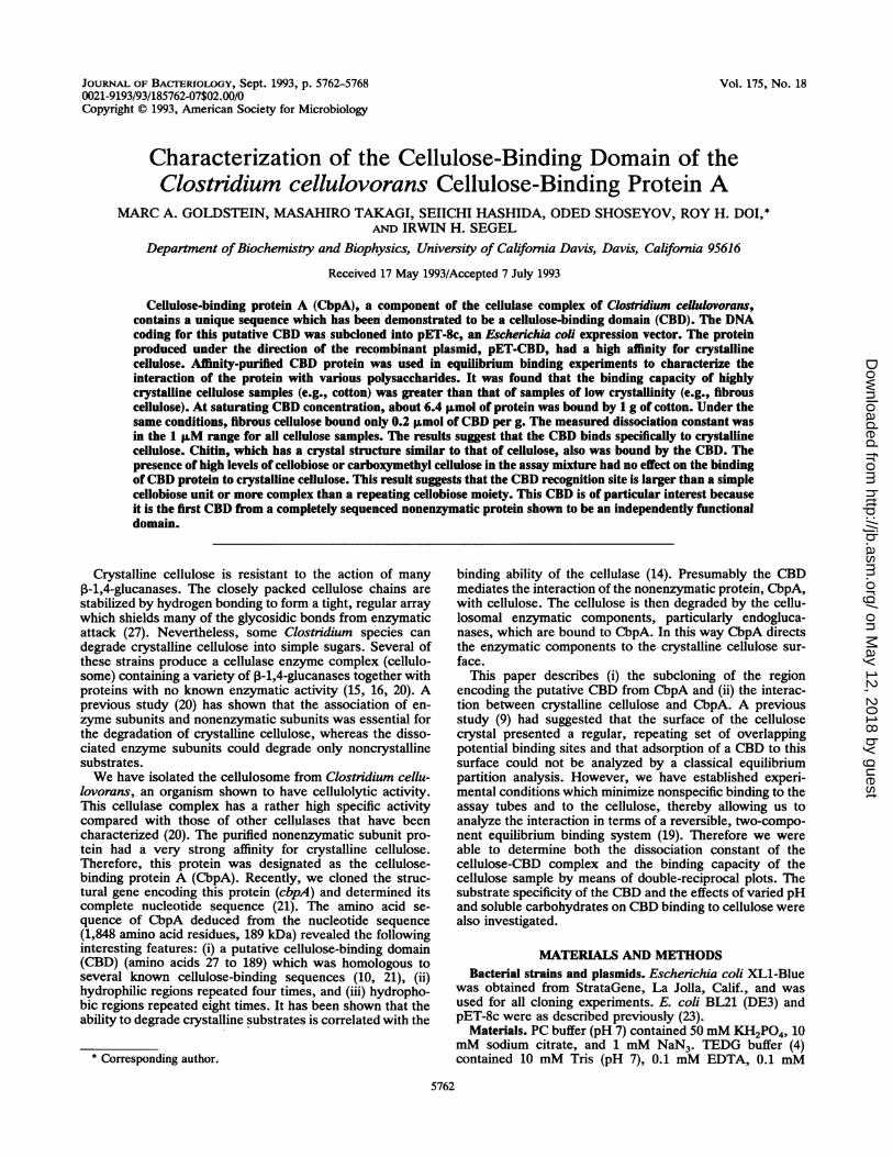
Purification of the CBD for binding analyses. To selectivelyproduce the putative CBD region of CbpA, residues 28 to189, we designed oligonucleotide primers complementary tobases 67 to 86 and 558 to 579 of cbpA (Fig. 1). As shown inFig. 1, these primers were designed with mismatches tocreate an NcoI site and an ATG start codon on one end ofthe PCR product and a TAG stop codon followed by a
TABLE 1. Adsorption of CBD protein to insoluble substrates
Substrate Observed Kd Observed [PCJma(Ij.M)' (~Lmol of CBD/gf'
a The values for Kd and [PC]m, were calculated as described in Materialsand Methods.
b Competition experiments including CMC or cellobiose were performedwith Avicel PH101 at 1 mg/ml. The values given for the competitionexperiments represent the dissociation constant and binding capacity of theCBD-Avicel pair.
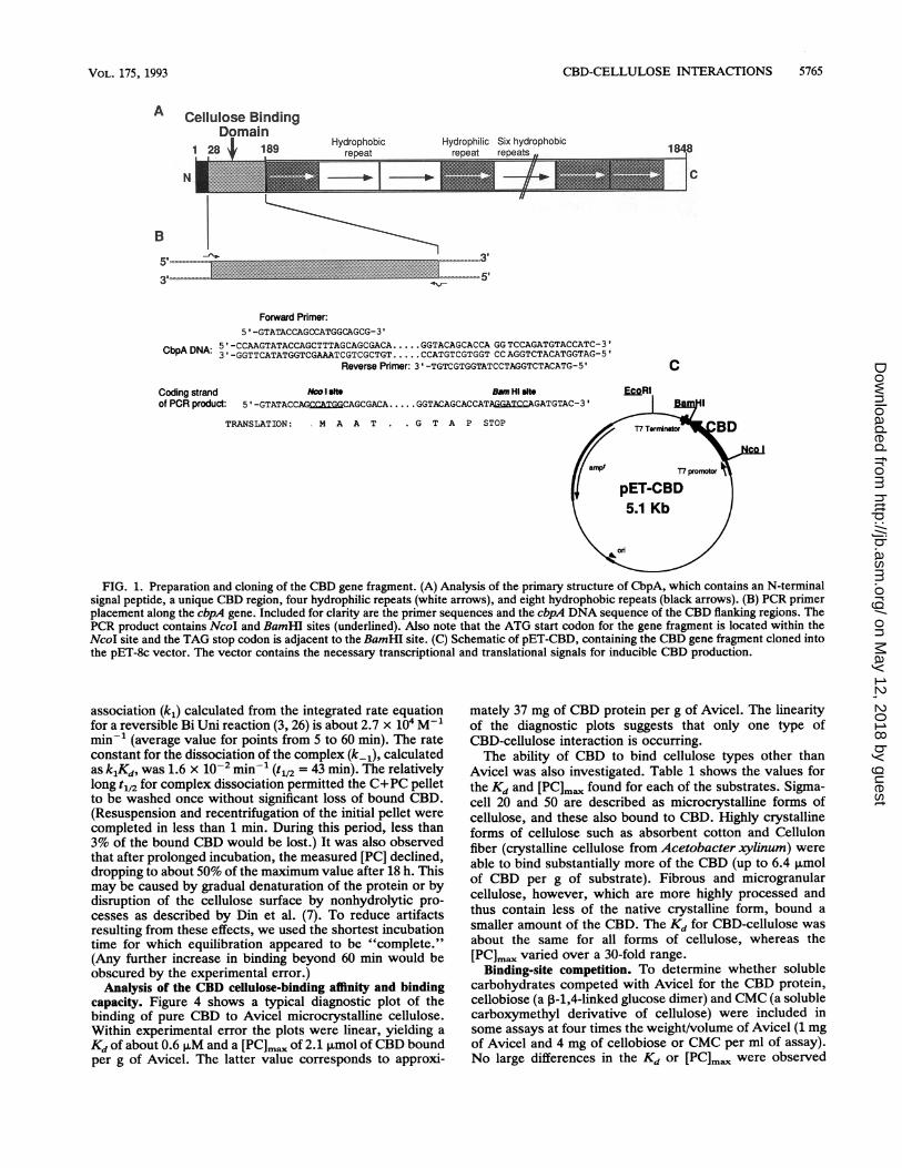
BamHI site at the other end. This gene fragment was thencloned into the T7 RNA polymerase expression plasmidpET-8c, resulting in plasmid pET-CBD. The cloned genefragment codes for a methionine at the N terminus of theCBD, but the rest of the CBD amino acid sequence corre-sponds to residues 28 to 189 of CbpA. The protein fragmenthas a molecular weight of 17,634. The insertion was verifiedby DNA sequencing. CBD protein was produced by E. coliBL21 (DE3) cells harboring pET-CBD. After the addition ofIPTG, this host strain produces T7 RNA polymerase, whichrecognizes the T7 promoter in the pET vector. The cbd genefragment was under the control of this inducible promoter,and CBD protein was synthesized in large amounts afterinduction (Fig. 2). After a 4-h production period, the solubleextract from the lysed cells contained only small amounts ofCBD protein, while most was found in the insoluble fraction.This protein was readily soluble in concentrated guanidinehydrochloride and was renatured by slow dilution intoTEDG buffer. It was found that protein prepared in thisfashion binds to Avicel, verifying the putative CBD. Al-though this fraction is mostly CBD protein, the assaysdescribed here require the protein to be very pure. Thispurity is obtained by a single cellulose affinity step, asdescribed in Materials and Methods. The affinity-purifiedCBD protein appears on acrylamide gels as a single bandwhen stained with Coomassie brilliant blue. Approximately70 mg of CBD protein can be recovered from the cellsharvested from a 1-liter culture.Time course ofbinding ofCBD to cellulose. The time course
of the interaction of Avicel with CBD (Fig. 3) disclosesseveral features of the process. (i) At initial concentrationsof 1.0 mg of Avicel per ml and 2.0 p.M CBD (i.e., [P]0), aplateau value of 1.2 ,uM complex (i.e., [PC]) is attained by 60min. A separate experiment established that the maximumCBD-binding capacity of the cellulose sample was 2.1 ,umolg-1, corresponding to an effective concentration of 2.1 puMtotal cellulose sites (i.e., [C]O). Assuming that an equilibriumwas established (verified below), Kd, defined as [P][C]/[PC],is about 0.6 puM. (ii) The second-order rate constant for
FIG. 1. Preparation and cloning of the CBD gene fragment. (A) Analysis of the primary structure of CbpA, which contains an N-terminalsignal peptide, a unique CBD region, four hydrophilic repeats (white arrows), and eight hydrophobic repeats (black arrows). (B) PCR primerplacement along the cbpA gene. Included for clarity are the primer sequences and the cbpA DNA sequence of the CBD flanking regions. ThePCR product contains NcoI and BamHI sites (underlined). Also note that the ATG start codon for the gene fragment is located within theNcoI site and the TAG stop codon is adjacent to the BamHI site. (C) Schematic of pET-CBD, containing the CBD gene fragment cloned intothe pET-8c vector. The vector contains the necessary transcriptional and translational signals for inducible CBD production.
association (k1) calculated from the integrated rate equationfor a reversible Bi Uni reaction (3, 26) is about 2.7 x 10⁴ M-1min-1 (average value for points from 5 to 60 min). The rateconstant for the dissociation of the complex (k-1), calculatedas klKd, was 1.6 x 10⁻² min-' (t112 = 43 min). The relativelylong t112 for complex dissociation permitted the C+PC pelletto be washed once without significant loss of bound CBD.(Resuspension and recentrifugation of the initial pellet werecompleted in less than 1 min. During this period, less than3% of the bound CBD would be lost.) It was also observedthat after prolonged incubation, the measured [PC] declined,dropping to about 50% of the maximum value after 18 h. Thismay be caused by gradual denaturation of the protein or bydisruption of the cellulose surface by nonhydrolytic pro-cesses as described by Din et al. (7). To reduce artifactsresulting from these effects, we used the shortest incubationtime for which equilibration appeared to be "complete."(Any further increase in binding beyond 60 min would beobscured by the experimental error.)
Analysis of the CBD cellulose-binding affinity and bindingcapacity. Figure 4 shows a typical diagnostic plot of thebinding of pure CBD to Avicel microcrystalline cellulose.Within experimental error the plots were linear, yielding a
Kd of about 0.6 ,uM and a [PC]m. of 2.1 ,umol ofCBD boundper g of Avicel. The latter value corresponds to approxi-
mately 37 mg of CBD protein per g of Avicel. The linearityof the diagnostic plots suggests that only one type ofCBD-cellulose interaction is occurring.The ability of CBD to bind cellulose types other than
Avicel was also investigated. Table 1 shows the values forthe Kd and [PC]m. found for each of the substrates. Sigma-cell 20 and 50 are described as microcrystalline forms ofcellulose, and these also bound to CBD. Highly crystallineforms of cellulose such as absorbent cotton and Cellulonfiber (crystalline cellulose from Acetobacter xylinum) were
able to bind substantially more of the CBD (up to 6.4 ,umolof CBD per g of substrate). Fibrous and microgranularcellulose, however, which are more highly processed andthus contain less of the native crystalline form, bound a
smaller amount of the CBD. The Kd for CBD-cellulose wasabout the same for all forms of cellulose, whereas the[PC]max varied over a 30-fold range.
Binding-site competition. To determine whether solublecarbohydrates competed with Avicel for the CBD protein,cellobiose (a P-1,4-linked glucose dimer) and CMC (a solublecarboxymethyl derivative of cellulose) were included insome assays at four times the weight/volume of Avicel (1 mgof Avicel and 4 mg of cellobiose or CMC per ml of assay).No large differences in the Kd or [PC]m. were observed
FIG. 2. Expression and purification of the CBD protein. Whole-cell proteins from cells harboring pET-8c (lane 2), whole-cell pro-teins from cells harboring pET-CBD (lane 3), cytosolic fraction fromlysed pET-CBD cells (lane 4), guanidine HCl-solubilized membrane/inclusion body fraction from lysed pET-CBD cells (lane 5), final PCbuffer wash of Avicel pellet (lane 6), and purified CBD protein (lane7) were loaded on a 15% acrylamide gel. Each lane was loaded with0.005% of the total protein of each fraction, except lane 6, which isa lOx concentrate. Prestained molecular mass markers (lanes 1 and8) have mobilities of approximately 2.6, 5, 12.7, 18.1, 29, and 44kDa.
(Table 1), indicating that these soluble carbohydrates hadlittle or no effect on the binding of the CBD to Avicel.
Effect of pH on dissociation constant. C. cellulovorans is aneutrophilic organism, thriving only around pH 7 (22), sothis pH was used for most of the binding assays. However,other experiments established that the Kd and [PC]mx didnot vary significantly with changes in pH over the range 5.0to 8.0. In addition, it was noted that PC buffers as acidic aspH 3.5 or as basic as pH 9.5 would not remove the CBD fromAvicel during 1-min washes (data not shown).
Binding of the CBD to other polysaccharides. Xylan,Sephadex G-75, nigeran, and chitin were used to explore thebinding specificity of the CBD. Of these, only chitin showedmeasurable binding of the CBD peptide (Table 1). Thechitin-CBD Kd and binding capacity were similar to theAvicel-CBD values.
1.4
1.2
-~0.8C
o0.60.4
0.2
0 20 40 60 80 100 120Incubation Time (min)
FIG. 3. Time course of CBD-Avicel binding. CBD (total protein,2.0 ,uM) and Avicel (1 mg/ml) were equilibrated as described inMaterials and Methods, except that a larger total volume was usedto provide samples taken at various time points. Each time pointsample was washed and assayed as described in Materials andMethods.
1.0 2.0 3.0 4.0 5.0 6.0
[PI1 (gM)1FIG. 4. Double-reciprocal plot of CBD binding to Avicel. Sym-
bols: * 0.5 mg of Avicel; *, 1 mg of Avicel; A, 2 mg of Avicel. Theinset shows [PC]mna plotted against the amount of Avicel used. Theassay volume was 1.0 ml.
DISCUSSION
Many cellulases and other hydrolytic enzymes, such aschitinases, have high affinities for their substrates (2, 20).Much interest has recently focused on identifying individualdomains of these enzymes that are responsible for thebinding (8). Several bacterial cellulases take the idea of abinding domain one step further and produce a cellulase-related protein that is nonenzymatic but mediates the inter-action between the endoglucanases and the insoluble sub-strate (15, 20, 21). It has previously been shown that theability of a cellulase to degrade crystalline cellulose is relatedto the strength of the binding between the cellulase andcrystalline cellulose (14), whereas strong binding is notrelated to the ability to degrade amorphous cellulose.Our results show that CbpA contains a domain responsible
for cellulose binding and that this domain could functionindependently from the rest of CbpA. Because the purifica-tion protocol involved denaturation and renaturation steps,the fact that the purified protein was functional indicates thatthe CBD protein sequence was sufficient for proper foldingof the protein fragment.
Previous attempts to determine the relative binding affinityof cellulases for the surface of cellulose have been made. Asreviewed by Klyosov (14), the basic concept involved deter-mining a partition coefficient (Kp) between the insolublecellulose and the soluble phase (i.e., moles protein per gramof cellulose/concentration of protein in the aqueous phase),which was typically expressed in units of liters per gram.These partition coefficients were used as a general estima-tion of the relative binding ability of the cellulase. Morerecent attempts to derive a more traditional measurement,using an actual affinity constant, Ka, have yielded nonlineardiagnostic plots (9). The authors suggested that the nonlin-earity was a result of mutually hindered binding (negativecooperativity) because the cellulose surface is a two-dimen-sional array of overlapping potential binding sites.There are several alternative explanations for the nonlin-
earity of diagnostic plots reported in earlier work. (i) Thefirst is nonspecific binding of the protein to the assay tubeand perhaps also to the surface of the cellulose. These
problems are compounded when the free protein rather thanthe bound protein is used for the measurements. When thefree-protein concentration is very low, the relative amountlost due to tube interactions is large; when the free-proteinconcentration is high enough to minimize the effects of suchnonspecific binding, the experimental error involved in theprotein determination itself may mask the relatively smallchanges in free-protein concentration being measured. (ii)The simultaneous weak binding of catalytic sites to amor-
phous cellulose can affect the linearity of diagnostic plots forCBD-containing cellulases. (iii) A cellulase or CBD mayhave different affinities for different crystal faces of crystal-line cellulose. As an example, it was suggested that the CBDfrom CenA binds preferentially to the 110 face of cellulosecrystals but that the 110 face, which has a higher bindingcapacity (but apparently lower affinity), is filled only at highligand concentration (9).We have found nonspecific binding of the CBD to the
assay tubes to be a problem in performing equilibriumbinding experiments, and we have developed an assay inwhich the CBD and cellulose are equilibrated in the presenceof excess BSA. The BSA effectively eliminates nonspecificCBD interactions with the tube. After equilibrium isreached, the cellulose and cellulose-protein complexes are
collected, washed, and assayed for bound proteins. Asdescribed above the dissociation of the CBD-cellulose isslow so that no detectable amount is removed during a rapidwash step.The bound CBD concentration was measured directly by
the protein assay, and the free CBD concentration was
calculated by subtracting the bound CBD concentrationfrom the total CBD concentration, as shown in equation 1.This has the advantage that any CBD molecules adsorbednonspecifically with low affinity to the cellulose would beremoved by the wash step, resulting in data that more
accurately reflect the specific, high-affinity interaction be-tween the CBD and the cellulose surface. As shown in Fig.4, data gathered by using this type of assay yields (withinexperimental error) linear diagnostic plots. The validity ofthe assay is supported by the observation that [PC]m.increases linearly with the amount of cellulose used,whereas Kd is independent of the amount of cellulose. Table1 shows the results obtained with several forms of celluloseas well as with other carbohydrates. The results indicate thatcellulose types described as crystalline have a higher CBD-binding capacity than do highly processed celluloses thathave lost much of their crystallinity. The fact that the[PC]ma of cellulose samples varies widely with differentcellulose types whereas the Kd remains constant indicatesthat we have measured one type of strong protein-celluloseinteraction occurring between the CBD and the cellulose.The lower [PC]ma of highly processed celluloses reflects asmaller number of potential protein interaction sites in thesample and seems to correlate with the crystallinity of thesample. This would indicate that there is some specialfeature present in crystalline cellulose that makes it accept-able as a binding substrate, whereas amorphous cellulose isfound lacking.To further characterize the substrate specificity of the
CBD, we measured the effect of added soluble substrates(cellobiose or CMC) on cellulose binding. Excess cellobioseor CMC had no effect on the CBD-Avicel Kd or [PC]ma, asshown in Table 1. This lack of competition confirms that theCBD recognition site is specific for something more complexthan a simple repeating glucose or cellobiose moiety and
suggests that a particular three-dimensional arrangement ofcellulose chains is needed.The specificity of the CBD for crystalline cellulose
prompts a consideration of chitinases, which are known tobind tightly to chitin, a polymer of N-acetylglucosamine in,B-1,4 linkage. Like cellulose, chitin comes in a variety offorms, depending on the source and the purification methodused in its isolation (1, 2). The chitin used for affinitypurification of chitinases is a-chitin, in which the chains arearranged in an antiparallel fashion. This form of chitin iscrystalline with a structure similar to that of native crystal-line cellulose (often referred to as cellulose I). Cellulose I isthe form in which the cellulose chains are arranged inparallel bundles, as opposed to cellulose II, in which thechains are in an antiparallel configuration. Processing ofcellulose I under harsh conditions causes its disruption,resulting in cellulose II (25). Both forms are crystalline,because of extensive hydrogen bond formation. Since ourisolated CBD binds to less highly processed forms of cellu-lose, i.e., largely cellulose I, we were interested in findingwhether the CBD would bind to a-chitin, which has a similarcrystal structure, although of opposite strand orientation.We found that the CBD did accept chitin as a bindingsubstrate with a Kd very similar to that for cellulose. Chitinis the only noncellulosic substrate that we have found that isable to bind the CBD. Xylan (P3-1,4-xylose), nigeran (alter-nating a-1,4- and a-1,3-glucose), and Sephadex G-75 (a-1,6-glucose with a-1,3 branches) (5) were also tried, but the CBDdid not show measurable binding to any of them under theconditions of the assay. Since chitin is the only one of thesesubstrates that is crystalline, we believe that this demon-strates the importance of crystallinity in the substrate. It ispossible that the CBD is specific for a long, rigid carbohy-drate chain or recognizes an area of adjacent chains withbond distances similar in both chitin and cellulose.A parallel can be drawn between endo-,-1,4-glucanases
and xylanases. Neither type of enzyme has been reported asbinding tightly to its noncrystalline substrate (amorphouscellulose and xylan, respectively). No xylan-binding do-mains have been reported, but some xylanases, like someendo-3-1,4-glucanases, have CBDs specific for crystallinecellulose (12). Although it seems illogical for an enzyme tohave a high affinity for a substrate it cannot hydrolyze, onfurther consideration, taking into account the proximity ofxylan and cellulose in native cellulosic materials, this factsuggests that crystalline cellulose makes a better anchoringsite for glycan hydrolases than does xylan or amorphouscellulose.To make use of the dissociation constant that we have
determined, we must compare the CBD-cellulose bindingwith that in other systems previously studied. However,earlier work on cellulases is reported mostly in the form ofpartition coefficients (e.g., the work of Klyosov [13]). Ourdata can be converted to this form, yielding a result of K, =1.5 liters/g. This places the tightness of the CBD-Avicelbinding fairly near the top of the logarithmic range (systemswith the weakest binding have Kp < 0.004, and those withthe strongest binding have Kp = 8). It must be noted thatdifferences in the experimental procedures may reduce thesignificance of this direct comparison, because of the prob-lems described above.Comparisons with systems for which true Kd values have
been published are likely to hold more meaning. The Kds ofproteins for various soluble carbohydrates have been deter-mined. As reported by Szmelcman et al. (24), the E. colimaltose-binding protein binds maltose with a Kd of 1 ,uM and
maltotriose binds with a Kd of 0.16 ,uM. Similarly, the E. colixylose-binding protein binds xylose with a Kd of 0.63 ,uM (6).Therefore, our finding of about 0.6 p,M for the Avicel-CBDKd is well within the reported range for carbohydrate-proteininteractions.A qualitative analysis of a CBD of the cellulosome subunit
S1 from C. thermocellum YS has been reported (17). TheCBD of C. cellulovorans CbpA has approximately 50%homology with the CBD region reported for C. thennocel-lum. The CBD region of C. cellulovorans CbpA is unique(21), whereas the number of CBD regions in C. thennocel-lum is unknown, since the complete sequence of subunit S1has not been reported.
ACKNOWLEDGMENTS
This research was supported in part by grant DE-FG03-92ER20069 from the Department of Energy (R.H.D.).We thank Phan Tran for her excellent technical assistance.
REFERENCES1. Blackwell, J. 1988. Physical methods for the determination of
chitin structure and conformation. Methods Enzymol. 161:435-442.
2. Cabib, E. 1988. Chitinase from Serratia marcescens. MethodsEnzymol. 161:460-462.
3. Capellos, C., and B. H. Bielski. 1980. Kinetic systems: mathe-matical description of chemical kinetics in solution, p. 43-45.Robert E. Krieger Publishing Co., Huntington, N.Y.
4. Chang, B. Y., and R. H. Doi. 1990. Overproduction, purifica-tion, and characterization of Bacillus subtilis RNA polymerasesigma A factor. J. Bacteriol. 172:3257-3263.
5. Coutinho, J. B., N. R. Gilkes, R. A. J. Warren, D. G. Kilburn,and R. C. Miller. 1992. The binding of Cellulomonas fimiendoglucanase C (CenC) to cellulose and Sephadex is mediatedby the N-terminal repeats. Mol. Microbiol. 6:1243-1252.
6. Dahms, A. S., W. Huisman, G. Neslund, and C. Ahlem. 1982.D-Xylose-binding protein (periplasmic) from Escherichia coli.Methods Enzymol. 90:473-476.
7. Din, N., N. R. Gilkes, B. Tekant, R. C. Miller, R. A. J. Warren,and D. G. Kilburn. 1991. Non-hydrolytic disruption of cellulosefibres by the binding domain of a bacterial cellulase. Bio/Technology 9:1096-1099.
8. Gilkes, N. R., B. Henrissat, D. G. Kilburn, R. C. J. Miller, andR. A. J. Warren. 1991. Domains in microbial beta-1,4-glyca-nases: sequence conservation, function, and enzyme families.Microbiol. Rev. 55:303-315.
9. Gilkes, N. R., E. Jervis, B. Henrissat, B. Tekant, R. C. J. Miller,R. A. J. Warren, and D. G. Kilburn. 1992. The adsorption of abacterial cellulase and its two isolated domains to crystallinecellulose. J. Biol. Chem. 267:6743-6749.
10. Hansen, C. K. 1992. Fibronectin type III-like sequences and anew domain type in prokaryotic depolymerases with insolublesubstrates. FEBS Lett. 305:91-96.
11. Innis, M. A., and D. H. Gelfand. 1990. Optimization of PCRs, p.
3-12. In M. A. Innis, D. H. Gelfand, J. J. Sninsky, and T. J.White (ed.), PCR protocols: a guide to methods and applica-tions. Academic Press, Inc., San Diego, Calif.
12. Kellett, L. E., D. M. Poole, L. M. A. Ferreira, A. J. Durrant,G. P. Hazlewood, and H. J. Gilbert. 1990. Xylanase B and anarabinofuranosidase from Pseudomonas flourescens subsp. cel-lulosa contain identical cellulose-binding domains and are en-coded by adjacent genes. Biochem. J. 272:369-376.
13. Klyosov, A. A. 1988. Cellulases of the third generation, p. 87-99.In J.-P. Aubert, P. Beguin, and J. Millet (ed.), Biochemistry andgenetics of cellulose degradation. Academic Press Ltd., Lon-don.
14. Klyosov, A. A. 1990. Trends in biochemistry and enzymology ofcellulose degradation. Biochemistry 29:10577-10585.
15. Lamed, R., and E. A. Bayer. 1988. The cellulosome concept,p. 101-117. In J.-P. Aubert, P. Beguin, and J. Millet (ed.),Biochemistry and genetics of cellulose degradation. AcademicPress Ltd., London.
16. Mayer, F., M. P. Coughlan, Y. Mori, and L. G. Ljungdahl. 1987.Macromolecular organization of the cellulolytic enzyme com-plex of Clostridium thermocellum as revealed by electron mi-croscopy. Appl. Environ. Microbiol. 53:2785-2792.
17. Poole, D. M., E. Morag, R. Lamed, E. A. Bayer, G. P.Hazlewood, and H. J. Gilbert. 1992. Identification of the cellu-lose-binding domain of the cellulosome subunit S1 from Clos-tridium thennocellum YS. FEMS Microbiol. Lett. 99:181-186.
18. Sambrook, J., E. F. Fritsch, and T. Maniatis. 1989. Molecularcloning: a laboratory manual, 2nd ed. Cold Spring HarborLaboratory Press, Cold Spring Harbor, N.Y.
19. Segel, I. H. 1975. Enzyme kinetics, p. 18-97. Wiley Inter-science, New York.
20. Shoseyov, O., and R. H. Doi. 1990. Essential 170-kDa subunit fordegradation of crystalline cellulose by Clostridium cellulo-vorans cellulase. Proc. Natl. Acad. Sci. USA 87:2192-2195.
21. Shoseyov, O., M. Takagi, M. A. Goldstein, and R. H. Doi. 1992.Primary sequence analysis of Clostridium cellulovorans cellu-lose binding protein A. Proc. Natl. Acad. Sci. USA 89:3483-3487.
22. Sleat, R., R. A. Mah, and R. Robinson. 1984. Isolation andcharacterization of an anaerobic, cellulolytic bacterium, Clos-tridium cellulovorans sp. nov. Appl. Environ. Microbiol. 48:88-93.
23. Studier, F. W., and B. A. Moffat. 1986. Use of bacteriophage T7RNA polymerase to direct selective high-level expression ofcloned genes. J. Mol. Biol. 189:113-130.
24. Szmelcman, S., M. Schwartz, T. J. Silhavy, and W. Boos. 1976.Maltose transport in E. coli K12. Eur. J. Biochem. 65:13-19.
25. Weimer, P. J., A. D. French, and T. A. Calamari. 1991.Differential fermentation of cellulose allomorphs by ruminalcellulolytic bacteria. Appl. Environ. Microbiol. 57:3101-3106.
26. Wilkdnson, F. 1980. Chemical kinetics and reaction systems, p.52-58. Van Nostrand Reinhold Co., New York.
27. Young, R. A., and R. M. Rowell. 1986. Cellulose structure andbiosynthesis, p. 3-50. In R. A. Young, and R. M. Rowell,Cellulose: structure, modification, and hydrolysis. Wiley Inter-science, New York.


![[PPT]Clostridium botulinium and Botulism - Santa Monica …homepage.smc.edu/.../presentations/clostridium-botulinum.ppt · Web viewTitle Clostridium botulinium and Botulism Author](https://static.documents.pub/doc/80x56/5ad21b467f8b9a0f198c0cca/pptclostridium-botulinium-and-botulism-santa-monica-viewtitle-clostridium.jpg)


