CHARACTERIZATION OF MESENCHYMAL STEM CELLS IN MUSCULOSKELETAL INJURY A Dissertation by ALEXIS MITCHELL Submitted to the Office of Graduate and Professional Studies of Texas A&M University in partial fulfillment of the requirements for the degree of DOCTOR OF PHILOSOPHY Chair of Committee, Ashlee Watts Committee Members, Dana Gaddy Akhilesh Gaharwar Friedhelm Schroeder Mark Lenox Head of Department, Allen Roussel August 2017 Major Subject: Biomedical Sciences Copyright 2017 Alexis Mitchell
Transcript
CHARACTERIZATION OF MESENCHYMAL STEM CELLS IN
MUSCULOSKELETAL INJURY
A Dissertation
by
ALEXIS MITCHELL
Submitted to the Office of Graduate and Professional Studies of Texas A&M University
in partial fulfillment of the requirements for the degree of
DOCTOR OF PHILOSOPHY
Chair of Committee, Ashlee Watts Committee Members, Dana Gaddy
Akhilesh Gaharwar Friedhelm Schroeder Mark Lenox
Head of Department, Allen Roussel
August 2017
Major Subject: Biomedical Sciences
Copyright 2017 Alexis Mitchell
ii
ABSTRACT
The main goal of regenerative medicine is to enhance the innate healing response
to more closely mimic tissue development. This may include cell-based, trophic, or
small molecule therapies. In cell-based therapies, the initial premise was one of tissue
replacement. Mesenchymal stem cells (MSCs) are adult-derived stem cells present
throughout the body and easily harvested from individual patients. After over a decade
of veterinary and human medical use of MSCs and medical research, the function of
MSCs after therapeutic use in injury remains unclear.
Elucidating engraftment location and longevity of MSCs post injection may
provide insight to their mechanism of action. We developed a protocol for labeling
MSCs with a fluorocarbon nanoparticle that allows for non-invasive longitudinal
tracking and evaluated cellular viability, proliferation, and morphology. A dose
dependent cell association of the nanoparticle was seen in our study but it was not
repeatable between individuals. Prior to use of this technique to track MSCs, further
study is needed to elucidate where the failure occurred: uptake of label by MSCs,
maintenance of label within the cell cytoplasm or loss of conjugation of the fluorophore.
Despite the use of MSCs as a therapeutic in horses for many years, there is little
information on the best techniques for cryopreserving these cells for immediate use post
thaw. We tested several freezing mediums for the short-term cryopreservation of equine
MSCs. We found that 95% autologous serum and 5% DMSO did not negatively affect
post thaw viability, growth kinetics, or morphology.
iii
Bisphosphonates were approved for use in horses in the United States in 2014.
Since, they have become wildly popular due to improvements in lameness in treated
horses. We suspected that the efficacy in lameness reduction could be due to an off
target effect such as an effect on the MSC because the approved dose is so low
compared to the anti-resorptive dose of the same drugs in people. We investigated the
impact of bisphosphonates on bone remodeling and bone cells including MSCs. We
observed reduction in lameness but no changes in bone turnover or MSC characteristics.
iv
ACKNOWLEDGEMENTS
I would like to thank my committee chair, Dr. Ashlee Watts, for her patience and
guidance during my graduate schooling and involving me in clinical research in
veterinary medicine.
Thank you to my committee members, Dr. Akhilesh Gaharwar, Dr. Friedhelm
Schroeder, and Dr. Mark Lenox for their guidance and support throughout the course of
this research.
I would like to extend a special thank you to Dr. Dana Gaddy who dedicated her
time and energy to develop my graduate experience and broaden my research experience
outside of my directed studies.
Thanks also to my friends and colleagues, and Dr. Larry Suva for making my
time at Texas A&M University a great experience.
Finally, thank you to my mom, dad and husband for their patience, love, and
unconditional support.
v
CONTRIBUTORS AND FUNDING SOURCES
CONTRIBUTORS
This work was supervised by a dissertation committee consisting of Dr. Ashlee
Watts, advisor of the Department of Large Animal Clinical Sciences, Dr. Dana Gaddy of
the Department of Veterinary Integrative Biosciences, Dr. Akhilesh Gaharwar of the
Department of Biomedical Engineering, Dr. Friedhelm Schroeder of the Department of
Veterinary Physiology & Pharmacology, and Dr. Mark Lenox of the Department of
Biomedical Engineering.
The data flow cytometry data in Chapters 2, 3 and 4 were analyzed by Dr. Gus
Wright of the Department of Veterinary Pathobiology. Clinical evaluations of lameness
in Chapter 4 were scored by Dr. Sarah Sampson of the Department of Large Animal
Clinical Sciences.
All other work conducted for the dissertation was completed by the student
independently.
FUNDING SOURCES
Research was supported by the Link Endowment for Equine Research at Texas
A&M University. Work conducted in Chapter 2 was funded by the American Quarter
Horse Association and in Chapter 3 by the Department of Large Animal Clinical
Sciences.
vi
TABLE OF CONTENTS
Page
ABSTRACT .............................................................................................................. ii
ACKNOWLEDGEMENTS ...................................................................................... iv
CONTRIBUTORS AND FUNDING SOURCES..................................................... v
TABLE OF CONTENTS .......................................................................................... vi
LIST OF FIGURES................................................................................................... viii
LIST OF TABLES .................................................................................................... x
CHAPTER I INTRODUCTION.......................................................................... 1
Regenerative Medicine: Clinical Applications ................................................... 3 Regenerative Medicine: The Horse Model ......................................................... 6 Regenerative Medicine: Future Applications...................................................... 7
CHAPTER II LONGITUDINAL EVALUATION OF FLUOROCARBON LABELED MSCS ..................................................................................................... 8
Introduction ......................................................................................................... 8 Materials and Methods ........................................................................................ 12
CHAPTER III LONGITUDINAL EVALUATION OF FLUOROCARBON LABELED CRYOPRESERVATION OF EQUINE MESENCHYMAL STEM CELLS IN 95% AUTOLOGOUS SERUM AND 5% DMSO DOES NOT ALTER POST-THAW GROWTH OR MORPHOLOGY IN VITRO COMPARED TO FETAL BOVINE SERUM OR ALLOGENEIC SERUM AT 20 OR 95% AND DMSO AT 10 OR 5% ............................................................................................... 27
Introduction ......................................................................................................... 27 Materials and Methods ........................................................................................ 29
CHAPTER IV INVESTIGATING THE IMPACT OF BISPHOSPHONATES, A MUSCULOSKELETAL THERAPY, ON BONE REMODELING AND BONE CELLS........................................................................................................... 56
Introduction ......................................................................................................... 56 Materials and Methods ........................................................................................ 62
1.3 Publications of MSC Clinical Trials in Humans as Referenced on PubMed ................................................................................................. 5 2.1 Trilineage Differentiation of Bone Marrow Derived MSCs ...................... 16
2.2 Time Dependent Response of Colony Forming Units Between Individuals .................................................................................................. 18 2.3 Dose Dependent Relationship of 19F Labeled MSCs on Brightfield and Fluorescent Imaging ............................................................................ 20 2.4 Aggregation of 19F Complicates MSC Labeling....................................... 20
2.5 Dose Dependent Relationship of 19F Labeled MSCs via Flow Cytometric Analysis ................................................................................... 22 2.6 No Dose Dependent Cell Association of 19F ............................................ 22 2.7 Dose Dependent Cell Association of 19F via Nuclear Magnetic Resonance................................................................................................... 23
3.1 Pilot Project Percentage of Viable Cells Post Thaw .................................. 34 3.2 Total Viable Cell Number .......................................................................... 40
3.3 Percentage of Viable Cells Post-Thaw....................................................... 41 3.4 Debris and Morphology Scores.................................................................. 42 3.5 Images of Monolayer Culture .................................................................... 43 3.6 Total Colony Number................................................................................. 44 3.7 Cell Generations Post-Thaw....................................................................... 46
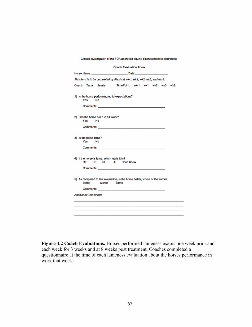
3.9 Debris Score vs Colony Forming Units ..................................................... 53 4.1 Study Design .............................................................................................. 63 4.2 Coach Evaluations...................................................................................... 67 4.3 Cell Recruitment and Differentiation......................................................... 69 4.4 Bone Resorption Ability of Clodronate Exposed Osteoclasts ................... 70 4.5 Trilineage Differentiation of MSCs Exposed to Clodronate...................... 71 4.6 CTX-I Levels After Clodronate Treatment ................................................ 73 4.7 Osteocalcin Levels After Clodronate Treatment........................................ 74 4.8 Front and Hind Limb Asymmetry.............................................................. 76 4.9 Front and Hind Limb Lameness................................................................. 77 4.10 Coach Evaluations of Performance ............................................................ 78
x
LIST OF TABLES
TABLE Page 2.1 Cell Surface Marker Expression of Bone Marrow Derived MSCs............ 16 2.2 Colony Forming Units Were Variable Between Individuals ..................... 17
2.3 Population Doubling Times of 19F Labeled MSCs ................................... 19
4.1 Immunophenotyping of MSCs Exposed to Clodronate ............................. 72
1
CHAPTER I
INTRODUCTION
Regenerative medicine is a new branch of medicine and since the 90s has
become commonplace in human and veterinary medicine. 1, 2 As a field, regenerative
medicine focuses on promoting the body’s innate healing response. 3 Traditional
treatments like surgical procedures and drugs focus on the modulation of symptoms
while regenerative medicine works to return tissues to normal function. 2 This novel field
of medicine encompasses a variety of strategies including cell-based therapies, tissue
engineering, and gene therapy.1, 3
Cell-based therapies are a large focus of regenerative medicine with stem cells
and progenitor cells being the dominant cell type.1, 4 Stem cells are defined by their
ability to self-renew and proliferate indefinitely while maintaining the ability to
differentiate into multiple cell types.5, 6 Asymmetrical division permits stem cells to self-
renew while generating a daughter cell that will terminally differentiate.7, 8 Stem cells are
classified based on their differentiation potential or potency. Stem cells with the ability
to give rise to all 3 germ layers and extra-embryonic tissues are referred to as totipotent.
Pluripotent stem cells, often referred to as embryonic stem cells, can only give rise to the
3 germ layers but not placental.8, 9 Multipotent stem cells, or adult stem cells, give rise to
one germ layer and are present in tissues throughout our bodies and therefore can be
isolated from many tissue types.7, 10 Multipotent stem cells give rise to progenitor cells
that can be multipotent or unipotent but lack the ability to self-renew.10 Cells and tissues
2
at the local environment are maintained by both progenitor cells and multipotent stem
cells in different regions of the body.7, 9
Mesenchymal stem cells (MSCs) are multipotent stem cells that give rise to
mesoderm tissues.11 First described by Friedenstein in 1976 as fibroblast precursor cells,
human MSCs are now defined by the international society for cellular therapy as plastic
adherent, express CD73, CD90, and CD105, lack expression for CD45, CD34, CD14,
CD79, and HLA-DR, and have trilineage differentiation potential for osteoblast,
adipocytes, and chondrocyte.12, 13 MSCs can be isolated from a variety of tissues
including bone marrow, fat, and umbilical cord blood.11, 14-16 MSCs have become
popular in regenerative medicine due to their ease of isolation and their availability for
autologous use without safety concerns. How MSCs improve healing is still unclear.
Initially, isolated MSCs used for regenerative medicine were thought to engraft at a
lesion site and differentiate into the needed cell type.17 More recent evidence suggests
that MSCs work as medicinal signaling cells to improve healing through functions other
than tissue differentiation which may include, growth factor production, inflammation,
immunomodulation, and other unknown factors.18-20
A classic example of MSCs as a signaling cell is their role supporting
hematopoiesis; a specific example being MSC production of rank ligand (RANK-L) and
macrophage colony stimulating factor (MCSF) to stimulate osteoclast differentiation of
hematopoietic lineage cells.21, 22 MSCs as immunomodulators secrete a variety of
molecules. MSCs have demonstrated release of prostaglandin E2 after interactions with
3
toll-like receptors, inhibition of T cell activity via cytokines like human growth factor,
and proliferation of lymphocytes via interleukin-7.19, 20, 23, 24
Another feature of MSCs is they might be able to be used in allogenic
applications without being immunophenotyped because that they are considered immune
privileged due to their lack of major histocompatibility complex II (MHC-II) expression,
T cell suppression, and decreased MHC-I expression.19, 20 Recently, increased
inflammatory reaction and MHC specific antibody reactions following allogenic
transplant of MSCs has been demonstrated in horse, murine and non-human primate
models. 25-29
REGENERATIVE MEDICINE: CLINICAL APPLICATIONS
The number of registered clinical trials and MSC therapy trials is steadily
increasing (Figure 1.1). Since 2005, MSC therapies have become a popular area of study
in regenerative medicine with applications in a variety of fields including cancers,
digestive system diseases, heart and blood diseases, and gland and hormone related
diseases. Muscle, bone, and cartilage studies comprise approximately one third of MSC
therapy clinical trials (Figure 1.2).
When “Mesenchymal Stem Cell Human Clinical Trial” was searched on PubMed
using the filters “humans”, “clinical trials”, and “01/01 to 12/31” for each year there was
an increasing trend in the number of publications (Figure 1.3).
4
Figure 1.1 Registered Clinical Trials From 2000-2017. A) Registered clinical trials in all phases are increasing across all areas with B) MSC therapeutic trials increasing proportionally.
Figure 1.2 Registered MSC Therapeutic Clinical Trials. One third of registered MSC therapeutic clinical trials are in musculoskeletal injuries including muscle, bone or cartilage.
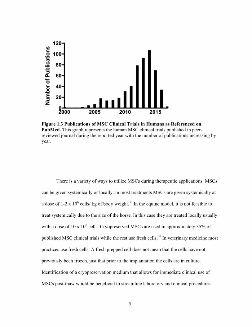
5
Figure 1.3 Publications of MSC Clinical Trials in Humans as Referenced on PubMed. This graph represents the human MSC clinical trials published in peer-reviewed journal during the reported year with the number of publications increasing by year.
There is a variety of ways to utilize MSCs during therapeutic applications. MSCs
can be given systemically or locally. In most treatments MSCs are given systemically at
a dose of 1-2 x 10⁶ cells/ kg of body weight.30 In the equine model, it is not feasible to
treat systemically due to the size of the horse. In this case they are treated locally usually
with a dose of 10 x 106 cells. Cryopreserved MSCs are used in approximately 35% of
published MSC clinical trials while the rest use fresh cells.30 In veterinary medicine most
practices use fresh cells. A fresh prepped cell does not mean that the cells have not
previously been frozen, just that prior to the implantation the cells are in culture.
Identification of a cryopreservation medium that allows for immediate clinical use of
MSCs post-thaw would be beneficial to streamline laboratory and clinical procedures
6
and reduce associated costs. MSCs are used in a variety of ways because we do not
understand what they are doing post injection. With some many diverse ways of utilizing
MSCs the use of a pre-clinical model is important for determining the most effective
ways to use MSCs to help generate a standardized protocol.
REGENERATIVE MEDICINE: THE HORSE MODEL
One of the benefits of studying stem cell therapies in the horse is that the horse
population, like that of man, is not homogenous in genotype or phenotype as most
laboratory species are. In addition to genotype and phenotype differences, individual
variation in MSC characteristics, especially in species with diversity, has been
reported.31, 32 It is important to assess MSCs in models that more accurately reflect the
inherent variability among human MSC preparations.
The equine athlete is a well-accepted model for stem cell therapies in
musculoskeletal injury suffering from similar injuries to those in humans.33, 34 The horse
ages similarly and experiences similar musculoskeletal injuries to human including
osteoarthritis, tendonitis, and stress fractures.33 MSCs as a treatment for tendon injuries
have been vigorously studied in the horse. In studies of naturally occurring tendon
lesions, horses treated with MSCs had a lower percentage of re-injury rates compared to
those receiving traditional treatments.35, 36 The horse is an important preclinical model
for musculoskeletal disease and will help elucidate the best way to utilize the MSC.
7
REGENERATIVE MEDICINE: FUTURE APPLICATIONS
The purpose of this dissertation is to better define the best ways to utilize MSCs.
Stem cells have the potential to be a powerful form of regenerative medicine if they
could be harnessed by clinicians to heal tissues without scar tissue and restore function.37
There are many diverse ways of using MSCs; however without knowing the best way to
use them clinicians cannot employ their full potential.
In order to optimize the potential of MSCs as a treatment we must fully
understand their role in the body after therapeutic application. To understand their
function we need to know their engraftment location and effects in the local
environment. Chapter 2 investigates a way to longitudinally evaluate cell engraftment,
engraftment duration, and quantify cell engraftment. The environment MSCs are
injected to and the state in they are in when they are injected could affect their function.
Chapter 3 investigates a clinically acceptable cryopreservation formula for MSCs
without xenogens and an almost completely autologous product. It is possible MSCs are
modulating the environment around them. Chapter 4 investigates the impact of
bisphosphonates, a musculoskeletal therapy, on bone remodeling and bone cells like
MSCs.
8
CHAPTER II
LONGITUDINAL EVALUATION OF FLUOROCARBON LABELED MSCS
INTRODUCTION
MSCs are an increasingly popular choice when treating musculoskeletal injuries
clinically in the horse due to their potential to be disease modifying osteoarthritic drugs
(DMOAD) based on MSC regenerative potential.38 MSC therapy has shown beneficial
effects in musculoskeletal injuries like osteoarthritis, cartilage defects, and tendonitis in
human and various animal models including caprine, porcine, and equine models.38-41
Despite the use of MSCs to treat musculoskeletal diseases in equine clinical practice, the
mechanism of action of these MSCs post injection is largely unknown.
MSCs have the potential to differentiation into multiple cell lineages.21 It is
possible MSCs act through direct cell differentiation based on their differentiation
potential and has been demonstrated in vivo.42 Reports have also demonstrated MSC
mechanism through immune-modulation or acting as medicinal signaling cells post
injection. MSCs have demonstrated modulation of cytokines via down regulation of
activated T-cells in diseased environments allowing for normal tissue healing without
scar tissue formation.43 Despite the promise of a potential DMOAD, MSCs cannot be
utilized fully without understanding if they act through cell differentiation, signaling, or
both. Identifying engraftment location of MSCs post injection, quantifying the number
of cells that engraft and the duration of engraftment could elucidate MSC function post
injection.
9
Tracking MSCs post injection provides the opportunity to identify cell
engraftment to help elucidate mechanism of action. Many forms of cell tracking after
implantation exist including transgenic mice, fluorescent labeling and Y chromosome
tracking.44 Unfortunately, most methods of tracking are not optimal for two main
reasons. First, these methods require tissue collection or biopsy for identification of
labeled cells. Tissue collection is invasive, affects tissue healing and is not always
possible depending on the location of cell injection. Second, it is impossible to evaluate
the cells longitudinally or to assess tissue healing when the tissues are constantly being
disrupted. Bioluminescence allows for in vivo longitudinal tracking without invasive
techniques however, this application can only be used in the murine model.45 A tracking
method that will allow longitudinal tracking in large animal models is required.
Iron oxide nanoparticles are a non-invasive form of in vivo cell tracking that has
been utilized over the last 10 years for longitudinal tracking. Theses nanoparticles
exhibit high magnetism. High magnetism causes a disruption in signal when using
magnetic imaging, which allows localization of labeled cells. Thus iron oxide labeled
cells can be imaged repeatedly over time using MRI and are seen as a loss of signal or
artifact on routine magnetic resonance imaging. Repeated MRI is an excellent modality
for longitudinal imaging because it does not cause tissue disruption or other changes to
healing.
Iron oxide nanoparticles exhibit small particle size allowing cellular uptake by
endocytosis that does not affect cell viability, proliferation, differentiation, or
migration.46, 47 Coatings like lipids and surfactants are needed to stabilize iron oxide
10
nanoparticles for proper dispersion of the particles.48 Cell death can cause exocytosis of
the iron oxide nanoparticle into the extracellular matrix where endocytosis by another
cell type is possible. These nanoparticles have been reported in ovine, mice, rats, swine
and have been traced using human MSCs in various mouse models.49-53
Limitations to iron oxide nanoparticles as a tracking modality are twofold. First
tissue healing cannot be assessed in areas iron oxide labeled MSCs are present. The
presence of the iron oxide labeled cells is seen through an acquisition artifact of the MRI
due to the nanoparticles magnetism, obscuring anatomical imaging and preventing
assessment of tissue healing. Second, the number of iron oxide labeled MSCS present
post injection cannot be quantified on MRI due to the production of signal voids larger
than the cell containing them.54 Only the presence or absence of label can be assessed.
Thus we wanted to utilize a label that allows for both longitudinal tracking of cells and
longitudinal assessment of tissue healing. If this were possible we would be able to
assess the effects of the cells on healing while also assessing their presence in the
tissues.
Perfluorocarbon nanoparticles (19F) are a new technology that have emerged in
the past 5 years and provide a new technique for in vivo cell tracking. Hydrogen and
fluorine resonate at different frequencies on magnetic imaging, allowing for evaluation
for the presence of hydrogen containing anatomy and fluorine containing label using
different sequences collected separately during an MRI exam. The overlay of H imaging
and F imaging can then be used to demonstrate both tissue characteristics and presence
or absence of fluorine label. An additional benefit is that the amount of fluorine signal
11
can be quantified, allowing not only confirmation of cell engraftment, but also
quantification of labeled cells. Like iron oxide, 19F label is imaged by MRI and
therefore can be imaged repeatedly in the same patient without invasive procedures.
Composed of 4- isotope19 fluorine molecules connected to a single carbon
molecule; 19F are small particles (160nm) stable in extreme environment.55 Endocytosis
of 19F nanoparticles has been demonstrated in human MSCs and neural stem cells
(NSC) cells without affecting cell viability, growth, or morphology in vitro. 42 Various
coatings including surfactants, transfecting agents, and targeting antibodies are used to
increase loading of the nanoparticles into the cell.56 Electroporation to open cell
membrane channels have been reported in non-phagocytic cells with difficulty
endocytosing 19F labels.56
Human neural stem cells successfully endocytosed 19F nanoparticle incubated
for 24 hours at 5 mg/mL before suspension in extracellular matrix and intracerebral
injection.42 Labeled hNSCs produced signal on MRI and distribution of cells in the
lesion cavity believed to form new tissue though it was unclear if formed tissue was
functional.42 In another study human MSCs were labeled with 19F nanoparticles in
culture for 24 hours at 2.5mg/mL for cellular uptake before being injected
intramuscularly into the hindlimbs of healthy mice.57 Labeled hMSCs produced signal
on MRI with a strong relationship between MRI quantification and number or real cells
in vitro.57
We wanted to develop a protocol to use 19F in horse. To allow a quick and
inexpensive validation of labeling in protocol development we needed a visual
12
assessment of labeling. Fluorescent labels can be utilized in a variety of ways including,
conjugation to antibodies that communicate with cell surface receptors and labeling cells
cytoplasm and organelles.58 This method is often used in conjugation with nanoparticle
tracking to validate MRI findings in research studies where tissues samples can be
collected.59 We used a perfluorocarbon emulsion conjugated with a Texas red (19F-TR)
or FITC fluorophore with an excitation/emission of 590/620 nm or 495/519 nm,
respectively (Celsense, INC., Pittsburgh, PA).
Our objective was to develop a protocol for labeling equine MSCs that 19F
labeled MSCs would allow for longitudinal evaluation of cell engraftment,
quantification, and duration in the same patient. Specifically we wanted to develop a
protocol that would not compromise viability, proliferation, or morphology in vitro and
quantify the number of 19F labeled equine MSCs in an ex vivo model using our 3Tesla
MRI.
MATERIALS AND METHODS
MSC Characterization
Cryopreserved MSCs isolated from sternal bone marrow were evaluated from 10
horses ranging in age from 2-16 years (median 11). Cryovials of MSCs were thawed in a
35ºC water bath until there was no longer an ice ball present. After thawing, 1mL of
DPBS (Lonza, Walkersville, MD ) was added to the cell suspension and allowed to sit
for 5 minutes. Following 5 minutes, the cell suspension was added in a drop wise
manner to 20mL of DPBS. A 100 µL aliquot of the cell suspension was used to
13
determine post thaw viability and a total cell count using fluorescein diacetate and
propidium iodide as previously described.60 The cell suspension was centrifuged (300G,
4ºC, 7 brake) for 5 minutes. Cells were seeded at 10,000 cells/cm2 in culture media
(Dulbecco’s modified Eagle’s medium 1g/L glucose (Corning, Corning, NY )
Corning, NY), and 10,000units/mL penicillin, 10,000 ug/mL streptomycin,
25microg/mL amphotericin B (Life Technologies, Grand Island, NY)) and allowed to
recover for 24 hours.
Three of the 9 horses underwent trilineage differentiation and
immunophenotyping as previously reported.60 Briefly, MSCs were immunophenotyped
for CD90, CD45RB, and MHCII, CD 44, CD 29 using dilutions of 1:400 1:10, and
1:100, and respectively. Multipotency was assessed by trilineage differentiation of
osteocytes, adipocytes, and chondrocytes. Osteogenic cultures were induced for 21 days
before staining with Alizarin red, adipogenic cultures for 6 days before staining with oil
red O and chondrogenic pellets for 21 days before sectioning and staining with toluidine
blue.
MSC Labeling & Visualization
MSC cultures were labeled with perfluorocarbon emulsion when cultures reached
70% confluence. Culture media was exchanged and 19F emulsion with either texas red
(19F-TR) fluorophore or FITC flourophore conjugate (19F-FITC) was added directly to
the cultures at concentrations of 0, 2.5, 5, 7.5, and 10 mg/mL of culture media and
14
incubated for 4, 8, 18, or 24 hours. At the end of incubation, cells were visualized by
microscopy and fluorescently photographed (Olympus, Center Valley, PA) using
commercially available software (cellSens, Olympus, Center Valley, PA). After
imaging, cultures were rinsed 3 times with HBSS (Lonza, Walkersville, MD), 1x trypsin
was added and incubation occurred at 37C for 5 minutes followed by serum
neutralization with 10% equine serum in HBSS. Cells were collected and centrifuged
(300G, 4ºC, 7 brake) for 5 minutes before being resuspended in culture media. A 100 µL
sample was taken and total cell number was determined by live dead assay. One
thousand of each set of labeled cells were seeded onto 10 cm plates for colony forming
unit (CFU-F) assays. CFU-F plates were maintained for 10 days with medium
exchanged every 3 days followed by staining with 3% crystal violet (Sigma Aldrich, St.
Louis, MO) and quantification of colony number.
Remaining cells were reseeded at 10,000 cells/cm2 to a 6 well plates, T75 flask,
or chambered slide (Corning, Corning, NY). Twenty-four hours after seeding cultures
were visualized by fluorescent imaging or taken for flow cytometric analysis of
fluorescent labeling. Forty-eight hours after reseeding, cultures were lifted and counted
as described above and population-doubling times were assessed. Washed pellets were
resuspended in 95% equine serum and 5% DMSO, transferred to cryovials (Thermo
Fisher, Waltham, MA) and were placed in a freeze container (Thermo Fisher, Waltham,
MA) and the container was placed into the -80 for 24 hours before being transferred to
liquid nitrogen.
15
Nuclear Magnetic Resonance
Cryopreserved MSCs labeled with fluorocarbon emulsion were thawed and
counted as previously described before being lysed by 125 µL of Triton-X 100. The
lysed cell suspension was transferred to a 5mm Class A 500 MHz nuclear magnetic
resonance (NMR) glass tube (Wilmad,Vineland, NJ) with 125 µL of D2O (Merck,
Kirkland, QC) and a fluorine control of 250 µL trifluoro acetic acid (TFA; AMRESCO,
Solon, OH). Tubes were capped and samples fluorine NMR was performed.
RESULTS
Characterized MSCs were able to differentiate into osteocytes, adipocytes, and
chondrocytes, expressed CD90 and CD29 and lacked expression for MHCII, CD45RB,
and CD44 (Figure 2.1; Table 2.1). One of three horses characterized expressed CD44.
Reports have shown mixed populations of CD44 in equine MSCs.
CFU assays of 19F-TR labeled MSCs were variable between individuals
(p=0.0001; Table 2.2). A time dependent trend was noted with longer incubation periods
producing CFUs with increased colony numbers (p=0.0007; Figure 2.2).
16
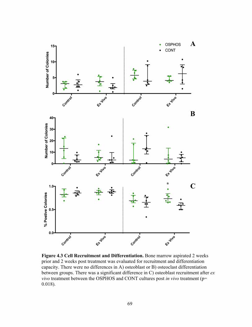
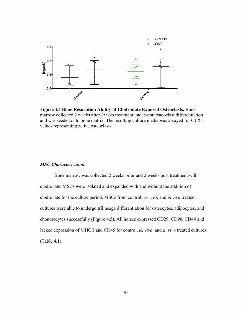
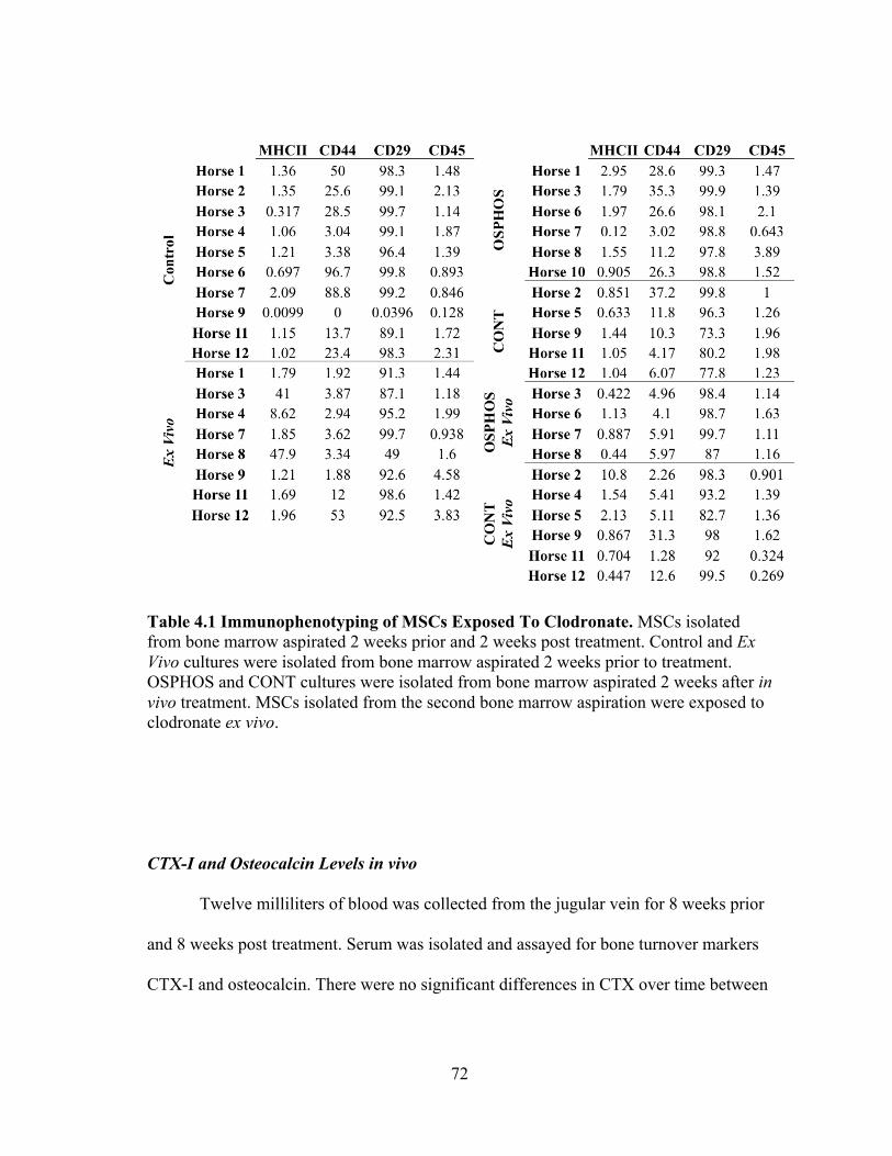
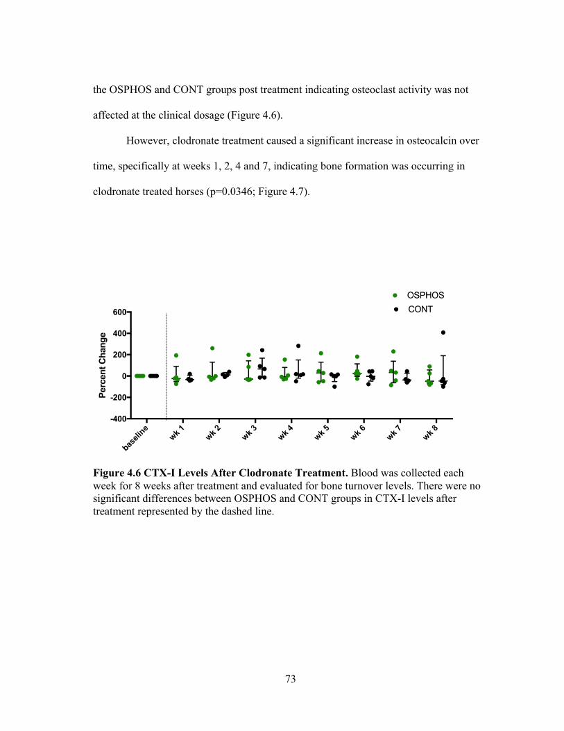
Figure 2.1 Trilineage Differentiation of Bone Marrow Derived MSCs. MSCs isolated from bone marrow were able to undergo trilineage differentiation. A) Adipogenic differentiated cells stained with Oil Red O; original magnification 20x scale bar 100um, B) osteogenic differentiated cells stained with Alizarin Red; original magnefication 4x scale bar 500um, C) chondrogenic differentiated cells stained with Toludine Blue; original magnification 20x scale bar 100um.
Table 2.1 Cell Surface Marker Expression of Bone Marrow Derived MSCs. MSCs isolated from bone marrow were immunophenotyped for known MSC related cell surface markers.
17
Table 2.2 Colony Forming Units Were Variable Between Individuals. Colony forming units of cells incubated for 4, 8, 18 and 24 hours at 0, 2.5, 5, 7.5 and 10 mg/mL produced high variability with large interquartile ranges (IQR).
18
Figure 2.2 Time Dependent Response of Colony Forming Units Between Individuals. Colony forming units of cells incubated for 4, 8, 18 and 24 hours at 0, 2.5, 5, 7.5 and 10 mg/mL produced high variability with an increase in number of colonies formed as incubation time increased.
Population doubling time was calculated based on incubation periods and
concentrations with shorter incubations of 4 hours and lower concentrations of
2.5mg/mL producing the shortest doubling times (Table 2.3).
Horse 1 19F-TR labeled MSCs were detectable using fluorescent imaging,
indicating cell association of the nanoparticle in a dose dependent manner (Figure 2.3).
Subsequent horses labeled with 19F-TR either did not produce signal under fluorescent
imaging or produced signal that appeared to be aggregated nanoparticle extracellular
(Figure 2.4). To test whether the lack of signal was due to the sensitivity of our
19
microscope, we used a microscope with better sensitivity and resolution. As the new
microscope lacked brightfield imaging, DAPI a nuclear stain was used to locate MSCs
and the presence of 19F-TR was assessed. Fluorescent signal of 19F-TR was not
detected using the new equipment (data not shown).
Table 2.3 Population Doubling Times of 19F Labeled MSCs. Populations doubling time in days of 19F labeled MSCs were variable between individuals.
20
Figure 2.3 Dose Dependent Relationship of 19F Labeled MSCs on Brightfield and Fluorescent Imaging. MSCs of Horse 1 were labeled with 19F for 18 hours and evaluated on A) brightfield and B) fluorescent imgaging; original magnification 10x scale bar 200 um.
Figure 2.4 Aggregation of 19F Nanoparticle Complicates MSC Labeling. Representative image of Horse 9 labeled with 19F for 24 hours and evaluated on A) brightfield and B) fluorescent imaging; original magnification 20x scale bar 100 um.
21
Two horses were labeled with 19F-FITC to allow for flow cytometry analysis of
fluoroscent signal. No singal was detected under fluorescent imaging of 19F-FITC
lableled cells. A dose dependent cell association was seen in 19F-FITC labeled cells
using flow cytometric analysis in the first horse (Figure 2.5). However the dose
dependent relationship was not repeated in the second horse indicating that individual
differences between donors exist (Figure 2.6). Either the labeling process was affecting
the fluorophore or the nanoparticle was being exocytosed by the cells.
We thought it was possible that the poor label uptake indicated by appearance (or
lack thereof) of the fluorophore conjugate was due to loss of the conjugate. If this was
happening, it was possible that there was adequate 19F label in the cells. Fluorine NMR
was used to assess the 19F nanoparticle independent of the fluorophore (TR or FITC).
Fluorine was present in 8 hour incubations at higher concentrations and in all
concentrations of 24 hour incubated MSCs indicating the nanoparticle was associated
with the cells in a dose and time dependent manner (Figure 2.7).
22
Figure 2.5 Dose Dependent Relationship of 19F Labeled MSCs Via Flow Cytometry. Horse 5 labled MSCs with 19F for A) 4 hours B) 8 hours and C) 24 hours demonstrated a dose and time dependent relationship. Gray peaks represent unstained cells while red, blue, green and purple peaks represent 2.5, 5, 7.5 and 10 mg/mL, respectively.
Figure 2.6 No Dose Dependent Cell Association of 19F. Horse 7 labeled with 19F for A) 4 hours B) 8 hours C) 18 hours and D) 24 hours did not demonstrate a dose or time dependent cell association. Gray peaks represent unstained cells while navy, green, orange and light blue peaks represent 2.5, 5, 7.5 and 10 mg/mL, respectively.
23
Figure 2.7 Dose Dependent Cell Association of 19F Via Nuclear Magnetic Resonance. Fluorine from 19F nanoparticles produced a signal at -92 ppm only for MSCs labeled for A) 8 hours, 7.5 mg/mL B) 8 hours, 10mg/mL C) 24 hours, 2.5 mg/mL D) 24 hours 5 mg/mL E) 24 hours, 7.5 mg/mL F) 24 hours 10 mg/mL. Fluorine control, Trifluoric Acid, produced a signal at -76 ppm.
DISCUSSION
Our objective was to develop a protocol for labeling equine MSCs with 19F that
would not compromise viability, proliferation, or morphology and allow for
quantification of 19F labeled MSCs in an ex vivo model using our 3Tesla MRI. Inability
to reliably detect the nanoparticle between individuals inhibited quantification of labeled
MSCs ex vivo.
24
Although we sometimes demonstrated a dose dependent amount of fluorescent
label it was inconsistent between horses, which is not what we expected. This may be
due to difficultly in labeling non-phagocytic cells like stem cells, which has been
demonstrated in hNSCs.42, 56, 57 The difference to non-phagocytic cells is because
phagocytic cells, like macrophages, have specialized receptors like mannose and
complement that activate cytoskeleton rearrangements by actin filaments leading to
internalization of products.61 In contrast to phagocytic cells, non-phagocytic cells like
MSCs do not have specialized receptors to enhance phagocytosis and do not readily
endocytose products. Endocytosis of nanoparticles by MSCs endocytosis of
nanoparticles has been aided through clathrin-mediated endocytosis where products are
packed into clathrin-coasted vesicles.62 It is possible insufficient label uptake was due to
lack of activation of clathrin recruitment to the plasma membrane by the 19F label
coating. Another possibility is that MSCs from specific horses did not adequately take
up the nanoparticle because of continuous exhaustion, which has been demonstrated in
MSCs loaded with nanoparticles leading to cell cycle arrest and inability to continue
uptake of nanoparticle.63
We used NMR to quantify 19F association with the cells independent of
fluorophore signal. A dose dependent cell association of fluorine was seen in 19F
labeled cells from all tested horses. Therefore we thought it was possible that lack of
signal on fluorescent imaging was due to loss or changes of the conjugate rather than
insufficient uptake of the nanoparticle. However, the possibility still exists that, NMR
detected nanoparticle attached to MSCs extracellularly.
25
Exocytosis of the nanoparticle is another possible reason for failure when
developing labeling methods with internalized labels. MSCs have demonstrated
relatively quick endocytosis and exocytosis process with material found in the
extracellular matrix as soon as 24 to 48 hours after uptake.63 Varying rates of exocytosis
between individuals could explain the dose dependent relationship seen by in some
assays that was not repeatable between individuals. This is similar to reports
demonstrating lack of specificity of tracking with 19F nanoparticles because of
exocytosis or cellular death of 19F labeled cells in vivo resulting in 19F release to the
extracellular environment. In one report of a stroke model in rats, nineteen percent of
cells containing the 19F nanoparticle were host cells and not the transplanted 19F
labeled hNSCs.42 Another study reported 19F signal was identified in macrophages
rather than 19F labeled MSCs when evaluated under fluorescent imaging.57
Identification of engraftment location or duration of engrafted cells may not be accurate
in vivo if 19F is exocytosed and endocytosed by a macrophage or other cell type.
A limitation to our study was the inability to expand cultures to a number that
would allow for characterization of 19F labeled MSCs. However, previous reports have
demonstrated 19F does not have an effect of MSC ability to undergo differentiation.57
CONCLUSIONS
Cell tracking with a flurocarbon nanoparticle has the potential to allow for
longitudinal identification of cell engraftment location and duration without disruption to
tissue healing or disrupted identification of tissue healing. However further research and
26
development of 109F is required prior to use for tracking of equine bmMSCs. Improved
reliability of label uptake and/or maintenance of label within cells to be tracked is
required. A mechanism to improve endocytosis could be coating activating clathrin
recruitment to the plasma membrane. A mechanism to to prevent exocytosis of the label
would prevent non-specific tracking of resident phagocytic cells. Identification of
engraftment, quantity and duration of MSCs post injection will increase the
understanding of the cells mechanism of injection in the lesion.
27
CHAPTER III
CRYOPRESERVATION OF EQUINE MESENCHYMAL STEM CELLS IN 95%
AUTOLOGOUS SERUM AND 5% DMSO DOES NOT ALTER POST – THAW
GROWTH OR MORPHOLOGY IN VITRO COMPARED TO FETAL BOVINE
SERUM OR ALLOGENEIC SERUM AT 20 OR 95% AND DMSO AT 10 OR 5%*
INTRODUCTION
The equine athlete is a well-accepted model for stem cell therapies in
musculoskeletal injury.33 This is because the horse suffers from naturally occurring
superficial digital flexor tendon injury that is similar to humans and culture derived and
expanded MSCs are being used to treat these injuries.34 Use of clinical practices in
equine cellular therapies that are acceptable in human medicine would be beneficial to
help ascertain the value of stem cell therapy for tendon injury in this naturally occurring
large animal model.
The ideal stem cell preparation, whether frozen or fresh, is an ongoing debate in
medicine.64-67 Cryopreserved MSCs are used in approximately 35% of published MSC
* This work has been adapted from the original article " Cryopreservation of equinemesenchymal stem cells in 95% autologous serum and 5% DMSO does not alter post – thaw growth or morphology in vitro compared to fetal bovine serum or allogeneic serum at 20 or 95% and DMSO at 10 or 5%" by Mitchell A, Atwell K, Smith R, Watts AE. Stem Cell Research and Therapy 2015, 6:231 (doi:10.1186/s13287-015-0230-y; https://stemcellres.biomedcentral.com/articles/10.1186/s13287-015-0230-y). The original article is an open access article distributed under the terms of the Creative Commons Attribution License (http://creativecommons.org/licenses/by/2.0), which permits unrestricted use, distribution, and reproduction in any medium, provided the original work is properly cited.60
28
clinical trials.30 However in veterinary medicine the majority of laboratories preparing
MSCs for horses throughout the world do so with fresh cells.68 This is not to state that
MSCs have not been previously frozen, but that immediately prior to implantation in the
patient, the MSCs are in monolayer culture and prepared for injection immediately prior
to clinical use with transport to the animal site in cooled media. Identification of a
cryopreservation medium that allows for immediate clinical use of MSCs post-thaw
would be beneficial to streamline laboratory and clinical procedures and reduce
associated costs. It is also possible that the cryopreservation process itself induces cell
selection of ‘stronger’ MSCs or induces greater MSC activity and expansion potential,
which could translate to improved stem cell efficacy.69
Because of the potential benefits of using cryopreserved MSCs, and the use of
cryopreserved MSCs in human clinical trials, cryopreserved MSCs should be
investigated in the treatment of naturally occurring tendon injury in horses. The first step
in using cryopreserved MSCs in equine veterinary patient is to identify the ideal medium
for cryopreservation. To do this, the effect on short-term viability and growth of MSCs
post-thaw must be understood.70 Our objective was to determine if a clinically
acceptable formulation and serum source for short-term cryopreservation of equine bone
marrow derived mesenchymal stem cells would preserve normal viability, morphology
and normal growth kinetics post-thaw. Six different freezing solutions were tested with
differing serum supplementation sources and concentrations of dimethyl sulfoxide
(DMSO). Different DMSO formulations were tested to determine if a low concentration
of DMSO was sufficient to preserve viability and growth of MSCs frozen in a slow-
29
freezing method. Different serum sources were tested to determine if an autologous
serum source was sufficient to preserve viability and growth. We hypothesized there
would be no differences in the post-thaw viability, morphology, cell growth kinetics in
MSCs cryopreserved in autologous, allogeneic or xenogeneic mediums or with different
concentrations of DMSO.
MATERIALS AND METHODS
Bone Marrow Derived MSC Isolation, Expansion, and Cryopreservation
All animal procedures were approved by the institution’s animal care and use
committee (IACUC 2012-079). No horses were euthanized for this study. Bone marrow
derived MSCs were isolated from 9 healthy horses ranging in age from 5 to 16 as
previously described. 71 Briefly, bone marrow was collected from mildly sedated horses
into heparinized syringes for a final concentration of 5,000 units of heparin per 30ml of
marrow. Red blood cell lysis was performed with ammonium chloride (7.7mg/mL
NH4CL; 2.06mg/mL Hydroxymethane-aminomethane; pH 7.2). 71 The remaining
nucleated cellular portion of the marrow was plated at 175microL of original raw
marrow volume per cm2 (Corning, Corning, NY) and maintained at 37ºC, 5% CO2 in
humidified air. Culture medium (Dulbecco’s modified Eagle’s medium 1g/L glucose
MO), 20nM/L dexamethasone, and 50 ug/mL L-ascorbic acid). Plates were stained with
2% alizarin red (Sigma Aldrich, St. Louis, MO).
Thawing
After storage in liquid nitrogen for 2-5 days, vials were thawed with gentle
agitation at 35ºC in a water bath until an ice ball was no longer present. Immediately
post thaw, an equal volume of DPBS was added to the cell suspension. Five minutes
later, the cell suspension was collected and added drop-wise to 20mL of DPBS (Lonza,
Walkersville, MD). This thawing method was defined in a pilot project to this
experiment where we determined the importance of avoiding osmotic shock in the 95/5
formulations (Figure 3.1). A 100 uL aliquot of thawed cell suspension in DPBS was
used for a total cell count and viability using fluorescein diacetate (67.57 mg/mL) and
34
propidium iodide (1.35mg/mL) in DPBS. Counting cell suspensions were plated on a
Nebauer hemocytometer and visualized by fluorescence microscopy (Olympus, Center
Valley, PA). The live (green) and dead (red) cells were counted. A total of 10 squares at
10x magnification were counted per sample. The cell suspension was carefully mixed by
pipetting and then centrifuged to pellet the cells for removal of freezing solutions (300G,
5 minutes, 4ºC, 7 brake).
Figure 3.1 Pilot Project Percentage of Viable Cells Post Thaw. Percentage of viable cells post-thaw of MSCs from 9 horses cryopreserved in 6 different solutions from our pilot project (median, quartile). In the pilot project, there was a minor variation in the thawing process. The viable MSCs were significantly lower when MSCs were frozen in 95/5 Allo and 95/5 Auto solutions.
35
Post Thaw Cell Staining with CellTrace™Label
Cell TraceTM violet dye was used to determine the speed of cellular proliferation.
CellTrace™ violet dye binds to intracellular amines without interfering with cellular
activity. As the cell divides, the dye is distributed equally between the 2 daughter cells,
resulting in dye dilution that reflects the number of cell divisions that have occurred
since labeling with CellTraceTM.77, 78 The amount of dye dilution in each cell and thus
the number of cell divisions that have occurred, or the generation of that cell, is
determined using flow cytometry. A cell that has dye dilution reflecting 2 cellular
divisions is a second-generation cell and so on. Post centrifugation, the supernatant was
removed and pelleted MSCs were resuspended in 1 mL DPBS to be labeled with
CellTrace™ violet dye, as per manufacturer instructions. Briefly, cells were labeled in
suspension by adding 1microL of staining solution to 1mL of DPBS containing ≤10x106
cells. CellTrace™ violet dye and MSCs were incubated for 20 minutes at room
temperature in the dark with gentle agitation every 3 minutes. Complete media was
added at 5 times the staining volume and incubated at room temperature in the dark for
an additional 5 minutes. The suspension was centrifuged, resuspended in complete
media at the same volume and incubated for another 10 minutes at room temperature in
the dark, with agitation every 3 minutes. Stained MSCs were then treated as described
below for post-thaw monolayer culture of MSCs.
36
Post-Thaw Monolayer Culture of MSCs
Following labeling, MSCs from each condition were seeded at 10,000 viable
MSCs/cm2 to tissue culture flasks (Corning, Corning, NY) to evaluate growth kinetics,
viability and morphology and 1,000 viable MSCs were seeded to a 10 cm plate for
colony-forming-unit assay. Cultures were maintained as outlined above prior to
cryopreservation.
Monolayer cultures of MSCs were visualized by microscopy and photographed
(Olympus, Center Valley, PA) using commercially available software (cellSens,
Olympus, Center Valley, PA). Each monolayer culture was given a morphology score of
excellent (cells were spindle shaped), good (cells were wider or more star shaped), fair
(cells were flattened and/or contained large vacuoles) or poor (cells were flattened,
vacuolar and foamy in appearance) and a debris score of none (<5 floating cells per 40x
field), mild (<20 floating cells per 40x field floating cells), moderate (<40floating cells
per 40x field), or severe (≥40 floating cells per 40x field) by an investigator blinded to
treatment group assignment (Table 3.1).
One week after seeding the 10 cm plates, colonies were stained with 3% crystal
violet (Sigma Aldrich, St. Louis, MO) and colonies were manually counted without
magnification. The evaluator was masked to treatment group assignment.
37
Table 3.1 Morphology and Debris Scoring Rubric for MSC Cultures. MSC cultures were scored as excellent, good, fair, poor or none, mild, moderate, severe for morphology and debris, respectively.
Cell Generation Assay
After 24 and 72 hours of monolayer culture post-thaw, cells were detached and
collected by addition of trypsinEDTA. Total cell number was determined. Cells were
co-labeled with propidium iodide and flow cytometry was used to assess the
concentration of remaining CellTraceTM cytoplasmic dye. Approximately 12,000 to
35,000 events were collected per condition. The cell generation with the greatest
concentration of cytoplasmic dye at 24 hours post-thaw was defined as the parent
generation. ModFit LT software program was used to determine division rates of the
38
MSCs (Verity Software House, Topsham, ME). Results were reported as the current cell
generation and the proportion of cells in that generation.
Statistical Analysis
Raw data were imported to a commercial statistical software program (Statistix
9, Analytical Software, Tallahassee, FL). Differences between the conditions for
continuous data were evaluated by 1-way-ANOVA with Tukey’s post hoc tests and by
Kruskal-Wallis ANOVA with pairwise comparisons as appropriate for data structure.
Differences in paired data within a group were evaluated by Wilcoxon-signed rank test.
Differences were considered significant when p≤0.05.
RESULTS
No differences were found in the post thaw viability, morphology and growth
kinetics of previously frozen MSCs with each of the tested solutions. Bone marrow was
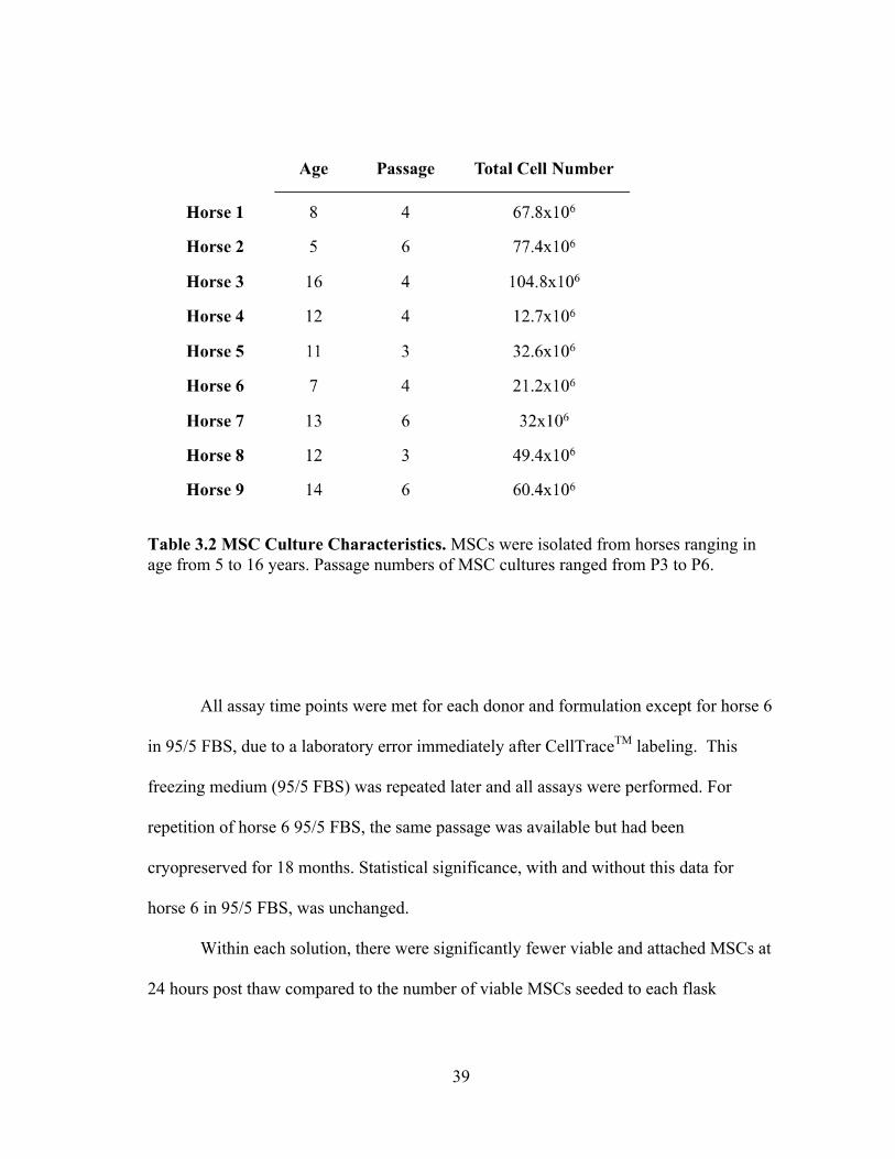
collected from 9 mixed breed mares aged 5-16 years. Passage number of MSCs ranged
from P3 to P6 (Table 3.2). Differences in passage number were due to the need for
different total numbers of MSCs from each horse for other experiments not outlined in
this manuscript.
39
Table 3.2 MSC Culture Characteristics. MSCs were isolated from horses ranging in age from 5 to 16 years. Passage numbers of MSC cultures ranged from P3 to P6.
All assay time points were met for each donor and formulation except for horse 6
in 95/5 FBS, due to a laboratory error immediately after CellTraceTM labeling. This
freezing medium (95/5 FBS) was repeated later and all assays were performed. For
repetition of horse 6 95/5 FBS, the same passage was available but had been
cryopreserved for 18 months. Statistical significance, with and without this data for
horse 6 in 95/5 FBS, was unchanged.
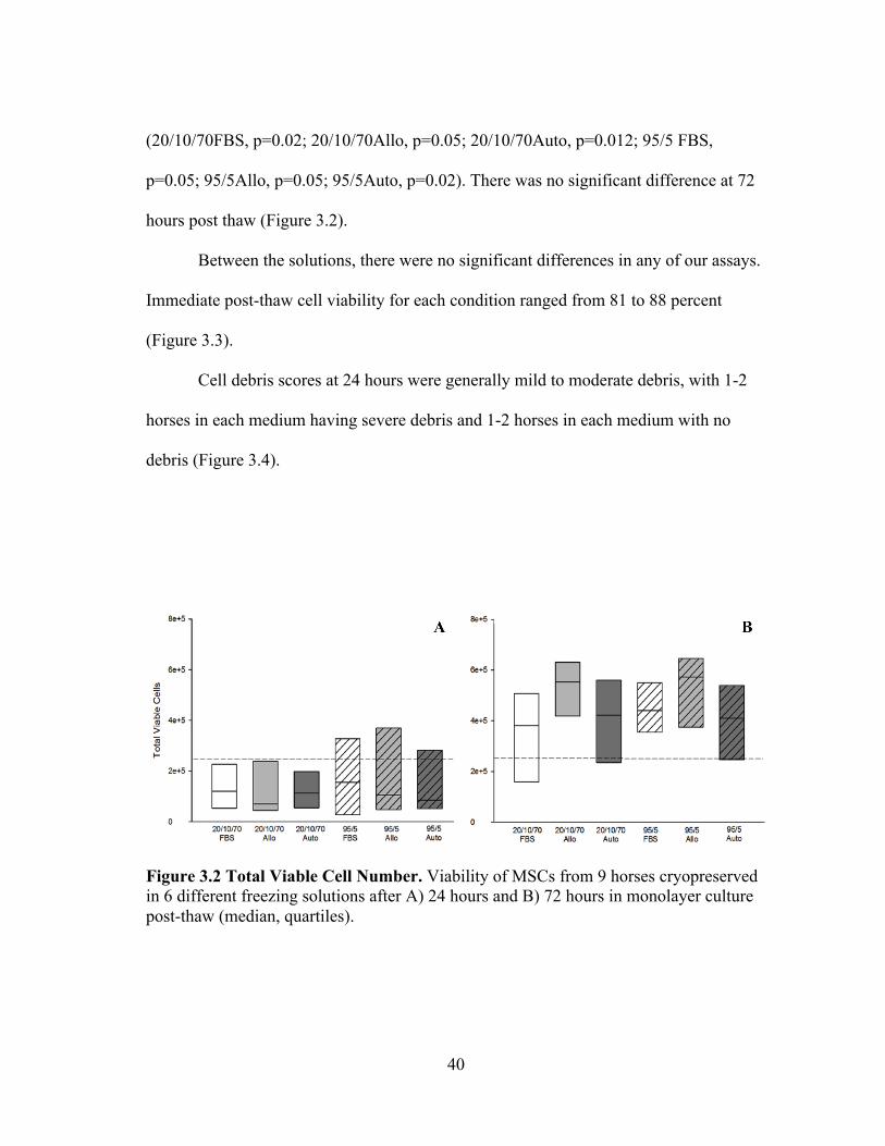
Within each solution, there were significantly fewer viable and attached MSCs at
24 hours post thaw compared to the number of viable MSCs seeded to each flask
p=0.05; 95/5Allo, p=0.05; 95/5Auto, p=0.02). There was no significant difference at 72
hours post thaw (Figure 3.2).
Between the solutions, there were no significant differences in any of our assays.
Immediate post-thaw cell viability for each condition ranged from 81 to 88 percent
(Figure 3.3).
Cell debris scores at 24 hours were generally mild to moderate debris, with 1-2
horses in each medium having severe debris and 1-2 horses in each medium with no
debris (Figure 3.4).
Figure 3.2 Total Viable Cell Number. Viability of MSCs from 9 horses cryopreserved in 6 different freezing solutions after A) 24 hours and B) 72 hours in monolayer culture post-thaw (median, quartiles).
41
Figure 3.3 Percentage of Viable Cell Post-Thaw. Viability immediately post-thaw of MSCs from 9 horses cryopreserved in 6 different solutions.
42
Figure 3.4 Debris and Morphology Scores. Frequency of A), B) debris and C), D) morphology scores of MSCs from 9 horses cryopreserved in 6 different solutions in monolayer cultures at A), C) 24 hours and B), D) 72 hours.
Mesenchymal stem cells receiving severe debris scores were from the same 2
individual donors. Horse 1 MSCs received a debris score of severe for all 6 freezing
solutions and the total viable MSCs at 24 hours were extremely low, ranging from
25,000 to 55,000 viable MSCs. Mesenchymal stem cells from 1 other individual donor
(horse 6) received severe debris scores in 2 formulations, 20/10/70FBS and 95/5Auto,
and scores of mild and moderate for all other formulations. The cell counts at 24 hours
were also very low for these formulations (145,000 and 45,000). In contrast to scores at
24 hours, cell debris scores at 72 hours were generally none to mild (Figure 3.4).
43
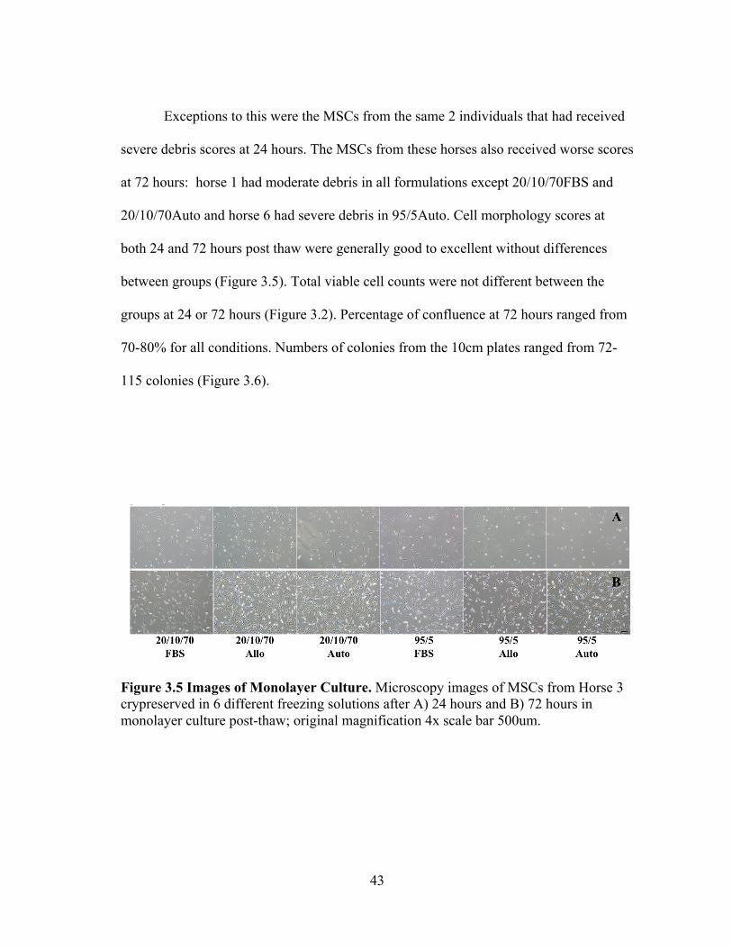
Exceptions to this were the MSCs from the same 2 individuals that had received
severe debris scores at 24 hours. The MSCs from these horses also received worse scores
at 72 hours: horse 1 had moderate debris in all formulations except 20/10/70FBS and
20/10/70Auto and horse 6 had severe debris in 95/5Auto. Cell morphology scores at
both 24 and 72 hours post thaw were generally good to excellent without differences
between groups (Figure 3.5). Total viable cell counts were not different between the
groups at 24 or 72 hours (Figure 3.2). Percentage of confluence at 72 hours ranged from
70-80% for all conditions. Numbers of colonies from the 10cm plates ranged from 72-
115 colonies (Figure 3.6).
Figure 3.5 Images of Monolayer Culture. Microscopy images of MSCs from Horse 3 crypreserved in 6 different freezing solutions after A) 24 hours and B) 72 hours in monolayer culture post-thaw; original magnification 4x scale bar 500um.
44
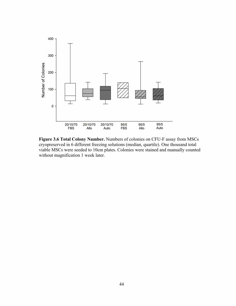
Figure 3.6 Total Colony Number. Numbers of colonies on CFU-F assay from MSCs cryopreserved in 6 different freezing solutions (median, quartile). One thousand total viable MSCs were seeded to 10cm plates. Colonies were stained and manually counted without magnification 1 week later.
45
At 24 hours post-thaw, the majority (mean; standard deviation) of MSCs
remained in their parent generation: 20/10/70 FBS (98.4%; 3.15), 20/10/70 Allo (98.2%;
2.36), 20/10/70 Auto (99.5%; 0.44), 95/5 FBS (98.8%; 1.48), 95/5 Allo (98.6%; 1.92),
and 95/5 Auto (98%; 2.96; Figure 3.7). At 72 hours post-thaw, the majority (mean;
standard deviation) of MSCs were in the fourth generation: 20/10/70 FBS (54.9%;
10.25), 20/10/70 Allo (55.1%; 7.94), 20/10/70 Auto (57%; 10.32), 95/5 FBS (51.5%;
15.17), 95/5 Allo (54.2%; 12.98), 95/5 Auto (59.1%; 11.78) (Figure 3.7).
When the number of MSCs contributing to the total cell number at 72 hours was
calculated, based upon the mean proportion of cells in each generation at 72 hours, it
was lower than the cell count at 24 hours (20/10/70 FBS, 65,372; 20/10/70 Allo, 95,865;
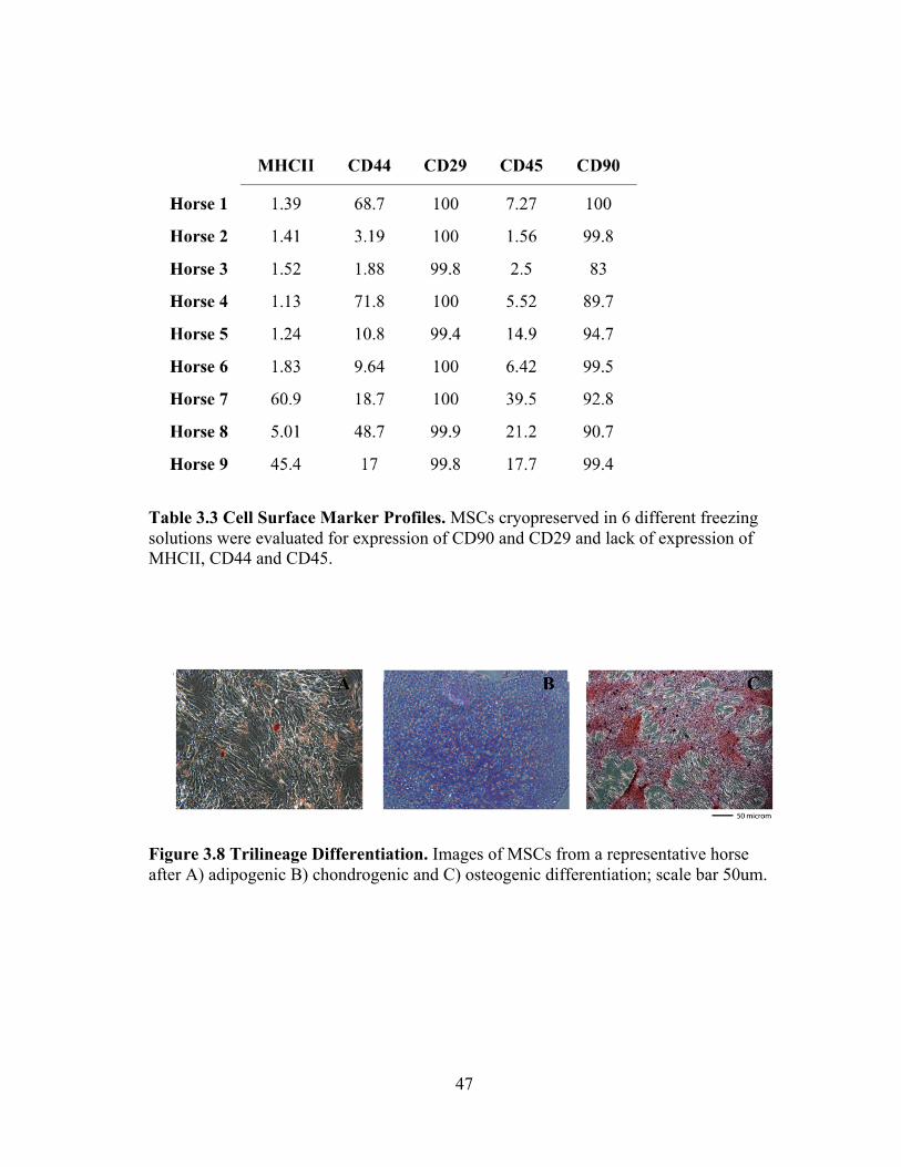
Mesenchymal stem cells for each horse were immunophenotyed (Table 3.3). The
majority of horses were negative for MHCII, CD44, CD45RB and positive for CD29 and
CD90. Eight of 9 horses underwent trilineage differentiation and were positive for
osteogenic, chondrogenic and osteogenic differentiation (Figure 3.8).
46
Figure 3.7 Cell Generations Post-Thaw. Percentage of MSCs cryopreserved in 6 different solutions in generations 1-5 at A) 24 hours and B) 72 hours post-thaw and monolayer culture (mean).
47
Table 3.3 Cell Surface Marker Profiles. MSCs cryopreserved in 6 different freezing solutions were evaluated for expression of CD90 and CD29 and lack of expression of MHCII, CD44 and CD45.
Figure 3.8 Trilineage Differentiation. Images of MSCs from a representative horse after A) adipogenic B) chondrogenic and C) osteogenic differentiation; scale bar 50um.
48
DISCUSSION
We sought to identify whether a clinically acceptable medium for short-term
cryopreservation of equine bone marrow-derived MSCs would preserve normal post-
thaw viability and growth. Mesenchymal stem cells from 9 middle-aged adult horses at a
broad range of passage numbers were utilized to best mimic the clinical scenario of
autologous MSC therapy where differing total numbers of MSCs might be required due
to differences in tendon lesion size and severity. Varying concentrations of autologous
serum, pooled equine serum or FBS; 2 concentrations of DMSO; and the presence or
absence of a cell culture media were tested. Standard immediate and longer-term post-
thaw viability assessments included total live and dead analysis, CFU-F assay and
assessment of MSC morphology and cellular debris. A more novel analysis we used to
assess growth was to stain MSC cytoplasm in a way that would not interfere with
cellular activity and could be accurately measured by flow cytometry, giving us the
number and frequency of cellular divisions for single cells. 79 Analysis of remaining
cytoplasmic dye 24 and 72 hours after staining, allowed evaluation of growth kinetics of
MSCs from each cryopreservation medium, and in combination with total cell numbers
and culture scoring, enabled indirect assessment of post-thaw apoptosis induction.
One of the benefits of studying stem cell therapies in the horse is that the horse
population, like that of man, is not homogenous in genotype or phenotype as most
laboratory species are. In addition to genotype and phenotype differences, individual
variation in MSC characteristics, especially in species with diversity, has been
reported.31, 32 It is important to assess MSCs in models that more accurately reflect the
49
inherent variability among human MSC preparations. Utilizing a greater number of
individuals in MSC experiments better reflects responses from a diverse population.
Using MSCs from 9 individual donors we found no differences between any of the
freezing medium formulations in the post-thaw viability or early growth and morphology
of MSCs by any of our assay methods. However, when we looked at individual horses,
there were marked differences in cell expansion between the media solutions 72 hours
post-thaw in a few of the horses. For example, 37% of MSCs from horse 4 frozen in
20/10/70Allo were in generation 5, while the other 5 freezing solutions were much
lower, ranging from 12-31% of the MSC population in generation 5. As a contrasting
example, only 0.5% of MSCs from horse 6 frozen in 20/10/70Allo had reached
generation 5, while the other 5 freezing mediums had much higher percentages of MSCs
in generation 5, ranging from 6-62%. Had we included one of these horses in a smaller
group size, we might have erroneously identified differences between the formulations.
The media formulations we tested were either 20% serum, 10% DMSO and 70%
cell culture media or 95% serum and 5% DMSO. The 20/10/70 formulation was elected
as the standard cryopreservation medium formulation used in cell culture for many cell
types. Within this group, our question was whether use of xenogen-free serum sources
was possible. The 95/5 formulation that has been recently reported was elected to answer
2 questions.80 Can an almost entirely autologous product (95%) and a reduced DMSO
concentration be used? The lack of deleterious effects when an autologous product was
used with a low concentration DMSO could move cryopreserved MSCs closer to an off-
the-shelf product and would also streamline preparation of autologous MSCs.
50
Culture and cryopreservation of MSCs in FBS has been a standard technique for
many years. Because of a desire to move toward an entirely xenogen-free product in
stem cell therapies, 2 equine serum sources were tested. Based upon other work in our
lab (data not shown) and that of others, we think there are individual differences in the
quality of serum for the growth of MSCs.30, 81 Because of these potential variations in
serum quality between individual horses, autologous serum and a commercially
available pooled equine serum were tested. If individual serum quality differences exist,
it does not appear to negatively affect the post-thaw viability and growth of MSCs
frozen in autologous serum at either concentration we report here. Therefore, either the
commercially available equine serum or autologous serum can be used for short-term
xenogen-free MSC cryopreservation. An entirely autologous product versus an
allogeneic, xenogen-free product would be desirable to minimize many more risks, both
known and unknown.
Despite being cytotoxic and potentially toxic to the patient who will receive the
cells, 10% DMSO is the most commonly used cryoprotectant agent with or without cell
washing for DMSO removal prior to cell infusion to patients.67, 82, 83 Because of its
cytotoxicity and varying reports of the effectiveness of lower DMSO concentrations in
human cell cryopreservation, 5% DMSO was also tested.84, 85 A lower DMSO
concentration, if effective, might minimize toxic effects that occur prior to freezing and
in the immediate post-thaw period when MSCs are in the cryopreservation medium.
Based upon our results, 5% DMSO is sufficient as a cryoprotectant for short-term MSC
cryopreservation. Using this lower concentration of DMSO would be especially
51
important if a post-thaw rinse of MSCs was delayed or avoided altogether prior to
clinical application.
Lack of differences among the cryopreservation mediums we tested is in stark
contrast to results of a pilot project in our lab. In the pilot project, the same 6 freezing
solutions and serum sources on MSCs from 6 middle-aged horses were tested, but we
utilized a very minor variation in the thawing process. The difference in the thawing
method was that post-thaw MSCs were slowly transferred in a drop-wise manner to a
large volume (20mL) of DPBS, as has been previously reported, rather than the step-
wise introduction to DPBS over 5 minutes we report here.68 This minor difference in
methods resulted in profound deleterious effects of cryopreservation mediums consisting
of 95% serum of both equine types with post-thaw viabilities of less than 60% (Figure
3.1). Susceptibility of all cell types to post-thaw osmotic shock is well known and
enhanced susceptibility has been suggested in human MSCs. 69, 86 Absence of balanced
isotonic solution and/or a lower concentration of cryoprotectant in our 95/5 formulation
both could have led to increased susceptibility to osmotic shock. Regardless, it appeared
in our pilot project, that the use of 95% FBS was somewhat protective of the enhanced
susceptibility to osmotic shock in the 95/5 formulation compared to either equine serum
source. In the experiment of this report, careful handling of MSCs to reduce osmotic
shock resulted in no differences among the 95/5 or 20/10/70 formulations. The
importance of MSC handling immediately after the thawing process should be
underscored.
52
First reported in 2011, post-thaw growth arrest of MSCs followed by a very rapid
proliferation rate of surviving MSCs was seen in our study.69 As originally suggested,
this might be selection of ‘better’ MSCs with a younger phenotype and faster
proliferation rate while inducing apoptosis of the ‘less strong’ MSCs post-thaw. Our
study demonstrated a lack of MSC division of the plastic adherent population in the first
24 hours with >95% of viable MSCs still in the defined parent generation, and a greater
number of non-adherent cells in the first 24 hours post-thaw reflected by the higher
debris scores at 24 hours and lower total cell count of adherent MSCs after 24 hours of
culture than the number of MSCs seeded for all groups. These floating cells were likely
apoptotic MSCs, rather than surviving but dysfunctional cells because much higher
numbers and monolayer densities would have occurred at 72 hours had the floating cells
recovered function after 24 hours. Additionally, the CFU-F assay colony number was
lower when debris scores at 24 hours were high (Figure 3.9).
53
Figure 3.9 Debris Score vs Colony Forming Units. Total number of colony forming units plotted against debris scores from MSCs cryopreserved in 6 different solutions and maintained in monolayer for 24 hours post-thaw.
This growth arrest seemed to recover between 24 and 72 hours, with the majority
of viable MSCs in the fourth generation, 48 hours later. However, we think there was
incomplete recovery with continued apoptosis in a portion of MSCs because the total
viable cell number at 72 hours was significantly lower than one would expect given our
cellular generation data. An assay of apoptosis would have been helpful to prove that
apoptosis occurred. Finally, although direct comparisons to growth of MSCs from the
same donors that had not been frozen were not made, our impression is that the growth
during the 72 hours post-thaw was much greater than we see during routine monolayer
expansion of fresh MSCs. This is in contrast to a recent report where post-thaw MSC
54
growth was not different to suspension stored MSCs where there was a steady
proliferation rate for 4 days.68
A limitation of our study was that cell surface markers, commonly used to
characterize the phenotype of MSCs, and tri-lineage differentiation potential in vitro,
were not assayed post thawing. These analyses were not performed for 2 main reasons.
First, others have reported lack of changes in cell surface markers in fresh versus post-
thaw human and porcine bone marrow-derived MSCs and that cryopreservation does not
change differentiation ability. Second, others have reported lack of changes in cell
surface marker profile due to serum type (autologous serum versus FBS). Therefore, we
thought the minimal exposure to different mediums during freezing and thawing was
unlikely to alter the cell surface marker expression or in vitro differentiation potential, so
long as viability and growth were unchanged.
Another step that is important to note in our design, is that DMSO was removed
from MSCs post-thaw with a post-thaw wash by centrifugation. In the clinical setting, if
one were to use any of our tested conditions immediately post-thaw, a post-thaw wash &
centrifugation step would be required if removal of DMSO was desired. This washing
step would require laboratory involvement in the clinical procedure, somewhat limiting
their off-the-shelf availability to the treating clinician.
55
CONCLUSIONS
In conclusion, we evaluated the short-term cryopreservation of equine bone
marrow-derived MSCs in solutions consisting of differing concentration and types of
serum and concentrations of DMSO. A low tech, commercially available freezing
system that would be affordable in veterinary services was used. In this system, equine
MSCs did not have differences in post-thaw viability and growth, regardless of the
cryopreservation formulation or serum source used. The importance of minimizing
osmotic shock of MSCs immediately post-thaw and the potential increased risk of
osmotic shock with different mediums for cryopreservation as found in our pilot project
should be noted. Additionally, immediately post-thaw, there was an apparent lag phase
of MSCs with little cellular division and assumed apoptosis in the first 24 hours post-
thaw, followed by rapid MSC growth over the next 48 hours. If a xenogen-free product
with lower concentration of cryoprotectant is clinically desirable to streamline clinical
and laboratory procedures by use of cryopreserved MSCs, the use of 95% autologous
serum and 5% DMSO for the short-term cryopreservation of equine bone marrow-
derived MSCs is recommended.
56
CHAPTER IV
INVESTIGATING THE IMPACT OF BISPHOSPHONATES, A
MUSCULOSKELETAL THERAPY, ON BONE REMODELING AND BONE
CELLS
INTRODUCTION
Bone is a dynamic tissue primarily composed of Type I collagen mineralized
with hydroxyapatite that must constantly adapt to various amounts of load.87 Osteoblasts
and osteoclasts are the cellular mechanisms that maintain bone homeostasis. Osteoblasts
lay down bone matrix containing mineral and non-collagenous protein before
mineralization, while osteoclasts resorb bone. Both osteoblastic and osteoclastic actions
are important for the remodeling, formation, and maintenance of healthy bone.
MSCs play an important role in bone homeostasis as both osteoblasts progenitors
and osteoclasts support cells. Although MSCs as osteoblast progenitors have recently
been questioned, osteoblasts are thought to be descendants of MSCs while osteoclasts
are differentiated from the hematopoietic lineage.88, 89 An additional role of MSCs is
they are known to support hematopoiesis; specifically in osteoclastogenesis by
producing RANK-L and MCSF stimulate osteoclast differentiation of hematopoietic
lineage cells.21, 22 When an imbalance in bone remodeling occurs a pathogenic state can
form. Osteoporosis is a chronic condition in which both osteoclast and osteoblast activity
is elevated.90 While osteoclast can resorb bone in a few weeks, it takes osteoblast months
to lay down new bone. This imbalance causes a net loss of bone density.90 Different
57
treatments have been developed to manage bone turnover disorders including the use of
bisphosphonate drugs.
Bisphosphonates were first reported in 1968 as a derivative of the pyrophosphate
family. Bisphosphonates differ from pyrophosphates as they contain a carbon ion rather
than oxygen which makes them more stable and resistant to chemical and enzymatic
hydrolysis.91 The first bisphosphonates were used as water softeners to inhibit calcium
carbonate precipitation.92 In medical applications, the high affinity for calcium by
bisphosphonates targets them to bone after administration. Bisphosphonates adhere to
bone and can be incorporated during mineralization. They can be classified into two
categories, non-nitrogen containing and nitrogen containing; with the latter having an
increased binding to hydroxyapatite therefore having a more potent affect.93 Nitrogenous
bisphosphonates cause cell apoptosis by inhibiting the mevalonate pathway, which is
important in many cellular processes leading to apoptosis.94 In contrast, non-nitrogenous
bisphosphonates are known to inhibit cell function by interfering with the mitochondria,
eventually leading to cell apoptosis. The non-nitrogenous equine approved
bisphosphonate clodronate, is metabolized into nonhydrolysable ATP analogs that
accumulate in the mitochondria resulting in cytotoxic effects.95 Osteoclasts contain a
higher number of mitochondria and therefore accumulate a higher number of
nonhydrolysable ATPs causing senescence preventing bone resorption followed by
apoptosis.95 Once released by the apoptotic cell, the non-hydrolysable ATP analogs have
a high affinity for calcium leading to storage in the bone through formation of
58
complexes with hydroxyapatite crystals that can be taken up by osteoclasts at a later
time.96
During osteoclast resorption collagenous and noncollagenous bone proteins are
released into the serum and urine, which can be used as biomarkers of bone
metabolism.97, 98 Two commonly used bone biomarkers are osteocalcin and CTX-I.99
Osteocalcin is a non-collagenous bone protein incorporated into the extracellular bone
matrix by osteoblasts during bone formation; osteocalcin levels reflect the active
osteoblast in bone.100 Osteocalcin levels also reflect active osteoclast in the bone as it is
liberated during resorption.100 CTX-I is a C terminal telopeptide of type I collagen, the
most abundant protein in bone. CTX-I is released during bone resorption by cathepsin K,
an enzyme produced by osteoclasts during bone resorption.97 Osteocalcin and CTX-I are
commonly used to monitor skeletal development, severity of bone disease, and effects of
bisphosphonates.101, 102
Today, bisphosphonates are potential disease modifying osteoarthritic drugs
(DMOAD) primarily used to treat diseases with increased bone resorption and bone
loss.103 To date, there are no DMOADs that have shown efficacy in slowing the
progression of osteoarthritis and all current treatments are simply symptom modifying.
Aside from their anti-resorptive effects, bisphosphonates have demonstrated
chondroprotective, anti-inflammatory and analgesic affects.103 Bisphosphonates have
commonly been utilized in humans to treat metastatic bone pain; however, the pathway
in which they can relieve pain has yet to be elucidated.104 Studies have shown pain relief
properties independent of known anti-resorptive action including anti-inflammatory
59
effects.103, 105-107 Tiludronate and clodronate have demonstrated inhibition of nitric oxide
and pro-inflammatory cytokines in macrophages in vitro as well as tiludronate inhibition
of vascular endothelial growth factor (VEGF) synthesis in osteoblasts.107, 108 Down
regulation of pro-inflammatory cytokines and formation of vessels by inhibition of
VEGF could modulate pain independent of resorption activity.
In humans, nitrogenous bisphosphonates have been used to treat hypercalcemia
and osteoporosis.109 Long-term bisphosphonate treatment can lead to oversuppresion of
bone turnover decreasing the skeleton’s ability to heal properly and reducing bone’s
mechanical properties.110 Rare but severe complications including include atypical
fractures and osteonecrosis of jaw have been reported in patients on long-term
bisphosphonate therapies due to over suppression of bone turnover.110-112 To prevent
over suppression of bone turnover and reduce chances of adverse events, patients are
now taking drug holidays in which they stop bisphosphonate usage for a determined
period of time.113 Yearly exams are recommended for patients on bisphosphonate
therapies evaluate bone turnover makers, like CTX-I and assess the likely hood of an
adverse event occurring. Though duration and length of holidays are specific to each
patient depending on bone mineral density, bone turnover markers like CTX-I and the
time in which the patient has been on the drug; generally this is every 3 to 5 years. 113
Bisphosphonate therapy is continued when bone turnover levels being to elevate to
levels consistent to those before treatment.113
In late 2014, the FDA approved the bisphosphonate drug OSPHOS®, containing
the active non-nitrogenous bisphosphonate clodronate, for the treatment of navicular
60
syndrome in horses. Navicular syndrome is a chronic disease affecting the podotrochlear
apparatus and is considered one of the most common causes of forelimb lameness in the
horse.114, 115 OSPHOS® has become a popular choice for veterinarians treating chronic
lameness issues in the horse due to the off target analgesic effects. Lameness, or limping,
in the horse is a result of musculoskeletal pain.116
Despite its clinical popularity, little research has been done to understand the
effects of clodronate in the horse. Without information on the effects of clodronate on
the bone after treatment at the clinical dosage we must wonder if we are putting our
clients at risk. The occurrence of one rare event like atypical fracture or osteonecrosis of
the jaw in the horse could be detrimental to the horse industry. CTX-I levels have been
evaluated in horses treated with the non-nitrogenous bisphosphonate tiludronate. Horses
treated with tiludronate intravenously once daily for 10 days were compared to horses
treated with a constant rate infusion and evaluated for CTX-I levels were evaluated on
days 1, 3, 6, 10, 20, 30, and 60 after treatment. A significant decrease in CTX-I levels
was noted between day 0 and day 1 and continued through day 3 for both treatment
groups.96 This is similar to CTX-I levels after treatment with bisphosphonates in human
reports.117
In a different study a force plate analysis was used to assess the effects of
tiludronate in horses treated intravenously (IV) compared to those receiving a regional
limb perfusion (RLP). Client owned horses with no previous history of tilduronate
treatment, scored grade 2 or 3 front limb lameness by the AAEP scale, and showed
clinical improvement in lameness after administration of local analgesic to the palmar
61
digital nerves were randomized for treatment selection for the study. After treatment
with tiludronate by either RLP or IV at one dose of 1 mg/kg, lameness by force plate
were evaluated on days 14, 30, 60, 120, and 200. A significant improvement in lameness
was noted on day 120 and 200 in IV treated horses compared to RLP treated horses. 115
Delayed lameness improvement was contributed to pain relief from decreased osteoclast
resorption.
Although tiludronate and clodronate are both non-nitrogenous bisphosphonates,
there are differences between them within the non-nitrogenous group. Clodronate is a
first generation bisphosphonate while tiludronate is a slightly more potent second
generation.118 Bisphosphonate potency is directly related to ability to bind to bone.
Clodronate has demonstrated lower binding affinity (806 µM) to bone compared to
tiludronate (173 µM) in vitro.119
Another difference between the two FDA approved equine bisphosphonates is
that tiludronate is administered IV while clodronate is administered intramuscularly
(IM). IV administration results in a higher peak of drug and a greater amount of drug
that binds bone allowing for rapid uptake of the bisphosphonate by the skeleton while
clodronate administered intramuscularly has decreased uptake by the skeleton.103, 118
There has been no research to date demonstrating efficacy of clodronate in
osteoclast function. In the New Animal Drug application of OSPHOS, a pharmokinetic
and 5x dosing study was conducted along with a field evaluation in which treated horses
were evaluated for lameness on days 28 and 56. Efficacy was demonstrated by a
reduction in lameness only and there was no data reported for the assessment of
62
osteoclast inhibition. A 75% success rate, determined by an improvement of a grade or
more by the American Association of Equine Practitioners (AAEP) scale, was noted in
clodronate treated horses.
Given the complications in human therapies, we believe it is critical to
understand the effects of clodronate in the horse, as atypical fractures could be
detrimental to the industry. Our goal was to determine if clodronate given at the
clinically relevant dose could alter MSC characterization and ultimately bone
remodeling. We hypothesized that there would be no charge in markers of bone
remodeling and no change in osteoclast and osteoblast recruitment and there would be an
improvement in lameness.
MATERIALS AND METHODS
Study Design
All animal procedures were approved by the institution’s animal care and use
committee (IACUC 2016-0122). No horses were euthanized for this study. Twelve
university-owned horses were used for this study. Inclusion criteria include horses on the
western horsemanship equestrian team that were competing during the fall 2016
semester and had not previously received bisphosphonate treatment. The head coach and
the equestrian team veterinarian identified eligible horses. Horses were randomly placed
into either the treatment group receiving 1.4mg/kg clodronate (OSPHOS®; n=6) or an
equivalent volume of LRS as a placebo (CONT; n=6). Patient allocation was controlled
in the pharmacy and remained unknown to all other parties. All horses had 12mls of
63
blood collected from the jugular vein weekly for 8 weeks prior to treatment and 8 weeks
post treatment. Serial clinical evaluations, lameness locator data, and coach
questionnaires of lameness were performed one week prior to treatment and each week
for 3 weeks and at 8 weeks following treatment (see below). Bone marrow was collected
from the sternum at 2 weeks prior to treatment and at 2 weeks post treatment for ex vivo
and in vitro bone marrow cultures (Figure 4.1).
Figure 4.1 Study Design. Horses were evaluated over a 16-week period with treatment at week 0. Each week from week -8 to week 8 blood was collected from the jugular vein. Two weeks prior and 2 weeks post treatment horses bone marrow was aspirated. One week prior and at each week for 3 weeks and at week 8 post treatment lameness was evaluated.
Differentiation & Bone Resorption
Bone marrow aspirate from 2 weeks prior and 2 weeks post treatment underwent
red blood cells lysis with ammonium chloride (7.7mg/mL NH4CL; 2.06mg/mL
64
Hydroxymethane-aminomethane; pH 7.2) before being washed and seeded into 12 well
plates with or without the presence of clodronate. Osteoblast cultures were maintained
for 28 days in α–MEM, 15% FBS, 1X antibiotic/antimycotic, 10nM dexamethasone, 50
uM L-Ascorbic Acid, and 10mM β-GlycerolPhosphate (basal media) followed by
alizarin red staining. Pre-osteoblast cultures were grown for 10 days and stained for
alkaline phosphatase positive cultures. Osteoclast cultures were maintained for 15 days
in basal media and 10-5M 1α, 25-Dihydroxycholecalciferol before TRAP staining.
Culture media was exchanged every 5 days. Colonies from each culture were counted
after the appropriate staining. Bone marrow aspirated post in vivo treatment was
additionally seeded onto an OsteoAssay kit containing bone mineral in basal media with
RANKL and MCSF, with and without 5x10⁻⁵M clodronate. Osteoassay cultures were
maintained for 10 days before TRAP staining. Prior to staining, media was collected and
an ELISA was performed for CTX-I levels.
MSC Characterization
Bone marrow aspirate from 2 weeks prior and 2 weeks post treatment underwent
red blood cells lysis before seeding onto flask with and without the addition of
clodronate. Cultures were maintained in 1g/L DMEM, 10% FBS, 2.5% hepes buffer, and
1X antibiotic/antimycotic until passage 3, or the needed cell number was reached with
media exchanged every 3 days. A passage occurred when the cells are 80% confluent.
At passage 3 MSCs were lifted and cryopreserved in 95% autologous serum and 5%
DMSO until trilineage differentiation and immunophenotyping could be performed.
65
Cryopreserved MSCs were thawed with gentle agitation at 35ºC in a water bath until an
ice ball was no longer present. Immediately post thaw, an equal volume of DPBS was
added to the cell suspension. Five minutes later, the cell suspension was collected and
added drop-wise to 20mL of DPBS (Lonza, Walkersville, MD A 100 microL aliquot of
thawed cell suspension in DPBS was used for a total cell count and viability using
fluorescein diacetate (67.57 mg/mL) and propidium iodide (1.35mg/mL) in DPBS.
Counting cell suspensions were plated on a Nebauer hemocytometer and visualized by
fluorescence microscopy (Olympus, Center Valley, PA). The live (green) and dead (red)
cells were counted. A total of 10 squares were counted per sample. The cell suspension
was carefully mixed by pipetting and then centrifuged to pellet the cells for removal of
performed by seeding MSCs into 10cm plates for adipogenic and osteogenic
differentiation and maintaining MSCs in pellets for chondrogenic differentiation as
previously described. 60 Immunophenotyping for CD44, CD45RB, CD29, CD90 and
MHCII was performed on isolated MSCs using flow cytometry as previously
described.60
CTX-I and Osteocalcin Levels in vivo
Twelve milliliters of blood was collected from the jugular vein using a 12mL
syringe and 18-gauge needle for 8 weeks prior and 8 weeks post treatment. Blood was
placed in red top glass tubes and allowed to clot for a minimum of 30 minutes before
centrifugation at 4000G. Serum was collected and stored in 1mL aliquots at -80°C.
66
Frozen aliquots were thawed and neat samples were used to assay for CTX-I and
osteocalcin using commercially available ELISAs.
Lameness Effects
Lameness exams were performed one week prior to treatment and each week for
3 weeks and at 8 weeks following treatment using 3 forms of evaluation; clinical
evaluations, Lameness Locator®, and coach evaluations, with all 3 forms of evaluation
blinded to the others. At each lameness exam the horse was hand walked in a 10-meter
straight line, hand jogged in a 20-meter straight line twice and hand jogged in two 10-
meter circles to the right and left on a concrete surface. Clinical evaluations of lameness
were scored while horses were jogged on concrete according to the AAEP scale with a
score of 0 being no asymmetry detected under any circumstances and a score of 5 being
the horse was minimally weight bearing or unable to move.120 The Lameness Locator®
is a computer based objective measure of lameness that detects asymmetry (lameness) in
the horse as it moves. Marker devices were place on top of the poll, between the hips and
on the right front pastern and data was collected during the 2 straight-line jogs.
Lameness Locator® data was collected concurrently with the clinical evaluations. Coach
evaluations were conducted after lameness exams. The coaches were asked a series of
questions to evaluate the horse’s work performance based on their observations of the
horse in practice in a sand arena (Figure 4.2).
67
Figure 4.2 Coach Evaluations. Horses performed lameness exams one week prior and each week for 3 weeks and at 8 weeks post treatment. Coaches completed a questionnaire at the time of each lameness evaluation about the horses performance in work that week.
68
Statistical Analysis
Raw data were imported to a commercial statistical software program (Prism,
GraphPad Software, La Jolla, CA). Differences between the conditions for longitudinal
data were evaluated by repeated measures ANOVA. Differences in paired data within a
group were evaluated by Wilcoxon-signed rank test. Coach evaluations were evaluated
using a Chi-Square test. Differences were considered significant when p≤0.05.
RESULTS
Differentiation & Bone Resorption
Bone marrow was collected 2 weeks prior and 2 weeks post treatment with
clodronate. Whole marrow underwent osteoblastogenesis and osteoclastogenesis with
and without the addition of clodronate. There were no significant differences in
osteoblastogenesis in control, ex vivo, or in vivo treated cultures or osteoclastogenesis
(Figure 4.3). However, there was a significant different in osteoblast recruitment for ex
vivo treated OSPHOS cultures after treatment in vivo (p= 0.018; Figure 4.3).
Bone marrow collected post treatment with clodronate was seeded onto bone
fragments with the addition of MCSF and RANK-L for 10 days before being fixed and
TRAP stained. Media was collected from the culture period and assayed for CTX-I.
There were no significant differences in CTX-I between the control, in vivo, and ex vivo
treated groups indicating osteoclast activity was no inhibited by clodronate ex vivo at
5x10⁻⁵M (Figure 4.4).
69
Figure 4.3 Cell Recruitment and Differentiation. Bone marrow aspirated 2 weeks prior and 2 weeks post treatment was evaluated for recruitment and differentiation capacity. There were no differences in A) osteoblast or B) osteoclast differentiation between groups. There was a significant difference in C) osteoblast recruitment after ex vivo treatment between the OSPHOS and CONT cultures post in vivo treatment (p= 0.018).
70
Figure 4.4 Bone Resorption Ability of Clodronate Exposed Osteoclasts. Bone marrow collected 2 weeks after in vivo treatment underwent osteoclast differentiation and was seeded onto bone matrix. The resulting culture media was assayed for CTX-I values representing active osteoclasts.
MSC Characterization
Bone marrow was collected 2 weeks prior and 2 weeks post treatment with
clodronate. MSCs were isolated and expanded with and without the addition of
clodronate for the culture period. MSCs from control, ex vivo, and in vivo treated
cultures were able to undergo trilineage differentiation for osteocytes, adipocytes, and
chondrocytes successfully (Figure 4.5). All horses expressed CD29, CD90, CD44 and
lacked expression of MHCII and CD45 for control, ex vivo, and in vivo treated cultures
(Table 4.1).
71
Figure 4.5 Trilineage Differentiation of MSCs Exposed To Clodronate. MSCs isolated from bone marrow aspirated 2 weeks prior and 2 weeks post treatment. Control and In Vitro cultures were isolated from bone marrow aspirated 2 weeks prior to treatment. In Vivo and Ex Vivo cultures were isolated from bone marrow aspirated 2 weeks after in vivo treatment. Representative images of A) adipocytes, 2) osteoblast and 3) chondrocytes.
72
Table 4.1 Immunophenotyping of MSCs Exposed To Clodronate. MSCs isolated from bone marrow aspirated 2 weeks prior and 2 weeks post treatment. Control and Ex Vivo cultures were isolated from bone marrow aspirated 2 weeks prior to treatment. OSPHOS and CONT cultures were isolated from bone marrow aspirated 2 weeks after in vivo treatment. MSCs isolated from the second bone marrow aspiration were exposed to clodronate ex vivo.
CTX-I and Osteocalcin Levels in vivo
Twelve milliliters of blood was collected from the jugular vein for 8 weeks prior
and 8 weeks post treatment. Serum was isolated and assayed for bone turnover markers
CTX-I and osteocalcin. There were no significant differences in CTX over time between
73
the OSPHOS and CONT groups post treatment indicating osteoclast activity was not
affected at the clinical dosage (Figure 4.6).
However, clodronate treatment caused a significant increase in osteocalcin over
time, specifically at weeks 1, 2, 4 and 7, indicating bone formation was occurring in
clodronate treated horses (p=0.0346; Figure 4.7).
Figure 4.6 CTX-I Levels After Clodronate Treatment. Blood was collected each week for 8 weeks after treatment and evaluated for bone turnover levels. There were no significant differences between OSPHOS and CONT groups in CTX-I levels after treatment represented by the dashed line.
74
Figure 4.7 Osteocalcin Levels After Clodronate Treatment. Blood was collected each week for 8 weeks after treatment and evaluated for bone turnover levels. There was a significant differences between OSPHOS and CONT groups in CTX-I levels after treatment represented by the dashed line at week 1, 2, 4 and 7 (p= 0.0346).
75
Lameness Effects
Lameness exams were performed one week prior to treatment and each week for
3 weeks and at 8 weeks following treatment. When lameness was evaluated objectively
by the Lameness Locator® no differences were found in front limb asymmetry due to
treatment. However a significant difference was found in hind limb asymmetry in
OSPHOS treated horses (p= 0.0252; Figure 4.8). When a clinician evaluated lameness,
no significant differences in for either front or hind limbs over time between the
OSPHOS and CONT groups when evaluated on a hard surface (Figure 4.9).
Interestingly, there was a significant difference in improvement between the
OSPHOS and CONT treated groups when evaluated by the coaches (Figure 4.10). The
coaches were able to successfully identify mild improvements in the horses’
performance on a soft surface. This improvement in performance seen by the coaches
contrasts with clinical lameness exams evaluated by a clinician or the Lameness
Locator®; each horse’s underlying chronic lameness was exacerbated by jogging on a
hard surface possibly masking any mild improvements due to treatment with clodronate.
76
Figure 4.8 Front and Hind Limb Asymmetry. Lameness evaluations were performed using the Lameness Locator® one week prior and each week for 3 weeks and at week 8 post treatment. There were no differences in A) front limb asymmetry after treatment; represented by the dashed line. There was a significant difference in B) hind limb asymmetry between OSPHOS and CONT groups with OSPHOS treated horses having a decrease in lameness (p= 0.0252).
77
Figure 4.9 Front and Hind Limb Lameness. Lameness evaluations were performed by a clinician using the AAEP scale one week prior and each week for 3 weeks and at week 8 post treatment. There were no differences in A) front limb or B) hind limb lameness between OSPHOS and CONT groups after treatment represented by the dashed line.
78
Figure 4.10 Coach Evaluations of Performance. Horse performance was evaluated by the coaches one week prior and each week for 3 weeks and at week 8 post treatment. The coaches identified improvement in 5 or 6 OSPHOS treated horses (p= 0.0395).
DISCUSSION
We sought to determine if clodronate given at the clinically relevant dose could
alter MSC characterization and ultimately bone remodeling in the horse. Twelve
university-owned horses ranging in age from 7 to 18 with a median age of 11 were used
for this study. Weekly blood collections for 8 weeks prior to treatment and 8 weeks post
treatment were used to analyze CTX-I and osteocalcin levels. Bone marrow was
collected from the sternum at 2 weeks prior to treatment and at 2 weeks post treatment
for ex vivo and in vitro bone marrow cultures for osteoblastogenesis, osteoclastogenesis,
79
bone resorption ability, and MSC characterization. Pain was evaluated by serial clinical
evaluations, lameness locator data, and coach questionnaires of lameness one week prior
to treatment and each week for 3 weeks and at 8 weeks following treatment.
CTX-I and osteocalcin are standard bone markers used to evaluate bone turnover
in vivo. From treatment on there was a significant difference in osteocalcin over time
between the OSPHOS and CONT groups post treatment, with the OSPHOS group
having higher levels of osteocalcin at weeks 1, 2, 4 and 7 following treatment. It is
possible osteoblast activity was stimulated in vivo by clodronate, leading to increased
matrix production and increased osteocalcin levels. Clodronate stimulated osteoblast
differentiation has been demonstrated in vitro cultures of osteoblast progenitors and
MSCs. 121 Treatment with clodronate had no or an undetectable effect on CTX-I and a
significant effect on osteocalcin. We believe there are 2 possible reasons for this. First,
we are working with skeletally mature horses that would not be expected to have high
levels of bone turnover. Therefore, there should not be large levels of CTX-I. Second,
the dose administered is likely lower than that necessary to elicit a bone resorption
response.
A reduction in forelimb lameness was seen in horses treated with OSPHOS in the
FDA approval study.122 Seventy five percent of OSPHOS treated horses demonstrated an
improvement in lameness by one or more grade of the AAEP scale 56 days after
treatment.122 Given these marked improvements in visible forelimb lameness we
expected to find similar improvements in lameness in our OSPHOS treated horses.
However, we saw no change in forelimb asymmetry. In contrast, we did see a significant
80
improvement in hindlimb asymmetry in OSPHOS treated horses. We think the
improvement in the hindlimb lameness versus lack of improvement in the forelimb
lameness could be because lameness assessment was made on a hard surface. The
forelimb lameness in our horses was due to foot pain, a condition known to be
exacerbated by a firm surface. We think the firm surface may have decreased the
likelihood of seeing a minor change in lameness. In contrast, hindlimb lameness is rarely
exacerbated to the same degree by a hard surface making it more likely to be able to
detect a mild improvement. No change in front or hind limb lameness was seen in horses
evaluated using by a clinician. We think the lack of change in lameness was due to
using the AAEP scale to score lameness. The AAEP scale defines a grade of 3 covers a
broad range of lameness as defined as “consistently observable at a trot under all
circumstances” covers a broad range of lameness. This means a horse with a clinically
relevant change in lameness from a severe grade 3 to a mild grade 3 would appear to be
unchanged using this scale.
The coach evaluations supported that a clinically relevant improvement in
lameness occurred with significantly more horses receiving the evaluation of
“improved” in the OSPHOS group as compared to CONT. Coaches observed the horses
under work throughout the week while the clinician and the Lameness Locator® only
evaluated lameness at the designated evaluation time points. It is likely the coaches are
more sensitive to changes in the horses’ performance and likely a better form of
evaluation of lameness in the horse. The improvement in lameness seen in this study is
similar to reports of the bisphosphonate tiludronate that produced a decrease in lameness
81
in treated horses. Interestingly, humans treated with intra-articular injections of
clodronate reported a decrease in pain associated with osteoarthritis, a musculoskeletal
injury.123
Analgesic effects of bisphosphonates have been reported in patients suffering
from pain caused by metastatic breast cancer.106 Tiludronate has demonstrated inhibition
of ATPases preventing the formation of an acidic environment created during
resorption.124 It is possible prevention of these acidic environments decreases acidosis in
the bone or in the body leading to decrease in perceived pain. Clodronate has
demonstrated effective analgesic effects at a similar or lower dose exerting anti-
resorptive affects.125 However the lameness reduction concurrent with no change in
CTX-I that we saw suggests that analgesia occurred independent of osteoclast inhibition.
Interestingly, non-nitrogenous bisphosphonates, like clodronate, have exhibited
more effective analgesic effects compared to nitrogenous bisphosphonates that exhibit
more potent anti-resorptive abilities.125 In a study evaluating clodronate as a treatment
for erosive osteoarthritis of the hand, intramuscular injection of 200mg of clodronate
daily 10 days followed by 6 days of injection at 90 and 180 days was effective at
reducing pain demonstrated by an increase in hand strength.126 Clodronate has
demonstrated a variety of analgesic effects including actions on glutamate and/or ATP-
related pain transmission and reducing secretion of inflammatory cytokines and matrix
metalloproteinase.107, 126
Osteoblastogeneis and osteoclastogenesis of control cultures and clodronate-
exposed ex vivo and in vivo cultures were able to undergo differentiation as expected. Ex
82
vivo treated OSPHOS cultures after in vivo treatment recruited more osteoblast cells
compared to ex vivo treated CONT cultures indicating in vivo and ex vivo treatment leads
to osteoblast recruitment. Bisphosphonate drugs have a high affinity for calcium and
adhere to bone. Therefore, clodronate should not have an effect of whole marrow ability
to undergo differentiation. Bone marrow seeded to bone mineral and grown in MCSF
and RANK-L did not have a significant difference in CTX-I availability in ex vivo
treated cultures compared to in vivo treated cultures. It is possible that there were so few
osteoclast in the in vivo treated cultures that the amount of CTX-I released was below
the detection limit of the assay compared to the ex vivo treated cultures who had
osteoclast inhibition. This is supported by microscopy which revealed more TRAP
staining and resorption pits in the in vivo treated cultures than the ex vivo treated (data
not shown).
MSCs were isolated from control and in vivo treated bone marrow were able to
undergo trilineage differentiation and exhibited the expected immunophenotype. As
previously mentioned bisphosphonates have a high affinity for calcium and adhere to the
bone. During osteoclastic bone resorption the bisphosphonate is resorbed rendering the
osteoclast inactive. MSCs isolated from whole marrow should not be affected by
bisphosphonate treatment.
A limitation to our study was two horses receiving bilateral forelimb coffin joint
injections with hyaluronate acid and triamcinolone and bilateral hock injections with
depo-medrol. However, both horses were in the control group so any improvements in
lameness from clodronate are not contributed to treatment by joint injection.
83
Lameness and bone turnover markers should be evaluated in horses continuing
treatment at the manufactures’ recommended dosing regimen of 3 months, to further
elucidate the long-term effects of clodronate on bone parameters. Due to the long half-
life of all the bisphosphonates, it is possible that clodronate will have a significant effect
on bone turnover after re-dosing.
CONCLUSIONS
Further dosing studies should be conducted; however, the findings herein
demonstrate that that clodronate improved lameness that was independent of its anti-
resorptive actions. If this can be confirmed in a larger study, clodronate should be safe to
use without worry of adverse skeletal effects.
84
CHAPTER V
SUMMARY AND CONCLUSIONS
The mechanistic action of MSCs was originally thought to be direct cell
differentiation due to their differentiation potential.42 Recent reports have indicated
MSCs functioning as medicinal signaling cells modulating the environment post
injection.24 MSCs have demonstrated secretion of chemokines, growth factors, other
immune modulators and signaling of endogenous progenitors to optimize tissue
regeneration.37, 43 In order to best utilize MSCs in a given circumstance, it is important to
determine whether MSCs are functioning to replace damaged tissue via direct cell
differentiation, act as medicinal signaling cells, or both.
Elucidating engraftment location and longevity of MSCs post injection may
provide insight to their mechanism of action. In our second chapter we evaluated
labeling MSCs with a fluorocarbon nanoparticle as a way to longitudinal track MSCs in
the post injection environment to elucidate engraftment quantity, duration and location
concurrently assessing tissue healing. However, dose dependent cell association of the
nanoparticle seen in our study was not repeatable between individuals.
Possible dissociation of the fluorophore conjugate inhibited visualization of the
nanoparticle and raised concerns about how the cells were labeling, intra- or extra-
cellularly. It is possible that non-phagocytic cells like MSCs did not reliably endocytose
the nanoparticle leading to differences observed between individuals. Varied metabolic
rates between cultures, and therefore possible exocytosis of the nanoparticle, could
85
explain inconsistent results of labeling efficiency between individuals. Possible
exocytosis of the nanoparticle raises concerns in our ability to sufficiently label the cells
as well as specificity of tracking.
Modifications of the fluorocarbon nanoparticle allowing for increased
endocytosis and retention of the label could improve MSC tracking. A tracking modality
that would allow for longitudinal evaluation of MSCs post injection to elucidate quantity
and duration of cell engraftment with concurrent assessment of tissue healing could help
identify MSC mechanism of action.
If MSCs are working as medicinal signaling cells to provide a favorable
environment allowing for normal tissue healing, the preparation of the cells prior to
injection could affect their ability to function post injection. In our third chapter, we
explored the ideal cell preparation for cryopreservation that would allow for injection of
MSCs immediately post thaw while avoiding contaminating xenogens.
In our study no differences in post thaw viability, growth kinetics, or morphology
were seen between 95% serum 5% DMSO and 20% serum 10% DMSO 70% complete
media freezing formulations for any serum source. A post thaw lag phase was noted at
24 hours for all formulations and serum sources with possible apoptosis of MSCs
indicated by our debris scores and ability to form colonies. It is possible this lag phase
cause selection of “stronger” MSCs followed by marked proliferation of the cells.69 A
formulation with lower DMSO content like our 95% serum 5% DMSO formulation and
an autologous serum source is recommended for clinical application to lower possible
cytotoxic effects of DMSO and mitigate immune reactions from non-autologus sources.
86
Endogenous MSCs at the level of the bone are known signaling or “support”
cells for osteoclastogenesis. Production growth factors like macrophage colony
stimulating factor and RANK ligand by MSCs and osteoblast progenitors drive
osteoclast differentiation of hematopoietic lineage cells.22, 37 In our fourth chapter, we
investigated the impact of bisphosphonates, a musculoskeletal therapy, on bone
remodeling and bone cells like MSCs. We hypothesized that clodronate treatment at the
clinically relevant dosage would provide beneficial effects on lameness (pain) without
exerting an effect on osteoclastic activity.
Bone homeostasis after in vivo clodronate treatment was evaluated through
detection of bone turnover levels, assessment of lameness, recruitment and
differentiation of osteoblast and osteoclast cells, and MSC characterization. Indeed, we
observed a clinical benefit, a reduction in lameness, without producing measurable
changes in bone turnover suggesting clodronate exerts analgesic effects independent of
anti-resorptive effects. Analgesic effects of clodronate have previously been
demonstrated, specifically anti-inflammatory effects via glutamate and/or ATP related
pain transmission and reduction in secretion of cytokines and chemokines by activated
macrophages.107, 126
FUTURE STUDIES
In one study we evaluated the labeling of MSCs with a fluorocarbon nanoparticle
as a way to longitudinal track MSCs in the post injection environment. It is possible
endocytosis of the fluorocarbon nanoparticle was inconsistent between individual cell
87
cultures. Evaluating metabolic activity of cell cultures compared to their ability to uptake
the nanoparticle could elucidate effects of the activity of the cells on their ability to
retain the nanoparticle. If exocytosis is an issue between cultures, understanding what
type of endocytosis is needed for retention of the nanoparticle could elucidate ideal
labeling methods.
We investigated a clinically acceptable cryopreservation formulation of MSCs
that allows for injection immediately post thaw. An initial lag phase was observed in all
formulations 24 hours post thaw. Apoptosis assays should be performed to confirm if the
lag phase was due to apoptosis or lack of cell attachment to the flask. Using autologous
serum during cryopreservation eliminated a possible immunogenic reaction due to a
xenogen (FBS). Studies have demonstrated a reduction in immune response after
injection with MSCs cultured with alternative serum sources for the last 48 hours of
culture.25 Identifying culture conditions that allow for MSC isolation and expansion in
xenogen free conditions should be evaluated. Platelet lysate could serve as an autologous
serum source when culturing cells.
Clordronate, a non-nitrogenous bisphosphonate, given at the FDA approved
dosage was studied to evaluate effects on bone turnover in vivo and potential off-target
effects, a reduction in lameness. We demonstrated mild reduction in lameness without
measureable changes in bone turnover. It is possible the dosage of clodronate needed to
elicit an anti-resorptive response is higher than the FDA approved dosage of 1.8mg/kg
needed to elicit an analgesic effect. In vitro assays of dosing response to clodronate
treated osteoclastogenic cells should be evaluated to identify the concentration needed to
88
elicit an anti-resorptive effect. Osteoclast seeded onto bone matrix with varying
concentrations of clodronate could evaluate concentrations needed to inhibit resorption.
Further studies should be conducted to evaluate the effect of clodronate on lameness and
bone turnover when horses are re-treated at the usual clinical regimen of 3 months.
FINAL REMARKS
Regenerative medicine is a growing field that focuses on developing methods of
natural healing allowing tissues to return to normal function. Tendon, ligament, and joint
related diseases are common in the horse, exhibiting similar musculoskeletal disease
pathogenesis to humans.33, 127 The prevalence of musculoskeletal injuries in the horse,
and their inability to heal without scar tissue development, leads to early retirement with
61% of horses failing to return to work at their previous level.115 Developing therapies
that allow for natural healing musculoskeletal tissues without scar tissue development
could enhance recovery these debilitating conditions and allow horses to return to work
and more productive lives, rather than retiring.
Stem cells have the potential to be a powerful method of regenerative medicine,
healing tissues without scar tissue formation, although their utility is not yet widespread
due to inconsistent results in clinical trials and difficulties in writing regulation laws.3
MSCs are an increasingly investigated therapy in regenerative medicine with the
potential to work as a disease modifying treatment.38 There are many diverse ways of
using MSCs; however to maximize their full potential requires better understanding of
their capacities for proliferation and differentiation within unique in vivo
89
microenvironments. The function of the MSC remains unknown as well as the original
question of whether MSCs contribute by direct cell differentiation or modulation of the
environment. MSC therapy can only grow and elicit answers if we continue to do
research that benefits both the horse and people.
90
REFERENCES
1. Mao AS, Mooney DJ. Regenerative medicine: Current therapies and future directions.