Characterization of pathogenic human monoclonalautoantibodies against GM-CSF
6
Characterization of pathogenic human monoclonal autoantibodies against GM-CSF Yanni Wang a,1 , Christy A. Thomson a,1,2 , Lenka L. Allan a,1,3 , Linda M. Jackson a , Melanie Olson a,4 , Timothy R. Hercus b , Tracy L. Nero c , Amanda Turner d , Michael W. Parker c,e , Angel L. Lopez b , Thomas K. Waddell f , Gary P. Anderson g , John A. Hamilton d , and John W. Schrader a,5 a The Biomedical Research Centre, University of British Columbia, Vancouver, BC, Canada V6T 1Z3; b Centre for Cancer Biology, SA Pathology, Adelaide, SA 5000, Australia; c Biota Structural Biology Laboratory, St. Vincent’s Institute of Medical Research, Fitzroy, VIC 3065, Australia; d Department of Medicine, Royal Melbourne Hospital, University of Melbourne, Parkville, VIC 3050, Australia; e Department of Biochemistry and Molecular Biology, Bio21 Molecular Science and Biotechnology Institute, University of Melbourne, Parkville, VIC 3010, Australia; f Department of Surgery, University of Toronto, Toronto, ON, Canada M5S 1A1; and g Department of Pharmacology, University of Melbourne, Parkville, VIC 3010, Australia Edited by Michel C. Nussenzweig, The Rockefeller University, New York, NY, and approved April 2, 2013 (received for review September 17, 2012) The origin of pathogenic autoantibodies remains unknown. Idio- pathic pulmonary alveolar proteinosis is caused by autoantibodies against granulocyte–macrophage colony-stimulating factor (GM- CSF). We generated 19 monoclonal autoantibodies against GM- CSF from six patients with idiopathic pulmonary alveolar proteino- sis. The autoantibodies used multiple V genes, excluding preferred V-gene use as an etiology, and targeted at least four nonoverlap- ping epitopes on GM-CSF, suggesting that GM-CSF is driving the autoantibodies and not a B-cell epitope on a pathogen cross-react- ing with GM-CSF. The number of somatic mutations in the autoanti- bodies suggests that the memory B cells have been helped by T cells and re-entered germinal centers. All autoantibodies neutralized GM-CSF bioactivity, with general correlations to affinity and off- rate. The binding of certain autoantibodies was changed by point mutations in GM-CSF that reduced binding to the GM-CSF receptor. Those monoclonal autoantibodies that potently neutralize GM-CSF may be useful in treating inflammatory disease, such as rheuma- toid arthritis and multiple sclerosis, cancer, and pain. autoantibody | biotherapeutic | cytokine | autoimmune | three-dimensional model A lthough autoantibodies that cause paroxysmal cold hemo- globinuria were demonstrated more than a century ago (1), the origin of pathogenic autoantibodies remains unknown, de- spite immense progress in understanding immunology and in the knowledge of genes that predispose to autoimmunity (2). Ehrlich proposed that mechanisms exist to prevent antibody production against components of self and consequent damage (“horror autotoxicus”) (1). Burnet showed five decades later that immu- nological tolerance was acquired during fetal development and developed the clonal selection theory of antibody formation. Burnet proposed that autoimmunity was caused by a “forbidden clone” (3). Antibodies are made up of a heavy (H) chain and a light (L) chain, which contribute to the antigen-binding site through six variable peptide loops, termed complementary de- termining regions (CDRH1–3 and CDRL1–3). The diversity of the antigen-binding sites is achieved partially by combinations of H and L chains. Each H chain gene is formed somatically and stochastically by recombination and joining of one each of the ∼55 immunoglobulin heavy chain variable (IGHV), ∼27 immu- noglobulin heavy chain diversity (IGHD), and 6 immunoglobulin heavy chain joining (IGHJ) genes. Each L chain gene is similarly formed by using one of either ∼40 immunoglobulin kappa vari- able (IGKV) or ∼30 immunoglobulin lambda variable (IGLV) genes, recombined with one of either 5 immunoglobulin kappa joining (IGKJ) or 4 immunoglobulin lambda joining (IGLJ) genes (4). There is evidence that certain V genes are more fre- quently used in autoantibodies, such as IGHV1-69 (5), and certain families of V genes are overexpressed in particular diseases, e.g., Graves disease (IGHV1 family), Hashimoto disease (IGHV3 fam- ily), myasthenia gravis, chronic idiopathic thrombocytopenic pur- pura (IGHV3-30), and Sjogren disease (IGHV1-69 and IGHV3-7) (6). Thus, one notion for the origin of pathogenic autoantibodies is the propensity of certain V genes or alleles or preferred combi- nations of H and L chains to give rise to high-affinity autoanti- bodies against self-antigens (6). An elegant study showed that authentic human monoclonal autoantibodies (mAbs) against transglutinaminase 2 (TG2) in celiac disease used preferred V genes (7). There were few somatic mutations in the mAbs so that germ-line V genes could produce high-affinity autoantibodies against TG2, but whether the autoantibodies against TG2 are pathogenic for celiac disease is unclear (7). Conversely, an alter- native origin of pathogenic autoantibodies is that the autoanti- bodies arise in response to a pathogen antigen that mimics a self- antigen (8). One autoimmune disease that is clearly caused by pathogenic autoantibodies that neutralize the bioactivity of the hemopoietic growth factor or cytokine, granulocyte–macrophage colony-stim- ulating factor (GM-CSF), is idiopathic pulmonary alveolar pro- teinosis (IPAP) (9–11). IPAP is a lung disease, caused by the accumulation of surfactant protein in the alveoli, that leads to severe respiratory distress. The typical treatment is periodic bronchio-alveolar lavage (BAL) (12). GM-CSF is produced by many types of cells when stimulated with microbial products or inflammatory cytokines and is also produced by antigen-stimu- lated T lymphocytes (13). GM-CSF stimulates the proliferation and differentiation of committed progenitors that generate neu- trophils, monocytes, macrophages and dendritic cells and also activates differentiated myeloid effector cells such as neutrophils, eosinophils, or monocytes to increase their activity or maturation or prolong their survival (14–17). Small amounts of GM-CSF produced in the lung are necessary for the maturation of alveolar macrophages, which normally catabolize surplus surfactant in the alveoli. Moreover, administration of GM-CSF ameliorates IPAP (18). Animals or humans that lack functional genes encoding ei- ther GM-CSF or the GM-CSF receptor have pulmonary alveolar proteinosis (19). Affinity-purified autoantibodies against GM-CSF from patients suffering from IPAP transferred to nonhuman primates cause the Author contributions: M.W.P., A.L.L., G.P.A., J.A.H., and J.W.S. designed research; Y.W., C.A.T., L.L.A., L.M.J., M.O., T.L.N., and A.T. performed research; T.K.W. contributed new reagents/analytic tools; C.A.T., T.R.H., M.W.P., A.L.L., and J.W.S. analyzed data; and J.W.S. wrote the paper. Conflict of interest statement: G.P.A. and J.A.H. have a relevant licensed patent, and JWS has a relevant patent application. This article is a PNAS Direct Submission. 1 Y.W., C.A.T., and L.L.A. contributed equally to this work. 2 Present address: Department of Biologic Discovery, Amgen Inc., Burnaby BC, Canada. 3 Present address: University of British Columbia, Vancouver General Hospital, Vancouver BC, Canada. 4 Present address: Stemcell Technologies Inc., Vancouver BC, Canada. 5 To whom correspondence should be addressed. E-mail: [email protected]. This article contains supporting information online at www.pnas.org/lookup/suppl/doi:10. 1073/pnas.1216011110/-/DCSupplemental. 7832–7837 | PNAS | May 7, 2013 | vol. 110 | no. 19 www.pnas.org/cgi/doi/10.1073/pnas.1216011110
Transcript
Characterization of pathogenic human monoclonalautoantibodies against GM-CSFYanni Wanga,1, Christy A. Thomsona,1,2, Lenka L. Allana,1,3, Linda M. Jacksona, Melanie Olsona,4, Timothy R. Hercusb,Tracy L. Neroc, Amanda Turnerd, Michael W. Parkerc,e, Angel L. Lopezb, Thomas K. Waddellf, Gary P. Andersong,John A. Hamiltond, and John W. Schradera,5
aThe Biomedical Research Centre, University of British Columbia, Vancouver, BC, Canada V6T 1Z3; bCentre for Cancer Biology, SA Pathology, Adelaide,SA 5000, Australia; cBiota Structural Biology Laboratory, St. Vincent’s Institute of Medical Research, Fitzroy, VIC 3065, Australia; dDepartment of Medicine,Royal Melbourne Hospital, University of Melbourne, Parkville, VIC 3050, Australia; eDepartment of Biochemistry and Molecular Biology, Bio21 MolecularScience and Biotechnology Institute, University of Melbourne, Parkville, VIC 3010, Australia; fDepartment of Surgery, University of Toronto, Toronto, ON,Canada M5S 1A1; and gDepartment of Pharmacology, University of Melbourne, Parkville, VIC 3010, Australia
Edited by Michel C. Nussenzweig, The Rockefeller University, New York, NY, and approved April 2, 2013 (received for review September 17, 2012)
The origin of pathogenic autoantibodies remains unknown. Idio-pathic pulmonary alveolar proteinosis is caused by autoantibodiesagainst granulocyte–macrophage colony-stimulating factor (GM-CSF). We generated 19 monoclonal autoantibodies against GM-CSF from six patients with idiopathic pulmonary alveolar proteino-sis. The autoantibodies used multiple V genes, excluding preferredV-gene use as an etiology, and targeted at least four nonoverlap-ping epitopes on GM-CSF, suggesting that GM-CSF is driving theautoantibodies and not a B-cell epitope on a pathogen cross-react-ingwith GM-CSF. The number of somaticmutations in the autoanti-bodies suggests that thememory B cells have been helped by T cellsand re-entered germinal centers. All autoantibodies neutralizedGM-CSF bioactivity, with general correlations to affinity and off-rate. The binding of certain autoantibodies was changed by pointmutations in GM-CSF that reduced binding to the GM-CSF receptor.Those monoclonal autoantibodies that potently neutralize GM-CSFmay be useful in treating inflammatory disease, such as rheuma-toid arthritis and multiple sclerosis, cancer, and pain.
autoantibody | biotherapeutic | cytokine | autoimmune |three-dimensional model
Although autoantibodies that cause paroxysmal cold hemo-globinuria were demonstrated more than a century ago (1),
the origin of pathogenic autoantibodies remains unknown, de-spite immense progress in understanding immunology and in theknowledge of genes that predispose to autoimmunity (2). Ehrlichproposed that mechanisms exist to prevent antibody productionagainst components of self and consequent damage (“horrorautotoxicus”) (1). Burnet showed five decades later that immu-nological tolerance was acquired during fetal development anddeveloped the clonal selection theory of antibody formation.Burnet proposed that autoimmunity was caused by a “forbiddenclone” (3). Antibodies are made up of a heavy (H) chain anda light (L) chain, which contribute to the antigen-binding sitethrough six variable peptide loops, termed complementary de-termining regions (CDRH1–3 and CDRL1–3). The diversity ofthe antigen-binding sites is achieved partially by combinations ofH and L chains. Each H chain gene is formed somatically andstochastically by recombination and joining of one each of the∼55 immunoglobulin heavy chain variable (IGHV), ∼27 immu-noglobulin heavy chain diversity (IGHD), and 6 immunoglobulinheavy chain joining (IGHJ) genes. Each L chain gene is similarlyformed by using one of either ∼40 immunoglobulin kappa vari-able (IGKV) or ∼30 immunoglobulin lambda variable (IGLV)genes, recombined with one of either 5 immunoglobulin kappajoining (IGKJ) or 4 immunoglobulin lambda joining (IGLJ)genes (4). There is evidence that certain V genes are more fre-quently used in autoantibodies, such as IGHV1-69 (5), and certainfamilies of V genes are overexpressed in particular diseases, e.g.,Graves disease (IGHV1 family), Hashimoto disease (IGHV3 fam-ily), myasthenia gravis, chronic idiopathic thrombocytopenic pur-pura (IGHV3-30), and Sjogren disease (IGHV1-69 and IGHV3-7)
(6). Thus, one notion for the origin of pathogenic autoantibodies isthe propensity of certain V genes or alleles or preferred combi-nations of H and L chains to give rise to high-affinity autoanti-bodies against self-antigens (6). An elegant study showed thatauthentic human monoclonal autoantibodies (mAbs) againsttransglutinaminase 2 (TG2) in celiac disease used preferred Vgenes (7). There were few somatic mutations in the mAbs so thatgerm-line V genes could produce high-affinity autoantibodiesagainst TG2, but whether the autoantibodies against TG2 arepathogenic for celiac disease is unclear (7). Conversely, an alter-native origin of pathogenic autoantibodies is that the autoanti-bodies arise in response to a pathogen antigen that mimics a self-antigen (8).One autoimmune disease that is clearly caused by pathogenic
autoantibodies that neutralize the bioactivity of the hemopoieticgrowth factor or cytokine, granulocyte–macrophage colony-stim-ulating factor (GM-CSF), is idiopathic pulmonary alveolar pro-teinosis (IPAP) (9–11). IPAP is a lung disease, caused by theaccumulation of surfactant protein in the alveoli, that leads tosevere respiratory distress. The typical treatment is periodicbronchio-alveolar lavage (BAL) (12). GM-CSF is produced bymany types of cells when stimulated with microbial products orinflammatory cytokines and is also produced by antigen-stimu-lated T lymphocytes (13). GM-CSF stimulates the proliferationand differentiation of committed progenitors that generate neu-trophils, monocytes, macrophages and dendritic cells and alsoactivates differentiated myeloid effector cells such as neutrophils,eosinophils, or monocytes to increase their activity or maturationor prolong their survival (14–17). Small amounts of GM-CSFproduced in the lung are necessary for the maturation of alveolarmacrophages, which normally catabolize surplus surfactant in thealveoli. Moreover, administration of GM-CSF ameliorates IPAP(18). Animals or humans that lack functional genes encoding ei-ther GM-CSF or the GM-CSF receptor have pulmonary alveolarproteinosis (19).Affinity-purified autoantibodies against GM-CSF from patients
suffering from IPAP transferred to nonhuman primates cause the
Author contributions: M.W.P., A.L.L., G.P.A., J.A.H., and J.W.S. designed research; Y.W.,C.A.T., L.L.A., L.M.J., M.O., T.L.N., and A.T. performed research; T.K.W. contributed newreagents/analytic tools; C.A.T., T.R.H., M.W.P., A.L.L., and J.W.S. analyzed data; and J.W.S.wrote the paper.
Conflict of interest statement: G.P.A. and J.A.H. have a relevant licensed patent, and JWShas a relevant patent application.
This article is a PNAS Direct Submission.1Y.W., C.A.T., and L.L.A. contributed equally to this work.2Present address: Department of Biologic Discovery, Amgen Inc., Burnaby BC, Canada.3Present address: University of British Columbia, Vancouver General Hospital, VancouverBC, Canada.
4Present address: Stemcell Technologies Inc., Vancouver BC, Canada.5To whom correspondence should be addressed. E-mail: [email protected].
This article contains supporting information online at www.pnas.org/lookup/suppl/doi:10.1073/pnas.1216011110/-/DCSupplemental.
development of the characteristic features of IPAP, includingmilky BAL, an increased concentration of surfactants in BALand serum, and increased pulmonary leucocytosis (11). Althoughtransfers of serum or IgG have recapitulated the disease in hemo-lytic anemia, pemphigus (20), and myasthenia gravis (21), IPAP isone of the few human autoimmune diseases in which affinity-purified autoantibodies against a specific protein have been dem-onstrated to be sufficient to cause the disease, thus warranting adetailed study of their pathogenic mechanism.We have generated 19 human mAbs against GM-CSF from
six patients with IPAP. We demonstrate that the autoantibodyresponse is polyclonal, uses multiple Ig V genes, and targetsat least four different nonoverlapping epitopes on GM-CSF,suggesting that GM-CSF itself is driving the antibody responseand thus the disease. The high number of somatic mutationssuggests that T cells are involved and that the memory B cellshave re-entered germinal centers and undergone somatic mu-tation and affinity selection multiple times. All of the auto-antibodies specifically neutralized GM-CSF bioactivity, andthe affinity of the antibodies to GM-CSF generally correlatedwith the neutralization of bioactivity. Epitope mapping andmolecular modeling demonstrated that the mAbs most likelyblock, by steric interference, the interaction of GM-CSF withthe GM-CSF receptor, thus preventing an effective signalingcomplex. These mAbs that potently neutralize GM-CSF maybe useful in treating diseases in which the bioactivity of GM-CSF is pathogenic.
ResultsGeneration of mAbs Against GM-CSF from Patients with IPAP. Wegenerated 19 mAbs against GM-CSF from six patients with IPAP(Table S1). All 19 mAbs were structurally and genetically un-related. The only sign of the preferred use of Ig V genes was thegreater use of IGHV1-2 or -3. However, some of these mAbsusing the same IGHV gene bound to different epitopes (below).We noted that some mAbs had a high number of somaticmutations in the IGHV gene, the highest number being 52,meaning that almost one in every five nucleotides had beenmutated (Table S1). The median number of somatic mutations inthe IGHV gene was 30 (Fig. S1). In contrast, the average mu-tation rate in human memory B cells and germinal center B cellsis 13.6 ± 4.8 (22).
Neutralization of Bioactivity of GM-CSF. We used the human eryth-roleukemia cell line TF-1, which depends on growth factors suchas GM-CSF or interleukin-3 (IL-3) to survive and proliferate, tocompare the ability of the mAbs to neutralize the bioactivity ofGM-CSF and IL-3 (10, 23). We found that all 19 mAbs had somecapacity to neutralize the ability of GM-CSF to promote pro-liferation and survival of TF-1 cells (Table S1) and had no
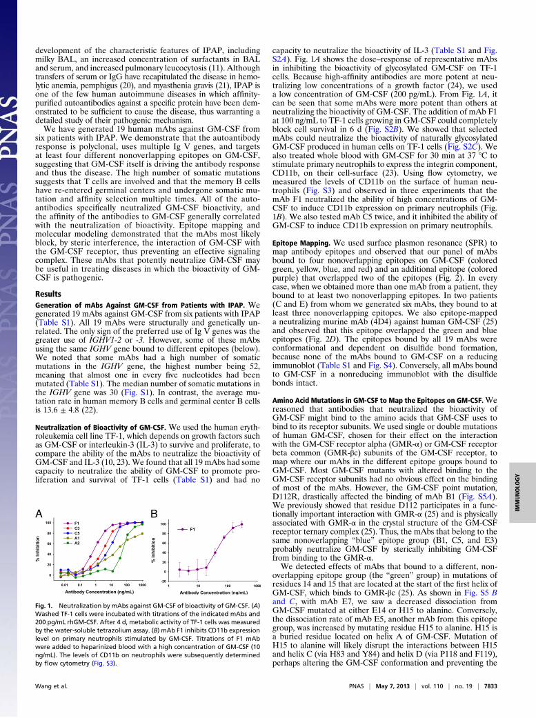
capacity to neutralize the bioactivity of IL-3 (Table S1 and Fig.S2A). Fig. 1A shows the dose–response of representative mAbsin inhibiting the bioactivity of glycosylated GM-CSF on TF-1cells. Because high-affinity antibodies are more potent at neu-tralizing low concentrations of a growth factor (24), we useda low concentration of GM-CSF (200 pg/mL). From Fig. 1A, itcan be seen that some mAbs were more potent than others atneutralizing the bioactivity of GM-CSF. The addition of mAb F1at 100 ng/mL to TF-1 cells growing in GM-CSF could completelyblock cell survival in 6 d (Fig. S2B). We showed that selectedmAbs could neutralize the bioactivity of naturally glycosylatedGM-CSF produced in human cells on TF-1 cells (Fig. S2C). Wealso treated whole blood with GM-CSF for 30 min at 37 °C tostimulate primary neutrophils to express the integrin component,CD11b, on their cell-surface (23). Using flow cytometry, wemeasured the levels of CD11b on the surface of human neu-trophils (Fig. S3) and observed in three experiments that themAb F1 neutralized the ability of high concentrations of GM-CSF to induce CD11b expression on primary neutrophils (Fig.1B). We also tested mAb C5 twice, and it inhibited the ability ofGM-CSF to induce CD11b expression on primary neutrophils.
Epitope Mapping. We used surface plasmon resonance (SPR) tomap antibody epitopes and observed that our panel of mAbsbound to four nonoverlapping epitopes on GM-CSF (coloredgreen, yellow, blue, and red) and an additional epitope (coloredpurple) that overlapped two of the epitopes (Fig. 2). In everycase, when we obtained more than one mAb from a patient, theybound to at least two nonoverlapping epitopes. In two patients(C and E) from whom we generated six mAbs, they bound to atleast three nonoverlapping epitopes. We also epitope-mappeda neutralizing murine mAb (4D4) against human GM-CSF (25)and observed that this epitope overlapped the green and blueepitopes (Fig. 2D). The epitopes bound by all 19 mAbs wereconformational and dependent on disulfide bond formation,because none of the mAbs bound to GM-CSF on a reducingimmunoblot (Table S1 and Fig. S4). Conversely, all mAbs boundto GM-CSF in a nonreducing immunoblot with the disulfidebonds intact.
Amino Acid Mutations in GM-CSF to Map the Epitopes on GM-CSF.Wereasoned that antibodies that neutralized the bioactivity ofGM-CSF might bind to the amino acids that GM-CSF uses tobind to its receptor subunits. We used single or double mutationsof human GM-CSF, chosen for their effect on the interactionwith the GM-CSF receptor alpha (GMR-α) or GM-CSF receptorbeta common (GMR-βc) subunits of the GM-CSF receptor, tomap where our mAbs in the different epitope groups bound toGM-CSF. Most GM-CSF mutants with altered binding to theGM-CSF receptor subunits had no obvious effect on the bindingof most of the mAbs. However, the GM-CSF point mutation,D112R, drastically affected the binding of mAb B1 (Fig. S5A).We previously showed that residue D112 participates in a func-tionally important interaction with GMR-α (25) and is physicallyassociated with GMR-α in the crystal structure of the GM-CSFreceptor ternary complex (25). Thus, the mAbs that belong to thesame nonoverlapping “blue” epitope group (B1, C5, and E3)probably neutralize GM-CSF by sterically inhibiting GM-CSFfrom binding to the GMR-α.We detected effects of mAbs that bound to a different, non-
overlapping epitope group (the “green” group) in mutations ofresidues 14 and 15 that are located at the start of the first helix ofGM-CSF, which binds to GMR-βc (25). As shown in Fig. S5 Band C, with mAb E7, we saw a decreased dissociation fromGM-CSF mutated at either E14 or H15 to alanine. Conversely,the dissociation rate of mAb E5, another mAb from this epitopegroup, was increased by mutating residue H15 to alanine. H15 isa buried residue located on helix A of GM-CSF. Mutation ofH15 to alanine will likely disrupt the interactions between H15and helix C (via H83 and Y84) and helix D (via P118 and F119),perhaps altering the GM-CSF conformation and preventing the
A B
Fig. 1. Neutralization by mAbs against GM-CSF of bioactivity of GM-CSF. (A)Washed TF-1 cells were incubated with titrations of the indicated mAbs and200 pg/mL rhGM-CSF. After 4 d, metabolic activity of TF-1 cells was measuredby the water-soluble tetrazolium assay. (B) mAb F1 inhibits CD11b expressionlevel on primary neutrophils stimulated by GM-CSF. Titrations of F1 mAbwere added to heparinized blood with a high concentration of GM-CSF (10ng/mL). The levels of CD11b on neutrophils were subsequently determinedby flow cytometry (Fig. S3).
Wang et al. PNAS | May 7, 2013 | vol. 110 | no. 19 | 7833
cytokine interacting with its receptor or mAbs. Thus, mAbs thatbelong to the green epitope group probably neutralize GM-CSFby binding to the surface of GM-CSF, which then stericallyinhibits GM-CSF from binding to the βc.
Fig. 3A depicts a model of the ternary complex, based on ourternary complex crystal structure (26), of a partially refined crystalstructure of the GM-CSF:GMR-α binary complex and the crystalstructure of the homologous IL-5 receptor alpha subunit (27).To visualize how anti–GM-CSF mAbs could interfere with thebioactivity of GM-CSF and its formation of a signaling complexwith the GM-CSF receptor, we used Pisa (Version 1.37; www.ebi.ac.uk/msd-srv/prot_int/pistart.html) to map and quantify the in-teraction interface residues of the ternary GM-CSF:receptorcomplex model. Fig. 3B shows the location of the E14 and D112mutations on the GM-CSF structure and the surface buried onGM-CSF by the two subunits of the GM-CSF receptor. Shown inTable S2 is the total solvent surface area of GM-CSF and thethree parts of the GM-CSF receptor with which GM-CSF inter-acts, namely the GMR-α and two domains of different βc mon-omers. It can be seen that 30% of the surface area of GM-CSF isburied by interaction with the receptor: 19% by the interactionwith the GMR-α, 7% with the D4 domain of βc (monomer 1), and4% with the D1 domain of βc (monomer 2). A typical antiproteinantibody buries between 600 and 1,000 Å2 of the surface area withits antigen-binding site (28). If the binding sites of anti–GM-CSFmAbs overlap the area buried by interaction with the GM-CSFreceptor, the mAbs could sterically interfere with the formationof the signaling complex.
Affinity and Kinetics of Binding to GM-CSF and Correlation withNeutralization of Bioactivity of GM-CSF. We used SPR to analyzethe kinetics of binding of selected mAbs to GM-CSF. Shown inFig. 4A are representative SPR data from a very high-affinitymAb, a high-affinity mAb, and a moderate-affinity mAb. Incases where we isolated multiple mAbs from the same patient,we observed a range of affinities (Fig. 4B). Fig. 5A shows theIC50 of representative mAbs from the TF-1 proliferation assay,in comparison with their binding kinetics. There was a generalcorrelation of a decreased IC50 with a higher affinity and loweroff-rate but there was no correlation with on-rate (Fig. 5B).However, we noted that a high affinity or a low off-rate did notstrictly correlate with the ability to neutralize the bioactivity ofGM-CSF. For example, mAb F1 has the best IC50 of 0.8 ng/mL,with mAb C3 the second best, with an IC50 of 2.7 ng/mL.However, mAb C3 has the highest affinity of all of the anti–GM-CSF mAbs (KD at 3.2 × 10⁻¹¹ M), whereas mAb F1 has analmost fourfold lower affinity (KD at 1.2 × 10⁻¹⁰ M). The kineticswere not a factor because both the on-rate of mAb C3 washigher than mAb F1 and the off-rate of mAb C3 was lower.Even for mAbs against the same overlapping epitopes, the af-finity did not correlate with the potency of neutralizing bio-activity of GM-CSF. For example, of the mAbs A2, E5, and E6
A B
C D
Fig. 2. Human mAbs against GM-CSF target mul-tiple epitopes. SPR was used to map the binding ofanti–GM-CSF mAbs to rhGM-CSF. (A) A schematicshowing how an anti-hIgG is immobilized on a CM5sensor chip to capture the first anti–GM-CSF mAbinjected. After blocking of free sites with an excessof hIgG, rhGM-CSF is injected and bound by the firstmAb. Subsequently, a second and third anti–GM-CSF mAb is injected, and the binding is monitored inreal time by SPR. (B) A summary of the epitope-mapping experiments, with “X” meaning that thetwo mAbs cannot bind simultaneously and “&”meaning they can bind simultaneously to rhGM-CSF. mAbs in the leftmost column denote the cap-tured mAb for each experiment. (C) Representativemapping experiments. (Left) After capture of C3and binding of rhGM-CSF, only E6, and not F1, canbind. (Right) After capture of A1 and binding ofrhGM-CSF, both C3 and B1 can bind simultaneously.(D) A map of the GM-CSF epitopes. Color coding ismaintained throughout. The 4D4 is a mouse mAbraised against rhGM-CSF.
A
B
Fig. 3. Mutations on the surface of GM-CSF and area buried by the subunits ofthe GM-CSF receptor. (A) The GM-CSF:receptor ternary model. GM-CSF is shownin purple ribbon, the GMR-α in yellowmolecular surface (with the NTD shown inlighter shade), and the βc shown in blue (different shades for the twomonomerchains of the βc dimer). (B) Two views of the surface of GM-CSF using the col-oring scheme in A. The surfaces that interact with the GMR-α are in yellow, withthe GMR-βc in blue and the remainder of the cytokine colored purple. Some ofthe residues targeted for mutation are highlighted, and key residues, shown bymutation to markedly disrupt antibody binding, are shown in red.
7834 | www.pnas.org/cgi/doi/10.1073/pnas.1216011110 Wang et al.
that bound to the green epitope, mAb A2 had the lowest IC50 of59 ng/mL and the highest Kd of 9.2 × 10⁻⁹ M, whereas the mAbE5 and E6 had dramatically higher IC50 of 2,650 and 2,550ng/mL and had 17- and 7-fold higher affinities (KD of 5.7 × 10⁻¹⁰
and 1.4 × 10⁻⁹ M), respectively. Likewise, mAbs E3 and C5bound to the same blue epitope; mAb E3 had a higher affinitythan mAb C5, but mAb C5 had a fourfold lower IC50.
DiscussionThe genetics and epitope specificity of the mAbs against GM-CSFgenerated from six IPAP patients (Table S1) makes several hy-potheses about their pathogenesis unlikely. That all 19 mAbsgenerated were genetically unrelated and used multiple V genes ineach patient shows that there was not preferred V-gene use likethose seen in autoantibodies against TG2 (7). The fact that theautoantibodies were polyclonal excludes a single forbidden clo-notype of pathogenic autoantibodies in each patient. The fact thatthere were at least three nonoverlapping conformational epitopesof mAbs against GM-CSF in two patients and a total of fournonoverlapping conformational epitopes in the six patients wouldappear to exclude a cross-reactive B-cell epitope on a pathogen
(“molecular mimicry”) (8) for the following reasons. Because thearea of a B-cell epitope on a protein is at least 600–935 Å2, theproper description of a B-cell epitope is through crystallography ofan antigen/antigen-binding site complex (28). The probability thata pathogen antigen should have four nonoverlapping, conforma-tional B-cell epitopes that are cross-reactive with GM-CSF issmall. If another unrelated antigen initiates the antibody responseand somatic mutations change the antigen-binding site so it bindsto an epitope on GM-CSF, it is unlikely that chance somaticmutations would change the antigen-binding sites to bind fournonoverlapping, conformational epitopes on GM-CSF. Twocrystallographic studies of affinity maturation of human anti-bodies (29, 30) establish that the increase in affinity is due tostabilization of the original antigen contacts and that the anti-gen-binding site of the original antibody has the same generalconformation as the affinity-matured, high-affinity antibodies. Ifsubsequent affinity maturation is driven by GM-CSF, the epitopeon the mutated antibody should still overlap the original epitopeon GM-CSF.Our tentative conclusion is that GM-CSF most likely drove
the original antibody response. There is no evidence for B-cell
A
B
Fig. 4. Affinities of the human anti–GM-CSF mAbs.(A) Shown are examples of SPR data and fit for arepresentative very-high-affinity (C3), high-affinity(C5), and moderate-affinity (B1) mAb. (B) Relativeaffinities of the human anti–GM-CSF mAbs.
A
B
Fig. 5. Relationship between kinetics of bindingand potency at inhibiting the bioactivity of GM-CSF.(A) Shown are the equilibrium dissociation constantsand rate constants for the rhGM-CSF and mAbcomplexes and comparison with their IC50 valuesfrom the TF-1 proliferation assay. (B) A graphicalrepresentation of the data from A analyzing thecorrelation between neutralization activity on the yaxis vs. the KD (Left), kd (Center), and ka (Right) onthe y axis. A nonparametric Spearman correlationtest was used to calculate the P values reported foreach comparison. ka, P = 0.4131; kd, P = 0.0436; KD,(P = 0.0315).
Wang et al. PNAS | May 7, 2013 | vol. 110 | no. 19 | 7835
tolerance or anergy for an antigen like GM-CSF that is notdetectable in the plasma (31); indeed, there is evidence to thecontrary (32, 33). There is evidence of B-cell anergy for solubleantigens at serum concentration of >0.1 nM (34). Even in in-flammatory conditions like rheumatoid arthritis, GM-CSF ispresent in synovial fluid at <35 pM (31, 35). One group hasreported that the frequency of autoantibodies in normal subjectsreaches 100% if cytokine–autoantibody complexes are dissoci-ated at low pH before autoantibodies are measured against IL-2,granulocyte colony-stimulating factor (G-CSF), VEGF, TNF, IL-8 (32), and GM-CSF (33). However, the latter report causedsome controversy, and Bazin et al. suggested that IgG, treatedwith low pH, can bind artifactually to GM-CSF (36). The ques-tion of the physiological production of autoantibodies againstGM-CSF could be answered by measuring the frequency ofmemory B cells in healthy humans that produce autoantibodiesagainst GM-CSF.The involvement of T follicular helper cells in the pathogenic
autoantibodies to GM-CSF is suggested by the high numbers ofmutations in the IGHV genes of mAbs that we cloned from IPAPpatients. We speculate that a pathogen, with a T-cell epitope thatis cross-reactive with GM-CSF, will present pathogen-associatedmolecular patterns (like endotoxin or viral RNA) to the den-dritic cells and activate T cells, breaking T-cell tolerance. If thepathogen stimulates the release of GM-CSF, the GM-CSF–specific B cells will endocytose GM-CSF and will present thecross-reactive T-cell epitope to T cells activated by the pathogen.The T-cell epitope on the pathogen need not correspond to theamino acid sequence of the T-cell epitope in GM-CSF, becausethe T-cell receptor (TCR) is polyreactive, and multiple peptidesof different sequences on the MHC II can be recognized by thesame TCR (37). In the future, it will be important to investigatethe frequency of the T cells that are stimulated by GM-CSF inIPAP. The importance of T-cell central tolerance as a mecha-nism for preventing the development of autoantibodies againstcytokines is suggested by the autoimmune polyendocrine syn-drome type I, in which subjects have a mutation in the gene forthe Autoimmune Regulator and make autoantibodies againstIL-17A, IL-17F, and IL-22 (38, 39).A paper that was published recently characterized pathogenic
mAbs that cause pemphigus vulgaris (40). They reverted the so-matic mutations from four mAbs against desmoglein-3 (DSG3)and found that the unmutated antibodies did not bind to DSG3.They concluded that autoantibodies against DSG3 were createdby somatic mutations generated in the response to an antigenunrelated to DSG3. However, an antibody with a KD of 3 × 10⁻⁴M could still activate B cells when expressed as a BCR in mice(41). In another study, although somatic mutations were revertedin three affinity-matured antibodies and the unmutated mAbs donot bind to the antigen, when the unmutatedmAbs were expressedin a B-cell line as a BCR, they signaled when antigen was added(42). Furthermore, the probability that antibodies that bind to oneunrelated antigen should have fortuitous somatic mutations thatbind to two or three nonoverlapping epitopes of a DSG3 is small.The mechanism for pathogenic autoantibodies that cause pem-phigus may be similar to the mechanism we propose for path-ogenic autoantibodies in IPAP, namely that a pathogen thatactivates T cells cross-reactive with peptides of DSG3 and thathumans exhibit no B-cell tolerance or anergy for DSG3 (43).There is one report of four human mAbs against GM-CSF
obtained from peripheral blood B cells from normal subjects orfrom patients with IPAP and with in vitro immunization ofpeptides from human GM-CSF (44). However, this publicationdoes not provide information as to whether the four antibodiesagainst GM-CSF were derived from IPAP patients or normaldonors or include the results from in vitro immunization. Nor didit provide data on the autoantibodies against GM-CSF aboutV-gene use, somatic mutations, or epitopes.All of the mAbs we examined neutralized the bioactivity of
GM-CSF, although they did not neutralize the bioactivity of IL-3(Table S1 and Fig. S2A). Each mAb when bound to GM-CSF
could sterically interfere with the formation of the dodecamersignaling complex (26) (Fig. 3). The relationship between affinityand off-rate with the IC50 generally correlated, but the epitopewhere the mAb bound to GM-CSF also mattered (Fig. 5). Be-cause we used GM-CSF binding to select specific memory Bcells and the ELISA to screen, we would have not generatedvery low-affinity antibodies. Because these were long-time patientswith IPAP, we expected to find high-affinity mAbs cloned from theGM-CSF–specific memory B cells. For example, in donors C andE, the affinities ranged, respectively, from a KD of 2.4 × 10⁻⁹ to3.7 × 10−11 M and from a KD of 1.2 × 10⁻⁸ to 1.8 × 10⁻¹⁰ M. Wespeculate that most of the high-affinity memory B cells wereretained in the lymphoid tissue and were not in the blood.Those mAbs that potently neutralize the biological activity of
GM-CSF may be useful in treating inflammatory diseases (45)like arthritis (46) and multiple sclerosis (47–49). Anti–GM-CSFmAbs may treat juvenile myelomonocytic leukemias (50) andepithelial cancers, which secrete GM-CSF that suppresses theimmune response against cancer (51, 52). These mAbs may alsobe useful in treating pain (53, 54). The risk of causing IPAP withthese therapeutic anti–GM-CSF mAbs is low because very smallamounts of GM-CSF are necessary to mature alveolar macro-phages, and IPAP is only caused by a critical threshold of auto-antibodies against GM-CSF (33).
Materials and MethodsGeneration of mAbs. Ethical clearance for samples from patients was obtainedfrom the ethics boards of theUniversities of British Columbia,Melbourne, andToronto. Blood samples were collected from individuals treated for IPAP.Individual memory B cells binding to GM-CSF were sorted into individualwells (55) (Fig. S6). After 1 wk, wells were screened for the production ofGM-CSF–specific antibodies by ELISA. RNA was purified from positive clones,and cDNA from clones was synthesized by using constant region primers fol-lowed by PCR amplification with primers taken from the leader sequences ofhuman V genes (Table S3). The International ImMunoGeneTics Informa-tion System (www.imgt.org) was used to determine Ig V gene use andsomatic mutations.
Proliferation Assay Using TF-1 Cells. mAbs were serially diluted, added toGM-CSF at a concentration of 400 pg/mL, and preincubated at 37 °C for 1 h.The mAb/GM-CSF mixtures were added to an equal volume of washed TF-1cells, 1,000 cells per well, yielding a final concentration of 200 pg/mL GM-CSF.KE5, which is a human mAb IgG1 molecule against human cytomegalovirus,was used as a negative control (56). To control that the mAbs did not inhibitTF-1 cells, the GM-CSF was replaced by a final concentration of 1% (vol/vol)gibbon IL-3–conditioned medium. After incubation at 37 °C for 4 d, water-soluble tetrazolium-1 reagent was added, and incubation was continued for4 h. The absorbance values at wavelength 450 nm, with reference wave-length of 690 nm values subtracted, were determined by using a plate reader.
Inhibition of CD11b Expression Level on Neutrophils Stimulated by GM-CSF.Triplicate samples of heparinized human whole blood (100 μL) were in-cubated with GM-CSF (10 ng/mL) with 2.34–300 ng/mL mAb. Cells werestained with anti-human CD11b-PE Ab, and FACS was performed to evaluateCD11b levels (Fig. S3).
SPR Analysis. The SPR experiments were performed at 25 °C on a Biacore 3000(GE Healthcare) by using a carboxymethylated dextran 5 (CM5) sensor chip. Ananti-human IgG (Fc) was immobilized similarly on the experimental and ref-erence flow cells by using standard amine coupling. For affinity measure-ments, recombinant human GM-CSF (rhGM-CSF) was injected over the mAb-coated cell and reference cell at the following concentrations: 100, 50, 25, 12.5,6.25, 3.12, 1.5, and 0.8 nM. The flow rate was 30 μL/min, and triplicate blankinjections and replicate injections of the 12.5 nM rhGM-CSF were included toaccount for any drift and to ensure assay reproducibility. Data were fit to a 1:1model with (for those with very fast on-rates) or without a mass transportterm by using the Biacore 3000 evaluation software.
Statistical Analysis.A nonparametric Spearman correlation test was used to assessthe correlation between mAb potency and affinity for GM-CSF using Statview.
Modeling the GM-CSF Ternary Complex. The ternary model shown in Fig. 3is based partly on the crystal structure of the published ternary GM-CSF:
7836 | www.pnas.org/cgi/doi/10.1073/pnas.1216011110 Wang et al.
receptor complex (Protein Data Bank ID code 3CXE). However, the GMR-αextracellular region in this complex was incomplete, with only the extra-cellular domain 3 (D3) of GMR-α well defined (26). Thus, extracellulardomains 1 [N-terminal domain (NTD)] and 2 (D2) of GMR-α were modeledbased on a partially refined crystal structure of the GM-CSF:GMR-α binarycomplex and the crystal structure of the homologous IL-5 receptor α subunit(27). Our GM-CSF:GMR-α binary complex data suggest that the GMR-α NTD–D2 linker is flexible and that the NTD may adopt a variety of orientationswith respect to D2 and D3.
ACKNOWLEDGMENTS. We thank the staff of the Royal Melbourne Hospitaland the Toronto General Hospital; Dr. John Seymour; Dr. Christopher Brown;and the University of British Columbia Centre for Biothermodynamics. Thisstudy was supported by the Canadian Institutes of Health Research and theCanadian Arthritis Network (J.W.S.); by a National Health and Medical Re-search Council of Australia (NHMRC) Development Grant (to J.A.H., J.W.S.,and G.P.A.) and Program Grant 565217 (to A.L.); and infrastructure supportfrom the Victorian State Government Operational Infrastructure SupportProgram (M.W.P.). J.W.S. is a Canada Research Chair and M.W.P. and J.A.H.are Senior Principal Research Fellows of the NHMRC.
1. Silverstein AM (2001) Autoimmunity versus horror autotoxicus: The struggle forrecognition. Nat Immunol 2(4):279–281.
2. Zenewicz LA, Abraham C, Flavell RA, Cho JH (2010) Unraveling the genetics of au-toimmunity. Cell 140(6):791–797.
3. Burnet FM (1972) A reassessment of the forbidden clone hypothesis of autoimmunedisease. Aust J Exp Biol Med Sci 50(1):1–9.
4. Watson CT, Breden F (2012) The immunoglobulin heavy chain locus: Genetic variation,missing data, and implications for human disease. Genes Immun 13(5):363–373.
5. Breden F, et al. (2011) Comparison of antibody repertoires produced by HIV-1 in-fection, other chronic and acute infections, and systemic autoimmune disease. PLoSONE 6(3):e16857.
6. Foreman AL, Van de Water J, Gougeon M-L, Gershwin ME (2007) B cells in autoim-mune diseases: Insights from analyses of immunoglobulin variable (Ig V) gene usage.Autoimmunity Rev 6(6):387–401.
7. Di Niro R, et al. (2012) High abundance of plasma cells secreting transglutaminase2-specific IgA autoantibodies with limited somatic hypermutation in celiac diseaseintestinal lesions. Nat Med 18(3):441–445.
8. Rowley D, Jenkin CR (1962) Antigenic cross-reaction between host and parasite asa possible cause of pathogenicity. Nature 193:151–154.
9. Kitamura T, et al. (1999) Idiopathic pulmonary alveolar proteinosis as an autoimmunedisease with neutralizing antibody against granulocyte/macrophage colony-stimu-lating factor. J Exp Med 190(6):875–880.
10. Uchida K, et al. (2004) High-affinity autoantibodies specifically eliminate granulocyte-macrophage colony-stimulating factor activity in the lungs of patients with idiopathicpulmonary alveolar proteinosis. Blood 103(3):1089–1098.
11. Sakagami T, et al. (2009) Human GM-CSF autoantibodies and reproduction of pul-monary alveolar proteinosis. N Engl J Med 361(27):2679–2681.
12. Trapnell BC, Carey BC, Uchida K, Suzuki T (2009) Pulmonary alveolar proteinosis,a primary immunodeficiency of impaired GM-CSF stimulation of macrophages. CurrOpin Immunol 21(5):514–521.
13. Metcalf D (2008) Hematopoietic cytokines. Blood 111(2):485–491.14. Handman E, Burgess AW (1979) Stimulation by granulocyte-macrophage colony-
stimulating factor of Leishmania tropica killing by macrophages. J Immunol 122(3):1134–1137.
15. Hamilton JA, Stanley ER, Burgess AW, Shadduck RK (1980) Stimulation of macrophageplasminogen activator activity by colony-stimulating factors. J Cell Physiol 103(3):435–445.
16. Lopez AF, et al. (1986) Recombinant human granulocyte-macrophage colony-stimu-lating factor stimulates in vitro mature human neutrophil and eosinophil function,surface receptor expression, and survival. J Clin Invest 78(5):1220–1228.
17. Hercus TR, et al. (2009) The granulocyte-macrophage colony-stimulating factor re-ceptor: Linking its structure to cell signaling and its role in disease. Blood 114(7):1289–1298.
19. Carey B, Trapnell BC (2010) The molecular basis of pulmonary alveolar proteinosis.Clin Immunol 135(2):223–235.
20. Anhalt GJ, Labib RS, Voorhees JJ, Beals TF, Diaz LA (1982) Induction of pemphigus inneonatal mice by passive transfer of IgG from patients with the disease. N Engl J Med306(20):1189–1196.
21. Toyka KV, et al. (1977) Myasthenia gravis. Study of humoral immune mechanisms bypassive transfer to mice. N Engl J Med 296(3):125–131.
22. Wrammert J, et al. (2008) Rapid cloning of high-affinity human monoclonal anti-bodies against influenza virus. Nature 453(7195):667–671.
23. Uchida K, et al. (2007) GM-CSF autoantibodies and neutrophil dysfunction in pul-monary alveolar proteinosis. N Engl J Med 356(6):567–579.
24. Rathanaswami P, et al. (2005) Demonstration of an in vivo generated sub-picomolaraffinity fully human monoclonal antibody to interleukin-8. Biochem Biophys ResCommun 334(4):1004–1013.
25. Hercus TR, et al. (1994) Identification of residues in the first and fourth helices of humangranulocyte-macrophage colony-stimulating factor involved in biologic activity and inbinding to the alpha- and beta-chains of its receptor. Blood 83(12):3500–3508.
26. Hansen G, et al. (2008) The structure of the GM-CSF receptor complex reveals a dis-tinct mode of cytokine receptor activation. Cell 134(3):496–507.
27. Patino E, et al. (2011) Structure analysis of the IL-5 ligand-receptor complex revealsa wrench-like architecture for IL-5Rα. Structure 19(12):1864–1875.
28. Laver WG, Air GM, Webster RG, Smith-Gill SJ (1990) Epitopes on protein antigens:Misconceptions and realities. Cell 61(4):553–556.
29. Thomson CA, et al. (2008) Germline V-genes sculpt the binding site of a family ofantibodies neutralizing human cytomegalovirus. EMBO J 27(19):2592–2602.
30. Schmidt AG, et al. (2013) Preconfiguration of the antigen-binding site during affinitymaturation of a broadly neutralizing influenza virus antibody. Proc Natl Acad Sci USA110(1):264–269.
31. Steidl S, Ratsch O, Brocks B, Dürr M, Thomassen-Wolf E (2008) In vitro affinity mat-uration of human GM-CSF antibodies by targeted CDR-diversification. Mol Immunol46(1):135–144.
32. Watanabe M, et al. (2007) Anti-cytokine autoantibodies are ubiquitous in healthyindividuals. FEBS Lett 581(10):2017–2021.
33. Uchida K, et al. (2009) Granulocyte/macrophage-colony-stimulating factor autoanti-bodies and myeloid cell immune functions in healthy subjects. Blood 113(11):2547–2556.
34. Adelstein S, et al. (1991) Induction of self-tolerance in T cells but not B cells oftransgenic mice expressing little self antigen. Science 251(4998):1223–1225.
35. Ottonello L, et al. (2002) Synovial fluid from patients with rheumatoid arthritis in-hibits neutrophil apoptosis: Role of adenosine and proinflammatory cytokines.Rheumatology (Oxford) 41(11):1249–1260.
36. Bazin R, St-Amour I, Laroche A, Lemieux R (2010) Activated cryptic granulocyte-macrophage colony-stimulating factor autoantibodies in intravenous immunoglob-ulin preparations. Blood 115(2):431.
37. Felix NJ, et al. (2007) Alloreactive T cells respond specifically to multiple distinctpeptide-MHC complexes. Nat Immunol 8(4):388–397.
38. Kisand K, et al. (2010) Chronic mucocutaneous candidiasis in APECED or thymomapatients correlates with autoimmunity to Th17-associated cytokines. J Exp Med207(2):299–308.
39. Puel A, et al. (2010) Autoantibodies against IL-17A, IL-17F, and IL-22 in patients withchronic mucocutaneous candidiasis and autoimmune polyendocrine syndrome type I.J Exp Med 207(2):291–297.
40. Di Zenzo G, et al. (2012) Pemphigus autoantibodies generated through somaticmutations target the desmoglein-3 cis-interface. J Clin Invest 122(10):3781–3790.
41. Dal Porto JM, Haberman AM, Kelsoe G, Shlomchik MJ (2002) Very low affinity B cellsform germinal centers, become memory B cells, and participate in secondary immuneresponses when higher affinity competition is reduced. J Exp Med 195(9):1215–1221.
42. Lingwood D, et al. (2012) Structural and genetic basis for development of broadlyneutralizing influenza antibodies. Nature 489(7417):566–570.
43. Akkaraju S, Canaan K, Goodnow CC (1997) Self-reactive B cells are not eliminated orinactivated by autoantigen expressed on thyroid epithelial cells. J Exp Med 186(12):2005–2012.
44. Li J, et al. (2006) Human antibodies for immunotherapy development generated viaa human B cell hybridoma technology. Proc Natl Acad Sci USA 103(10):3557–3562.
45. Hamilton JA (2008) Colony-stimulating factors in inflammation and autoimmunity.Nat Rev Immunol 8(7):533–544.
46. Cook AD, Braine EL, Campbell IK, Rich MJ, Hamilton JA (2001) Blockade of collagen-induced arthritis post-onset by antibody to granulocyte-macrophage colony-stimu-lating factor (GM-CSF): Requirement for GM-CSF in the effector phase of disease.Arthritis Res 3(5):293–298.
47. McQualter JL, et al. (2001) Granulocyte macrophage colony-stimulating factor: A newputative therapeutic target in multiple sclerosis. J Exp Med 194(7):873–882.
48. Codarri L, et al. (2011) RORγt drives production of the cytokine GM-CSF in helperT cells, which is essential for the effector phase of autoimmune neuroinflammation.Nat Immunol 12(6):560–567.
49. El-Behi M, et al. (2011) The encephalitogenicity of T(H)17 cells is dependent on IL-1-and IL-23-induced production of the cytokine GM-CSF. Nat Immunol 12(6):568–575.
50. Iversen PO, et al. (1997) Inhibition of granulocyte-macrophage colony-stimulatingfactor prevents dissemination and induces remission of juvenile myelomonocyticleukemia in engrafted immunodeficient mice. Blood 90(12):4910–4917.
51. Bayne LJ, et al. (2012) Tumor-derived granulocyte-macrophage colony-stimulatingfactor regulates myeloid inflammation and T cell immunity in pancreatic cancer.Cancer Cell 21(6):822–835.
52. Pylayeva-Gupta Y, Lee KE, Hajdu CH, Miller G, Bar-Sagi D (2012) Oncogenic Kras-induced GM-CSF production promotes the development of pancreatic neoplasia.Cancer Cell 21(6):836–847.
53. Schweizerhof M, et al. (2009) Hematopoietic colony-stimulating factors mediate tu-mor-nerve interactions and bone cancer pain. Nat Med 15(7):802–807.
54. Cook AD, et al. (2013) Granulocyte-macrophage colony-stimulating factor is a keymediator in inflammatory and arthritic pain. Ann Rheum Dis 72(2):265–270.
55. Thomson CA, et al. (2012) Pandemic H1N1 influenza infection and vaccination inhumans induces cross-protective antibodies that target the hemagglutinin stem.Front Immunol 3:87.
56. McLean GR, et al. (2005) Recognition of human cytomegalovirus by human primaryimmunoglobulins identifies an innate foundation to an adaptive immune response.J Immunol 174(8):4768–4778.
Wang et al. PNAS | May 7, 2013 | vol. 110 | no. 19 | 7837