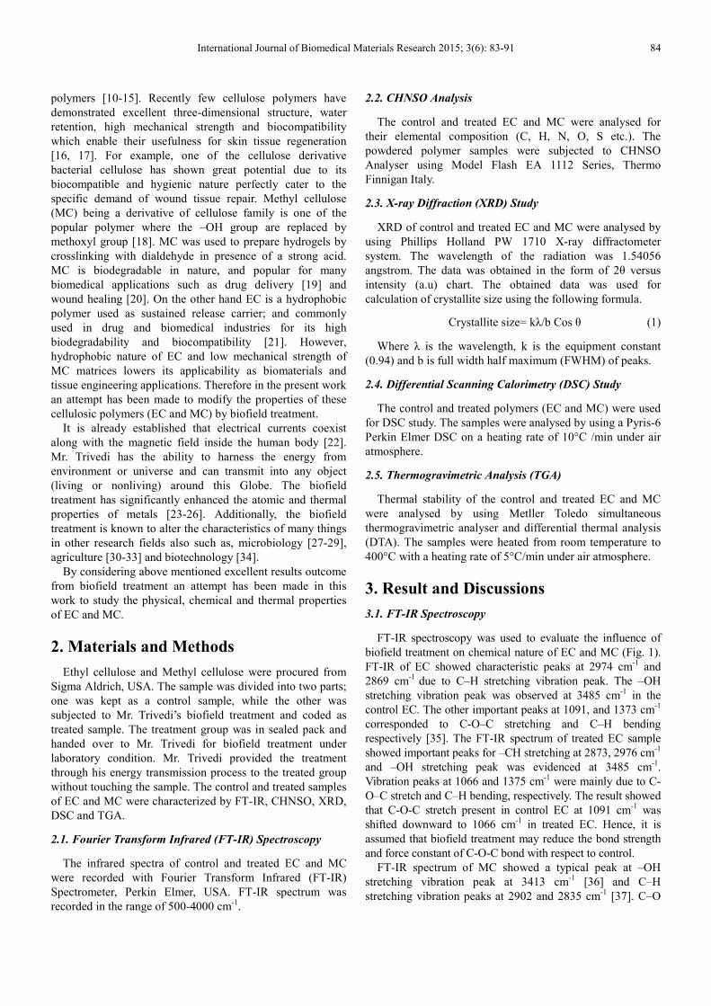
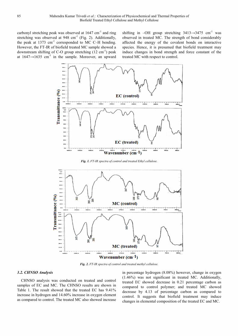
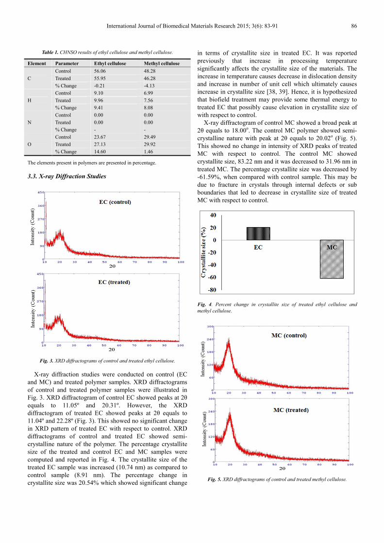
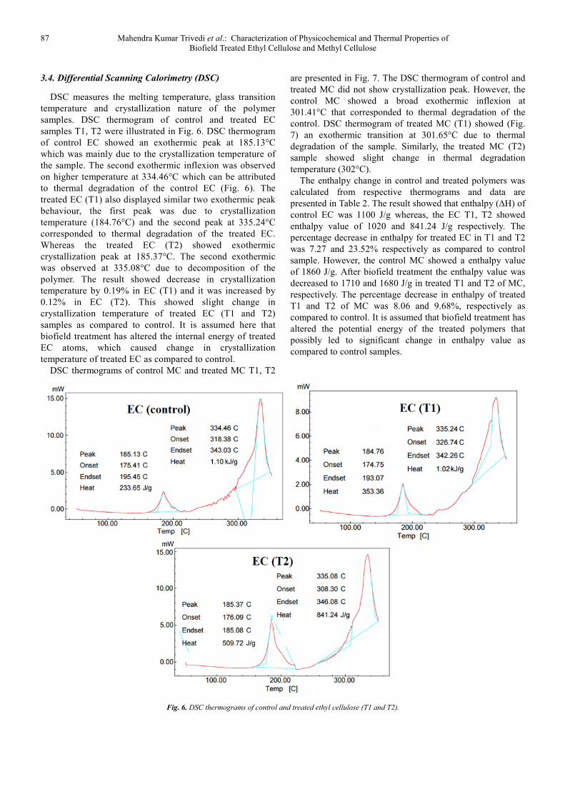
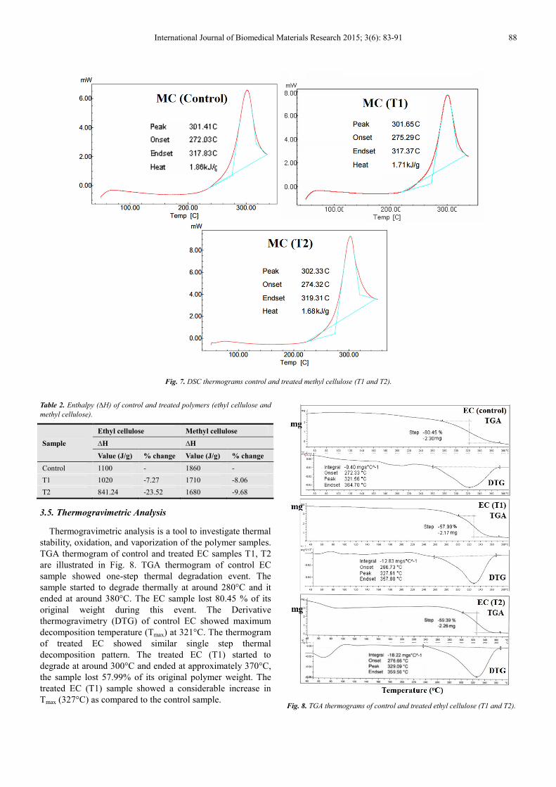
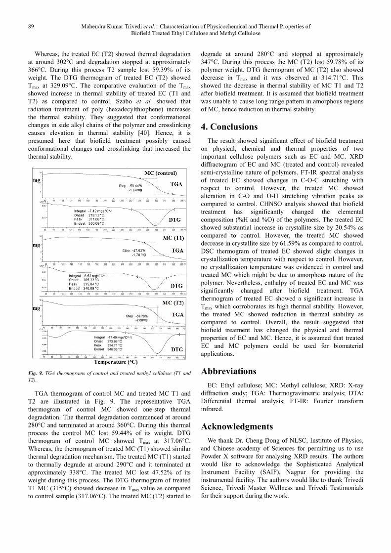
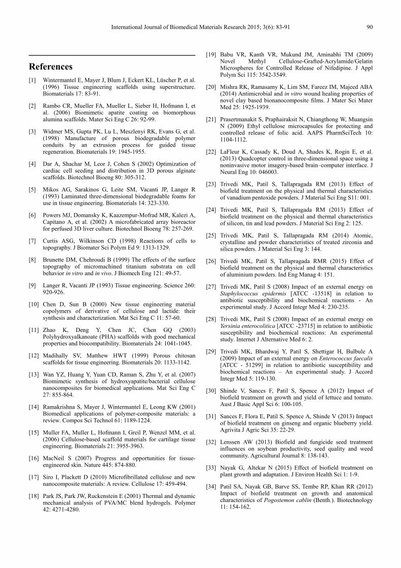
International Journal of Biomedical Materials Research 2015; 3(6): 83-91 Published online December 21, 2015 (http://www.sciencepublishinggroup.com/j/ijbmr) doi: 10.11648/j.ijbmr.20150306.12 ISSN: 2330-7560 (Print); ISSN: 2330-7579 (Online) Characterization of Physicochemical and Thermal Properties of Biofield Treated Ethyl Cellulose and Methyl Cellulose Mahendra Kumar Trivedi 1 , Alice Branton 1 , Dahryn Trivedi 1 , Gopal Nayak 1 , Rakesh Kumar Mishra 2 , Snehasis Jana 2, * 1 Trivedi Global Inc., Henderson, USA 2 Trivedi Science Research Laboratory Pvt. Ltd., Bhopal, Madhya Pradesh, India Email address: [email protected] (S. Jana) To cite this article: Mahendra Kumar Trivedi, Alice Branton, Dahryn Trivedi, Gopal Nayak, Rakesh Kumar Mishra, Snehasis Jana. Characterization of Physicochemical and Thermal Properties of Biofield Treated Ethyl Cellulose and Methyl Cellulose. International Journal of Biomedical Materials Research. Vol. 3, No. 6, 2015, pp. 83-91. doi: 10.11648/j.ijbmr.20150306.12 Abstract: Cellulose and its derivatives are used as potential matrices for biomaterials and tissue engineering applications. The objective of present research was to investigate the influence of biofield treatment on physical, chemical and thermal properties of ethyl cellulose (EC) and methyl cellulose (MC). The study was performed in two groups (control and treated). The control group remained as untreated, and biofield treatment was given to treated group. The biofield treated polymers are characterized by Fourier transform infrared spectroscopy (FT-IR), CHNSO analysis, X-ray diffraction study (XRD), Differential Scanning calorimetry (DSC), and thermogravimetric analysis (TGA). FT-IR analysis of treated EC showed downward shifting in C-O-C stretching peak from 1091→1066 cm -1 with respect to control. However, the treated MC showed upward shifting of –OH stretching (3413→3475) and downward shifting in C-O stretching (1647→1635 cm -1 ) vibrations with respect to control MC. CHNSO analysis showed substantial increase in percent hydrogen and oxygen in treated polymers with respect to control. XRD diffractogram of EC and MC affirmed the typical semi-crystalline nature. The crystallite size was substantially increased by 20.54% in treated EC with respect to control. However, the treated MC showed decrease in crystallite by 61.59% with respect to control. DSC analysis of treated EC showed minimal changes in crystallization temperature with respect to control sample. However, the treated and control MC did not show any crystallization temperature in the samples. TGA analysis of treated EC showed increase in thermal stability with respect to control. However, the TGA thermogram of treated MC showed reduction in thermal stability as compared to control. Overall, the result showed substantial alteration in physical, chemical and thermal properties of treated EC and MC. Keywords: Biofield Treatment, Ethyl Cellulose, Methyl Cellulose, X-ray Diffraction Study, Differential Scanning Calorimetry, Thermogravimetric Analysis, Fourier Transform Infrared Spectroscopy 1. Introduction Tissue engineering is an interdisciplinary area that uses cells, materials, engineering and life sciences toward the design and fabrication of biological substitutes that restore, support, or improve tissue or a whole organ function. Biomedical implant scaffolds are an excellent example of tissue engineering substrates composed of biodegradable polymers or inert materials coated with bioactive biomaterials which allows growth of new tissues of particular types of cells [1, 2]. Biomaterials need to be properly modified by introducing controlled porosity, design of three- dimensional structure and surface modification to achieve better cell packing and control cell network architecture [3- 8]. The biomaterial scaffold should have biodegradable and biocompatible nature. After the formation of the new tissue, polymeric scaffolds are slowly degraded into small molecular weight compounds, which was absorbed by the body or excreted out of the body [9]. In last few years hunt for new classes of biomaterials, with specific properties to be used as scaffolds for tissue engineering, has attained great interest, like cellulose, polyhydroxyalkanoates, polylactates and blends of these
Transcript
International Journal of Biomedical Materials Research 2015; 3(6): 83-91
Published online December 21, 2015 (http://www.sciencepublishinggroup.com/j/ijbmr)
doi: 10.11648/j.ijbmr.20150306.12
ISSN: 2330-7560 (Print); ISSN: 2330-7579 (Online)
Characterization of Physicochemical and Thermal Properties of Biofield Treated Ethyl Cellulose and Methyl Cellulose
Mahendra Kumar Trivedi1, Alice Branton
1, Dahryn Trivedi
1, Gopal Nayak
1,
Rakesh Kumar Mishra2, Snehasis Jana
2, *
1Trivedi Global Inc., Henderson, USA 2Trivedi Science Research Laboratory Pvt. Ltd., Bhopal, Madhya Pradesh, India
We thank Dr. Cheng Dong of NLSC, Institute of Physics,
and Chinese academy of Sciences for permitting us to use
Powder X software for analysing XRD results. The authors
would like to acknowledge the Sophisticated Analytical
Instrument Facility (SAIF), Nagpur for providing the
instrumental facility. The authors would like to thank Trivedi
Science, Trivedi Master Wellness and Trivedi Testimonials
for their support during the work.
International Journal of Biomedical Materials Research 2015; 3(6): 83-91 90
References
[1] Wintermantel E, Mayer J, Blum J, Eckert KL, Lüscher P, et al. (1996) Tissue engineering scaffolds using superstructure. Biomaterials 17: 83-91.
[2] Rambo CR, Mueller FA, Mueller L, Sieber H, Hofmann I, et al. (2006) Biomimetic apatite coating on biomorphous alumina scaffolds. Mater Sci Eng C 26: 92-99.
[3] Widmer MS, Gupta PK, Lu L, Meszlenyi RK, Evans G, et al. (1998) Manufacture of porous biodegradable polymer conduits by an extrusion process for guided tissue regeneration. Biomaterials 19: 1945-1955.
[4] Dar A, Shachar M, Leor J, Cohen S (2002) Optimization of cardiac cell seeding and distribution in 3D porous alginate scaffolds. Biotechnol Bioeng 80: 305-312.
[5] Mikos AG, Sarakinos G, Leite SM, Vacanti JP, Langer R (1993) Laminated three-dimensional biodegradable foams for use in tissue engineering. Biomaterials 14: 323-330.
[6] Powers MJ, Domansky K, Kaazempur-Mofrad MR, Kalezi A, Capitano A, et al. (2002) A microfabricated array bioreactor for perfused 3D liver culture. Biotechnol Bioeng 78: 257-269.
[7] Curtis ASG, Wilkinson CD (1998) Reactions of cells to topography. J Biomater Sci Polym Ed 9: 1313-1329.
[8] Brunette DM, Chehroudi B (1999) The effects of the surface topography of micromachined titanium substrata on cell behavior in vitro and in vivo. J Biomech Eng 121: 49-57.
[10] Chen D, Sun B (2000) New tissue engineering material copolymers of derivative of cellulose and lactide: their synthesis and characterization. Mat Sci Eng C 11: 57-60.
[11] Zhao K, Deng Y, Chen JC, Chen GQ (2003) Polyhydroxyalkanoate (PHA) scaffolds with good mechanical properties and biocompatibility. Biomaterials 24: 1041-1045.
[12] Madihally SV, Matthew HWT (1999) Porous chitosan scaffolds for tissue engineering. Biomaterials 20: 1133-1142.
[13] Wan YZ, Huang Y, Yuan CD, Raman S, Zhu Y, et al. (2007) Biomimetic synthesis of hydroxyapatite/bacterial cellulose nanocomposites for biomedical applications. Mat Sci Eng C 27: 855-864.
[14] Ramakrishna S, Mayer J, Wintermantel E, Leong KW (2001) Biomedical applications of polymer-composite materials: a review. Compos Sci Technol 61: 1189-1224.
[15] Muller FA, Muller L, Hofmann I, Greil P, Wenzel MM, et al. (2006) Cellulose-based scaffold materials for cartilage tissue engineering. Biomaterials 21: 3955-3963.
[16] MacNeil S (2007) Progress and opportunities for tissue-engineered skin. Nature 445: 874-880.
[17] Siro I, Plackett D (2010) Microfibrillated cellulose and new nanocomposite materials: A review. Cellulose 17: 459-494.
[18] Park JS, Park JW, Ruckenstein E (2001) Thermal and dynamic mechanical analysis of PVA/MC blend hydrogels. Polymer 42: 4271-4280.
[20] Mishra RK, Ramasamy K, Lim SM, Fareez IM, Majeed ABA (2014) Antimicrobial and in vitro wound healing properties of novel clay based bionanocomposite films. J Mater Sci Mater Med 25: 1925-1939.
[21] Prasertmanakit S, Praphairaksit N, Chiangthong W, Muangsin N (2009) Ethyl cellulose microcapsules for protecting and controlled release of folic acid. AAPS PharmSciTech 10: 1104-1112.
[22] LaFleur K, Cassady K, Doud A, Shades K, Rogin E, et al. (2013) Quadcopter control in three-dimensional space using a noninvasive motor imagery-based brain–computer interface. J Neural Eng 10: 046003.
[23] Trivedi MK, Patil S, Tallapragada RM (2013) Effect of biofield treatment on the physical and thermal characteristics of vanadium pentoxide powders. J Material Sci Eng S11: 001.
[24] Trivedi MK, Patil S, Tallapragada RM (2013) Effect of biofield treatment on the physical and thermal characteristics of silicon, tin and lead powders. J Material Sci Eng 2: 125.
[25] Trivedi MK, Patil S, Tallapragada RM (2014) Atomic, crystalline and powder characteristics of treated zirconia and silica powders. J Material Sci Eng 3: 144.
[26] Trivedi MK, Patil S, Tallapragada RMR (2015) Effect of biofield treatment on the physical and thermal characteristics of aluminium powders. Ind Eng Manag 4: 151.
[27] Trivedi MK, Patil S (2008) Impact of an external energy on Staphylococcus epidermis [ATCC -13518] in relation to antibiotic susceptibility and biochemical reactions - An experimental study. J Accord Integr Med 4: 230-235.
[28] Trivedi MK, Patil S (2008) Impact of an external energy on Yersinia enterocolitica [ATCC -23715] in relation to antibiotic susceptibility and biochemical reactions: An experimental study. Internet J Alternative Med 6: 2.
[29] Trivedi MK, Bhardwaj Y, Patil S, Shettigar H, Bulbule A (2009) Impact of an external energy on Enterococcus faecalis [ATCC - 51299] in relation to antibiotic susceptibility and biochemical reactions – An experimental study. J Accord Integr Med 5: 119-130.
[30] Shinde V, Sances F, Patil S, Spence A (2012) Impact of biofield treatment on growth and yield of lettuce and tomato. Aust J Basic Appl Sci 6: 100-105.
[31] Sances F, Flora E, Patil S, Spence A, Shinde V (2013) Impact of biofield treatment on ginseng and organic blueberry yield. Agrivita J Agric Sci 35: 22-29.
[32] Lenssen AW (2013) Biofield and fungicide seed treatment influences on soybean productivity, seed quality and weed community. Agricultural Journal 8: 138-143.
[33] Nayak G, Altekar N (2015) Effect of biofield treatment on plant growth and adaptation. J Environ Health Sci 1: 1-9.
[34] Patil SA, Nayak GB, Barve SS, Tembe RP, Khan RR (2012) Impact of biofield treatment on growth and anatomical characteristics of Pogostemon cablin (Benth.). Biotechnology 11: 154-162.
91 Mahendra Kumar Trivedi et al.: Characterization of Physicochemical and Thermal Properties of
Biofield Treated Ethyl Cellulose and Methyl Cellulose
[35] Suthar V, Pratap A, Raval H (2000) Studies on poly (hydroxy alkanoates)/ (ethylcellulose) blends. Bull Mater Sci 23: 215-219.
[36] Shi P, Li Y, Zhang L (2008) Fabrication and property of chitosan film carrying ethyl cellulose microspheres. Carbohydr Polym 72: 490-499.
[37] Viera RGP, Filho GR, de Assunção RMN, da S Meireles C, Vieira JG, et al. (2007) Synthesis and characterization of methylcellulose from sugar cane bagasse cellulose. Carbohydr Polym 67: 182-189.
[38] Gaber A, Abdel-Rahim MA, Abdel-Latief AY, Abdel-Salam
MN (2014) Influence of calcination temperature on the structure and porosity of nanocrystalline SnO2 synthesized by a conventional precipitation method. Int J Electrochem Sci 9: 81-95.
[39] Raj KJA, Viswanathan B (2009) Effect of surface area, pore volume, particle size of P25 titania on the phase transformation of anatase to rutile. Indian J Chem 48A: 1378-1382.
[40] Szabo L, Cik G, Lensy J (1996) Thermal stability increase of doped poly (hexadecylthiophene) by γ-radiation. Synt Met 78: 149-153.