2
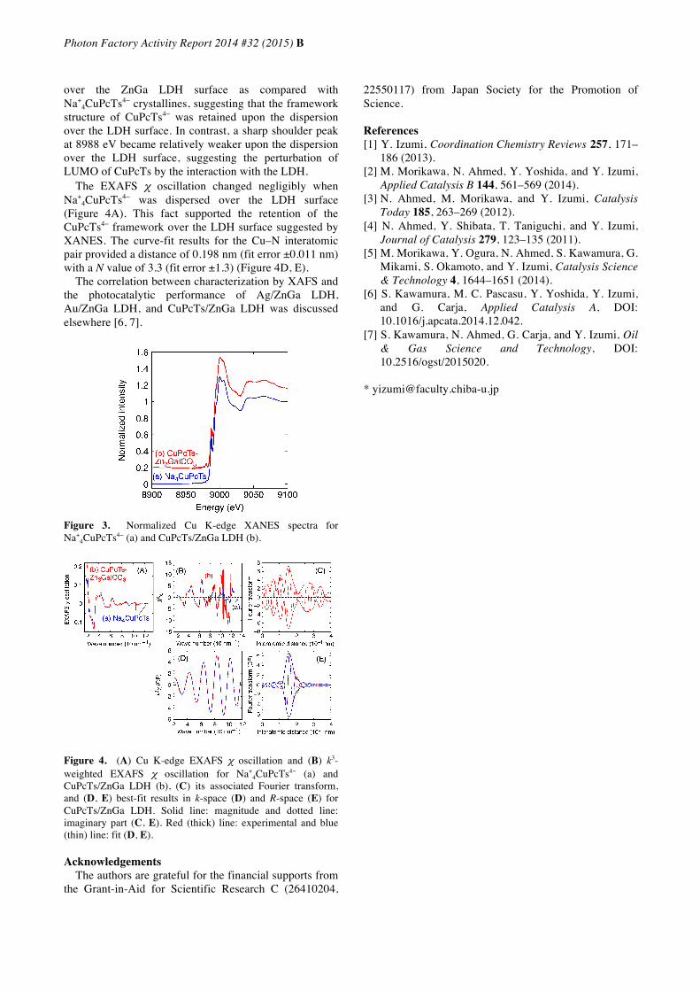
Photon Factory Activity Report 2014 #32 (2015) B 9C, 12C, NW10A/2014G631 Characterization of Plasmonic Nanoparticles and Dyes Assembled with Layered Double Hydroxides to Promote the Photoreduction of Carbon Dioxide into Fuels Shogo Kawamura and Yasuo Izumi * Graduate School of Science, Chiba University, Yayoi, 1-33, Inage-ku, Chiba 263-8522 1 Introduction The photoreduction of CO 2 to fuels using natural light can contribute simultaneously to reduction of the major greenhouse gas and the development of sustainable energy [1]. Efficient photoreduction of CO 2 into methanol (or CO) was reported using layered double oxides (LDHs) [2–4]. Furthermore, the combination of WO 3 and LDH photocatalysts as a photoanode to oxidize water and a photocathode to reduce CO 2 , respectively, was demonstrated [5]. The wide band gap of LDHs is advantageous to reduce high-potential compound, e.g. CO 2 . Conversely, wide band gap is also drawback because UV light is required to excite LDHs. In this study, plasmonic nanoparticles of Au and Ag and Cu phthalocyanine were assembled with LDHs to enable CO 2 photoreduction utilizing visible light. 2 Experimental Section Ag/ZnGa LDH was prepared by ion exchange at 373 K for 15–180 min [6]. The samples were denoted as Ag/ZnGa LDH-15 or Ag/ZnGa LDH-180. Au/ZnGa LDH was prepared by ion exchange and liquid phase reduction using NaBH 4 [6]. CuPcTs/ZnGa LDH was prepared by ion exchange of sodium salt of Cu phthalocyanine tetrasulfonate hydrate (Na + 4 CuPcTs 4− ) with ZnGa LDH [7]. Ag K, Au L 3 , and Cu K-edge XAFS spectra were measured in transmission mode at 290 K. 3 Results and Discussion Ag K-edge XANES spectra for Ag/ZnGa LDH-15 and Ag/ZnGa LDH-180 were depicted in Figure 1A-b and d, respectively. The XANES patterns for these samples were quite similar to that for Ag metal (a) rather than Ag 2 O (e). Thus, Ag 0 nanoparticles were combined with LDH. The metallic Ag 0 state of Ag/ZnGa LDH-15 negligibly changed after the photocatalytic test in CO 2 and H 2 (Figure 1A-c) in comparison to spectrum b for the as- synthesized sample. Similarly, metallic Au 0 state of fresh Au/ZnGa LDH unchanged after the photocatalytic test in CO 2 and H 2 (Figure 1B-b, c). The k 3 -weighted EXAFS function was depicted in Figure 2a for Ag/ZnGa LDH-15 (row 1) and Ag/ZnGa LDH-180 (row 2). The peaks at 0.27 nm (phase shift uncorrected) in the associated Fourier transform (b) were nicely curve-fit by the parameters of Ag–Ag interatomic pair (Figure 2c, d). The N value for Ag/ZnGa LDH-15 was 7.6 (particle size ~1.6 nm), significantly smaller than N of 10.1 (particle size ~3.7 nm) for Ag/ZnGa LDH-180, demonstrating the gradual growth of Ag nanoparticles during the synthesis at 373 K. The N(Au–Au) value of 8.3 (particle size ~1.9 nm) for fresh Au/ZnGa LDH (Figure 2, row 3) was consistent with mean particle size of 3.2 nm obtained based on the high-resolution transmission electron microscopy. Figure 1. (A) Normalized Ag K-edge XANES spectra for Ag metal (a), Ag/ZnGa LDH-15 (b), sample b after photocatalytic test in CO 2 and H 2 for 5 h (c; dotted line), Ag/ZnGa LDH-180 (d), and Ag 2 O (e). (B) Normalized Au L 3 -edge XANES spectra for Au metal (a), Au/ZnGa LDH (b), sample b after photocatalytic test in CO 2 and H 2 for 5 h (c; dotted line), and Au 2 O 3 (d). Figure 2. Ag K-edge EXAFS for Ag/ZnGa LDH-15 (1) and Ag/ZnGa LDH-180 (2) and Au L 3 -edge EXAFS for Au/ZnGa LDH (3). k 3 -weighted EXAFS oscillation (a), its associated Fourier transform (b), and best-fit results in k-space (c) and R- space (d). The XANES spectra for Na + 4 CuPcTs 4− and CuPcTs/ZnGa LDH are shown in Figure 3. The spectrum pattern changed negligibly by the dispersion of CuPcTs 4−