Doctoraatsproefschrift nr. 998 aan de faculteit Bio-ingenieurswetenschappen van de K.U.Leuven CHARACTERIZATION OF RADOPHOLUS SIMILIS RESISTANCE IN MUSA SPP. WITH EMPHASIS ON PHYTOCHEMICAL ANALYSIS Suganthagunthalam DHAKSHINAMOORTHY Supervisor: Prof. D. De Waele, K.U.Leuven Co-supervisor: Prof. A. Elsen, BDB & UGent Members of the Examination Committee: Prof. E. Decuypere, Chairman, K.U.Leuven Prof. A. Aertsen, K.U.Leuven Prof. J. Coosemans, K.U.Leuven Prof. R. Swennen, K.U.Leuven Prof. G. Gheysen, UGent Dr. D. Hölscher, MPI-CE, Germany Dissertation presented in partial fulfilment of the requirements for the degree of Doctor in Bioscience Engineering November 2011
Transcript
Doctoraatsproefschrift nr. 998 aan de faculteit Bio-ingenieurswetenschappen van de K.U.Leuven
CHARACTERIZATION OF RADOPHOLUS SIMILIS RESISTANCE IN MUSA SPP.
WITH EMPHASIS ON PHYTOCHEMICAL ANALYSIS
Suganthagunthalam DHAKSHINAMOORTHY
Supervisor:
Prof. D. De Waele, K.U.Leuven
Co-supervisor: Prof. A. Elsen, BDB & UGent
Members of the Examination Committee: Prof. E. Decuypere, Chairman, K.U.Leuven
Prof. A. Aertsen, K.U.Leuven
Prof. J. Coosemans, K.U.Leuven
Prof. R. Swennen, K.U.Leuven Prof. G. Gheysen, UGent
Dr. D. Hölscher, MPI-CE, Germany
Dissertation presented in partial fulfilment of the
Doctoraatsschool, W. de Croylaan 6, 3001 Heverlee, België
Alle rechten voorbehouden. Niets uit deze uitgave mag worden vermenigvuldigd en/of
openbaar gemaakt worden door middel van druk, fotokopie, microfilm, elektronisch of
op welke andere wijze ook zonder voorafgaandelijke schriftelijke toestemming van de
uitgever.
All rights reserved. No part of the publication may be reproduced in any form by print, photoprint, microfilm, electronic or any other means without written permission from
the publisher.
Cover illustration: Top: Light microscopic image of Musa genotypes Long Tavoy (left)
and Yangambi km5 (right) roots showing the accumulation of phenolic phytoalexins
due to R. similis infection.
Bottom: Light microscopic image of a moulting Radopholus similis juvenile that had
killed by the uptake of phenylphenalenone type-pheytoalexin anigorufone during a
laboratory bio-assay.
ISBN 978-90-8826-216-6 Legal deposit number D/2011/11.109/49
I
ACKNOWLEDGEMENTS
My deep sense of gratitude is extended to my Promotor Prof. Dirk De
Waele for his trust in my capabilities to conduct and finish the doctoral research
as well as for critically editing this manuscript. Dirk, I deeply appreciate your
invaluable inputs and time on improving the first drafts of this thesis.
My sincere thanks to my co-promotor Prof. Annemie Elsen, who guided
me starting from the proposal of this research project with critical comments,
continuous supervision and valuable thoughts and discussions.
Financial support by the Interfaculty Council for Development Co-
operation (IRO), K.U.Leuven as a PhD fellowship and a grant from the COST
action 872 for a short term scientific mission to the Max-Planck-Institute for
Chemical Ecology are gratefully acknowledged.
I extend my sincere appreciation to the members of the examination
committee for the invaluable time and contribution to improve this text.
A special word of gratitude is extended to Prof. Rony Swennen for the
introduction and the interest on the phenylphenalenones part of this research as
well as for sharing his extensive knowledge on bananas and plantains.
My deep sense of gratitude is extended to Dr. Dirk Hölscher for
introducing me to the world of phytochemicals and metabolite profiling. My
special appreciation to Dr. Hölscher for the numerous hours of brain storming
to start, build and complete the phenylphenalenones project and for his
unconditional support remotely as well as during my stay at Max-Planck-
Institute of Chemical Ecology (MPI-CE), Jena, Germany.
My appreciation is extended to the collaborative partners at the NMR and
MS research groups of MPI-CE as well as at the Laboratory of Organic
Chemistry and Macromolecular Chemistry of the Friedrich Schiller University
in Jena, Germany. Special word of gratitude is extended to Mrs. Katrin Knop
and Ravikumar Madulla for the productive collaboration and technical support.
II Acknowledgements
Thanks to the thesis students Erwin Galon, Mariama Salifu and Els Heylen
for their dedication on their thesis. It was a challenging but also very enriching
experience to guide you all. I enjoyed working with all my colleagues of the
Laboratory of Tropical crop Improvement, K.U.Leuven. Kind assistance of
Marleen and Suzy is highly appreciated.
The Nematology group members of the LabTrop: Christine, Lieselot, Lut,
Preeti and Wim gain special thanks for the technical, moral support and
friendship. Special thanks to Christine for the warm friendship, introducing me
to many aspects of Belgian life and for the scientific discussions and for the
translation of this thesis summary to Dutch. Thanks to the sandwitch PhD
students, Tuyet, Nguyet, Thuy, Maung, Nordalyn and Pa Pa for your kind
friendship and knowledge sharing. Word of thanks is extended to the past PhD
student, Annelies Vertommen for her warm friendship and for answering all my
queries about the PhD academic procedures.
My friends in Leuven (TFL) made my leisure time full of fun, laughter and
happiness. The time that we spent together during weekends charged me to
work harder over the week.
Moral support from my parents, sister and grandmother are invaluable.
Without your support, listening ears, constant encouragements and motivation,
this work would not have been possible.
My hearty thanks and love to my husband Sudhakar for the great support,
patience and encouragements in the last phase of the PhD. Your support and
love made my writing phase more relaxed and enjoyable. Love and thanks to
the little addition to come in our family for the excellent co-operation in the last
months of my PhD.
Thank you all
நன்றி
Sugantha
November 2011, Leuven
III
TABLE OF CONTENTS
ACKNOWLEDGEMENTS............................................................................. I
TABLE OF CONTENTS ............................................................................. III
LIST OF TABLES ....................................................................................... IX
LIST OF FIGURES ..................................................................................... XI
LIST OF ABBREVIATIONS ................................................................... XVI
1.3.1.1. The burrowing nematode Radopholus similis ......................................... 7 1.3.1.2. Radopholus similis host range and economical importance .................... 7 1.3.1.3. Radopholus similis as a major root pathogen of bananas ....................... 7 1.3.1.4. Control and management of Radopholus similis on bananas .................. 8
1.4. HOST PLANT RESISTANCE IN MUSA SPP. TO RADOPHOLUS SIMILIS .............. 9 1.4.1. HOST-NEMATODE INTERACTIONS ........................................................... 10
1.4.1.1. Localisation of the host plant by the nematode ................................... 10 1.4.1.2. Penetration and entry of the nematode in the host plant .................... 11 1.4.1.3. Nematode development and reproduction in the host......................... 12
1.4.2. POSSIBLE MECHANISMS OF PLANT RESISTANCE TO NEMATODES ............ 12 1.4.2.1. Preformed resistance mechanisms ...................................................... 12 1.4.2.2. Induced resistance mechanisms .......................................................... 13
IV 1.5. PLANT SECONDARY METABOLITES INVOLVED IN HOST PLANT RESISTANCE 15
1.5.3.1. Origin and natural occurrence ..............................................................17 1.5.3.2. Biosynthesis of phenylphenalenones ....................................................19 1.5.3.3. Biosynthesis of phenylphenalenones in Musaceae................................20 1.5.3.4. Phenylphenalenones as phytoalexins and phytoanticipins ....................21 1.5.3.5. Phenylphenalenones as antibiotic compounds .....................................23 1.5.3.6. Phenylphenalenones as phytoalexins in plant-nematode interactions...24
1.5.4. PHENYLPROPANOIDS...............................................................................24 1.5.4.1. Phenylpropanoids and nematode resistance in Musa spp. ....................25
CHAPTER 2: IDENTIFICATION OF COMBINED RESISTANCE TO RADOPHOLUS SIMILIS AND MELOIDOGYNE INCOGNITA IN MUSA GERMPLASM ............................................................................................ 27
2.2. MATERIALS AND METHODS .........................................................................29 2.2.1. EXPERIMENTAL SET-UP ............................................................................29 2.2.2. PLANTING MATERIAL ...............................................................................29 2.2.3. NEMATODE INOCULUM AND INOCULATION ............................................31 2.2.4. EVALUATION OF THE HOST PLANT RESPONSE ..........................................32
Radopholus similis ............................................................................................32 Meloidogyne incognita .....................................................................................33
2.2.5. STATISTICAL DATA ANALYSIS ...................................................................34
2.3. RESULTS ......................................................................................................34 2.3.1. HOST RESPONSE TO R. SIMILIS .................................................................34 2.3.2. HOST RESPONSE TO M. INCOGNITA .........................................................35
CHAPTER 3: DEVELOPMENT OF AN AUTOTROPHIC IN VITRO MODEL SYSTEM TO STUDY RADOPHOLUS SIMILIS HOST LOCATION AND PENETRATION .......................................................... 41
3.2. MATERIALS AND METHODS .........................................................................43 3.2.1. PLANTING MATERIAL ...............................................................................43
V
3.2.2. NEMATODE INOCULUM .......................................................................... 43 3.2.3. THE AUTOTROPHIC IN VITRO SYSTEM ..................................................... 43 3.2.4. EXPERIMENTAL SET-UP ........................................................................... 45
3.2.4.1. First experiment: attraction ................................................................. 46 3.2.4.2. Second experiment: penetration ......................................................... 46 3.2.4.3. Third experiment: attraction and penetration ...................................... 46 3.2.4.4. Fourth experiment: attraction and penetration in a two-compartment system ............................................................................................................. 47
3.2.5. STATISTICAL DATA ANALYSIS................................................................... 48
3.3. RESULTS ..................................................................................................... 48 3.3.1. FIRST (OPTIMISATION) EXPERIMENT: ATTRACTION ................................. 48 3.3.2. SECOND (OPTIMISATION EXPERIMENT): PENETRATION........................... 50 3.3.3. THIRD EXPERIMENT: ATTRACTION AND PENETRATION ............................ 52 3.3.4. FOURTH EXPERIMENT: NEMATODE ATTRACTION IN A TWO-COMPARTMENT SYSTEM ...................................................................................... 55
4.2. MATERIALS AND METHODS ........................................................................ 63 4.2.1. EXPERIMENTAL SET-UP ........................................................................... 63 4.2.2. PLANTING MATERIAL .............................................................................. 63 4.2.3. NEMATODE INOCULUM .......................................................................... 63 4.2.4. FIRST EXPERIMENT: NEMATODE ATTRACTION AND PENETRATION .......... 64 4.2.5. SECOND EXPERIMENT: POST-INFECTIONAL NEMATODE DEVELOPMENT AND REPRODUCTION ........................................................................................... 64 4.2.6. STATISTICAL DATA ANALYSIS................................................................... 65
4.3. RESULTS ..................................................................................................... 65 4.3.1. FIRST EXPERIMENT: NEMATODE ATTRACTION AND PENETRATION .......... 65 4.3.2. SECOND EXPERIMENT: NEMATODE DEVELOPMENT AND REPRODUCTION.........................................................................................................69
CHAPTER 5: LIGNIN AND PHENOLS INVOLVED IN THE INTERACTIONS BETWEEN RADOPHOLUS SIMILIS AND MUSA SPP. ..................................................................................................................... 73
5.2. MATERIALS AND METHODS .........................................................................75 5.2.1. PLANTING MATERIAL ...............................................................................75 5.2.2. NEMATODE INOCULUM...........................................................................75 5.2.3. EXPERIMENTAL SET-UP ............................................................................75 5.2.4. SAMPLING AND ASSESSMENT OF NEMATODE INFECTION ........................75 5.2.5. HISTOCHEMICAL STAINING OF ROOT CROSS SECTIONS ............................76 5.2.6. EXTRACTION AND QUANTIFICATION OF LIGNIN .......................................77 5.2.7. FOLIN-CIOCALTEU ASSAY FOR TOTAL PHENOLICS .....................................78 5.2.8. STATISTICAL ANALYSIS .............................................................................78
5.3. RESULTS ......................................................................................................79 5.3.1. ROOT AND SHOOT WEIGHT .....................................................................79 5.3.2. ROOT ANATOMY AND CELLULAR DAMAGE IN INFECTED ROOTS...............80 5.3.3. NEMATODE NUMBERS IN NECROTIC LESIONS ..........................................82 5.3.4. HISTOCHEMICAL STAINING OF MUSA ROOT CROSS SECTIONS FOR LIGNIFIED CELL WALLS ..........................................................................................83 5.3.5. LIGNIN CONTENT OF ROOT CELL WALLS...................................................87 5.3.6. HISTOCHEMICAL STAINING OF MUSA ROOT SECTIONS FOR TOTAL PHENOLS ..............................................................................................................89 5.3.7. PHENOLIC CONTENT OF MUSA ROOTS .....................................................89 5.3.8. LOCALISATION OF FLAVONOIDS ...............................................................90
CHAPTER 6: PHENYLPHENALENONE-TYPE PHYTOALEXINS INVOLVED IN THE PLANT RESISTANCE TO PARASITIC NEMATODES ............................................................................................ 97
RATIONALE AND OUTLINE .........................................................................................98
CHAPTER 6.1: CELL-SPECIFIC LOCALISATION AND PHYTOCHEMICAL PROFILING OF PHENYLPHENALENONE-TYPE PHYTOALEXINS IN RADOPHOLUS SIMILIS-RESISTANT AND -SUSCEPTIBLE MUSA ROOTS ........................................................................... 100
6.1.1. INTRODUCTION................................................................................ 100 6.1.2. MATERIALS AND METHODS ................................................................... 101
6.1.2.10. UPLC-MS analysis ............................................................................ 105 6.1.2.11. Fixation of plant material for LDI-MSI .............................................. 106 6.1.2.12. LDI-MSI on the ultraflex III
® mass spectrometer ................................ 106
CHAPTER 6.2: ANTI-NEMATODE PROPERTIES OF THE PHENYLPHENALENONES . 122 6.2.1. INTRODUCTION .................................................................................... 122 6.2.2. MATERIALS AND METHODS .................................................................. 123
6.2.2.1. Experimental set-up .......................................................................... 123 6.2.2.2. Chemicals .......................................................................................... 123 6.2.2.3. Nematodes ....................................................................................... 123 6.2.2.4. Effect of phenylphenalenones on R. similis motility bio-assay ............ 123 6.2.2.5. Dosage effect of anigorufone (1) on R. similis motility bio-assay ........ 124 6.2.2.6. Statistical data analysis ...................................................................... 124
6.2.3. RESULTS ............................................................................................... 125 6.2.3.1. Effect of phenylphenalenones on R. similis motility bio-assay ............ 125 6.2.3.2. Dosage effect of anigorufone (1) on R. similis motility bio-assay ........ 130
Table 2.3. Host response of selected Musa genotypes and the reference cultivars
to Radopholus similis, measured at 8 weeks after inoculation with 1,000 vermiform nematodes per plant (n = 8).................................................37
Table 2.4. Host response of selected Musa genotypes and the susceptible
reference cultivar Grande Naine to Meloidogyne incognita measured at 8 weeks after inoculation with 4,000 eggs and second-stage juveniles
per plant (n = 8).....................................................................................38
Table 3.1. Nematode attraction zones in the two-compartment autotrophic in
vitro model system.................................................................................48
Table 3.2. Fresh root and shoot weights of 4-weeks-old plants of the Musa
genotypes Saba and Yangambi km5 at 2 and 4 days after inoculation
(DAI) with 30 mature females of Radopholus similis...........................50
Table 3.3. Fresh root and shoot weights of 4-weeks-old plants of the Musa
genotypes Grande Naine and Yangambi km5 at 1 and 2 days after
inoculation (DAI) with 30 mature females of Radopholus similis........53
Table 4.1. Fresh root and shoot weights of the Musa genotypes at 4, 8 and 12
days after inoculation (DAI) with 1,000 adults and juveniles of Radopholus similis. The nematodes were inoculated around the roots of
Table 4.2. Fresh root and shoot weights of the Musa genotypes at 26 days after
inoculation (DAI) with 1,000 adults and juveniles of Radopholus
similis. The nematodes were inoculated around the roots of six-weeks-
old plants. (n=8).....................................................................................69
Table 4.3. Number of Radopholus similis eggs, juveniles, females, males, final
population density and reproduction factor (Rf) in the roots of the resistant (Long Tavoy, Saba, Yangambi km5) and the susceptible
X
(Grande Naine) Musa genotypes at 26 days after inoculation with 1,000
adults and juveniles of Radopholus similis. (n=8).................................70
Table 5.1. Mean fresh root and shoot weights of Radopholus similis-infected
and uninfected Musa genotypes at 3 and 6 weeks after inoculation with
1,000 adults and juveniles of R. similis.................................................79
Table 5.2. Mean number of Radopholus similis in the necrotic lesions of three
resistant (Saba, Long Tavoy & Yangambi km5) and one susceptible
(Grande Naine) Musa genotypes at 6 weeks after inoculation with 1,000 adults and juveniles of R. similis (n=4)..................................................83
Table 6.1. Weights of the sub-fractions isolated from the Musa root extracts.................................................................................................109
Table 6.2. The occurrence of identified phenylphenalenones in the resistant and
susceptible Musa cultivars based on HPLC and 1H NMR analyses....112
Table 6.3. Effect of the phenylphenalenone-type phytoalexins on the motility of
Table 6.4. Percentage quiescent Radopholus similis caused by the
phenylphenalenones at two concentrations (conc.) after 24, 48 and 72 h incubation (n=3)...................................................................................127
Table 6.5. Percentage quiescent Radopholus similis caused by anigorufone (1) at six different concentrations over three days (n=6)..........................132
Table 6.6. The motility inhibitive concentrations (IC50) of anigorufone (1) on
Radopholus similis after 24, 48 and 72 h of incubation.......................133
XI
LIST OF FIGURES
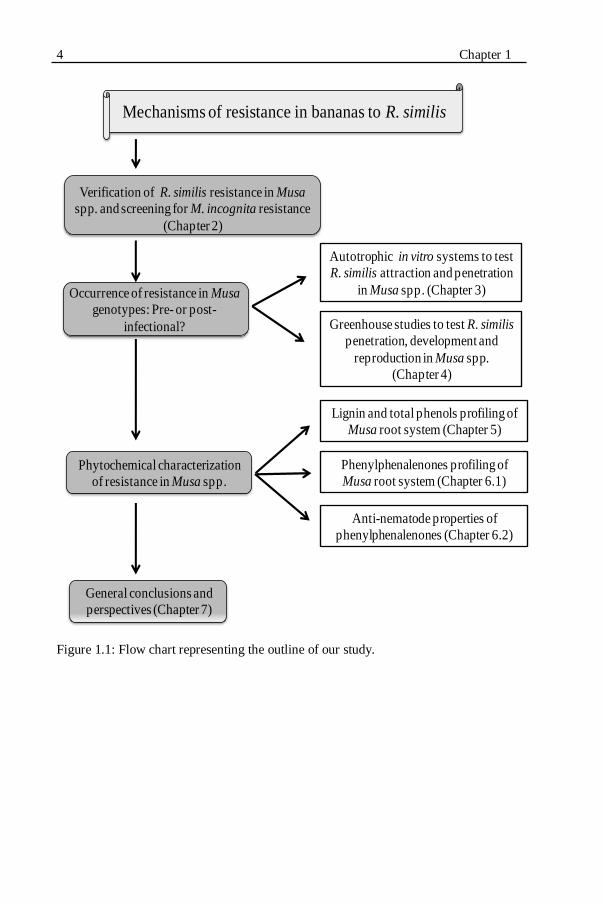
Figure 1.1: Flow chart representing the outline of our study................................4
Figure 1.2: A) Food served on a banana leaf in southern India. B) A small shop
in a local market showing a high diversity of bananas for sale...............5
Figure 1.3: Host plant/pathogen relationship terminology used in nematology.............................................................................................10
Figure 1.4: Possible mechanisms of plant resistance to nematodes...................13
Figure 1.5: Host-nematode interaction and active plant responses....................14
Figure 1.6: Structure of A) phenalen, B) phenalen-1-one and C) 9-
Figure 3.1: Banana plants cultured in an autotrophic in vitro model system.....44
Figure 3.2: Radopholus similis inoculation spots (arrows) to study nematode attraction and penetration in an autotrophic in vitro model system.......44
Figure 3.3: Two-compartment autotrophic in vitro model system to study
nematode attraction to banana plants.....................................................47
Figure 3.4: Two-compartment autotrophic in vitro model system to study
nematode attraction to banana plants.....................................................47
Fig. 3.5: Migration of Radopholus similis females towards the roots of A)
Yangambi km5 and B) Saba at 1 h after inoculation.............................49
Figure 3.6: Attraction of Radopholus similis females (expressed as a percentage
of 30 inoculated mature females) towards the roots of 4-weeks-old plants of the Musa genotypes Yangambi km5 and Saba at 1, 2, 4 and 24
h after inoculation..................................................................................49
Figure 3.7: A) Penetration of Radopholus similis females (expressed as a percentage of 30 inoculated mature females) in the roots of 4-weeks-old
plants of the Musa genotypes Yangambi km5 and Saba at 2 and 4 days
after inoculation. B) Number of eggs laid by the penetrated R. similis females at 4 days after inoculation.........................................................51
Figure 3.8: Penetration and egg laying of Radopholus similis females in banana roots grown in an autotrophic in vitro system. Roots of Saba...............52
Figure 3.9: Attraction of Radopholus similis females (expressed as a percentage
of 30 inoculated mature females) towards the roots of 4-weeks-old plants of the susceptible Musa genotype Grande Naine and the resistant
Musa genotype Yangambi km5 at 1, 3, 4 and 6 h after inoculation......53
Figure 3.10: Penetration of Radopholus similis females (expressed as a
percentage of 30 inoculated mature females) in the roots of 4-weeks-old
plants of the susceptible Musa genotype Grande Naine and the resistant Musa genotype Yangambi km5 at 1 and 2 days after inoculation.........54
Figure 3.11: Migration tracks of Radopholus similis females near banana roots in an autotrophic in vitro system observed at 24 h after inoculation.....55
Figure 3.12: Percentages of Radopholus similis females present in the attraction
zones of the two-compartment autotrophic in vitro system at 30 min, 1, 2, 3 and 6 h after inoculation of 30 mature females at the inoculation
Figure 5.2: Necrotic root cross sections of Radopholus similis-resistant and
susceptible Musa genotypes at 6 weeks after inoculation with R. similis.....................................................................................................82
Figure 5.3: Lignifications in uninfected Musa root cells at 3 and 6 weeks........84
Figure 5.4: Lignifications in Radopholus similis-infected Musa root cells at 3
and 6 weeks after inoculation with 1,000 adults and juveniles of R.
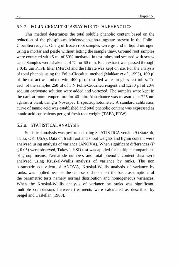
Figure 5.7: Lignin content (mg/g of fresh root weight) in root cell walls of
Radopholus similis-infected and uninfected plants of resistant and
susceptible Musa genotypes at 3 and 6 weeks after inoculation with 1,000 adults and juveniles of R. similis.................................................88
XIV
Figure 5.8: Tissue localisation of total phenols by toluidine blue staining in root
cross sections of Musa genotypes at 6 weeks after inoculation with
1,000 adults and juveniles of Radopholus similis..................................89
Figure 5.9: Total phenol contents (mg TAE/g roots) in the Radopholus similis-
infected and uninfected roots of resistant and susceptible Musa
genotypes at 3 and 6 weeks after inoculation with 1,000 adults and juveniles of R. similis.............................................................................91
Figure 5.10: Fluorescence of phenolic compounds in root cross sections of Radopholus similis-infected susceptible and resistant Musa genotypes at
3 and 6 weeks after inoculation (WAI)..................................................92
Figure 6.1: Musa plants arranged in a randomized block design in the
greenhouse to study the phytoalexins synthesis in response to
Radopholus similis infection................................................................102
Figure 6.2: Scheme followed for the isolation, cellular distribution and
structural identification of phenylphenalenones..................................103
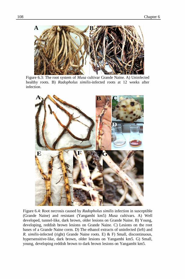
Figure 6.3: The root system of Musa cultivar Grande Naine. A) Uninfected
healthy roots. B) Radopholus similis-infected roots at 12 weeks after
Figure 6.4: Root necrosis caused by Radopholus similis infection in susceptible
(Grande Naine) and resistant (Yangambi km5) Musa cultivars.........108
Figure 6.5: TLC chromatogram of chloroform subfractions............................109
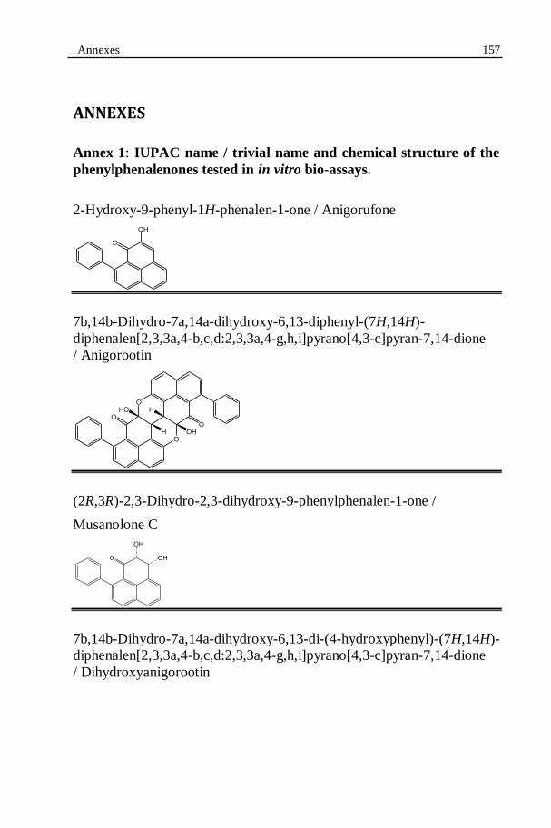
Figure 6.6: Structure of all the isolated phenylphenalenone-type phytoalexins.........................................................................................110
Figure 6.7: Analytical HPLC chromatograms of chloroform sub-fractions separated from the ethanol extracts of Musa spp.............................…111
Figure 6.8: Identification of anigorufone (1) based on the comparison of 1H
NMR spectrum of authentic compounds.............................................113
Figure 6.9: The 1H NMR spectrum of anigorufone (1)....................................113
Figure 6.10: The LDI mass spectra of Musa roots...........................................115
Figure 6.11: Mass images of the necrotic lesions on Yangambi km5 caused by
Radopholus similis infection................................................................116
XV
Figure 6.12: The heat profile of LDI-MSI for the m/z 271 in the necrotic lesions
of Yangambi km5 roots showing the distribution of anigorufone only in
the necrotic lesion................................................................................116
Figure 6.13: Mass images of the necrotic lesions on Grande Naine caused by
Radopholus similis infection................................................................117
Figure 6.14: Light microscopic images of Radopholus similis during the bio-
assay with anigorufone (1)...................................................................128
Figure 6.15: Light microscopic images of all life stages of Radopholus similis
that had died because of ingesting anigorufone (1) during the bio-
Figure 6.18: Percentage quiescent Radopholus similis observed during the
motility bio-assay on a concentration gradient of anigorufone after 24, 48 and 72 h of incubation....................................................................132
(carbofuran) and Terbufos (Counter) which are applied 2-4 times a year
(Villanueva, 2003; Quénéhervé, 2009). However, the intensive application of
these organophosphate and carbamate nematicides continues to pose a threat to
the environment and the health of agricultural workers (Atkinson et al., 2004).
Moreover, strict regulations are exercised in many countries on the use of these
nematicides that are still in use (Villanueva, 2003; Quénéhervé, 2009). Cultural
practices such as fallowing, crop rotation, etc. are also used to control
nematodes but are not applicable in banana fields. Also, they are often not
adequate enough as a sole practice to control nematodes (Lilley et al., 2007).
Complete eradication of R. similis from infested banana plantations requires a
5-years-crop free fallow as the nematode can survive on alternative weeds
(EPPO, 2008). Using resistant cultivars to manage nematode populations in
severely infested fields offers an efficient and economical alternative nematode
management strategy (Fuller et al., 2008; Zasada et al., 2010).
Introduction 9
1.4. HOST PLANT RESISTANCE IN MUSA SPP. TO
RADOPHOLUS SIMILIS
In nematology, resistance is defined as the ability of a host plant to
prevent or suppress nematode multiplication while in a susceptible host plant
nematodes can multiply. Tolerance is independent of resistance and is defined
as the ability of a host plant to suffer little damage even when quite heavily
infected with nematodes while a sensitive host plant will suffer much injury
even when relatively lightly infected with nematodes (Bos & Parleviet, 1995).
This terminology is illustrated in Figure 1.3.
Using natural host plant resistance for the management of plant-parasitic
nematodes offers a sustainable, ecologically-friendly and cost-effective
alternative to the use of pesticides (Roberts, 1992; Fuller et al., 2008).
However, most known resistance sources are specific only to certain nematode
species. Also, resistance sources to the predominant nematode species have not
been identified yet in many agricultural crops (Young, 1992; Atkinson et al.,
2003; Lilley et al., 2007).
In bananas, numerous studies were carried out around the world to
identify Musa genotypes with natural resistance to R. similis (Quénéhervé et
al., 2008b). Wehunt et al. (1978) were the first to report that Musa cultivars
belonging to the subgroup Pisang Jari Buaya (AA genome) were resistant to R.
similis. This discovery intensified the search for additional resistant sources
(see for instance Price, 1994; Fogain & Gowen, 1998; Stoffelen et al., 2000;
Viaene et al., 2003; Dochez et al., 2005, 2006; Quénéhervé et al., 2008a,
2008b, Dizon et al., 2010; Herradura et al., 2011). Yangambi km5 (AAA) was
another R. similis-resistant source that was studied elaborately (Price, 1994;
Fogain & Gowen, 1998).
10 Chapter 1
Figure 1.3: Host plant/pathogen relationship terminology used in
nematology. Source: Roberts, 2002.
1.4.1. HOST-NEMATODE INTERACTIONS
An in-depth understanding of the interactions between a host plant and its
pathogen(s) is crucial for the development of a successful management
strategy. Plant infection by nematodes is a continuous and dynamic process. In
the nematode infection cycle the following phases can be distinguished:
1) localisation of the host plant by the nematode (including migration
by the nematode towards the host plant)
2) penetration and entry of the nematode in the host plant
3) nematode development
4) nematode reproduction.
The host-nematode interactions can be either successful or unsuccessful
for the nematode. In the first case the plant is susceptible. In the second case
the plant is resistant or a non-host.
1.4.1.1. Localisation of the host plant by the nematode
Plant-released cues are exploited by plant-parasitic nematodes to guide
themselves towards the roots of their host plants. Plant roots release
chemically-rich exudates and diffusates. Additionally, several other gradients
existing in the rhizosphere, such as a CO2 gradient, microbial density, redox
potential or an electric field, can serve as guiding signals for nematodes to
locate their host plants (Perry, 1996; Spence et al., 2008). Nematodes possess
Susceptible Non-host Resistant Tolerant host
Intolerant host
Terminology
Host growth
Nematode population increase
Introduction 11 sensory receptors in their head region. The pair of amphids is considered as a
primary chemosensory receptor along with inner and outer labial sensilla
which are considered as vital in nematode host finding and identification of
suitable penetration sites (Perry, 1996). Host localisation or nematode
attraction takes place over a distance of several centimeters. Nematodes sense
the long-distance, short-distance and local attractants operating at several
centimeters, in the close proximity of roots and at the direct contact of the host
plant, respectively (Spence et al., 2008).
Long-distance cues are, in general, non-specific plant signals such as a
CO2 gradient (Robinson, 1995). Short-distance cues are usually host-specific.
Nematodes with a broader host range, such as Meloidogyne spp., seem to
respond to short-distance but non-specific plant signals (Robinson, 2002).
Nematode host finding can be inhibited by blocking either the release of host-
signaling molecules or the nematode‟s chemoreceptors (Zuckerman, 1983).
Lectins were shown to inhibit the attraction of R. similis towards the banana
roots in vitro (Wuyts et al., 2003). However, inconsistent effects were observed
as treating R. similis with lectins resulted in enhanced root penetration by the
nematode (Kaplan & Davis, 1991). Certain nematicides, such as aldicarb,
impair nematode orientation at low concentration without affecting nematode
movement (Perry, 1996).
1.4.1.2. Penetration and entry of the nematode in the host plant
Once nematodes have found the host roots, they invade the roots using
their specialised feeding apparatus, the stylet and pharyngeal gland secretions.
The stylet is a thin, hollow, needle-like tube located at the anterior end of the
nematodes. Nematodes attack the plant‟s epidermal cells by repeated stylet
thrusts to pierce the plant cell wall. In addition to physical damage, they also
produce pharyngeal gland secretions to dissolve the cell wall. Radopholus
similis possesses three large pharyngeal glands secreting different enzymes
including cell wall degrading enzymes such as endo-1,4-β-glucanases
(Haegeman et al., 2008) and endoxylanase (Haegeman et al., 2009). These two
enzymes aid R. similis in penetrating the host and in migrating inside the host
as they degrade the major cell wall components cellulose and xylan,
respectively. Radopholus similis is capable of invading the plant root at any
place albeit that they seem to prefer root tips and tender roots (Sarah et al.,
1996). In a successful penetration, nematodes breach the first-line physical and
chemical barriers of the host plant and penetrate it. In an incompatible
interaction, plants are able to block nematode penetration by early defence
12 Chapter 1 responses such as cell wall strengthening by lignifications or pre-existing
specialised cell layers, etc.
Biochemicals that may be involved in resistance of plants to nematodes
are discussed later in this chapter, in the section 1.5.
1.4.1.3. Nematode development and reproduction in the host
Following penetration, R. similis migrates through the cortex both inter-
and intra-cellularly and continuously feeds on the cortical cells by sucking and
digesting the cytoplasm using enzymatic secretions. Radopholus similis keeps
migrating to new cortical cells to feed on (Sarah et al., 1996; Gowen et al.,
2005). Juveniles develop into further developmental stages while females start
laying one to six eggs per day (Haegeman et al., 2010). Resistant plants
prevent nematode development and reproduction through hypersensitive
responses, anti-nematode toxins or by inhibiting nematode feeding.
1.4.2. POSSIBLE MECHANISMS OF PLANT RESISTANCE TO
NEMATODES
Natural plant resistance mechanisms towards nematodes (and other
pathogens) can be classified into a) preformed resistance mechanisms and b)
induced resistance mechanisms (Fig. 1.4).
1.4.2.1. Preformed resistance mechanisms
Pre-existing (passive) structural features such as root thickness, waxiness
of the cuticle, degree of secondary wall thickenings, vascular structure, etc.,
have been reported to contribute to plant resistance to pathogens including
plant-parasitic nematodes (Hutcheson, 1998). For instance, coffee clones
resistant to the root-lesion nematode P. coffeae possessed more hairy roots,
thicker epidermal and endodermal cell walls, and higher total polyphenol
contents than susceptible clones (Toruan-Mathius et al., 1995).
Introduction 13
Figure 1.4: Possible mechanisms of plant resistance to nematodes. HR: hypersensitive
response.
Constitutive antimicrobial secondary metabolites present in the plants may
contribute to the preformed chemical mechanisms of nematode resistance. The
polythienyl compounds extracted from Tagetes spp. are well documented as
anti-nematode compounds. A phytochemical, thiophene α-terthienyl isolated
from T. erecta plants was reported as toxic to the potato cyst nematode
Globodera rostochiensis, the wheat seed gall nematode Anguina tritici and the
stem and bulb nematode Ditylenchus dipsaci. In soil, the isothiocyanates and
glucosinolates released by plants belonging to the family Brassicaceae are
active against a variety of nematodes and insects (Chitwood, 2002)
1.4.2.2. Induced resistance mechanisms
Induced resistance mechanisms include active, energy-requiring responses
of plants after infection by a pathogen. The triggering of these strong responses
requires specific recognition of certain molecules from the invading pathogen
which are collectively known as elicitors. Experiments have shown that in the
absence of the pathogen, elicitors are capable of initiating the active plant
defence response (Garcia-Brugger et al., 2006; Sanchez-Estrada et al., 2009).
When resistant plants recognise matching elicitors, intracellular signal
transduction pathways are activated resulting in an integrated series of plant
responses leading to eventual neutralization of the invading pathogen (Fig. 1.5)
(Dixon et al., 1994; Williamson & Hussey, 1996; Das et al., 2008; Fuller et al.,
2008).
Resistant mechanisms
Preformed
(Passive)Infection-Induced
(Active)
Structural barriers e.g. waxy cuticle, branched roots
Chemical barriers
e.g.phytoanticipins
Hypersensitive responses (HR)
Antibiotics, toxins production
- Phytoalexins
Systemic acquired responses
Structural e.g. cell wall
strengthening by lignin, suberin
Other Induced phytochemicals
e.g.
14 Chapter 1
Figure 1.5: Host-nematode interaction and active plant responses. Nematode-released
elicitors activate signal transduction pathways in the plant resulting in many induced plant responses to limit nematode development and reproduction. Source: Williamson
and Hussey, 1996 (modified).
Key reactions of plants primarily include the hypersensitive response
(HR) that consists of rapid death of plant cells in the immediate vicinity of the
pathogen restricting the pathogen development. This is the primary response of
the cell which comes in contact with the pathogenic organism (Gilchrist, 1998;
Lam et al., 2001; Fuller et al., 2008).
Secondary responses are induced in the adjacent cells surrounding the
initial infection site in response to diffusible signal molecules that are produced
during the primary interaction. Secondary responses include the production and
accumulation of toxic natural products known as phytoalexins and hydrolytic
enzymes to antagonise the development of the pathogen (Hammerschmidt,
1999; Gheysen & Fenoll, 2002). Toxic secondary metabolites are deployed to
site of pathogen challenge by vesicle-mediated trafficking (Field et al., 2006).
The third category of active plant defense responses is associated with
systemically acquired resistance (SAR) that is hormonally induced throughout
the plant (Hutcheson, 1998; Conrath, 2006). For instance nicotine is
biosynthesised in root cells following insect attack or wounding. After its
biosynthesis, the nicotine is translocated to the leaves of tobacco via the xylem
IDENTIFICATION OF COMBINED RESISTANCE TO RADOPHOLUS SIMILIS AND MELOIDOGYNE INCOGNITA
IN MUSA GERMPLASM2
2 The results presented in this chapter were published as: SUGANTHAGUNTHALAM, D.,
ELSEN, A. AND DE WAELE, D. (2010). Identification of combined resistance to
Radopholus similis and Meloidogyne incognita in Musa germplasm. International
Journal of Nematology 20:19-24.
28 Chapter 2
2.1. INTRODUCTION
Burrowing and root-knot nematodes are root pathogens that can cause
serious yield losses to Musa spp. (Gowen et al., 2005). Worldwide, the
burrowing nematode Radopholus similis (Cobb, 1893) Thorne, 1949 is
considered as the most damaging nematode species in Musa-based cropping
systems (Sarah et al., 1996; Gowen et al., 2005). However, under field
conditions, usually several nematode species co-exist in the roots of bananas
and plantains (De Waele & Elsen, 2007), and in the absence of R. similis, other,
less important nematode species such as the root-knot nematode Meloidogyne
incognita (Kofoid and White, 1919) Chitwood, 1949 can become more
damaging to Musa spp. (De Waele & Davide, 1998; De Waele, 2000; Brentu et
al., 2004; Cofcewicz et al., 2005; Van den Bergh et al., 2006).
The use of host plant resistance is considered as an economical and
environmental-friendly nematode management strategy in the low-input
cropping systems prevailing in developing countries (Roberts, 1992). This
strategy also ensures maximal land use efficiency without changing the existing
cropping systems (Atkinson et al., 2003). In Musa, a relatively high number of
studies have been carried out to identify natural sources of resistance to R.
similis resulting in the identification of some potential R. similis-resistant Musa
genotypes (see for instance, Wehunt et al., 1978; Fogain & Gowen, 1998;
Stoffelen et al., 2000; Viaene et al., 2003; Dochez et al., 2005, 2006;
Quénéhervé et al., 2008a, 2008b; Herradura et al., 2011). However, the
efficient use of natural sources of resistance to nematodes is limited due to
several reasons (Roberts, 1992). A major limitation is that, in most cases, the
resistance is restricted to one nematode species or a few pathotypes of the same
nematode species. Planting of species-specific or pathotype-specific resistant
cultivars under field conditions has resulted in a shift in nematode population
composition causing less important nematode species to become more
damaging and, sometimes, even a major problem (Young, 1992). To overcome
this loss of efficiency, identification of plant genotypes with resistance to more
than one nematode species or pathotype is necessary. Moreover, if these plant
genotypes can be used in breeding programmes, their identification could lead
to the development of cultivars with multiple nematode resistances.
The objective of this part of our study was to evaluate the host response of
selected Musa genotypes to both R. similis and M. incognita. The Musa
genotypes studied were selected based on their resistant host response to R.
similis as reported by Dochez et al. (2006). For their experiments, Dochez et al.
(2006) used sucker-derived planting material. Some studies have demonstrated
Combined resistance to R. similis and M. incognita 29
that tissue culture-derived Musa planting material can be more susceptible to R.
similis compared to sucker-derived planting material (Blomme et al., 2004;
Viaene et al., 2003) apparently due to their more fragile root system. In spite of
this, tissue culture-derived plants are preferred as planting material for the
initiation of new or the rejuvenation of existing commercial plantations and also
for research purposes because of their homogeneity, availability throughout the
year and because they are free of pathogens. In the first part of our study we re-
confirmed the host response of the R. similis-resistant Musa genotypes reported
by Dochez et al. (2006) using this time tissue culture-derived plants. In the
second part of our study, we examined the host response of the same Musa
genotypes to M. incognita, also using tissue culture-derived plants.
2.2. MATERIALS AND METHODS
2.2.1. EXPERIMENTAL SET-UP
Two experiments were carried out in the greenhouse at 27 and 20 °C day
and night temperatures, respectively, 12 h photoperiod and 80% relative
humidity. In the first experiment, seven Musa genotypes were examined for
their host response to R. similis. This experiment was carried out in two
separate batches. The host response of each genotype was compared to the host
response of a susceptible (Grande Naine) and two resistant (Yangkambi km5,
Pisang Jari Buaya) reference cultivars (Speijer & De Waele, 1997). In the
second experiment, eight Musa genotypes were examined for their host
response to M. incognita. In this experiment, Grande Naine was included as the
susceptible reference cultivar (Speijer & De Waele, 1997). There was no
resistant reference cultivar included as no Musa genotypes with resistance to M.
incognita were known. In both experiments, all treatments were replicated eight
times and a randomized block design was used. Some important characteristics
of the selected genotypes and reference cultivars are listed in Table 2.1.
2.2.2. PLANTING MATERIAL
All Musa genotypes were initially obtained from the Musa germplasm
collection maintained at the International Transit Centre (ITC), K.U.Leuven,
Belgium. The plant material was proliferated, regenerated and rooted in test
tubes on Murashige and Skoog medium including vitamins, 30 g/l sugar, 10
mg/l ascorbic acid and 2 g/l gelrite with pH 6.12 (Murashige & Skoog, 1962).
Proliferation was obtained by adding 10-6
M indole-3-acetic acid (IAA) and 10-5
M 6-benzylaminopurine (BAP). For rooting, 0.05% (w/v) active charcoal was
30 Chapter 2
added (Banerjee & De Langhe, 1985). No plant growth regulators were added.
The plantlets were grown in growth chambers at 28 °C and 16 h photoperiod.
Table 2.1. Characteristics of selected Musa genotypes and the reference cultivars*
evaluated for their host response to Radopholus similis and Meloidogyne
incognita.
ITC: International Transit Centre, K.U.Leuven, Belgium.
S: susceptible; R: resistant.
Eight-weeks-old rooted tissue culture plantlets were planted in 1 l pots
filled with sand and potting soil (2:1). The potted plants were maintained under
greenhouse conditions (Fig. 2.1) and fertilized at 10 days intervals throughout
the experiments.
Host response
Genotype
(subgroup)
ITC Genome Use R. similis M. incognita
Gia Hui
(Pisang Awak)
1143
ABB
cooking
-
-
Kokopo 1243 AA cooking/
dessert
- -
Long Tavoy
(Burbannica)
0283
AA
wild
-
-
Marau 0772 AAA
(AAB)
cooking - -
Pisang Mas
(Sucrier)
0653
AA
dessert
-
-
Pora Pora 0868 AA cooking/
dessert
- -
Saba 1138 ABB cooking - -
Vudu Papau 0590 AA dessert - -
Grande Naine*
(Cavendish)
1256
AAA
dessert
S
S
Yangambi km5*
(Ibota)
1123
AAA
dessert
R
-
Pisang Jari Buaya* 0312 AA dessert R -
Combined resistance to R. similis and M. incognita 31
Figure 2.1: Experimental set-up of the Radopholus similis host response experiment
in the greenhouse.
2.2.3. NEMATODE INOCULUM AND INOCULATION
A population of R. similis originally isolated from banana roots in Uganda
was used in the first experiment. This population was maintained and multiplied
monoxenically on sterile carrot discs at 25+1 °C in the dark (Speijer & De
Waele, 1997). The population from Uganda was characterized by a high
reproductive fitness (Fallas et al., 1995). A M. incognita population originally
isolated from banana roots in Malaysia was used in the second experiment. This
population was maintained in vitro on Ri T-DNA-transformed tomato roots
(Verdejo et al., 1988). This population was multiplied in vivo on tomato cv.
Marmande roots under greenhouse conditions.
For the first experiment, R. similis was extracted from the carrot discs by
the maceration-sieving technique (Speijer & De Waele, 1997). Eight weeks
after planting, approximately 1,000 living vermiform nematodes were
inoculated per plant by pipetting 4 ml of the nematode suspension into
inoculation holes made in the soil near the plant root zone. For the second
experiment, M. incognita was extracted from the in vivo tomato cv. Marmande
roots by treating the roots with 1% NaOCl for 4 min to dissolve the gelatinous
matrix of the egg masses followed by the maceration-sieving method (Speijer &
De Waele, 1997; Hooper et al., 2005). Eight weeks after planting,
32 Chapter 2
approximately 4,000 eggs and juveniles were inoculated per plant by pipetting 4
ml of the nematode suspension into inoculation holes made in the soil near the
plant root zone.
2.2.4. EVALUATION OF THE HOST PLANT RESPONSE
Eight weeks after nematode inoculation (i.e. 16 weeks after planting),
plants were uprooted and gently washed free of soil under running tap water.
The fresh root and shoot weights, root damage and nematode reproduction were
assessed.
Radopholus similis
Estimation of the percentage root necrosis caused by R. similis was based
on the scoring of five randomly selected, 10-cm-long, longitudinally sliced
functional primary roots (Speijer & De Waele, 1997). Then, the same roots
were cut into 1-cm-long pieces, weighed and, if necessary, additional roots
were sampled to make up 15 g of roots for nematode extraction by the
maceration-sieving technique (Speijer & De Waele, 1997). The roots were
macerated in a kitchen blender for three periods of 10 sec each separated by a 5
sec interval. The macerated suspension was passed through a series of 250, 100,
40 and 25 µm aperture sieves. Nematodes were collected from the 40 and 25
µm aperture sieves and pooled. Eggs, juveniles, females and males were
counted separately from a 2 ml aliquot out of the homogenised suspension. The
final nematode population densities were determined by calculating the sum of
all developmental stages of the nematodes present in the total root system. The
reproduction factor (Rf) was calculated as the final nematode population
density divided by the inoculum. The host response of each genotype to R.
similis was determined based on a comparison of the final nematode population
density of each genotype with the reference cultivars as explained in Table 2.2.
Combined resistance to R. similis and M. incognita 33
Table 2.2. Identification of the host response of selected Musa genotypes to Radopholus similis based on a comparison with the host response of a susceptible
(Grande Naine) and a resistant (Yangambi km5) reference cultivar.
Significance was measured according to the Dunnett‟s test (P < 0.05).
Meloidogyne incognita
Roots were cut into 1- to 2-cm-long pieces; a sample of 5 g roots was taken
at random and stained with phloxine B for visual observation of the egg masses
(Hooper et al., 2005). The red stained egg masses were counted as egg-laying
females using a stereoscopic microscope. After counting the egg-laying
females, the same roots were used to extract the eggs and juveniles by
solubilising the gelatinous matrix with 1% NaOCl followed by the maceration-
sieving technique (Speijer & De Waele, 1997). The roots were macerated in a
kitchen blender for three periods of 10 sec each separated by a 5 sec interval.
The macerated suspension was passed through a series of 250, 100, 40 and 25
µm aperture sieves. Eggs and juveniles retained on the 40 and 25 µm aperture
sieves were collected, pooled and counted in a 2 ml aliquot out of the
homogenised suspension using a stereoscopic microscope. The final M.
incognita population density (Pf) was calculated by the sum of eggs and
(mostly) second-stage juveniles (J2) present in the whole root system. The
reproduction factor (Rf) was calculated as the final number of eggs and J2
divided by the inoculum. The host response of each genotype to M. incognita
was determined as follows: the genotypes with a significantly lower final
nematode population density compared to the susceptible reference cultivar
Grande Naine were categorised as resistant while the genotypes with a final
nematode population density which was not significantly different compared to
the susceptible reference cultivar Grande Naine were categorised as susceptible.
Statistical analysis was performed using STATISTICA version 7 (StatSoft,
Tulsa, OK, USA). Prior to each analysis, the basic assumptions for parametric
statistics, namely normal distribution and homogeneity of variances were tested
(Anonymous, 2007). Based on these tests, the nematode population densities
were log10 (x+1) transformed. Analysis of variance was performed to determine
the host response of the Musa genotypes to R. similis and M. incognita. The
final nematode population density of each genotype was compared to the final
nematode population densities of the reference cultivars by the two-sided
Dunnett‟s test (P < 0.05).
2.3. RESULTS
2.3.1. HOST RESPONSE TO R. SIMILIS
The results of the experiment on the host response to R. similis are
summarised in Table 2.3. The results show that the plants had good growing
conditions during the experiment and this resulted in fresh root weights ranging
from an average of 22.8 g in Gia Hui to an average of 76.3 g in Pisang Jari
Buaya.
Significantly (P < 0.05) higher numbers of nematodes were present in the
susceptible reference cultivar Grande Naine compared to the resistant reference
cultivar Yangambi km5. The final nematode population density was about 4
and 5 times higher in Grande Naine than in Yangambi km5 in the first and
second batch, respectively. Lesions on Grande Naine were long, continuous and
tunnel-like (Fig. 2.2A). Lesions on Yangambi km5 were short and
discontinuous resembling hypersensitive lesions (Fig. 2.2B). The other resistant
reference cultivar, Pisang Jari Buaya, expressed an inconsistent host response
since it was resistant to R. similis in the first batch but its host response was
inconclusive in the second batch.
Combined resistance to R. similis and M. incognita 35
Figure 2.2: Root necrosis caused by Radopholus similis at 8 weeks after
inoculation with 1,000 nematodes. A) Long, continuous and tunnel-like root
lesions on the susceptible reference cultivar Grande Naine. B) Short,
discontinuous, hypersensitive-like lesions on the resistant reference cultivar
Yangambi km5.
Out of the seven Musa genotypes tested, Long Tavoy, Saba, Pisang Mas
and Pora Pora were resistant to R. similis based on their final nematode
population density. Marau expressed a partial resistance to R. similis. The host
response of Kokopo was inconclusive and Gia Hui was susceptible to R. similis.
2.3.2. HOST RESPONSE TO M. INCOGNITA
The results of the experiment on the host response to M. incognita are
summarised in Table 2.4.
Figure 2.3: Egg masses of Meloidogyne incognita in Musa plants stained with phloxin B. A) Egg masses on a secondary root (x40). B) Egg mass inside a thick
primary root surrounded by a layer of phenolic root cells (x100).
A B
A)
B)
A)
36 Chapter 2
The results show that the plants had good growing conditions during the
experiment and this resulted in fresh root weights ranging from an average of
11.6 g in Kokopo to an average of 45.2 g in Pisang Mas. Although
characteristic galls were not very conspicuous for direct observation in our
study, many egg-laying females and egg masses were observed during counting
(Fig. 2.3). The susceptible reference cultivar Grande Naine had a high number
of egg-laying females per 5 g roots and the highest final nematode population
density per 5 g roots and per root system. Out of the eight Musa genotypes
tested, Vudu Papau and Pisang Mas were resistant to M. incognita based on
their final nematode population density. The number of egg-laying females per
5 g roots was low in Pisang Mas but in Vudu Papau as high as in Grande Naine.
Nevertheless, the final nematode population density per 5 g roots and per root
system was about 5 to 6 times lower in Vudu Papau than in Grande Naine. The
other genotypes tested were susceptible to M. incognita.
2.4. DISCUSSION
The resistance to R. similis of Long Tavoy, Saba, Pisang Mas and Pora
Pora observed when using sucker-derived plants (Dochez et al., 2006) was
confirmed by our study with tissue culture-derived plants. However, in our
study Gia Hui was susceptible to R. similis while in the study by Dochez et al.
(2006) this genotype was resistant. Our study confirms other reports (De Waele
et al., 1998; Stanton, 1999; Viaene et al., 2003; Blomme et al., 2001, 2003,
2004) that tissue culture-derived plants of some Musa genotypes could be
susceptible and sensitive to R. similis while sucker-derived plants of the same
genotype are resistant. It is possible that the R. similis resistance genes are not
yet expressed by tissue-cultured plants at the time of infection while they are
being expressed at the same time in sucker-derived plants. Previously, the host
response of Pisang Mas to R. similis was reported as less susceptible (Binks &
Gowen, 1997) and partially resistant (Marin et al., 2000). Conversely, another
study (Stoffelen et al., 1999) reported Pisang Mas as susceptible to R. similis.
These observations underline the necessity to confirm the resistant host
response observed in Musa genotypes to R. similis under a range of
experimental conditions, including different types of planting material
(Herradura, 2009).
Combined resistance to R. similis and M. incognita 37
Table 2.3. Host response of selected Musa genotypes and the reference cultivars† to Radopholus similis, measured at 8 weeks after inoculation
with 1,000 vermiform nematodes per plant (n = 8). Plants were inoculated at 8 weeks after planting.
Genotype Fresh root
weight
Pf Rf Comparison of Pf/root system with Host
response (g) per g roots per root system Grande Naine Yangambi km5
Batch 1
Long Tavoy
62.2
86
5331
5.3
*
n.s.
R Long Tavoy 62.2 86 5,331 5.3 * n.s. R Saba 61.2 151 9,521 9.5 * n.s. R
Pisang Mas 53.6 277 15,079 15.1 * n.s. R Marau 43.5 375 15,457 15.5 * * PR Grande Naine† 54.7 485 26,499 26.5 - * S
Yangambi km5† 56.4 134 7,332 7.3 * - R Pisang Jari Buaya† 76.3 150 11,350 11.4 * n.s. R
Batch 2
Pora Pora
69.7
558
37958
38.0
*
n.s.
R Pora Pora 69.7 558 37,958 38.0 * n.s. R
Kokopo 49.9 811 38,404 38.4 n.s. n.s. I Gia Hui 22.8 3,008 62,115 62.1 n.s. * S Grande Naine† 45.2 1,543 69,444 69.4 - * S Yangambi km5† 64.7 245 13,885 13.9 * - R
Pisang Jari Buaya† 65.7 631 40,748 40.7 n.s. n.s. I
Pf: final nematode population density; Rf: reproduction factor (Pf/Pi; Pi: initial nematode inoculum density).
*: significantly different; n.s.: not significantly different according to Dunnett‟s test (P < 0.05).
Means within a same column followed by the same letter are not significantly (P ≤
0.05) different according to Tukey‟s HSD test.
No significant differences were observed between the numbers of
nematodes that had penetrated the four Musa genotypes at 4 and 8 DAI (Fig.
4.3). At 12 DAI, a significantly (P ≤ 0.05) lower number of nematodes had
penetrated the roots of Long Tavoy compared to Grande Naine. No significant
differences were observed between the number of nematodes that had
penetrated Saba, Yangambi km5 and Grande Naine at 12 DAI (Fig. 4.3). A
decrease in the number of penetrated nematodes was observed in all genotypes
at 12 DAI.
Eggs were laid by the females that had penetrated in Grande Naine at 8
DAI but not in any of the three resistant Musa genotypes at 8 DAI. At 12 DAI,
eggs were also observed in the roots of Long Tavoy but the number of eggs was
significantly (P ≤ 0.05) lower than in Grande Naine. No eggs were observed in
Saba and Yangambi km5 (Fig. 4.4).
R. similis penetration, development and reproduction 67
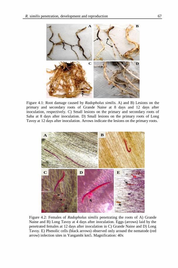
Figure 4.1: Root damage caused by Radopholus similis. A) and B) Lesions on the
primary and secondary roots of Grande Naine at 8 days and 12 days after
inoculation, respectively. C) Small lesions on the primary and secondary roots of
Saba at 8 days after inoculation. D) Small lesions on the primary roots of Long
Tavoy at 12 days after inoculation. Arrows indicate the lesions on the primary roots.
Figure 4.2: Females of Radopholus similis penetrating the roots of A) Grande
Naine and B) Long Tavoy at 4 days after inoculation. Eggs (arrows) laid by the
penetrated females at 12 days after inoculation in C) Grande Naine and D) Long
Tavoy. E) Phenolic cells (black arrows) observed only around the nematode (red
arrow) infection sites in Yangambi km5. Magnification: 40x
A B
C D
A
C D E
B
68 Chapter 4
Figure 4.3: Number of Radopholus similis that had penetrated the roots of the resistant (Long Tavoy, Saba, Yangambi km5) and the susceptible (Grande Naine) Musa
genotypes at 4, 8 and 12 days after inoculation with 1,000 adults and juveniles of
Radopholus similis. Data were log10(x+1) transformed prior to statistical analysis. The
original means are presented between parentheses. Means within a same time (DAI)
followed by the same letter are not significantly (P ≤ 0.05) different from each other
according to Tukey‟s HSD test. Error bars represent confidence intervals. (n=8).
Figure 4.4: Number of Radopholus similis eggs observed in the roots of the resistant
(Long Tavoy, Saba, Yangambi km5) and the susceptible (Grande Naine) Musa
genotypes at 12 days after inoculation with 1,000 adults and juveniles of Radopholus
similis. Means are presented between parentheses. Means followed by the same letter
are not significantly (P ≤ 0.05) different from each other according to Tukey‟s HSD test.
Error bars represent confidence intervals. (n=8).
R. similis penetration, development and reproduction 69
4.3.2. SECOND EXPERIMENT: NEMATODE DEVELOPMENT AND
REPRODUCTION
The fresh root and shoot weights of the Musa genotypes at 26 days after
inoculation are presented in Table 4.2. Long Tavoy had the highest root and
shoot weights while Grande Naine had the lowest root and shoot weights.
Table 4.2. Fresh root and shoot weights of the Musa genotypes at 26 days after
inoculation (DAI) with 1,000 adults and juveniles of Radopholus similis. The
nematodes were inoculated around the roots of six-weeks-old plants. (n=8).
methyl ether. The resultant pellets are the purified cell walls. The purified cell
wall pellets were freeze-dried overnight.
Approximately 15 mg of the cell wall preparations were taken and
suspended in a mixture of 1 ml HCl (2 M) and 200 µl thioglycolic acid.
Suspensions were incubated for 4 h in a water bath at 95+2 ºC. After cooling
down to room temperature, samples were centrifuged for 10 min at 16,000 x g.
Pellets were washed three times with MilliQ water, suspended in 1 ml NaOH
(0.5 M) and vigorously shaken overnight to extract the lignothioglycolic acid
(LTGA). Samples were centrifuged for 10 min at 16,000 x g and the
supernatants were collected. The pellets were washed with an additional 500 µl
of NaOH (0.5 M). The combined supernatants (alkali extracts) were acidified
with 300 µl concentrated HCl and incubated for 4 h at 4 ºC to precipitate the
LTGA from the alkali extracts. Samples were centrifuged for 10 min at 16,000
x g and pellets were dried in a SpeedVac centrifuge. Dry brown pellets were
dissolved in 1 ml NaOH (0.5 M) to measure the absorbance.
Absorbance of the samples was measured against the blank at 280 nm in a
NanoDrop spectrophotometer using the UV-Vis high absorbance mode (ND-
1000, NanoDrop Technologies, Inc., Wilmington, USA). The lignin content
(LTGA) was expressed as mg lignin per g fresh root weight using a calibration
curve of alkali lignin (Sigma-Aldrich, Inc., Borem, Belgium).
78 Chapter 5
5.2.7. FOLIN-CIOCALTEU ASSAY FOR TOTAL PHENOLICS
This method determines the total soluble phenolic content based on the
reduction of the phospho-molybdene/phospho-tungstate present in the Folin–
Ciocalteu reagent. One g of frozen root samples were ground in liquid nitrogen
using a mortar and pestle without letting the sample thaw. Ground root samples
were extracted with 5 ml of 50% methanol in test tubes and secured with screw
caps. Samples were shaken at 4 ºC for 60 min. Each extract was passed through
a 0.45 µm PTFE filter (Merck) and the filtrate was kept on ice. For the analysis
of total phenols using the Folin-Ciocalteu method (Makkar et al., 1993), 100 µl
of the extract was mixed with 400 µl of distilled water in glass test tubes. To
each of the samples 250 µl of 1 N Folin-Ciocalteu reagent and 1,250 µl of 20%
sodium carbonate solution were added and vortexed. The samples were kept in
the dark at room temperature for 40 min. Absorbance was measured at 725 nm
against a blank using a Novaspec II spectrophotometer. A standard calibration
curve of tannic acid was established and total phenolic content was expressed as
tannic acid equivalents per g of fresh root weight (TAE/g FRW).
5.2.8. STATISTICAL ANALYSIS
Statistical analysis was performed using STATISTICA version 9 (StatSoft,
Tulsa, OK, USA). Data on fresh root and shoot weights and lignin content were
analysed using analysis of variance (ANOVA). When significant differences (P
≤ 0.05) were observed, Tukey‟s HSD test was applied for multiple comparisons
of group means. Nematode numbers and total phenolic content data were
analysed using Kruskal-Wallis analysis of variance by ranks. The non
parametric equivalent of ANOVA, Kruskal-Wallis analysis of variance by
ranks, was applied because the data set did not meet the basic assumptions of
the parametric tests namely normal distribution and homogeneous variances.
When the Kruskal-Wallis analysis of variance by ranks was significant,
multiple comparisons between treatments were calculated as described by
Siegel and Castellan (1988).
Lignin and Phenols involved in plant-nematode interactions 79
5.3. RESULTS
5.3.1. ROOT AND SHOOT WEIGHT
The effect of R. similis infection on the root and shoot fresh weights of the
Musa genotypes are summarised in Table 5.1.
Table 5.1. Mean fresh root and shoot weights of Radopholus similis-infected and
uninfected Musa genotypes at 3 and 6 weeks after inoculation with 1,000 adults and
juveniles of R. similis.
Treatments Saba Long Tavoy Yangambi km5 Grande Naine
Root fresh weight (g)
3 weeks
+ R. similis 33.8* 44.4* 32.1 17.3
- R. similis 33.3 38.4* 28.7 19.5
6 weeks
+ R. similis 53.4* 64.0* 69.5†* 34.7
- R. similis 51.7 55.2* 46.1† 37.0
Shoot fresh weight (g)
3 weeks + R. similis 88.1* 107.8* 91.0* 51.6
- R. similis 87.1* 113.1* 83.0 58.5
6 weeks
+ R. similis 114.0* 131.8* 116.2* 73.4
- R. similis 113.3* 116.7* 102.7 79.0
Means followed by † indicates significant differences (P ≤ 0.05) between the R. similis-
infected and uninfected plants of the same genotype within the same time (3 or 6 weeks). Means followed by * indicates significant differences (P ≤ 0.05) compared to the mean
shoot or root weight of the susceptible reference genotype Grande Naine in the same
row. Significance of the differences was tested using the Tukey‟s HSD test.
Root weights of infected Yangambi km5 plants were significantly (P ≤
0.05) higher than the root weights of the uninfected plants of the same genotype
at 6 weeks after infection. No significant differences in root weight between
infected and uninfected plants were observed for the three other Musa
genotypes. No significant differences in shoot weight between infected and
uninfected plants were observed. The fresh root and shoot weights of Grande
Naine were always lowest compared with the other three Musa genotypes.
80 Chapter 5
5.3.2. ROOT ANATOMY AND CELLULAR DAMAGE IN INFECTED
ROOTS
Thin, fresh, hand-cut root cross sections were made from all the Musa
genotypes to compare the anatomical structures of the uninfected and R. similis-
infected plants. In the infected plants, nematode damage and lesion patterns
were studied. The anatomical root structures in the uninfected Musa root cross
sections are shown in Figure 5.1. The anatomy of the healthy roots was similar
to previous descriptions of Musa roots (Acquarone, 1930; Wuyts, 2006).
The outermost layer is the root epidermis lining the root cortex. Few
outermost cortical layers are thick cork cells. The outer cortex layers are
composed of large cells, arranged in irregular radial rows with small
intercellular spaces. The inner side of the outer cortex continues to form
aerenchyma previously described as lacunae by Acquarone (1930). The
aerenchyma is wide and the cavities are lined by radial cortical cell plates. The
inner cortex follows the aerenchyma internally. The inner cortical cells are
smaller, cylindrical and arranged in more regular radial rows with well defined
larger intercellular spaces (Fig. 5.1A). The inner cortical cells become smaller
for every inner row. The innermost cortex layer is a layer of very small, well
packed cells called endodermis. Suberin deposition thickens the lateral sides of
endodermal cells.
The pericycle is the outermost layer of the vascular bundle made of a
single layer of cells lying just below the endodermis. As a monocotyledon root,
the vascular bundle is a polyarch type and radial in arrangement. The outer
protoxylem vessels lie against the pericycle. The metaxylem vessels are
strikingly large. Each xylem vessel is lined by a layer of sclerenchyma (wood
parenchyma). The peripheral and central phloem strands are found in between
the xylem vessels (Fig. 5.1B).
Lignin and Phenols involved in plant-nematode interactions 81
Figure 5.1: Anatomical root structures of Musa spp. cv. Saba (cross sections).
A) Epidermis, inner and outer cortex, aerenchyma and vascular bundle (stele)
(x40). B) The vascular bundle with its different structures (x100); en:
The necrotic lesions in R. similis-infected root cross sections were
observed under a compound, transmitted light microscope. The images of the
lesions are presented in Figure 5.2. The necrosis started from the epidermis and
extended into the outer cortex, aerenchyma and inner cortex but it was more
extensive in the aerenchyma of all the four Musa genotypes (Fig. 5.2C).
Necrosis was also observed in the endodermis, extending into the outer layers
of vascular bundle of Yangambi km5 at 3 weeks after infection. Observation of
the lesions showed that the cells were dark brown coloured in Yangambi km5
and Saba (Figs 5.2B, E & F) and reddish brown coloured in Grande Naine (Fig.
5.2A).
epidermis
Outer cortex
Aerenchyma
Inner cortexStele
pc
mxpx
en
ph
A
B
82 Chapter 5
Figure 5.2: Necrotic root cross sections of Radopholus similis-resistant and susceptible
Musa genotypes at 6 weeks after inoculation with R. similis.
A) Young lesions extending from the cortex to the aerenchyma cells of Grande Naine.
B) Dark brown necrosis in the cortex of Saba. C) Lesions in the aerenchyma cells of
Long Tavoy. D) Phenolic compounds starting to accumulate in a newly developing
lesion in Long Tavoy. E) Developing lesions with high phenol contents (dark brown
cells) in the necrotic areas of Yangambi km5. F) Well developed lesions in Yangambi
km5. Magnification of the images except D: 100x; D: 400x
5.3.3. NEMATODE NUMBERS IN NECROTIC LESIONS
The numbers of R. similis in the necrotic lesions of the resistant and
susceptible Musa genotypes at 6 weeks after infection are summarised in Table
5.2. The mean number of nematodes extracted from the necrotic lesions of
susceptible Musa genotype Grande Naine was significantly (P ≤ 0.05) higher
A
FE
DC
B
Lignin and Phenols involved in plant-nematode interactions 83
than the number of nematodes extracted from the resistant Musa genotypes
Saba, Long Tavoy and Yangambi km5.
Table 5.2. Mean number of Radopholus similis in the necrotic lesions of three resistant
(Saba, Long Tavoy & Yangambi km5) and one susceptible (Grande Naine) Musa
genotypes at 6 weeks after inoculation with 1,000 adults and juveniles of R. similis
(n=4).
Genotype Nematodes/g of necrotic lesion
Saba 583 a
Long Tavoy 254 a
Yangambi km5 255 a
Grande Naine 1057 b
Means within the same column followed by the same letter are not
significantly (P ≤ 0.05) different from each other according to the Kruskal-
Wallis analysis of variance by ranks.
5.3.4. HISTOCHEMICAL STAINING OF MUSA ROOT CROSS SECTIONS
FOR LIGNIFIED CELL WALLS
Lignified cell walls fluoresce under UV light when stained with DPBA.
When the root section is viewed through a FITC filter, the syringyl units
fluoresce in green and the guaiacyl units fluoresce in bright yellow colour. The
uninfected plants of all Musa genotypes sampled at 6 weeks showed intense
fluorescence (Figs 5.3B, D, F, G & I) compared to the plants sampled at 3
weeks after infection (Figs 5.3A, C, E & H) i.e. older roots showed more
extensive secondary wall thickening due to lignin deposition. The uninfected
plants of the R. similis-resistant genotype Saba showed distinct lignification of
aerenchyma with syringyl units (Fig. 5.3D).
Radopholus similis-infected plants (Fig. 5.4) showed much intense
fluorescence in the vascular bundles and endodermis compared to the
uninfected plants of the same age (Fig. 5.3). The R. similis-infected plants of
Long Tavoy showed the most extensive secondary wall lignification of vascular
parenchyma (sclerenchyma) cells (Figs 5.4I & J) compared to all other
genotypes. Lignification starts at the endodermis (Fig. 5.4E), extending to the
peripheral tissues of vascular system especially the xylem walls, xylem-
accompanying parenchyma cells (Fig. 5.4H) and eventually the lignification
progress to the vascular parenchyma (sclerenchyma) cells in the centre (Figs
5.4I & J).
84 Chapter 5
Figure 5.3: Lignifications in uninfected Musa root cells at 3 and 6 weeks.
Fresh, hand-cut root cross sections were stained with DPBA and observed under
epifluorescence microscope using FITC (all images except G) and DAPI filter
(G). A) Vascular bundle of Grande Naine at 3 weeks showing no cell wall
lignification and B) initial lignification of protoxylem cells of Grande Naine at 6
weeks. C) Initial lignifications in the xylem-accompanying vascular parenchyma
cells of Saba at 3 weeks and D) lignified aerenchymatic cells at 6 weeks in Saba.
E) Initial lignifications of parenchyma cells surrounding xylem cells of Long
Tavoy at 3 weeks and F) & G) lignified vascular cell (sclerenchyma) walls of
Long Tavoy at 6 weeks. H) Initial lignifications of xylem cells of Yangambi km5 at 3 weeks and I) strong lignifications progressing to the centre of the vascular
bundles of Yangambi km5 at 6 weeks. Arrows are pointing at the lignified cell
walls. Magnification of the images: 100x.
A B
C D
F GE
IH
Lignin and Phenols involved in plant-nematode interactions 85
Figure 5.4: Lignifications in Radopholus similis-infected Musa root cells at 3 and 6 weeks after inoculation with 1,000 adults and juveniles of R. similis
Fresh, hand-cut root cross sections were stained with DPBA and observed under
epifluorescence microscope using FITC (except C, G & J) and DAPI filter (C, G & J).
A) & B) Initial lignifications of parenchyma cells surrounding protoxylem of Grande
Naine at 3 weeks. C) & D) Extended lignifications of vascular bundles of Grande Naine
at 6 weeks. E) Localisation of syringyl lignin (arrow) in the endodermis of Saba roots at
3 weeks. F) & G) Lignifications in the xylem accompanying parenchyma cells of Saba
at 6 weeks. H) Intense syringyl lignifications (arrows) in the peripheral vascular cells of
Long Tavoy at 3 weeks. I) & J) Intense guaiacyl lignification in the vascular
parenchyma cells of Long Tavoy at 6 weeks. K) Intense lignifications in sclerenchyma
of Yangambi km5 at 3 weeks when the nematode damage extended into vascular bundles. L) Guaiacyl lignin deposition in the vascular sclerenchyma of Yangambi km5
at 6 weeks. M) Intense deposition of syringyl (greenish) and guaiacyl (yellowish
fluorescence) lignifications in Yangambi km5 vascular cells against a lesion at 6 weeks.
Arrows are pointing at the lignified cell walls. Magnification of the images: 100x.
A B C D
FE G
JIH
MLK
Lesion
Lesion
86 Chapter 5
Figure 5.5: Tissue localisation of lignin in the root cross sections of Radopholus
similis-resistant Musa genotype Yangambi km5 at 6 weeks after inoculation with
1,000 adults and juveniles of R. similis.
Thin, fresh, hand-cut sections were stained with Wiesner reagent to stain the lignin
and observed under bright field transmitted light microscope. A) Lignified endodermis (arrow). B) Lignified inner cortical cells (black arrows) and endodermis
(white arrow). C) & D) Extensive lignification of the endodermis (black arrows) and
the sclerenchyma cell walls (white arrows). Magnification of images A & B: 400x;
C & D: 100x.
Figure 5.6: Tissue localisation of syringyl units of lignin in the root cross sections of
Radopholus similis-resistant Musa genotype Long Tavoy at 6 weeks after inoculation
with 1000 adults and juveniles of R. similis.
Thin, fresh, hand-cut sections were stained with Mäule reagent for syringyl lignin and
observed under bright field transmitted light microscope. A) Secondary cell wall
lignification of central vascular sclerenchyma cells (arrow) (x400) with syringyl lignin
units. B) Lignification with syringyl units in the peripheral vascular sclerenchyma
(arrows) (x100).
A
C D
B
A B
Lignin and Phenols involved in plant-nematode interactions 87
Cell wall lignifications in the Musa genotypes were confirmed by
histochemical staining of the root cross sections with lignin specific dyes. When
reacted with the Wiesner reagent, the lignified cells develop burgundy-red
colour. With the Mäule reagent, the syringyl units of lignin develop bright or
deep red colour.
Endodermis of all Musa genotypes became deep red coloured when treated
with Wiesner reagent indicating the endodermal cell wall lignification
especially after infection with R. similis (Figs 5.5C & D). Distinct pink
colouration was observed in the endodermis of Yangambi km5 (Figs 5.5A &
B). The reactions with Maule‟s reagents showed high syringyl units of lignin
present in the central (Fig. 5.6A) and peripheral (Fig. 5.6B) vascular
parenchyma (sclerenchyma) of R. similis-infected plants.
5.3.5. LIGNIN CONTENT OF ROOT CELL WALLS
The total lignin contents in the isolated roots cell walls of R. similis-
infected and uninfected Musa genotypes are shown in the Figure 5.7. The
results show that R. similis infection significantly (P ≤ 0.05) increased the
lignin content of Yangambi km5 roots at 6 weeks after inoculation. The root
lignin content in the uninfected Yangambi km5 was lower than the root lignin
content in uninfected Grande Naine at 6 weeks after inoculation (Fig. 5.7B).
The post infection lignin content in Yangambi km5 was significantly (P ≤ 0.05)
higher than the post infection lignin content in Grande Naine at 6 weeks after
inoculation.
88 Chapter 5
Figure 5.7: Lignin content (mg/g of fresh root weight) in root cell walls of Radopholus similis-infected and uninfected plants of resistant and susceptible
Musa genotypes at 3 and 6 weeks after inoculation with 1,000 adults and
juveniles of R. similis.
Mean lignin contents are presented between parentheses. Means followed by †
indicate a significant difference (P ≤ 0.05) between the R. similis-infected and
uninfected plant of the same genotype. Means followed by * indicate a
significant difference (P ≤ 0.05) compared to the susceptible reference genotype
Grande Naine. Error bars represent confidence intervals. (n=8).
G. Naine: Grande Naine; Ykm5: Yangambi km5.
Lignin and Phenols involved in plant-nematode interactions 89
There were no significant differences in lignin content among the Musa
genotypes and between the R. similis-infected and uninfected plants of each
genotype at 3 weeks after inoculation (Fig. 5.7A). In the histochemical staining,
extensive lignification was observed constitutively in the older roots (6 weeks)
compared to the younger (3 weeks) roots (Figs 5.3 & 5.4). This increase in the
constitutive lignin content was observed only in Grande Naine and Long Tavoy
during quantification (Fig. 5.7).
5.3.6. HISTOCHEMICAL STAINING OF MUSA ROOT SECTIONS FOR
TOTAL PHENOLS
Observation of toluidine blue stained fresh root cross sections of R. similis-
resistant and susceptible Musa genotypes illustrated the greenish blue stained
lignified cell walls of vascular parenchyma (Fig. 5.8A). In the infected roots,
the lesions were well stained with toluidine blue. The nematode infected
necrotic cells were filled with phenolic substances (Fig. 5.8B).
Figure 5.8: Tissue localisation of total phenols by toluidine blue staining in
root cross sections of Musa genotypes at 6 weeks after inoculation with 1,000
adults and juveniles of Radopholus similis.
A) Blue-green staining of lignified vascular cell walls of uninfected Grande
Naine root (x100). B) Phenols in the necrotic cells of R. similis-infected
Grande Naine root (x100).
5.3.7. PHENOLIC CONTENT OF MUSA ROOTS
The total root phenol content was determined using the Folin-Ciocalteu
method. Total phenol content was significantly (P ≤ 0.05) lower in uninfected
roots of all resistant Musa genotypes compared to the necrotic regions of R.
similis-infected Musa roots at 3 and 6 weeks after inoculation. In contrast, in the
susceptible genotype Grande Naine, the phenolic contents of uninfected and
infected necrotic regions were comparable at 6 weeks after infection (Fig.
5.9B). The constitutive total phenol contents of all three resistant genotypes
A B
Lesion
Lesion
90 Chapter 5 were significantly (P ≤ 0.05) lower than the constitutive phenol content in
Grande Naine at 6 weeks after inoculation. No significant differences were
observed between the total root phenol contents of the resistant and susceptible
genotypes at 3 weeks after inoculation.
5.3.8. LOCALISATION OF FLAVONOIDS
Observations of DPBA treated root sections under an epifluorescence
microscope with a FITC filter showed various fluorescing compounds. Green,
bright yellow, yellow-gold, and orange fluorescence were observed in cells
surrounding the necrosis in the cortex and aerenchyma of the root sections (Fig.
5.10). These fluorescent compounds are indicative of lignin and certain
flavonoids according to the literature. After DPBA staining, lignin shows
similar green or bright yellow fluorescence when observed under an
epifluorescence microscope with FITC filter. Identification of the compounds
based on the fluorescence is highly ambiguous. The fluorescing compounds
were always observed in the cells and the intercellular spaces surrounding the
necrotic lesions. They were never observed in the lesions.
Lignin and Phenols involved in plant-nematode interactions 91
Figure 5.9: Total phenol contents (mg TAE/g roots) in the Radopholus similis-
infected and uninfected roots of resistant and susceptible Musa genotypes at 3 and
6 weeks after inoculation with 1,000 adults and juveniles of R. similis.
Mean total phenol contents are presented between parentheses. Means followed by
† indicate a significant differences (P ≤ 0.05) between the R. similis-infected and
uninfected plants of the same genotype. Means followed by * indicate a significant
differences (P ≤ 0.05) compared to the susceptible reference genotype Grande
Naine. Error bars represent confidence intervals. G. Naine: Grande Naine; Ykm5:
Yangambi km5.
92 Chapter 5
Figure 5.10: Fluorescence of phenolic compounds in root cross sections of Radopholus
similis-infected susceptible and resistant Musa genotypes at 3 and 6 weeks after
inoculation (WAI).
Thin, fresh, hand-cut sections were stained with DPBA and viewed under an
epifluorescence microscope with a FITC filter. A) Bright and golden yellow fluorescing
compounds in the intercellular spaces bordering the necrotic lesions in Grande Naine at
3 WAI. B) & C) Bright yellow and pale orange fluorescing compounds in the cell walls and intercellular spaces bordering the lesions in Grande Naine at 6 WAI. D) & E)
Orange fluorescing compounds present in cells adjacent to lesions in Long Tavoy at 3
WAI. F) Bright and golden yellow compounds in the cell walls of Long Tavoy at 6
WAI. G) Green and yellow fluorescing cells next to a large lesion in Saba at 3 WAI. H)
Bright yellow and green fluorescing compounds border the lesions in Saba at 6 WAI. I)
& J) Bright, golden yellow and orange fluorescing compounds are bordering the lesions
in Yangambi km5 at 3 WAI. K) & L) Green, yellow and orange fluorescing compounds
present in the cell walls and intercellular spaces adjacent to lesions in Yangambi km5 at
6 WAI. Magnification of the images except G: 100x; G: 40x.
A
J K L
G H I
D E F
B C
LesionLesion
Lesion
Lesion
LesionLesion
Lesion
Lesion
Lesion
Lesion
Lesion
Lignin and Phenols involved in plant-nematode interactions 93
5.4. DISCUSSION
Lignin and phenols were tissue localised by histochemical analysis and
quantitatively assessed in the R. similis-resistant and susceptible Musa roots to
understand their involvement in the R. similis-resistance of Musa spp. The
effect of R. similis infection on lignin and phenols accumulation was studied by
comparing their constitutive levels in uninfected plants.
In our study, nematode infection was associated with a significant increase
in the root weight of the resistant reference genotype Yangambi km5. Talwana
et al. (2006) observed higher number of roots in the R. similis-infected banana
genotypes Nabusa, Pisang Awak and Sukali Ndizi compared to the uninfected
plants. This stimulation in root growth is a mechanism to compensate for the
nematode damage by the dormant root primordia (Talwana et al., 2006).
Moreover, the nematode infection has increased the lignin content of Yangambi
km5. The increase in root weight has been associated with an increase in the
root secondary cell wall lignification (Fogain & Gowen, 1996). However, the
increased root damage by nematodes results in reduced root system and loss of
root weight and anchorage upon completion of the many nematode life cycles
in the host roots.
No difference in the general structural root anatomy was found between
the R. similis-resistant and susceptible Musa genotypes. The anatomical
structures were similar to the previous descriptions of Musa roots (Acquarone,
1930; Wuyts, 2006). The nematode damage and necrosis were extensive in the
outer cortex and aerenchyma. The necrosis has been extended to the
endodermis and vascular bundle of a Yangambi km5 plant at early stage.
Invasion of the vascular bundles by R. similis in Musa spp. has been
occasionally reported (Mateille, 1994; Sarah et al., 1996; Valette et al., 1998).
Poor lignification in the young roots might be the reason for this vascular
invasion by the nematodes
Based on the histochemical staining, it is observed in our study that lignin
was a major constituent of the cell walls of vascular bundles and endodermis.
The cortex and aerenchyma cells were lignified to a very small extent. Similar
observations were made previously (Fogain & Gowen, 1996; Wuyts et al.,
2007). This shows that the increased lignification is only a general defense
response to protect the vascular bundle. Therefore, the damage to the plant can
be reduced. Extensive lignification is not associated with the cortex cells that
are directly involved in the defense to R. similis. Hence lignification is more
likely to be associated with the plant‟s tolerance to R. similis preventing damage
94 Chapter 5 to the plants and less likely to be associated with the resistance to nematode
development, reproduction and multiplication.
Histochemical studies indicated a more extensive lignification of vascular
bundles in older plants than in younger plants. Increased cell wall lignification
with increase in age is documented in Musa spp. (Wuyts et al., 2007) and in
other crops. For instance, the proximal roots of wheat contain more lignin than
the distal younger roots (Rengel et al., 1994). The higher levels of indole acetic
Switzerland; Pump system: Vaccubrand Diaphragm Vaccum Pump type MZ
2C, Vaccubrand, Wertheim, Germany) to remove the solvent. The resultant was
sub-fractioned between chloroform/H2O, followed by partition between ethyl
acetate/H2O and n-butanol/H2O using a separating funnel. All three sub-
fractions obtained from each sample were collected separately, filtered and
evaporated to remove the solvents and stored in small vials at room
temperature.
6.1.2.6. Thin layer chromatography (TLC) analysis
TLC was used to obtain a preliminary idea about the compounds in the
sub-fractions prior to using other analytical methods. The chloroform sub-
fraction from Grande Naine was developed in the TLC system along with the
chloroform sub-fraction of Strelitzia reginae roots as a standard for comparison
(Hölscher & Schneider, 2000). About 15 µl of the samples were loaded on the
TLC sheets (TLC aluminium sheets, 20 x 20 cm, silica gel 60 F254, Merck,
Darmstadt, Germany). The TLC developing system consisted of 3:2 toluene and
acetone. The retention factors (Rf) of the separated compounds from Grande
Naine were compared with the Rf of known compounds from S. reginae for a
tentative identification.
6.1.2.7. Preparative HPLC (pHPLC)
Purifications of the sub-fractions were done on a Merck-Hitachi
LiChrograph preparative chromatography system with an L-6200A gradient
pump, L-4250 UV-VIS detector (Merck-Hitachi, Darmstadt, Germany). The
preparative HPLC runs were performed on a reverse phase RP18 column
(LiChrospher® 100 RP18; 250 x 10 mm, 10 μm) at a constant flow rate of 3.5
ml/min using a binary solvent system: solvent A, water with 0.1%
trifluoroacetic acid (TFA) and solvent B, acetonitrile. The HPLC gradient
system was started with 15% B and linearly increased to 95% B at 120 min and
Phenylphenalenones in the plant resistance to nematodes 105
held for 5 min, then brought back to the 15% B initial condition at 135 min and
held for 5 min for the column re-equilibration for the next injection.
Wavelength studied at 284 nm. All the peaks individually collected and purified
using pHPLC were subjected to analytical HPLC, 1H NMR and UPLC-MS to
detect phenylphenalenones.
6.1.2.8. Analytical HPLC (aHPLC)
Tentative identification and purification of phenylphenalenones from the
Musa root extracts were carried out by analyzing the sub-fractions using an
analytical HPLC system (Agilent series HP1100; binary pump G1312A; auto-
sampler G1313A; diode array detector (DAD) G1315B). The aHPLC was
performed using a reverse phase analytical column, LiChrospher® 100 RP18
(Merck, Darmstadt, Germany). Each sub-fraction was dissolved in methanol
mixture at a concentration of 10 mg/ml. In each run, 25 μl of the sub-fraction
was separated at room temperature at a constant flow rate of 0.8 ml/min using a
binary solvent system: solvent A, water with 0.1% TFA and solvent B,
acetonitrile. The chloroform sub-fractions were separated using the aHPLC
gradient started with 20% of B and linearly increased to 50% B at 60 min, 95%
B at 65 min and back to 20% B initial condition at 70 min for the column re-
equilibration for the next injection. The diode array detector (Agilent diode
array detector (DAD) G1315B) was used to record the online spectra at 254,
270, 284, 360 and 450 nm wavelengths.
6.1.2.9. 1H NMR spectroscopy
1H NMR spectra were recorded on a Bruker Avance DRX 500 NMR
spectrometer (Bruker, Karlsruhe, Germany) in acetone-d6 at 24 °C. The
resonance frequency was 125.13 MHz for 1H. Chemical shifts () are given
relative to tetramethylsilane (TMS), which was used as an internal standard for
referencing 1H NMR spectra. The spectrometer was equipped with a broad-
band probe (5 mm, z-gradient, automatic tuning and matching). XwinNMR 3.1
software (Bruker-Biospin) was used to control the spectrometer and data
processing.
6.1.2.10. UPLC-MS analysis
The LC-MS system consisted of an Ultimate 3000 series RSLC (Dionex,
Sunnyvale, California) system and orbitrap mass spectrometer (Thermo Fisher
Scientific, Bremen, Germany). HPLC was performed using Acclaim C18
106 Chapter 6
Column, (150 x 2.1 mm, 2.2 μm, Dionex) at a constant flow rate of 300 μl/min
using a binary solvent system: solvent A, water with 0.1% formic acid and
solvent B, acetonitrile with 0.1% formic acid. The HPLC gradient system
started with 5% B, linearly increased to 98% B at 28 min and held for 3 min,
then brought back to the 5% B initial condition and held for 5 min for the
column re-equilibration for the next injection. Full scan mass spectra were
generated using 30,000 resolving power.
6.1.2.11. Fixation of plant material for LDI-MSI
Initially, the cryo-sectioning and fixation performed at -25 °C produced
root cross sections of 60 m thickness (Cryostat CM 1850, Leica
Microsystems, Nußloch, Germany). However, the fixative used in cryo-
sectioning obstructed the LDI-MSI procedure. Alternatively, thin, small
longitudinal sections of root epidermis at the region of nematode infection were
performed using a razor blade. The root sections were fixed on a carbon
conductive adhesive tape (Plano, Wetzlar, Germany). The sample on the
conductive tape was fixed on the conductive, indium tin oxide (ITO) coated
glass slides (Bruker Daltonic, Bremen, Germany). A gel-liner (Staedtler triplus
gel-liner, silver, 0.4 mm; Staedtler-Mars, Nürnberg, Germany) was used to
place marks surrounding the sections to define their position. The gel marked
points were used as reference points or guidelines to teach the LDI-MSI system
to position the regions of root samples to be analyzed.
6.1.2.12. LDI-MSI on the ultraflex III®
mass spectrometer
An Ultraflex III® 421 (Bruker Daltonics, Bremen, Germany) was used for
the analysis. The instrument was equipped with a Nd:YAG laser with an
emitting wavelength of 355 nm. All spectra were measured in the negative
reflectron mode. For measuring the pixels, the minimum laser focus setting was
used (corresponding to a diameter of about 10 µm laser, under usage of
oversampling for the measurement with a raster of 20 x 20 µm). For each raster
point, a spectrum was accumulated with 100 laser shots, a shot rate of 40 Hz
and fixed laser intensity. For image reconstruction, the FlexImaging v.2.0
software (Bruker Daltonics, Bremen, Germany) was used. Within all prepared
mass images, the signal intensity is represented by the corresponding color
intensity for the represented mass.
Phenylphenalenones in the plant resistance to nematodes 107
6.1.3. RESULTS
6.1.3.1. Root damage and root lesions
The root damage caused by R. similis infection was severe in the
susceptible Musa cultivar Grande Naine compared to the resistant Musa cultivar
Yangambi km5 (Figs 6.3 & 6.4).
Visual observation of R. similis-infected roots of Yangambi km5 showed
that the young developing as well as the old lesions were small, discontinuous
and dark coloured and non-expanding as in hypersensitive reactions (Figs 6.4E-
G). The lesions were large, continuous and tunnel-like in Grande Naine (Figs
6.4A & B). Moreover, the root damage was extended to the root bases in the
Grande Naine corms (Fig. 6.4C). No corm infection was observed in Yangambi
km5.
6.1.3.2. Sub-fraction weights
The mean weights of each sub-fraction isolated from the root extracts of
Yangambi km5 and Grand Naine are listed in Table 6.1. Higher amounts of all
the sub-fractions were isolated from the nematode-infected plants of both
cultivars. The chloroform sub-fraction extracted from the nematode-infected
Yangambi km5 roots weighed about 2.7 times of the weight of chloroform sub-
fraction of uninfected plants. In Grande Naine, the chloroform sub-fraction of
nematodes infected roots weighed 1.6 times of the weight of the chloroform
sub-fraction from uninfected plants. A striking difference was observed in the
colour intensity between the ethanol extracts of R. similis-infected and
uninfected plants (Fig. 6.4D). The chloroform sub-fractions of both cultivars
showed signals of candidate phenylphenalenones in TLC and HPLC analyses.
108 Chapter 6
Figure 6.3: The root system of Musa cultivar Grande Naine. A) Uninfected
healthy roots. B) Radopholus similis-infected roots at 12 weeks after
infection.
Figure 6.4: Root necrosis caused by Radopholus similis infection in susceptible
(Grande Naine) and resistant (Yangambi km5) Musa cultivars. A) Well
developed, tunnel-like, dark brown, older lesions on Grande Naine. B) Young,
developing, reddish brown lesions on Grande Naine. C) Lesions on the root bases of a Grande Naine corm. D) The ethanol extracts of uninfected (left) and
R. similis-infected (right) Grande Naine roots. E) & F) Small, discontinuous,
hypersensitive-like, dark brown, older lesions on Yangambi km5. G) Small,
young, developing reddish brown to dark brown lesions on Yangambi km5.
A B
A
FE
B C
G
D
Phenylphenalenones in the plant resistance to nematodes 109
Table 6.1. Weights of the sub-fractions isolated from the Musa root extracts.
Details Weight of the sub-fractions (mg/g of roots)
Chloroform Ethyl acetate n-Butanol
Grande NaineS
Infected region 1.97 0.85 2.58
Uninfected plant 1.25 0.30 2.08
Yangambi km5R
Infected region 4.67 1.65 2.55
Uninfected plant 1.74 0.31 2.42 S Susceptible Musa cultivar; R Resistant Musa cultivar; Infected: Radopholus similis-
infected.
6.1.3.3. Thin layer chromatography (TLC)
The chloroform sub-fraction separated from the ethanol extracts of the R.
similis-infected Grande Naine roots were analysed by TLC for the occurrence
of phenylphenalenones. A chloroform sub-fraction isolated from S. reginae
plants was used as a standard to compare the retention factor (Rf) of the known
phenylphenalenones. The known phenylphenalenones anigorufone and
hydroxyanigorufone were tentatively identified in the chloroform sub-fraction
of Grande Naine (Fig. 6.5).
Figure 6.5: TLC chromatogram of chloroform subfractions.
GN CHCl3: Grande Naine chloroform sub-fraction; SR CHCl3: S. reginae
chloroform sub-fraction. 1: anigorufone and 2: hydroxyanigorufone.
6.1.3.4. Purification of phenylphenalenones by HPLC
The chloroform sub-fractions of R. similis-infected and uninfected root
extracts of Yangambi km5 and Grande Naine were purified by pHPLC.
Nineteen fractions from Yangambi km5 and 16 fractions from Grande Naine
were eluted from the chloroform sub-fractions of R. similis-infected plants
during the pHPLC runs.
GN CHCl3 SR CHCl3
1 1
22
110 Chapter 6
All these fractions were individually collected and subjected to aHPLC,
1H
NMR and UPLC-MS analyses for identification. Nine phenylphenalenones-type
phytoalexins were structurally identified based on 1H NMR analyses. The
structures of the identified compounds are illustrated in Figure 6.6. The
compounds will be numbered throughout the chapter as in Figure 6.6 for the
ease of identification.
Figure 6.6: Structure of all the isolated phenylphenalenone-type phytoalexins. (1)
irenolone (5), methoxyanigorufone and perinaphthenone showed moderate anti-
nematode activity as they caused a non-persistent nematode quiescence at only
one of the tested concentrations. The solvent 1% ethanol exhibited no
significant effect on nematode movement compared to the sterile distilled water.
A notable observation was made while counting the quiescent nematodes.
The nematodes ingested anigorufone (1) in such large amounts that the ingested
anigorufone (1) molecules were clearly visible inside the nematodes under light
microscopic observation (Figs 6.14 & 6.15). This is due to the natural colour of
anigorufone (1) and the natural transparency of nematode cuticle. No coloured
spots were observed in the nematodes in the controls (Fig. 6.16).
Due to this interesting observation, the bio-assay with anigorufone (1) was
repeated many times. The percentages of nematodes ingesting visible amounts
of anigorufone (1) were variable from one experiment to the other. However, all
the nematodes that ingested visibly large amounts of anigorufone (1) molecules
were always dead (Fig. 6.15).
Phenylphenalenones in the plant resistance to nematodes 127
Table 6.4. Percentage quiescent Radopholus similis caused by the phenylphenalenones
at two concentrations (conc.) after 24, 48 and 72 h incubation (n=3).
All the presented mean percentage quiescent nematodes are significantly (P ≤ 0.05)
different from the percentage quiescent nematodes present in the negative control
according to Kruskal-Wallis analysis.
- : No significant inhibitory effect was observed.
Compound Conc.
(ppm)
Quiescent nematodes (%)
24 h 48 h 72 h
Anigorufone (1) 100 70.0 86.1 89.1
50 51.4 70.7 67.9
4-Hydroxy-2-methoxy-9-
phenylphenalenone
100 38.1 76.7 75.9
50 - 74.9 72.2
Isoanigorufone (4) 100 25.5 74.4 72.8
50 - 71.9 68.8
Hydroxyanigorufone (2) 100 47.7 67.6 68.7
50 22.3 55.4 69.3
4-(4-Hydroxyphenyl)-1H,3H-
benzo[de]isochromene-1,3-dione (7)
100 26.6 66.1 66.3
50 - 54.7 65.2
Monohydroxyanigorootin 100 35.9 59.4 51.2
50 23.7 57.8 54.3
Irenolone (5) 100 - 55.4 -
Anigorootin (9) 50 - 51.3 -
Methoxyanigorufone 100 41.5 - -
Perinaphthenone 100 33.4 - -
(2R,3R)-2,3-Dihydro-2,3-dihydroxy-9-
phenylphenalenone
100
-
31.9
26.1
128 Chapter 6
Figure 6.14: Light microscopic images of Radopholus similis during the bio-
assay with anigorufone (1). A) Quiescent nematodes resulted from 24 h
incubation in anigorufone (1). B) & D) Live, active nematodes present in the
negative control, 1% ethanol after 24 h of incubation. C) Dead nematodes
after ingesting anigorufone (1) molecules during the bio-assay; ingested
molecules (yellow coloured in the gut) stored along the entire length of the
nematode gut. Scale bar: C & D: 20 µm; A & B: 40 x magnification.
A
C
B
D
Phenylphenalenones in the plant resistance to nematodes 129
Figure 6.15: Light microscopic images of all life stages of Radopholus similis that had
died because of ingesting anigorufone (1) during the bio-assays. A) Intense
accumulation of the compound in a moulting juvenile. B) & F) Accumulation of
anigorufone (1) in small vacuoles throughout the body of a juvenile. C) & D) Adult females; C) shows that the compound is present in the digestive tract not in the
pseudocoelum. E) Male nematode. G) Female nematodes showing anigorufone (1)
accumulated near the ovary. H) Anigorufone (1) accumulation at the end of the
digestive tract but not in the tail. Scale bar: A: 25µm; B: 40µm; C, D & E): 20µm.
A B C D E
F G H
130 Chapter 6
Figure 6.16: Light microscopic images of Radopholus similis from the negative
control, 1% ethanol. Nematodes were temporarily fixed for imaging purpose. A)
The head & B) the tail of a R. similis showing normal digestive tract. C) Juvenile
& D) adult female R. similis after 72 h incubation in the negative control.
6.2.3.2. Dosage effect of anigorufone (1) on R. similis motility bio-
assay The dosage effects of anigorufone (1) on R. similis motility after 24, 48
and 72 h of incubation are listed in Table 6.5 and shown in the Figure 6.18. A
sharp increase in the effect of concentration on nematode motility was observed
after an initial lag phase (Fig. 6.17). At the lowest concentration of 10 ppm,
A B
C D
Phenylphenalenones in the plant resistance to nematodes 131
anigorufone (1) caused no effect on nematode motility after 24 to 72 h of
exposure compared to the negative control, 1% ethanol.
Figure 6.17: Percentage quiescent Radopholus similis caused by a gradient of six
different concentrations of anigorufone (1) after 72 h exposure. Data are the
averages of six replications. Error bars represent the confidence interval.
A slightly higher concentration of 20 ppm caused significant (P ≤ 0.05)
nematode quiescence after 24 h exposure compared to the negative control. But
the effect was lost after 48 and 72 h. Significant (P ≤ 0.05) and persistent
percentages of quiescent nematodes were observed starting from 40 ppm
concentration onwards compared to the negative control (Fig. 6.18 & Table
6.5).
At all the observed incubation times, anigorufone (1) showed the highest
percentage of quiescence to R. similis at 100 ppm. Even a higher concentration
of 150 ppm showed a slightly lower inhibition especially at 72 h where, the
percentage quiescent nematodes was significantly (P < 0.05) lower than in 100
ppm (Fig. 6.18 & Table 6.5).
The inhibitory concentrations (IC) of anigorufone (1) which caused 50% of
nematode quiescence (IC50) values are listed in Table 6.6. Anigorufone (1)
showed the strongest motility inhibition to R. similis at 72 h of incubation
causing quiescence to 50% of the nematodes at a low concentration of 23 µg/ml
(ppm).
132 Chapter 6
Table 6.5. Percentage quiescent Radopholus similis caused by anigorufone (1) at
six different concentrations over three days (n=6).
Concentration of anigorufone
(ppm)
Quiescent nematodes (%)
24h 48h 72h
10 25.9 a† 30.2 a† 32.7 a†
20 31.4 a 28.6 a† 35.9 a†
40 50.2 b 68.6 b 79.3 bc
50 63.8 c 79.3 c 85.6 c
100 74.3 d 85.0 c 86.8 c
150 70.1 cd 82.8 c 77.0 b
1% ethanol 17.6 21.0 27.3
Means within each column followed by the same letters are not significantly (P ≤
0.05) different from each other according to Tukey‟s HSD test. Means followed by † are not significantly different from the mean percentage quiescent
nematodes present in the negative control (1% ethanol).
Figure 6.18: Percentage quiescent Radopholus similis observed during the
motility bio-assay on a concentration gradient of anigorufone after 24, 48 and
72 h of incubation. Error bars represent the confidence interval (n=6).
24h 48h 72h10
20
30
40
50
60
70
80
90
% R
. si
mil
is q
uie
scen
ce
150 PPM 100PPM 50 PPM 40 PPM 20 PPM 10 PPM
Phenylphenalenones in the plant resistance to nematodes 133
Table 6.6. The motility inhibitive concentrations (IC50) of anigorufone (1) on
Radopholus similis after 24, 48 and 72 h of incubation.
Hours of incubation in anigorufone (1) IC50 (µg/ml)
24 59
48 38
72 23
The IC50 values were obtained by interpolating the corresponding percentage of
quiescent nematodes vs. anigorufone (1) concentration in scatter plots.
6.2.4. DISCUSSION
Biological activities of phenylphenalenones against bacteria, fungi, algae
and diatoms have been previously reported (Winters et al., 1977; Kamo et al.,
1998; Quiñones et al., 2000; Lazzaro et al., 2004; Qin et al., 2006; Otálvaro et
al. 2007). To the best of our knowledge, our study presents the first evidence of
anti-nematode properties of phenylphenalenones based on systematic bio-
assays. Eleven of the 13 tested phenylphenalenones were active against the
burrowing nematode R. similis. Especially anigorufone (1) along with 4-
hydroxy-2-methoxy-9-phenylphenalenone and isoanigorufone (4) were
remarkably powerful anti-nematode compounds assayed in our study. The
nematode quiescence caused by anigorufone (1) was the strongest of all tested
compounds.
Concentration of the compounds plays a key role in causing the quiescence
as we could see that the increase in concentration of anigorufone (1) caused a
sharp increase in the nematode quiescence after an initial lag phase. However, it
also reached a stationary phase at higher concentrations. Starting from 50 ppm
concentration onwards, it caused nematode mortality due to the ingestion of
large amounts of anigorufone (1) molecules by the nematodes. The cellular
concentration of the anigorufone in resistant plants could be much higher than
the concentrations tested in the bio-assay. So the toxicity effect is very much
applicable to the real time situation.
The intake of the anigorufone (1) molecules seems to be both oral and
transcuticular as the anigorufone (1) molecules are found inside the moulting
juveniles. It was observed that a few nematodes with high reserve food particles
seem to avoid ingestion of the anigorufone (1) molecules as they sustain from
the reserve food particles. Consequently, they stay alive at the early hours of
exposure and/or at low concentrations. However, after a prolonged exposure
such as after 48 or 72 h, they also tend to ingest the compound even at a lower
concentration of 50 ppm.
134 Chapter 6
At initial observation, the yellow coloured spots were distributed in small
putative lipid-like globules. However, with time, the globules joined with each
other and formed a channel of yellow globules accupying a large space of the
nematode body volume. This might also imposs a large constraint in nematode
body affecting the nematode viability. The possibility of anigorufone forming a
complex with the nematode‟s putative lipid molecules could be of interest for
future studies. Lipids are reported in the free-living nematode Caenorhabditis
elegans (Klapper et al., 2011). The reason why the nematodes did not excrete
but accumulate the anigorufone remains to be studied further. Biogenic amines
and trace amines such as tyramine and octopamine were shown to affect the
behaviour of free living nematode Caenorhabditis elegans. These amines were
shown to inhibit the aversive response of C. elegans to 100% octanol (Wragg et
al., 2007). Possibility of such behavioural modulation by the nematode-ingested
phytoalexins influencing the behaviour of R. similis could be of interest to study
in future. Lipophilic compounds are highly membrane disruptive (Cowan,
1999). Thus, there is also a possibility that the ingestion of the lipophilic
anigorufone (1) by nematodes could be associated with the toxicity.
Bio-assays conducted by Quiñones et al. (2000) and Otálvaro et al. (2007)
showed that anigorufone (1) was also a notable antifungal compound which
significantly inhibited the mycelial growth but, moderately inhibited the spore
germination of M. fijiensis, the causal agent of Black Sigatoka leaf disease in
Musa spp. Anigorufone (1) was also active against F. oxysporum in dark but
inactive in light (Lazzaro et al., 2004).
In previous reports, isoanigorufone (4) showed strong inhibition of F.
oxysporum in the dark but was less active in light (Lazzaro et al., 2004).
However, the same compound showed only mild and non-persistent inhibition
146 References Elsen, A., Stoffelen, R., Tuyet, N.T., Baimey, H., Dupré de Boulois, H. and De
Waele, D. 2002. In vitro screening for resistance to Radopholus
similis in Musa spp. Plant Science 163: 407-416.
European and Mediterranean Plant Protection Organization (EPPO) Diagnostics Report. 2008. Radopholus similis. EPPO Bulletin 38: 374–378.
Fallas, G.A., Sarah, J.L. and Fargette, M. 1995. Reproductive fitness and
pathogenicity of eight Radopholus similis isolates on banana plants (Musa AAA cv. Poyo). Nematropica 25: 135-141.
FAOSTAT. 2010. Food and Agriculture Organization of United
Nations://faostat.fao.org/ Faske, T.R. and Starr, J.L. 2009. Mechanism of resistance to Meloidogyne
incognita in resistant cotton genotypes. Nematropica 39: 281-288.
Ferrer, J.L., Austin, M.B., Stewart, Jr.C. and Noel, J.P. 2008. Structure and
function of enzymes involved in the biosynthesis of phenylpropanoids. Plant Physiology and Biochemistry 46: 356-370.
Field, B., Jordan, F. and Osbourn, A. 2006. First encounters – deployment of
defence-related natural products by plants. New Phytologist 172: 193-207.
Fogain, R. 2000a. Effect of Radopholus similis on plant growth and yield of
plantains (Musa, AAB). Nematology 2: 129-133. Fogain, R. 2000b. Evaluation of Musa spp. for susceptibility to nematodes and
study of resistance mechanisms. In: Craenen, K., Ortiz, R., Karamura,
E.B. and Vuylsteke, D.R. (Eds). Proceedings of the First International
Conference on Banana and Plantain for Africa, Kampala, Uganda, 14-18 October 1996. Acta Horticulturae 540: 215-224.
Fogain, R. and Gowen, S.R. 1996. Investigations on possible mechanisms of
resistance to nematodes in Musa. Euphytica 92: 375-381. Fogain, R. and Gowen, S.R. 1998. “Yangambi km5” (Musa AAA, Ibota
subgroup): a possible source of resistance to Radopholus similis and
Pratylenchus coffeae. Fundamental and Applied Nematology 21: 75-80.
Forge, T.A. and MacGuidwin, A.E. 1989. Nematode autofluorescence and its use as an indicator of viability. Journal of Nematology 21: 399-403.
Fouré, E., Moulium, P.A. and Mourichon, X. 1990. Etude de la sensibilité
variétale des bananiers et plantains à Mycospharella fijiensis Morelet au Cameroon. Caractérisation de la résistance au champ de bananiers
appartenant à divers groupes génétiques. Fruits, 45: 339-345.
Fourie, H. 2005. In vivo evaluation of resistance to Meloidogyne incognita race 2 (Nematode: Tylenchida) and identification of genetic markers for this
trait in soybean (Glycine max). PhD thesis. K.U. Leuven, Belgium.
Fuller, V.L., Lilley, C.J. and Urwin, P.E. 2008. Nematode resistance. New
Phytologist 180: 27-44. Garcia-Brugger, A., Lamotte, O., Vandelle, E., Bourque, S., Lecourieux, D.,
Poinssot, B., Wendehenne, D. and Pugin, A. 2006. Early signaling
events induced by elicitors of plant defenses. Molecular Plant-Microbe
References 147
Interactions 19: 711–724.
Gheysen, G. and Fenoll, C. 2002. Gene expression in nematode feeding sites.
Annual Review of Phytopathology 40:191–219.
Gilchrist, D.G. 1998. Programmed cell death in plant disease: The purpose and promise of cellular suicide. Annual Review of Phytopathology 36: 393–
414.
Gowen, S. R., Quénéhervé, P. and Fogain, R. 2005. Nematode parasites of bananas and plantains. In: Luc, M., Sikora, A., and Bridge, J. (Eds).
Plant Parasitic Nematodes in Subtropical and Tropical Agriculture.
Oxfordshire, UK, CABI Publishing. Pp. 611-644. Grunewald, W., Cannoot, B., Friml, J. and Gheysen, G. 2009. Parasitic
nematodes modulate PIN-mediated auxin transport to facilitate
infection. Plos Pathogens 5: e1000266.
Doi:10.1371/journal.ppat.1000266. Haegeman, A., Jacob, J., Vanholme, B., Kyndt, T. and Gheysen, G. 2008. A
family of GHF5 endo-1,4-beta-glucanases in the migratory plant-
parasitic nematode Radopholus similis. Plant Pathology 57: 581–590. Haegeman, A., Vanholme, B. and Gheysen, G. 2009. Characterization of a
putative endoxylanase in the migratory plant-parasitic nematode
Radopholus similis. Molecular Plant Pathology 10: 389-401. Haegeman, A., Elsen, A., De Waele, D. and Gheysen, G. 2010. Emerging
molecular knowledge on Radopholus similis, an important nematode
pest of banana. Molecular Plant Pathology 11: 315-323. ublishing Ltd Hahlbrock, K. and Scheel, D. 1989. Physiology and molecular biology of
phenylpropanoid metabolism. Annual Review of Plant Physiology and Plant Molecular Biology 40: 347-369.
Hammerschmidt, R. 1999. Phytoalexins: what have we learned after 60 years?
Annual Review of Phytopathology 37: 285-306. Hammerschmidt, R. 2005. Phenols and plant–pathogen interactions: The saga
continues. Physiological and Molecular Plant Pathology 66: 77–78.
Harman, R.E., Cason, J., Stodola, F.H. and Adkins, A.L. 1955. Structural features of herqueinone, a red pigment from Penicillium herquei.
Journal of Organic Chemistry 20: 1260-1269.
Hartmann, T. 2007. From waste products to ecochemicals: fifty years research
of plant secondary metabolism. Phytochemistry 68: 2831-2846. Herradura, L. 2009. Host-plant response of Eumusa varieties to a Radopholus
similis population from the Philippines. PhD dissertation. Katholieke
Universiteit Leuven, Leuven, Belgium. Herradura, L.E., Lobres, Ma.A., De Waele, D., Davide, R.G. and Van den
Bergh, I. 2011. Host response of Southeast Asian Musa genotypes to
Radopholus similis. International Journal of Nematology 21: 225-233. Heslop-Harrison, J.S., Schwarzacher. T. 2007. Domestication, genomics and
the future for banana. Annals of Botany 100: 1073-1084.
Hirai, N., Ishida, H. and Koshimizu, K. 1994. A phenalenones-type phytoalexin