Characterization of SOC1’s Central Role inFlowering by the Identification of Its Upstreamand Downstream Regulators1[C][W]
Richard G.H. Immink2*, David Posé2, Silvia Ferrario, Felix Ott, Kerstin Kaufmann, Felipe Leal Valentim,Stefan de Folter3, Froukje van der Wal, Aalt D.J. van Dijk, Markus Schmid, and Gerco C. Angenent
Plant Research International, Bioscience, 6708 PB Wageningen, The Netherlands (R.G.H.I., S.F., K.K., F.L.V.,S.d.F., F.v.d.W., A.D.J.v.D., G.C.A.); Consortium for Improved Plant Yield, 6700 AB Wageningen, TheNetherlands (R.G.H.I., G.C.A.); Department of Molecular Biology, Max Planck Institute for DevelopmentalBiology, 72076 Tuebingen, Germany (D.P., F.O., M.S.); and Laboratory of Molecular Biology, WageningenUniversity, 6708 PB Wageningen, The Netherlands (K.K., G.C.A.)
The transition from vegetative to reproductive development is one of the most important phase changes in the plant life cycle.This step is controlled by various environmental signals that are integrated at the molecular level by so-called floral integrators.One such floral integrator in Arabidopsis (Arabidopsis thaliana) is the MADS domain transcription factor SUPPRESSOR OFOVEREXPRESSION OF CONSTANS1 (SOC1). Despite extensive genetic studies, little is known about the transcriptionalcontrol of SOC1, and we are just starting to explore the network of genes under the direct control of SOC1 transcriptionfactor complexes. Here, we show that several MADS domain proteins, including SOC1 heterodimers, are able to bind SOC1regulatory sequences. Genome-wide target gene analysis by ChIP-seq confirmed the binding of SOC1 to its own locus and showsthat it also binds to a plethora of flowering-time regulatory and floral homeotic genes. In turn, the encoded floral homeoticMADS domain proteins appear to bind SOC1 regulatory sequences. Subsequent in planta analyses revealed SOC1 repression byseveral floral homeotic MADS domain proteins, and we show that, mechanistically, this depends on the presence of the SOC1protein. Together, our data show that SOC1 constitutes a major hub in the regulatory networks underlying floral timing andflower development and that these networks are composed of many positive and negative autoregulatory and feedback loops.The latter seems to be crucial for the generation of a robust flower-inducing signal, followed shortly after by repression of theSOC1 floral integrator.
Plants have an impressive capacity to adapt to chang-ing environmental conditions. An important charac-teristic is their ability to control flowering time and toflower under the most optimal conditions (Frankset al., 2007; Izawa, 2007). Plants sense their environmentcontinuously and act on signals such as light quality,
daylength, temperature, and the availability of nutri-ents. The individual environmental signals are inte-grated with the endogenous flowering program, whichultimately gives rise to a flower-inducing stimulus(Parcy, 2005; Turck et al., 2008). Various members ofthe MADS box transcription factor (TF) family playessential roles in the molecular signaling cascadesunderlying the environmental sensing and functioneither as activators or repressors of the floweringprocess (for review, see Michaels, 2009; Yant et al.,2009; Kaufmann et al., 2010a). In the model speciesArabidopsis (Arabidopsis thaliana), the FLOWERINGLOCUS T (FT), LEAFY (LFY), and SUPPRESSOR OFOVEREXPRESSION OF CONSTANS1 (SOC1) genesact as central floral integrators (Blazquez et al., 1998;Nilsson et al., 1998; Samach et al., 2000). Of these, theFT protein appears to be the flowering stimulus thatmoves from leaves into the shoot apical meristem re-gion, where it evokes the transition from vegetative toreproductive meristem identity (Corbesier et al., 2007;Mathieu et al., 2007).
The SOC1 floral integrator is a member of the MADSbox TF family, and its expression is regulated by thedaylength and vernalization pathways (Hepworth et al.,2002). SOC1 expression is up-regulated under long-day
1 This work was supported by the European Molecular BiologyOrganization (grant to S.F.), the European Research Area in PlantGenomics BLOOMNET project (grant no. SCHM1560/7–1 to D.P.,M.S., R.G.H.I., K.K., and G.C.A.), and the Centre for Biosystems Ge-nomics in The Netherlands.
2 These authors contributed equally to the article.3 Present address: Laboratorio Nacional de Genómica para la Bio-
diversidad, Centro de Investigación y de Estudios Avanzados delInstituto Politécnico Nacional, CP 36821 Irapuato, Guanajuato,Mexico.
* Corresponding author; e-mail [email protected] author responsible for distribution of materials integral to the
findings presented in this article in accordance with the policy de-scribed in the Instructions for Authors (www.plantphysiol.org) is:Richard G.H. Immink ([email protected]).
[C] Some figures in this article are displayed in color online but inblack and white in the print edition.
[W] The online version of this article contains Web-only data.www.plantphysiol.org/cgi/doi/10.1104/pp.112.202614
Plant Physiology�, September 2012, Vol. 160, pp. 433–449, www.plantphysiol.org � 2012 American Society of Plant Biologists. All Rights Reserved. 433 www.plantphysiol.orgon December 27, 2019 - Published by Downloaded from
(LD) conditions by CONSTANS, in a process that to alarge extent depends on the presence of the FT protein(Samach et al., 2000; Yoo et al., 2005). Furthermore, SOC1integrates the GA-mediated flowering-time signal withthese environmental cues (Moon et al., 2003). A recentgenetic study showed that the Arabidopsis SOC1 clademembers AGAMOUS-LIKE42 (AGL42), AGL71, andAGL72 also contribute to the GA-mediated transition toflowering; however, SOC1 appears to be the majorplayer in this response (Dorca-Fornell et al., 2011).FLOWERING LOCUS C (FLC) is the central player inthe vernalization pathway, and this MADS domain TFrepresses the expression of SOC1 via direct binding tothe SOC1 promoter region (Hepworth et al., 2002;Deng et al., 2011). FLC interacts with the MADS do-main protein SHORT VEGETATIVE PHASE (SVP),which also acts as a floral repressor and binds SOC1regulatory sequences as well (Hartmann et al., 2000; Liet al., 2008; Tao et al., 2012). These floral repressors arecounteracted by AGL24, which acts as a direct inducerof SOC1 and flowering (Michaels et al., 2003). In ad-dition, SOC1 is controlled by an age-dependent mecha-nism involving SQUAMOSA-BINDING FACTOR-LIKE9andmicroRNA156 (miR156;Wang et al., 2009). Besides thistranscriptional control, SOC1 mRNA levels appear to beposttranscriptionally regulated by the RNA-binding pro-tein EARLY FLOWERING9 (ELF9), which supposedlytargets SOC1 transcripts for nonsense-mediated mRNAdecay (Songet al., 2009).Recently, itwas shown that SOC1activity is also regulatedat theprotein level byaPIN1-typeparvulin, which is involved in cis/trans-isomerizationof phosphorylated Ser/Thr-Promotifs (Wang et al., 2010).Jointly, these complex regulatorymechanisms ensure thatSOC1 reaches its threshold value for triggering the floraltransition at the right moment during plant developmentand under environmental conditions that are favorablefor reproduction.
Because of the important and central role of SOC1 inthe integration of flowering-time signals, it is of highinterest to identify the genes under its direct control. Asmall set of genes that act downstream of SOC1 areknown from a microarray experiment (Seo et al., 2009),and recently, a genome-wide analysis reported targetgenes based on a ChIP-chip experiment using a SOC1overexpression line (Tao et al., 2012). Furthermore, apeak of SOC1 expression in the shoot apical meristem(SAM) just prior to the floral transition precedes LFYup-regulation, which could be explained by directbinding of SOC1 to LFY regulatory sequences (Leeet al., 2008), although LFY was not identified as adirect SOC1 target in the study by Tao et al. (2012). Inturn, LFY is involved in the activation of the floralmeristem identity gene APETALA1 (AP1), resulting inflower meristem initiation (Liljegren et al., 1999; Benllochet al., 2011; Moyroud et al., 2011; Pastore et al., 2011;Winter et al., 2011). SOC1 is expressed initially withinthe emerging flower meristems, but this signal dis-appears in stage 1 and 2 flowers (Borner et al., 2000;Samach et al., 2000). SOC1 expression in the floralmeristem is proposed to prevent the precocious
expression of B- and C-class floral organ identity genesvia SEPALLATA3 (SEP3) and, hence, the maintenanceof meristematic activity (Liu et al., 2009). In parallel,SOC1 may repress the B-type genes by direct bindingto their regulatory sequences (Gregis et al., 2009).Shortly after the emergence of the floral meristem,SOC1 gets repressed in the floral meristem due to in-creased AP1 levels (Liu et al., 2007). This transient re-pression of SOC1 during the early stages of flowermeristem development is essential because ectopicexpression of SOC1 affects further floral developmentseverely (Borner et al., 2000; Liu et al., 2007). Fromstage 3 of flower development onward, some SOC1 ex-pression reappears in the central part of the floral meri-stem and later on in developing stamens and carpels, butat much lower levels than in the SAM at the floral tran-sition. In conclusion, SOC1 plays a pivotal role in theintegration of multiple flowering signals and in main-taining meristematic activity in young floral meristems ina redundant manner with AGL24 and SVP (Gregis et al.,2009; Liu et al., 2009). Altogether, this demands tightcontrol of its activity by a plethora of different regulatoryfactors (for review, see Lee and Lee, 2010).
In this study, we unraveled part of the transcriptionalregulatory network integrating SOC1 activity by theidentification of upstream and downstream factors.We focused on the transition from vegetative to repro-ductive development, as at this transition point SOC1functions both as an integrator of flowering signals andas a mediator of meristematic activity in the initiatedfloral meristems. Loci bound by SOC1 were identifiedby a ChIP-seq-based genome-wide target gene analysis,making use of a line in which a GFP-tagged version ofSOC1 is expressed from its native regulatory sequences.For the identification of SOC1 upstream regulators, amatrix-based yeast one-hybrid approach was employed.Because SOC1 has been shown to be under the control ofvarious other MADS domain proteins and these pro-teins are known to regulate each other’s activity viacomplex regulatory loops (for review, see de Folter andAngenent, 2006), we focused on SOC1 regulation bymembers of this TF family. In planta reporter-genestudies were used to determine the effects of the floralMADS domain proteins AP1, AGAMOUS (AG), andSEP3 on SOC1 expression. Our results provide strongevidence for the down-regulation of SOC1 in flowers bya number of floral MADS box proteins and attribute animportant role to the SOC1 protein in its own repression.Furthermore, the important central role of SOC1 inflowering-time regulation is emphasized by the presenceof a large number of well-known flowering-time regu-lators among SOC1’s direct targets.
RESULTS
Complementation of soc1-2 by gSOC1:GFP
SOC1 is an important regulator of the transition toflowering and integrates diverse flowering-time signals
in leaves and at the shoot meristem. To identify directtargets of SOC1, we created a C-terminal GFP-tagged8.2-kb genomic construct (gSOC1:GFP) that was able tocompletely rescue the late flowering of the soc1-2 mu-tant. Whereas the soc1-2 mutant produced on average19.0 6 0.5 (23 SE) leaves under inductive long daysat 23°C, transgenic mutant plants carrying either thegSOC1:GFP or the untagged gSOC1 construct produced12 6 0.6 and 14 6 0.4 leaves, respectively. The rescuedmutants were indistinguishable from wild-type ecotypeColumbia (Col-0) controls, which produced 12 6 0.5leaves before transitioning to flowering (Fig. 1, A and B).Next, we analyzed the expression domain of gSOC1:GFP by confocal microscopy. For this purpose, plantswere initially grown under short-day (SD) conditions tokeep them in the vegetative state. Under these condi-tions, SOC1:GFP signal is low in young leaf primordiaand hardly detectable in the shoot meristem (Fig. 1C).Subsequently, the plants were transferred to LD condi-tions, resulting in a fast increase in SOC1 protein accu-mulation in the shoot meristem, and the transition toflowering occurs (Fig. 1, D–F). Already after 3 to 5 d inLD conditions, the switch to flowering is morphologi-cally visible: the shoot meristem is enlarged and pro-duces floral meristems on its flanks instead of leaves(Fig. 1, D and E). SOC1 signal is absent from young
stage 1 and 2 floral meristems (Fig. 1F), but signal re-appears in the central region of the flower buds duringlater developmental stages (Fig. 1, F and G). In general,the observed SOC1:GFP localization follows the patterndescribed for SOC1 mRNA (Lee et al., 2000; Samachet al., 2000). Taken together, these findings indicate thatthe genomic SOC1 construct used in this study containsall elements required for SOC1 expression and that theSOC1:GFP fusion protein is fully functional.
Genome-Wide Identification of SOC1 Target Genes
To better understand the role of SOC1 in regulatingthe reproductive phase transition and during flowerdevelopment, chromatin immunoprecipitation (ChIP)was performed in triplicate on transition apices isolatedfrom gSOC1;soc1-2 (control) and gSOC1:GFP;soc1-2 lines.Isolated DNA was subjected to high-throughput se-quencing, and after filtering for read quality, between0.6 and 2.9 million sequencing reads per sample wereuniquely mapped to The Arabidopsis Information Re-source (TAIR) 10 genome (Supplemental Table S1). Intotal, 363 regions in the genome, representing putativebinding sites, exhibited statistically significant enrich-ment in gSOC1:GFP over the control samples at a false
Figure 1. Analysis of gSOC1:GFP lines. A,Col-0, soc1-2, gSOC1;soc1-2, and gSOC1:GFP;soc1-2 30-d-old plants grown in LDconditions at 23˚C. B, gSOC1;soc1-2 andgSOC1:GFP lines show complementation ofthe soc1-2 late-flowering phenotype. Errorbars indicate 23 SE of the total leaf number. Cto G, Analysis of SOC1 expression at theswitch from vegetative to reproductive devel-opment in gSOC1:GFP transgenic plants.gSOC1:GFP signal is shown in green andpTUB6:TagRFP signal is shown in red. Cshows SOC1 localization in the shoot meri-stem region of a representative 3-week-oldplant grown under SD conditions and inthe vegetative state of development. Subse-quently, the plants were switched to LD con-ditions and SOC1 signal was imaged after 3 d(D), 5 d (E), and 7 d (F). G shows SOC1 ex-pression in a stage 3 floral bud. Some signalreappears in the center of the floral meristem.H, AP1:GFP expression in an inflorescence ata developmental stage similar to F. IM, Inflo-rescence meristem; LP, leaf primordium; M,shoot meristem; S, sepal. Numbers 1 to 5 in-dicate floral meristem stages. Bars = 50 mm inC to F and H and 25 mm in G.
discovery rate (fdr) of 0.1 in all three biological replicates(Supplemental Table S2). In almost all of these regions oflocal enrichment (352), at least one protein-coding ormicroRNA gene could be identified within a distance of63 kb of the peak. Most of the peaks (281) were locatedin either the promoters or downstream regions of genes,and several (71) were associated with exons or introns.Only 11 peaks were not directly associated with genes(i.e. were more than 3 kb distant from an annotatedgene; Supplemental Table S2). A MEME search forenriched sequence motifs in the 100 top-ranking peaksreveals the presence of a perfect consensus MADSdomain TF-binding site (CArG box [de Folter andAngenent, 2006]; Fig. 2I).
Among the high-confidence genes bound by SOC1(Fig. 2, A–H; Supplemental Table S3) were many TFsknown to be involved in flowering-time regulation(Table I). Strikingly, the best-ranked peak obtained in
the ChIP-seq experiment is located approximately 90bp upstream of the SOC1 59 untranslated region (UTR;Figs. 2A and 3C), indicative of strong feedback regu-lation of SOC1 by its own gene product (see below).Besides SOC1, several other MADS box genes alsoshowed binding by SOC1. These include the floral re-pressors SVP, AGL15, and AGL18 (Fig. 2G; Adamczyket al., 2007; Li et al., 2008). In addition to SVP, whichregulates flowering in response to temperature (Leeet al., 2007), SOC1 also targeted CRT/DRE-BINDINGFACTOR1 (CBF1), CBF2, and CBF3 (Fig. 2B), proteinsthat contain AP2-like DNA-binding domains and thatare involved in the response to low temperature. SOC1down-regulates CBFs, which induce the expression ofthe SOC1 repressor FLC, generating a positive feed-back loop that promotes SOC1 expression under warmconditions (Seo et al., 2009). Besides the CBF genes,SOC1 was found to directly bind to regulatory regions
Figure 2. Targets of SOC1 identified by ChIP-seq. A to H, Examples of flowering-time and flower development loci directlybound by SOC1. The graphs in each panel show the local enrichment of SOC1 binding in gSOC1:GFP;soc1-2 (top graph) overthe control experiment (gSOC1;soc1-2; bottom graph). Chromosomal position (TAIR 10) and models of the genes flanking thepeaks are given at the top of the panels. Each panel shows a 10-kb window centered around the flanking genes. I, CArG boxmotif overrepresented in the 100 top-ranking peaks. [See online article for color version of this figure.]
of a number of other AP2-like genes involved inflowering-time regulation, such as TEMPRANILLO2(TEM2; Castillejo and Pelaz, 2008), AP2, TARGET OFEAT3 (TOE3), SCHLAFMüTZE (SMZ; Fig. 2C), andSCHNARCHZAPFEN (SNZ; Aukerman and Sakai,2003; Mathieu et al., 2009).Apart from regulators of flowering time, SOC1 also
binds to the regulatory regions of several floral ho-meotic genes, such as the MADS box genes SEP3 (Fig.2E), which is one of the most strongly enriched SOC1targets, AP3 (Fig. 2H), PISTILLATA (PI), and SHAT-TERPROOF2 (SHP2; Fig. 2F). In addition, the zinc-finger TF gene SUPERMAN (SUP), which is involvedin the control of cell proliferation in stamen and carpelprimordia and in ovules (Ito et al., 2003), is boundby SOC1. The same holds for AUXIN RESPONSETRANSCRIPTION FACTOR3 (ARF3/ETTIN), which im-parts regional identity in the floral meristems affectingperianth organ number spacing, stamen formation,and regional differentiation in stamens and gynoecium(Sessions et al., 1997).To verify the quality of our ChIP-seq data set, the list
of highly confident targets (Supplemental Table S3)
was compared with a recently published genome-wideSOC1 target gene list (Tao et al., 2012). It is importantto realize that we identified SOC1 targets by a ChIP-seq approach at the moment of transition from vege-tative to reproductive development and that SOC1was expressed from its native regulatory sequences. Incontrast, Tao et al. (2012) identified SOC1 targets in9-d-old seedlings using a ChIP-chip approach and aSOC1 overexpression line. It is well known that MADSdomain TFs act in a dynamic manner and can havedifferent target genes depending on the developmentalstage, as was shown for AP1 (Kaufmann et al., 2010b).Nevertheless, almost 30% of the SOC1 target genes(130 out of 474) identified by Tao et al. (2012) were alsoidentified in our screen. A subsequent BiologicalNetworks Gene Ontology analysis for overrepresentedplant ontology terms (Maere et al., 2005) revealed theoverrepresentation of genes supposed to be involvedin “response to stress,” “response to endogenous stim-ulus,” and “response to external stimulus” in the 130commonly identified SOC1 targets (fdr = 0.05). Theseoverrepresented ontology classes include, for example,genes involved in the floral timing pathways. The
Table I. Known and potential flowering-time and floral organ identity genes targeted by SOC1
Above the boldface data line, all highly confident SOC1 binding positions are indicated. The binding events in and below the boldface line scoredabove the set threshold level in at least one of the three replicates.
Orp Ranka Up/Down Gene No.b Gene Namec Genomic Positionb,d fdr Biological Replicate 1e fdr Biological Replicate 2e fdr Biological Replicate 3e
aRank of the binding peak based on the product of the P value ranks for the three replicates. bAll downstream events are italic, and all upstreamevents are roman. cGene name abbreviations not defined in the text: BRI1, BRASSINOSTEROID INSENSITIVE1; PNY, PENNYWISE; TEM1,TEMPRANILLO1; ATH1, ARABIDOPSIS HOMEOBOX1; CDF3, CYCLING DOF FACTOR3; NFYC9, NUCLEAR TRANSCRIPTION FACTOR Y SUB-UNIT C-9. dDistance of the closest genes upstream/downstream from the center of the peak. eBenjamini-Hochberg adjusted fdr of the peak.
ontologies “postembryonic development” and “flowerdevelopment” were only overrepresented in ourChIP-seq data set (fdr = 0.05). This observation reflectswell the difference in sampled material, in which weused apices in the transition from vegetative to re-productive development or just switched to flowerdevelopment.
In order to obtain evidence for the transcriptionalregulation of genes that are bound by SOC1 in ourChIP-seq experiments, we initially analyzed a publiclyavailable microarray data set representing the tran-scriptional effects of SOC1 overexpression (Seo et al.,2009). This analysis shows differential expression forabout 50 of the genes bound by SOC1 in our genome-wide target gene analysis (Supplemental Table S3).Subsequently, a number of putative SOC1 target geneswere selected, and quantitative reverse transcription-PCR was performed to show the differential expres-sion of these genes comparing wild-type Col-0 plants,
pCaMV35S:SOC1 overexpression plants, and soc1-2mutant plants grown under the same conditions asused in the ChIP-seq experiments (3-d LD induction;Supplemental Fig. S1). These analyses reveal a SOC1-dependent response of the analyzed putative targetgenes that is in accordance with the functions of SOC1and these genes. Taken together, our ChIP-seq datademonstrate that SOC1 binds to the regulatory se-quences of numerous important flowering-time andflower developmental genes. These findings confirmthe importance of SOC1 as a central integrator offlowering-time signals but also highlight the role ofSOC1 at later stages of flower development.
Characterization of SOC1 Regulatory Sequences
Because of the central role of SOC1 as a flowering-time regulator, its expression is under the tight control of
Figure 3. Genomic structure and regulation ofthe SOC1 locus. A, Schematic representation ofthe Arabidopsis SOC1 promoter region and 59UTR. The numbering is relative to the first posi-tion of the 59 UTR sequence (position 0). The 59UTR is indicated in blue, and the upstream pro-moter region is indicated in green. The positionsof seven putative CArG box sequences are indi-cated. The three fragments that have been usedfor the yeast one-hybrid assays (pARC1046,pARC1047, and CZN2030) are presented belowthe schematic representation of the SOC1 up-stream region. B, Likelihood ratios under a fast-versus slow-mutation regime for the ArabidopsisSOC1 upstream genomic region. The x axis rep-resents the position in the sequence, and the yaxis represents the log-likelihood ratio at thatposition. A relative lower ratio indicates a higherdegree of constraint on the mutability of thatposition. The numbers in red represent perfectmatches with the CArG box (CC[W]6GG)and CArG box-like (C[W]7GG, CC[W]7G, andC[W]6G) consensus sequences, located inslow-mutated regions that overlap with AP1,SEP3, or SOC1ChIP-seq binding regions. C,Chip-seq scores (peaks) for AP1, SEP3 (Kaufmannet al., 2009, 2010b), and SOC1 are shown by thelines in gray, blue, and black, respectively.
a plethora of flowering-time signals. At the molecularlevel, this is, among others, enforced by various MADSdomain TFs (Hepworth et al., 2002; Liu et al., 2007, 2008;Li et al., 2008). Additionally, our ChIP-seq analysisshows binding of the SOC1 locus by SOC1 itself(Figs. 2A and 3C). Analysis of the SOC1 genomic regionupstream of the translational start site reveals the pres-ence of a number of consensus binding sites for MADSdomain proteins (CArG box [Treisman, 1986; Shore andSharrocks, 1995]; Fig. 3, A and B). In order to unveil theconservation of these and other putative binding sites,we applied phylogenetic footprinting (Fig. 3B) to thepromoter regions of SOC1 orthologs from seven eudicotspecies (Supplemental Fig. S2). This analysis revealedthe presence of various regions with a local higher de-gree of constraint on mutability, suggesting selectionpressure. Remarkably, in the majority of these con-served regions, a CArG box was located in the center ofthe peak, providing additional evidence for the impor-tance of SOC1 regulation by MADS domain proteins.
Identification of SOC1 Upstream Regulators
A large number of known SOC1 regulators belong tothe MADS domain TF family, whose members are
known to bind DNA as dimers (Huang et al., 1996;Riechmann et al., 1996). Based on this knowledge, weperformed a comprehensive matrix-based yeast one-hybrid assay to identify MADS domain proteins thatare able to bind SOC1 regulatory sequences. For thispurpose, all type II MADS domain protein dimers(135) were selected from the available ArabidopsisMADS dimer collection in yeast (Immink et al., 2009).This subset of dimers was screened in a modified yeastone-hybrid assay against two different SOC1 promoterfragments (pARC1046 and pARC1047) and a SOC1 59UTR fragment (CZN2030; Figs. 3A and 4). As shown inFigure 4 and Supplemental Table S4, specific bindingwas obtained for all fragments tested. Because of thesetup of the yeast one-hybrid assay, we cannot rule outthat in some cases only the MADS domain proteinexpressed as a GAL4-AD fusion protein is bound tothe regulatory DNA sequence as a homodimer. Nev-ertheless, in most cases, differences in binding wereobtained depending on the second MADS domainprotein that is expressed from the pTFT1 vector, whichstrongly suggests binding by a multiprotein complexconsisting of two MADS domain proteins expressed inyeast. In line with the ChIP-seq data, we could identifybinding of dimers containing the SOC1 protein tothe SOC1 proximal promoter fragment and 59 UTR.
Figure 4. Binding of SOC1 regulatory sequencesby particular MADS domain protein dimers. Thedrawing at the top represents the SOC1 upstreamsequence (promoter and 59 UTR). Below that,the three fragments are indicated that wereused in the yeast one-hybrid assay (pARC1046,pARC1047, and CZN2030). Only dimers ofMADS domain proteins involved in flowering-time regulation or floral organ identity specifica-tion are shown. MADS domain protein dimersbinding to the indicated SOC1 regulatory se-quences have been categorized according to theirsupposed function (flowering time, autoregula-tion, or control of SOC1 inside flowers mediatedby AP1, SEP3, or AG). For a complete overview ofSOC1 yeast one-hybrid results, see SupplementalTable S4. [See online article for color version ofthis figure.]
Furthermore, complexes consisting of proteins in-volved in floral timing and transition are binding tothe SOC1 regulatory sequences (e.g. SVP-AGL15 andFRUITFUL-SOC1). Remarkably, a large number ofinteractions also are found with complexes consistingof “ABC-class” MADS domain proteins involved infloral organ specification (e.g. AG-SEP3) and com-plexes consisting of a protein involved in floral tran-sition and a protein that plays a role in floral organdevelopment (e.g. AGL24-AP1 and SOC1-SEP3). Pre-viously, it was shown that AP1 is involved in the re-pression of SOC1 in the floral meristem (Liu et al.,2007). Our yeast one-hybrid data confirm the bindingof AP1 in combination with particular dimerizationpartners to the SOC1 promoter. In addition, our yeastone-hybrid data suggest a role for other floral home-otic MADS domain proteins in SOC1 regulation di-rectly after the switch to reproductive development orduring later steps of flower development.
Repression of SOC1 Expression by FloralMADS Domain TFs
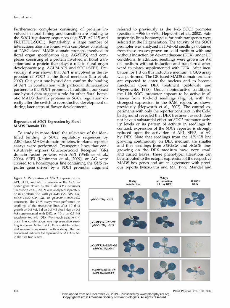
To study in more detail the relevance of the iden-tified binding to SOC1 regulatory sequences byABC-class MADS domain proteins, in planta reporterassays were performed. Transgenic lines that con-stitutively express Glucocorticoid Receptor (GR)domain fusion proteins with AP1 (Wellmer et al.,2006), SEP3 (Kaufmann et al., 2009), or AG werecrossed to a homozygous line containing the GUS re-porter gene driven by a SOC1 promoter fragment
referred to previously as the 1-kb SOC1 promoter(positions 2966 to +960; Hepworth et al., 2002). Sub-sequently, lines homozygous for both transgenes wereselected in the F2 generation. The activity of the SOC1promoter was analyzed in 10-d-old seedlings obtainedfrom these crosses grown on solid medium with andwithout induction by dexamethasone (DEX) under LDconditions. In addition, seedlings were grown for 9 don medium without induction and transferred after-ward to plates supplemented with DEX. After incu-bation for 1 d on this inductive medium, a GUS assaywas performed. The GR-fused MADS domain proteinsare expected to enter the nucleus and to becomefunctional upon DEX treatment (Sablowski andMeyerowitz, 1998). Under noninductive conditions,the 1-kb SOC1 promoter appears to be active in alltissues from 10-d-old seedlings (Fig. 5), with thestrongest expression in the SAM region, as shownpreviously (Hepworth et al., 2002). The control ex-periments with only the reporter construct in the Col-0background revealed that DEX treatment as such doesnot have a substantial effect on SOC1 promoter activ-ity levels or its pattern of activity in seedlings. Incontrast, expression of the SOC1 reporter is stronglyreduced upon the activation of AP1, SEP3, or AGby DEX. Note that seedlings from the AP1:GR linegrowing continuously on DEX medium are smallerand that seedlings from SEP3:GR and AG:GR linesgrowing on the DEX medium have very smalland curled leaves. These phenotypic alterations canbe attributed to the ectopic expression of the respectiveMADS box genes and are in agreement with previ-ous reports (Mizukami and Ma, 1992; Mandel and
Figure 5. Repression of SOC1 expression byAP1, SEP3, and AG. Expression of the GUS re-porter gene driven by the 1-kb SOC1 promoter(Hepworth et al., 2002) was analyzed separatelyor in combination with pCaMV35S::AP1:GR,pCaMV35S::SEP3:GR, or pCaMV35S::AG:GRconstructs. The GUS assays were performed onseedlings of the respective lines after 10 d ofgrowth on 0.5 MS, 9 d on 0.5 MS plus 1 day on 0.5MS supplemented with DEX, or 10 d on 0.5 MSsupplemented with DEX. From each treatment 3plant line combination, one representative seed-ling is shown. Note that GUS is a stable proteinand represents repression with a delay. The redarrowhead indicates the repression of SOC1 by AGin the first true leaves.
Yanofsky, 1995; Honma and Goto, 2001). Previously, itwas shown that AP1 is indeed able to repress SOC1 inearly floral meristems (Liu et al., 2007). To obtainfurther evidence for the repression of SOC1 by SEP3and AG, additional DEX treatments were performedon these specific lines at the moment of the transitionto reproductive development. For this purpose, plantswere grown in SD conditions for 3 weeks and sub-sequently induced to flower by transfer to LD con-ditions. At the same time, the plants were treatedwith DEX, and this treatment was repeated daily.Three days after the switch, a GUS assay was per-formed revealing the repression of SOC1 promoteractivity by AG and SEP3 at this developmental stage(Supplemental Fig. S3). All together, the in planta re-porter assays show that the floral homeotic MADSdomain proteins AP1, SEP3, and AG are able to re-press SOC1.
In Vivo Binding of MADS Domain TFs to the SOC1Promoter Region
Combining the yeast one-hybrid assay results(Fig. 4; Supplemental Table S4) with the results of thereporter assays (Fig. 5; Supplemental Fig. S3) suggeststhat the analyzed floral MADS domain proteinsrepress SOC1 expression by direct binding to itsupstream regulatory sequences. Previously, Liu et al.(2007) showed that this is in fact the case for AP1. Theymade use of ChIP followed by quantitative PCR(qPCR) to identify protein-bound genomic regions.Recently, we performed genome-wide target geneanalyses for the AP1 and SEP3 proteins by ChIP-seq,which confirmed the binding of AP1 and identifiedSEP3 as an additional binding factor of the SOC1 locus(Fig. 3C; Kaufmann et al., 2009, 2010b). One of theCArG box sequences in the SOC1 promoter region,designated “CArG box III” (Fig. 3A), appears to bedirectly bound by AP1 and SEP3 and also by the floralrepressor MADS domain protein FLC (Hepworthet al., 2002). Further analyses of a SOC1-reporter con-struct with mutations in this binding site indicated apivotal role for this CArG box in the down-regulationof SOC1 expression by FLC during the vegetative stageof development in nonvernalized plants (Hepworthet al., 2002). These observations led us to hypothesizethat CArG box III might be a central mediator of SOC1repression, including its repression in floral meristems.To test this hypothesis, we analyzed the activity of thewild-type 1-kb SOC1 promoter and the expression ofpSOC1(1kb)DCArG-III::GUS inside flowers (Hepworthet al., 2002). The intact promoter element gives weakreporter gene expression in the anther locules, whereasall other full-grown floral tissues contain hardly anyGUS signal (Fig. 6A). In contrast to this weak and re-stricted GUS signal, reporter lines with the mutatedCArG-III sequence appear to give substantial floralexpression (Fig. 6B). In these transgenic lines, strongGUS expression was obtained in sepals, anther filaments,
and style and stigma tissues. This result supports theidea that CArG-III is important for limiting SOC1 ex-pression inside floral tissues.
Autoregulatory Feedback Loops for SOC1 Repression
We postulated previously that down-regulation offlowering time genes inside the flower is mediated byMADS domain protein complexes consisting of floraltiming MADS domain proteins, such as SOC1, and flo-ral organ identity proteins, such as AP1 (de Folter et al.,2005). Autoregulatory feedback loops are common forplant MADS domain proteins, and well-known exam-ples are the feedback loops involved in the maintenanceof expression for the B-type MADS box genes (Schwarz-Sommer et al., 1992; Tröbner et al., 1992; Goto andMeyerowitz, 1994; Jack et al., 1994), for the C-type geneAG (Gómez-Mena et al., 2005), and for the E-type geneSEP3 (Kaufmann et al., 2009). Elaborating on this pos-tulated hypothesis, we expect that the repression ofSOC1 by the ABC-class MADS domain proteins is de-pendent on a complex consisting of SOC1 and the floralhomeotic MADS domain proteins. Indeed, various di-mers and higher order complexes have been identifiedcontaining these proteins (de Folter et al., 2005; Imminket al., 2009), and the ChIP-seq experiments and yeastone-hybrid assays showed binding of the SOC1 locus byat least AP1, SEP3, and SOC1 itself, in combination witha variety of dimerization partners (Figs. 3C and 4;Supplemental Table S4). To provide further evidence forthe proposed role of the SOC1 protein in repressing itsown expression, in combination with floral homeoticMADS domain proteins, in planta reporter assays wereperformed in a soc1-6 mutant background (Fig. 6C). Forthis purpose, the homozygous lines described before,containing the pSOC1(1kb)::GUS reporter in combina-tion with a pCaMV35S::MADS:GR construct, wereintrogressed into the soc1-6 T-DNA insertion line. Sub-sequently, GUS assays were performed on plants fromthe selected lines that had been grown on standardmedium or medium supplemented with DEX (Fig. 6C).Both AP1 and AG appeared to down-regulate the SOC1promoter only when the wild-type SOC1 allele is pre-sent (compare Figs. 5 and 6C). In contrast, the SEP3protein can apparently act on the SOC1 promoter in-dependently of SOC1, because the pSOC1-reporterconstruct is still down-regulated in a soc1 mutant back-ground (Fig. 6C). We then investigated whether CArGbox III is essential for the repression of SOC1 by SEP3and crossed the pSOC1(1kb)DCArG-III::GUS constructinto the pCaMV35S::SEP3GR;soc1-6 background. In thiscase, no down-regulation could be obtained for theGUS reporter upon DEX treatment (Fig. 6D), showingthat this binding site is essential for SEP3 protein-mediated repression of the SOC1 promoter in vivo.These experiments provide strong evidence for the pres-ence of negative autoregulatory loops, in which SOC1represses its own expression in combination with AGand AP1.
In this study, we unraveled part of the regulatorynetwork controlling the SOC1 gene and target genesbeing controlled by the SOC1 MADS domain TF.SOC1 is one of the key integrating factors of flowering-time signals and, hence, translates input from thevarious flowering-time pathways into a unique flower-inducing signal (Lee and Lee, 2010). After fulfilling thisrole as hub in the network and shortly after the switchfrom vegetative to reproductive development, it is ofimportance that SOC1 activity is repressed to avoidthe malformation of flowers, which would result in anegative effect on plant fitness (Borner et al., 2000).
SOC1 Is at the Center of the Flowering-TimeRegulatory Network
SOC1 is known as an integrator of different flow-ering-time signals and, therefore, is placed geneticallyat the end of the floral timing gene cascade (Lee andLee, 2010). However, SOC1 also binds numerous keyflowering-time regulatory genes (summarized in TableI; Tao et al., 2012), which genetically are supposed toact upstream of SOC1 and for which in some casesdirect binding of the encoded protein to the SOC1 lo-cus has been shown. For example, SOC1 binds to theregulatory regions of the floral repressors AP2, TOE3,SMZ (Fig. 2C), and SNZ, four of the six TFs with a
Figure 6. Role of CArG box III and dependencyon SOC1 for SOC1 repression by floral MADSdomain proteins. A, Expression of pSOC1(1kb)::GUS in floral organs. B, Expression of pSOC1(1kb)DCArG-III::GUS in floral organs. Note theectopic expression in sepals (green arrowhead),anther filaments (yellow arrowhead), and styleand stigma tissues (red arrowhead). C, Expressionof the GUS reporter gene driven by the 1-kbSOC1 promoter fragment (Hepworth et al., 2002)in the soc1-6 mutant background and in soc1-6mutant seedlings containing the pCaMV35S::AP1:GR, pCaMV35S::SEP3:GR, or pCaMV35S::AG:GR construct. The GUS assays were per-formed on seedlings of the respective lines after10 d of growth on 0.5 MS, 9 d on 0.5 MS plus 1 don 0.5 MS supplemented with DEX, or 10 d on0.5 MS supplemented with DEX. From eachtreatment 3 plant line combination, one repre-sentative seedling is shown. D, Expression of theGUS reporter gene driven by a 1-kb SOC1 pro-moter fragment containing a mutation in CArGbox III (see Fig. 3) in the soc1-6/pCaMV35S::SEP3:GR background. Seedlings were grown onthe same media as in C.
miR172 target site in the Arabidopsis genome. TheseTFs constitute an important group of floral repressors,where AP2 and SMZ have been shown to bind to theSOC1 locus, resulting in its repression (Mathieu et al.,2009; Yant et al., 2010). As expected, the performedexpression studies revealed that SOC1 is able to re-press the expression of this group of AP2 TFs(Supplemental Fig. S1; Tao et al., 2012). Furthermore,and similar to AP2 (Yant et al., 2010), SOC1 targetsMIR156e (Wang et al., 2009), which indirectly regulatesAP2 expression via the SPL and MIR172 genes. AGL15is another floral repressor that binds the SOC1 locus(Supplemental Table S4; Zheng et al., 2009), which isalso targeted by SOC1. In addition, AGL15 is under thecontrol of AP2, which activates its expression and addsan additional layer of regulatory complexity (Yantet al., 2010). Besides AGL15, SOC1 binds to otherflowering-time repressor loci belonging to the MADSdomain TF family, such as SVP and AGL18 (Table I),both of which seem to be repressed by SOC1(Supplemental Fig. S1). Based on these findings andthe antagonistic expression patterns of the floral re-pressors and SOC1 during the vegetative stage of de-velopment (Schmid et al., 2005), it is tempting tospeculate that SOC1 is repressing the majority of itsown repressors. This type of double-negative feedbackloop is frequently observed in developmental path-ways and serves as a molecular switch with twosteady states: either the flowering repressors are onand SOC1 is off, or the opposite (Alon, 2007). A(transient) signal, which could be a flowering-inducingfactor or reaching a particular temperature, could in-duce SOC1 above a threshold level, after which thenetwork irreversibly and independently of the signallocks into the flowering state (SOC1 on) by suppress-ing the repressors. In summary, SOC1 binds loci of alarge number of known flowering-time regulators(Table I), which places SOC1 molecularly in the centerof the flowering regulatory network. Furthermore, ourChIP-seq and expression analysis data suggest thatthis network contains multiple regulatory loops andfeedback mechanisms.
SOC1 Binds Loci with a Potential Role in FloweringTime Regulation
In addition to the well-described flowering-timeregulators discussed above, SOC1 binds loci encodinggenes with a potential role in the flowering response.One example is SMALL UBIQUITUN-LIKE MODI-FIER3 (SUM3), which belongs to a small gene family ofubiquitin-like posttranslational modifiers (van denBurg et al., 2010). In contrast to single mutants forSUM1 or SUM2, sum3 plants appear to be late flow-ering, whereas overexpression of SUM3 results in earlyflowering. Based on this knowledge and the ChIP-seq data, one could assume that SOC1 binding ofSUM3 results in SUM3 induction and hence a flow-ering-stimulating signal. Besides these examples for
protein-coding genes, SOC1 appears to bind the lociof various microRNA genes, such as the above-mentioned MIR156e. Interestingly, another micro-RNA gene, MIR319c, was the second-best overalltarget of SOC1. miR319 and some of their targets, theTEOSINTE BRANCHED1, CYCLOIDEA, AND PCFTRANSCRIPTION FACTORS (TCP) genes, have beenshown to be expressed in the developing flower(Cubas et al., 1999; Wellmer et al., 2006; Nag et al.,2009). Besides MIR319c, SOC1 also binds to regula-tory sequences of the miR319 target TCP3, which isexpressed throughout the young floral meristem atfloral stages 2 and 3 and is more restricted to the sepalprimordia at stage 4 (Cubas et al., 1999; Wellmer et al.,2006). As the expression pattern of TCP3 is comple-mentary to that of SOC1, SOC1 might repress TCP3 inthe developing flowers, both directly and indirectly viaMIR319c. Additionally, plants ectopically expressingMIR319a show a late-flowering phenotype (Palatniket al., 2003; Schommer et al., 2008), suggesting that themiR319 targets play a role in floral timing as well,which could also in part be under the control of SOC1.
A Direct Role for SOC1 in the Repression of the FloralHomeotic MADS Box Genes?
Previous studies revealed the importance of main-taining SEP3 and the B- and C-class floral organidentity genes in a suppressed state in the inflorescencemeristem and young floral meristems (Liu et al., 2009).The repression in very young flowers is key to avoidinga precocious differentiation of the floral meristemduring the first stages of development. SVP seems toplay a major role in this, although SOC1 and AGL24also have been shown to be important (Gregis et al.,2008; Liu et al., 2009). Our ChIP-seq data suggestan alternative mechanism, in which SOC1 directlyrepresses the B-class genes AP3 and PI in the inflo-rescence and flower meristems to prevent prematurefloral meristem differentiation. It seems that SOC1,SVP, and AGL24 jointly provide the floral meristem ashort lag time in which differentiation is suppressed,allowing the establishment of sufficient cells for theinner floral whorls, from which the important repro-ductive organs will develop.
SOC1 Repression in Young Developing Floral Meristems
Analysis of SOC1 expression at the mRNA (Samachet al., 2000) and protein (Fig. 1) levels reveals that itsexpression in developing young floral meristems isturned off, in contrast to the maintained strong SOC1signals in the inflorescence meristem. This expressionpattern is fully complementary to the expression of thefloral meristem identity gene AP1 (Fig. 1H) and can beexplained by the direct repression of SOC1 by AP1 (Liuet al., 2007). Here, we show that in addition to AP1, thefloral organ identity proteins SEP3 and AG have the
potential to repress SOC1 as well. SOC1 repression bymultiple factors allows robust control over this geneduring early flower development. However, despite thepotential repression of SOC1 by SEP3 and AG, someSOC1 expression reappears during later stages of flowerdevelopment in the center of the floral meristem (Fig.1G). This suggests that SOC1 repression by SEP3 andAG is less rigorous than repression by AP1 in the peri-anth primordia or, alternatively, that competing induc-ing factors cause some SOC1 expression in the center ofdeveloping flowers. Nevertheless, the facts that SOC1 isexpressed only at low levels in full-grown flowers inpart of the anthers (Fig. 6A) and that a mutation ofCArG box III results in ectopic SOC1 expression in an-ther filaments and stylar tissue (Fig. 6B) suggest thatSOC1 expression is actively repressed in later stages ofwhorl 3 and 4 development. Based on our results, SEP3and AG, in combination with specific dimerizationpartners, are possible candidates for this function. Adifference in SOC1 repression by AP1, SEP3, and AG isseen in the dependence on the SOC1 protein itself. BothAP1 and AG appear to depend fully on SOC1 for therepression of SOC1. In contrast, SEP3 is able to repressSOC1 expression in the absence of the SOC1 protein.The latter might be explained by differences in proteincomplex formation capacity: SEP3 has a large number ofinteraction partners and is able to interact with variousflowering-time-regulating MADS domain proteins, suchas AGL24 and SVP (de Folter et al., 2005; Immink et al.,2009). Based on this knowledge, we hypothesize that therole of SOC1 in SOC1 repression by SEP3 can be takenover by another interacting MADS domain protein. Inline with this hypothesis, we saw various SEP3 dimersbinding the SOC1 regulatory sequences in our yeastone-hybrid assay (Fig. 4; Supplemental Table S4). Thenegative autoregulation, as shown here for SOC1, is acore transcriptional network component that is oftenseen in developmental regulatory networks to facilitatefast switches (Rosenfeld et al., 2002). Such an adequateand fast response is essential for SOC1 suppression justafter transition to the reproductive stage, where a rapidsuccession of developmental transitions take place.Analysis of the existence of autoregulatory loops showsconservation and overrepresentation of this networkmotif in various kingdoms, suggesting that this motifhas been maintained in evolution (Kiełbasa andVingron, 2008).
Molecular Mode of Action for SOC1 Repression by SOC1and Floral Homeotic MADS Domain Proteins
A remaining question is what is the exact stoichi-ometry and composition of the transcriptional repres-sion complex in the negative autoregulatory SOC1loop. MADS domain proteins are able to bind DNA asdimers; hence, the most simple molecular unit for theSOC1 negative feedback loop would be a heterodimerconsisting of, for example, SOC1 and AP1. Yeast two-hybrid experiments reveal that SOC1-AP1 and SOC1-
SEP3 dimers potentially can be formed (de Folter et al.,2005). Although SOC1-SEP3DC (dimer 283) appears tobind the proximal SOC1 promoter region in yeast, thisdimer is not exclusively responsible for the down-regulation of SOC1. In contrast, no binding couldbe detected for the SOC1-AP1 dimer (dimer 46;Supplemental Table S4), while we know from the re-porter studies that the SOC1 suppression by AP1 isSOC1 dependent (Fig. 6). Taking into account the ca-pacity of plant MADS domain proteins to assembleinto multimeric complexes, it might be that SOC1 getsrepressed by a higher order complex consisting of atleast one SOC1 dimer and one AP1 dimer binding tothe proximal SOC1 promoter region. Identification ofSOC1 and AP1 in the same protein complex isolatedfrom native inflorescence material shows that thesetwo proteins assemble into complexes in vivo(Smaczniak et al., 2012). The fact that AG is not foundas a dimerization partner of SOC1 in yeast (de Folteret al., 2005) but interacts with SOC1 in a higher ordercomplex (Immink et al., 2009) suggests that a similarmodel could hold for SOC1 repression by the combi-nation of AG and SOC1. Alternatively, the ABC-MADS domain proteins and SOC1 bind the regulatorysequences independently, and both binding events areessential for SOC1 repression. A careful analysis of theChIP-seq peaks for AP1 (Kaufmann et al., 2010b), SEP3(Kaufmann et al., 2009), and SOC1 shows that theSOC1 locus is bound at various positions by theseproteins or their interaction partners; hence, differentcombinations of CArG boxes, including CArG box III,could be involved in mediating SOC1 repression. Inthe latter case, it is expected that the DNA from theSOC1 promoter loops around such a higher ordertranscriptional repression complex. In both scenarios,the question remains whether additional general tran-scriptional repressors are essential. The repression ofSEP3 by SOC1 is mediated by at least two chromatinregulators, TERMINAL FLOWER2/LIKE HETERO-CHROMATIN PROTEIN1 and SAP18 (Liu et al.,2009). Furthermore, different chromatin regulatorswere identified in the protein complex isolations for thefloral homeotic MADS domain proteins (Smaczniak et al.,2012), supporting the idea that cofactors with tran-scriptional repression activity play a role in this type ofnegative feedback loop.
A Complex Regulatory Network of MADS Proteins inFlower Development
Genome-wide target gene analyses for a number ofMADS domain proteins that control flowering time(Zheng et al., 2009; Deng et al., 2011; Tao et al., 2012)and floral organ identities (Gómez-Mena et al., 2005;Kaufmann et al., 2009, 2010a, 2010b) reveal that thesemaster regulators target a large number and wide va-riety of genes, ranging from other regulatory factors tostructural genes. Furthermore, these TFs control them-selves and each other via direct regulatory interactions,
resulting in a complex interconnected network. LikeSOC1, AGL24 and SVP also are directly repressed byAP1 (Liu et al., 2007), SEP3, and AG (Gregis et al.,2008). All together, these results show that a tightlycontrolled balance exists between the activity andfunctioning of floral timing and floral organ identity-specifying MADS TFs around the transition fromvegetative to reproductive development, resulting in arobust phase switch and the development of flowersand reproductive organs under optimal environmentalconditions.
MATERIALS AND METHODS
Plant Material and Plant Transformation
Arabidopsis (Arabidopsis thaliana) Col-0 was used as the genetic back-ground for all experiments. For the soc1 mutation, soc1-2 (Lee et al., 2000) andsoc1-6 (SALK_138131) T-DNA insertion lines were used (Alonso et al., 2003).Homozygous mutant plants were selected based on their late-flowering phe-notype, and the presence of the T-DNA was confirmed by PCR on genomicDNA. For detection of the wild-type SOC1 allele in the soc1-2 and soc1-6mutants, the primer pairs G-20046 (59-CTTTTGGTTTGAACTAATCTTTGTC-39)/G-19924 (59-ATATCACAAACCGTTTAGAAGCTTC-39) and PDS606 (59-ATCTCATGAAAGGAGGTTGC-39)/PDS607 (59-GTCACTTGTCTGCTTGTT-GC-39) were used, respectively. For the T-DNA insertion alleles, the primerpairs G-11003 (59-GTTCACGTAGTGGGCCATC-39)/G-19924 for the soc1-2mutant and lba1 (Alonso et al., 2003) and PDS607 for soc1-6 were used. Allgenerated constructs were transformed into Col-0 plants, making use ofAgrobacterium tumefaciens strain GV3101 or ASE and the floral dip method(Clough and Bent, 1998). Transgenic plants were identified by selective ger-mination on one-half-strength Murashige and Skoog (0.5 MS) medium sup-plemented with kanamycin (50 mg L21) or on soil watered with BASTA(0.1%).
Plasmid Constructions
AGateway destination vector suitable for the expression of genes of interestfused to the coding region of the rat GR domain was obtained by removing theAGL11 coding region from vector NOB221. For this purpose, the BamHI andNcoI restriction enzymes were used. Subsequently, the digested vector wasblunted, followed by introduction of the Gateway conversion cassette (Invi-trogen) downstream of the GR coding region and upstream of the CaMV35Spromoter. This complete expression cassette was cloned as an AscI/PacIfragment into the binary vector pGD121 (de Folter et al., 2006), resulting in theGateway-compatible GR destination vector pARC146. The open readingframes from SEP3 and AG (Immink et al., 2009) were cloned into pARC146 byLR reactions.
The bait constructs for the yeast one-hybrid assays were obtained bycloning the SOC1 regulatory sequences as NotI/SpeI fragments into thevector pINT-HIS3NB (Meijer et al., 1998). Two fragments from the SOC1promoter were selected: one from 2663 to +82 (Fig. 3A) and the other from2864 to 2397. Fragments were amplified with the primers PDS497 (59-AGACACGTCGCTACTTAACG-39)/PDS499 (59-TCTTCTCGTTGTAGT-TATGG-39) and PDS496 (59-CGAAATAATTAGTTTGTGTGG-39)/PDS498(59-ATATCTTTCCATCCCAACAG-39), respectively. These two fragmentshave overlap in sequence, but this region does not contain putative con-sensus CArG box-binding sites. Together, these two promoter elementsrepresent the upstream region of the 2966 to +960 1-kb SOC1 promoterthat has been described by Hepworth et al. (2002). The obtained bait vec-tors containing these promoter fragments were designated pARC1046(pSOC1 2663 to +82) and pARC1047 (pSOC1 2864 to 2397; Fig. 3A). Inaddition, a bait plasmid was generated covering the SOC1 59 UTR. Thisfragment was amplified with PDS2572 (59-TTATCTTTCTCCAAGAAA-TAAAAT-39)/PDS2573 (59-CATGACGAAGAGATCTTACC-39) and spansthe genomic region from +1 to +409. This fragment was cloned into pINT-HIS3NB, resulting in the bait construct CZN2030 (59-UTR-SOC1 +1 to+409). All the above indicated constructs were controlled by restriction
analyses and sequencing of the inserted fragment (DETT sequencing kit;Amersham).
The 8,186-bp genomic SOC1 (At2g45660; TAIR 10, Chromosome 2:18806523.0.18814708) rescue fragment, which includes an approximately 3.7-kb upstream sequence, exons, introns, UTR sequences, and an approximately1.2-kb downstream sequence, was amplified by PCR from genomic DNAisolated from Col-0 using Phusion polymerase (New England Biolabs), primerG-27271 (59-AAACTCGTATAATAAAACCATATAGTTAA-39), and primer G-27272 (59-ACCAACATTTTCCAAATGAAATAAAC-39). The resulting PCRproduct was purified and cloned into the pCR8/GW/TOPO Gateway entryvector (Invitrogen) to create pDP29, which was confirmed by Sanger se-quencing. Subsequently, the SOC1 genomic fragment was recombined frompDP29 into a Gateway-compatible pGREEN-IIS binary destination vector(pFK387), which provides resistance to BASTA for selection in plants, result-ing in pDP36 (gSOC1). For visualization of the SOC1 protein and to facilitateChIP, SOC1 was tagged with mGFP6-6xHIS. For this purpose, we amplified agenomic subfragment of SOC1 ranging from exon 2, which contains a uniqueAgeI restriction site, to the last coding triplet before the stop codon of SOC1using primer G-27342 (59-GTTATCTGAGGCATACTAAG-39) and primer G-27264 (59-CTGTCGGCCGCAGAACCGGATCCAGATCCAGATCCCTTTCT-TGAAG-39), and the SOC1 39 region, which contains a unique AatII restrictionsite, starting with the stop codon using primer G-26333 (59-CAAA-CACCACCACCACCACCACTGATCTCCACTCAACAA-39) and primer G-27272. The sequence encoding mGFP6-6xHIS was amplified from the pMD107plasmid (Curtis and Grossniklaus, 2003) using primer G-26331 (59-GGTTCTGCGGCCGACAGTAAAGGAGAAGAAC-39) and primer G-26332(59-TTGTTGAGTGGAGATCAGTGGTGGTGGTGGTGGTGTTTG-39). Finally,the three fragments were combined in an overlapping fusion PCR employingprimers G-27342 and G-27272. The resulting PCR product was cut with AgeIand AatII, cloned into the corresponding sites of pDP29, and verified bySanger sequencing. Finally, the SOC1:mGFP6 genomic fragment was recom-bined into pFK387 to create pDP37 (gSOC1:GFP). Then, a red fluorescence-based construct was generated as a reference marker for the imaging of thegSOC1:GFP lines. For this purpose, the TagRFP coding region (Merzlyak et al.,2007) was tagged with a nuclear localization signal-encoding sequence andplaced under the control of the constitutive BETA-6 TUBULIN (TUB6) genepromoter (At5g12250) in a binary vector backbone.
Yeast One-Hybrid Assays
Integration of the bait vectors into the yeast genome from the strain PJ69-4(mating type a; James et al., 1996) was performed as described before (Meijeret al., 2000). In order to determine the background levels of expression for theHIS3 reporter gene, various colonies from the independent yeast integrationswere suspended in 100 mL of Milli Q and spotted as 5-mL droplets onto a seriesof plates with selective synthetic dropout medium lacking His but supple-mented with 0, 5, 10, 15, 20, 25, 30, 40, 50, or 60 mM 3-amino-1,2,4-triazole(3-AT). Plates were incubated at 20°C for 7 d and then scored for growth(activation of the HIS3 reporter gene). For each bait construct, three inde-pendent colonies were selected that had low levels of background growth(growth up to 10 mM 3-AT maximum). Screening of binding by MADSbox type II TF dimers was performed by mating. The available collection oftype II dimers in strain PJ69-4 (mating type A; Immink et al., 2009) was grownovernight at 30°C in liquid synthetic dropout medium lacking Trp. At thesame time, the yeast bait clones also were grown in liquid synthetic dropoutmedium lacking His. All possible dimer-bait combinations were made byspotting 5-mL droplets on top of each other in a grid of 96 spots on syntheticdropout agar plates containing all essential amino acids. After mating andgrowth for 1 night at 30°C, the yeast spots were transferred to syntheticdropout plates lacking Trp/His by a 96-pin replicator. These plates weregrown over 2 nights at 30°C, and afterward, yeast spots were suspended on a96-well plate with 100 mL of sterile Milli Q water in each well. Finally, thesesuspensions were spotted onto a series of synthetic dropout selection plateswithout Trp/His and supplemented with a range of 3-AT concentrations(10–60 mM). Dimer-DNA interaction events were scored after 7 d of incubationat 20°C. Each dimer-bait combination was screened at least two times in in-dependent experiments.
Reporter and GR Induction Assays
Seedlings for the reporter assays were grown on plates with 0.5 MSmedium(Duchefa Biochemie) in a growth chamber under LD conditions (16/8-h day/
night regime, 21°C). For GR induction, the medium was supplemented withDEX (Sigma-Aldrich) at a final concentration of 10 mM. Before germination,seeds were vapor-phase sterilized, followed by stratification at 4°C for 3 d. TheGUS assay was performed on 10-d-old seedlings (LD conditions) or on plantsgrown for 3 weeks under SD conditions followed by 3 d in LD conditions. TheGUS assays were performed based on the protocol described previously (deFolter et al., 2006). Plant material was incubated in the GUS staining solutionfor 16 h at 37°C.
SOC1 Loci Sequence Data
Putative orthologous genes of SOC1 were selected by BLAST search ofthe Arabidopsis SOC1 locus sequence (AT2G45660) against plant genomicsequences available on the Phytozome Web site (http://www.phytozome.net) and in the Brassica database (Cheng et al., 2011). Next, two filteringcriteria were applied to identify true orthologs among the BLAST hits: (1)reciprocal best hit; and (2) the locus containing the hit has to have annota-tions for at least the first and last SOC1 exons. As a result, SOC1 orthologousgenes were identified in the genome of seven species: Arabidopsis (TAIR10, Chromosome 2:18807538-18811045), Arabidopsis lyrata (V1.0, scaf-fold_4:22217070-22221788), Brassica rapa (V1.1, A05:2530045-2533747), Citrussinensis (V1.0, scaffold00001:1502952-1509395), Citrus clementina (V0.9, scaf-fold_3:991596-998038), Cucumis sativus (V1.0, scaffold02229:5073026-5076945),and Mimulus guttatus (V1.0, scaffold_27:797423-801044). Then, for each gene,the 3.0-kb sequence upstream of the first exon was taken as the promotersequence.
Phylogenetic Analysis of the SOC1 Promoter Sequence
The promoter sequences of SOC1 from Arabidopsis and its orthologousgenes were aligned using MUSCLE (Edgar, 2004), and maximum-likelihoodphylogenetic trees were generated from the resulting alignments usingfastDNAmL (Olsen et al., 1994). The DNA substitution model Hasegawa-Kishino-Yano (HKY85) was selected by modeltest (Posada and Crandall,1998) using the Akaike informational criterion based on log-likelihoodscores of the alignment. The training set for the mutation rates comprisedsequences containing exon 1 of SOC1 and flanking regions (6100 bp),using the known location of that exon in each of the seven species. Modelsfor “fast” and “slow” regimes were learned by training the HKY85 modelon the training set alignment using the regions with and without gaps,respectively. Subsequently, the models for “slow” and “fast” regimeswere used to calculate the likelihood for each column of the multiplealignments. These likelihood values were employed to calculate the log-likelihood ratio under a fast versus a slow mutation regime. This ratiorepresents the relative likelihood that any given nucleotide site is sub-jected to a faster or slower mutation rate and is related to functionalconstraints imposed on each site (Boffelli et al., 2003). The correspondinglikelihood ratio curves were used to describe the mutation profile of theArabidopsis SOC1 promoter sequence. The curve is smoothed by means ofa 20% trimmed mean over the 50-base window centered at each alignedsite.
ChIP was performed in triplicate using 1 g of tissue enriched for transitionapices collected at zeitgeber 4 from plants grown for 15 d under SD conditions,followed by a shift to long days for 3 consecutive days to synchronously in-duce flowering. DNA was precipitated using 2.5 mL of a polyclonal anti-GFPantibody (Abcam; no. 290) from gSOC1:GFP, soc1-2, and gSOC1;soc1-2 plants,the latter of which serve as negative controls. Precipitated DNA was frag-mented on a Covaris S2 machine (duty cycle, 20%; intensity, 5; cycles perburst, 200; cycle time, 2 min) and tested for the enrichment of presumed SOC1targets such as SEP3 and SOC1 itself by qPCR, using the primers G-31798 (59-TTTGAGGCAATGTCGTGAAG-39) and G-31799 (59-CCCTTCCCAT-TACGTCTTGA-39) for SEP3, G-31800 (59-ATGATGGACGCTTGAAACCT-39)and G-31801 (59-GACAGGCATTTCCATCCAAC-39) for SOC1, and G-47 (59-GGCTGTTGTCCTGGTATTATTTCTC-39) and G-15952 (59-GAGGACTAAG-GCAATAGTACATGTT-39) for ARR7 (negative control). The qPCRs wereperformed using the Bio-Rad Real-Time PCR SYBR Green Mix. Libraries for
high-throughput sequencing were prepared as described previously (Yantet al., 2010), and 40-bp single-end sequencing was performed on an IlluminaGAIIx instrument following the manufacturer’s instructions.
ChIP-seq Analyses
ChIP-seq peak calling was essentially performed as described (Moyroudet al., 2011), except that the filtering parameters were slightly modified asfollows: potential peaks were discarded if their mean coverage for any controlwould exceed the median average control coverage plus a tolerance of 6 SD
in all peak regions. A minimum normalized fold change of at least 2-foldbetween sample and control was required in at least one replicate, as wellas a shift in peak location between forward and reverse strand of 10 bp ormore.
Expression Analysis
Total RNA was isolated using Trizol (Ambion) from Col-0, soc1-2, and 35S::SOC1 plants grown in the same conditions and using the same tissue as in theChIP experiment (15 d of SD conditions + 3 d of LD conditions). One micro-gram of total RNA was DNase I treated, and single-stranded complementaryDNA was synthesized using oligo(dT) and the RevertAid first-strand com-plementary DNA synthesis kit (Fermentas). Quantitative real-time PCR wasperformed on an Opticon continuous fluorescence detection system (Bio-Rad[MJ Research Models]) using the Platinum SYBR Green qPCR Supermix-UDG(Invitrogen). Gene expression was calculated relative to b-tubulin using theDDCt method (Livak and Schmittgen, 2001). Three biological and technicalreplicates were used for the quantification. Oligonucleotide primers used areas follows: for b-TUB2, N-0078 (59-GAGCCTTACAACGCTACTCTGTCTGTC-39) and N-0079 (59-ACACCAGACATAGTAGCAGAAATCAAG-39); for SOC1,G-0628 (59-ATAGGAACATGCTCAATCGAGGAGCTG-39) and G-0629 (59-TTTCTTGAAGAACAAGGTAACCCAATG-39); for SVP, G-20863 (59-CAAG-GACTTGACATTGAAGAGCTTCA-39) and G-20864 (59-CTGATCTCACTCA-TAATCTTGTCAC-39); for AGL18, G-33582 (59-ACCATTCCGACACTTCCTTG-39) and G-33583 (59-GAAGCCACTTGACTCCCAGA-39); for TEM2, G-22652 (59-GACTAGAGCGGCAGTTATATATTGAT-39) and G-22653 (59-CTTTCCACCG-CAAACGGCCA-39); for AP2, G-26366 (59-TACACGTACTTCGCCGACAA-39)and G-26367 (59-GGTGTCGAACAAACCCAAAT-39); for SNZ, G-0658(59-AGGGAGAAGGAGCCATGAAGTTTGGTG-39) and G-0659 (59-GTCTTCA-GAGGTTTCATGGTTGCCATG-39); for SMZ, G-4476 (59-ATAAAATACAA-TGAGTTGGGAAAGGGA-39) and G-4477 (59-TGGTTGCCATGGGTAAAAA-TATCGATG-39).
Confocal Imaging
Confocal laser scanning microscopy was performed to determine the ex-pression and localization pattern of the SOC1 protein. The generated gSOC1:GFP lines in the Col-0 background were crossed with the pTUB6::TagRFPtransgenic plants. From the progeny of this cross, a few lines were selectedcontaining both constructs and showing the expected SOC1 expression patternand constitutive expression of TagRFP. Seeds from these lines were sown, andthe seedlings were grown for 21 d in SD conditions and 21°C to maintain theplants in the vegetative state. Subsequently, plants were transferred to LDconditions (21°C) to induce SOC1 expression in the SAM and, hence, flow-ering. Images from the SAM region were taken 0, 3, 5, and 7 d after thetransfer to LD conditions. Imaging of the living plant tissue was performedwith a Leica SPE DM5500 upright microscope as described previously(Urbanus et al., 2009).
Supplemental Data
The following materials are available in the online version of this article.
Supplemental Figure S1. qPCR to show the effect of SOC1 on theexpression of seven loci bound by SOC1 according to the ChIPexperiments.
Supplemental Figure S2. Maximum-likelihood phylogenetic tree ofprotein sequences encoded by SOC1 orthologs found in floweringplants.
Supplemental Figure S3. Repression of SOC1 by SEP3 and AG at themoment of floral transition.
Supplemental Table S2. SOC1 ChIP-seq quality and position of peaks.
Supplemental Table S3. Summary of SOC1 ChIP-seq analyses and micro-array expression studies.
Supplemental Table S4. Summary of yeast one-hybrid analyses.
ACKNOWLEDGMENTS
We thank Pieter Ouwerkerk for providing the pINT-HIS3NB plasmid andhelpful advice on the yeast one-hybrid system; Iain Searle and GeorgeCoupland for seeds from the pSOC1(1kb)::GUS and pSOC1(1kb)DCArG1::GUS lines; Isabella Nougalli Tonaco for generating the SEP3 and AG GR con-structs and transgenic lines; Martin Kater for providing vector NOB221 con-taining a GR expression cassette; Frank Wellmer and Elliot Meyerowitz forproviding seeds from the AP1:GR line; and Christa Lanz for assistance withIllumina sequencing.
Received June 26, 2012; accepted July 10, 2012; published July 12, 2012.
LITERATURE CITED
Adamczyk BJ, Lehti-Shiu MD, Fernandez DE (2007) The MADS domainfactors AGL15 and AGL18 act redundantly as repressors of the floraltransition in Arabidopsis. Plant J 50: 1007–1019
Alon U (2007) Network motifs: theory and experimental approaches. NatRev Genet 8: 450–461
Alonso JM, Stepanova AN, Leisse TJ, Kim CJ, Chen HM, Shinn P,Stevenson DK, Zimmerman J, Barajas P, Cheuk R, et al (2003) Ge-nome-wide insertional mutagenesis of Arabidopsis thaliana. Science301: 653–657
Aukerman MJ, Sakai H (2003) Regulation of flowering time and floralorgan identity by a microRNA and its APETALA2-like target genes.Plant Cell 15: 2730–2741
Benlloch R, Kim MC, Sayou C, Thevenon E, Parcy F, Nilsson O (2011)Integrating long-day flowering signals: a LEAFY binding site is essen-tial for proper photoperiodic activation of APETALA1. Plant J 67:1094–1102
Blazquez MA, Green R, Nilsson O, Sussman MR, Weigel D (1998) Gib-berellins promote flowering of Arabidopsis by activating the LEAFYpromoter. Plant Cell 10: 791–800
Boffelli D, McAuliffe J, Ovcharenko D, Lewis KD, Ovcharenko I,Pachter L, Rubin EM (2003) Phylogenetic shadowing of primate se-quences to find functional regions of the human genome. Science 299:1391–1394
Borner R, Kampmann G, Chandler J, Gleissner R, Wisman E, Apel K,Melzer S (2000) A MADS domain gene involved in the transition toflowering in Arabidopsis. Plant J 24: 591–599
Castillejo C, Pelaz S (2008) The balance between CONSTANS and TEM-PRANILLO activities determines FT expression to trigger flowering.Curr Biol 18: 1338–1343
Cheng F, Liu S, Wu J, Fang L, Sun S, Liu B, Li P, Hua W, Wang X (2011)BRAD, the genetics and genomics database for Brassica plants. BMCPlant Biol 11: 136
Clough SJ, Bent AF (1998) Floral dip: a simplified method for Agro-bacterium-mediated transformation of Arabidopsis thaliana. Plant J 16:735–743
Corbesier L, Vincent C, Jang S, Fornara F, Fan Q, Searle I, Giakountis A,Farrona S, Gissot L, Turnbull C, et al (2007) FT protein movementcontributes to long-distance signaling in floral induction of Arabidopsis.Science 316: 1030–1033
Cubas P, Lauter N, Doebley J, Coen E (1999) The TCP domain: a motiffound in proteins regulating plant growth and development. Plant J 18:215–222
Curtis MD, Grossniklaus U (2003) A Gateway cloning vector set for high-throughput functional analysis of genes in planta. Plant Physiol 133:462–469
de Folter S, Angenent GC (2006) trans meets cis in MADS science. TrendsPlant Sci 11: 224–231
de Folter S, Immink RGH, Kieffer M, Parenicová L, Henz SR, Weigel D,Busscher M, Kooiker M, Colombo L, Kater MM, et al (2005)
Comprehensive interaction map of the Arabidopsis MADS box tran-scription factors. Plant Cell 17: 1424–1433
de Folter S, Shchennikova AV, Franken J, Busscher M, Baskar R,Grossniklaus U, Angenent GC, Immink RGH (2006) A Bsister MADS-box gene involved in ovule and seed development in petunia andArabidopsis. Plant J 47: 934–946
Deng W, Ying H, Helliwell CA, Taylor JM, Peacock WJ, Dennis ES (2011)FLOWERING LOCUS C (FLC) regulates development pathwaysthroughout the life cycle of Arabidopsis. Proc Natl Acad Sci USA 108:6680–6685
Dorca-Fornell C, Gregis V, Grandi V, Coupland G, Colombo L, KaterMM (2011) The Arabidopsis SOC1-like genes AGL42, AGL71 andAGL72 promote flowering in the shoot apical and axillary meristems.Plant J 67: 1006–1017
Edgar RC (2004) MUSCLE: multiple sequence alignment with high accur-acy and high throughput. Nucleic Acids Res 32: 1792–1797
Franks SJ, Sim S, Weis AE (2007) Rapid evolution of flowering time by anannual plant in response to a climate fluctuation. Proc Natl Acad SciUSA 104: 1278–1282
Gómez-Mena C, de Folter S, Costa MMR, Angenent GC, Sablowski R(2005) Transcriptional program controlled by the floral homeotic geneAGAMOUS during early organogenesis. Development 132: 429–438
Goto K, Meyerowitz EM (1994) Function and regulation of the Arabidopsisfloral homeotic gene PISTILLATA. Genes Dev 8: 1548–1560
Gregis V, Sessa A, Colombo L, Kater MM (2008) AGAMOUS-LIKE24 andSHORT VEGETATIVE PHASE determine floral meristem identity inArabidopsis. Plant J 56: 891–902
Gregis V, Sessa A, Dorca-Fornell C, Kater MM (2009) The Arabidopsisfloral meristem identity genes AP1, AGL24 and SVP directly repressclass B and C floral homeotic genes. Plant J 60: 626–637
Hartmann U, Höhmann S, Nettesheim K, Wisman E, Saedler H, Huijser P(2000) Molecular cloning of SVP: a negative regulator of the floraltransition in Arabidopsis. Plant J 21: 351–360
Hepworth SR, Valverde F, Ravenscroft D, Mouradov A, Coupland G(2002) Antagonistic regulation of flowering-time gene SOC1 byCONSTANS and FLC via separate promoter motifs. EMBO J 21:4327–4337
Honma T, Goto K (2001) Complexes of MADS-box proteins are sufficient toconvert leaves into floral organs. Nature 409: 525–529
Huang H, Tudor M, Su T, Zhang Y, Hu Y, Ma H (1996) DNA bindingproperties of two Arabidopsis MADS domain proteins: binding consen-sus and dimer formation. Plant Cell 8: 81–94
Immink RG, Tonaco IA, de Folter S, Shchennikova A, van Dijk AD,Busscher-Lange J, Borst JW, Angenent GC (2009) SEPALLATA3: the‘glue’ for MADS box transcription factor complex formation. GenomeBiol 10: R24
Ito T, Sakai H, Meyerowitz EM (2003) Whorl-specific expression of theSUPERMAN gene of Arabidopsis is mediated by cis elements in thetranscribed region. Curr Biol 13: 1524–1530
Izawa T (2007) Adaptation of flowering-time by natural and artificial se-lection in Arabidopsis and rice. J Exp Bot 58: 3091–3097
Jack T, Fox GL, Meyerowitz EM (1994) Arabidopsis homeotic gene APE-TALA3 ectopic expression: transcriptional and posttranscriptional reg-ulation determine floral organ identity. Cell 76: 703–716
James P, Halladay J, Craig EA (1996) Genomic libraries and a host straindesigned for highly efficient two-hybrid selection in yeast. Genetics 144:1425–1436
Kaufmann K, Muiño JM, Jauregui R, Airoldi CA, Smaczniak C,Krajewski P, Angenent GC (2009) Target genes of the MADS tran-scription factor SEPALLATA3: integration of developmental and hor-monal pathways in the Arabidopsis flower. PLoS Biol 7: e1000090
Kaufmann K, Pajoro A, Angenent GC (2010a) Regulation of transcriptionin plants: mechanisms controlling developmental switches. Nat RevGenet 11: 830–842
Kaufmann K, Wellmer F, Muiño JM, Ferrier T, Wuest SE, Kumar V,Serrano-Mislata A, Madueño F, Krajewski P, Meyerowitz EM, et al(2010b) Orchestration of floral initiation by APETALA1. Science 328:85–89
Kiełbasa SM, Vingron M (2008) Transcriptional autoregulatory loops arehighly conserved in vertebrate evolution. PLoS ONE 3: e3210
Lee H, Suh S-S, Park E, Cho E, Ahn JH, Kim S-G, Lee JS, Kwon YM, Lee I(2000) The AGAMOUS-LIKE 20 MADS domain protein integrates floralinductive pathways in Arabidopsis. Genes Dev 14: 2366–2376
Lee J, Lee I (2010) Regulation and function of SOC1, a flowering pathwayintegrator. J Exp Bot 61: 2247–2254
Lee J, Oh M, Park H, Lee I (2008) SOC1 translocated to the nucleusby interaction with AGL24 directly regulates leafy. Plant J 55:832–843
Lee JH, Yoo SJ, Park SH, Hwang I, Lee JS, Ahn JH (2007) Role of SVP inthe control of flowering time by ambient temperature in Arabidopsis.Genes Dev 21: 397–402
Li D, Liu C, Shen L, Wu Y, Chen H, Robertson M, Helliwell CA,Ito T, Meyerowitz E, Yu H (2008) A repressor complex governsthe integration of flowering signals in Arabidopsis. Dev Cell 15:110–120
Liljegren SJ, Gustafson-Brown C, Pinyopich A, Ditta GS, Yanofsky MF(1999) Interactions among APETALA1, LEAFY, and TERMINAL FLOWER1specify meristem fate. Plant Cell 11: 1007–1018
Liu C, Chen H, Er HL, Soo HM, Kumar PP, Han JH, Liou YC, Yu H (2008)Direct interaction of AGL24 and SOC1 integrates flowering signals inArabidopsis. Development 135: 1481–1491
Liu C, Xi W, Shen L, Tan C, Yu H (2009) Regulation of floral patterningby flowering time genes. Dev Cell 16: 711–722
Liu C, Zhou J, Bracha-Drori K, Yalovsky S, Ito T, Yu H (2007) Specificationof Arabidopsis floral meristem identity by repression of flowering timegenes. Development 134: 1901–1910
Livak KJ, Schmittgen TD (2001) Analysis of Relative Gene Expression DataUsing Real-Time Quantitative PCR and the 2−DDCT Method. Methods 25:402–408
Maere S, Heymans K, Kuiper M (2005) BiNGO: a Cytoscape plugin toassess overrepresentation of Gene Ontology categories in biologicalnetworks. Bioinformatics 21: 3448–3449
Mathieu J, Warthmann N, Küttner F, Schmid M (2007) Export of FTprotein from phloem companion cells is sufficient for floral induction inArabidopsis. Curr Biol 17: 1055–1060
Mathieu J, Yant LJ, Mürdter F, Küttner F, Schmid M (2009) Repression offlowering by the miR172 target SMZ. PLoS Biol 7: e1000148
Meijer AH, Ouwerkerk PBF, Hoge JH (1998) Vectors for transcriptionfactor cloning and target site identification by means of genetic selectionin yeast. Yeast 14: 1407–1415
Meijer AH, Schouten J, Ouwerkerk PBF, Hoge JHC (2000) Yeast as ver-satile tool in transcription factor research. In SR Gelvin, ed, Plant Mo-lecular Biology Manual, Ed 2, Suppl IV. Kluwer Academic Publishers,Dordrecht, The Netherlands, pp E3: 1–28
Merzlyak EM, Goedhart J, Shcherbo D, Bulina ME, Shcheglov AS,Fradkov AF, Gaintzeva A, Lukyanov KA, Lukyanov S, Gadella TWJ,et al (2007) Bright monomeric red fluorescent protein with an extendedfluorescence lifetime. Nat Methods 4: 555–557
Michaels SD (2009) Flowering time regulation produces much fruit. CurrOpin Plant Biol 12: 75–80
Michaels SD, Ditta G, Gustafson-Brown C, Pelaz S, Yanofsky M,Amasino RM (2003) AGL24 acts as a promoter of flowering inArabidopsis and is positively regulated by vernalization. Plant J 33:867–874
Mizukami Y, Ma H (1992) Ectopic expression of the floral homeotic geneAGAMOUS in transgenic Arabidopsis plants alters floral organ identity.Cell 71: 119–131
Moon J, Suh SS, Lee H, Choi KR, Hong CB, Paek NC, Kim SG, Lee I(2003) The SOC1 MADS-box gene integrates vernalization and gibber-ellin signals for flowering in Arabidopsis. Plant J 35: 613–623
Moyroud E, Minguet EG, Ott F, Yant L, Posé D, Monniaux M, BlanchetS, Bastien O, Thévenon E, Weigel D, et al (2011) Prediction ofregulatory interactions from genome sequences using a biophysicalmodel for the Arabidopsis LEAFY transcription factor. Plant Cell 23:1293–1306
Nag A, King S, Jack T (2009) miR319a targeting of TCP4 is critical for petalgrowth and development in Arabidopsis. Proc Natl Acad Sci USA 106:22534–22539
Nilsson O, Lee I, Blázquez MA, Weigel D (1998) Flowering-time genesmodulate the response to LEAFY activity. Genetics 150: 403–410
Olsen GJ, Matsuda H, Hagstrom R, Overbeek R (1994) fastDNAmL: a toolfor construction of phylogenetic trees of DNA sequences using maxi-mum likelihood. Comput Appl Biosci 10: 41–48
Palatnik JF, Allen E, Wu X, Schommer C, Schwab R, Carrington JC,Weigel D (2003) Control of leaf morphogenesis by microRNAs. Nature425: 257–263
Parcy F (2005) Flowering: a time for integration. Int J Dev Biol 49: 585–593Pastore JJ, Limpuangthip A, Yamaguchi N, Wu MF, Sang Y, Han SK,
Malaspina L, Chavdaroff N, Yamaguchi A, Wagner D (2011) LATEMERISTEM IDENTITY2 acts together with LEAFY to activate APE-TALA1. Development 138: 3189–3198
Posada D, Crandall KA (1998) MODELTEST: testing the model of DNAsubstitution. Bioinformatics 14: 817–818
Riechmann JL, Krizek BA, Meyerowitz EM (1996) Dimerization specificityof Arabidopsis MADS domain homeotic proteins APETALA1, APE-TALA3, PISTILLATA, and AGAMOUS. Proc Natl Acad Sci USA 93:4793–4798
Rosenfeld N, Elowitz MB, Alon U (2002) Negative autoregulationspeeds the response times of transcription networks. J Mol Biol 323:785–793
Sablowski RWM, Meyerowitz EM (1998) A homolog of NO APICALMERISTEM is an immediate target of the floral homeotic genes APE-TALA3/PISTILLATA. Cell 92: 93–103
Samach A, Onouchi H, Gold SE, Ditta GS, Schwarz-Sommer Z,Yanofsky MF, Coupland G (2000) Distinct roles of CONSTANS targetgenes in reproductive development of Arabidopsis. Science 288:1613–1616
Schmid M, Davison TS, Henz SR, Pape UJ, Demar M, Vingron M,Schölkopf B, Weigel D, Lohmann JU (2005) A gene expression map ofArabidopsis thaliana development. Nat Genet 37: 501–506
Schommer C, Palatnik JF, Aggarwal P, Chételat A, Cubas P, Farmer EE,Nath U, Weigel D (2008) Control of jasmonate biosynthesis and senes-cence by miR319 targets. PLoS Biol 6: e230
Schwarz-Sommer Z, Hue I, Huijser P, Flor PJ, Hansen R, Tetens F, LönnigWE, Saedler H, Sommer H (1992) Characterization of the Antirrhinumfloral homeotic MADS-box gene deficiens: evidence for DNA bindingand autoregulation of its persistent expression throughout flower de-velopment. EMBO J 11: 251–263
Seo E, Lee H, Jeon J, Park H, Kim J, Noh YS, Lee I (2009) Crosstalk be-tween cold response and flowering in Arabidopsis is mediated throughthe flowering-time gene SOC1 and its upstream negative regulator FLC.Plant Cell 21: 3185–3197
Sessions A, Nemhauser JL, McColl A, Roe JL, Feldmann KA, ZambryskiPC (1997) ETTIN patterns the Arabidopsis floral meristem and repro-ductive organs. Development 124: 4481–4491
Shore P, Sharrocks AD (1995) The MADS-box family of transcription fac-tors. Eur J Biochem 229: 1–13
Smaczniak C, Immink RGH, Muiño JM, Blanvillain R, Busscher M,Busscher-Lange J, Dinh QD, Liu S, Westphal AH, Boeren S, et al (2012)Characterization of MADS-domain transcription factor complexes inArabidopsis flower development. Proc Natl Acad Sci USA 109: 1560–1565
Song H-R, Song J-D, Cho J-N, Amasino RM, Noh B, Noh Y-S (2009)The RNA binding protein ELF9 directly reduces SUPPRESSOROF OVEREXPRESSION OF CO1 transcript levels in Arabidopsis, possiblyvia nonsense-mediated mRNA decay. Plant Cell 21: 1195–1211
Tao Z, Shen L, Liu C, Liu L, Yan Y, Yu H (2012) Genome-wide identifi-cation of SOC1 and SVP targets during the floral transition in Arabi-dopsis. Plant J 70: 549–561
Treisman R (1986) Identification of a protein-binding site that mediatestranscriptional response of the c-fos gene to serum factors. Cell 46:567–574
Tröbner W, Ramirez L, Motte P, Hue I, Huijser P, Lönnig WE, Saedler H,Sommer H, Schwarz-Sommer Z (1992) GLOBOSA: a homeotic genewhich interacts with DEFICIENS in the control of Antirrhinum floralorganogenesis. EMBO J 11: 4693–4704
Turck F, Fornara F, Coupland G (2008) Regulation and identity of florigen:FLOWERING LOCUS T moves center stage. Annu Rev Plant Biol 59:573–594
Urbanus SL, de Folter S, Shchennikova AV, Kaufmann K, Immink RG,Angenent GC (2009) In planta localisation patterns of MADS domainproteins during floral development in Arabidopsis thaliana. BMC PlantBiol 9: 5
van den Burg HA, Kini RK, Schuurink RC, Takken FLW (2010) Arabi-dopsis small ubiquitin-like modifier paralogs have distinct functions indevelopment and defense. Plant Cell 22: 1998–2016
Wang JW, Czech B, Weigel D (2009) miR156-regulated SPL transcriptionfactors define an endogenous flowering pathway in Arabidopsis thali-ana. Cell 138: 738–749
Wang Y, Liu C, Yang D, Yu H, Liou Y-C (2010) Pin1At encoding a peptidyl-prolyl cis/trans isomerase regulates flowering time in Arabidopsis. MolCell 37: 112–122
Wellmer F, Alves-Ferreira M, Dubois A, Riechmann JL, Meyerowitz EM(2006) Genome-wide analysis of gene expression during early Arabi-dopsis flower development. PLoS Genet 2: e117
Winter CM, Austin RS, Blanvillain-Baufumé S, Reback MA, MonniauxM, Wu MF, Sang Y, Yamaguchi A, Yamaguchi N, Parker JE, et al (2011)LEAFY target genes reveal floral regulatory logic, cis motifs, and a linkto biotic stimulus response. Dev Cell 20: 430–443
Yant L, Mathieu J, Dinh TT, Ott F, Lanz C, Wollmann H, Chen X, SchmidM (2010) Orchestration of the floral transition and floral development inArabidopsis by the bifunctional transcription factor APETALA2. Plant Cell22: 2156–2170
Yant L, Mathieu J, Schmid M (2009) Just say no: floral repressors helpArabidopsis bide the time. Curr Opin Plant Biol 12: 580–586
Yoo SK, Chung KS, Kim J, Lee JH, Hong SM, Yoo SJ, Yoo SY, Lee JS, Ahn JH(2005) CONSTANS activates SUPPRESSOR OF OVEREXPRESSION OFCONSTANS 1 through FLOWERING LOCUS T to promote flowering inArabidopsis. Plant Physiol 139: 770–778
Zheng Y, Ren N, Wang H, Stromberg AJ, Perry SE (2009) Global identi-fication of targets of the Arabidopsis MADS domain protein AGAMOUS-Like15. Plant Cell 21: 2563–2577
Immink R.G.H., Pose D., Ferrario S., Ott F., Kaufmann K., Valentim F.L., de Folter S., van derWal F., van Dijk A.D.J., Schmid M., and Angenent G.C. Characterization of SOC1’s CentralRole in Flowering by the Identification of Its Upstream and Downstream Regulators.
The authors neglected to make the raw ChIP-seq data from this article available in a publicrepository. The raw data are available at http://www.ncbi.nlm.nih.gov/geo/query/acc.cgi?acc=GSE45846.
The processed datasheet is provided in Supplemental Table S3 of the online article.