The genome of mammalian reoviruses consists of 10double-stranded RNA segments enclosed in an innerand an outer protein shell (reviewed by Nibert et al.,1996). Each of these RNA segments encodes a viralprotein, with the exception of the dicistronic S1 gene,which produces two proteins encoded in different read-ing frames. The nature of the viral genome and its modeof replication has precluded the development of a re-verse-genetic strategy for the study of reovirus proteinfunctions. However, the segmented nature of the ge-nome has allowed the generation of reassortant virusesand facilitated the assignment of temperature-sensitive(ts) mutations to specific genes (Ramig and Fields, 1983;Nibert et al., 1996). Hence, reovirus thermosensitive mu-tants represent an interesting alternative approach tostudy the functional roles of viral proteins. Such a con-ditional temperature-sensitive mutant is the ts453 virusderived from reovirus serotype 3 Dearing (T3D) and be-longing to complementation group G (Cross and Fields,1972); the mutation was assigned to the S4 gene, knownto encode the s3 viral protein (Mustoe et al., 1978).
The s3 protein is present in about 600 molecules
per mature virion and, along with m1C, is one of thetwo major components of the outer capsid (Metcalf etal., 1991; Nibert et al., 1996). Its association with m1contributes to the stability of m1, thus allowing itscleavage to m1C (Tillotson and Shatkin, 1992; Mabroukand Lemay, 1994b). In addition to its structural role, s3harbors two functional domains that can be separatedfollowing V8 protease digestion: an amino-terminaldomain that has a zinc-binding motif and a carboxy-terminal region that has affinity for double-strandedRNA (dsRNA) (Schiff et al., 1988; Miller and Samuel,1992; Wang et al., 1996).
The amino-terminal domain of s3 appears to be in-volved in the interaction with m1, as first suggested by thestudy of the ts453 thermosensitive mutant of the virus.The mutant is defective in outer capsid assembly whengrown at nonpermissive temperature and exhibits a non-conservative mutation near the zinc-binding motif of s3(Danis et al., 1992). It appears that misfolding of thethermosensitive s3 protein expressed at the nonpermis-sive temperature could be involved in phenotypicchanges associated with this mutation (Shing andCoombs, 1996). When coexpressed in COS cells, the s3protein presenting this unique amino acid substitutionfails to interact with m1, thereby preventing m1C accu-mulation (Mabrouk and Lemay, 1994b). Additional stud-ies using either cotransfection of m1 and s3 expressionvectors or in vitro translated proteins further support theimportance of s3 amino-terminal domain in its interac-
1 To whom reprint requests should be addressed at Departement deMicrobiologie et Immunologie, Universite de Montreal, P. O. Box 6128,Station Centre-ville, Montreal, Quebec, Canada, H3C 3J7. Fax (514)-343-5701. E-mail:[email protected].
tion with m1 (Mabrouk and Lemay, 1994b; Shepard et al.,1996).
As for the dsRNA-binding activity of s3, it is believed tobe involved in translational regulation through an inhibi-tion of the cellular double-stranded RNA-dependent pro-tein kinase (PKR) (Giantini and Shatkin, 1989; Lloyd andShatkin 1992; Seliger et al., 1992; Martin and McCrae,1993; Beattie et al., 1995; Mabrouk et al., 1995). Viralinfection and treatment of cells with interferon are knownto induce a higher PKR expression level, the proteinacting as a host–cell antiviral mechanism; accordingly,numerous viruses have evolved strategies to counteractPKR action (reviewed by: Samuel, 1991; Katze, 1992,1993; Hovanessian, 1993). In addition to its anti-PKRaction, the affinity of s3 for double-stranded RNA ap-pears important for the partial nuclear localization of theprotein (Yue and Shatkin, 1996).
The property of s3 to bind dsRNA is conferred by thepresence of two basic motifs, within a portion of the;85-amino-acid carboxy-terminal domain (Miller andSamuel, 1992; Denzler and Jacobs, 1994; Mabrouk et al.,1995; Wang et al., 1996). Northwestern blot analyses offull-length and amino-truncated proteins have revealedthat the second basic motif is more important for RNA–protein interaction (Miller and Samuel, 1992; Wang et al.,1996); in the absence of the N-terminal portion of theprotein, mutations affecting the first basic motif appearedto have only minimal effect on RNA binding (Wang et al.,1996). Northwestern blot has not only revealed that thezinc finger is dispensable for dsRNA binding, but alsosuggested that the amino-terminal portion of s3 couldexert an inhibitory effect on the dsRNA affinity conferredby the carboxy-terminal portion of the protein (Schiff etal., 1988; Miller and Samuel, 1992; Wang et al., 1996).
In the present study the thermosensitive mutant pro-tein was found to exhibit an increased dsRNA bindingand an altered subcellular distribution. The effect ofthese properties on interferon sensitivity was thus inves-tigated. Mouse L929 cells treated with interferon wereinfected with either virions or ISVPs (infectious subviralparticles); the synthesis of viral proteins in ts453-infectedcells was found to be resistant to interferon when com-pared to the sensitivity in cells infected with the wild-typevirus. This further supports the idea that the dsRNA-binding activity of s3 can act as a major determinant inreovirus resistance to interferon. The difference in inter-feron sensitivity between wild-type and ts453 virus wasalso found to be mostly abolished when infections werecarried out with infectious subviral particles.
RESULTS
Effects of amino-terminal mutations on dsRNAbinding
In an effort to elucidate the effects of the amino-terminal portion of s3 on dsRNA binding, pET expression
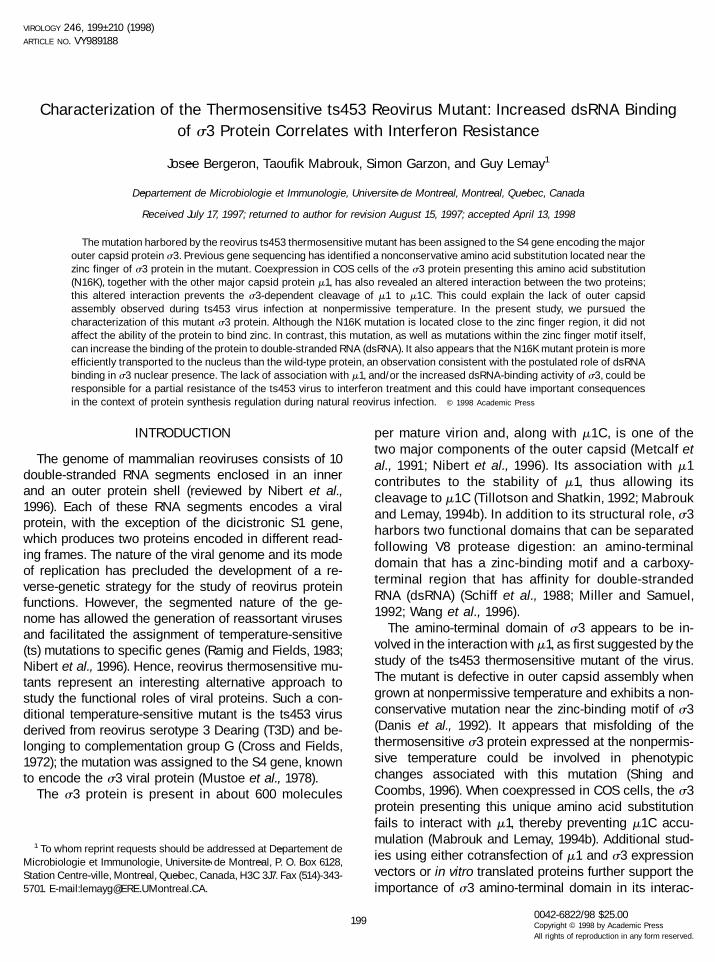
vectors were first designed to harbor mutations withinthe region encoding the N-terminal domain of the pro-tein. The amino acid substitutions used were the onesalready described as affecting zinc binding (C51A andC73A) (Mabrouk et al., 1994a) and the nonconservativesubstitution (N16K) previously found in the thermosensi-tive ts453 virus (Danis et al., 1992). In turn, these substi-tutions were combined to mutations affecting the firstC-terminal basic motif of the protein (K,R234–240A orR236A basic amino acid substitutions) (Wang et al., 1996).The positions of the different amino acid substitutionsare summarized in Fig. 1. The different proteins wereexpressed in bacterial cells by taking advantage of theT7 promoter/polymerase system (Studier et al., 1990).The capacity of each mutant to bind dsRNA was ana-lyzed by performing a Northwestern blot assay on full-length proteins. The amino-terminal amino acid substi-tutions (C51A, C73A, or N16K) did increase s3 ability tobind dsRNA by two- to four-fold compared to the wild-type protein (Figs. 2A and 2C); the presence of identicalamounts of filter-bound proteins was verified by fastgreen staining (Fig. 2B). Moreover, the mutations affect-ing the first C-terminal motif, known to reduce RNA bind-ing, had less effect when combined to the N-terminalamino acid substitutions. In fact, it was possible to re-cover RNA binding to an even higher than wild-type levelby associating the R236A substitution with either one ofthe mutations affecting the amino-terminal region of theprotein (Fig. 2A, lanes 6, 9, and 12, and Fig. 2C). However,only limited recovery was observed when the more dras-tic substitution of four basic amino acids was introducedin the same C-terminal motif (Figs. 2A, lanes 5, 8, and 11,and 2C).
Two additional results also indicate an increasedaffinity of N16K for dsRNA. Affinity chromatography ona synthetic dsRNA column, using s3 proteins pro-duced in COS cells, has shown that the binding of thes3 mutant is more resistant to an increase in ionicstrength from 0.125 to 0.4 M (Fig. 3A). Also, the maxi-mal activity of the N16K protein in the COS cell co-transfection assay, using stimulation of expression ona reporter CAT gene, was reached at lower amounts ofcotransfected s3 expression vector (Fig. 3B). This as-say, in which s3 exerts a translational effect via PKRinhibition, was previously shown to be related to theability of s3 to bind dsRNA (Mabrouk et al., 1995; Yueand Shatkin, 1997).
Effect of the thermosensitive mutation on zinc binding
Initially, the ts453 mutant was shown to be defective inouter capsid assembly (Morgan and Zweerink, 1974; Da-nis et al., 1992). In addition to preventing s3–m1 interac-tion (Mabrouk and Lemay, 1994b), the preceding exper-iments showed that the N16K substitution increasesdsRNA binding. Since the N16K substitution is located
200 BERGERON ET AL.
near the zinc finger and since it has been recently re-ported that alterations of the zinc-binding motif can alsopreclude s3–m1 association (Shepard et al., 1996), itshould be determined whether the N16K substitutiondirectly interferes with the ability of the protein to bindzinc. This point was examined by an assay based on amodification of zinc-chelate affinity chromatography us-ing radiolabeled proteins produced from transfectedCOS-7 cells. In this assay, proteins retained on the col-
umn through their interaction with zinc ions are elutedwith a chelating agent (EDTA) and analyzed by SDS–PAGE and autoradiography. A similar percentage of s3wild-type (13%) or N16K protein (19%) was retained ontothe column (Fig. 4B, lanes 2 and 3). In contrast, aminoacid substitutions introduced directly into the zinc fingerprevented binding even when larger amounts of theprotein were applied on the column (Mabrouk andLemay, 1994a; Fig. 4B, lane 4, and data not shown).
FIG. 1. Schematic representation of the different mutations within the N-terminal portion and the first C-terminal basic motif of s3 expressionvectors. Underlined bold amino acids constitute the four amino acids responsible for the tetracoordination of zinc atom and the basic amino acidsof the two C-terminal motifs, previously shown to be involved in dsRNA binding. The mutations used in this study are also indicated as bold underlinedletters.
201REOVIRUS ts453 THERMOSENSITIVE MUTANT
Subcellular localization of the thermosensitive mutants3 protein
Although mechanistic details are still lacking, recentresults suggest that s3 is partly transported to the nu-cleus by means of its dsRNA-binding activity (Yue andShatkin, 1996). The thermosensitive s3 mutant having anincreased affinity for dsRNA, it was thus of interest todetermine whether this property could actually influencethe subcellular localization of the protein. COS-7 cellswere transfected with either wild-type or N16K expres-
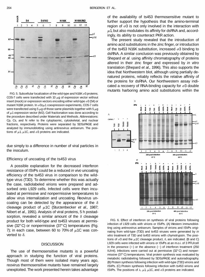
sion vectors. Cell fractionation showed nearly equalamounts of wild-type s3 in all three subcellular fractions(cytoplasmic, cytoskeletal, and nuclear) (Fig. 5, lanes2–4), whereas a larger proportion of the mutant proteinwas found in the nuclear fraction, 50% compared to30–35% for the wild-type protein (Fig. 5, lanes 8–10).Immunoreactive proteins of lower molecular weight werealso observed in the nuclear fraction and likely corre-spond to cleavage products of the s3 protein. Whenwild-type s3 was coexpressed with m1, s3–m1 associa-tion resulted in m1 to m1C cleavage (Fig. 5, lane 5). Incontrast, coexpression of N16K with m1 did not producem1C and, as previously reported (Mabrouk et al., 1994b),there was a reduction in total accumulation of m1–m1C inthe absence of interaction with the s3 N16K mutant (Fig.5, lane 11). The results also indicated that s3–m1 inter-action occurs exclusively in the cytoplasm and that s3alone is transported to the nucleus; m1 and m1C arerestricted to the cytoplasmic fraction and absent fromboth the cytoskeletal and nuclear fractions. The intranu-clear presence of s3 was further verified by immunoelec-tron microscopy using a monospecific anti-s3 antibody.The presence of s3 in both cytoplasmic and nuclearcompartments was easily detected (data not shown).Although not strictly quantitative, these latter observa-tions also support the idea of a selective nuclear enrich-ment of s3 N16K protein.
Effect of interferon on synthesis of viral proteins incells infected with wild-type or ts453 virus
By means of its dsRNA-binding activity, s3 is appar-ently involved in translational regulation by inhibition ofthe cellular interferon-inducible double-stranded RNA-dependent, protein kinase (PKR) (Imani and Jacobs, 1988;Lloyd and Shatkin, 1992; Beattie et al., 1995). In order todetermine if the ts453 virus could further modulate PKR,L929 cells were infected at permissive and nonpermis-sive temperatures following interferon treatment. Themultiplicity of infection (3 PFU/cell) was chosen so as toensure infection of most cells in the culture at eithertemperature and the dose of interferon corresponds tonearly saturating amount on L cells (Jacobs and Fergu-son, 1991; and our own unpublished data). Synthesis ofviral proteins was then detected by metabolic radiolabel-ing followed by autoradiography. Synthesis of viral pro-teins was chosen as a criterion to determine the effect ofinterferon. This synthesis of viral protein is mostly unaf-fected in the thermosensitive mutant; in contrast, com-parisons of viral yields between wild-type and ts virusesis rendered difficult by the decreased viral titer of thislatter virus and this approach was not pursued. At non-permissive temperature (37°C), cells infected with ts453virus subsequent to interferon treatment exhibited noapparent decrease in synthesis of viral proteins com-pared to untreated infected cells; in contrast, synthesis of
FIG. 2. Northwestern blot analysis of double-stranded RNA bindingin different s3 mutant proteins. The s3 mutants were expressed inbacterial cells following IPTG induction and bacterial pellets wereanalyzed by SDS–PAGE (see Materials and Methods). A is the autora-diogram of the Northwestern analysis using radiolabeled reovirusdsRNA. B is the result of fast green staining of the proteins transferredonto the filter, indicating similar expression levels of the differentmutants used in the assay. The position of the s3 proteins is indicated.As previously noticed, proteins harboring the K,R234–240 mutationexhibit a reduced electrophoretic mobility. C shows quantitation ofdsRNA binding relative to the wild-type value.
202 BERGERON ET AL.
viral proteins was reduced by 25% consecutive to inter-feron treatment at 32°C (Figs. 6B and 6C). As usuallyobserved, cells previously treated with interferon andinfected with wild-type reovirus (T3D) exhibited an im-portant decrease in synthesis of viral proteins at bothtemperatures, 50% at 37°C and 25% at 32°C (Figs. 6Band 6C). The presence of the N16K amino acid substitu-tion thus confers a resistance to interferon specifically at
the nonpermissive temperature at which expression ofthe corresponding phenotype is expected to occur.
In the context of natural gastrointestinal reovirus in-fection, partially uncoated intermediate subviral particles(ISVPs), lacking the s3 outer capsid protein, appear to beresponsible for infection rather than complete double-shelled virions (Borsa et al., 1979; Bodkin et al., 1989;Bass et al., 1990; Nibert et al., 1991; Amerongen et al.,1994). In order to determine if increased resistance ob-served with the ts453 virus is maintained when infectionis carried out with single-shelled particles, cells wereinfected with ISVPs, which were generated by treatmentof purified virions with chymotrypsin (Joklik, 1972; Borsaet al., 1973, 1979). In addition to their lack of s3, ISVPscan be recognized by the presence of a cleaved form ofm1–m1C, known as the d cleavage product. In our hands,and under the conditions used, infectious titer was notaffected by chymotrypsin treatment despite the fact thatvirions to ISVPs conversion appeared essentially com-plete as judged by disappearance of s3 and completedigestion of m1C to d (Fig. 6A). It was thus possible touse the same multiplicity of infection with either virus orISVPs stocks.
The results show that the effect of interferon treatmenton synthesis of viral proteins consecutive to the infectionwith the ISVPs derived from either wild-type or ts453virions was the same as the effect on wild-type virions(Figs. 6B and 6C). The increased interferon resistanceobserved during ts453 infection is clearly lost upon re-moval of the outer capsid. This also provides us with acontrol indicating that the difference in interferon resis-tance observed upon infection with virions could not be
FIG. 3. Increased dsRNA binding by the s3 N16K mutant. (A) COS cells transfected with either the wild-type or N16K expression construct wereradiolabeled and proteins subjected to dsRNA affinity chromatography as described under Materials and Methods. Protein eluted at different ionicstrengths were analyzed by gel electrophoresis and bound proteins quantitated relative to the original input. (B) Translational stimulation wascompared by cotransfecting CAT expression vector (0.2 mg) with 1 or 4 mg of expression vector for either the wild-type or the N16K s3 expressionvector. CAT activity was quantitated by scanning of the autoradiogram following separation of acetylated products from nonacetylated chloramphen-icol substrate. Results are presented as relative CAT activity compared to a control of cells transfected with 10 mg of CAT expression vector alone.
FIG. 4. Zinc-binding ability of the s3 protein harboring the thermo-sensitive mutation (N16K). COS-7 cells were transfected with 10 mg ofplasmid expression vector without insert (mock) or expression vectorsencoding wild-type s3 (wt), mutant N16K protein, or zinc finger mutant(C73A), as negative control. Labeled COS-7 cells were lysed andprotein extracts submitted to zinc chelate affinity chromatography asdescribed under Materials and Methods. Proteins were analyzed bySDS–PAGE followed by autoradiography. (A) Aliquots of the startingmaterial loaded onto the zinc column; (B) proteins retained onto thecolumn after washing at decreasing pH were eluted with acidic 0.2 MEDTA solution. The position of the s3 proteins is indicated.
203REOVIRUS ts453 THERMOSENSITIVE MUTANT
due simply to a difference in number of viral particles inthe inoculum.
Efficiency of uncoating of the ts453 virus
A possible explanation for the decreased interferonresistance of ISVPs could be a reduced in vivo uncoatingefficiency of the ts453 virus in comparison to the wild-type virus (T3D). To determine whether this was actuallythe case, radiolabeled virions were prepared and ad-sorbed onto L929 cells. Infected cells were then incu-bated at permissive and nonpermissive temperatures toallow virus internalization and uncoating. Reovirus un-coating can be detected by the appearance of the dcleavage product of m1C (Sturzenbecker et al., 1987;Nibert et al., 1991). Analysis of viral proteins, 5 h postad-sorption, revealed a similar amount of the d cleavageproduct for both wild-type and ts453 viruses at permis-sive (32°C) or nonpermissive (37°C) temperatures (Fig.7); in each case, between 60 to 70% of m1C was con-verted to d.
DISCUSSION
The use of thermosensitive mutants is a powerfulapproach in studying the function of viral proteins.Though most of them were isolated many years ago,reovirus thermosensitive mutants have remained largelyunexploited. The work presented herein takes advantage
of the availability of ts453 thermosensitive mutant tofurther support the hypothesis that the amino-terminalregion of s3 is not only involved in the interaction withm1, but also modulates its affinity for dsRNA and, accord-ingly, its ability to counteract PKR action.
The present study revealed that the introduction ofamino acid substitutions in the zinc finger, or introductionof the ts453 N16K substitution, increased s3 binding todsRNA. A similar conclusion was previously obtained byShepard et al. using affinity chromatography of proteinsaltered in their zinc finger and expressed by in vitrotranslation (Shepard et al., 1996). This also supports theidea that Northwestern blot, although using partially de-natured proteins, reliably reflects the relative affinity ofthe proteins for dsRNA. Our Northwestern assay indi-cated a recovery of RNA-binding capacity for s3 doublemutants harboring amino acid substitutions within the
FIG. 5. Subcellular localization of the wild-type and N16K s3 proteins.COS-7 cells were transfected with 10 mg of expression vector withoutinsert (mock) or expression vectors encoding either wild-type s3 (S4) ormutant N16K protein. In s3–m1 coexpression experiments, COS-7 cellswere transfected using 5 mg of those same plasmids together with 5 mgof m1 expression vector (M2). Cell fractionation was done according tothe procedure described under Materials and Methods. Abbreviations:Cp, Cs, and N refer to the cytoplasmic, cytoskeletal, and nuclearfractions, respectively. Proteins were separated by SDS–PAGE andanalyzed by immunoblotting using antireovirus antiserum. The posi-tions of m1, m1C, and s3 proteins are indicated.
FIG. 6. Effect of interferon on synthesis of viral proteins followinginfection of L929 cells with virions or ISVPs. (A) Western immunoblot-ting using antireovirus antiserum. Samples of virions and ISVPs origi-nating from wild-type (T3D) and ts453 viruses were generated by invitro treatment of T3D and ts453 virions with chymotrypsin. The posi-tions of s3 and the m1C cleavage product, d, are indicated. (B and C)L929 cells were infected with virions or ISVPs at an m.o.i. of 3 PFU/cellin the presence (1) or the absence (2) of interferon treatment (250IU/ml). Infections were carried out at permissive (32°C) and nonper-missive (37°C) temperatures. Viral protein synthesis was evaluated bymetabolic radiolabeling followed by SDS–PAGE and autoradiography.(B) Protein synthesis following infection with wild-type (T3D) virions andISVPs. (C) Protein synthesis following infection with ts453 virions andISVPs. The positions of l, m1, m1C, and s3 proteins are indicated.
204 BERGERON ET AL.
zinc finger or the N16K substitution, together with muta-tions affecting the first C-terminal basic motif. In additionto Northwestern analysis, binding of the N16K protein topoly(rl):poly(rC) agarose columns at higher salt concen-tration is another evidence for an increased ability tobind dsRNA. Finally, the better affinity of N16K for dsRNAis also supported by its increased ability to stimulateexpression of the CAT gene in COS cell cotransfectionexperiments. Various studies have demonstrated the cor-relation between dsRNA binding and the ability of s3 tostimulate translation via PKR inhibition in this assay.
The two C-terminal basic motifs are predicted to adoptan a-helical configuration using standard secondarystructure prediction methods (Chou and Fasman, 1974;Garnier et al., 1978). Yet, prediction algorithms indicatethat the first C-terminal motif is less prone to form ana-helical structure than is the second basic motif (un-published data). Although both basic motifs exhibitamino acid sequence similarity with other dsRNA-bind-ing proteins (see among others: Green and Mathews,1992; McCormack et al., 1992; Chang and Jacobs, 1992),it was observed that the first motif also shares lesssequence homology and displays a basic stretch whichis less aligned with its analogous counterparts (Yue andShatkin, 1996). Altogether, these findings support theidea that the first C-terminal motif can become dispens-able, either upon removal of the N-terminal domain orsubsequent to mutations introduced near or within thezinc finger; the first basic motif may be acting as a‘‘buffer’’ to prevent the inhibitory effect of the wild-type
N-terminal region. Although it is unlikely that truncatedforms of s3 could be found in replication-competentvirus, it is possible that amino acid changes in theamino-terminal portion of the protein and affectingdsRNA binding and susceptibility to interferon couldeventually be identified in viral isolates.
Affinity chromatography clearly showed that zinc bind-ing is not directly affected by the N16K substitution (Fig.3). In addition, contrary to zinc finger mutants, which areunstable when transiently expressed in COS cells(Mabrouk and Lemay, 1994a) or significantly protease-sensitive when expressed by in vitro translation (Shep-ard et al., 1996), the N16K mutant was apparently stablein COS cells; its total accumulation being similar to thatof wild-type. Nevertheless, it cannot be completely ex-cluded that the s3 produced during multiplication ofts453 virus is more unstable than the wild-type protein,since viral assembly could likely play a role in proteinstability; recently published data did suggest a reducedstability of the s3 protein during viral infection with thets453 virus (Shing and Coombs, 1996). It thus appearsthat an alteration of the zinc-binding ability of the proteinis not responsible for the stimulatory effect on dsRNA-binding activity, but that it is rather a conformationalchange exerted by amino acid substitutions within thezinc finger or surrounding this motif of the protein. In fact,it is well known that zinc fingers can be important deter-minants of protein conformation (see for example: McIn-tyre et al., 1993).
The presence of s3 in the nucleus has been recentlyreported and the dsRNA-binding activity of the proteinwas held responsible for this subcellular distribution(Yue and Shatkin, 1996). Nuclear presence of s3 appearsrather surprising considering that the nucleus does notseem to intervene during reovirus multiplication (Follettet al., 1975; Zarbl and Millward, 1983), although somestudies have indicated morphological alterations of thenucleus in infected cells (Chaly et al., 1980). It should bementioned that other dsRNA-binding proteins, such asthe cellular PKR (RNA-dependent protein kinase) or theE3L protein of vaccinia virus (a DNA virus replicating inthe cytoplasm of infected cells), were also shown to bepartly localized to the nucleus (Yuwen et al., 1993; Jeffreyet al., 1995). In the present study, cell fractionation wasused instead of immunofluorescence analysis to allowmore precise determination of the relative amount of s3found within three distinct subcellular compartments(cytoplasm, cytoskeleton, and nucleus). The fact that s3N16K mutant was preferentially found in the nucleuscompared to the wild-type protein further supports theimportance of dsRNA-binding activity in nuclear trans-port. Although mechanistic details are lacking, our re-sults with the N16K mutant are in good agreement with amodel in which the affinity of s3 is of crucial importancefor nuclear presence, while blockage of this affinity, con-secutive to m1 binding, prevents this subcellular local-
FIG. 7. Uncoating efficiency of the ts453 virus. Radiolabeled wild-type(T3D) or ts453 virus was adsorbed onto L929 cells. Radiolabeled viralproteins were then recovered immediately after the 1-h adsorptionperiod at 4°C (T0) or after 5 h incubation (T5) at either permissive(32°C) or nonpermissive (37°C) temperature. The positions of viralproteins l, m1C, d, and s3 are indicated.
205REOVIRUS ts453 THERMOSENSITIVE MUTANT
ization (Yue and Shatkin, 1996). Immunoelectron micros-copy of L929 cells infected with reovirus serotype 3 alsorevealed the nuclear presence of s3 (unpublished data),the labeling being most intense near the nucleolus, thesite of rRNA synthesis and ribosome assembly. Consid-ering that cytoplasmic s3 was previously found enrichedin fractions prepared from infected L929 cells by high-salt wash of ribosomes (RSW) (Lemay and Millward,1986; Lemieux et al., 1987), it is tempting to speculatethat the presence of s3 in the nucleus could be some-how related to PKR inhibition.
Previous studies have indicated that the formation ofs3–m1 protein complexes interferes with the ability of s3to bind dsRNA and to inhibit PKR autophosphorylationpathway. These studies include coexpression experi-ments in which m1 interfered with the ability of s3 tostimulate reporter gene expression (Tillotson and Shat-kin, 1992) and the observation that s3–m1 complexespresent in infected cells were never isolated by affinityon dsRNA columns (Huismans and Joklik, 1976; Lemieuxet al., 1987). The binding of s3 with m1 results in aconformational change rendering s3 more sensitive toproteolytic degradation; this conformational change maybe an important event conducing to the shift of s3 fromits translational control role to its structural role (Shepardet al., 1995; Yue and Shatkin, 1997; Schmechel et al.,1997). PKR is considered as one of the main antiviralmechanism induced by interferon and is believed to bethe main intracellular factor interfering with reovirus mul-tiplication (Wiebe and Joklik, 1975; Miyamoto and Sam-uel, 1980; Gupta et al., 1982; Nilsen et al., 1982; DeBenedetti et al., 1985). Our finding that the ts453 viruswas resistant to the effect of interferon at nonpermissivetemperature is a further evidence for the importance ofs3 in PKR regulation. It is not clear whether the in-creased dsRNA binding or lack of interaction with m1 isresponsible for this increased interferon resistance ofts453 virus. However, an increased dsRNA binding couldbe sufficient for effective PKR inhibition, since the s3N16K mutant was found to be more active than wild-typefor the stimulation of a reporter gene in a cotransfectionassay. A better affinity of s3 for dsRNA could certainlyincrease its ability to inhibit PKR, especially consideringthat a recent report has established that this affinity is atleast 50-fold lower than that of PKR (Yue and Shatkin,1997).
Finally, the fact that ISVPs have a reduced interferonresistance suggests that viral proteins from infectingvirions can play a key role in translational regulation atearly stages of viral infection. A similar mechanism hasbeen proposed for other viruses, such as mengoviruses(King and Simon, 1993), and this point certainly deservesto be further studied. Since infection by ISVPs, lackings3, is believed to be the main route of natural reovirusinfection at mucosal surfaces (Bodkin et al., 1989; Basset al., 1990; Amerongen et al., 1994), our observations
raise the possibility that interferon can play a greater rolein the control of reovirus infection in this context.
Altogether our results demonstrate that the amino-terminal region located near the zinc-binding motif of s3is an essential determinant in viral protein interactionand PKR regulation through modulation of dsRNA bind-ing. This work further stresses the importance of tsmutants for the study of viral proteins in Reoviridae. Inthe absence of an adequate reverse-genetics system,the ts mutants represent the best tool for correlating invitro biochemical data with effects observed during theviral multiplication cycle.
MATERIALS AND METHODS
Construction of bacterial expression vectors
The cloned S4 gene encoding s3 was subjected tosite-directed mutagenesis on double-stranded DNA us-ing the unique site elimination procedure (Deng andNickoloff, 1992). Mutants of the amino-terminal domainwere previously cloned in plasmid vectors suitable fortransient expression in COS cells (Mabrouk and Lemay,1994a), whereas mutants of the carboxy-terminal regionwere already available in bacterial expression vectors(Wang et al., 1996). To obtain mutants harboring pointmutations affecting both the amino-terminal and car-boxy-terminal domains, the mutated S4 genes of theeukaryotic expression vectors were amplified by poly-merase chain reaction (PCR) performed with a GeneAmp TM DNA Amplification reagent kit (Perkin–Elmer)using Ampli Taq DNA polymerase. Oligonucleotidesused were 5’ CCGATTGTCCATATGGAGGTGTGC and 59GGAACGGCTGCGCTGCGACAGTG 3’. This procedurewas used to introduce a NdeI site at the 5’ end of the S4coding sequence, as underlined. The N-terminal frag-ments were then obtained following digestion with NdeIand SpeI and subcloned to replace the homologousfragments in bacterial vectors, which provided the C-terminal mutations. The final constructs were verified bydirect sequencing of denatured plasmid DNA.
Protein expression and Northwestern blot assay
Bacterial expression vector pET21a and bacterialEscherichia coli strain BL21(DE3) were originally ob-tained from Novagen Inc. Plasmids harboring mutatedS4 genes under the control of the T7 promoter weretransformed in the BL21(DE3) strain expressing the T7polymerase under the control of an IPTG-inducible pro-moter. Induction of bacterial cultures was performed aspreviously described (Wang et al., 1996). For Northwest-ern blot analysis, proteins were resolved by SDS–poly-acrylamide gel electrophoresis (SDS–PAGE) and trans-ferred onto nitrocellulose filters (Laemmli, 1970; Towbinet al., 1979). The s3 dsRNA-binding assay was per-formed using radioactive reovirus RNA (Wang et al.,
206 BERGERON ET AL.
1996). Quantification of the autoradiograms was donewith a laser personal densitometer (Molecular Dynam-ics). The filter was then stained with 0.1% fast green infixing solution (40% methanol–10% acetic acid) to revealfilter-bound proteins.
Cells and viruses
Mouse L929 fibroblasts and initial inoculum of wild-type reovirus serotype 3 Dearing (T3D) were originallyobtained from the American Type Culture Collection,whereas the initial inoculum of the ts453 virus was agenerous gift from Dr. W. K. Joklik (Department of Micro-biology and Immunology, Duke University Medical Cen-ter, Durham, NC). COS-7 cells were provided by Dr. E. A.Cohen (Departement de Microbiologie et Immunologie,Universite de Montreal). Viral stocks of T3D and ts453were propagated at permissive temperature (32°C) onL929 cell monolayers, whereas L929 and COS-7 cellswere maintained as previously described (Danis andLemay, 1993; Mabrouk and Lemay, 1994b).
Transfection and metabolic radiolabeling
COS-7 cells were seeded the day before transfectionat a concentration of 5 3 105 cells per 100-mm-diametertissue culture dishes. Cells were then transfected with 10mg of DNA using the standard calcium phosphate copre-cipitation procedure (Graham and van der Eb, 1973).Expression vectors harboring an SV40 replication originand reovirus genes under the control of SV40 promoterhave been previously described (Mabrouk and Lemay,1994b). After 16 h of contact with the precipitate, cellswere subjected to three washes in phosphate-bufferedsaline (PBS) and fresh medium was added. For meta-bolic radiolabeling of proteins, cell monolayers wereused 48 h posttransfection; they were washed threetimes in PBS and starved for 30 min in a methionine-freemedium (MEM without methionine, Gibco/BRL) supple-mented with 1% glutamine. Labeling was performed for1 h with Tran35S-label (ICN; 1000 Ci/mmol) at a concen-tration of 50 mCi/ml.
dsRNA affinity chromatography
Following metabolic radiolabeling, COS-7 cells werewashed in PBS and harvested in HKM buffer [10 mMHEPES–KOH (pH 8.0), 10 mM potassium acetate, 1.5 mMmagnesium acetate]. Samples were then brought tovarying concentrations of NaCl before being appliedonto poly(rl):poly(rC)–agarose (Pharmacia) as previouslydescribed (Mabrouk et al., 1995). Proteins retained on theagarose beads following extensive washing in the samebuffer were then analyzed by SDS–PAGE, autoradiogra-phy, and densitometric analysis.
Chloramphenicol acetyltransferase (CAT) assay
COS cells were transfected with either CAT expressionvector alone or in combination with varying amounts ofexpression vector for the wild-type or the N16K mutant ofs3. In each case, the total amount of plasmid was main-tained constant (10 mg per 100-mm-diameter petri dish)by adding appropriate amounts of homologous vectorwith no insert. Cells were lysed by repeated cycles offreezing and thawing and CAT assay was performed oncell lysates using standard procedures (Gorman et al.,1982; Leahy et al., 1995).
Zinc affinity chromatography
Following metabolic radiolabeling, COS-7 cells werewashed in PBS and harvested in 300 ml of column buffer[50 mM Tris–HCl (pH 8.0), 150 mM NaCl] containing 0.1%NP-40. Zinc chelate affinity adsorbent (Boehringer-Mann-heim) was used as described (Porath et al., 1975). Briefly,samples were loaded onto a zinc affinity column and leftfor 2 h at 4°C. Thereafter, the column was extensivelywashed with buffer containing 150 and 800 mM NaCl toreduce nonspecific binding. Selective elution was per-formed using 0.1 M NaPO4, 0.8 M NaCl with decreasingpH (7.5, 7.0, 6.5, 6.0 and 5.5), and final elution was per-formed with 0.2 M EDTA (pH 6.0). Protein samples wereanalyzed by SDS–PAGE and autoradiography. Quantifi-cation of the autoradiograms was done with a laserpersonal densitometer (Molecular Dynamics).
Cell fractionation and immunoblotting
Transfected COS-7 cells were recovered in PBS bycentrifugation at 5000 rpm for 15 min. Cell pellets wereresuspended in hypotonic buffer A [10 mM HEPES-KOH (pH 8.0), 10 mM potassium acetate, 1.5 mM mag-nesium acetate, and 1 mM DTT], incubated 10 min at4°C, and centrifuged at 10,000 rpm for 10 min. Thesupernatants were saved as the cytoplasmic fractions.Following the same procedure, pellets were washed inbuffer B [10 mM PIPES–HCl (pH 6.8), 100 mM KCl, 2.5mM MgCl2, 0.3 M sucrose, 1% Triton X-100] and resus-pended in buffer C [10 mM Tris–HCl (pH7.5), 10 mMNaCl, 1.5 mM MgCl2, 1% Tween 20, 0.5% sodium de-oxycholate]. The supernatant obtained subsequentlyto the treatment with buffer C was considered thecytoskeletal fraction. Finally, the remaining pellets,composed of nuclei, were resuspended in Laemmli’ssample buffer (Laemmli, 1970). The different proteinfractions were analyzed by SDS–PAGE and immuno-blotting using rabbit antireovirus antiserum (Lee Bio-molecular) and goat anti-rabbit IgG–alkaline phospha-tase conjugate (GIBCO/BRL) as previously described(Mabrouk and Lemay, 1994b).
207REOVIRUS ts453 THERMOSENSITIVE MUTANT
In vitro generation of infectious subviral particles(ISVPs)
Viral stocks were prepared following infection of L929cells in the absence of serum and were treated with 20mg/ml of chymotrypsin (Sigma, bovine pancreas type I-S)for 30 min at 37°C. As a control, half of each viral stockwas submitted to the same procedure in the absence ofchymotrypsin. Viral titers of treated and untreated viralstocks were determined by using the ‘‘Tissue CultureInfection Dose 50%’’ method (Reed and Muench, 1938).For protein analysis, L929 cells were seeded at a con-centration of 5 3 105 cells per 60-mm-diameter tissueculture dish the day before infection.
Viral infection and interferon treatment
When necessary, cells were pretreated with mouseb-interferon (Lee Biomolecular) at a saturating concen-tration of 250 international units (IU)/ml for 16 h. Subse-quently, cell monolayers were infected at an m.o.i. of 3PFU/cell with either ISVPs or complete virions; metabolicradiolabeling was performed 20 h postinfection at 37°C.Cells were then recovered by centrifugation and lysed inTris-buffered saline [10 mM Tris–HCl (pH7.5), 150 mMNaCl] containing 0.1% NP-40. Labeled proteins were an-alyzed by SDS–PAGE followed by autoradiography. Forthe infections carried out at 32°C, incubation times forinfection and radiolabeling were doubled.
In vivo uncoating of viral particles
Radiolabeled viral particles were obtained by infectingL929 cells with either wild-type (T3D) or ts453 viruses atan m.o.i. of 25 PFU/cell. Cells were then incubated for12 h at 32°C in minimal Eagle’s medium without methi-onine (MEM, Gibco/BRL). Infected cells were subse-quently incubated 16 h in MEM reduced to 1/10 of normalmethionine concentration and supplemented with 100mCi/ml of Tran35S-label (ICN; 1000 Ci/mmol). Viral parti-cles were recovered by freon extraction of infected cells(Smith et al., 1969) and dialyzed overnight against 100 volof MEM. The radiolabeled viral particles were adsorbed1 h at 4°C onto cell monolayers and removed. Cells werethen washed three times in nonradioactive medium andincubated at the corresponding temperature (either 32°or 37°C) for 5 h and recovered. Viral proteins weredetected by SDS–PAGE followed by autoradiography.
ACKNOWLEDGMENTS
We thank Dr. A. J. Shatkin (Center for Advanced Biotechnology andMedicine, Piscataway, NJ) for his generous gift of the m1 expressionvector and anti-s3 monospecific antibody and Dr. W. K. Joklik (Depart-ment of Microbiology and Immunology, Duke University Medical Cen-ter, Durham, NC) for his initial gift of the ts453 mutant. We also thankCarole Danis for technical support conducing to smooth operation ofthe laboratory. This work was supported by a grant to G.L. from theMedical Research Council of Canada. G.L. is also the recipient of a
scholarship from the ‘‘Fonds de la Recherche en Sante du Quebec.’’ J.B.was the recipient of a studentship from the ‘‘Fonds pour la Formation deChercheurs et l’Aide a la Recherche,’’ and T.M was the recipient of astudentship from the ‘‘Agence Canadienne de Developpement Interna-tional.’’
REFERENCES
Amerongen, H. M., Wilson, G. R., Fields, B. N., and Neutra, M. R. (1994).Proteolytic processing of reovirus is required for adherence to intes-tinal M cells. J. Virol. 68, 8428–8432.
Bass, D. M., Bodkin, D., Dambrauskas, R., Trier, J. S., Fields, B. N., andWolf, J. L. (1990). Intraluminal proteolytic activation plays an importantrole in replication of type 1 reovirus in the intestines of neonatalmice. J. Virol. 64, 1830–1833.
Beattie, E., Denzler, K. L., Tartaglia, J., Perkus, M. E., Paoletti, E., andJacobs, B. L. (1995). Reversal of the interferon-sensitive phenotype ofa vaccinia virus lacking E3L by expression of the reovirus S4 gene.J. Virol. 69, 499–505.
Bodkin, D. K., Nibert, M. L., and Fields, B. N. (1989). Proteolytic digestionof reovirus in the intestinal lumens of neonatal mice. J. Virol. 63,4676–4681.
Borsa, J., Sargent, M. D., Copps, T. P., Long, D. G., and Chapman, J. D.(1973). Specific monovalent cation effects on modification of reovirusinfectivity by chymotrypsin digestion in vitro. J. Virol. 11, 1017–1019.
Borsa, J., Morash, B. D., Sargent, M. D., Copps, T. P., Lievaart, P. A., andSzekely, J. G. (1979). Two modes of entry of reovirus particles into Lcells. J. Gen. Virol. 45, 161–170.
Chaly, N., Johnstone, M., and Hand, R. (1980). Alterations in nuclearstructure and function in reovirus-infected cells. Clin. Invest. Med. 2,141–152.
Chang, H.-W., and Jacobs, B. L. (1992). The E3L gene of vaccinia virusencodes an inhibitor of the interferon-induced double-stranded RNA-dependent protein kinase. Proc. Natl. Acad. Sci. USA 89, 4825–4829.
Chou, P. Y., and Fasman, G. D. (1974). Prediction of protein conforma-tion. Biochemistry 13, 222–245.
Cross, R. K., and Fields, B. N. (1972). Temperature-sensitive mutants ofreovirus type 3: Studies on the synthesis of viral RNA. J. Virol. 50,799–809.
Danis, C., Garzon, S., and Lemay, G. (1992). Further characterization ofthe ts453 mutant of mammalian orthoreovirus serotype 3 and nucle-otide sequence of the mutated S4 gene. Virology 190, 494–498.
Danis, C., and Lemay, G. (1993). Protein synthesis in different cell linesinfected with orthoreovirus serotype 3: Inhibition of host-cell proteinsynthesis correlates with accelerated viral multiplication and cellkilling. Biochem. Cell Biol. 71, 81–85.
DeBenedetti, A., Williams, G. J., and Baglioni, C. (1985). Inhibition ofbinding to initiation complexes of nascent reovirus mRNA by double-stranded RNA-dependent protein kinase. J. Virol. 54, 408–413.
Deng, W. P., and Nickoloff, J. A. (1992). Site-directed mutagenesis ofvirtually any plasmid by eliminating a unique site. Anal. Biochem.200, 81–88.
Denzler, K. L., and Jacobs, B. L. (1994). Site-directed mutagenic analysisof the reovirus sigma3 protein binding to double-stranded RNA.Virology 204, 190–199.
Follet, E. A., Pringle, C. R., and Pennington, T. H. (1975). Virus develop-ment in enucleated cells: echovirus, poliovirus, pseudorabies virus,reovirus, respiratory syncytial virus and Semliki forest virus. J. Gen.Virol. 26, 183–196.
Garnier, J., Osguthorpe, D. J., and Robson, B. (1978). Analysis of theaccuracy and implications of simple methods for predicting thesecondary structure of globular proteins. J. Mol. Biol. 120, 97–120.
Giantini, M., and Shatkin, A. J. (1989). Stimulation of chloramphenicolacetyltransferase mRNA translation by reovirus capsid polypeptidesigma 3 in cotransfected COS cells. J. Virol. 63, 2415–2421.
Gorman, C. M., Moffat, L. F., and Howard, B. H. (1982). Recombinant
208 BERGERON ET AL.
genomes which express chloramphenicol acetyltransferase in mam-malian cells. Mol. Cell. Biol. 2, 1044–1051.
Graham, F. L., and van der Eb, A. J. (1973). A new technique for theassay of infectivity of human adenovirus 5 DNA. Virology 52, 151–157.
Green, S. R., and Mathews, M. B. (1992). Two RNA-binding motifs in thedouble-stranded RNA-activated protein kinase, DAI. Genes Dev. 6,2478–2490.
Gupta, S. L., Holmes, S. L., and Mehra, L. L. (1982). Interferon actionagainst reovirus: Activation of interferon-induced protein kinase inmouse L929 cells upon reovirus infection. Virology 120, 495–499.
Hovanessian, A. G. (1993). Interferon-induced dsRNA-activated proteinkinase: Antiproliferative, antiviral and antitumoral functions. Semin.Virol. 4, 237–246.
Huismans, H., and Joklik, W. K. (1976). Reovirus coded polypeptides ininfected cells: Isolation of two native monomeric polypeptides withaffinity for single-stranded and double-stranded RNA, respectively.Virology 70, 411–424.
Imani, F., and Jacobs, B. L. (1988). Inhibitory activity for the interferon-induced protein kinase is associated with the reovirus serotype 1sigma 3 protein. Proc. Natl. Acad. Sci. USA 85, 7887–7891.
Jacobs, B. L., and Ferguson, R. E. (1991). The Lang strain of reovirusserotype 1 and the Dearing strain of reovirus serotype 3 differ in theirsensitivities to beta interferon. J. Virol. 65, 5102–5104.
Jeffrey, I. W., Kadereit, S., Meurs, E. F., Metzger, T., Bachmann, M.,Schwemmle, M., Hovanessian, A. G., and Clemens, M. J. (1995).Nuclear localization of the interferon-inducible protein kinase PKR inhuman cells and transfected mouse cells. Exp. Cell Res. 218, 17–27.
Joklik, W. K. (1972). Studies on the effect of chymotrypsin on reovirions.Virology 49, 700–715.
Katze, M. G. (1992). The war against the interferon-induced dsRNA-activated protein kinase: Can viruses win? J. Interf. Res. 12, 241–248.
Katze, M. G. (1993). Games viruses play: A strategic initiative againstthe interferon-induced dsRNA-activated 68,000 Mr protein kinase.Semin. Virol. 4, 259–268.
King, R. W., and Simon, E. H. (1993). The virion of mengovirus containsanti-interferon activity. J. Interferon Res. 13, 1–7.
Laemmli, U. K. (1970). Cleavage of structural proteins during the as-sembly of the head of bacteriophage T4. Nature 227, 680–685.
Leahy, P., Carmichael, G. G., and Rossomando, E. F. (1995). Effects ofethanol concentration and incubation period at 65°C on Cat activityin mammalian cell extracts. BioTechniques 19, 894.
Lemay, G., and Millward, S. (1986). Expression of the cloned S4 gene ofreovirus serotype 3 in transformed eucaryotic cells: Enrichment ofthe viral protein in the crude initiation factor fraction. Virus Res. 6,133–140.
Lemieux, R., Lemay, G., and Millward, S. (1987). The viral protein sigma3 participates in translation of late viral mRNA in reovirus-infected Lcells. J. Virol. 61, 2472–2479.
Lloyd, R. M., and Shatkin, A. J. (1992). Translational regulation byreovirus polypeptide s3: Substitution for VAI RNA and inhibition ofphosphorylation of the a subunit of eukaryotic initiation factor 2.J. Virol. 66, 6878–6884.
Mabrouk, T., and Lemay, G. (1994a). Mutations in a CCHC zinc-bindingmotif of the reovirus s3 protein decrease its intracellular stability.J. Virol. 68, 5287–5290.
Mabrouk, T., and Lemay, G. (1994b). The sequence similarity of reoviruss3 protein to picornaviral proteases is unrelated to its role in m1 viralprotein cleavage. Virology 202, 615–620.
Mabrouk, T., Danis, C., and Lemay, G. (1995). Two basic motifs ofreovirus s3 protein are involved in double-stranded RNA binding.Biochem. Cell Biol. 73, 137–145.
Martin, P. E. M., and McCrae, M. A. (1993). Analysis of the stimulationof reporter gene expression by the sigma3 protein of reovirus inco-transfected cells. J. Gen. Virol. 74, 1055–1062.
McCormack, S. J., Thomis, D. C., and Samuel, C. E. (1992). Mechanismof interferon action: Identification of a RNA binding domain within the
N-terminal region of the human RNA-dependent P1/elF-2 alpha pro-tein kinase. Virology 188, 47–56.
McIntyre, M. C., Frattini, M. G., Grossman, S. R., and Laimins, L. A.(1993). Human papillomavirus type 18 E7 protein requires intactCys-X-X-Cys motif for zinc binding, dimerization, and transformationbut not for Rb binding. J. Virol. 67, 3142–3150.
Metcalf, P., Cyrklaff, M., and Adrian, M. (1991). The three-dimensionalstructure of reovirus obtained by cryo-electron microscopy. EMBO J.10, 3129–3136.
Miller, F. E., and Samuel, C. E. (1992). Proteolytic cleavage of thereovirus sigma 3 protein results in enhanced double-stranded RNA-binding activity: Identification of a repeated basic amino acid motifwithin the C-terminal binding region. J. Virol. 66, 5347–5356.
Miyamoto, N. G., and Samuel, C. E. (1980). Mechanism of interferonaction: Interferon-mediated inhibition of reovirus mRNA translation inthe absence of detectable mRNA degradation but in the presence ofprotein phosphorylation. Virology 107, 461–475.
Morgan, E. M., and Zweerink, H. J. (1974). Reovirus morphogenesis:Core-like particles in cells infected at 39 degrees with wild-typereovirus and temperature sensitive mutants of groups B and G.Virology 59, 556–565.
Mustoe, T. A., Ramig, R. F., Sharpe, A. H., and Fields, B. N. (1978). Agenetic map of reovirus. III. Assignment of the double-stranded RNAmutant groups A, B, and G to genome segments. Virology 85, 545–556.
Nibert, M. L., Furlong, D. B., and Fields, B. N. (1991). Mechanisms ofviral pathogenesis: Distinct forms of reoviruses and their roles duringreplication in cells and host. J. Clin. Invest. 88, 727–734.
Nibert, M. L., Schiff, L. A., and Fields, B. N. (1996). Reoviruses and theirreplication. In ‘‘Fundamental Virology’’ (B. N. Fields, D. M. Knipe, andP. M. Howley, Eds.), pp. 691–730. Lippincott–Raven, Philadelphia.
Nilsen, T. W., Maroney, P. A., and Baglioni, C. (1982). Inhibition of proteinsynthesis in reovirus-infected HeLa cells with elevated levels ofinterferon-induced protein kinase activity. J. Biol. Chem. 257, 14593–14596.
Porath, J., Carlsson, J., Olsson, I., and Belfrage, G. (1975). Metal chelateaffinity chromatography, a new approach to protein fractionation.Nature 258, 598–599.
Ramig, R., and Fields, B. N. (1983). Genetics of reovirus. In ‘‘TheReoviridae’’ (W. K. Joklik, Ed.), pp. 197–228. Plenum, New York.
Reed, L. J., and Muench, H. (1938). A simple method for estimating fiftyper cent endpoints. Am. J. Hyg. 27, 493–497.
Samuel, C. E. (1991). Antiviral actions of interferon: Interferon-regulatedcellular proteins and their surprisingly selective antiviral activities.Virology 183, 1–11.
Schiff, L. A., Nibert, M. L., Co, M. S., Brown, E. G., and Fields, B. N.(1988). Distinct binding sites for zinc and double-stranded RNA in thereovirus outer capsid protein sigma 3. Mol. Cell. Biol. 8, 273–283.
Schmechel, S., Chute, M., Skinner, P., Anderson, R., and Schiff, L. (1997).Preferential translation of reovirus mRNA by a s3-dependent mech-anism. Virology 232, 62–73.
Seliger, L. S., Giantini, M., and Shatkin, A. J. (1992). Translational effectsand sequence comparisons of the three serotypes of the reovirus S4gene. Virology 187, 202–210.
Shepard, D. A., Ehnstrom, J. G., and Schiff, L. A. (1995). Association ofreovirus outer capsid protein s3 and m1 causes a conformationalchange that renders s3 protease sensitive. J. Virol. 69, 8180–8184.
Shepard, D. A., Ehnstrom, J. G., Skinner, P. J., and Schiff, L. A. (1996).Mutations in the zinc-binding motif of the reovirus capsid protein s3eliminate its ability to associate with capsid protein m1. J. Virol. 70,2065–2068.
Shing, M., and Coombs, K. M. (1996). Assembly of the reovirus outercapsid requires m1/s3 interactions which are prevented by mis-folded s3 protein in temperature-sensitive mutant tsG453. Virus Res.46, 19–29.
Smith, R. E., Zweerink, H. J., and Joklik, W. K. (1969). Polypeptide
209REOVIRUS ts453 THERMOSENSITIVE MUTANT
components of virions, top component and cores of reovirus 3.Virology 89, 791–810.
Studier, F. W., Rosenberg, A. H., Dunn, J. J., and Dubendorff, J. W. (1990).Use of T7 RNA polymerase to direct expression of cloned genes.Methods Enzymol. 185, 60–89.
Sturzenbecker, L. J., Nibert, M., Furlong, D., and Fields, B. N. (1987).Intracellular digestion of reovirus particles requires a low pH and isan essential step in the viral infectious cycle. J. Virol. 61, 2351–2361.
Tillotson, L., and Shatkin, A. J. (1992). Reovirus polypeptide sigma 3 andN-terminal myristoylation of polypeptide mu1 are required for site-specific cleavage to mu1c in transfected cells. J. Virol. 66, 2180–2186.
Towbin, H., Staehelin, T., and Gordon, J. (1979). Electrotransfer of pro-teins from polyacrylamide gel to nitrocellulose sheets: Procedureand some applications. Proc. Natl. Acad. Sci. USA 76, 4350–4354.
Wang, Q., Bergeron, J., Mabrouk, T., and Lemay, G. (1996). Site-directed
mutagenesis of the double-stranded RNA binding domain of bacte-rially expressed s3 protein. Virus Res. 41, 141–151.
Wiebe, M. E., and Joklik, W. K. (1975). The mechanism of inhibition ofreovirus replication by interferon. Virology 66, 229–240.
Yue, Z., and Shatkin, A. J. (1996). Regulated, stable expression andnuclear presence of reovirus double-stranded RNA-binding proteins3 in HeLa cells. J. Virol. 70, 3497–3501.
Yue, Z., and Shatkin, A. J. (1997). Double-stranded RNA-dependentprotein kinase (PKR) is regulated by reovirus structural proteins.Virology 234, 364–371.
Yuwen, H., Cox, J. H., Yewdell, J. W., Bennink, J. R., and Moss, B. (1993).Nuclear localization of a double-stranded RNA-binding protein en-coded by the vaccinia virus E3L gene. Virology 195, 732–744.
Zarbl, H., and Millward, S. (1983). The reovirus multiplication cycle. In‘‘The Reoviridae’’ (W. K. Joklik, Ed.), pp. 197–228. Plenum, New York.