1 1 FORUM REVIEW Characterizing the Dynamics of Proteasome Complexes by Proteomics Approaches Robyn M. Kaake, Athit Kao, Clinton Yu, Lan Huang* Department of Physiology and Biophysics, University of California, Irvine, CA 92697, *Correspondence should be addressed to Dr. Lan Huang ([email protected]) Medical Science I, D233 Department of Physiology & Biophysics University of California, Irvine Irvine, CA 92697-4560 Phone: (949) 824-8548 Fax: (949) 824-8540 Running Head: Proteomics of proteasome complexes Page 1 of 39 Antioxidants & Redox Signaling Characterizing the Dynamics of Proteasome Complexes by Proteomics Approaches (doi: 10.1089/ars.2013.5815) This article has been peer-reviewed and accepted for publication, but has yet to undergo copyediting and proof correction. The final published version may differ from this proof.
Transcript
1
1
FORUM REVIEW
Characterizing the Dynamics of Proteasome Complexes by Proteomics Approaches
Robyn M. Kaake, Athit Kao, Clinton Yu, Lan Huang*
Department of Physiology and Biophysics, University of California, Irvine, CA 92697,
*Correspondence should be addressed to Dr. Lan Huang ([email protected])
Medical Science I, D233
Department of Physiology & Biophysics
University of California, Irvine
Irvine, CA 92697-4560
Phone: (949) 824-8548
Fax: (949) 824-8540
Running Head: Proteomics of proteasome complexes
Page 1 of 39
Ant
ioxi
dant
s &
Red
ox S
igna
ling
Cha
ract
eriz
ing
the
Dyn
amic
s of
Pro
teas
ome
Com
plex
es b
y Pr
oteo
mic
s A
ppro
ache
s (d
oi: 1
0.10
89/a
rs.2
013.
5815
)T
his
artic
le h
as b
een
peer
-rev
iew
ed a
nd a
ccep
ted
for
publ
icat
ion,
but
has
yet
to u
nder
go c
opye
ditin
g an
d pr
oof
corr
ectio
n. T
he f
inal
pub
lishe
d ve
rsio
n m
ay d
iffe
r fr
om th
is p
roof
.
2
2
ABSTRACT
Significance: The proteasome is the degradation machine of the ubiquitin-proteasome system
(UPS), which is critical in controlling many essential biological processes. Aberrant regulation
of proteasome-dependent protein degradation can lead to various human diseases and general
proteasome inhibitors have shown efficacy for cancer treatments. Though clinically effective,
current proteasome inhibitors have detrimental side effects and thus better therapeutic strategies
targeting proteasomes are needed. Therefore, comprehensive characterization of proteasome
complexes will provide the molecular details essential for developing new and improved drugs.
Recent Advances: New mass spectrometry-based proteomics approaches have been developed
to study protein interaction networks and structural topologies of proteasome complexes. The
results have helped define the dynamic proteomes of proteasome complexes, thus providing new
insights to the mechanisms underlying proteasome function and regulation.
Critical Issues: The proteasome exists as heterogeneous populations in tissues/cells and its
proteome is highly dynamic and complex. In addition, proteasome complexes are regulated by
various mechanisms under different physiological conditions. Consequently, complete proteomic
profiling of proteasome complexes remains a major challenge for the field.
Future Directions: We expect that proteomic methodologies enabling full characterization of
proteasome complexes will continue to evolve. Further advances in mass spectrometry
instrumentation and protein separation techniques will be needed to facilitate the detailed
proteomic analysis of low abundance components and subpopulations of proteasome complexes.
The results will help us understand proteasome biology as well as provide new therapeutic
targets for disease diagnostics and treatment.
Page 2 of 39
Ant
ioxi
dant
s &
Red
ox S
igna
ling
Cha
ract
eriz
ing
the
Dyn
amic
s of
Pro
teas
ome
Com
plex
es b
y Pr
oteo
mic
s A
ppro
ache
s (d
oi: 1
0.10
89/a
rs.2
013.
5815
)T
his
artic
le h
as b
een
peer
-rev
iew
ed a
nd a
ccep
ted
for
publ
icat
ion,
but
has
yet
to u
nder
go c
opye
ditin
g an
d pr
oof
corr
ectio
n. T
he f
inal
pub
lishe
d ve
rsio
n m
ay d
iffe
r fr
om th
is p
roof
.
3
3
Proteomes exist in a state of constant flux—a dynamic equilibrium of protein synthesis
and degradation in order to maintain cellular homeostasis. The ubiquitin-proteasome system
(UPS) represents the major intracellular pathway for selective degradation of regulatory,
misfolded, and damaged proteins in eukaryotic cells (24,27,36). Aberrant UPS regulation can
result in irregular protein turnover and accumulation of dysfunctional proteins, thus leading to
severe physiological repercussions and cytotoxicity. Not surprisingly, the disruption of normal
UPS functions has been implicated in a broad range of human diseases including various cancers
and neurological disorders (28). Given their critical importance in cell biology, components of
the ubiquitin-proteasome degradation pathway have recently become attractive drug targets for
therapeutic intervention of a variety of human diseases (15,28,29). Therefore, a comprehensive
characterization of the ubiquitin-proteasome system is vital for our understanding of the
molecular mechanisms underlying the pathologies of associated human diseases and for allowing
us to design more effective treatment strategies targeting the UPS.
There are two major steps involved in the ubiquitin-dependent proteasome degradation
pathway: 1) substrate polyubiquitination and 2) substrate recognition and degradation by the
proteasome. In the first step, a cascade of ubiquitination enzymes (E1, E2, and E3) mediates the
conjugation of ubiquitin (Ub) chains to target proteins. A variety of ubiquitin chains have been
identified, in which Ub is conjugated to one of 7 internal lysine residues on the Ub molecule
(24,43,83). Conventionally, K48-linked ubiquitin chains have been established as the major
signal for targeted proteasomal degradation. However, recent studies have highlighted the
importance of non-canonical linkages (K6, K11, K27, K29, K33, K63) in both proteasome and
non-proteasome associated cellular processes (44-46,83), notably with K11-linked ubiquitin
chains being shown to be important in directing protein substrates for proteasome dependent
Page 3 of 39
Ant
ioxi
dant
s &
Red
ox S
igna
ling
Cha
ract
eriz
ing
the
Dyn
amic
s of
Pro
teas
ome
Com
plex
es b
y Pr
oteo
mic
s A
ppro
ache
s (d
oi: 1
0.10
89/a
rs.2
013.
5815
)T
his
artic
le h
as b
een
peer
-rev
iew
ed a
nd a
ccep
ted
for
publ
icat
ion,
but
has
yet
to u
nder
go c
opye
ditin
g an
d pr
oof
corr
ectio
n. T
he f
inal
pub
lishe
d ve
rsio
n m
ay d
iffe
r fr
om th
is p
roof
.
4
4
degradation. In the second step, a group of proteins called ubiquitin receptors have been
suggested to recognize and transport ubiquitinated substrates to proteasomes for degradation
(24).
The degradation machine of the UPS is the 26S proteasome, a ~2.5 MDa macromolecular
protein complex composed of at least 33 subunits (24,60). The 26S holocomplex contains two
subcomplexes: the 20S catalytic core particle (CP) and the 19S regulatory particle (RP). The
20S CP is responsible for various proteolytic activities, and has a highly conserved ’barrel’-like
structure consisting of two copies each of 14 non-identical subunits (α1-7, β1-7) which are
arranged into four heptameric rings stacked in the order of α7β7β7α7 (32,54). While the 20S CP
is capable of indiscriminately degrading peptides and small proteins in an ATP-independent
manner, protein degradation carried out by the 26S proteasome complex is strictly ATP and
ubiquitin dependent. In contrast to the 20S CP, the structures and functions of the 19S RP and
26S holocomplex are less well characterized. The latest innovative studies have revealed the
topologies of the 19S RP and/or 26S holocomplex (8,17,42,47,48), thus providing new structural
insights on molecular mechanisms underlying the diverse functions of the 19S RP including
substrate recognition and deubiquitination, protein unfolding, and translocation to the 20S CP for
degradation.
Apart from the 19S RP, the 20S proteasome can be activated by three other known
regulatory protein complexes, i.e. PA28α/β (also known as REG and the 11S regulator),
PA28γ/REGgamma, and PA200/Blm10, to form distinct functional subspecies of proteasomes
(24,60). In contrast to the 19S RP, these proteasome activator complexes do not have ATPase
activity but can only assist ubiquitin-independent protein degradation with varied proteolytic
cleavage specificities. PA28α/β is mostly present in cytosol and is IFN-γ inducible and
Page 4 of 39
Ant
ioxi
dant
s &
Red
ox S
igna
ling
Cha
ract
eriz
ing
the
Dyn
amic
s of
Pro
teas
ome
Com
plex
es b
y Pr
oteo
mic
s A
ppro
ache
s (d
oi: 1
0.10
89/a
rs.2
013.
5815
)T
his
artic
le h
as b
een
peer
-rev
iew
ed a
nd a
ccep
ted
for
publ
icat
ion,
but
has
yet
to u
nder
go c
opye
ditin
g an
d pr
oof
corr
ectio
n. T
he f
inal
pub
lishe
d ve
rsio
n m
ay d
iffe
r fr
om th
is p
roof
.
5
5
responsible for generating MHC class I peptides for antigen presentation (68). In comparison,
PA28γ/REGgamma is localized in the nucleus, and regulates the degradation of nuclear proteins
such as steroid hormone receptor coactivator SRC-3 and cell cycle regulator p21 (52,53).
Interestingly, PA200/Blm10 is also a nuclear proteasome regulator, and has been suggested to
play an important role in modulating normal spermatogenesis, DNA repair and maintenance of
mitochondria function (24,64). In addition to multiple proteasome activators, the three 20S
catalytic subunits (β1, β2, and β5) can be replaced by three inducible subunits (β1i, β2i, and β5i)
in mammalian systems to form immunoproteasomes with altered proteolytic activities and
functions. Recently, a novel and thymic specific variant of β5, i.e. β5t, has also been identified
(57). Together with β1i and β2i, they can replace the three canonical catalytic β1, β2, and β5
subunits to form thymoproteasomes critical for thymic education. Apart from subunit
composition, proteasomes can be further modulated by posttranslational modifications (PTMs)
and proteasome interacting proteins (PIPs) (1,11,16,20,24,65,79). It is evident that proteasome
complexes in eukaryotic cells represent a dynamic and heterogeneous population, whose
proteomes and functions can change depending on cell or tissue types, subcellular localization
and in response to extracellular cues (Figure 1). One of the major goals in proteasome biology is
to fully characterize proteasome subtypes in regards to their structures, compositions,
posttranslational modifications, and associated proteins, and thus understand how the dynamics
of proteasomal proteomes correlate with their diverse functionalities.
Despite its biological importance, our understanding of the regulation of the UPS and its
associated components, especially proteasome complexes, remains elusive. Various
technological advancements have made mass spectrometry (MS)-based proteomic approaches
the primary method for characterizing and quantifying the dynamics of the proteomes of protein
Page 5 of 39
Ant
ioxi
dant
s &
Red
ox S
igna
ling
Cha
ract
eriz
ing
the
Dyn
amic
s of
Pro
teas
ome
Com
plex
es b
y Pr
oteo
mic
s A
ppro
ache
s (d
oi: 1
0.10
89/a
rs.2
013.
5815
)T
his
artic
le h
as b
een
peer
-rev
iew
ed a
nd a
ccep
ted
for
publ
icat
ion,
but
has
yet
to u
nder
go c
opye
ditin
g an
d pr
oof
corr
ectio
n. T
he f
inal
pub
lishe
d ve
rsio
n m
ay d
iffe
r fr
om th
is p
roof
.
6
6
complexes (7). Such strategies have proven to be powerful and effective, and have been
successfully applied to unravel the molecular details of the UPS. For example, a series of
proteomic studies have been carried out to map posttranslational modifications of proteasomes
(55,66), define the contents of ubiquitomes (43,83), elucidate protein complex composition and
structure, and decipher interaction landscapes of various protein complexes such as
deubiquitinases, E3 ubiquitin ligases, and proteasomes (6,8,19,31,33,38,42,48,70,77,80,81). The
vast amount of information gained by proteomics studies has dramatically enhanced our current
understanding of the UPS on a more systems level. The PTMs of proteasomes and their roles in
regulating the proteasome function has been nicely reviewed recently by Cui et al (16). Specific
aspects on the phosphorylation and oxidation of proteasome complexes in cardiac tissues and
their impact on proteasome structures and activities are described by Drews et al (18) and
Soriano et al (69) respectively in this special issue. Therefore, in this review, we focus on the
recent developments in proteomics studies of proteasome complexes, particularly in the area of
mapping protein interaction networks and protein complex structural topology. These studies
represent a significant step forward towards full understanding of the dynamic proteome of
proteasome complexes and proteasomal biology.
1. Functional characterization of protein interaction networks of proteasome complexes by
quantitative proteomics
Most proteins function in combination with other proteins via protein-protein interactions
(PPIs). It is known that disruption of endogenous PPIs, through environmental or genetic means,
can have drastic effects on cell homeostasis. Many emerging therapeutic treatment strategies are
now targeting protein interactions with new drugs being designed to disrupt harmful or disease
Page 6 of 39
Ant
ioxi
dant
s &
Red
ox S
igna
ling
Cha
ract
eriz
ing
the
Dyn
amic
s of
Pro
teas
ome
Com
plex
es b
y Pr
oteo
mic
s A
ppro
ache
s (d
oi: 1
0.10
89/a
rs.2
013.
5815
)T
his
artic
le h
as b
een
peer
-rev
iew
ed a
nd a
ccep
ted
for
publ
icat
ion,
but
has
yet
to u
nder
go c
opye
ditin
g an
d pr
oof
corr
ectio
n. T
he f
inal
pub
lishe
d ve
rsio
n m
ay d
iffe
r fr
om th
is p
roof
.
7
7
causing PPIs (67,82). Therefore, mapping the PPIs of macromolecular protein complexes is
critical not only for better understanding of health and disease, but also for predicting response to
drug treatments and the design of future drug therapies.
Many studies have clearly shown that PPIs play a significant role in modulating
proteasome functions (31,35,49,65,75,77,81). Given their dynamic nature, effective isolation of
proteasome complexes from tissues and cells has been a major challenge in proteomic studies.
Current approaches employed for isolating proteasome complexes for mass spectrometric
analysis are summarized in Figure 2. There exists a delicate balance between isolating specific
proteasome subtypes from heterogeneous populations, and maintaining associated proteins, as
many interactions are transient and/or weak in nature. This tradeoff can impede comprehensive
characterization of the proteasome interactome. Recent advances in protein purification
strategies, especially the incorporation of chemical cross-linking, have provided researchers with
the tools needed to expand our knowledge of proteasome interaction networks using mass
spectrometry based quantitative proteomics (11,39). Although various sample preparation
strategies have been developed to facilitate the purification of proteasome complexes for MS
characterization, each strategy has its own advantages and is beneficial for specific applications.
For example, conventional biochemical approaches are best suited for isolating proteasomes
from tissues and clinical samples, whereas affinity tag-based strategies are mostly attractive
when cells can express tagged baits. While purifications under native conditions allow the
isolation of functional proteasome entities for determining their subunit composition,
stoichiometry, heterogeneity, PTMs and activities (31,77-80), purifications under fully
denaturing conditions permit better preservation of PTMs prior to MS analysis and can also be
coupled with in vivo cross-linking to capture weak/transient protein interactions (33,34,39).
Page 7 of 39
Ant
ioxi
dant
s &
Red
ox S
igna
ling
Cha
ract
eriz
ing
the
Dyn
amic
s of
Pro
teas
ome
Com
plex
es b
y Pr
oteo
mic
s A
ppro
ache
s (d
oi: 1
0.10
89/a
rs.2
013.
5815
)T
his
artic
le h
as b
een
peer
-rev
iew
ed a
nd a
ccep
ted
for
publ
icat
ion,
but
has
yet
to u
nder
go c
opye
ditin
g an
d pr
oof
corr
ectio
n. T
he f
inal
pub
lishe
d ve
rsio
n m
ay d
iffe
r fr
om th
is p
roof
.
8
8
Previous developments in proteasome purification strategies have been reviewed elsewhere
(11,20,39,79). In the following sections we review the recent advances made towards the
mapping and functional characterization of various proteasome interaction networks including
those associated with cell cycle or stress response, as well as tissue and cell type specificities.
Mapping cell cycle specific 26S proteasome interaction networks
Transitions between phases of the eukaryotic cell cycle are tightly controlled to maintain
genome integrity and prevent uncontrolled cell proliferation. The UPS is key to the regulation of
cell cycle checkpoints and phase transitions (2,5). By specifically and irreversibly degrading
cyclins and other important cell cycle regulatory proteins, the UPS ensures the precise timing
and unidirectional progression through the phases of the cell cycle. In order to better understand
the molecular mechanisms underlying UPS regulation during the cell cycle, and specifically to
identify novel regulators and targets of the 26S proteasome, our group developed and utilized the
QTAX (quantitative analysis of tandem affinity purified in vivo cross-linked (x) protein
complexes) method to purify 26S proteasome complexes from G1-, S-, and M-phase
synchronized yeast cells, and then performed a comparative proteomic analysis on the resulting
PPI networks (38). The schematic diagram of the QTAX method is displayed in Figure 3, which
uniquely combines the following benefits: 1) stabilization of weak/transient interactions by in
vivo chemical cross-linking, 2) reduction of purification background by tandem affinity
purification (TAP) under fully denaturing (8M urea) conditions via the His-Bio tag (71), and 3)
unambiguous identification and quantitative characterization of specific PIPs by SILAC (stable
isotope labeling of amino acids in cell culture)-based quantitative mass spectrometry (33,34,38).
The SILAC-based quantitation method allows us to distinguish specific PIPs from non-specific
Page 8 of 39
Ant
ioxi
dant
s &
Red
ox S
igna
ling
Cha
ract
eriz
ing
the
Dyn
amic
s of
Pro
teas
ome
Com
plex
es b
y Pr
oteo
mic
s A
ppro
ache
s (d
oi: 1
0.10
89/a
rs.2
013.
5815
)T
his
artic
le h
as b
een
peer
-rev
iew
ed a
nd a
ccep
ted
for
publ
icat
ion,
but
has
yet
to u
nder
go c
opye
ditin
g an
d pr
oof
corr
ectio
n. T
he f
inal
pub
lishe
d ve
rsio
n m
ay d
iffe
r fr
om th
is p
roof
.
9
9
background proteins based on their relative abundance ratios (i.e. L/H or SILAC ratios) when
comparing purified samples from cells expressing a tagged proteasome subunit and from
untagged control cells (Figure 3) (34). Essentially, if a protein is a background protein, it is
purified in equal amounts from both the tagged and control cells and all peptides representing
that protein will elute as a pair with a SILAC ratio of approximately 1. In contrast, proteins
enriched in the tagged sample and have a defined SILAC ratio (>1.5) are considered as putative
PIPs (34). As for highly specific PIPs, they have the same SILAC profiles as proteasome
subunits and are only found in tagged cells, not from control cells.
Using the QTAX method we were able to capture, identify, and quantitatively compare
677 PIPs, 266 of which were not previously identified from unsynchronized cells, thus providing
the largest detailed interaction map of the 26S proteasome to date (38). In comparison, 93% of
the PIPs identified from unsynchronized cells in our previous reports (33,34) were also present in
our cell cycle study, confirming that QTAX-based experimental approaches are reproducible and
robust. To identify trends within the data, each of the 677 PIPs was then classified into clusters
based on their cell cycle specific SILAC ratio profiles (38). As a result, 20 functionally
significant groups of PIPs have been clustered, and 3 of them are enriched with cell cycle related
functions. Most excitingly, we have demonstrated for the first time that Fus3, a MAP kinase,
physically interacts with the proteasome in a cell cycle phase specific manner. Together with our
results from studies on cell cycle specific phosphorylation of proteasome subunits, we suspect
that proteasomes may be regulated by Fus3-mediated phosphorylation through their direct
physical interaction during pheromone induced G1 arrest.
Stress-mediated dynamic changes in proteasome interaction networks
Page 9 of 39
Ant
ioxi
dant
s &
Red
ox S
igna
ling
Cha
ract
eriz
ing
the
Dyn
amic
s of
Pro
teas
ome
Com
plex
es b
y Pr
oteo
mic
s A
ppro
ache
s (d
oi: 1
0.10
89/a
rs.2
013.
5815
)T
his
artic
le h
as b
een
peer
-rev
iew
ed a
nd a
ccep
ted
for
publ
icat
ion,
but
has
yet
to u
nder
go c
opye
ditin
g an
d pr
oof
corr
ectio
n. T
he f
inal
pub
lishe
d ve
rsio
n m
ay d
iffe
r fr
om th
is p
roof
.
10
10
Oxidative stress has been implicated in aging as well as a number of pathologies
including neurodegenerative disorders and various cancers (13). Reactive oxygen species can
cause oxidative damage to lipids, proteins, and DNA, and in the case of proteins, these modified
molecules can undergo chemical fragmentation or form large cytotoxic aggregates. Many
studies have indicated the importance of the proteasome in the removal of oxidatively damaged
proteins (1,14). It appears that the 26S proteasome is more susceptible to oxidative stress while
the 20S proteasome is more resistant and plays a critical role in degrading oxidized proteins.
Upon acute H2O2-induced oxidative stress, the activity of the 26S proteasome is inhibited and
20S activity is enhanced (14,81). Proteomic analysis of yeast 26S proteasome interacting
proteins using affinity purification coupled with SILAC-based quantitative mass spectrometry
has revealed that oxidative stress triggers the dissociation of the 19S RP from the 20S CP, which
is important for cell viability and cellular recovery from oxidative stress (81). Similarly, stress-
induced disassembly of the 26S proteasome complex has also been demonstrated in mammalian
cells, suggesting a general regulatory mechanism of the proteasome complex in response to
oxidative stress (14,61,81). Interestingly, the dissociation of the 19S RP from the 20S CP in
yeast is dependent on a known PIP, Ecm29, which is recruited to the 19S RP upon H2O2 stress
(81). Recently, it has been shown that another PIP, Hsp70, appears to be responsible for
mediating the dissociation and re-association of the 26S proteasome upon mild H2O2 treatment in
mammalian cells (14). During cell recovery following H2O2 stress, regulatory proteins (PA28
αβ, PA28, PA 200) were transcriptionally up-regulated, while 19S RP subunits remained
unchanged (14,61). Although the role for PA200 during oxidative stress response needs to be
further addressed, studies have suggested that free 20S CP can be activated by poly(ADP ribose)
polymerase and/or PA28 in the nucleus and by PA28α/β in the cytoplasm to facilitate the ATP-
Page 10 of 39
Ant
ioxi
dant
s &
Red
ox S
igna
ling
Cha
ract
eriz
ing
the
Dyn
amic
s of
Pro
teas
ome
Com
plex
es b
y Pr
oteo
mic
s A
ppro
ache
s (d
oi: 1
0.10
89/a
rs.2
013.
5815
)T
his
artic
le h
as b
een
peer
-rev
iew
ed a
nd a
ccep
ted
for
publ
icat
ion,
but
has
yet
to u
nder
go c
opye
ditin
g an
d pr
oof
corr
ectio
n. T
he f
inal
pub
lishe
d ve
rsio
n m
ay d
iffe
r fr
om th
is p
roof
.
11
11
independent degradation of oxidized proteins (14,61). In addition, overexpression of PA28α has
been shown to enhance proteasome-mediated removal of misfolded and oxidized proteins, and
protect against H2O2-induced oxidative stress in cardiomyocytes (51). Taken together, these
results have shown that the proteome of the proteasome complex changes dynamically in
response to oxidative stress, which is associated with the recruitment of specific PIPs and/or
reorganization of proteasome subpopulations. In addition, multiple regulatory mechanisms of
the 26S and the 20S proteasomes do exist and are important in cell survival, adaptation and
recovery in response to stress. However, whether Ecm29 and Hsp70 function similarly in yeast
and mammalian cells and whether they work independently or in concert during oxidative stress
require further investigation.
In addition to H2O2 stress, long-term alcohol treatment is cytotoxic and can lead to
decreased proteasome activity and accumulation of ubiquitinated proteins (3,12). To understand
the effects of chronic ethanol feeding, changes of rat liver proteasome subunit composition and
PIPs was investigated by analyzing endogenous proteasomes immunoprecipitated from ethanol-
fed and control-fed rats. Several known PIPs were found to be differentially regulated with lower
abundance in ethanol-fed proteasome samples: Ecm29, PA28α, PA28β, PA200, Usp14 and
UCHL5/Uch37 (12). Decreased interactions between proteasome activators PA28αβ and PA200
with 20S proteasomes may lessen proteasomal ability to degrade oxidatively damaged proteins.
In addition, abundance changes in the two different forms of proteasome subunit Adrm1 (native
vs. cleaved forms) may affect ADRM1-mediated substrate translocation prior to their
degradation. Though the molecular details underlying alcohol-induced regulation of the
proteasome needs to be further explored, the results suggest that changes in proteomic profiles of
Page 11 of 39
Ant
ioxi
dant
s &
Red
ox S
igna
ling
Cha
ract
eriz
ing
the
Dyn
amic
s of
Pro
teas
ome
Com
plex
es b
y Pr
oteo
mic
s A
ppro
ache
s (d
oi: 1
0.10
89/a
rs.2
013.
5815
)T
his
artic
le h
as b
een
peer
-rev
iew
ed a
nd a
ccep
ted
for
publ
icat
ion,
but
has
yet
to u
nder
go c
opye
ditin
g an
d pr
oof
corr
ectio
n. T
he f
inal
pub
lishe
d ve
rsio
n m
ay d
iffe
r fr
om th
is p
roof
.
12
12
proteasome complexes including proteasome interacting proteins and composition may
contribute to the observed decrease in proteasome activities.
Proteasome interacting proteins and subpopulations from various tissues/cell types
Recently, proteasome inhibitors have been presented as effective treatment strategies for
cancer therapy (62). Bortezomib is the first general proteasome inhibitor approved for clinical
use, and has marked a new era for translational proteasome biology (29,62). Following its
success, new proteasome inhibitors have been continuously developed to achieve better efficacy
in cancer treatment. Though effective, treatment using general proteasome inhibitors does have
adverse and often dramatic side effects to other organs which make long term administration
unfeasible. Therefore, in order to better understand and predict how different organs will react to
proteasome inhibitors, various studies have set out to determine proteomic profiles of tissue
specific proteasomes.
In order to study endogenous proteasomes from tissues or cells, isolation of proteasomes
is often carried out using conventional biochemical approaches (31,86) and/or antibody-based
immunoaffinity purification (Figure 2) (10,21,22). Early proteomic characterization of murine
heart proteasomes has identified their interactions with phosphatase PP2A subunits (α, β, and ς),
and PKA (31,86). It appears that PP2A and PKA have opposite effects on proteolytic activities,
as the addition of PP2A inhibitor or recombinant active PKA in vitro both led to increased
proteolytic activity of cardiac proteasomes by modulating the phosphorylation of 20S
proteasome subunits. Interestingly, a separate study by Zhang, et al has shown that PKA can
phosphorylate 19S proteasome subunit Rpt6 (85), and this phosphorylation has a direct impact
on proteasome activities, further suggesting PKA is an important proteasomal regulator. In
Page 12 of 39
Ant
ioxi
dant
s &
Red
ox S
igna
ling
Cha
ract
eriz
ing
the
Dyn
amic
s of
Pro
teas
ome
Com
plex
es b
y Pr
oteo
mic
s A
ppro
ache
s (d
oi: 1
0.10
89/a
rs.2
013.
5815
)T
his
artic
le h
as b
een
peer
-rev
iew
ed a
nd a
ccep
ted
for
publ
icat
ion,
but
has
yet
to u
nder
go c
opye
ditin
g an
d pr
oof
corr
ectio
n. T
he f
inal
pub
lishe
d ve
rsio
n m
ay d
iffe
r fr
om th
is p
roof
.
13
13
addition to PKA and PP2A, a separate proteomic analysis of the murine cardiac and hepatic
proteasomes have identified 7 additional PIPs including elongation factor 2, 90kDa heat shock
and PP1(30). Except Nedd8 and PKA, orthologs of the other 7 PIPs have also been identified in
yeast proteasome interaction networks (33,38), implying that these PIPs may have similar
functional connections with proteasomes from both yeast and mammalian systems. Whether
these PIPs contribute to observed differences in proteolytic activities between cardiac and
hepatic proteasomes requires further investigation (30). Due to abundance differences in
proteasomal regulatory proteins and inducible beta subunits, more complex mechanisms may be
adopted for controlling functional diversity in proteasomes from different tissues.
In searching of novel therapeutic targets in the myocardium (19), Drews et al has
undertaken a detailed proteomic analysis to dissect the functional and compositional diversity of
subpopulations of 20S proteasomes in murine hearts. With the development of in-solution
isoelectric focusing electrophoresis of multi-protein complexes that have an average resolution
of 0.04 pH units, subpopulations of cardiac 20S proteasomes including constitutive proteasomes
and immunoproteasomes were isolated for functional characterization. The separated subgroups
of proteasomes displayed different proteolytic activities, which correlate with compositional
differences in their β subunits. In addition, cardiac and hepatic proteasomes appear to have
similar subunit compositions but different pIs, and this is partially attributed to differences in
proteasome phosphorylation states (19).
To further understand proteasome compositional heterogeneity, biochemical based
purification strategies have been used to isolate 20S proteasome complexes from mice heart,
kidney, liver, lung, thymus and spleen respectively (59). Subunit abundance was determined
Page 13 of 39
Ant
ioxi
dant
s &
Red
ox S
igna
ling
Cha
ract
eriz
ing
the
Dyn
amic
s of
Pro
teas
ome
Com
plex
es b
y Pr
oteo
mic
s A
ppro
ache
s (d
oi: 1
0.10
89/a
rs.2
013.
5815
)T
his
artic
le h
as b
een
peer
-rev
iew
ed a
nd a
ccep
ted
for
publ
icat
ion,
but
has
yet
to u
nder
go c
opye
ditin
g an
d pr
oof
corr
ectio
n. T
he f
inal
pub
lishe
d ve
rsio
n m
ay d
iffe
r fr
om th
is p
roof
.
14
14
using a label-free quantitation method based on intensities of extracted ion chromatograms of
peptides identified by LC MS/MS. With this approach, it was determined that seven α subunits
(α1-α7) and five non-catalytic β subunits (i.e. β3, β4, β6 and β7) have similar abundance across
the tissues analyzed, suggesting that the total amount of the 20S proteasome complex present in
each tissue is similar. In comparison, the three constitutive catalytic subunits (β1, β2, β5), their
inducible counterparts (β1i, β2i, β5i) and a thymus specific subunit β5t have demonstrated
preferential abundance in different tissues. As expected, β5t is most abundant in thymus, and the
three inducible β subunits appears to be elevated in the spleen and thymus that are more involved
in immune response. In agreement with the study by Drews et al (19), the relative abundance
between the immunosubunits and their constitutive counterparts varies with tissues, suggesting
the existence of tissue-variable hybrid classes of immunoproteasomes. These results add another
layer of complexity that contributes to proteasome heterogeneity. In addition to 20S
proteasomes, Wang et al have recently investigated the dynamic proteomes of cardiac 19S
proteasomes (77). With multi-dimensional chromatography based purification strategy, two
functionally distinct subpopulations of 19S regulatory complexes from murine hearts have been
isolated and characterized (77). The major compositional difference between these two groups is
heat shock protein 90 (Hsp90), which specifically attenuates the ability of one subgroup of the
cardiac 19S proteasomes in regulating the 20S proteasome activities. Collectively, these studies
have provided a strong molecular basis for designing specific agents against proteasome
subpopulations for enhanced specificity in disease treatment.
Apart from tissues, human proteasomes have been isolated for proteomic analysis from
human red blood cells using immuoprecipitation with a specific antibody against the 20S
proteasome subunit α2 (MCP21 antibody) (10,22). This antibody-based purification procedure
Page 14 of 39
Ant
ioxi
dant
s &
Red
ox S
igna
ling
Cha
ract
eriz
ing
the
Dyn
amic
s of
Pro
teas
ome
Com
plex
es b
y Pr
oteo
mic
s A
ppro
ache
s (d
oi: 1
0.10
89/a
rs.2
013.
5815
)T
his
artic
le h
as b
een
peer
-rev
iew
ed a
nd a
ccep
ted
for
publ
icat
ion,
but
has
yet
to u
nder
go c
opye
ditin
g an
d pr
oof
corr
ectio
n. T
he f
inal
pub
lishe
d ve
rsio
n m
ay d
iffe
r fr
om th
is p
roof
.
15
15
can also be coupled with low % formaldehyde (~0.1%) cross-linking to improve the capture of
proteasome interacting proteins (10). In total, 86 proteins were identified, including all of the
26S proteasome subunits, the 20S inducible subunits, proteasome activators, inhibitors, and
assembly proteins, as well as other proteins involved in the UPS (10). One of the novel putative
PIPs Usp7, or herpes virus-associated ubiquitin-specific protease, was validated as a specific
interactor of human 20S proteasomes, however, functional consequence of this interaction is not
clear. Compared to previous proteomic analyses of 26S proteasomes affinity purified from
human HEK293 cells (78,80), only 48 PIPs were found to overlap. This is most likely due to
differences in affinity purification procedures, respective specific interactions with 20S and 26S
proteasome, as well as cell types.
Recently the same purification strategy has been coupled with a subcellular fractionation
technique to study the proteomes of proteasome complexes in different subcellular compartments
(22). In combination with label-free quantitative mass spectrometry, the subcellular distribution
of the different proteasome subtypes was characterized (22). Quantitative comparison revealed a
higher proportion of 19S regulator subunits as well as PA28γ associated with nuclear 20S
proteasomes, while a greater proportion of PA28α/β was found in cytosolic fractions as expected
(22). In addition, proteasome subunit Rpn11 and deubiquitinases Usp14 and UCHL5/Uch37 all
showed highest association with nuclear 20S proteasomes, and the lowest association with
cytosolic 20S proteasomes (22). In contrast, PI31, an inhibitor of proteasomes, was found to
associate more with cytosolic proteasomes, and least with nuclear proteasomes. These results
further support the notion that proteasomes exist as heterogeneous populations containing
subcellular localization dependent subproteomes in cells.
Page 15 of 39
Ant
ioxi
dant
s &
Red
ox S
igna
ling
Cha
ract
eriz
ing
the
Dyn
amic
s of
Pro
teas
ome
Com
plex
es b
y Pr
oteo
mic
s A
ppro
ache
s (d
oi: 1
0.10
89/a
rs.2
013.
5815
)T
his
artic
le h
as b
een
peer
-rev
iew
ed a
nd a
ccep
ted
for
publ
icat
ion,
but
has
yet
to u
nder
go c
opye
ditin
g an
d pr
oof
corr
ectio
n. T
he f
inal
pub
lishe
d ve
rsio
n m
ay d
iffe
r fr
om th
is p
roof
.
16
16
In addition to conventional biochemical and antibody-based immunoprecipitation
methods, affinity purification strategies based on immobilized UBL domains have been used as
alternatives for isolating endogenous proteasomes from rat brains (72). In this work,
proteasomes were isolated and proteomes were compared from cytosolic and synaptosomal
cellular fractions. LC-MS/MS analysis demonstrated that there was no detectable difference in
the proteasome subunit composition for cytosolic and synaptosomal proteasomes (72). In
addition to proteasome subunits, an additional 35 putative PIPs were captured and identified,
including one cytosolic PIP (ECM29) and five synaptosomal PIPs (TAX1BP1, SNAP-25,
drebin,GRASP-1, and 14-3-3γ) (72). Expectedly, most of the shared PIPs were involved in UPS
pathways and functions. Interestingly, when cultured hippocampal neurons were treated with
glutamate receptor agonist NMDA (72), disassembly of 26S proteasomes was also observed,
which correlates well with a prolonged decrease in the activity of the ubiquitin-proteasome
system. Whether proteasome regulation during NMDA-induced synaptic plasticity is similar to
that mediated by oxidative stress or combinatory regulatory mechanisms exist to counter
different stresses remains to be explored. This study has shown that the proteomic profile of
proteasomes can be altered by neuronal activity, and such interplay may affect synaptic plasticity
and learning.
Structural characterization of the 26S proteasome complex
Enormous efforts have been taken to uncover the structure of the 26S proteasome
complex since its discovery, however due to its heterogeneous and dynamic nature as well as the
limitations in existing technologies; this task has proven extremely challenging. As an
Page 16 of 39
Ant
ioxi
dant
s &
Red
ox S
igna
ling
Cha
ract
eriz
ing
the
Dyn
amic
s of
Pro
teas
ome
Com
plex
es b
y Pr
oteo
mic
s A
ppro
ache
s (d
oi: 1
0.10
89/a
rs.2
013.
5815
)T
his
artic
le h
as b
een
peer
-rev
iew
ed a
nd a
ccep
ted
for
publ
icat
ion,
but
has
yet
to u
nder
go c
opye
ditin
g an
d pr
oof
corr
ectio
n. T
he f
inal
pub
lishe
d ve
rsio
n m
ay d
iffe
r fr
om th
is p
roof
.
17
17
alternative, various attempts have been undertaken to define the structural details of the 20S CP
and 19S RP subcomplexes respectively. In the late 1990s, these efforts gave rise to the first
crystal structure of the 20S CP from T. acidophilum which established it as a 28-mer complex
consisting of four heptameric rings assembled in the barrel-like shape (54). Unlike the 20S CP
which has a highly conserved and ordered structure, the 19S RP is less ordered, highly dynamic,
and heterogeneous in nature and thus has proven to be much more difficult to characterize by
traditional techniques such as X-ray crystallography and nuclear magnetic resonance (NMR).
Therefore, alternative strategies based on low-resolution structural tools such as cryo-electron
microscopy (cryoEM) and cross-linking mass spectrometry (XL-MS), have been developed and
utilized to elucidate structures of the 19S RP and 26S holocomplex
(4,8,9,17,26,42,47,48,58,63,73). These studies have provided new insights on the structural
framework of the 26S proteasome thus significantly improving our understanding of proteasome
function (56). In this section, we review the latest technological advancements for structural
characterization of proteasome complexes.
Mapping the structural topology of proteasome complexes using XL-MS techniques
In addition to being powerful techniques for mapping PPI networks (23,33,34,38), XL-
MS strategies have the ability to define protein interaction interfaces through the identification of
cross-linked peptides, and thus permit structural topology modeling of protein complexes
(37,40,42,48). Though XL-MS strategies have proven successful in the past, it is only recently
that they have been recognized as a robust alternative for protein structure analysis (50). This is
largely due to innovative developments in cross-linking reagents, and substantial advancements
in mass spectrometry instrumentation and bioinformatics tools for data interpretation.
Page 17 of 39
Ant
ioxi
dant
s &
Red
ox S
igna
ling
Cha
ract
eriz
ing
the
Dyn
amic
s of
Pro
teas
ome
Com
plex
es b
y Pr
oteo
mic
s A
ppro
ache
s (d
oi: 1
0.10
89/a
rs.2
013.
5815
)T
his
artic
le h
as b
een
peer
-rev
iew
ed a
nd a
ccep
ted
for
publ
icat
ion,
but
has
yet
to u
nder
go c
opye
ditin
g an
d pr
oof
corr
ectio
n. T
he f
inal
pub
lishe
d ve
rsio
n m
ay d
iffe
r fr
om th
is p
roof
.
18
18
The unique combination of chemical cross-linking coupled with mass spectrometric and
computational analysis for the elucidation of three dimensional protein structures offers distinct
advantages over traditional structural biology methods due to its speed, sensitivity, and
versatility. Despite many advantages that XL-MS strategies possess, several challenges exist for
this type of analysis, primarily due to the low abundance of cross-linked products and the
inherent complexity of sequencing inter-linked peptides by MS (Figure 4A). The complexity in
peptide mixtures often impedes MS detection of low abundance cross-linked peptides due to the
presence of significantly more abundant non-cross-linked peptides. In addition, heterogeneous
populations of cross-linked products, i.e., inter-linked, intra-linked, and dead-end modified
peptides further complicates the analysis. This challenge can be overcome by a variety of
methods with the use of enrichable and/or isotope-coded cross-linkers (50,74). Apart from the
detection of cross-linked peptides, unambiguous identification of inter-linked peptides by peptide
sequencing is challenging when non-cleavable cross-linkers are used. This is due to the
difficulty in interpreting convoluted tandem mass spectra resulted from the fragmentation of two
inter-linked peptides, though recent developments in new bioinformatics tools have made such
data analysis possible with improved accuracy (76,84). In order to circumvent these challenges
and uncover the structural topologies of proteasome complexes, we have recently developed a
novel integrated XL-MS strategy which facilitates MS detection and identification of cross-
linked products (41). This new strategy utilizes a novel homobifunctional amine reactive, low-
energy MS-cleavable cross-linker, disuccinimidyl sulfoxide (DSSO), and integrates chemical
cross-linking with multistage tandem mass spectrometry (MSn) and new bioinformatics tools.
DSSO contains MS-cleavable sites that permit the preferential cleavage of the linker region in
DSSO cross-linked peptides over the breakage of peptide bonds during collision induced
Page 18 of 39
Ant
ioxi
dant
s &
Red
ox S
igna
ling
Cha
ract
eriz
ing
the
Dyn
amic
s of
Pro
teas
ome
Com
plex
es b
y Pr
oteo
mic
s A
ppro
ache
s (d
oi: 1
0.10
89/a
rs.2
013.
5815
)T
his
artic
le h
as b
een
peer
-rev
iew
ed a
nd a
ccep
ted
for
publ
icat
ion,
but
has
yet
to u
nder
go c
opye
ditin
g an
d pr
oof
corr
ectio
n. T
he f
inal
pub
lishe
d ve
rsio
n m
ay d
iffe
r fr
om th
is p
roof
.
19
19
dissociation (CID), thus allowing physical separation of a DSSO inter-linked peptide (-) into
two single peptide chain fragment ions ( and ) during MS2 analysis (Figure 4B). The
resulting MS2 peptide fragment ions can be then subjected for peptide sequencing by MS3,
which can be interpreted using existing database searching tools for easy peptide identification
(41). The general workflow of the new DSSO based XL-MS strategy for elucidating structural
topologies of proteasome complexes is illustrated in Figure 5. As shown, integrative analysis of
three types of MSn data (MS1, MS2 and MS3) provides three lines of evidence to allow the
identification of DSSO cross-linked peptides with much higher confidence than using
conventional non-cleavable cross-linkers.
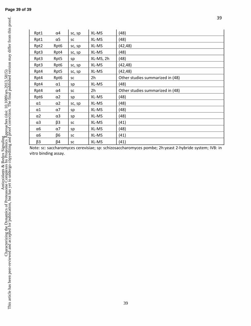
Our initial analysis of the 20S CP from S. cerevisiae using the new DSSO based XL-MS
strategy revealed 13 unique lysine-lysine linkages among the 20S CP subunits which were
mapped onto its crystal structure within expected distances (<26 Å) (41). The same strategy was
also successfully applied to map subunit interaction interfaces of the 19S RP from S. cerevisiae
(42). In total, 43 inter-subunit lysine-lysine inter-links were identified, representing 24 unique
subunit-subunit binary interactions between the 19S subunits (42). In comparison to existing
knowledge of protein subunit interactions, eight novel pair-wise interactions were determined for
the first time in the yeast 19S RP (Table 1). In order to determine the architecture of the 19S RP,
we developed a rigorous probabilistic analysis framework to generate a rationalized prediction of
topological ordering of protein complexes based solely on experimentally derived cross-link data
(42). The probabilistic analysis of identified lysine-lysine linkages within the ATPase base ring
(Rpt1-6) of the 19S RP determined its topological ordering as Rpt1-2-6-3-4-5, which
corroborated previous reports (8,25,58,73). Although the architecture of the ATPase base ring
was known at the time of our study, the topology of the remainder of the 19S RP was not
Page 19 of 39
Ant
ioxi
dant
s &
Red
ox S
igna
ling
Cha
ract
eriz
ing
the
Dyn
amic
s of
Pro
teas
ome
Com
plex
es b
y Pr
oteo
mic
s A
ppro
ache
s (d
oi: 1
0.10
89/a
rs.2
013.
5815
)T
his
artic
le h
as b
een
peer
-rev
iew
ed a
nd a
ccep
ted
for
publ
icat
ion,
but
has
yet
to u
nder
go c
opye
ditin
g an
d pr
oof
corr
ectio
n. T
he f
inal
pub
lishe
d ve
rsio
n m
ay d
iffe
r fr
om th
is p
roof
.
20
20
resolved. Therefore, we carried out a similar analysis to predict the spatial organization of the
PCI (Proteasome-COP9 Signalosome-eIF3) domain-containing-heterohexamer, part of the lid
subcomplex of the 19S RP and composed of Rpn3, Rpn5, Rpn6, Rpn7, Rpn9, and Rpn12. The
top scoring topology of the PCI-heterohexamer was determined as Rpn9-5-6-7-3-12 (42), in
perfect agreement with recent results provided by other structural methods (47,48). These results
demonstrate the feasibility of combining XL-MS strategy with probabilistic modeling to derive
unknown spatial subunit organization of protein complexes.
Elucidating molecular architectures of the proteasome complexes using integrated approaches
Recently, two studies utilizing cryoEM based approaches elegantly defined the subunit
architecture of the 19S RP and the 26S holocomplex (47,48). Lander et al (47) developed a new
heterologous expression system, which was incorporated with cryoEM and single particle
analysis to derive the topological structure of the yeast 19S RP and 26S proteasome. This
integrated approach facilitated the localization of all subunits within the 19S RP and the
delineation of their approximate subunit boundaries, thus providing a complete architectural
picture of the proteasome. In comparison, Lasker et al (48) employed a different and more
comprehensive approach to probe the structure of the 26S holocomplex, integrating data
obtained from cryoEM, X-ray crystallography, and XL-MS, as well as previously known subunit
interactions with comparative/homology modeling. This combinatory approach incorporated a
XL-MS strategy involving a commercially available non-cleavable cross-linker disuccinimidyl
suberate (DSS) to determine protein interaction interfaces of purified S. pombe 26S proteasomes.
In contrast to the DSSO based XL-MS method (42), DSS cross-linked peptides result in complex
MS2 spectra as shown in Figure 4A, which requires special database searching tools for data
interpretation to eliminate false positives (76). The data obtained from all analyses was then
Page 20 of 39
Ant
ioxi
dant
s &
Red
ox S
igna
ling
Cha
ract
eriz
ing
the
Dyn
amic
s of
Pro
teas
ome
Com
plex
es b
y Pr
oteo
mic
s A
ppro
ache
s (d
oi: 1
0.10
89/a
rs.2
013.
5815
)T
his
artic
le h
as b
een
peer
-rev
iew
ed a
nd a
ccep
ted
for
publ
icat
ion,
but
has
yet
to u
nder
go c
opye
ditin
g an
d pr
oof
corr
ectio
n. T
he f
inal
pub
lishe
d ve
rsio
n m
ay d
iffe
r fr
om th
is p
roof
.
21
21
translated into spatial restraints that allowed for fitting of atomic models into the density of the
EM reconstruction (48), thus uncovering the molecular architecture of the 26S holocomplex.
These studies have determined that the lid subcomplex (consisting of Rpn3, Rpn5-9,
Rpn11, Rpn12) of the 19S RP is organized in a modular fashion with a horseshoe-shaped
heterohexamer (Rpn3/5/6/7/9/12) and a heterodimer (Rpn8/Rpn11) (47,48). Based on single-
particle EM reconstructions of proteasome complexes, the 19S lid subcomplex was determined
to localize on one side of the regulatory particle and interact extensively with the base
subcomplex, placing it also in close contact with the 20S CP. The PCI domain-containing-
heterohexamer forms a horseshoe-shaped anchor structure, which possibly serves as a scaffold
for the assembly of other 19S subunits since they are determined as the hinge between the base
and the rest of the lid (47,48). Rpn11, the only essential deubiquitinase of the proteasome, is
located at the mouth of the horse-shoe structure and interacts extensively with Rpn8, Rpn9 and
Rpn5. Rpn2 contacts the Rpn8/11 dimer at its torus-shaped region and it interacts with Rpn12
and Rpn3 at its distal end (48). In addition, Rpn2 associates with Rpt2 and Rpt6, and its C-
terminus physically interacts with Rpn13 (42,48). In comparison, Rpn1 is conformationally
variable and positioned at the periphery of the ATPase ring (48). Moreover, the two ubiquitin
receptors (Rpn10 and Rpn13) and a deubiquitinase (Rpn11) appear to be in a forked arrangement
with Rpn11 in the center-bottom and the two receptors in the top corners (9,48). This suggests an
arrangement where the polyUb chain of a protein substrate is bound to the distal receptors and
the base of the chain is exposed to the deubiquitinase. Furthermore, the extensive and
unexpected contacts between the 19S lid and 20S CP may be important in stabilizing the entire
holocomplex assembly, and/or be part of an allosteric network that modulates the activities of
either subcomplex (47,48). Taken together, these studies have not only determined the subunit
Page 21 of 39
Ant
ioxi
dant
s &
Red
ox S
igna
ling
Cha
ract
eriz
ing
the
Dyn
amic
s of
Pro
teas
ome
Com
plex
es b
y Pr
oteo
mic
s A
ppro
ache
s (d
oi: 1
0.10
89/a
rs.2
013.
5815
)T
his
artic
le h
as b
een
peer
-rev
iew
ed a
nd a
ccep
ted
for
publ
icat
ion,
but
has
yet
to u
nder
go c
opye
ditin
g an
d pr
oof
corr
ectio
n. T
he f
inal
pub
lishe
d ve
rsio
n m
ay d
iffe
r fr
om th
is p
roof
.
22
22
organization of the 19S RP structure, but also defined the entire architecture of the 26S
proteasome for the first time (47,48). The structural details obtained offer novel insight into the
mechanisms of ubiquitin binding, deubiquitination, substrate unfolding and translocation by the
proteasome.
In comparison to these reports, it is noted that our DSSO XL-MS strategy was able to
determine two additional interactions between the small Rpn15/Sem1 subunit to Rpn3 and Rpn7
respectively, which were not detected by other approaches (47,48). This finding was later
confirmed by a more targeted approach through EM difference mapping between wild type
Rpn15 and an Rpn15 deletion strain (9). This further indicates that XL-MS analysis can provide
complimentary information to EM-based structural analysis, and combinatory approaches
integrating various technologies are beneficial in structural characterization of heterogeneous and
dynamic protein complexes such as proteasomes.
SUMMARY
MS-based proteomic studies have revealed that the dynamic proteome of proteasome
complexes is much more complicated than anticipated. Given the importance that PIPs play in
modulating proteasome assembly, PTMs, activity, and function, more detailed proteomic studies
mapping the reorganization of proteasome interaction networks, induced by various cytotoxic
stresses from different cell types, tissues and organisms, are clearly needed and essential to
address many unanswered questions in our understanding of proteasome regulation. In addition,
profiling of dynamic proteomes of proteasome complexes at different disease states will help
unravel molecular mechanisms underlying human pathologies.
Page 22 of 39
Ant
ioxi
dant
s &
Red
ox S
igna
ling
Cha
ract
eriz
ing
the
Dyn
amic
s of
Pro
teas
ome
Com
plex
es b
y Pr
oteo
mic
s A
ppro
ache
s (d
oi: 1
0.10
89/a
rs.2
013.
5815
)T
his
artic
le h
as b
een
peer
-rev
iew
ed a
nd a
ccep
ted
for
publ
icat
ion,
but
has
yet
to u
nder
go c
opye
ditin
g an
d pr
oof
corr
ectio
n. T
he f
inal
pub
lishe
d ve
rsio
n m
ay d
iffe
r fr
om th
is p
roof
.
23
23
Significant progress has been made to define the structural topologies of the 19S RP and
the 26S holocomplex, largely attributed to innovative development of novel low-resolution
structural methods. Integration of improved technologies including cryoEM, XL-MS, and
computational modeling has allowed the characterization of the architectures of proteasome
complexes possible, and has made major contributions to our current structural understanding of
the 26S proteasome. The work presented here represents a huge step forward towards the full
understanding of the heterogeneous and dynamic proteasome complex. We expect technological
advancements in structural and proteomic methods will be further developed to allow full
characterization of proteasome complexes, and thus increase our understanding of proteasomal
biology as well as provide new therapeutic targets for disease diagnostics and treatment.
ACKNOWLEDGEMENTS
This work is supported by NIH R21CA161807 and R01GM074830 to L.H., and
R01GM106003 to L.H. and S. R..
AUTHOR DISCLOSURE STATEMENT
No competing financial interests exist.
Page 23 of 39
Ant
ioxi
dant
s &
Red
ox S
igna
ling
Cha
ract
eriz
ing
the
Dyn
amic
s of
Pro
teas
ome
Com
plex
es b
y Pr
oteo
mic
s A
ppro
ache
s (d
oi: 1
0.10
89/a
rs.2
013.
5815
)T
his
artic
le h
as b
een
peer
-rev
iew
ed a
nd a
ccep
ted
for
publ
icat
ion,
but
has
yet
to u
nder
go c
opye
ditin
g an
d pr
oof
corr
ectio
n. T
he f
inal
pub
lishe
d ve
rsio
n m
ay d
iffe
r fr
om th
is p
roof
.
24
24
ABBREVIATIONS
2h: yeast 2-hybride system
CID: collision induced dissociation
CP: core particle
cryoEM: cryo-electron microscopy
DSSO: disuccinimidyl sulfoxide
HB tag: His-Bio tag
IVB: in vitro binding assay
MS: mass spectrometry
MSn: multistage tandem mass spectrometry
NMR: nuclear magnetic resonance
PCI domain: Proteasome–CSN–eIF3 domain
PIP: proteasome interacting protein
PPI: protein-protein interactions
PTMs: posttranslational modifications
QTAX: quantitative analysis of tandem affinity purified in vivo cross-linked (x) protein
complexes
RP: regulatory particle
SILAC: stable isotope labeling of amino acids in cell culture
sc: saccharomyces cerevisiae
sp: schizosaccharomyces pombe
TAP: tandem affinity purification
Ub: ubiquitin
Page 24 of 39
Ant
ioxi
dant
s &
Red
ox S
igna
ling
Cha
ract
eriz
ing
the
Dyn
amic
s of
Pro
teas
ome
Com
plex
es b
y Pr
oteo
mic
s A
ppro
ache
s (d
oi: 1
0.10
89/a
rs.2
013.
5815
)T
his
artic
le h
as b
een
peer
-rev
iew
ed a
nd a
ccep
ted
for
publ
icat
ion,
but
has
yet
to u
nder
go c
opye
ditin
g an
d pr
oof
corr
ectio
n. T
he f
inal
pub
lishe
d ve
rsio
n m
ay d
iffe
r fr
om th
is p
roof
.
25
25
UPS: ubiquitin-proteasome system
XL-MS: cross-linking mass spectrometry
Page 25 of 39
Ant
ioxi
dant
s &
Red
ox S
igna
ling
Cha
ract
eriz
ing
the
Dyn
amic
s of
Pro
teas
ome
Com
plex
es b
y Pr
oteo
mic
s A
ppro
ache
s (d
oi: 1
0.10
89/a
rs.2
013.
5815
)T
his
artic
le h
as b
een
peer
-rev
iew
ed a
nd a
ccep
ted
for
publ
icat
ion,
but
has
yet
to u
nder
go c
opye
ditin
g an
d pr
oof
corr
ectio
n. T
he f
inal
pub
lishe
d ve
rsio
n m
ay d
iffe
r fr
om th
is p
roof
.
26
26
REFERENCES:
1. Aiken CT, Kaake RM, Wang X, Huang L. Oxidative stress-mediated regulation of proteasome complexes. Mol Cell Proteomics 10: R110 006924, 2011.
2. Ang XL, Wade Harper J. SCF-mediated protein degradation and cell cycle control. Oncogene 24: 2860-70, 2005.
3. Bardag-Gorce F. Effects of ethanol on the proteasome interacting proteins. World J Gastroenterol 16: 1349-57, 2010.
4. Beck F, Unverdorben P, Bohn S, Schweitzer A, Pfeifer G, Sakata E, Nickell S, Plitzko JM, Villa E, Baumeister W, Forster F. Near-atomic resolution structural model of the yeast 26S proteasome. Proc Natl Acad Sci U S A 109: 14870-5, 2012.
5. Benanti JA. Coordination of cell growth and division by the ubiquitin-proteasome system. Semin Cell Dev Biol 23: 492-8, 2012.
7. Bensimon A, Heck AJ, Aebersold R. Mass spectrometry-based proteomics and network biology. Annu Rev Biochem 81: 379-405, 2012.
8. Bohn S, Beck F, Sakata E, Walzthoeni T, Beck M, Aebersold R, Forster F, Baumeister W, Nickell S. Structure of the 26S proteasome from Schizosaccharomyces pombe at subnanometer resolution. Proc Natl Acad Sci U S A 107: 20992-7, 2010.
9. Bohn S, Sakata E, Beck F, Pathare GR, Schnitger J, Nágy I, Baumeister W, Förster F. Localization of the regulatory particle subunit Sem1 in the 26S proteasome. Biochemical and Biophysical Research Communications, 2013.
10. Bousquet-Dubouch MP, Baudelet E, Guerin F, Matondo M, Uttenweiler-Joseph S, Burlet-Schiltz O, Monsarrat B. Affinity purification strategy to capture human endogenous proteasome complexes diversity and to identify proteasome-interacting proteins. Mol Cell Proteomics 8: 1150-64, 2009.
11. Bousquet-Dubouch MP, Fabre B, Monsarrat B, Burlet-Schiltz O. Proteomics to study the diversity and dynamics of proteasome complexes: from fundamentals to the clinic. Expert Rev Proteomics 8: 459-81, 2011.
12. Bousquet-Dubouch MP, Nguen S, Bouyssie D, Burlet-Schiltz O, French SW, Monsarrat B, Bardag-Gorce F. Chronic ethanol feeding affects proteasome-interacting proteins. Proteomics 9: 3609-22, 2009.
13. Breusing N, Grune T. Regulation of proteasome-mediated protein degradation during oxidative stress and aging. Biol Chem 389: 203-9, 2008.
14. Chondrogianni N, Petropoulos I, Grimm S, Georgila K, Catalgol B, Friguet B, Grune T, Gonos ES. Protein damage, repair and proteolysis. Mol Aspects Med, 2012.
15. Ciechanover A. Intracellular protein degradation: from a vague idea thru the lysosome and the ubiquitin-proteasome system and onto human diseases and drug targeting. Biochim Biophys Acta 1824: 3-13, 2012.
16. Cui Z, Scruggs SB, Gilda JE, Ping P, Gomes AV. Regulation of cardiac proteasomes by ubiquitination, SUMOylation, and beyond. J Mol Cell Cardiol, 2013.
17. da Fonseca PC, He J, Morris EP. Molecular model of the human 26S proteasome. Mol Cell 46: 54-66, 2012.
18. Drews O, Taegtmeyer H. Regulation of the Ubiquitin-Proteasome System in Heart Disease: The Basis for New Therapeutic Strategies. Antioxid Redox Signal, 2013.
Page 26 of 39
Ant
ioxi
dant
s &
Red
ox S
igna
ling
Cha
ract
eriz
ing
the
Dyn
amic
s of
Pro
teas
ome
Com
plex
es b
y Pr
oteo
mic
s A
ppro
ache
s (d
oi: 1
0.10
89/a
rs.2
013.
5815
)T
his
artic
le h
as b
een
peer
-rev
iew
ed a
nd a
ccep
ted
for
publ
icat
ion,
but
has
yet
to u
nder
go c
opye
ditin
g an
d pr
oof
corr
ectio
n. T
he f
inal
pub
lishe
d ve
rsio
n m
ay d
iffe
r fr
om th
is p
roof
.
27
27
19. Drews O, Wildgruber R, Zong C, Sukop U, Nissum M, Weber G, Gomes AV, Ping P. Mammalian proteasome subpopulations with distinct molecular compositions and proteolytic activities. Mol Cell Proteomics 6: 2021-31, 2007.
20. Drews O, Zong C, Ping P. Exploring proteasome complexes by proteomic approaches. Proteomics 7: 1047-58, 2007.
21. Ducoux-Petit M, Uttenweiler-Joseph S, Brichory F, Bousquet-Dubouch MP, Burlet-Schiltz O, Haeuw JF, Monsarrat B. Scaled-down purification protocol to access proteomic analysis of 20S proteasome from human tissue samples: comparison of normal and tumor colorectal cells. J Proteome Res 7: 2852-9, 2008.
22. Fabre B, Lambour T, Delobel J, Amalric F, Monsarrat B, Burlet-Schiltz O, Bousquet-Dubouch MP. Subcellular distribution and dynamics of active proteasome complexes unraveled by a workflow combining in vivo complex cross-linking and quantitative proteomics. Mol Cell Proteomics 12: 687-99, 2013.
23. Fang L, Kaake RM, Patel VR, Yang Y, Baldi P, Huang L. Mapping the Protein Interaction Network of the Human COP9 Signalosome Complex Using a Label-free QTAX Strategy. Mol Cell Proteomics 11: 138-47, 2012.
24. Finley D. Recognition and processing of ubiquitin-protein conjugates by the proteasome. Annu Rev Biochem 78: 477-513, 2009.
25. Forster F, Lasker K, Beck F, Nickell S, Sali A, Baumeister W. An atomic model AAA-ATPase/20S core particle sub-complex of the 26S proteasome. Biochem Biophys Res Commun 388: 228-33, 2009.
26. Forster F, Lasker K, Nickell S, Sali A, Baumeister W. Toward an integrated structural model of the 26S proteasome. Mol Cell Proteomics 9: 1666-77, 2010.
27. Goldberg AL. Protein degradation and protection against misfolded or damaged proteins. Nature 426: 895-9, 2003.
28. Goldberg AL. Functions of the proteasome: from protein degradation and immune surveillance to cancer therapy. Biochem Soc Trans 35: 12-7, 2007.
29. Goldberg AL. Development of proteasome inhibitors as research tools and cancer drugs. J Cell Biol 199: 583-8, 2012.
30. Gomes AV, Young GW, Wang Y, Zong C, Eghbali M, Drews O, Lu H, Stefani E, Ping P. Contrasting proteome biology and functional heterogeneity of the 20 S proteasome complexes in mammalian tissues. Mol Cell Proteomics 8: 302-15, 2009.
31. Gomes AV, Zong C, Edmondson RD, Li X, Stefani E, Zhang J, Jones RC, Thyparambil S, Wang GW, Qiao X, Bardag-Gorce F, Ping P. Mapping the murine cardiac 26S proteasome complexes. Circ Res 99: 362-71, 2006.
32. Groll M, Ditzel L, Löwe J, Stock D, Bochtler M, Bartunik HD, Huber R. Structure of 20S proteasome from yeast at 2.4 A resolution. Nature 386: 463-71, 1997.
33. Guerrero C, Milenkovic T, Przulj N, Kaiser P, Huang L. Characterization of the proteasome interaction network using a QTAX-based tag-team strategy and protein interaction network analysis. Proc Natl Acad Sci U S A 105: 13333-8, 2008.
34. Guerrero C, Tagwerker C, Kaiser P, Huang L. An integrated mass spectrometry-based proteomic approach: quantitative analysis of tandem affinity-purified in vivo cross-linked protein complexes (QTAX) to decipher the 26 S proteasome-interacting network. Mol Cell Proteomics 5: 366-78, 2006.
35. Guo X, Engel JL, Xiao J, Tagliabracci VS, Wang X, Huang L, Dixon JE. UBLCP1 is a 26S proteasome phosphatase that regulates nuclear proteasome activity. Proc Natl Acad Sci U S A 108: 18649-54, 2011.
36. Hershko A, Ciechanover A. The ubiquitin system. Annu Rev Biochem. 67: 425-79, 1998.
Page 27 of 39
Ant
ioxi
dant
s &
Red
ox S
igna
ling
Cha
ract
eriz
ing
the
Dyn
amic
s of
Pro
teas
ome
Com
plex
es b
y Pr
oteo
mic
s A
ppro
ache
s (d
oi: 1
0.10
89/a
rs.2
013.
5815
)T
his
artic
le h
as b
een
peer
-rev
iew
ed a
nd a
ccep
ted
for
publ
icat
ion,
but
has
yet
to u
nder
go c
opye
ditin
g an
d pr
oof
corr
ectio
n. T
he f
inal
pub
lishe
d ve
rsio
n m
ay d
iffe
r fr
om th
is p
roof
.
28
28
37. Herzog F, Kahraman A, Boehringer D, Mak R, Bracher A, Walzthoeni T, Leitner A, Beck M, Hartl FU, Ban N, Malmstrom L, Aebersold R. Structural probing of a protein phosphatase 2A network by chemical cross-linking and mass spectrometry. Science 337: 1348-52, 2012.
38. Kaake RM, Milenkovic T, Przulj N, Kaiser P, Huang L. Characterization of Cell Cycle Specific Protein Interaction Networks of the Yeast 26S Proteasome Complex by the QTAX Strategy. J Proteome Res 9: 2016-2029, 2010.
39. Kaake RM, Wang X, Huang L. Profiling of protein interaction networks of protein complexes Using affinity purification and quantitative mass spectrometry. Mol Cell Proteomics, 2010.
40. Kalisman N, Adams CM, Levitt M. Subunit order of eukaryotic TRiC/CCT chaperonin by cross-linking, mass spectrometry, and combinatorial homology modeling. Proc Natl Acad Sci U S A, 2012.
41. Kao A, Chiu C, Vellucci D, Yang Y, Patel V, Guan S, Randall A, Baldi P, Rychnovsky S, Huang L. Development of a novel cross-linking strategy for fast and accurate identification of cross-linked peptides of protein complexes. Molecular & Cellular Proteomics, 2011.
42. Kao A, Randall A, Yang Y, Patel VR, Kandur W, Guan S, Rychnovsky SD, Baldi P, Huang L. Mapping the structural topology of the yeast 19S proteasomal regulatory particle using chemical cross-linking and probabilistic modeling. Mol Cell Proteomics 11: 1566-77, 2012.
43. Kim W, Bennett EJ, Huttlin EL, Guo A, Li J, Possemato A, Sowa ME, Rad R, Rush J, Comb MJ, Harper JW, Gygi SP. Systematic and quantitative assessment of the ubiquitin-modified proteome. Mol Cell 44: 325-40, 2011.
44. Kravtsova-Ivantsiv Y, Ciechanover A. Non-canonical ubiquitin-based signals for proteasomal degradation. J Cell Sci 125: 539-48, 2012.
45. Kravtsova-Ivantsiv Y, Sommer T, Ciechanover A. The lysine48-based polyubiquitin chain proteasomal signal: not a single child anymore. Angew Chem Int Ed Engl 52: 192-8, 2013.
46. Kulathu Y, Komander D. Atypical ubiquitylation - the unexplored world of polyubiquitin beyond Lys48 and Lys63 linkages. Nat Rev Mol Cell Biol 13: 508-23, 2012.
47. Lander GC, Estrin E, Matyskiela ME, Bashore C, Nogales E, Martin A. Complete subunit architecture of the proteasome regulatory particle. Nature 482: 186-91, 2012.
48. Lasker K, Forster F, Bohn S, Walzthoeni T, Villa E, Unverdorben P, Beck F, Aebersold R, Sali A, Baumeister W. Molecular architecture of the 26S proteasome holocomplex determined by an integrative approach. Proc Natl Acad Sci U S A 109: 1380-7, 2012.
49. Leggett DS, Hanna J, Borodovsky A, Crosas B, Schmidt M, Baker RT, Walz T, Ploegh H, Finley D. Multiple associated proteins regulate proteasome structure and function. Mol Cell. 10: 495-507, 2002.
50. Leitner A, Walzthoeni T, Kahraman A, Herzog F, Rinner O, Beck M, Aebersold R. Probing native protein structures by chemical cross-linking, mass spectrometry and bioinformatics. Molecular & Cellular Proteomics (Accepted for Publication), 2010.
51. Li J, Powell SR, Wang X. Enhancement of proteasome function by PA28α overexpression protects against oxidative stress. Faseb J 25: 883-93, 2011.
52. Li X, Amazit L, Long W, Lonard DM, Monaco JJ, O'Malley BW. Ubiquitin- and ATP-independent proteolytic turnover of p21 by the REGgamma-proteasome pathway. Mol Cell 26: 831-42, 2007.
53. Li X, Lonard DM, Jung SY, Malovannaya A, Feng Q, Qin J, Tsai SY, Tsai MJ, O'Malley BW. The SRC-3/AIB1 coactivator is degraded in a ubiquitin- and ATP-independent manner by the REGgamma proteasome. Cell 124: 381-92, 2006.
54. Löwe J, Stock D, Jap B, Zwickl P, Baumeister W, Huber R. Crystal structure of the 20S proteasome from the archaeon T. acidophilum at 3.4 Å resolution. Science 268: 533-539, 1995.
Page 28 of 39
Ant
ioxi
dant
s &
Red
ox S
igna
ling
Cha
ract
eriz
ing
the
Dyn
amic
s of
Pro
teas
ome
Com
plex
es b
y Pr
oteo
mic
s A
ppro
ache
s (d
oi: 1
0.10
89/a
rs.2
013.
5815
)T
his
artic
le h
as b
een
peer
-rev
iew
ed a
nd a
ccep
ted
for
publ
icat
ion,
but
has
yet
to u
nder
go c
opye
ditin
g an
d pr
oof
corr
ectio
n. T
he f
inal
pub
lishe
d ve
rsio
n m
ay d
iffe
r fr
om th
is p
roof
.
29
29
55. Lu H, Zong C, Wang Y, Young GW, Deng N, Souda P, Li X, Whitelegge J, Drews O, Yang PY, Ping P. Revealing the dynamics of the 20 S proteasome phosphoproteome: a combined CID and electron transfer dissociation approach. Mol Cell Proteomics 7: 2073-89, 2008.
56. Matyskiela ME, Lander GC, Martin A. Conformational switching of the 26S proteasome enables substrate degradation. Nat Struct Mol Biol 20: 781-8, 2013.
57. Murata S, Sasaki K, Kishimoto T, Niwa S, Hayashi H, Takahama Y, Tanaka K. Regulation of CD8+ T cell development by thymus-specific proteasomes. Science 316: 1349-53, 2007.
58. Nickell S, Beck F, Scheres SH, Korinek A, Forster F, Lasker K, Mihalache O, Sun N, Nagy I, Sali A, Plitzko JM, Carazo JM, Mann M, Baumeister W. Insights into the molecular architecture of the 26S proteasome. Proc Natl Acad Sci U S A 106: 11943-7, 2009.
59. Pelletier S, Schuurman KG, Berkers CR, Ovaa H, Heck AJ, Raijmakers R. Quantifying cross-tissue diversity in proteasome complexes by mass spectrometry. Mol Biosyst 6: 1450-3, 2010.
60. Pickart CM, Cohen RE. Proteasomes and their kin: proteases in the machine age. Nat Rev Mol Cell Biol. 5: 177-87, 2004.
61. Pickering AM, Davies KJ. Differential roles of proteasome and immunoproteasome regulators Pa28alphabeta, Pa28gamma and Pa200 in the degradation of oxidized proteins. Arch Biochem Biophys 523: 181-90, 2012.
62. Rastogi N, Mishra DP. Therapeutic targeting of cancer cell cycle using proteasome inhibitors. Cell Div 7: 26, 2012.
63. Sakata E, Bohn S, Mihalache O, Kiss P, Beck F, Nagy I, Nickell S, Tanaka K, Saeki Y, Forster F, Baumeister W. Localization of the proteasomal ubiquitin receptors Rpn10 and Rpn13 by electron cryomicroscopy. Proc Natl Acad Sci U S A 109: 1479-84, 2012.
64. Savulescu AF, Glickman MH. Proteasome activator 200: the heat is on. Mol Cell Proteomics 10: R110 006890, 2011.
65. Schmidt M, Hanna J, Elsasser S, Finley D. Proteasome-associated proteins: regulation of a proteolytic machine. Biol Chem 386: 725-37, 2005.
66. Scruggs SB, Zong NC, Wang D, Stefani E, Ping P. Post-translational modification of cardiac proteasomes: functional delineation enabled by proteomics. Am J Physiol Heart Circ Physiol 303: H9-18, 2012.
67. Shangary S, Wang S. Small-molecule inhibitors of the MDM2-p53 protein-protein interaction to reactivate p53 function: a novel approach for cancer therapy. Annu Rev Pharmacol Toxicol 49: 223-41, 2009.
68. Sijts EJ, Kloetzel PM. The role of the proteasome in the generation of MHC class I ligands and immune responses. Cell Mol Life Sci 68: 1491-502, 2011.
69. Soriano GP, de Bruin G, Overkleeft HS, Florea BI. Towards understanding induction of oxidative stress and apoptosis by proteasome inhibitors. Antioxid Redox Signal, 2013.
70. Sowa ME, Bennett EJ, Gygi SP, Harper JW. Defining the human deubiquitinating enzyme interaction landscape. Cell 138: 389-403, 2009.
71. Tagwerker C, Flick K, Cui M, Guerrero C, Dou Y, Auer B, Baldi P, Huang L, Kaiser P. A tandem affinity tag for two-step purification under fully denaturing conditions: application in ubiquitin profiling and protein complex identification combined with in vivocross-linking. Mol Cell Proteomics 5: 737-48, 2006.
72. Tai HC, Besche H, Goldberg AL, Schuman EM. Characterization of the Brain 26S Proteasome and its Interacting Proteins. Front Mol Neurosci 3, 2010.
73. Tomko RJ, Jr., Funakoshi M, Schneider K, Wang J, Hochstrasser M. Heterohexameric ring arrangement of the eukaryotic proteasomal ATPases: implications for proteasome structure and assembly. Mol Cell 38: 393-403, 2010.
Page 29 of 39
Ant
ioxi
dant
s &
Red
ox S
igna
ling
Cha
ract
eriz
ing
the
Dyn
amic
s of
Pro
teas
ome
Com
plex
es b
y Pr
oteo
mic
s A
ppro
ache
s (d
oi: 1
0.10
89/a
rs.2
013.
5815
)T
his
artic
le h
as b
een
peer
-rev
iew
ed a
nd a
ccep
ted
for
publ
icat
ion,
but
has
yet
to u
nder
go c
opye
ditin
g an
d pr
oof
corr
ectio
n. T
he f
inal
pub
lishe
d ve
rsio
n m
ay d
iffe
r fr
om th
is p
roof
.
30
30
74. Vellucci D, Kao A, Kaake RM, Rychnovsky SD, Huang L. Selective Enrichment and Identification of Azide-tagged Cross-Linked Peptides Using Chemical Ligation and Mass Spectrometry. J Am Soc Mass Spectrom, 2010.
75. Verma R, Chen S, Feldman R, Schieltz D, Yates J, Dohmen J, Deshaies RJ. Proteasomal proteomics: identification of nucleotide-sensitive proteasome-interacting proteins by mass spectrometric analysis of affinity-purified proteasomes. Mol Biol Cell. 11: 3425-39, 2000.
76. Walzthoeni T, Claassen M, Leitner A, Herzog F, Bohn S, Forster F, Beck M, Aebersold R. False discovery rate estimation for cross-linked peptides identified by mass spectrometry. Nat Methods 9: 901-3, 2012.
77. Wang D, Zong C, Koag MC, Wang Y, Drews O, Fang C, Scruggs SB, Ping P. Proteome dynamics and proteome function of cardiac 19S proteasomes. Mol Cell Proteomics 10: M110 006122, 2011.
78. Wang X, Chen CF, Baker PR, Chen PL, Kaiser P, Huang L. Mass spectrometric characterization of the affinity-purified human 26S proteasome complex. Biochemistry 46: 3553-65, 2007.
79. Wang X, Guerrero C, Kaiser P, Huang L. Proteomics of proteasome complexes and ubiquitinated proteins. Expert Rev Proteomics 4: 649-65, 2007.
80. Wang X, Huang L. Identifying dynamic interactors of protein complexes by quantitative mass spectrometry. Mol Cell Proteomics 7: 46-57, 2008.
81. Wang X, Yen J, Kaiser P, Huang L. Regulation of the 26S proteasome complex during oxidative stress. Sci Signal 3: ra88, 2010.
82. Wells JA, McClendon CL. Reaching for high-hanging fruit in drug discovery at protein-protein interfaces. Nature 450: 1001-9, 2007.
83. Xu P, Duong DM, Seyfried NT, Cheng D, Xie Y, Robert J, Rush J, Hochstrasser M, Finley D, Peng J. Quantitative proteomics reveals the function of unconventional ubiquitin chains in proteasomal degradation. Cell 137: 133-45, 2009.
84. Yang B, Wu YJ, Zhu M, Fan SB, Lin J, Zhang K, Li S, Chi H, Li YX, Chen HF, Luo SK, Ding YH, Wang LH, Hao Z, Xiu LY, Chen S, Ye K, He SM, Dong MQ. Identification of cross-linked peptides from complex samples. Nat Methods 9: 904-6, 2012.
85. Zhang F, Hu Y, Huang P, Toleman CA, Paterson AJ, Kudlow JE. Proteasome Function Is Regulated by Cyclic AMP-dependent Protein Kinase through Phosphorylation of Rpt6. J Biol Chem 282: 22460-71, 2007.
86. Zong C, Gomes AV, Drews O, Li X, Young GW, Berhane B, Qiao X, French SW, Bardag-Gorce F, Ping P. Regulation of murine cardiac 20S proteasomes: role of associating partners. Circ Res 99: 372-80, 2006.
Page 30 of 39
Ant
ioxi
dant
s &
Red
ox S
igna
ling
Cha
ract
eriz
ing
the
Dyn
amic
s of
Pro
teas
ome
Com
plex
es b
y Pr
oteo
mic
s A
ppro
ache
s (d
oi: 1
0.10
89/a
rs.2
013.
5815
)T
his
artic
le h
as b
een
peer
-rev
iew
ed a
nd a
ccep
ted
for
publ
icat
ion,
but
has
yet
to u
nder
go c
opye
ditin
g an
d pr
oof
corr
ectio
n. T
he f
inal
pub
lishe
d ve
rsio
n m
ay d
iffe
r fr
om th
is p
roof
.
31
31
Page 31 of 39
Ant
ioxi
dant
s &
Red
ox S
igna
ling
Cha
ract
eriz
ing
the
Dyn
amic
s of
Pro
teas
ome
Com
plex
es b
y Pr
oteo
mic
s A
ppro
ache
s (d
oi: 1
0.10
89/a
rs.2
013.
5815
)T
his
artic
le h
as b
een
peer
-rev
iew
ed a
nd a
ccep
ted
for
publ
icat
ion,
but
has
yet
to u
nder
go c
opye
ditin
g an
d pr
oof
corr
ectio
n. T
he f
inal
pub
lishe
d ve
rsio
n m
ay d
iffe
r fr
om th
is p
roof
.
32
32
FIGURE LEGENDS
Page 32 of 39
Ant
ioxi
dant
s &
Red
ox S
igna
ling
Cha
ract
eriz
ing
the
Dyn
amic
s of
Pro
teas
ome
Com
plex
es b
y Pr
oteo
mic
s A
ppro
ache
s (d
oi: 1
0.10
89/a
rs.2
013.
5815
)T
his
artic
le h
as b
een
peer
-rev
iew
ed a
nd a
ccep
ted
for
publ
icat
ion,
but
has
yet
to u
nder
go c
opye
ditin
g an
d pr
oof
corr
ectio
n. T
he f
inal
pub
lishe
d ve
rsio
n m
ay d
iffe
r fr
om th
is p
roof
.
33
33
Figure 1. Defining proteasome dynamics to understand proteasome biology.
Page 33 of 39
Ant
ioxi
dant
s &
Red
ox S
igna
ling
Cha
ract
eriz
ing
the
Dyn
amic
s of
Pro
teas
ome
Com
plex
es b
y Pr
oteo
mic
s A
ppro
ache
s (d
oi: 1
0.10
89/a
rs.2
013.
5815
)T
his
artic
le h
as b
een
peer
-rev
iew
ed a
nd a
ccep
ted
for
publ
icat
ion,
but
has
yet
to u
nder
go c
opye
ditin
g an
d pr
oof
corr
ectio
n. T
he f
inal
pub
lishe
d ve
rsio
n m
ay d
iffe
r fr
om th
is p
roof
.
34
34
Figure 2. The general workflow for isolating and analyzing proteasome complexes.