96
KNEE DISTORTION CHARLES UNIVERSITY IN PRAGUE Faculty of Physical Education and Sport Diploma Osama Hamed Aljeheny, March 2016 CHARLES UNIVERSITY IN PRAGUE

KNEE DISTORTION
CHARLES UNIVERSITY IN PRAGUE
Faculty of Physical Education and Sport
Diploma
Osama Hamed Aljeheny, March 2016
CHARLES UNIVERSITY IN PRAGUE
KNEE DISTORTION 2
FACULTY OF PHYSICAL EDUCATION AND SPORT
Department of physiotherapy
Case Study of Physiotherapy Treatment of Patient after Distortion of the
Right Knee
BACHELOR DEGREE PROGRAM IN PHYSIOTHERAPY
Author Osama Hamed Aljeheny
Supervisor PhDr. Ivana Vláčilová Ph.D.
March 2016, Prague

KNEE DISTORTION 3
ABSTRACT
Title of the thesis: Case Study of Physiotherapy Treatment of Patient after Distortion of the
Right Knee
Author: Osama Hamed Aljeheny
Work placement: Centrum léčby pohybového aparátu Mediterra
Summary
In the bachelor thesis, which was written by myself, it is divided in two parts, general
part and case study. The general part describes anatomy of knee joint, its bones, muscles,
ligaments, nerves and blood supply and surgical and non-surgical approaches of therapy of the
knee joint after this injury.
Information about kinesiological and biomechanical point of view were discussed as
well. In the practical part I analyzed procedures I have done with the patient, all examinations,
conclusions, therapies and results.
Last part of the bachelor thesis contains list of literature used in the bachelor thesis, it
contains list of figures and tables used in the thesis, abbreviations and the ethics committee.
Key words: knee joint, physiotherapy treatment, knee distortion, knee exercises, knee ligaments
KNEE DISTORTION 4
ABSTRAKT
Název: Případová studie fyzioterapeutické péče o pacienta po distorzi pravého kolene
Autor: Osama Hamed Aljeheny
Pracoviště: Centrum léčby pohybového aparátu Mediterra
Souhrn
Tuto bakalářskou práci, kterou jsem napsal sám, jsem rozdělil do dvou částí - obecná část
a případová studie. Obecná část popisuje anatomii kolenního kloubu (kosti, svaly, ligamenta,
nervy a krevní zásobení kolenního kloubu), chirurgickou a konzervativní léčbu kolene po jeho
zranění.
Dále byla diskutována i kineziologie a biomechanika. V praktické části práce je popsána
práce s pacientem (všechna vyšetření, závěry, terapie a její výsledek).
Závěrečná část práce obsahuje seznam literatury použité v této bakalářské práci, seznam
obrázků, tabulek, zkratek a také souhlas etické komise.

KNEE DISTORTION 5
DECLARATION
I declare that the bachelor thesis was written by me and under supervising of PhDr. Ivana
Vláčilová Ph.D. This is an original research, which refers to practice with patient after distortion
of the right knee, under supervising of Mgr.Zahir Elali, the practice took a place at Centrum
léčby pohybového aparátu (CLPA) Mediterra.
I confirm that all written information, examinations, and therapeutic treatments, which
are presented in the bachelor thesis, were performed based on my own knowledge that I got from
professors of Charles University Faculty of Physical Education and Sport and supervisors in the
hospitals. Information in the bachelor thesis were sourced from the list of literature, which is
placed below at the end of this thesis.
Finally I confirm that there were no invasive methods used during my practice and that
patient was fully aware of examinations and therapies at any time.

KNEE DISTORTION 6
ACKNOWLEDGMENT
At this space I would like to thank to all my professors, who taught me for the three years
of my studying at Faculty of Physical Education and Sport. Many thanks to PhDr. Ivana
Vláčilová Ph.D. for her help and support during my study and for her supervising of my bachelor
theses. Special thanks belong to my supervisor in CLPA Mediterra Mgr.Zahir Elali who helped
me with the practice of the bachelor theses.

KNEE DISTORTION 7
DEDICATION
I would like to dedicate this bachelor thesis to my parents because I would not be at this
place without their help and support. And I would not forget my professors who helped me along
my study of physiotherapy during the last three years. And at last I would like to thank the
supervisors at the hospitals I visited during my practice and also everybody who helped me
during my study.

KNEE DISTORTION 8
ABSTRACT .................................................................................................................................................. 3
ABSTRAKT ................................................................................................................................................. 4
DECLARATION .......................................................................................................................................... 5
ACKNOWLEDGMENT ............................................................................................................................... 6
DEDICATION .............................................................................................................................................. 7
1. INTRODUCTION .............................................................................................................................. 12
2. KNEE’S ANATOMY ......................................................................................................................... 13
2.1. Bones........................................................................................................................................... 13
2.2. Patella .......................................................................................................................................... 13
2.3. Ligaments .................................................................................................................................... 15
2.3.1. Medial Meniscus ................................................................................................................. 15
2.3.2. Lateral Meniscus ................................................................................................................. 15
2.3.3. Meniscal Blood Supply ....................................................................................................... 15
2.3.4. Stabilizing Ligaments ......................................................................................................... 15
2.3.5. Cruciate Ligaments ............................................................................................................. 16
2.3.6. Posterior Cruciate Ligament ............................................................................................... 16
2.3.7. Capsular and Collateral Ligaments ..................................................................................... 16
2.3.8. Medial Collateral Ligament ................................................................................................ 16
2.3.9. Deep Medial Capsular Ligaments ....................................................................................... 17
2.4. Lateral Collateral Ligament and Associated Structures .............................................................. 18
2.5. Joint Capsule ............................................................................................................................... 19
2.6. Knee Musculature ....................................................................................................................... 20
2.7. Bursae ......................................................................................................................................... 20
2.8. Fat Pads ....................................................................................................................................... 21
2.9. Nerve Supply .............................................................................................................................. 21
2.10. Blood Supply .......................................................................................................................... 22
2.11. Functional Anatomy ................................................................................................................ 22
3. THE KNEE WITHIN THE KINETIC CHAIN .................................................................................. 24
4. BIOMECHANICAL OF THE CRUCIATE ....................................................................................... 24
4.1. Biomechanics and Kinematic of the Knees Joints ...................................................................... 24
4.2. Passive Motion of the Knees ....................................................................................................... 25
4.3. The Functional Biomechanics of the Ligament .......................................................................... 25
4.4. Biomechanical of the Anterior Cruciate Ligament ..................................................................... 25
KNEE DISTORTION 9
4.5. Biomechanics of Posterior Cruciate Ligament ........................................................................... 26
4.6. The Interaction of the Cruciate Ligament ................................................................................... 27
5. DIAGNOSIS AND MANAGEMENT OF KNEE DISTORTIONS ................................................... 29
5.1. Classification ............................................................................................................................... 29
5.2. Mechanism of Injuries ................................................................................................................ 31
5.3. Associated Injuries ...................................................................................................................... 31
6. FIRST EVALUATION AND MANAGEMENT ............................................................................... 33
6.1. General Considerations ............................................................................................................... 33
6.2. Imaging Studies .......................................................................................................................... 33
6.3. Reduction .................................................................................................................................... 34
6.4. Physical Examination .................................................................................................................. 34
6.5. Vascular Injuries ......................................................................................................................... 35
6.6. Absolute Surgical Indications ..................................................................................................... 36
7. DEFINITIVE SURGICAL MANAGEMENT .................................................................................... 36
7.1. Historical Management ............................................................................................................... 36
7.2. Sports Injury Clinic Experience .................................................................................................. 37
7.3. Surgical timing ............................................................................................................................ 37
7.4. Graft Selection ............................................................................................................................ 38
7.5. Surgical Approach ...................................................................................................................... 39
7.6. Graft Tensioning and Fixation .................................................................................................... 39
8. PHYSIOTHERAPY ............................................................................................................................ 40
8.1. Examples of Physiotherapies ...................................................................................................... 40
8.1.1. Mobility Exercises .............................................................................................................. 40
8.1.2. Heel slides ........................................................................................................................... 41
8.1.3. Flexion Extension Exercise ................................................................................................. 41
8.1.4. Strengthening Exercises ...................................................................................................... 42
8.2. Static squad contractions ............................................................................................................. 43
8.3. Hip Abduction ............................................................................................................................. 43
8.4. Half Squats .................................................................................................................................. 44
8.5. Squat with Swiss Ball ................................................................................................................. 44
8.6. Lunges ......................................................................................................................................... 45
8.7. Bridge exercises .......................................................................................................................... 46
8.8. Leg press ..................................................................................................................................... 46
8.9. Calf Raises .................................................................................................................................. 47
8.10. Leg Curl .................................................................................................................................. 47

KNEE DISTORTION 10
8.11. Proprioception exercises ......................................................................................................... 48
8.12. Balance board exercise ............................................................................................................ 48
8.13. Plan for sprain exercise ........................................................................................................... 48
8.14. Functional Exercises ............................................................................................................... 49
8.15. Plyometric exercise ................................................................................................................. 50
9. NON-SURGICAL THERAPY ........................................................................................................... 50
9.1. Heat/Cold therapies ..................................................................................................................... 50
9.2. Assistive Devices ........................................................................................................................ 50
9.3. Balancing .................................................................................................................................... 51
9.4. Avoidance ................................................................................................................................... 51
9.5. Mental heath ................................................................................................................................ 51
9.6. Injections ..................................................................................................................................... 51
9.7. Technical Hints ........................................................................................................................... 51
10. POSTOPERATIVE REHABILITATION ...................................................................................... 52
10.1. Complications ......................................................................................................................... 52
11. DATA ANALYSIS ......................................................................................................................... 53
12. RESULTS ....................................................................................................................................... 53
13. DISCUSSION ................................................................................................................................. 54
14. CONCLUSION ............................................................................................................................... 55
15. CASE STUDY ................................................................................................................................ 56
15.1. Report on Clinical Work ......................................................................................................... 56
16. DIAGNOSIS ................................................................................................................................... 56
17. SUBJECTIVE FEELING OF THE PATIENT ............................................................................... 56
18. STATUS PRESENS ....................................................................................................................... 56
19. HISTORY OF PROBLEM ............................................................................................................. 57
20. SOCIAL ANAMNESIS .................................................................................................................. 57
21. ANAMNESIS ................................................................................................................................. 57
22. RHB INDICATIONS ...................................................................................................................... 58
23. INITIAL EXAMINATIONS .......................................................................................................... 58
23.1. Postural examination (by Kendal) ........................................................................................... 58
23.2. Gait examination (by Kendal) ................................................................................................. 59
23.3. Modification of gait examination: ........................................................................................... 59
23.4. Soft tissue examination (by Lewit) ......................................................................................... 59
23.5. Pelvis examination (by Kendal) .............................................................................................. 60
23.6. Special tests ............................................................................................................................. 60

KNEE DISTORTION 11
23.7. Palpation examination (by Kendal) ......................................................................................... 61
23.8. Range of motion examination (by Kendal) ............................................................................. 62
23.9. Muscle strength examination (by Kendal) .............................................................................. 64
23.10. Muscle length examination (by Janda) ................................................................................... 64
23.11. Neurological examination (by Lewit) ..................................................................................... 64
23.12. Joint Play Examination (by Lewit) ......................................................................................... 65
24. EXAMINATIONS CONCLUSION ............................................................................................... 66
25. SHORT-TERM PLAN .................................................................................................................... 66
26. LONG-TERM PLAN ...................................................................................................................... 66
27. PHYSICAL THERAPY TOOLS .................................................................................................... 67
28. PHYSICAL THERAPY SESSIONS .............................................................................................. 67
29. FINAL KINESIOLOGIC EXAMINATION .................................................................................. 80
29.1. Postural examination (by Kendal) ........................................................................................... 80
29.2. Modification of gait examination: ........................................................................................... 81
29.3. Soft tissue examination (by Lewit): ........................................................................................ 81
29.4. Pelvis examination (by Kendal) .............................................................................................. 81
29.5. Romberg test: .......................................................................................................................... 81
29.6. Anthropometry examination (by Kendal) ............................................................................... 82
29.7. Palpation examination (by Kendal) ......................................................................................... 82
29.8. Range of motion examination (by Kendal) ............................................................................. 83
29.9. Muscle strength examination (by Kendal) .............................................................................. 85
29.10. Muscle length examination (by Janda) ................................................................................... 85
29.11. Neurological examination (by Lewit) ..................................................................................... 85
29.12. Joint Play Examination (by Lewit) ......................................................................................... 86
30. THERAPY EFFECT EVALUATION, PROGNOSIS .................................................................... 87
31. REFERENCES ............................................................................................................................... 88
LIST OF ATTACHMENTS ....................................................................................................................... 91
Attachment No. 3: List of Tables ................................................................................................................ 92
Attachment No. 4: List of Figures .............................................................................................................. 93
Attachment No. 5: List of Abbreviations .................................................................................................... 95

KNEE DISTORTION 12
1. INTRODUCTION
Since several activities place life-threatening stress on the knee, it remains one of the
greatest traumatized joints within the physical activity populace. The knee is usually considered
a hinge joint as its two main movements are extension and flexion. Nevertheless, since the
torsion of the tibia is an important component of knee movement, the knee is not an actual hinge
joint. The knee joint's stability depends mostly on the ligaments, the muscles, and the joint
capsule surrounding the joint. The knee is designed principally to offer stability in weight
posture and mobility in locomotion; nevertheless, it is particularly unstable medially and
laterally.
Although several knee injuries are rare, they are serious injuries that frequently lead to
the loss of the active and passive knee stabilizers plus frequently being linked with the
compromise of neurovascular structures. Treating these injuries is contentious, and results
following surgery are usually poor (Zhang, 2010). Once sustaining injuries to manifold
ligaments, the knee is said to be at a biomechanical weakness which poses a rehabilitative and
reconstructive difficulty to even the highest experienced orthopedic surgeons. Surgeons
conducting reconstructions in patients with these injuries have to possess a comprehensive
understanding of the Knees’ normal anatomical view and biomechanics to enhance the timing of
the surgery, tunnel preparation, surgical approach and anatomic implants of grafts. This chapter
highlights the biomechanics and the atomy of cruciate ligaments and their surgical insinuations
(Finerman & Noyes, 2013). The form and structure of the posterior and anterior cruciate
ligaments, structural properties of cruciate ligament and graft replacements, a pattern of injury,
functional biomechanics and interaction between the cruciate ligament, and the surgical
implications associated with anatomic reconstruction of the posterior and anterior cruciate are all
reviewed exhaustively.

KNEE DISTORTION 13
2. KNEE’S ANATOMY
2.1. Bones
In relation to Pedowitz and Akeson (2013) classifications, bones knee joints complex
comprise of the tibia, patella, femur, and fibula. The femur’s distal ending extends and forms the
medial condyles and convex lateral that are designed to communicate via patella along with the
tibia. The medial condyles’ articular facade is elongated from anterior to posterior than the
surface of the laterals condyles (Yeager, 2010). Anteriorly, the two condyles create a hollowed
femoral trochlea, or grove, to receive the patellas. A proximal ending of the tibial plateau, the
tibia, articulates with the femur’s condyle. On the flat tibial plateau are two shallow concavities,
which articulate with their individual condyles and separated by a thepopliteal notch. Dividing
these concavities, or articular facets, roughed area in which the cruciate ligament is fixed and
from which a procedure commonly referred as the tibial spine rises (Chaudhari, 2013).
2.2. Patella
It is the biggest sesamoid bone inside the humanoid body. It is sited inside the tendon
femoris muscle divided into a lateral facet and three medial facets that articulate with the femur
(Figure 1). The patella’s lateral feature of is broader than the medial feature. The patella
articulates between the concavities formed by femoral condyle. Tracking in this groove relies on
the pull of patellar tendon and quadriceps muscle, the patella’s shape, and the femoral condyles’
depth (Bianchi, 2012).

KNEE DISTORTION 14
Figure 1: The knee joint bones. A is anterior poor blood supply view. B is posterior view (Bianchi, 2012, p.
231)
Articulations
The knee-joint complex comprises of 4 articulations between the femur and the patella,
femur and the tibia, the tibia and the fibula, and the femur and fibula (Jenkins & Hollinshead,
2012).
Menisci
The menisci (Figure 2A) are two semilunar (oval) fibrocartilages that hollow out the articular
facets of the tibia, mitigate any stress subjected over the knees intersection, and sustain spacing
between the tibial plateau and femoral condyles. The stability of the menisci is much identical
that of the intervertebral disk. The menisci distribute one-half of the contact force within the
medial section and even higher proportion of the contact load inside most lateral sections. The
menisci aid stabilizes the knee, particularly the medial meniscus, in case the knee is flexed at 90
degrees (Jakob & Hassler, 2013).

KNEE DISTORTION 15
2.3. Ligaments
2.3.1. Medial Meniscus
It is a C-shaped brocartilage, the edge of which is mounted rigidly to the joint capsules
via the coronary ligaments and medial articular facet of the tibia. In is moreover attached to the
fibers of the semi-membranous muscle posteriorly (Greenfield, 2011).
2.3.2. Lateral Meniscus
It is more of O-shaped and is mounted on lateral articular facets on the superior facet of
the tibia. The lateral meniscus similarly attaches loosely to the popliteal tendon and the lateral
articular capsule. The ligaments of the Wrisberg are the lateral meniscus’s compartment that
points upward, adjacent to the attachment of the posterior cruciate ligaments. The crosswise
ligaments join the frontal parts of the menisci (medial and lateral) (Yeager, 2010).
2.3.3. Meniscal Blood Supply
Blood is circulated in every meniscus through the media genicular artery. Every meniscus
may be split into three circumferential regions, such as the red-red zone, which is the peripheral
or exterior, one-third and characterizes a better supply; and white-white on the one-third interior
zone is avascular (Figure 2B) (Fotopoulos, 2015).
Figure 2: A, Menisci and blood supply of the knees. B is 3-vascular zones (Yeager, 2010, p. 81)
2.3.4. Stabilizing Ligaments
The knee’s main stabilizing ligaments include the cruciate ligaments, the capsular ligaments,
and the collateral ligaments (Figure 3).

KNEE DISTORTION 16
2.3.5. Cruciate Ligaments
It accounts for a significant amount of knee steadiness. They comprise two ligamentous bands
that transverse each other in the knee’s joint capsule. The anterior capsule ligament attaches
beneath and in frontal of the tibia, then passing backward. It attaches laterally to the lateral
condyles’ interior surface. The posterior cruciate ligament, the resilient of the two, transverses
from the posterior of the tibia in a forward, upward, and medial bearing and mounts to the lateral
surface’s medial condyles anterior part (Cantrell, 2013).
Anterior Cruciate Ligament - It incorporates three twisted bands such as the intermediate,
posterolateral, and anteromedial bands. The anterior ligaments inhibit the femur from shifting
posteriorly during weight exertion and restrict anterior translation of the tibia within the non-
weight bearing. It likewise stabilizes against extreme inner tortion and act as a secondary
limitation for varus or valgus stress with collateral ligament impairment. When the knee is
completely stretched, the posterolateral part of the cruciate tightens. In flexion, the posterolateral
fibers slacken, and the anteromedial fiber tightens. The anterior cruciate ligament functions in
combination with the thigh muscles, particularly the hamstring muscle group, to make the knees
joints stable (Marshall, 2011).
2.3.6. Posterior Cruciate Ligament
Some section of the posterior cruciate ligament is stretched all through the full range of
motion. The posterior cruciate ligaments resist the interior torsion of the tibia, inhibits knee’s
hyperextension, constraints the femur’s anterior translation during weight posture, and
constraints posterior translation of the tibia during non-weight bearing (Heerwaarden, 2013).
2.3.7. Capsular and Collateral Ligaments
Further knee stabilization is offered by the lateral and capsular ligaments. Apart from
providing stability, they likewise direct motion in the right path. Even though they move in
synchrony, they are split into lateral and medial complexes.
2.3.8. Medial Collateral Ligament
The superficial loca of the tibial (medial) collateral ligament is isolated from the deeper
capsular ligament over the joint line. It mounts above the joint line over the medial epicondyle
and beneath the tibia, just beneath the connection of the pesanserinus (Bianchi, 2012). The
posterior façade of the ligament curves into the deeper semi-membranous muscle and posterior

KNEE DISTORTION 17
capsular ligament. Fibers of the semi-membranous muscles cross the capsule and attach to the
medial meniscus’s posterior aspects, towing it backward during knee flexion. A number of its
fibers are taut via extension and flexion. Its primary function is to shield the knee from valgus
and outside rotating forces. The medial collateral ligaments were understood to be the main
knees stabilizer within a valgus position when integrated with rotation. It is identified that other
structures, like the anterior cruciate ligaments, play an equivalent or better part in the function
(Finerman et al., 2013).
2.3.9. Deep Medial Capsular Ligaments
It is disintegrated into three sections, which include posterior, medial and anterior
capsular ligaments. The anterior capsular ligaments join with the medial meniscus along with the
extensor system via the coronary ligaments. It tightens during knee flexion and loosens during
knee extension. The medial capsular ligament principal uses are to connect the medial meniscus
to the femur and enable the tibia to son the meniscus inferiorly. The posterior capsule ligament is
occasionally called the posterior oblique ligament and connects to the posterior medial facet of
the meniscus and overlaps with the semimembranous muscle (Johnson, 2012).

KNEE DISTORTION 18
Figure 3: The knee ligaments. A, is anterior view. B is Posterior view. C is Capsular ligament, posterior view
(Fotopoulos, 2015, p. 127)
2.4. Lateral Collateral Ligament and Associated Structures
It is around the fibrous cord that is approximately the size of a pencil. It is joined to the
head of the fibula, and to the femur’s lateral epicondyle. The lateral collateral ligaments are
tough during knees extensions but loosened during flexion. The arcuate ligaments are formed by
a thickening of the posterior articular capsules (Zhang, 2010). Its posterior feature attachés to the
posterior horn and the lateral meniscus’s popliteal muscle. Additional structures that stabilize the
knees laterally include the biceps femoris, iliotibial band, and popliteus muscle. The iliotibial
band, a tendon of the gluteus medius and tensor fasciae latae, connects to the tibial tubercle and
femur’s lateral epicondyle. It becomes tense during both flexion and extension. The popliteus
muscle stabilizes the knees during flexion, and while contracting, protects the lateral meniscus
KNEE DISTORTION 19
via pulling it posteriorly. The bicep femoris muscle similarly stabilizes the knee laterally through
implanting into the fibular head, capsule, and iliotibial band (Kurosawa, 2013).
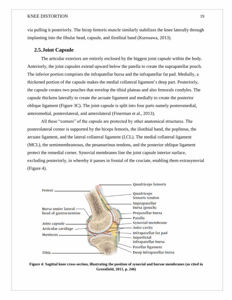
2.5. Joint Capsule
The articular exteriors are entirely enclosed by the biggest joint capsule within the body.
Anteriorly, the joint capsules extend upward below the patella to create the suprapatellar pouch.
The inferior portion comprises the infrapatellar bursa and the infrapatellar fat pad. Medially, a
thickened portion of the capsule makes the medial collateral ligament’s deep part. Posteriorly,
the capsule creates two pouches that envelop the tibial plateau and also femorals condyles. The
capsule thickens laterally to create the arcuate ligament and medially to create the posterior
oblique ligament (Figure 3C). The joint capsule is split into four parts namely posteromedial,
anteromedial, posterolateral, and anterolateral (Finerman et al., 2013).
All these “corners” of the capsule are protected by other anatomical structures. The
posterolateral corner is supported by the biceps femoris, the iliotibial band, the popliteus, the
arcuate ligament, and the lateral collateral ligament (LCL). The medial collateral ligament
(MCL), the semimembranosus, the pesanserinus tendons, and the posterior oblique ligament
protect the remedial corner. Synovial membranes line the joint capsule interior surface,
excluding posteriorly, in whereby it passes in frontal of the cruciate, enabling them extrasynovial
(Figure 4).
Figure 4: Sagittal knee cross-section, illustrating the position of synovial and bursae membranes (as cited in
Greenfield, 2011, p. 246)

KNEE DISTORTION 20
2.6. Knee Musculature
For the knees to operate suitably, several muscles have to collaborate in a complex way (Sick &
Burguet, 2012). Below is a list of knee activities and the muscles that induce them:
Knee flexion is performed by the semimembranosus, semitendinosus, biceps femoris,
popliteus, gracilis, gastrocnemius, plantaris, and sartorius muscles.
Knee extension is accomplished by the quadriceps muscles of the thigh, comprising of
three vasti, for instance, vastus lateralis, vastus medialis, rectus femoris, and vastus
intermedius (Sick & Burguet, 2012).
Internal rotation is executed by the popliteal, semimembranosus, gracilis, sartorius, and
semitendinosus muscles. Torsion of the tibia is restricted and can happen only when the
knees is in a flexion position.
The biceps femoris regulate the exterior torsion of the tibia. The bony structure also
generates external tibial rotation when the knee moves into extension.
On the literal side, the iliotibial band principally serves as a dynamic lateral stabilizer.
2.7. Bursae
A bursa consists of pieces of synovial tissue divided by a thin layer of fluid. The
responsibility of a bursa is to decrease the friction between anatomical alignments. Bursae are
found between bone and muscle, bone and tendon, alignment and tendons, and many others. As
many as 2–dozen bursae have been recognized around the knees joints. The infrapatellar,
suprapatellar, prepatellar, pretibial, and gastrocnemius bursae are perchance the leading
frequently injured around the knees joints (Figure 4).

KNEE DISTORTION 21
Figure 5: Knee muscles. A is anterior view. B is posterior view. C, is deep posterior view (Trippel, 2014, p.
208)
2.8. Fat Pads
There are numerous fat pads about the knees. Infrapatellar fat pad remains the largest. It
cats as a protection to the knee façade and isolates patellar tendon againt the joint capsules. Other
dominant fat pads within the knee incorporate the posterior and anterior suprapatellar, and the
popliteal. Specific fat pads fill the synovial capsule (Figure 4) (Pedowitz & Akeson, 2013).
2.9. Nerve Supply
The tibial nerves innervate several gastrocnemius and the hamstrings. The mutual
peroneal nerves innervate the short head of the bicep femoris and later courses via the popliteal
fossa and spirals around the proximal head of the fibula. Since the peroneal nerve is visible at the
fibula’s head, contusion of the nerve may contribute distal sensory and motor shortfalls. The
femoral nerves innervate the sartorius and the quadriceps muscles (Figure 6).

KNEE DISTORTION 22
Figure 6: Nerve supply to the knee (Chaudhari, 2013 p. 312)
2.10. Blood Supply
The major blood supply to the knee instigates from popliteal arteries that stem from the
femoral artery. From the popliteal arteries, four branches serve the knee, which includes medials
and laterals inferior genicular, and medials and laterals superior genicular arteries (Figure 7, A
and B). Blood flows through the tiny saphenous vein into the popliteal vein and afterward to the
femoral veins (Figure 7C).
Figure 7: The knees Blood supply. A, is anterior arteries. B is posterior arteries. C, venous supply
(Heerwaarden, 2013, p. 98)
2.11. Functional Anatomy
Motion between the femur and tibia entails the physiological movements of extensions,
flexion, and torsion besides arthrokinematic mobility as well as gliding and rolling. As the tibia

KNEE DISTORTION 23
stretches on the femur, the tibia rolls and glides anteriorly. If the femur is stretching on the tibia,
gliding takes place in an anterior direction, while rolling happens posteriorly.
Axial torsion of tibia proportional to the femur is an essential constituent of knees
movement. As the knee lengthens, the tibia outwardly rotates. The torsion takes place since the
medial femoral condyle is bigger than the lateral condyles. Therefore, during weight bearing, the
tibia the tibia has to rotate exteriorly to attain full extension. The torsional constituent provides a
great compact to knee stability in complete extension. In the case of weight bearing, the
popliteus muscle has to contract and externally alternate to the femur to unlock the knees for
flexion to occur (Greenfield, 2011).
The capsular ligaments are extended during full extension and rather a slacker during
flexion. This is especially true of laterals collateral ligament; nevertheless, medial collateral
ligaments parts relax while flexion arises. Relaxation of the other superficial collateral ligament
lets rotation take place. On the other hand, the deeper capsular ligaments tighten to stop the
excessive turning of the tibia (Sick et al., 2012).
During the final 15-degrees of stretching, the tibia outwardly rotates, and the anterior
cruciate ligaments relax. In the full stretch, the posteriolateral section of the anterior cruciate
ligament is extended, and it relaxes during flexion. While femurs glide on the tibia, the posterior
cruciate ligaments become tight and inhibit additional gliding. Overall, the anterior cruciate
ligaments prevent extreme internal torsion, stabilize knees in full extension, and inhibit
hyperextension. The posterior cruciate ligaments prevent excessive inner torsion of the tibia,
constraints the femur’s anterior translation on the attached tibia, and constraints posterior
translation of the tibia within the non-weight bearing (Stenstrom, 2010).
In full flexion, about 140 degrees, the variation of knee mobility is restricted by the
excessively shortened situation of the hamstring muscles, the bulk of hamstring muscles, and
quadriceps muscles extensibility. In this situation, femorals condyles rest on their equivalent
menisci at a point that allows a slight degree of internal rotation.
Consistent with Stannard and Cook (2013) assessment, the patella helps the knee during
extension by expanding quadriceps muscles lever arms. It transmits the compressive stresses on
the femur through widening contact area between the femur and patellar tendons. It similarly
shields the patellar tendon against friction. In full extension, the patella lies somewhat proximal
and lateral to the trochlea or femoral groove. At 20 degrees of knee flexion, tibial rotation

KNEE DISTORTION 24
occurs, and the patella moves into the trochlea. Patella remains most prominent at 30-degrees.
Also at 30-degrees and above, the patella moves deeper into the femoral groove. At 90 degrees,
the patella yet again becomes situated laterally. When knees flexion is 135-degrees, the patella
has moved laterally above the femoral groove (Yeager, 2010).
3. THE KNEE WITHIN THE KINETIC CHAIN
The knee is directly affected by forces and motion arising and being transmitted from the
ankle, foot, and lower leg. As a result, the knee has to transmit forces to pelvis, thigh, spine, and
hip. The tissue must absorb anomalous forces that cannot be transmitted. When the foot interacts
with the ground, a locked kinetic chain occurs. In a locked kinetic chain, forces should either be
absorbed in a more distal joint or be transmitted to proximal segments. The incapability of this
closed system to disperse these forces characteristically causes a breakdown in some portion of
the system. The knees joints are prone to vulnerable to injury originating from the absorption of
these forces (Pedowitz et al., 2013).
4. BIOMECHANICAL OF THE CRUCIATE
4.1. Biomechanics and Kinematic of the Knees Joints
The aim of the joints is to permit for the motion of the bony parts surrounding the joint
whereas resisting the loads against gravity inflicted by these movements. Biomechanics can be
defined as the science of the act of forces on the living being. The complex collaboration of
patella, femur, and tibia enables knees joints to resist extreme forces during normal stages of
ambulation (Marshall, 2011). Kinematics can define as the study of body movement without
concern for the contributor of that motion. Six planes of motion are present for the knee, such as
medial/lateral translation, anterior/posterior translation, flexion/extension, cephalad/caudad
translation, varus/valgus angulation, and internal/external torsion as has been outlined above.
The knees joints has to provide a normal degree of motion without losing stability during static
actions like standing to more dynamic activities like running, walking, pivoting, jogging, and
descending or ascending stairs. These goals are accomplished by the interplay of the osseous
anatomy, ligaments, articular surface, menisci, and surrounding musculature around the knee.
Modifications of any of these elements can change the knee joint biomechanics, largely
increasing loads and operational demands positioned on the rest of the structures. Understanding

KNEE DISTORTION 25
the normal interplays of these structures is important before trying any reconstructive operations
(Kurosawa, 2013).
4.2. Passive Motion of the Knees
According to Trippel (2014) evaluation, the prime motion of the knees is extension and
flexion. The knee junction totals from 0 to 135o of flexion in the sagittal surface. The passive
movement of the knees linkages is dictated by the articular surfaces structure and the adjacent
soft-tissue capsule ligament. Due to the distal between the lateral and medial femoral condyles,
mobility between 20o of flexion and full extension is accompanied by gently sloping of the
laterals femoral condyle posteriorly greater than the medial femoral condyle. This enables the
tibia and femur to unlock from full extension and ensue with no assistance from any dynamic
muscle participation. Following 20o of flexion, knee joint passive flexion happens by a sliding
motion, with comparative tibial mobility on the femur (Pedowitz et al., 2013).
4.3. The Functional Biomechanics of the Ligament
Among the knees ligament, the cruciate is the most essential in offering passive limit to
the posterior/anterior knee motions. If a single or both cruciate are interrupted, the biomechanics
during ambulatory practices can be interrupted. The knee interaction between the collateral
ligaments, cruciate ligaments and other dynamic and static stabilizer is complicated, and
appreciation for the meniscal, osseous, tendinous, articular, and other soft-tissue components
causes the entire knee motion and stability is vital (Cantrell, 2013).
4.4. Biomechanical of the Anterior Cruciate Ligament
The principal function anterior cruciate ligament (ACL) is to prevent anterior translation
of the tibia. It serves as a secondary stabilizer against valgus angulation at the knee and interior
torsion of the tibia. In full extension, the ACL uptakes 75% of the anterior translation weight and
85% between 30-90o flexion. Loss of ALC results in reduced magnitude of this coupled rotation
in an unstable knee and during flexion. Numerous researches have been executed to discover the
biomechanical aspects of ACL. Nevertheless, uniform analysis on strain rates and alignment is
improbable. A number of recent studies have showed that the anterior bundles (both lateral and
medial) have higher maximal strain and stress than the posterior bundles. The tensile strength of
KNEE DISTORTION 26
ACL is about 2,2 N though it is changed with age and repetitive loads. When the magnitude of
the anterior drawers force rises, the in situ force of the ALC similarly rises (Bianchi, 2012).
4.5. Biomechanics of Posterior Cruciate Ligament
The main task of the posterior cruciate ligaments (PCL) is to withstand posterior
translation of the tibia on the femur at any bearing of the knees flexion. It is a secondary
stabilizer against exterior rotation of the tibia and extreme valgus or varus angulation of the
knees. The anterolateral band is rigid in flexion and is extremely important in withstanding
posterior distortion of the tibia in 70o to 90o of flexion. The posteromedial section is taut in
extension; therefore, it withstands posterior distortion of the tibia in this position. Whereas the
PCL is the major limit to the posterior translation of the tibia, this ro9le is largely enhanced by
other atomies. The recent cadaveric research has reported that extreme posterior translation of
the tibia needs damage to one or more structures as well as the PCL (Johnson, 2012).
Isolated PCL ruptures can contribute a mild increase in external torsion at about 90o
knees flexion. That is to say, they do not significantly alter the valgus/varus angulation or tibial
rotation, however, because of the tight extracapsular ligaments and tissues. With both
posterolateral corner and PCL rupture, there is a noticeable increase in external tibia torsion to
the absence of reinforcing restraints. It has been established that the anterolateral component
characterized a greater tensile and stiffness strength than the meniscofemoral ligaments and the
posteromedial bundle. Moreover, it has been established that at a different degree of knee
flexion, variance in situ forces occurred. At 0o, the PCL characterized a total tensile strength of
6.1 N, whereas, at 90o, it characterized a tensile strength of approximately 112.3 N. The
posteromedial bundle reached a maximum force of 67.9 N at 90o of the knees flexion, whereas
the anterolateral bundle attained a maximum force of 478 N at 60o. Knowledge of these
associations is crucial in the reconstructive surgery to make sure that the grafts are tensioned
appropriately (Chaudhari, 2013).

KNEE DISTORTION 27
Figure 8: The four-bar cruciate link system (Kurosawa, 2013, p. 134)
As well as to its well-known function in the sagittal plane, the PCL affects knee motion
in the anterior plane. This happens because the PCL attaches onto the medial femoral condyle’s
lateral facet of and is aligned indirectly. This alignment of the PCL assists in the articular
unevenness between the lateral and medial femoral condyles and allows sufficient tensioning of
the PCL in the course of the laterals femoral condyle rolling posteriorly in initial flexion. The
popliteus muscle helps the PCL in withstanding posterior tibial translation and improving
stability. In PCL-deficient knees, the popliteus muscle decreased posterior translation of the tibia
by about 36% (Fotopoulos, 2015).
4.6. The Interaction of the Cruciate Ligament
The complex interplay between PCL and ACL at different degrees of extension and
flexion aids account for the knee joint dynamic balance. The tension and stretch of the PCL and
ACL alter during extension and flexion because of their asymmetric insertion positions. In full
extension, the ACL is tight, whereas the PCL is relatively loose. When an individual stands up
with his knees in hyperextension, the joint is partially stable, with little requirement for muscular
support. When the knees flex, the posterolateral section of the ACL turns to lax, whereas the
PCL remains taut, particularly the anterolateral bundle. Stability is delicate between 0-50o of
flexion because neither cruciate ligament is tremendously tight. The alteration in the orientation
of the PCL and ACL fibers during knee flexion permits for dynamic stability within the sagittal
surface. With augmenting flexion, the ACL shifts from an upright position to a more horizontal
alignment with respect to the joint line. The PCL's alignment is opposite to the ACL's during
extension and flexion (Pedowitz et al., 2013).

KNEE DISTORTION 28
Thus, as the knee attains a higher degree of flexion, the PCL changes to more important
in inhibiting damage to the joint. This interaction between PCL and ACL is often known as the
four-bar cruciate link system. The connection of these ligaments illustrates that the epicenter of
joint rotation moves posteriorly in the company of the knees flexion. This enables for both the
femur rolling and sliding movements in the course of flexion and prevents the femurs from
rolling off the tibial plateau at excesses of flexion. In the varied phase of the bearing cycle, the
force vector surrounding the knee is the sagittal plane alteration. The mechanical loads
throughout the knees intersection are modified by variations in foot position besides by the type
and intensity of ambulatory action (Kean, 2012).
In normal ambulation, a joint responsive force of 2 to 5 times the body mass is generated;
this force is approximately 24-times the body mass during running period. Dynamic muscle
forces aid to stabilize these functional weights and joint responsive forces, specifically as knee
flexes and the load-bearing axis moves from a site anterior to the knees linkage to a single
posterior. If a muscular, ligamentous, and bony injury happens that interrupts this weak balance
of forces, the joint is ineffective at resisting these loads, quickening the knees waning process
(Marshall, 2011).
According to Muscolino (2014) insinuation, the dynamic activities of the adjoining
muscles are controlled by the cruciate ligaments during knee extension and flexion. The
quadriceps muscles, via the patella tendon, eventually join onto the anterior tibia, and, thus, the
tibia is translated anteriorly by way of the exterior mechanism and constrained by way of the pull
of the ACL. The biomechanical benefit is capitalized on when the axis of knee rotation is vertical
to the joint line. When anterior translation takes place on the sagittal surface during ambulation,
the epicenter of torsion is altered as with ACL deficiency, and the resulting escalation of forces
across the knee linkage exerts increased stress following the secondary limitations. The moment
arm of the knees extensor device is reduced, leading to an increase in muscle forces needed to
maintain stability all over the knee linkage. This causes an increase in joint responsive forces
and, finally, injured or stressed reinforcing structures. Within ACL-deficient knees, increased
stress is exerted on the secondary restraints of anterior translations, encompassing the menisci
and the adjacent soft-tissue capsule. In case, the quadriceps become atrophied once ACL
raptures, the extensor pull on the tibia decreases, lessening the stresses exerted on the secondary
stabilizer (Heerwaarden, 2013).

KNEE DISTORTION 29
The screw-home method again indicates the essential the dynamic of muscle in knee
movement. As the lateral femoral condyles move posteriorly during early flexion, the extensor
apparatus's moment arm increases. This provides a mechanical advantage to the knees during
running and stair climbing, when there is a maximum demand on the knee joints (Jakob et al.,
2013).
Figure 9: Knee illustration of the in 0o (left) and 30o (right) of flexion showing femoral rotation associated
with the tibia in early flexion (Muscolino, 2014, p. 124)
5. DIAGNOSIS AND MANAGEMENT OF KNEE DISTORTIONS
Analysis by Greenfield (2011) explicate that acute knee distortion is a rare diagnosis in
the orthopedics, with a high rate of related injuries and possible limb-threatening problems. The
reported occurrence is 0.02% of musculoskeletal shock, even though this is likely an underrating
due to an unidentified number of spontaneous decreases briefly after injury. Reports of
permanent instability and suffering are common after a diagnosis of knee distortion. Though
management principles have evolved over the last two decades, optimal therapy of these injuries
is still controversial. Few high-level substantiation studies are obtainable to aid guide
management. The low occurrence and varied nature of the injury enable randomized controlled
experiments challenging to facilitate. An elementary knowledge of the topic, with special
attention to physical evaluation and first management, will let the treating doctor to manage
patient's knees distortion properly, with a possibly decreased risk of complications (Lee, 2013).
5.1. Classification
The knee distortion classification is mainly centered on the direction the tibia distorts
comparative to the femur. This leads to diverse categories including posterior, anterior, medial,
lateral, or rotatory. The posterior –medial, anterior-medial, and lateral distortion can be classified

KNEE DISTORTION 30
as “rotatory” dislocation. Other parameters to be taken into consideration comprise whether the
knee is totally dislocated or subluxated, the injury is open or closed, there is neurovascular
involvement, and the low-energy or high-energy trauma. Also, one ought to be acutely mindful
of the fact that a total distortion can spontaneously moderate, and triple-ligament knee damage
constitutes a forthright distortion (Bianchi, 2012).
Reports differ, but anterior and posterior distortion seems to be the most frequent
direction of distortion. There is 70% occurrence rate of posterior, 5% rotatory, and 25% anterior
distortions. Rotatory distortion incidence is less frequent, but the posterolateral distortion appears
to be the most frequent combination. This specific pattern can be complex due to the medial
femoral condyle turning button-hollowed via the anteromedial joint capsule. Additionally, the
MCL invaginates into the joints space, preventing reduction. This button-hollowing lead to a
skin furrow along the medial joint line as the subcutaneous tissue connections to the joint capsule
pull the skin into the joint. Efforts at a reduction in this situation make the skin furrow more
marked (Pedowitz et al., 2013).
The real incidence of various directional displacements is not essential as properly
diagnosing the direction of the damage, and how it associates to the possible neurovascular
damage. Posterior dislocations or hyperextension injuries, due to the tethered popliteal vein and
artery, can encounter the highest incidence of related vascular injury; nevertheless, any
displacement, if the original distortion is severe enough, will cause impairment to the popliteal
artery. The normal peroneal nerve is less endangered since it has a higher excursion rate than the
popliteal vessels, although it remains susceptible in case a verus force is subjected to the knee.
Posterolateral distortion is linked with a high incidence of damage to the common peroneal nerve
(Pedowitz et al., 2013).
Open knee dislodgements are not common. The reported occurrence is range from 19-
35% of all dislodgements. An open knee distortion, generally, carries an inferior prognosis of the
serious harm to the soft-tissues envelopes. Consequently, an open injury can need a staged
reconstruction, or an open ligament reconstruction, as arthroscopically assisted methods cannot
work in the acute environment with these open wounds (Jenkins et al., 2012).
Differentiating between high- and low-energy injuries is essential. Low-energy injuries,
often connected with sports injuries, have a reduced incidence of related vascular injury. High-
energy injuries, linked with motor vehicle crashes or falls from an elevation, tend to have

KNEE DISTORTION 31
augmented prevalence of vascular compromise. With reduced pulses in a wounded limb and the
history of a high-velocity or high energy injury, one ought to acquire vascular studies
immediately (Zhang, 2010).
5.2. Mechanism of Injuries
The mechanism of the injury of two most frequent knee distortions patterns, posterior and
anterior, are considerably well discussed. There is some query as to whether the PCL or the ACL
fails initially with hyperextension, though in clinical results, both posterior and anterior cruciate
ligament fails with displacement. A posterior-directed force exerted to the proximal tibias if the
knee is flexed to 90o is alleged to produce a posterior distortion, what is known as “dash-board”
injury. Lateral and medial distortions arise from valgus/varus stresses inflicted to the knee. A
combination of hyperextension/blow with valgus/varus stress to proximal tibia will potentially
give rise to one of the rotatory distortions.
5.3. Associated Injuries
As stressed by Lee (2013), numerous anatomic structures are at danger in the distorted
knee. The knee’s four primary ligaments along with the lateral corners and posterior medial may
be compromised. Nerve and vascular injuries are regular. There can also be associated bony
lesions; such as distal femur condylar or frank tibial plateau fractures, avulsion breakages of the
PCL or ACL, or femoral shaft or ipsilateral tibial breakages.
There is confirmation in the literature that an actual distortion cannot lead to the complete
rapture of three out of four primary ligaments; nevertheless, this appears to be exclusion instead
of the rule. Many investigators have identified that actual knee distortion habitually causes
rapture of a minimum 3 from the 4 primary ligaments. With frank knee distortion, cautious
ligament analysis is required to completely diagnose the degree of the damage (Wilkinson,
(2014).
The occurrence of vascular compromise in knee displacements has been approximated to
be 32%. When constrained to posterior or anterior dislocation, the occurrence can be increased
by 50%. Latest studies prove the important incidence of arterial injuries, echoing the demand for
careful vascular examination. The popliteal artery is also called “end artery” to the legs, with
least collateral circulation via the genicular arteries. Consequently, the popliteal vein account for

KNEE DISTORTION 32
the majority of the venous discharge from the knee. If either anatomy is compromised to the
point of protracted impairment, ischemia and ultimate amputation are usually the outcomes.
Two mechanisms have been discussed for damage to the popliteal artery: once include a
stretching mechanism, observed with hyperextension, pending the vessel ruptures. This can
ensue secondary to the tethered condition of the artery positioned at the adductor hiatus and the
inlet via the gastrocnemius-soleus component. This kind of injury should be expected with
anterior distortion. Posterior dislodgments can result in direct contusion of the vessels by the
posterior plateau, causing damage. Under no condition should the compromised vascular state be
associated with arterial spasm; in this condition, there is normally intimal injury and future
thrombosis formation. Initial evaluation can be common; however, thrombus formation may
occur hours or days later, and previous studies have realized delayed thrombus formation. Also,
bicruciate ligament ruptures showing a decreased dislocated knee could have a high incidence of
arterial damage as a frank distortion (Lee, 2013).
Potential vein damage takes place much less often, or as minimum historically had not
been observed. Regardless of this, venous occlusion should similarly be identified and
adequately treated. Factually, whether to repair venous injury is looked contentious. Ligating the
popliteal vein, regular practice during Vietnam War, caused severe phlebitis, edema, and chronic
stasis alterations. The venous restoration was believed to contribute to pulmonary embolism and
thrombophlebitis. Today, if the obstruction to outflow is discovered, surgical repair of the
popliteal vein is necessary (Finerman et al., 2013).
Injury to either tibial nerve or peroneal nerve has been reported with an incidence rate of
approximately 20% to 30%. The nervous anatomies around the knee are not as rigidly attached to
the popliteal vessels; this is possibly accounted for the lower occurrence of injury in comparison
with surrounding vascular structures. The mechanism of damage is often the stretch. The
peroneal nerves appear to be more regularly engaged than the tibial nerve, certainly due to its
anatomic site. With any varus knees weighing, the peroneal is positioned under tension. Posterior
distortion contributed most of the nerve injuries. Provided the reality that knee displacement is
often contributed by violent trauma, related fractures are frequent; the incidence can be as great
as 60%, Tibial plateau ligament and fractures avulsion fracture from the proximal-distal or tibia-
femur are frequent. Acknowledgment of these injuries is similarly vital since extra bony
involvement has insinuations on the absolute treatment.

KNEE DISTORTION 33
Related distal femur ruptures and proximal tibial ruptures treated with intramedullary
nailing make bone shaft dislocation for PCL and ACL reconstruction problematic. With violent
trauma, any avulsion or fracture imaginable can take place with a distorted knee, but there is a
report that lateral and medial distortions are related to some elevated frequency of bony minor
lesions. Fracture distortions signify a different entity within the range of pure knee displacement
to tibial plateau ruptures. Pure knee displacement necessitates only soft tissue reconstruction to
acquire balance; tibial plateau raptures need purely bony stabilization. Fracture knee distortions
commonly involve both ligamentous and bony reconstruction. A permanent result of fracture-
distortion damages to the knee linkage is someplace between pure distortions or tibial plateau
fractures, with tibial plateau fractures doing better and distortion the worst (Cantrell, 2013).
6. FIRST EVALUATION AND MANAGEMENT
6.1. General Considerations
Evident deformity can exist during the initial evaluation. However, in a poly-trauma,
patients who are sedated and intubated, the injury can escape initial examination. Contusions or
abrasions about the knee, laxity, or gross crepitus can allude to injury in an or else normal
looking knee (Heerwaarden, 2013). This significance of instant recognition of the knees
distortion or fracture-distortion lay not with the treatment variability, but the recognition of
possible vascular injury and potential vascular compromise. The neurovascular condition must
be evaluated on both lower limits. Neurologic assessment can be tough in poly-trauma patients,
and is not as significant initially as is the series neurologic assessment. Vascular evaluation is
more important since ischemia lasting greater than 8 hours normally lead to amputation. In the
decreased knee, a white could limb, which is noticeable on the physical evaluation and represents
arterial damage, needed an instant arteriogram. Nevertheless, Doppler signals, normal pulses,
and capillary refill do not exclude an arterial damage. Thrombosis can occur hours to days after,
calling for serial evaluation. If there is any query of perfusion limb, an arteriogram is necessary.
6.2. Imaging Studies
Before any manipulation, lateral and anteroposterior radiographs of the interrupted
constraint are completed. This is vital to verify the direction of distortion and any related
fractures and helps in planning the attenuation of maneuver. With the existence of cyanosis,

KNEE DISTORTION 34
pallor, weak capillary refill, pallor, and reduced peripheral following decrease, arteriography
must be taken into consideration. Venography can be warranted if the clinical images display
sufficient limb perfusion but blockade of outflow. Following the acute treatment of the distorted
knee, magnetic resonance imaging can be carried out subacutely to validate and assist in
strategizing the reconstruction of affected ligamentous structures (Pedowitz et al., 2013).
6.3. Reduction
According to Fotopoulos (2015) studies, unrestrained distorted knee establishes an
orthopedic emergency, and reduction must be made as soon as possible, rather in the emergency
ward. Prior to manipulation, adequate anteroposterior and lateral radiographic examination is
conducted. This permits for identification of the direction of distortion, any related fractures, and
aids in planning the direction movement. In the separated knee distortion, conscious sedation or
intravenous morphine is often needed. Slow longitudinal traction is inflicted to the leg from
ankle, and a proximal tibia is repositioned in the suitable direction to effect moderation. Once
decreased, the radiographic analysis is applied to confirm tibiofemoral congruency, as well as
repeated neurovascular evaluation. The limb is then put in either knee extension immobilizer or a
long leg split. It is imperious to perform radiographic examination following placement in the
brace or splint, as posterior subluxations of the tibia on the femur is normal. A "bump"
comprising a pad or towel behind the gastrocnemius-soleus composite can help in maintaining
reduction.
The “dimple sign” demonstrates a posterolateral distortion, and closed reduction might
not be prosperous. The medial femoral condyle infiltrates the medial joint, contributing
interposition of soft tissue within the joint, necessitating open reduction (Jenkins et al., 2012).
6.4. Physical Examination
Physical examination properties of the PCL/ACL/PLC injured knee incorporate abnormal
posterior and anterior translation at equally 25o and 90o of the knees flexion that is typically
higher than 15 mm. The tibial step-off is missing at 90o, and the posterior drawer test is two or
more, indicating higher than 10 mm of pathologic posterior tibial distortion. The pivot-shift and
Lachman test phenomenon are positive, presenting ACL disruption, and there could be knee
hyperextension. Three types of posterolateral instability, A, B, and C, have been identified and
described.

KNEE DISTORTION 35
Posterolateral imbalance (PLI) in the multiple-ligament Wounded Knee consists, at least,
10o of enhanced tibial external rotation in comparison with the typical knee at 30o-90o of flexion,
and adjustable degree of varus imbalance relying on the injured anatomic structures.
Posterolateral unsteadiness PLI type A has augmented external torsion only, relative to damage
to the popliteus tendon, and popliteofibula ligaments only. PLI type B indicates with increased
external alternation, and slight varus of about 5 mm augmented lateral joint opening to varus
stress at 30-degrees knee flexion. This takes place after harm to the popliteus tendon,
popliteofibular ligament, and decline of the fibular collateral ligaments. PLI type C indicates
with augmented tibial exterior rotation and varus imbalance of 10 mm more than the normal
knee examined at 30o flexion with varus stress. This takes place after damage to the popliteus
tendon, popliteofibular ligament, lateral capsular avulsion, fibular collateral ligament, as well as
cruciate ligament disruption.
The MCL is verified with valgus stress between 0o-30o flexion to evaluate the superficial
MCL, the posterior medial capsule, and the posterior oblique ligament. Extensor mechanism
stability is evaluated by medials and laterals patellar glide to examine the veracity of the laterals
and medial retinaculum.
6.5. Vascular Injuries
As indicated by Johnson (2012) studies, a complete spectrum of vascular injuries can be
experienced. The overall clinical pictures can fluctuate from an uncomplicated, bicruciate
ligament damage, with a potential internal injury with a normal physical evaluation to a poly-
trauma patient, having intra-abnormal bleeding, a closed head injury, and distorted knee with
vascular complication. Severe injuries treated first. The orthopaedic surgeon must be aware of
the of the overall limb ischemia time. If the suspicious arterial damage is observed, a vascular
consult is obtained straightaway. The reduction is conducted to confirm if this restores blood
circulation to the limb. When the overall ischemia time nears 6 hours, there is an urgency to
reinstate circulation to the lower end. An intraoperative angiogram during vascular examination
and shutting might be needed at the expense of a first-rate preoperative angiogram (Stenstrom,
2010). Mechanism of damage also needs to be considered. A high-energy injury could be more
suspicious of vascular damage, and one can opt to obtain arteriograms in spite of a normal
vascular test.

KNEE DISTORTION 36
When an isolated displaced knee with suspected arterial injury arises, arteriography is
performed as the normal availability of pulses does not exclude vascular mutilation. Any
suspicion necessitates a vascular operation consultation. When the limb is appropriately
perfused, and all indices are well, one can opt to forego a formal arteriogram in care there are
recurrent neurovascular examinations to the lower extremity. In spite of the historical preference
to receive an arteriogram in the existence of knee distortion as screening model, it has been
demonstrated that arteriography after considerable blunt pain to the lower extremity with typical
vascular check exhibit a low yield ratio for discovering surgical vascular lesion. Popliteal vein
damage is also likely. When the clinical picture is required, a venogram could be supportive.
6.6. Absolute Surgical Indications
A situation of vascular injury and irreducibility warrants instant surgical intervention.
Four-partition fasciotomy of the limb is taken into account when ischemia time is higher than 5
hours. The incapability to sustain reduction similarly obligates early ligamentous reconstruction
or external skeletal fixation to stabilize the knee to prevent potential repeated vascular
compromise. Open displacements and open fracture-displacements warrant immediate clinical
debridement to disinfect the wound. An exterior fixator can be a rational alternative in the
situation of open distortion with large soft-tissue impairment or open fracture distortion. In this
scenario, access to soft-tissue would be sustained for surgical debridement (Pedowitz et al.,
2013).
7. DEFINITIVE SURGICAL MANAGEMENT
7.1. Historical Management
Knee distortions were originally managed using a cylinder cast for many months. Early
reports indicate meaningful results for nonoperatively treated knee displacements. However,
there was the recommendation that surgically stabilized distortioned knees would be quite better
in the long-standing. A recent finding compared surgically stabilized knee with conservative
therapy and resolved that the conservative therapy was similar to surgical intervention. Despite
the same results, the conservatively treated knee was grossly compared with surgically stabilized
knee. The survey was reflective from 1963-1988 and the distinctive surgical treatment in this

KNEE DISTORTION 37
period was in many cases open direct reconstruction of the ligaments. The same results were
arrived at by comparing four conservatively treated knees using 16-direct suture reconstruction
of torn ligaments. Likewise, examined early (in 5 days of damage) direct reconstruction (with or
without increment) of torn ligament parts in 13 out of 17 patients. They resolved that better
outcomes were attained with early versus after direct repair of the torn ligament. This research
backs surgical treatment of the distortioned knees and presents the idea of benefit from
ligamentous stabilized knee (Stannard et al., 2013).
In the past decade, the method of arthroscopically assisted PCL/ACL repair has become
common. Numerous advancements have enabled these techniques successful such as better
sterilization, procurement, and storage of allograft tissue, better graft fixation techniques,
improved arthroscopic surgical instrumentation method, and improved knowledge of knees
ligamentous biomechanics and anatomy. Few reports of integrated PCL/ACL repair are
accessible in the literature though surgical repair seems to afford identical outcomes, if not
better, than the direct reconstruction of ligaments (Marshall, 2011).
7.2. Sports Injury Clinic Experience
There is the 38-percent incidence of PCL tears in acute knee wounds; with 45% of these
PCL wounded knees being linked PCL/ACL tears. Careful evaluation and treatment of vascular
wounds are critical in these acute multiple-ligaments. There is an 11% occurrence of vascular
injury related to these acute multiple-ligaments injured knees. Most preferred to technique to link
PCL/ACL repair with the transtibial method, with capsular/collateral ligament surgery as
specified. Not all incidences are agreeable to the arthroscopic technique, and the surgical
physician must examine each case independently (Pedowitz et al., 2013).
7.3. Surgical timing
Surgical timing is reliant on the vascular situation, skin condition, reduction stability,
open versus closed knees injury, systematic injury, articular and meniscus surface injuries, other
orthopaedic damages, and capsular/collateral ligaments concerned. Particular PCL/ACL/MCL
damages are treated through bracing of the MCL succeeded by arthroscopic combined PCL/ACL
repair in 46 weeks after recovery of the MCL. Other cases could warrant medial structures
reconstruction and have to be examined individually.

KNEE DISTORTION 38
Combined PCL/ACL/posterolateral damages are addressed timely as safe as conceivable.
ACL/PCL/posterolateral reconstruction-repair carried out between 2-3 weeks following injury
enables closing of capsular tissues to allow an arthroscopic intervention, and still allows primary
reconstruction of damaged posterolateral structures. Open multiple-ligament knee distortions
may need staged procedures. The capsular/collateral structures are reconstructed after methodical
debridement and irrigation and combined PCL/ACL repair performed at a later date once wound
healing has occurred. Care must be considered in all cases of postponed reconstruction to
guarantee the attenuation of tibiofemoral joint by lateral radiograph and serial anteroposterior.
The surgical timing procedures highlighted above must be taken into account in the
context of each patient. Some patients having multiple-ligament knee damages are seriously
wounded multiple-trauma patients with multisystem wounds. Modifiers to the best timing
protocols highlighted earlier encompass the reduction stability, the vascular state of the involved
extremity, open or close injury, skin condition, and other systemic and orthopaedic injuries.
These extra considerations can contribute the knee ligament surgery to be done earlier or later
than expected (Kean, 2012).
7.4. Graft Selection
The suitable graft material is strong, gives safe fixation, is simple to pass, readily
obtainable, and has low donor-site movement. The available alternatives in America are allograft
and autograft sources. The most preferred graft for PCL includes the Achilles tendon allograft
due to its broad cross-sectional area and strength, an easy passage with safe fixation, and lack of
donor mobility. Also, bone allograft, bone patellar tendons or Achilles tendon allograft are
preferred for ACL repair. For a posterolateral corner, the preferred graft material is split biceps
tendon transfer, allograft tissue, or free autograft (semitendinosus) when biceps tendon cannot be
obtained (Pedowitz et al., 2013).
Cases necessitating MCL and posteromedial corner operation could have reconstruction,
primary repair, or a combination of both. Open favored approach for MCL and posteromedial
reconstruction involves a posteromedial capsular shift with allograft or autograft
supplementation as required (Moreno, 2012).

KNEE DISTORTION 39
7.5. Surgical Approach
The principal surgical approach includes a single-stage arthroscopic combined PCL/ACL
reconstruction with the transtibial method with capsular/collateral ligament procedure as
presented. The posterolateral corner is reconstructed and then amplified with an allograft tissue,
semitendinosus free gap, split biceps tendon transfer, or biceps tendon transfer. An acute medial
injury, not amenable responsive to brace treatment go through the posteromedial capsular shift,
primary repair, or allograft reconstruction as specified. The surgery surgeon should be prepared
to adapt to an open procedure or a dry arthroscopic procedure in case fluid extravasation
becomes an issue (Manske, 2015).
7.6. Graft Tensioning and Fixation
The PCL is the first to be reconstructed followed by ACL, and then the medial ligament
complex and posterolateral complex follows. Tension is exerted on the PCL graft distally with
the Arthrotek knee ligaments tensioning instrument along with the tension set for 20 lb (Figure
15). This reinstates the anatomic tibial step-off.
Figure 10: The mechanical graft knee ligament tensioning instrument (Tortora et al., 2014, p. 499)
The mechanical graft knee ligament tensioning instrument is employed to accurately
tension ACL and PCL grafts. In PCL reconstruction, the tensioning instrument is inserted to the
tibial ending of the graft and the torque wrench ratchet set to 20 lb. This reinstates the anatomic
tibial step-off (Tortora et al., 2014).
The operated knee is cycled via a complete range of movement 25-times to enable
pretensioning and positioning of the graft. The knee is position in 70o of flexion, and concluding
fixation is accomplished with the ACL graft using a bioabsorbable interference screw and spikey

KNEE DISTORTION 40
ligament washer and a screw stoppage fixations. Tensioning the ACL graft of the knees flexion
allows surgeons to maintain the knee’s neutral location of by checking the tibial step-off at the
period of final graft in 70o fixations. The MCL restoration is tensioned with the knees at 30o of
flexions with the leg within the fugure-of-4 bearing. (Moreno, 2012).
Table 1: Tensile strength comparison (Cantrell, 2013, p. 366)
Material Maximum load (N)
Antrior cruciate ligament 2,0
Posterios cruciate ligament 4,0
Bone-patellar tendon-bone (10 mm) 2,9
Semitendinosus and gracilis (2-strand) 1,9
Semitendinosus and gracilis (4-strand) 2,8
8. PHYSIOTHERAPY
Physiotherapists are experienced in the prescription of exercises. They will offer you
specified exercises to improve your flexibility, strength, posture and balance as designated by a
detailed examination. Thus, your program will be individualized to make sure the best probable
result for your knee, especially for ligament treatment. Physiotherapists also employ other
treatment methods for knee pain resulting from distortion. These may incorporate tissue massage
and joint mobilizing, knee taping, education and advice on how well to manage knee injury
condition (Cantrell, 2013).
8.1. Examples of Physiotherapies
8.1.1. Mobility Exercises
Mobility exercises are conducted immediately pain will enable with the aim of regaining
or maintaining complete joint mobility (Fotopoulos, 2015).
Plan for mobility exercises

KNEE DISTORTION 41
Table 2: Plan for mobility exercises (Moreno, 2012, p. 120)
Mobility Exercises per days
sets Reps Time in seconds per set
Heel slides 3 2-4 30 sec/se
Flexion Extension Exercise 3 10-20 60 sec/set
8.1.2. Heel slides
To carry out this gentle knee range workout, the athlete lies on a hard surface or on their
back. The heel is gradually moved upwards the buttocks, as much as this is comfortable
(Wilkinson, (2014). Sock may be worn to make sure that foot slides. Once a minute, advance
movement can be possible. A strap or a towel wrapped about the ankle may be used to aid
though this must not be forced during the early stages in case there is a lot of pain (Yeager,
2010). Do 2-4 reps several times during the day.
Figure 11: Heel slides (Manske, 2015, p. 625)
8.1.3. Flexion Extension Exercise
The exercise is tremendously crucial to improve the movement of injured knee though it
will also assist to maintain the hamstrings and quadriceps during early phases of rehabilitation. It
can similarly help to lessen swelling around the knee.
Try to bend your knee as much as you can easily. This may be done while sitting, lying or
standing on your front. Aim three sets of 10 to 20 repetitions as pain tolerates. Progress this
workout to holding the in position at the ending of likely range, both in extension and flexion.
This will help in strengthening muscles (Bianchi, 2012).

KNEE DISTORTION 42
Figure 12: Flexion Extension Exercise (as cited in Johnson, 2012, p. 313)
8.1.4. Strengthening Exercises
Table 3: Strengthening Exercises (Manske, 2015, p. 633)
Strengthening Exercises
sets Repetitions Time in seconds per set
Static squad contractions 3 2-4 30 sec/se
Hip Abduction 3 10-20 60 sec/set
Half Squats 3 10-20 30 sec/set
Squat with Swiss Ball 3 10 60 sec/set
Lunges 2 10 60 sec/set
Bridge exercises 3 8-13 30 sec/set
Leg press 3 6 60 sec/set
Calf Raises 3 2-20 60 sec/set
Leg Curl 3 10-20 30 sec/set
These should start immediately as pain allows starting with static or somatic
shrinkages/contractions. Follow your trainer or physiotherapist rehabilitation program. Do not do
it if it hurts. Knee strengthening ought to be done 1-3 times for each week given they do not

KNEE DISTORTION 43
increase or cause pain. Idyllically they ought not to be carried out on successive days, to enable
muscle recovery. The exercises may be advanced by slowly augmenting the repetitions numeral
of sets and resistance of the exercises so long as they do not increase or cause pain (Fotopoulos,
2015).
8.2. Static squad contractions
This exercise can be commenced the moment pain will allow and can be performed every
day. Contract the quadriceps muscles at the anterior of the thigh. Hold for ten sec. Relax and rest
for 3 sec., repeat 10-20 times. You can also exercise with foam or rolled up towel under a tree as
shown below. Contraction will contribute the foot to take off the floor while the knee strengthens
(Cantrell, 2013).
Figure 13: Static squad contractions (Moreno, 2012, p. 148)
8.3. Hip Abduction
Stand on the unhurt leg only and move the injured leg out of the sideways as much as
possible. Gradually bring it back toward the center. Ensure you have something to grip. Use
ankle weights or resistance band for increase strain. Aim for three sets of 10-12 reps (Yeager,
2010).

KNEE DISTORTION 44
Figure 14: Hip Abduction (as cited in Johnson, 2012, p. 356)
8.4. Half Squats
Sand using feet wider than shoulder width, back straight. Then, squat down half way to
about 45o or horizontal and resume standing. Target three sets of 10-20 repetitions while in
rehabilitation. Continue with this workout by increasing weight, adding the depth of squat to 90o
or close to horizontal thighs or single leg squats (Bianchi, 2012).
Figure 15: Half Squats (Kean, 2012, p. 210)
8.5. Squat with Swiss Ball
Start this workout while standing with your shoulder and feet width spaced out. Your feet
should face forward and a Swiss ball positioned between your lower back and a wall. Gradually
do a squat. Maintain the back straight. Your knees must be consistent with your medium toes and

KNEE DISTORTION 45
must not move forward beyond your toes. Do 3 sets of 10 repetitions given you do not feel pain
(Wilkinson, 2014).
Figure 16: Squat with Swiss Ball (Kean, 2012, p. 277)
8.6. Lunges
They are a slightly simpler version of squat and are some cases referred as split squats.
Begin with a broad posture. Bend the back knee towards the ground, but do not allow it touch.
Maintain you’re back upright all through and do not let the frontal knee move frontward pas the
toes. Begin with 2 sets of 10 reps, with the harmed leg forward in the back. Slowly increase to
three sets of fifteen. To make it tougher, you may add either a dumbbell per hand of a barbell on
the shoulder (Fotopoulos, 2015).
Figure 17: Lunges (Moreno, 2012, p. 165)

KNEE DISTORTION 46
8.7. Bridge exercises
The sportsperson lies on the back, knees twisted/bent and pushes hips upwards to work
hamstrings and gluteual muscle. Use all the feet over the floor pushing up to start with. Hold the
place shortly and then lower. Commence with 3 sets of 8 reps increasing to 3 of 13 reps,
afterward continue the exercise to single leg bridges (Yeager, 2010).
Single leg bridges are done typically making sure press the gluteal muscles to keep a
straight line interval from the shoulder on the floor to the knees at the climax of the workout.
Figure 18: Bridge exercises (as cited in Johnson, 2012, p. 416)
8.8. Leg press
Posture yourself on a seat. The feet hip should be apart over the platform. There should
be roughly 90o angle at knee. You may adjust the seat correspondingly. Push using the legs to
strengthen your knees. Do not close the knees but leave a slight bend/curve. Gradually bend the
knees back towards the initial position, but do not let the weights in the pile to touch. This
sustains tension on the muscle (Cantrell, 2013).
Figure 19: Leg press (Manske, 2015, p. 648)
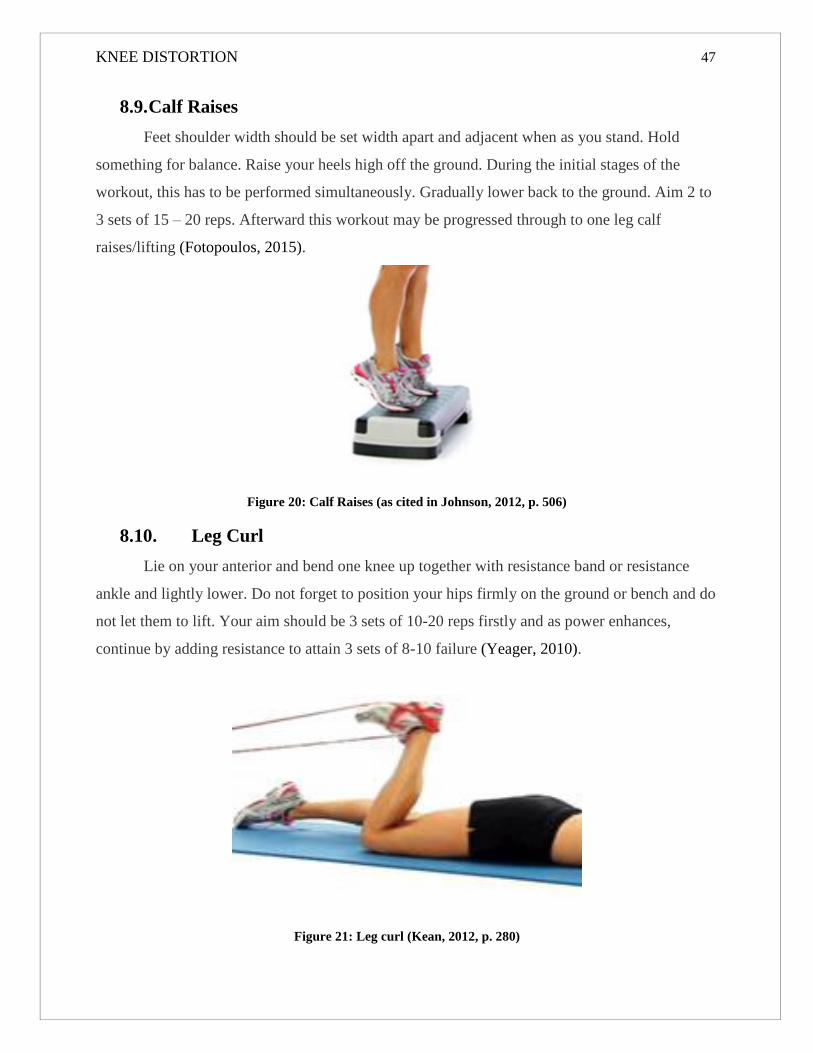
KNEE DISTORTION 47
8.9. Calf Raises
Feet shoulder width should be set width apart and adjacent when as you stand. Hold
something for balance. Raise your heels high off the ground. During the initial stages of the
workout, this has to be performed simultaneously. Gradually lower back to the ground. Aim 2 to
3 sets of 15 – 20 reps. Afterward this workout may be progressed through to one leg calf
raises/lifting (Fotopoulos, 2015).
Figure 20: Calf Raises (as cited in Johnson, 2012, p. 506)
8.10. Leg Curl
Lie on your anterior and bend one knee up together with resistance band or resistance
ankle and lightly lower. Do not forget to position your hips firmly on the ground or bench and do
not let them to lift. Your aim should be 3 sets of 10-20 reps firstly and as power enhances,
continue by adding resistance to attain 3 sets of 8-10 failure (Yeager, 2010).
Figure 21: Leg curl (Kean, 2012, p. 280)

KNEE DISTORTION 48
8.11. Proprioception exercises
These entail balance and co-ordination that is frequently impaired with knee hurt.
Table 4: Proprioception exercises (Pedowitz et al., 2013, p. 158)
Proprioception exercises
sets Repetitions Time in seconds per set
Balance board exercise 4 6 60 sec/se
8.12. Balance board exercise
Wobble board is most largely used in recovery of ankle damages like ankle sprains, even
though they ought to be utilized for lower led and knees injuries (Wilkinson, (2014).
8.13. Plan for sprain exercise
As soon as probable, and for 72hrs, injuries, apply the RICE method
Table 5: Plan for sprain exercise (Pedowitz et al., 2013, p. 191)
RICE METHOD within 72 hours
Rest Take it easy and only move within your limit of pain
Ice Immediately, and for 20 min every 2 hours, place ice or a frozen
cream pack draped in a damp towel. This aids to stop pain, bleeding
and diminishes secondary tissue harm.
Compression Steadily bandage the knees and add 5 cm below and above the joint to
control swelling.
Elevation raise your leg above the level of your heart to diminish inflammation

KNEE DISTORTION 49
Figure 22: Balance board exercise (as cited in Johnson, 2012, p. 753)
8.14. Functional Exercises
They tend to be complex sports particular workouts which close the cavity between
standard restoration and reverting to complete sports playing and training. Specific exercises are
reliance on the kind of sport take and you must look for professional guidance from a
physiotherapist (Wilkinson, 2014).
Table 6: Functional Exercises (Moreno, 2012, p. 215)
Functional Exercises
sets Repetitions Time in seconds per set
Plyometric exercise 4 8 120 sec/se
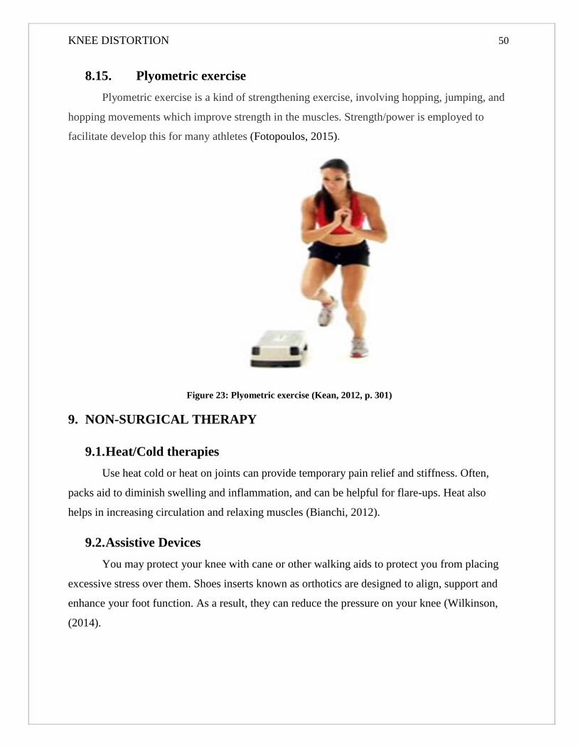
KNEE DISTORTION 50
8.15. Plyometric exercise
Plyometric exercise is a kind of strengthening exercise, involving hopping, jumping, and
hopping movements which improve strength in the muscles. Strength/power is employed to
facilitate develop this for many athletes (Fotopoulos, 2015).
Figure 23: Plyometric exercise (Kean, 2012, p. 301)
9. NON-SURGICAL THERAPY
9.1. Heat/Cold therapies
Use heat cold or heat on joints can provide temporary pain relief and stiffness. Often,
packs aid to diminish swelling and inflammation, and can be helpful for flare-ups. Heat also
helps in increasing circulation and relaxing muscles (Bianchi, 2012).
9.2. Assistive Devices
You may protect your knee with cane or other walking aids to protect you from placing
excessive stress over them. Shoes inserts known as orthotics are designed to align, support and
enhance your foot function. As a result, they can reduce the pressure on your knee (Wilkinson,
(2014).

KNEE DISTORTION 51
9.3. Balancing
Diverse forms of braces can aid you lessen knee pain and boost mobility and function. A
“support” braces resist the overall load on your knee, whereas “unloader” withstand the weight
sideway, in case one side on the knees got injured (Fotopoulos, 2015).
9.4. Avoidance
Specific activities to shun comprise impact-loading sports like downhill skiing, jogging,
high-impact aerobics; excessive stair climbing; physical activities engaging impact-stresses or
twisting, quick stop-start motion; excessive kneeling and betting; pushing or lifting heavy object;
sitting on low chairs and surfaces. You can also try resting for some time (Yeager, 2010).
9.5. Mental heath
Conversing about how you feel with friends and family members, performing mental
exercises like staying positive and medication, and joining support groups can help you manage
knee pain.
9.6. Injections
At times hyaluronic acid, a product produced in natural, healthy joints, can be utilized to
give ease or relief knee pains. Anesthetics also can be injected suing a corticosteroid anti-
inflammatory prescription to distressed knee pain at bay (Fotopoulos, 2015).
9.7. Technical Hints
The posteromedial secure incision protects the neurovascular anatomies, confirms precise
tibial tunnel positioning and permits more expeditious accomplishment of surgical operation.
The single-incision ACL reconstruction method inhibits lateral convex congestion and eradicates
multiple through-and-through drill openings in the distal femur that diminishes stress riser
impact. It is vital to know the –tibial directions and to maintain 1 cm bridge between ACL and
PCL tibial tunnels. This will minimize the likelihood of fracture. It is advantageous to utilize
back-up and primary fixation. Back-up fixation is attained with spiked ligament washer and a
screw while primary fixation is with resorbable interference screws.

KNEE DISTORTION 52
Figure 24: Anteroposterior and lateral radiographs once combined ACL/PCL are reconstructed (Manske,
2015, p. 666)
The sequence of tensioning includes the PCL, posterior lateral corner, the ACL, and the
MCL respectively. Reinstatement of the normal tibia step-off in 70o of flexion provides the
greatest reproducible technique of setting the neutral site of the tibia-femoral correlation.
10. POSTOPERATIVE REHABILITATION
The knee is kept in full extension for 3-weeks non-weight direction. Advanced range of
movement occurs in week 4 through 6. Advanced weight direction takes place at the ending of
week 6. Advanced closed-chain kinetic strength exercises continual motion training is done. The
brace is suspended after the 10th week. Return to heavy labor and sports happen following 9
months when adequate strength and range movement has resumed (Trippel, 2014).
10.1. Complications
Possible complications in treatment of injured knee consist of failure to identify and treat
vascular injuries (both venous and arterial), iatrogenic neurovascular damage during repair,
iatrogenic tibial plateau fractures during reconstruction, failure to identify and treat complexes of
the instability, knee motion loss, postoperative anterior knee pain, and postoperative medial
femoral condyle osteonecrosis.

KNEE DISTORTION 53
11. DATA ANALYSIS
Descriptive statistics, comprising means and standard deviation for continual variables
and frequency averages for ordinary and nominal level variables were computed. Individual tests
were conducted for continual variables, and chi-square tests were performed for many and
nominal level variables to identify the importance of variances between patients treated with
acute replacement and those treated with chronic replacement (Fotopoulos, 2015).
12. RESULTS
About 31 of 33 patients were availed for examination at a mean of 44-months after the
operation. The patients' mean age at surgery was about 28.4 years (range, 16-50 years). 17-
patients were injured during sports sessions, 4 in a car accident, 4 in a motorcycle accident, 4 in
work-associated accidents, and 2 in a fall. Also, 19 patients went through surgery less than 3-
weeks following injury. 12 patients went through surgery more than 3-weeks after injury.
Injury ranges were variable and were recognized by magnetic resonance imaging, a
checkup with arthroscopy and the patient under anesthesia. 15 of the 18 patient suffering acute
injury underwent reconstruction of both the posterior and anterior cruciate ligament with fresh-
frozen allograft. One patient experienced a grade- 2+ wound of the anterior cruciate ligament,
which was not replaced, and 2-patients had a grade- 2+ wound of the posterior cruciate ligament,
which was not replaced. The remaining cruciate ligament was restored in all 3-patients. One
patient encountered a peel-off wound of the posterior cruciate ligaments which was reconstructed
primarily plus a wound of the anterior cruciate ligament that was replaced with an allograft
(Pedowitz et al., 2013).
Of the 15-patients who were treated, 10 had collective injuries of posterior cruciate,
anterior cruciate and medial collateral without lateral injury. 8 of those 10 patients had a medial
ligament replacement of grade- 3+ damages and 2 did not go through replacement of a grade- 2+
injury (Simonian & Bach, 2011). 7 of the 19 patients had damages of the posterior cruciate
ligament, anterior cruciate ligament, and posterolateral corner. 5 of the 7 went through lateral
collateral ligament allograft replacement with the reconstruction of posterolateral corner
portions. 1 patient had a reconstruction of an avulsion that incorporated the lateral collateral
ligaments, and the biceps femoris anchoring onto the fibular head. 1 patient had an intact

KNEE DISTORTION 54
posterolateral corner and lateral collateral ligament, though the iliotibial band and anterolateral
capsule were avulsed and repaired. The rest of the 2 acutely treated patients were found to have a
low-energy knee distortion. 1 of these patients suffered graded- 3+ injuries of the posterior and
anterior cruciate ligament but only grade- 1+ damages of lateral collateral and medial collateral
ligaments. The remaining patient had the grade- 3+ injuries of the posterior and anterior cruciate
ligament and medial collateral ligaments with grade- 1+ injuries of the posterolateral corner
which did not warrant surgical intervention (Wilkinson, (2014).
The laxity structures in patients going through delayed procedure were determined with
intraoperative physical evaluation at the period of ligament reconstruction. 11 of the 12 patients
having chronic injury went through allograft replacement of the posterior and anterior cruciate
ligaments (Sick et al., 2012). One 17-year-old patient submitted to primary repair of an avulsion
damage of the anterior cruciate ligament and a fresh allograft reinstatement of the posterior
cruciate ligaments 1 month following injury. 5 patients who were subjected to chronic treatment
encountered injuries of medial collateral ligaments, anterior cruciate, and posterior cruciate. 3
out of 3 patients suffered medial collateral ligaments grade- 3+ laxities necessitating
reconstruction. 2 out of the 5 experienced grade- 2+ laxities of the posterior and anterior cruciate
ligaments, and medial collateral ligaments were replaced with no medial collateral ligaments
repair. 2 patients who underwent chronic procedure had grade- 3+ injuries of posterior and
anterior cruciate ligaments and laxity of posterolateral corner (Pedowitz et al., 2013).
13. DISCUSSION
It was found that allograft reconstruction presents a good operational result in most
patients. Subjective functional results were tolerable for patients who went through the ligament
surgery in the first 3-weeks following injury. The grades on all 3 rating scales applied in
examination demonstrate this pattern. Many patients suffered only functional limitations and
slight symptoms during activities of day-to-day living. Patients who were placed on surgery in
the first 3-weeks following operation tended to have higher subjective grades. Nevertheless, only
the variance in the score on sports actions of the outcome of knee review attained significance (p
= 0.04) (Johnson, 2012).
Objectively, all the chronically and acutely treated patients received a positive range of
movement. On evaluation, most knees were stable. Enduring laxity of the posterior cruciate

KNEE DISTORTION 55
ligaments was very prevalent in chronically treated patients. As well, residual valgus laxity was
more common in patients who went through delayed medial collateral ligament reconstruction.
These results are in line with those other studies concerning the treatment of dislocated knee
(Bianchi, 2012).
Clinically, nevertheless, most patients did not show instability unless they tried strenuous
sports or manual labor activities warranting aggressing shifts in pivoting or direction.
14. CONCLUSION
Knee distortions are serious injuries as they can result in disruption of numerous
ligaments, neurovascular structures, and surrounding musculature. Acute treatment and diagnosis
may be difficult, and the changing techniques that are employed to reconstruct the PCLs and
anterior structures may be controversial. These injuries, as a result of surrounding tissue damage
and ligamentous disruption, can cause biomechanical hindrances of the knees intersections
before or following reconstruction efforts made.
To avoid abnormal angulations and translations in the reconstructed knees, surgeons in
charge of repairs in patients having multiple ligament damages must have a complete knowledge
of biomechanics and normal anatomy of the entire knee plus the anterior and PCLs. This
understanding ought to help optimize the order of ligament reconstructions, the timing of the
surgery, the rehabilitation of the neighboring musculature, and the anatomic placement of grafts.

KNEE DISTORTION 56
15. CASE STUDY
15.1. Report on Clinical Work
Done by: Osama Aljeheny Year: 3rd year Workplace: C.L.P.A
Supervisor: PhDr.Ivana Vláčilová Ph.D., Mgr.Zahir Elali
Year of birth: 1967
Examined person: J.K.
16. DIAGNOSIS
Distortion of the right knee joint
17. SUBJECTIVE FEELING OF THE PATIENT
The patient feels slight pain just some times when he tries to perform a full range of
motion in the right knee joint in the direction of maximum flexion and extension. The patient
feels bad about the weakness he has in his thigh muscles and he feels also decreased knee joint
stability.
18. STATUS PRESENS
Height: 180cm Weight: 85kg BMI: 26.2 Blood pressure: 119 over 80 Heart rate: 70
He felt good and no pain when he walked into the room but later the patient had some pain
during the therapy session during exercising of isometric training but there was not any special
movement that can provoke the pain. The patient was in the 1st day walking without the knee
orthosis so he didn’t feel stable with walking. The patient used the orthosis for one month.
KNEE DISTORTION 57
19. HISTORY OF PROBLEM
The injury happened on 15.12.2015 when the patient was running outdoor. As the patient
described while running his right knee was rotated inward suddenly then he lost balance and fell
down on the ground with sharp pain in the knee.
Medications: Insulin injection for diabetic patients
Excerpt from patient’s health care file: NO
20. SOCIAL ANAMNESIS
He is living in the 3rd floor in house in Prague with elevator.
21. ANAMNESIS
Family History: No special diseases related to the patient injury.
Operations: NO
Injuries: NO
Allergies: Soap
Abuses: Social drinker
Hobbies: Tennis, jogging, football and cycling sometimes
Occupation: Psychiatrist at the state police (sitting most of the time at work almost 5 hours)
Prior rehabilitation: NO. The patient used knee brace for one month after this injury.

KNEE DISTORTION 58
22. RHB INDICATIONS
- Soft tissue techniques for skin and fascia
- Exercises for limited Range of motion (ROM) of knee joint
- Mobilization for restricted joints of the lower extremity
- PNF technique
- Relaxing hypertonic muscles using Post isometric relaxation (PIR) technique
- Sensomotoric training to improve proprioception level
- Stretching of shorted muscles
- Strengthening and stimulation of weak muscles of lower extremity
- Correct posture and gait
23. INITIAL EXAMINATIONS
23.1. Postural examination (by Kendal)
Posterior view:
- Both lower extremities are in slight internal rotation
- Right knee joint is in semi-flexed position
- Whole spine is in mid line
- Both scapulas are slightly abducted.
- Both upper extremities are in slightly external rotation
- Left shoulder is slightly higher than the right shoulder
Lateral view:
- More loading on heels
- Right knee joint is in semi-flexed position

KNEE DISTORTION 59
- Normal curves of whole spine
- Both upper extremities are in slight external rotation
Anterior view:
- Both lower extremities are in slight external rotation
- Right knee is in semi-flexed position
- The navel is at mid line
- Left shoulder is slightly higher than the right shoulder
23.2. Gait examination (by Kendal)
Patient is on the 1st day after using the knee brace (orthosis) which was supporting
the right knee.
- Whole body limping to the left side
- He walks more on the lateral aspects of the foot
- There is not full extension in the right knee
- Pain in the 1st metatarsal bone during walking in the right foot
- More loading on the left side
- The patient’s gait is symmetrical
23.3. Modification of gait examination:
Walking on tip toes: Patient is able to provide but there is not full extension of his right
knee joint.
Walking with squats: Patient is able to provide it. But he felt not so comfortable due to some
pain in the right knee.
23.4. Soft tissue examination (by Lewit)
- Skin and connective tissue of lower extremity (shin and calf) is restricted in both directions
(medial and lateral) in the right leg.

KNEE DISTORTION 60
- Deep facia of the thigh is restricted in both directions (medial and lateral) around the axis of the
lower extremity in both legs.
23.5. Pelvis examination (by Kendal)
The position of the pelvic is in slight anterior tilt.
23.6. Special tests
2 scales test: R 43 L 52
Trandelburg’s test: The patient was able to perform the test on left side but on the right side the
patient was slightly shaking and the pelvic drooped (which indicates weakness in the right hip
abductors) and the stability was worse too. So the test was positive in the right side.
Romberg test:
I negative
II negative
III negative
Special tests for stability of knee joint
Anterior drawer test: Negative
Posterior drawer test: Negative
Lachman test: Negative

KNEE DISTORTION 61
Anthropometry examination (by Kendal)
Table 7: Anthropometry examination (by Kendal)
Measurement Right lower extremity Left lower extremity
Anatomical length 89 cm 89 cm
Functional length 92 cm 92cm
Length of the thigh 49 cm 49 cm
Length of the middle leg 40 cm 40 cm
Length of the foot 26 cm 26 cm
Circumference of thigh Quadriceps 45 cm 46 cm
Vastus Medialis 40 cm 42 cm
Circumference of knee joint 39 cm 37 cm
Circumference of the calf 36 cm 36 cm
Circumference of ankle joint 27 cm 27 cm
Circumference of the foot 25 cm 25 cm
23.7. Palpation examination (by Kendal)
Muscles:
Right lower extremity:
M. Quadriceps (m. rectus femoris): hypotonic
M. Tensor fasciae latae: hypertonic
M. Adductors (m. adductor longus, magnus, brevis): normal tonus
M. Hamstrings: hypertonic
M. Iliopsoas: normal tonus
M. Gastrocnemius: normal tonus
M. Gluteus maximus: normal tonus

KNEE DISTORTION 62
M. Piriformis: hypertonic
Left lower extremity:
M. Quadriceps (m. rectus femoris): normal tonus
M. Tensor fasciae latae: hypertonic
M. Adductors (m. adductor longus, magnus, brevis): normal tonus
M. Hamstrings: normal tonus
M. Iliopsoas: normal tonus
M. Gastrocnemius: normal tonus
M. Gluteus maximus: normal tonus
M. Piriformis: normal tonus
23.8. Range of motion examination (by Kendal)
Knee joint:
Table 8: Range of motion examination - Knee joint
Right side Left side
Motion Active
movement
(degrees)
Passive
movement
(degrees)
Active
movement
(degrees)
Passive
movement
(degrees)
Extension -10° -5° 0° 0°
Flexion 110° 125° 130° 135°

KNEE DISTORTION 63
Ankle joint:
Table 9: Ankle joint
Right side Left side
Motion Active
movement
(degrees)
Passive
movement
(degrees)
Active
movement
(degrees)
Passive
movement
(degrees)
Plantar Flexion 40° 45° 45° 45°
Dorsiflexion 15° 20° 15° 20°
Hip joint:
Table 10: Hip joint
Right side Left side
Motion Active
movement
(degrees)
Passive
movement
(degrees)
Active
movement
(degrees)
Passive
movement
(degrees)
Extension 10° 15° 10° 15°
Flexion 100° 110° 110° 120°
Abduction 40° 45° 40° 45°
Adduction 10° 15° 10° 15°
External rotation 40° 40° 40° 40°
Internal rotation 35° 40° 40° 40°

KNEE DISTORTION 64
23.9. Muscle strength examination (by Kendal)
Table 11: Muscle strength examination (by Kendal)
Muscle Right lower extremity Left lower extremity
Quadriceps 4- 5
Hamstrings 4- 5
Adductors 5 5
Tensor fasciae latae 4 5
Gastrocnemius 4+ 5
Iliopsoas 5 5
Gluteus maximus 4 4
Gluteuse medius -4 4
23.10. Muscle length examination (by Janda)
Table 12: Muscle length examination (by Janda)
Muscle Right lower extremity Left lower extremity
Quadriceps 0 0
Hamstrings 2 2
Adductors 0 0
Tensor fasciae latae 0 0
Gastrocnemius 0 0
Iliopsoas 0 0
23.11. Neurological examination (by Lewit)
Deep sensation test: Sterognosis: it was felt by hands and feet. Graphesthesia: it was
applied on both hands and both lower extremities. Both are Negative position sense: it was
applied of both lower extremities it was negative in all joints except of the right knee joint the
sense was slightly decreased.

KNEE DISTORTION 65
Rhomberg test: I,II,III are Negative
Superficial sensation test: light touch: it was applied on both lower extremities on the inner and
outer aspects. The test is Negative
Examination of deep tendon reflexes (by Lewit):
Patellar tendon: 2 normal reflex
Achilles tendon: 2 normal reflex
23.12. Joint Play Examination (by Lewit)
Table 13: Joint Play Examination (by Lewit)
Joint Left side Right side
Sacroiliac No restriction in all
directions
No restriction in all
directions
Patella No restriction in all
directions
Restricted in the lateral
direction
Head of fibula No restriction in all
directions
No restriction in all
directions
Talocrural No restriction No restriction
Subtalar No restriction No restriction
Chopar’t No restriction Restricted cranial direction
Lisfranc’s No restriction Restricted cranial direction
Metatarsophalangeal No restriction in all
directions
No restriction in all
directions
Interphalangeal No restriction in all
directions
No restriction in all
directions
KNEE DISTORTION 66
24. EXAMINATIONS CONCLUSION
From the case of the patient we can see that the patient has pain sometimes when he
reaches his maximum ROM of his injured knee, bad stabilization of the knee joint due to
decreased level of proprioception in the joint after the injury. Due to the position of the right
lower extremity (semi flexed knee) and the restricted joins Chopar’t and Lisfranc’s the patient
has a wrong contact with the foot and the ground which results in a fault gait pattern. One more
major problem for the patient at this time there is the swelling around the right knee joint and the
restriction of the deep fascia at this area too. The patient has no sensation problems in superficial
and deep except of the position sense in the right knee joint. As a result of his injury now the
patient suffers from weakness of muscles of right lower extremity (quadriceps, hamstrings and
gastrocnemius) with shortness of both hamstrings and some hypertonic muscles (Tensor fasciae
latae, Hamstrings).
25. SHORT-TERM PLAN
- Reduce pain (applying hot rolls and PIR)
- Relax hyper tonic muscles (using PIR technique)
- Increase ROM in limited joints (using passive and active ROM training)
- Stretch shorted muscles (using PIR with stretching and stretching techniques)
- Mobilization for restricted joints
- Activation of hypotonic muscles
- Strengthening of weak muscles
- Correct faulty gait with instructions
26. LONG-TERM PLAN
- Keep strengthening exercises for weak muscles
- Improve stability of the knee

KNEE DISTORTION 67
- Stretching exercises for shortened muscles
- Relaxing hypertonic muscles
- ADL training
- Improve gait pattern
- Advices to improve posture daily activities
27. PHYSICAL THERAPY TOOLS
- Kinesio-tape for decreasing the swelling and improving stability
- Laser therapy: biostimulation efect for 14 minutes, power 3 W, area 25 cm2, total energy 2500
J.
28. PHYSICAL THERAPY SESSIONS
Day to day therapy
Date 20.1.2016
Status praesens subjective: the patient feels weakness and limitation in the range of motion and
some pain during maximum range of motion.
Status praesens objective: the biggest problems for the patient now is the limited range of
motion in the right knee, the swelling and the weakness of the mentioned muscles above. And for
sure we still need to work on the gait to improve it and the restricted joints and fascia.
Goal of today’s therapy unit
The goal of therapy is to increase range of motion of the right knee joint. Strengthening of right
knee extensors and flexors and hip joint abductors. Mobilization of the fascia of the thighs
(around the axis of the leg). Mobilization of patella and Chopar’t Lisfranc’s joints.
Procedure
For mobilization of patella and Chopar’t and Lisfranc’s joints in cranial direction.
While patient on bed supine with legs extended, we do mobilization of patella which was
restricted in lateral direction in right knee, by moving patella by therapist hand to the restriction

KNEE DISTORTION 68
direction and hold it until release.
For mobilization of Chopar’t and Lisfranc’s joints, when patient is supine with slightly flexed
knees, we reach the barrier in the cranial direction then we apply repetitive movements in the
same direction till we feel releas.
PIR techniques for hypertonic muscles according to lewit M. Tensor fasciae latae,M.
Hamstrings, M. Iliopsoas and M. Piriformis.
for mobilization of deep fascia of thigh , both side, while patient is in bed, with flexed knees, we
move whole thigh by whole hand (around the axis of leg) to one direction which is restricted and
wait for release
For increase ROM of knee joints
Patient supine, putting small ball under heel and roll it by doing flexion and extension of knee.
For strengthening knee extensors muscles
at the bed, post isometric contraction, when patient is supine, putting small ball under knee, ask
patient to lift foot up (extend knee) and hold for few seconds and then relax
At the gym
Walk on unstable surfaces, and holding small ball and pass it around the body
Standing on bosu, with holding small ball and throw it from hand to hand
Lifting weight
For abductors patient is side lying, weight on leg above ankle, and doing abduction
For hamstring patient is prone, weight on leg above ankle and doing flexion of knee

KNEE DISTORTION 69
At the end of training patient do cycling for 10 min with slight resistance.
Laser therapy: biostimulation efect for 14 minutes, power 3 W, area 25 cm2, total energy 2500 J.
Results
Patient felt good, doing exercises in good form as he already learned from physiotherapist, he
had good stability and strength of lower extremities muscles.
Self-therapy
The patient is out-patient, and does not have therapy every day at hospital, but we taught him
ROM exercises at bed by rolling small ball under heel while doing flexion and extension of knee
and hip, we instruct her also to do isometric contraction of quadriceps by putting small ball under
knee and extend the knee. He can also exercise at the home same exercises we have done.
Date 22.1.2016
Status praesens subjective: the patient still feels weakness and limitation in the range of motion
and some pain during maximum range of motion.
Status praesens objective: the biggest problems for the patient now still the limited range of
motion in the right knee, the swelling and the weakness of the mentioned muscles above. And for
sure we still need to work on the gait to improve it and the restricted joints and fascia.
Goal of today’s therapy unit
We checked the muscles tone, joint play of patella and Chopar’t and Lisfranc’s joints, ROM of
knee joints. Mobilization for right patella which is restricted in lateral direction, mobilization of
deep fascia of thighs, lymph drainage, gait training, strengthening exercises of weak muscles and
general relaxation of muscles and soft tissues.
Procedure
For mobilization of patella and Chopar’t and Lisfranc’s joints.
While patient on bed supine with legs extended, we do mobilization of patella which was
restricted in lateral direction in right knee, by moving patella by therapist hand to the restriction
direction and hold it until release.
KNEE DISTORTION 70
For mobilization of Chopar’t and Lisfranc’s joints, when patient is supine with slightly flexed
knees, we reach the barrier in the cranial direction then we apply repetitive movements in the
same direction till we feel releas.
PIR techniques for hypertonic muscles according to lewit M. Tensor fasciae latae,M.
Hamstrings, M. Iliopsoas and M. Piriformis
For mobilization of deep fascia of thigh , both side, while patient is in bed, with flexed knees,
we move whole thigh by whole hand (around the axis of leg) to one direction which is restricted
and wait for release
For increase ROM of knee joints
Patient supine, putting small ball under heel and roll it by doing flexion and extension of knee.
For gait training
Forward walking, with slow walk, and semi flexed knees, contact heel sole tiptoes
Backward walking with slow walk, contact toes sole heels
Side to side walk
Tiptoes walking, contact on tiptoes
Heel walking, contact on heel
For strengthening knee extensors muscles
at the bed, post isometric contraction, when patient is supine, putting small ball under knee, ask
patient to lift foot up (extend knee) and hold for few seconds and then relax
Adductors
Putting small ball between knees and squeezing it, while patient supine with flexed knees
KNEE DISTORTION 71
Standing on bosu, and throw small ball from hand to hand, while patient is standing with semi
flexed knees and hips, and slight external rotation of hip
Knee and hip extensors, bridging
By putting medicine ball under calves and heels, arms at sides, lifting pelvis upward, and hold
for few seconds then relax
Abductors, lifting weight
2 kg weight on leg above ankle, patient side lying, doing abduction
Hamstring, lifting weight while patient is prone, 2 kg weight on leg above ankle, doing knee
flexion
For lymph drainage, after exercising
Lymph therapy (massage), duration 25 min, pressure 9.5 kpa, program 37
Cycling, for 5 min with slight resistance
Stretching exercises for quadriceps and hamstring
For quadriceps, patient prone, flexed knees, belt around foot and heel, and in patient hand,
withdraw foot in direction of flexion of knee and hold it for few seconds then relax
For hamstring, patient is supine, with extended knees, belt around foot and heel, and and patient
is holding it by her hand, withdraw the belt toward trunk, while doing flexion of hip and dorsal
flexion of foot, hold the belt for few seconds and then relax.
All exercises 3 times 10 repetitions.
Laser therapy: biostimulation efect for 14 minutes, power 3 W, area 25 cm2, total energy 2500 J.
Results
Patient did not have pain or difficulties in exercising, he is doing exercises well, and there is
improvement in ROM of right knee, moveable patella and better mobility of Chopar’t and
Lisfranc’s joints, improvement in muscle strength of weak muscles.

KNEE DISTORTION 72
Self-therapy
Patient can do the same self-therapies was instructed before, he knows it well and can perform it
in good form.
Date 25.1.2016
Status praesens subjective: the patient still feels weakness and limitation in the range of motion
and some pain during maximum range of motion.
Status praesens objective: the biggest problems for the patient now still the limited range of
motion in the right knee, the swelling and the weakness of the mentioned muscles above. And for
sure we still need to work on the gait to improve it and the restricted joints and fascia.
Goal of today’s therapy
We do evaluation checking for muscle tone of affected muscles, soft tissues of thigh, joint play
for restricted joints and ROM of knees. The goal of therapy is increase ROM of the right knee,
strengthening of weak muscles (knee extensors) , lymph drainage, gait training, stretching
shorten muscles (hamstring)
Procedure
Lymph massage therapy
Lymph therapy (massage), duration 25 min, pressure 11 kpa, program 37
For gait training
Forward walking, with slow walk, and semi flexed knees, contact heel sole tiptoes
Backward walking with slow walk, contact toes sole heels
Side to side walk
Tiptoes walking, contact on tiptoes
Heel walking, contact on heel
PIR techniques for hypertonic muscles according to lewit M. Tensor fasciae latae,M.
Hamstrings, M. Iliopsoas and M. Piriformis.
For strengthening exercises

KNEE DISTORTION 73
Standing on bosu, and throw small ball from hand to hand, while patient is standing with semi
flexed knees and hips, and slight external rotation of hip
Knee and hip extensors, bridging
By putting medicine ball under calves and heels, arms at sides, lifting pelvis upward, and hold
for few seconds then relax
Cycling, for 10 min with slight resistance
Stretching exercises for quadriceps and hamstring
For quadriceps, patient prone, flexed knees, belt around foot and heel, and in patient hand,
withdraw foot in direction of flexion of knee and hold it for few seconds then relax
for hamstring, patient is supine, with extended knees, belt around foot and heel, and and patient
is holding it by his hand, withdraw the belt toward trunk, while doing flexion of hip and dorsal
flexion of foot, hold the belt for few seconds and then relax
Laser therapy: biostimulation efect for 14 minutes, power 3 W, area 25 cm2, total energy 2500 J.
All exercises 3 times 10 repetitions
Results
Patient is feeling good, no pain, no complication, he does exercises well, there is improvement in
ROM in knees (F, E), free and moveable patella and Chopar’t and Lisfranc’s joints.
Self-therapy
We instruct patient to continue self-therapy he was instructed before.
Date 26.1.2016
Status praesens subjective: the patient still feels weakness and limitation in the range of motion
and some pain during maximum range of motion.
Status praesens objective: the biggest problems for the patient now still the limited range of
motion in the right knee, the swelling and the weakness of the mentioned muscles above. And for

KNEE DISTORTION 74
sure we still need to work on the gait to improve it and the restricted joints and fascia.
Goal of today’s therapy
We do evaluation checking for muscle tone, soft tissues (fascia), joint play of patella and head of
fibula, ROM knees and hips. The goal of therapy is increase ROM of right knee, strengthening of
weak muscles (knee extensors) and hip abductors, gait training, stretching shorten muscles
(hamstring).
Procedure
For gait training
Forward walking, with slow walk, and semi flexed knees, contact heel sole tiptoes
Backward walking with slow walk, contact toes sole heels
Side to side walk
Tiptoes walking, contact on tiptoes
Heel walking, contact on heel
Walking on treadmill, for 5 min
Walking on unstable surfaces, holding small ball and throw it from hand to hand
PIR techniques for hypertonic muscles according to lewit M. Tensor fasciae latae,M.
Hamstrings, M. Iliopsoas and M. Piriformis.
For strengthening exercises
Standing on bosu, and throw small ball from hand to hand, while patient is standing with semi
flexed knees and hips, and slight external rotation of hip
Knee and hip extensors, bridging
By putting medicine ball under calves and heels, arms at sides, lifting pelvis upward, and hold
for few seconds then relax
Hip abductors
By placing a weight around the ankle in side laying position and perform abduction in the hip
joint.
Cycling, for 10 min with slight resistance

KNEE DISTORTION 75
Stretching exercises for quadriceps and hamstring
For quadriceps, patient prone, flexed knees, belt around foot and heel, and in patient hand,
withdraw foot in direction of flexion of knee and hold it for few seconds then relax
For hamstring, patient is supine, with extended knees, belt around foot and heel, and patient is
holding it by his hand, withdraw the belt toward trunk, while doing flexion of hip and dorsal
flexion of foot, hold the belt for few seconds and then relax.
Laser therapy: biostimulation efect for 14 minutes, power 3 W, area 25 cm2, total energy 2500 J.
All exercises 3 times 10 repetitions.
Results
Patient felt fine, no pain or fatigue, doing exercises well, there is improvement in ROM of knee
(F, E ) free patella and Chopar’t and Lisfranc’s joints. , free deep fascia of thighs both side.
Self-therapy
Patient continue the same self-therapy was instructed.
Date 27.1.2016
Status praesens subjective: the patient feels better and more confident and stronger with no pain.
Status praesens objective: the paiten is so much better by this time but we still need to work on
the same things as before to reach the optimal condition. (Still the limited range of motion in the
right knee, the swelling and the weakness of the mentioned muscles above. And for sure we still
need to work on the gait to improve it and the restricted joints and fascia).
Goal of today’s therapy
We do evaluation checking for muscle tone of affected muscles, soft tissues of thigh, joint play
and ROM of right knee. The goal of therapy is increase ROM of right knee, strengthening of
weak muscles (knee extensors) and hip abductors and hamstrings, lymph drainage, gait training,
stretching shorten muscles (hamstring).
KNEE DISTORTION 76
Procedure
Lymph massage therapy
Lymph therapy (massage), duration 25 min, pressure 12.5 kpa, program 37
For gait training
Forward walking, with slow walk, and semi flexed knees, contact heel sole tiptoes
Backward walking with slow walk, contact toes sole heels
Side to side walk
Tiptoes walking, contact on tiptoes
Heel walking, contact on heel
PIR techniques for hypertonic muscles according to lewit M. Tensor fasciae latae,M.
Hamstrings, M. Iliopsoas and M. Piriformis.
For strengthening exercises
Standing on bosu, and throw small ball from hand to hand, while patient is standing with semi
flexed knees and hips, and slight external rotation of hip
Knee and hip extensors, bridging
By putting medicine ball under calves and heels, arms at sides, lifting pelvis upward, and hold
for few seconds then relax
Hip abductors
By placing a weight around the ankle in side laying position and perform abduction in the hip
joint.
Jumping on trampoline, for few minutes
Cycling, for 10 min start with resistance

KNEE DISTORTION 77
Stretching exercises for quadriceps and hamstring
For quadriceps, patient prone, flexed knees, belt around foot and heel, and in patient hand,
withdraw foot in direction of flexion of knee and hold it for few seconds then relax
For hamstring, patient is supine, with extended knees, belt around foot and heel, and patient is
holding it by his hand, withdraw the belt toward trunk, while doing flexion of hip and dorsal
flexion of foot, hold the belt for few seconds and then relax
Laser therapy: biostimulation efect for 14 minutes, power 3 W, area 25 cm2, total energy 2500 J.
All exercises 3 times 10 repetitions. Patient is going to gym twice a day doing the same
exercises.
Results
Patient felt perfect, no pain or fatigue, doing exercises well, there is improvement in ROM of
knee (F, E), free patella and Chopar’t and Lisfranc’s joints, free deep fascia of thighs both side,
muscle strength is improvement.
Self-therapy
Patient can continue the same exercises, and can add jumping on trampoline.
Date 29.1.2016
Status praesens subjective: the patient feels better and more confident and stronger with no pain.
Status praesens objective: the patient is so much better by this time but we still need to work on
the same things as before to reach the optimal condition. (Still the limited range of motion in the
right knee, the swelling and the weakness of the mentioned muscles above. And for sure we still
need to work on the gait to improve it and the restricted joints and fascia).
Goal of today’s therapy
We do evaluation checking for muscle tone of affected muscles, soft tissues of thigh, joint play
KNEE DISTORTION 78
and ROM of knees. The goal of therapy is increase ROM of right knees, strengthening of weak
muscles (knee extensors, hip extensors) and hamstrings and hip abductors, lymph drainage, gait
training, stretching shorten muscles (hamstring).
Procedure
For gait training
Forward walking, with slow walk, and semi flexed knees, contact heel sole tiptoes
Backward walking with slow walk, contact toes sole heels
Side to side walk
Tiptoes walking, contact on tiptoes
Heel walking, contact on heel
PIR techniques for hypertonic muscles according to lewit M. Tensor fasciae latae,M.
Hamstrings, M. Iliopsoas and M. Piriformis.
For strengthening exercises
Standing on bosu, and throw small ball from hand to hand, while patient is standing with semi
flexed knees and hips, and slight external rotation of hip.
Hip abductors
By placing a weight around the ankle in side laying position and perform abduction in the hip
joint.
Knee and hip extensors, bridging
By putting medicine ball under calves and heels, arms at sides, lifting pelvis upward, hold for
few Seconds then relax.
First time to do extension of knee with weight (machine), 1.5 kg (3 times 8 repetitions)
First time to do extension of hip with weight (machine), 1, 5 kg (3 times 8 repetitions)
KNEE DISTORTION 79
Jumping on trampoline, for few minutes
Cycling, for 10 min with resistance
Stretching exercises for quadriceps and hamstring
For quadriceps, patient prone, flexed knees, belt around foot and heel, and in patient hand,
withdraw foot in direction of flexion of knee and hold it for few seconds then relax
for hamstring, patient is supine, with extended knees, belt around foot and heel, and and patient
is holding it by his hand, withdraw the belt toward trunk, while doing flexion of hip and dorsal
flexion of foot, hold the belt for few seconds and then relax
Lymph massage therapy, after exercising
Lymph therapy (massage), duration 25 min, pressure 17 kpa, program 37
Laser therapy: biostimulation efect for 14 minutes, power 3 W, area 25 cm2, total energy 2500 J.
All exercises 3 times 10 repetitions.
Results
We can see notable improvement in ROM of knee (F, E) improvement strength of weak muscles
(quadriceps, hamstring) and hip abductors, moveable patella and Chopar’t and Lisfranc’s joints,
moveable deep fascia of thigh.
Self-therapy
Patient felt great, we instruct him to continue past self-therapy.

KNEE DISTORTION 80
29. FINAL KINESIOLOGIC EXAMINATION
29.1. Postural examination (by Kendal)
Posterior view:
- Both lower extremities are in slight internal rotation
- Right knee joint is in slightly semi-flexed position
- Whole spine is in mid line
- Both scapulas are slightly abducted.
- Both upper extremities are in slightly external rotation
- Left shoulder is slightly higher than the right shoulder
Lateral view:
- More loading on heels
- Right knee joint is in semi-flexed position
- Normal curves of whole spine
- Both upper extremities are in slight external rotation
Anterior view:
- Both lower extremities are in slight external rotation
- Right knee is in slightly semi-flexed position
- The navel is at mid line
- Left shoulder is slightly higher than the right shoulder
Gait examination (by Kendal)
Patient is on the 1st day after using the knee brace (orthosis) which was supporting
the right knee.

KNEE DISTORTION 81
- Whole body slightly limping to the left side
- He walks slightly more on the lateral aspects of the foot
- There is almost full extension in the right knee
- The pain in the 1st metatarsal bone during walking in the right foot is disappeared
- The patient’s gait is symmetrical
29.2. Modification of gait examination:
Walking on tip toes: Patient is able to provide but there is not full extension of his right
knee joint.
Walking with squats: Patient is able to provide it.
29.3. Soft tissue examination (by Lewit):
- Skin and connective tissue of lower extremity (shin and calf) is not restricted in both directions
(medial and lateral) in the right leg.
- Deep facia of the thighs is not restricted in both directions (medial and lateral) around the axis
of the lower extremity in both legs.
29.4. Pelvis examination (by Kendal)
The position of the pelvic is in slight anterior tilt.
Special tests:
2 scales test: R 45 L 50
Trandelburg’s test: The patient was able to perform the test on left and right side with no
problems. So the test was negative in both side.
29.5. Romberg test:
I negative
II negative

KNEE DISTORTION 82
III negative
Special tests for stability of knee joint:
Anterior drawer test: Negative
Posterior drawer test: Negative
Lachman test: Negative
29.6. Anthropometry examination (by Kendal)
Table 14: Anthropometry examination (by Kendal)
Measurement Right lower extremity Left lower extremity
Anatomical length 89 cm 89 cm
Functional length 92 cm 92cm
Length of the thigh 49 cm 49 cm
Length of the middle leg 40 cm 40 cm
Length of the foot 26 cm 26 cm
Circumference of thigh Quadriceps 45 cm 46 cm
Vastus Medialis 40 cm 42 cm
Circumference of knee joint 37 cm 37 cm
Circumference of the calf 36 cm 36 cm
Circumference of ankle joint 27 cm 27 cm
Circumference of the foot 25 cm 25 cm
29.7. Palpation examination (by Kendal)
Muscles:
Right lower extremity:
M. Quadriceps (m. rectus femoris): normal tone

KNEE DISTORTION 83
M. Tensor fasciae latae: normal tone
M. Adductors (m. adductor longus, magnus, brevis): normal tonus
M. Hamstrings: hypertonic
M. Iliopsoas: normal tonus
M. Gastrocnemius: normal tonus
M. Gluteus maximus: normal tonus
M. Piriformis: hypertonic
Left lower extremity:
M. Quadriceps (m. rectus femoris): normal tonus
M. Tensor fasciae latae: normal tone
M. Adductors (m. adductor longus, magnus, brevis): normal tonus
M. Hamstrings: normal tonus
M. Iliopsoas: normal tonus
M. Gastrocnemius: normal tonus
M. Gluteus maximus: normal tonus
M. Piriformis: normal tonus
29.8. Range of motion examination (by Kendal)
Knee joint:
Table 15: Knee joint
Right side Left side
Motion Active
movement
(degrees)
Passive
movement
(degrees)
Active
movement
(degrees)
Passive
movement
(degrees)
Extension -5° 0° 0° 0°
Flexion 125° 130° 130° 135°

KNEE DISTORTION 84
Ankle joint:
Table 16: Ankle joint
Right side Left side
Motion Active
movement
(degrees)
Passive
movement
(degrees)
Active
movement
(degrees)
Passive
movement
(degrees)
Plantar Flexion 40° 45° 45° 45°
Dorsiflexion 15° 20° 15° 20°
Hip joint:
Table 17: Hip joint
Right side Left side
Motion Active
movement
(degrees)
Passive
movement
(degrees)
Active
movement
(degrees)
Passive
movement
(degrees)
Extension 10° 15° 10° 15°
Flexion 100° 110° 110° 120°
Abduction 40° 45° 40° 45°
Adduction 10° 15° 10° 15°
External rotation 40° 40° 40° 40°
Internal rotation 35° 40° 40° 40°

KNEE DISTORTION 85
29.9. Muscle strength examination (by Kendal)
Table 18: Muscle strength examination (by Kendal)
Muscle Right lower extremity Left lower extremity
Quadriceps 5 5
Hamstrings 5 5
Adductors 5 5
Tensor fasciae latae 4+ 5
Gastrocnemius 4+ 5
Iliopsoas 5 5
Gluteus maximus 4 4
Gluteuse medius 4+ 4+
29.10. Muscle length examination (by Janda)
Table 19: Muscle length examination (by Janda)
Muscle Right lower extremity Left lower extremity
Quadriceps 0 0
Hamstrings 1 1
Adductors 0 0
Tensor fasciae latae 0 0
Gastrocnemius 0 0
Iliopsoas 0 0
29.11. Neurological examination (by Lewit)
Deep sensation test: Sterognosis: it was felt by hands and feet. Graphesthesia: it was applied on
both hands and both lower extremities. Both are Negative position sense: it was applied of both

KNEE DISTORTION 86
lower extremities it was negative in all joints.
Rhomberg test: I,II,III are Negative
Superficial sensation test: light touch: it was applied on both lower extremities on the inner and
outer aspects. The test is Negative
Examination of deep tendon reflexes (by Lewit):
Patellar tendon: 2 normal reflex
Achilles tendon: 2 normal reflex
29.12. Joint Play Examination (by Lewit)
Table 20: Joint Play Examination (by Lewit)
Joint Left side Right side
Sacroiliac No restriction in all
directions
No restriction in all
directions
Patella No restriction in all
directions
No restricted in all
directions
Head of fibula No restriction in all
directions
No restriction in all
directions
Talocrural No restriction No restriction
Subtalar No restriction No restriction
Chopar’t No restriction No restricted
Lisfranc’s No restriction No restricted
Metatarsophalangeal No restriction in all
directions
No restriction in all
directions
Interphalangeal No restriction in all
directions
No restriction in all
directions

KNEE DISTORTION 87
30. THERAPY EFFECT EVALUATION, PROGNOSIS
After all the therapy sessions we had with patient. We strongly notice the next, increasing
in range of motion in the right knee joint flexion 125° and extension -5° in the active movement.
But passively we can see its better flexion 130° and extension 0°. The swelling around the knee
cap is much better, now it is 37 cm in circumference. Increasing in strength of quadriceps and
hamstrings on the right leg (grade 5 by Kendall). No more restriction of joint play in patella,
Chopar’t and Lisfranc’s joints. Restriction of connective tissues and fascia on the thighs and legs
is gone. Gait of patient was slightly better as it was described above. Change of loading on both
extremities was improved and now the difference between both legs is 5 kg in the two scales test.
The proprioception on the right knee was improved too. At last the hypertonic muscles were
relaxed after PIR techniques.
The overall condition of the patient is very good and we expect him to be normal soon and this
good results is referred to the good fitness level of the patient before the injury.

KNEE DISTORTION 88
31. REFERENCES
Bianchi, A. B. (2012, June 12). A Sagittal Plane Model of the Knees and Cruciate Ligaments
With Application of a Sensitivity Analysis. Retrieved March 03, 2016, from
http://biomechanical.asmedigitalcollection.asme.org/article.aspx?articleid=1400415
Cantrell, K. B. (2013, September 8). Biomechanical evaluation of augmented and nonaugmented
primary repair. Retrieved March 03, 2016, from
http://link.springer.com/article/10.1007/s00264-013-2098-8/fulltext.html
Chaudhari, A. M. (2013). Development and application of biomechanical methods to study non-
contact injuries of the anterior cruciate ligament.
Pedowitz, R. A., & Akeson, W. H. (2013). Daniel's knee injuries: Ligament and cartilage:
Structure, function, injury, and repair. Philadelphia: Lippincott Williams & Wilkins.
Finerman, G., & Noyes, F. R. (2013). Biology and biomechanics of the traumatized synovial
joint: The knee as a model. Rosemont, IL: American Academy of Orthopaedic Surgeons.
Fotopoulos, W. (2015). Posterior dislocation of the flexed knees: A biomechanical study of
progressive ligament injuries, especially of the anterior cruciate ligament.
Greenfield, B. H. (2011). Rehabilitation of the knees: A problem-solving approach. Philadelphia:
F.A. Davis.
Heerwaarden, R. V. (2013). Effects of pretension in reconstructions of the anterior cruciate
ligament: Clinical, biomechanical and computer model analyses. S.l.: S.n.
Jakob, R., & Hassler, H. (2013). Observations on rotatory instability of the laterals compartment
of the knees: Experimental studies on the functional anatomy and the pathomechanism of
the true and the reversed pivot shift sign. Copenhagen: Munksgaard.
Jenkins, D. B., & Hollinshead, W. H. (2012). Hollinshead's functional anatomy of the limbs and
back. Philadelphia: Saunders.
Johnson, S. G. (2012). System behavior of cranial cruciate reconstruction autografts commonly
used in the canine stifle: A thesis. College Station, TX: Texas A & M University.
Kean, C. O. (2012). Effects of high tibial osteotomy and resistance training on neuromuscular
and biomechanical function of patients with varus gonarthrosis. Ottawa: Library and
Archives Canada = Bibliotheque et Archives Canada.

KNEE DISTORTION 89
Kurosawa, H. (2013). Conservative Repair for Acute Anterior Cruciate Ligament Injury.
Reconstruction of the Knees Joints, 28-35. Retrieved March 3, 2016.
Lee, T. Q. (2013). Biomechanical Effects of Kneeling With Posterior Cruciate Retaining and
Posterior Cruciate Substituting Total Knee Arthroplasty. ASME 2009 4th Frontiers in
Biomedical Devices.
Manske, R. C. (2015). Postsurgical orthopedic sports rehabilitation: Knee & shoulder. St.
Louis, MO: Mosby.
Marshall, C. (2011). Biomechanical characteristics of the healthy and ACL-reconstructed female
knee.
Moreno, A. (2012). Influence of a dexterity training protocol on biomechanical parameters of
the knees joints among adolescent female basketball players.
Muscolino, J. E. (2014). Kinesiology: The skeletal system and muscle function. St. Louis, MO:
Mosby Elsevier.
Pedowitz, R. A., & Akeson, W. H. (2013). Daniel's knee injuries: Ligament and cartilage:
Structure, function, injury, and repair. Philadelphia: Lippincott Williams & Wilkins.
Sick, H., & Burguet, J. (2012). Imaging anatomy of the knee region: Anatomy-CT-NMR, frontal
slices, sagittal slices, horizontal slices. Munchen: J.F. Bergmann.
Simonian, P. T., & Bach, B. R. (2011). Sports medicine of the knee: Surgical approaches. New
York: Thieme.
Stannard, J. P., & Cook, J. L. (2013). Articular cartilage injury of the knee: Basic science to
surgical repair. New York: Thieme.
Stenstrom, R. (2010). Arthrography of the knees joints in children. Roentgenologic anatomy,
diagnosis and the use of multiple discriminant analysis. Stockholm.
Tortora, G. J., & Nielsen, M. (2014). A brief atlas of the skeleton, surface anatomy, and selected
medical images. Hoboken, NJ: John Wiley & Sons.
Trippel, S. B. (2014). Articular Cartilage Injury and Repair. Traumatic Disorders of the
Knees, 19-36. Retrieved March 3, 2016.
Wilkinson, R. R. (2014, June 10). Biomechanics of ACL. Retrieved March 03, 2016, from
http://www.wheelessonline.com/ortho/biomechanics_of_acl
Winslow, V. L. (2014). Classic human anatomy: The artist's guide to form, function, and
movement. New York: Watson-Guptill.

KNEE DISTORTION 90
Yeager, C. R. (2010). Anterior cruciate ligament (ACL): Causes of injury, adverse effects, and
treatment options. New York: Nova Science.
Zhang, Y. (2010). Biomechanical Analysis of Anterior Cruciate Ligament Elongations. 2010 4th
International Conference on Bioinformatics and Biomedical Engineering. Retrieved
March 3, 2016.

KNEE DISTORTION 91
LIST OF ATTACHMENTS
Attachment No. 1: Ethics Committee
Attachment No. 2: Agreement of the Patient
Attachment No. 3: List of Tables
Attachment No. 4: List of Figures
Attachment No. 5: List of Abbreviations

KNEE DISTORTION 92
Attachment No. 3: List of Tables
Table 1: Tensile strength comparison (Cantrell, 2013, p. 366) ................................................................... 40
Table 2: Plan for mobility exercises (Moreno, 2012, p. 120) ..................................................................... 41
Table 3: Strengthening Exercises (Manske, 2015, p. 633) ......................................................................... 42
Table 4: Proprioception exercises (Pedowitz et al., 2013, p. 158) .............................................................. 48
Table 5: Plan for sprain exercise (Pedowitz et al., 2013, p. 191) ................................................................ 48
Table 6: Functional Exercises (Moreno, 2012, p. 215) ............................................................................... 49
Table 7: Anthropometry examination (by Kendal) ..................................................................................... 61
Table 8: Range of motion examination - Knee joint ................................................................................... 62
Table 9: Ankle joint .................................................................................................................................... 63
Table 10: Hip joint ...................................................................................................................................... 63
Table 11: Muscle strength examination (by Kendal) .................................................................................. 64
Table 12: Muscle length examination (by Janda) ....................................................................................... 64
Table 13: Joint Play Examination (by Lewit) ............................................................................................. 65
Table 14: Anthropometry examination (by Kendal) ................................................................................... 82
Table 15: Knee joint .................................................................................................................................... 83
Table 16: Ankle joint .................................................................................................................................. 84
Table 17: Hip joint ...................................................................................................................................... 84
Table 18: Muscle strength examination (by Kendal) .................................................................................. 85
Table 19: Muscle length examination (by Janda) ....................................................................................... 85
Table 20: Joint Play Examination (by Lewit) ............................................................................................. 86
KNEE DISTORTION 93
Attachment No. 4: List of Figures
Figure 1: The knee joint bones. A is anterior poor blood supply view. B is posterior view (Bianchi, 2012,
p. 231) ......................................................................................................................................................... 14
Figure 2: A, Menisci and blood supply of the knees. B is 3-vascular zones (Yeager, 2010, p. 81) ........... 15
Figure 3: The knee ligaments. A, is anterior view. B is Posterior view. C is Capsular ligament, posterior
view (Fotopoulos, 2015, p. 127) ................................................................................................................. 18
Figure 4: Sagittal knee cross-section, illustrating the position of synovial and bursae membranes (as cited
in Greenfield, 2011, p. 246) ........................................................................................................................ 19
Figure 5: Knee muscles. A is anterior view. B is posterior view. C, is deep posterior view (Trippel, 2014,
p. 208) ......................................................................................................................................................... 21
Figure 6: Nerve supply to the knee (Chaudhari, 2013 p. 312) .................................................................... 22
Figure 7: The knees Blood supply. A, is anterior arteries. B is posterior arteries. C, venous supply
(Heerwaarden, 2013, p. 98) .........................................................................................................................22
Figure 8: The four-bar cruciate link system (Kurosawa, 2013, p. 134) ...................................................... 27
Figure 9: Knee illustration of the in 0o (left) and 30o (right) of flexion showing femoral rotation
associated with the tibia in early flexion (Muscolino, 2014, p. 124) .......................................................... 29
Figure 10: The mechanical graft knee ligament tensioning instrument (Tortora et al., 2014, p. 499) ........ 39
Figure 11: Heel slides (Manske, 2015, p. 625) ........................................................................................... 41
Figure 12: Flexion Extension Exercise (as cited in Johnson, 2012, p. 313) ............................................... 42
Figure 13: Static squad contractions (Moreno, 2012, p. 148) ..................................................................... 43
Figure 14: Hip Abduction (as cited in Johnson, 2012, p. 356) ................................................................... 44
Figure 15: Half Squats (Kean, 2012, p. 210) .............................................................................................. 44
Figure 16: Squat with Swiss Ball (Kean, 2012, p. 277) .............................................................................. 45
Figure 17: Lunges (Moreno, 2012, p. 165) ................................................................................................. 45
Figure 18: Bridge exercises (as cited in Johnson, 2012, p. 416) ................................................................. 46
Figure 19: Leg press (Manske, 2015, p. 648) ............................................................................................. 46
Figure 20: Calf Raises (as cited in Johnson, 2012, p. 506) ......................................................................... 47
Figure 21: Leg curl (Kean, 2012, p. 280) .................................................................................................... 47

KNEE DISTORTION 94
Figure 22: Balance board exercise (as cited in Johnson, 2012, p. 753) ...................................................... 49
Figure 23: Plyometric exercise (Kean, 2012, p. 301) ................................................................................. 50
Figure 24: Anteroposterior and lateral radiographs once combined ACL/PCL are reconstructed (Manske,
2015, p. 666) ............................................................................................................................................... 52

KNEE DISTORTION 95
Attachment No. 5: List of Abbreviations
CLPA - Centrum léčby pohybového aparátu
LCL - Lateral collateral ligament
MCL - Medial collateral ligament
ACL - Anterior cruciate ligament
PCL - Posterior cruciate ligaments
ROM - Range of motion
PIR - Post isometric relaxation
m. – muscle
PNF – Proprioceptive neuromuscular facilitation
PLI - Posterolateral imbalance
F – Flexion
E – Extension

KNEE DISTORTION 96


