Chemical Basis of Peptidoglycan Discrimination by PrkC, a KeyKinase Involved in Bacterial Resuscitation from DormancyFlavia Squeglia,†,⊥ Roberta Marchetti,‡,⊥ Alessia Ruggiero,†,⊥ Rosa Lanzetta,‡ Daniela Marasco,†
Jonathan Dworkin,§ Maxim Petoukhov,∥ Antonio Molinaro,‡ Rita Berisio,*,† and Alba Silipo*,‡
†Institute of Biostructures and Bioimaging, Consiglio Nazionale delle Ricerche (CNR), Via Mezzocannone 16, I-80134 Napoli, Italy‡Department of Organic and Biological Chemistry, University of Naples “Federico II”, Via Cinthia 4, I-80126 Napoli, Italy§Department of Microbiology, College of Physicians and Surgeons, Columbia University, New York, New York 10032, United States∥European Molecular Biology Laboratory, Hamburg Outstation, c/o DESY, Notkestrasse 85, 22607 Hamburg, Germany
*S Supporting Information
ABSTRACT: Bacterial Ser/Thr kinases modulate a widenumber of cellular processes. In Bacillus subtilis, the Ser/Thr kinase PrkC has been shown to induce germination ofbacterial spores in response to DAP-type but not Lys-typecell wall muropeptides. Muropeptides are a clear molecularsignal that growing conditions are promising, since theyare produced during cell wall peptidoglycan remodelingassociated with cell growth and division of neighboringbacteria. However, whether muropeptides are able to bindthe protein physically and how the extracellular region isable to distinguish the two types of muropeptides remainsunclear. Here we tackled the important question of howthe extracellular region of PrkC (EC-PrkC) sensesmuropeptides. By coupling NMR techniques and proteinmutagenesis, we exploited the structural requirementsnecessary for recognition and binding and proved thatmuropeptides physically bind to EC-PrkC through DAP-moiety-mediated interactions with an arginine residue,Arg500, belonging to the protein C-terminal PASTAdomain. Notably, mutation of this arginine completelysuppresses muropeptide binding. Our data provide the firstmolecular clues into the mechanism of sensing ofmuropeptides by PrkC.
During growth, bacteria turn over their cell wall materialthrough the actions of peptidoglycan hydrolases and
amidases.1 Peptidoglycan (PGN) is an essential bacterial cellwall polymer formed by glycan chains of β(1−4)-linked N-acetylglucosamine (GlcNAc) and N-acetylmuramic acid(MurNAc) cross-linked by short peptide stems. Dependingon the amino acid located at the third position of the peptidestem, PGN is classified as either Lys-type or meso-diaminopimelic acid (DAP)-type. Release of PGN fragments(muropeptides) in the bacterial milieu is also associated withkey peptidoglycan hydrolases in bacterial revival fromdormancy, a metabolically inactive state that allows them tosurvive adverse physicochemical conditions or in case ofnutrient starvation.2−4 Consistently, PGN-derived muropep-tides induce resuscitation of Bacillus subtilis (germination ofspores).5 Muropeptide-driven exit from dormancy requires amember of the serine/threonine kinase (STPK) family,
denoted as PrkC.5 Proteins of this family are expressed inmany prokaryotes, including a broad range of pathogens, andmodulate a wide number of cellular processes, such as biofilmformation,6 cell wall biosynthesis and cell division,7 sporula-tion,6,8 and stress response.9
PrkC is a membrane protein that comprises an intracellularkinase domain joined by a transmembrane segment to a largeextracellular region.5 Notably, B. subtilis spores germinate inresponse to DAP-type muropeptides, which constitute the B.subtilis cell wall, but not in response to L-Lys-typemuropeptides.5 This finding suggests that PrkC extracellulardomains exhibit specificity of muropeptide sensing.5 However,whether muropeptides are able to bind the protein physicallyand how the extracellular region is able to distinguish the twotypes of muropeptides is hitherto unknown.In a previous study, we determined the crystal structure of
the extracellular region of a close homologue of PrkC fromStaphylococcus aureus.10 This structure shows that theextracellular part of PrkC is formed by three penicillin-binding-associated and Ser/Thr kinase-associated (PASTA)domains and an unpredicted immunoglobulin (IG)-likedomain.10 However, the absence of significant sequenceconservation of surface residues did not allow for identificationof a binding pocket. In the present work, we have addressed agap in our initial study, as we address the important question ofhow the extracellular region of PrkC (EC-PrkC) sensesmuropeptides. To this end, we isolated and identified by 2DNMR spectroscopy those muropeptides responsible forbacterial revival [GlcNAcMurNAcAla2GluDAP (1) andGlcNAc2MurNAc2Ala4Glu2DAP2 (2); Figure S1 in theSupporting Information (SI)] from B. subtilis peptidoglycan(see the SI for a complete NMR discussion and Tables S1 andS2).5 Thus, here we report saturation transfer difference (STD)NMR, biophysical, and biochemical experiments that reveal thefirst full picture of the binding of muropeptides to EC-PrkCand the chemical basis for the discrimination of PGN types.STD NMR spectroscopy is among the most efficient and
established methods for obtaining structural details ofsubstrate−protein complexes by epitope mapping.11−16 1HNMR and STD NMR analysis of 1 in the presence of EC-PrkC
Received: September 4, 2011Published: November 23, 2011
clearly revealed that 1 binds EC-PrkC (Figure 1). To determinethe binding epitope of 1, relative STD effects were calculated
from the STD amplification factors. STD NMR signals wereobserved for side chains of peptide stems (1−2 ppm),indicating that this region makes the closest contacts with theEC-PrkC binding site (Figure 1B,C). In particular, thestrongest signals involve the DAP residue (Hβ protons at1.65 ppm; Figure 1C). Significant STD NMR signals were alsoobserved for other side-chain protons of the peptide stem,whereas low-intensity STD signals were recorded for thecarbohydrate moieties (Table 1 and Figure 1).
STD NMR spectra of muropeptide 2 that revealed itsbinding epitope to EC-PrkC (Figure 2) confirmed a majorinvolvement of its peptide stem upon binding (Figure 2 andTable 2). Because of signal overlap in the region of thespectrum between 3 and 5.2 ppm (Figure S2), we also carriedout STD−total correlation spectroscopy (TOCSY) experi-ments (Figure 2C). The spectra showed the highest STDcontribution for proton signals of DAP (Hβ, ISTD = 100%;
Figure 2) and only a small involvement (<20%) of the sugarbackbone (Figure 2). For both muropeptides, rotationalOverhauser spectroscopy (ROESY) and transverse ROESY(tr-ROESY) spectra showed no significant conformationalchange upon protein binding (Figure S3).The key involvement of the DAP residue in protein
recognition agrees well with the previous finding that onlymuropeptides containing DAP in their peptide stem resuscitateB. subtilis, whereas L-Lys-type muropeptides do not.5 In furthersupport of the above-proposed model of binding, we studiedthe interaction of EC-PrkC with a DAP-containing tripeptide(L-Ala-γ-D-Glu-m-DAP) lacking the carbohydrate moiety and,as negative control, a lysine-containing tripeptide (L-Ala-γ-D-Glu-L-Lys) (see the SI for a complete discussion, Figures S4−S8 and Tables S3 and S4). The NMR binding data showedunambiguous albeit weak binding of EC-PrkC with the DAP-type peptide (Figure S4), whereas the same experiments carriedout for the Lys-type peptide showed no binding (Figure S5).Overall, these data confirm a key role of DAP in the
interaction with EC-PrkC (Figure S6) and provide a rationalefor the ability of EC-PrkC from B. subtilis to recognize DAP-type but not Lys-type PGN.5 On the other hand, the weaknessof the NMR signals observed for the DAP-type peptide (FigureS4) evidence the importance of the carbohydrate moiety in the
Figure 1. (A) Chemical structure and epitope binding of 1 to EC-PrkC; the percentages refer to the relative STD effects. (B) 1H NMRspectrum of 1. (C−E) 1D STD NMR spectra of 1 in the presence of(C) EC-PrkC (1:50 ratio) and the (D) R500A and (E) R500Emutants (1:100 ratio). Signals at about 2.9 and 0.5 ppm in (C−E) areresidual protein resonances.
Table 1. Relative STD Effects of 1 Bound to EC-PrkC
Figure 2. (A) 1H NMR spectrum of 2. (B) 1D STD NMR spectrumand (C) 2D STD TOCSY spectrum of 2 in the presence of EC-PrkC.(D) Chemical structure and epitope binding of 2 to EC-PrkC; thepercentages refer to the relative STD effects.
Table 2. Relative STD Effects of 2 Bound to EC-PrkC
Journal of the American Chemical Society Communication
dx.doi.org/10.1021/ja208080r | J. Am. Chem.Soc. 2011, 133, 20676−2067920677
overall recognition process, consistent with the finding that thispeptide fails to induce germination.5
Prompted by the observed key role of DAP in proteinrecognition, we carried out a statistical survey in the ProteinData Bank (PDB) to identify the structural determinantsresponsible for DAP binding to proteins. Notably, in allstructures of protein complexes with DAP, the carboxylate endof DAP forms a salt bridge with an arginine side chain (FigureS9). This finding, together with our STD data (Figures 1 and2), suggests that muropeptide binding occurs mainly throughthe interaction of DAP with an arginine residue of EC-PrkC.We derived a homology model of EC-PrkC from B. subtilis
using the structure of EC-PrkC from Staphylococcus aureus as atemplate.10 EC-PrkC contains two arginine residues: Arg614,which is in the IG-like domain and involved in a salt bridge withGlu604 (Figure 3), and Arg500, which is in the PASTA3
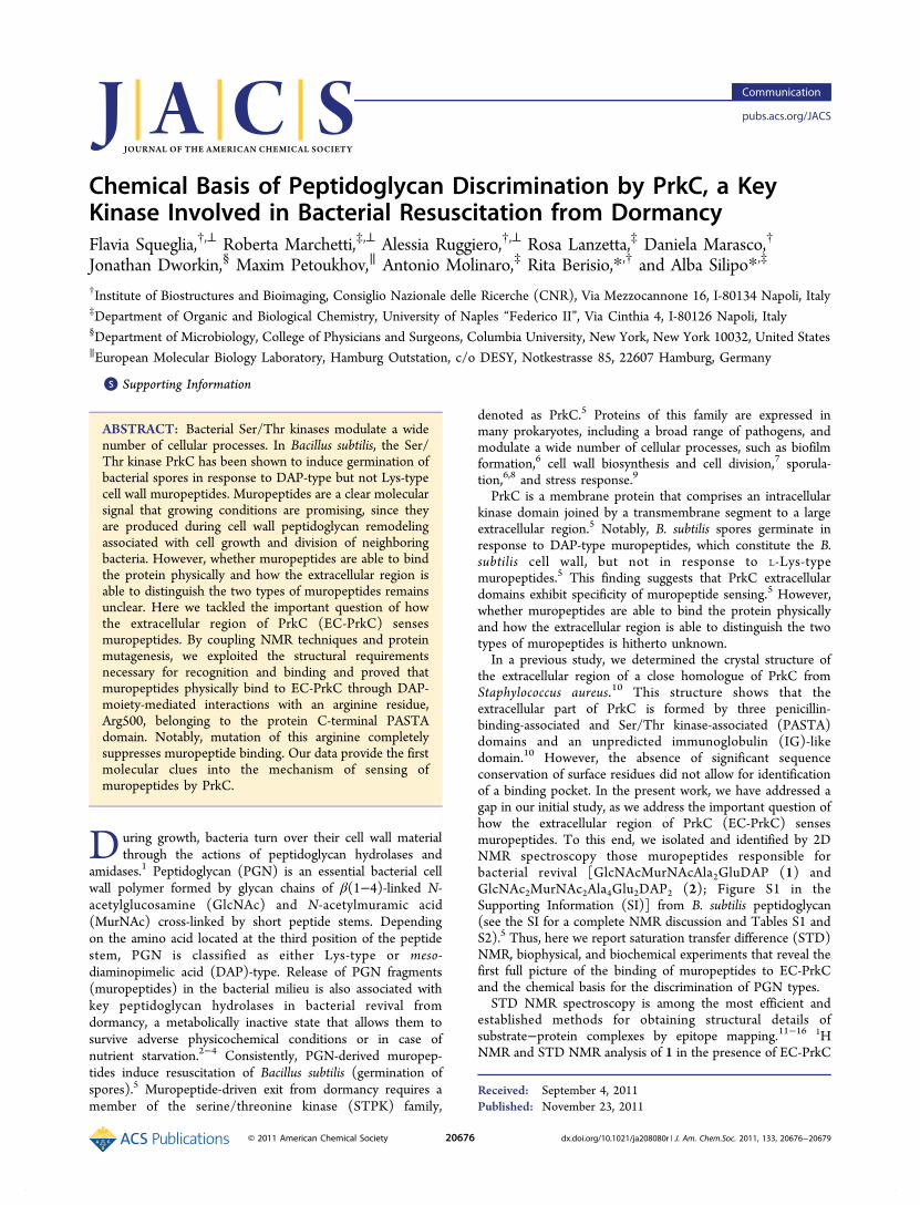
domain and solvent-exposed (Figure 3). These considerationsled us to hypothesize that Arg500 is involved in muropeptidebinding in a fashion similar to those observed in the PDB(Figure S9). To corroborate this hypothesis, we mutatedArg500 to alanine (R500A) and to glutamic acid (R500E),which we predicted to be more disruptive of the Arg−DAPinteraction. STD experiments unequivocally showed thatneither R500E nor R500A are able to bind muropeptides, asno STD signals were observed in either case (Figure 1D,E).Circular dichroism (CD) spectroscopy excluded the
possibility that the differences in the muropeptide binding ofthe two mutants were due to defects in protein folding inducedby the mutations (Figure 4). The inability of the R500E andR500A mutants to bind muropeptides shows that Arg500 is theprimary site of interaction with muropeptides. The combina-tion of these results with those from the STD experiments onwild-type EC-PrkC (Figures 1 and 2) proves that PrkC senses
muropeptides through interactions of the negatively chargedDAP side chain with the side chain of Arg500. Furthermore, theinability of the R500A mutant to bind muropeptides (Figure1D,E) shows that the contribution of sugar moieties, althoughmeasurable (Figure S4), is not sufficient for binding in theabsence of DAP−Arg interactions.Using a set of biophysical techniques, we also analyzed the
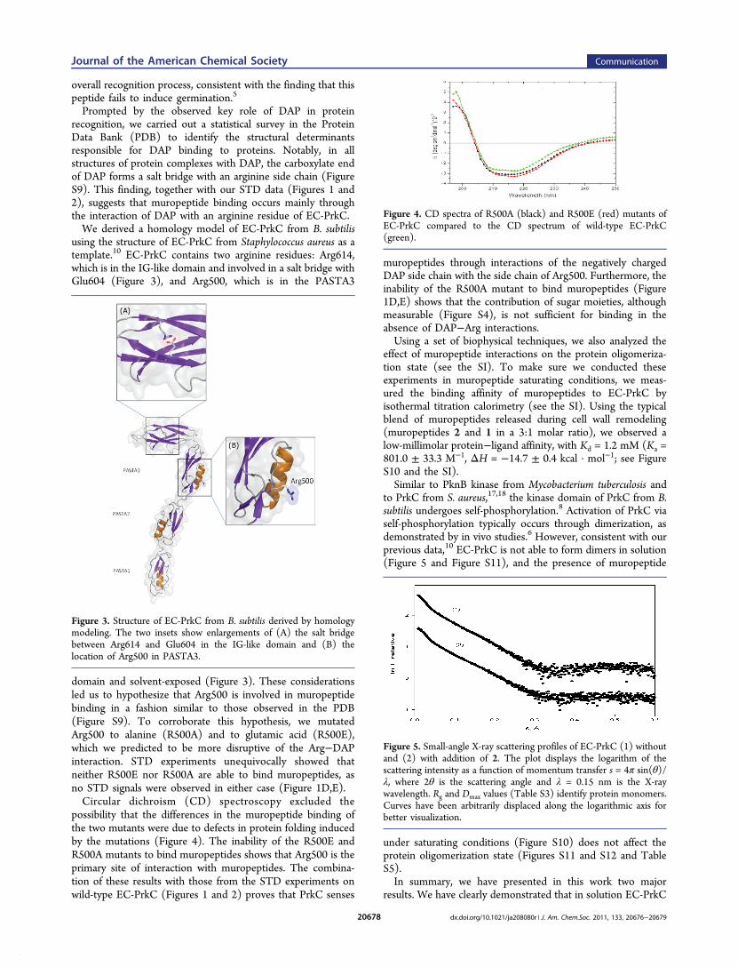
effect of muropeptide interactions on the protein oligomeriza-tion state (see the SI). To make sure we conducted theseexperiments in muropeptide saturating conditions, we meas-ured the binding affinity of muropeptides to EC-PrkC byisothermal titration calorimetry (see the SI). Using the typicalblend of muropeptides released during cell wall remodeling(muropeptides 2 and 1 in a 3:1 molar ratio), we observed alow-millimolar protein−ligand affinity, with Kd = 1.2 mM (Ka =801.0 ± 33.3 M−1, ΔH = −14.7 ± 0.4 kcal · mol−1; see FigureS10 and the SI).Similar to PknB kinase from Mycobacterium tuberculosis and
to PrkC from S. aureus,17,18 the kinase domain of PrkC from B.subtilis undergoes self-phosphorylation.8 Activation of PrkC viaself-phosphorylation typically occurs through dimerization, asdemonstrated by in vivo studies.6 However, consistent with ourprevious data,10 EC-PrkC is not able to form dimers in solution(Figure 5 and Figure S11), and the presence of muropeptide
under saturating conditions (Figure S10) does not affect theprotein oligomerization state (Figures S11 and S12 and TableS5).In summary, we have presented in this work two major
results. We have clearly demonstrated that in solution EC-PrkC
Figure 3. Structure of EC-PrkC from B. subtilis derived by homologymodeling. The two insets show enlargements of (A) the salt bridgebetween Arg614 and Glu604 in the IG-like domain and (B) thelocation of Arg500 in PASTA3.
Figure 4. CD spectra of R500A (black) and R500E (red) mutants ofEC-PrkC compared to the CD spectrum of wild-type EC-PrkC(green).
Figure 5. Small-angle X-ray scattering profiles of EC-PrkC (1) withoutand (2) with addition of 2. The plot displays the logarithm of thescattering intensity as a function of momentum transfer s = 4π sin(θ)/λ, where 2θ is the scattering angle and λ = 0.15 nm is the X-raywavelength. Rg and Dmax values (Table S3) identify protein monomers.Curves have been arbitrarily displaced along the logarithmic axis forbetter visualization.
Journal of the American Chemical Society Communication
dx.doi.org/10.1021/ja208080r | J. Am. Chem.Soc. 2011, 133, 20676−2067920678
is able to bind DAP-type muropeptides physically (Figures 1and 2) and that DAP is the critical element in the binding tothe protein. We have also shown that this recognition occursthrough interactions of DAP with Arg500, as a mutation of thisamino acid in EC-PrkC completely impaired muropeptidebinding (Figure 1D,E). This finding agrees well with the keyrole played by arginine in the specific recognition of DAP-typemuropeptides by peptidoglycan recognition proteins.19 In thisscenario, the key role of Arg500 in binding provides a clearexplanation for the ability of PrkC from B. subtilis todiscriminate between DAP- and Lys-type muropeptides inbacterial revival.5 Using this mechanism, B. subtilis bacteria,which possess a DAP-type PGN, can cross-talk and triggerresuscitation by its own cell wall turnover.Consistent with our results, very recent data have shown that
PknB kinase from M. tuberculosis also possesses a strongpreference for DAP-type muropeptides similar to the bacterialcell wall fragments, although the protein interaction site andreasons for specificity are yet to be identified.20 Furthermore,the observed inability of EC-PrkC to form dimers in vitropoints to a more complex protein dimerization mechanism,which may either involve the PrkC transmembrane portion orrequire a third molecule, available in vivo, in a manner similarto that observed for the fibroblast growth factor receptor(FGFR).21 Finally, the definition of structural determinants ofmuropeptide-driven revival from dormancy is precious to thedevelopment of low-molecular-weight entities of therapeuticinterest.
■ ASSOCIATED CONTENT*S Supporting InformationExperimental details, supporting results, and supporting figures.This material is available free of charge via the Internet athttp://pubs.acs.org.
■ ACKNOWLEDGMENTSA.M., R.B., and A.S. acknowledge the COST action BM1003“Microbial cell surface determinants of virulence as targets fornew therapeutics in Cystic Fibrosis”. R.B. would like toacknowledge MIUR (PRIN 2009 - prot. 200993WWF9).
■ REFERENCES(1) Hendrickx, A. P.; Budzik, J. M.; Oh, S. Y.; Schneewind, O. Nat.Rev. Microbiol. 2011, 9, 166.(2) Ruggiero, A.; Tizzano, B.; Pedone, E.; Pedone, C.; Wilmanns, M.;Berisio, R. J. Mol. Biol. 2009, 385, 153.(3) Ruggiero, A.; Marasco, D.; Squeglia, F.; Soldini, S.; Pedone, E.;Pedone, C.; Berisio, R. Structure 2010, 18, 1184.(4) Kaprelyants, A. S.; Mukamolova, G. V.; Ruggiero, A.; Makarov, V.A.; Demina, G. R.; Shleeva, M. O.; Potapov, V. D.; Shramko, P. ProteinPept. Lett. 2011in press.(5) Shah, I. M.; Laaberki, M. H.; Popham, D. L.; Dworkin, J. Cell2008, 135, 486.(6) Madec, E.; Laszkiewicz, A.; Iwanicki, A.; Obuchowski, M.; Seror,S. Mol. Microbiol. 2002, 46, 571.(7) Fiuza, M.; Canova, M. J.; Zanella-Cleon, I.; Becchi, M.; Cozzone,A. J.; Mateos, L. M.; Kremer, L.; Gil, J. A.; Molle, V. J. Biol. Chem.2008, 283, 18099.
(8) Madec, E.; Stensballe, A.; Kjellstrom, S.; Cladiere, L.;Obuchowski, M.; Jensen, O. N.; Seror, S. J. J. Mol. Biol. 2003, 330, 459.(9) Absalon, C.; Obuchowski, M.; Madec, E.; Delattre, D.; Holland, I.B.; Seror, S. J. Microbiology 2009, 155, 932.(10) Ruggiero, A.; Squeglia, F.; Marasco, D.; Marchetti, R.; Molinaro,A.; Berisio, R. Biochem. J. 2011, 435, 33.(11) Meyer, B.; Peters, T. Angew. Chem., Int. Ed. 2003, 42, 864.(12) Angulo, J.; Langpap, B.; Blume, A.; Biet, T.; Meyer, B.; Krishna,N. R. H.; Peters, H.; Palcic, M.; Peters, T. J. Am. Chem. Soc. 2006, 128,13529.(13) Roldos, V.; Canada, F. J.; Jimenez-Barbero, J. ChemBioChem2011, 12, 990.(14) Mari, S.; Serrano-Gomez, D.; Canada, F. J.; Corbi, A. L.;Jimenez-Barbero, J. Angew. Chem., Int. Ed. 2004, 44, 296.(15) Mayer, M.; Meyer, B. J. Am. Chem. Soc. 2001, 123, 6108.(16) Hu, X.; Zhang, W.; Carmichael, I.; Serianni, A. J. Am. Chem. Soc.2010, 132, 4641.(17) Mieczkowski, C.; Iavarone, A. T.; Alber, T. EMBO J. 2008, 27,3186.(18) Debarbouille, M.; Dramsi, S.; Dussurget, O.; Nahori, M. A.;Vaganay, E.; Jouvion, G.; Cozzone, A.; Msadek, T.; Duclos, B. J.Bacteriol. 2009, 191, 4070.(19) Lim, J. H.; Kim, M. S.; Kim, H. E.; Yano, T.; Oshima, Y.;Aggarwal, K.; Goldman, W. E.; Silverman, N.; Kurata, S.; Oh, B. H. J.Biol. Chem. 2006, 281, 8286.(20) Mushtaq, M.; Asong, J.; Li, X.; Cardot, J.; Boons, G.-J.; Husson,R. N. PLOS Pathog. 2011, 7, No. e1002182.(21) Schlessinger, J.; Plotnikov, A. N.; Ibrahimi, O. A.; Eliseenkova,A. V.; Yeh, B. K.; Yayon, A.; Linhardt, R. J.; Mohammadi, M. Mol. Cell2000, 6, 743.
Journal of the American Chemical Society Communication
dx.doi.org/10.1021/ja208080r | J. Am. Chem.Soc. 2011, 133, 20676−2067920679