Chemistry and Physics of Lipids 164 (2011) 727–731
Contents lists available at SciVerse ScienceDirect
Chemistry and Physics of Lipids
journa l homepage: www.e lsev ier .com/ locate /chemphys l ip
hort communication
echanoformation of neutral giant phospholipid vesicles in high ionic strengtholution
anez I. Pavlic a,b,∗, Julia Genovac, George Popkirovc, Veronika Kralj-Iglic d, Ales Iglic a, Marin D. Mitovc
University of Ljubljana, Faculty of Electrical Engineering, Trzaska 25, 1000 Ljubljana, SloveniaUniversity of Ljubljana, Faculty of Health Sciences, Zdravstvena pot 5, 1000 Ljubljana, SloveniaBulgarian Academy of Sciences, Institute of Solid State Physics, Tzarigradsko Chaussee Blvd. 72, 1784 Sofia, BulgariaUniversity of Ljubljana, Faculty of Medicine, Lipiceva 2, 1000 Ljubljana, Slovenia
r t i c l e i n f o
rticle history:eceived 3 May 2011eceived in revised form 26 August 2011ccepted 29 August 2011
a b s t r a c t
We present new and simple method for formation of giant unilamellar vesicles (GUVs) in high ionicstrength solutions, such as phosphate buffered saline (PBS). Mechanoformation method is an alternativemethod to electroformation method. The advantage of the mechanoformation procedure is that thereare no limitations with respect to the ionic strength of the aqueous solutions, because there is no applied
Nowadays there are many different methods for formation ofUVs, as described in recent review paper (Walde et al., 2010). Theethod of the electroformation is well known and the preferred
echnique for the formation of giant unilamellar vesicles (Angelovand Dimitrov, 1986; Dimitrov and Angelova, 1988). The main draw-ack of electroformation method is the inability to form GUVs inigh ionic strength solutions (PBS, saline). These physiologicallyelevant solutions are used to achieve stable and well-controlledonditions. Such conditions are of great interest in research fields,ike medicine, physical chemistry and biophysics.
Some different techniques have been developed for high ionictrength solutions, such as sonication or extrusion techniquesLapinski et al., 2007). Though some attempts to form GUVs in highonic strength solutions has been performed (Pott et al., 2008; Pavlict al., 2010; Montes et al., 2007; Estes and Mayer, 2005; Akashit al., 1996), still there is no easy and suitable method that does not
epend on the ionic strength of the aqueous solution.
In this work we propose a new formation method that is inde-endent of the aqueous solution used, during GUV formation and
∗ Corresponding author at: University of Ljubljana, Faculty of Electrical Engineer-ng (Laboratory of Biophysics), Trzaska 25, 1000 Ljubljana, Slovenia.el.: +386 1 476 8825; fax: +386 1 476 8850.
does not induce chemical changes to the vesicle’s membrane envi-ronment due to hydrolysis caused by applied voltage.
2. Materials and methods
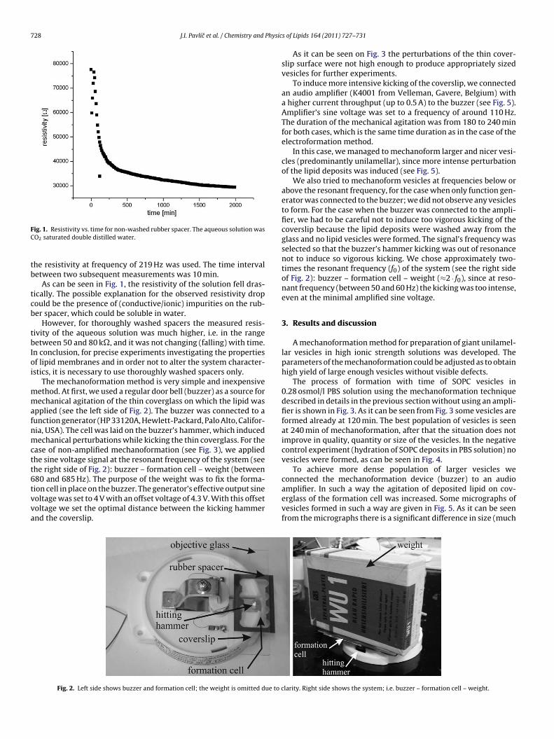
For vesicle preparation we used 1-stearoyl-2-oleoyl-sn-glycero-3-phosphocholine (SOPC) lipid (Avanti Polar Lipids, Alabaster,Alabama, USA). The lipid was dissolved in chloroform in concen-tration of 1 mg/ml. SOPC lipid was applied in small drops overthe thin coverglass (coverslip) and subsequently subjected to vac-uum for 15–20 min to evaporate the organic solvent (chloroform).After the entire evaporation of the organic solvent, the cell wasfilled with phosphate buffer saline (PBS) solution with osmolar-ity of 0.28 osmol/l. The PBS tablet (Sigma–Aldrich Chemie GmbH,Buchs, Switzerland) was dissolved in double distilled water. Thecell was assembled from thin coverglass, a rubber spacer takenfrom perfusion chamber (Electron Microscopy Sciences, Hatfield,Pennsylvania, USA) and an objective glass as seen on the left sidein Fig. 2.
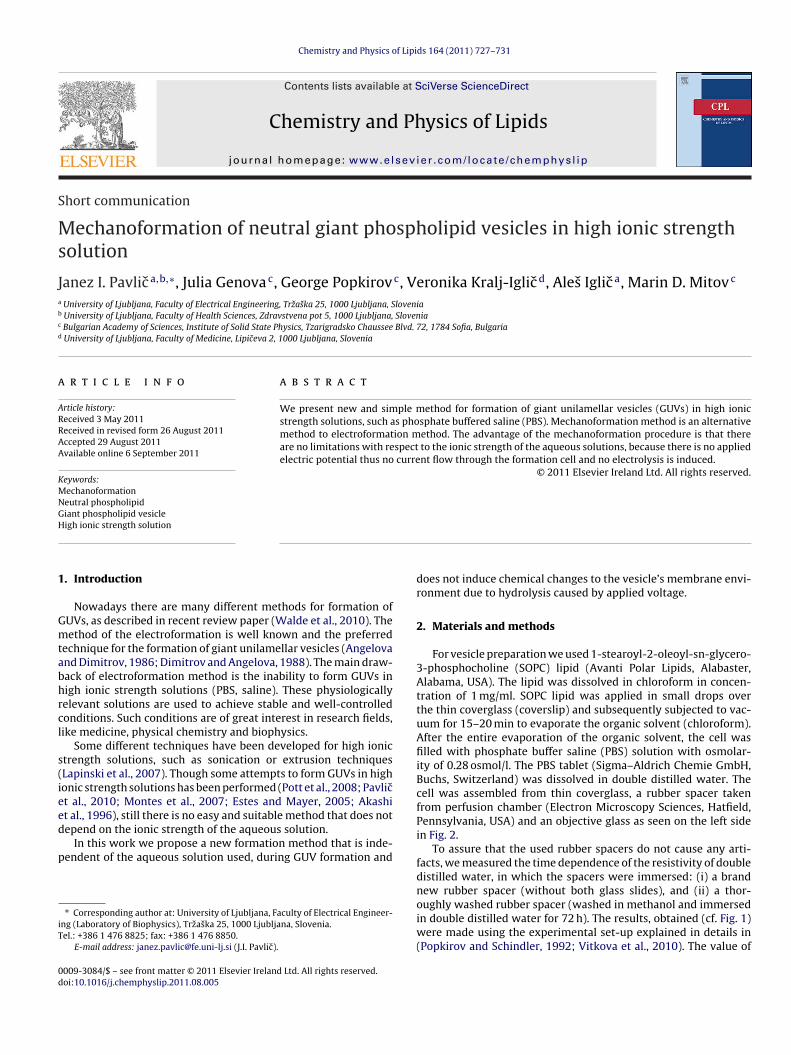
To assure that the used rubber spacers do not cause any arti-facts, we measured the time dependence of the resistivity of doubledistilled water, in which the spacers were immersed: (i) a brandnew rubber spacer (without both glass slides), and (ii) a thor-
oughly washed rubber spacer (washed in methanol and immersedin double distilled water for 72 h). The results, obtained (cf. Fig. 1)were made using the experimental set-up explained in details in(Popkirov and Schindler, 1992; Vitkova et al., 2010). The value of
amplifier. In such a way the agitation of deposited lipid on cov-
ig. 1. Resistivity vs. time for non-washed rubber spacer. The aqueous solution wasO2 saturated double distilled water.
he resistivity at frequency of 219 Hz was used. The time intervaletween two subsequent measurements was 10 min.
As can be seen in Fig. 1, the resistivity of the solution fell dras-ically. The possible explanation for the observed resistivity dropould be the presence of (conductive/ionic) impurities on the rub-er spacer, which could be soluble in water.
However, for thoroughly washed spacers the measured resis-ivity of the aqueous solution was much higher, i.e. in the rangeetween 50 and 80 k�, and it was not changing (falling) with time.
n conclusion, for precise experiments investigating the propertiesf lipid membranes and in order not to alter the system character-stics, it is necessary to use thoroughly washed spacers only.
The mechanoformation method is very simple and inexpensiveethod. At first, we used a regular door bell (buzzer) as a source forechanical agitation of the thin coverglass on which the lipid was
pplied (see the left side of Fig. 2). The buzzer was connected to aunction generator (HP 33120A, Hewlett-Packard, Palo Alto, Califor-ia, USA). The cell was laid on the buzzer’s hammer, which inducedechanical perturbations while kicking the thin coverglass. For the
ase of non-amplified mechanoformation (see Fig. 3), we appliedhe sine voltage signal at the resonant frequency of the system (seehe right side of Fig. 2): buzzer – formation cell – weight (between80 and 685 Hz). The purpose of the weight was to fix the forma-ion cell in place on the buzzer. The generator’s effective output sine
oltage was set to 4 V with an offset voltage of 4.3 V. With this offsetoltage we set the optimal distance between the kicking hammernd the coverslip.
Fig. 2. Left side shows buzzer and formation cell; the weight is omitted due to c
s of Lipids 164 (2011) 727–731
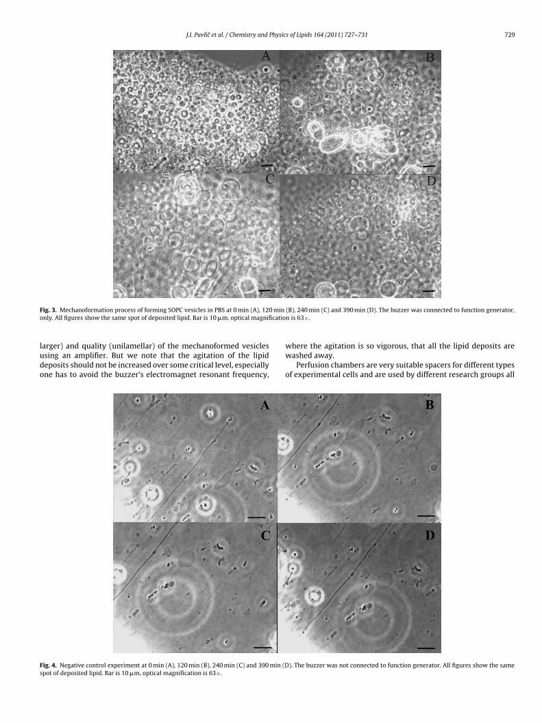
As it can be seen on Fig. 3 the perturbations of the thin cover-slip surface were not high enough to produce appropriately sizedvesicles for further experiments.
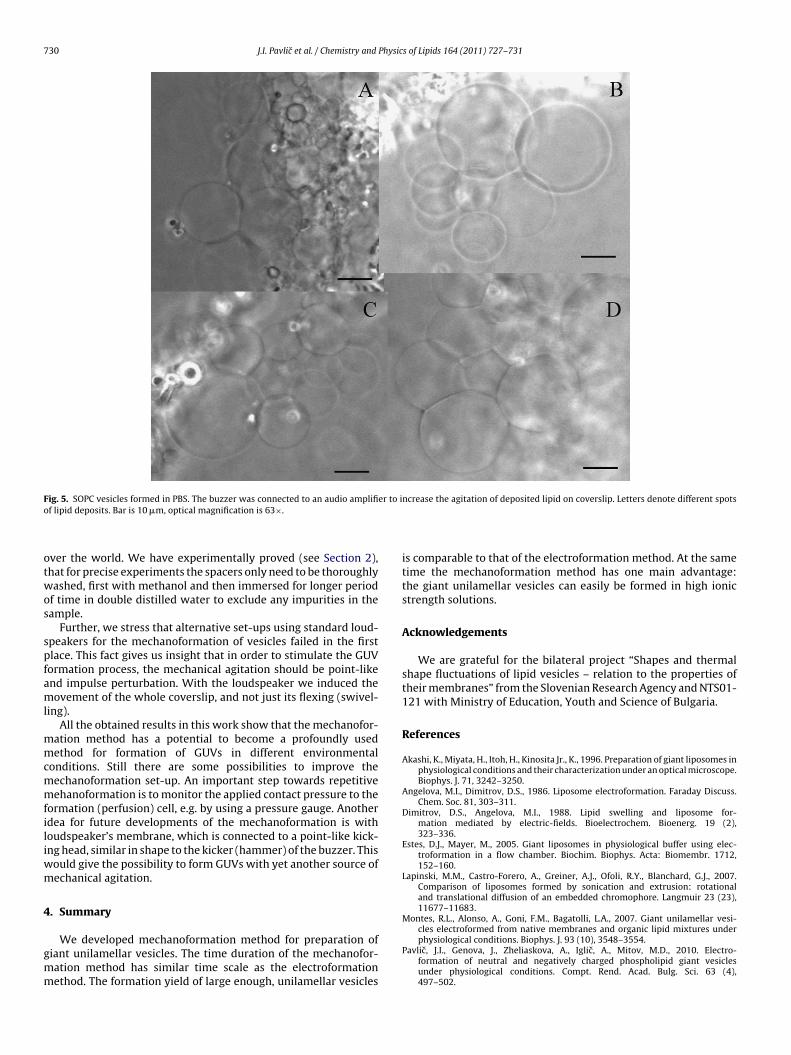
To induce more intensive kicking of the coverslip, we connectedan audio amplifier (K4001 from Velleman, Gavere, Belgium) witha higher current throughput (up to 0.5 A) to the buzzer (see Fig. 5).Amplifier’s sine voltage was set to a frequency of around 110 Hz.The duration of the mechanical agitation was from 180 to 240 minfor both cases, which is the same time duration as in the case of theelectroformation method.
In this case, we managed to mechanoform larger and nicer vesi-cles (predominantly unilamellar), since more intense perturbationof the lipid deposits was induced (see Fig. 5).
We also tried to mechanoform vesicles at frequencies below orabove the resonant frequency, for the case when only function gen-erator was connected to the buzzer; we did not observe any vesiclesto form. For the case when the buzzer was connected to the ampli-fier, we had to be careful not to induce too vigorous kicking of thecoverslip because the lipid deposits were washed away from theglass and no lipid vesicles were formed. The signal’s frequency wasselected so that the buzzer’s hammer kicking was out of resonancenot to induce so vigorous kicking. We chose approximately two-times the resonant frequency (f0) of the system (see the right sideof Fig. 2): buzzer – formation cell – weight (≈2 · f0), since at reso-nant frequency (between 50 and 60 Hz) the kicking was too intense,even at the minimal amplified sine voltage.
3. Results and discussion
A mechanoformation method for preparation of giant unilamel-lar vesicles in high ionic strength solutions was developed. Theparameters of the mechanoformation could be adjusted as to obtainhigh yield of large enough vesicles without visible defects.
The process of formation with time of SOPC vesicles in0.28 osmol/l PBS solution using the mechanoformation techniquedescribed in details in the previous section without using an ampli-fier is shown in Fig. 3. As it can be seen from Fig. 3 some vesicles areformed already at 120 min. The best population of vesicles is seenat 240 min of mechanoformation, after that the situation does notimprove in quality, quantity or size of the vesicles. In the negativecontrol experiment (hydration of SOPC deposits in PBS solution) novesicles were formed, as can be seen in Fig. 4.
To achieve more dense population of larger vesicles weconnected the mechanoformation device (buzzer) to an audio
erglass of the formation cell was increased. Some micrographs ofvesicles formed in such a way are given in Fig. 5. As it can be seenfrom the micrographs there is a significant difference in size (much
larity. Right side shows the system; i.e. buzzer – formation cell – weight.
J.I. Pavlic et al. / Chemistry and Physics of Lipids 164 (2011) 727–731 729
F 0 mino catio
ludo
Fs
ig. 3. Mechanoformation process of forming SOPC vesicles in PBS at 0 min (A), 12nly. All figures show the same spot of deposited lipid. Bar is 10 �m, optical magnifi
arger) and quality (unilamellar) of the mechanoformed vesiclessing an amplifier. But we note that the agitation of the lipideposits should not be increased over some critical level, especiallyne has to avoid the buzzer’s electromagnet resonant frequency,
ig. 4. Negative control experiment at 0 min (A), 120 min (B), 240 min (C) and 390 min (Dpot of deposited lipid. Bar is 10 �m, optical magnification is 63×.
(B), 240 min (C) and 390 min (D). The buzzer was connected to function generator,n is 63×.
where the agitation is so vigorous, that all the lipid deposits arewashed away.
Perfusion chambers are very suitable spacers for different typesof experimental cells and are used by different research groups all
). The buzzer was not connected to function generator. All figures show the same
730 J.I. Pavlic et al. / Chemistry and Physics of Lipids 164 (2011) 727–731
Fig. 5. SOPC vesicles formed in PBS. The buzzer was connected to an audio amplifier to increase the agitation of deposited lipid on coverslip. Letters denote different spotso
otwos
spfaml
mmcmmfiliwm
4
gmm
f lipid deposits. Bar is 10 �m, optical magnification is 63×.
ver the world. We have experimentally proved (see Section 2),hat for precise experiments the spacers only need to be thoroughlyashed, first with methanol and then immersed for longer period
f time in double distilled water to exclude any impurities in theample.
Further, we stress that alternative set-ups using standard loud-peakers for the mechanoformation of vesicles failed in the firstlace. This fact gives us insight that in order to stimulate the GUVormation process, the mechanical agitation should be point-likend impulse perturbation. With the loudspeaker we induced theovement of the whole coverslip, and not just its flexing (swivel-
ing).All the obtained results in this work show that the mechanofor-
ation method has a potential to become a profoundly usedethod for formation of GUVs in different environmental
onditions. Still there are some possibilities to improve theechanoformation set-up. An important step towards repetitiveehanoformation is to monitor the applied contact pressure to the
ormation (perfusion) cell, e.g. by using a pressure gauge. Anotherdea for future developments of the mechanoformation is withoudspeaker’s membrane, which is connected to a point-like kick-ng head, similar in shape to the kicker (hammer) of the buzzer. This
ould give the possibility to form GUVs with yet another source ofechanical agitation.
. Summary
We developed mechanoformation method for preparation ofiant unilamellar vesicles. The time duration of the mechanofor-ation method has similar time scale as the electroformationethod. The formation yield of large enough, unilamellar vesicles
is comparable to that of the electroformation method. At the sametime the mechanoformation method has one main advantage:the giant unilamellar vesicles can easily be formed in high ionicstrength solutions.
Acknowledgements
We are grateful for the bilateral project “Shapes and thermalshape fluctuations of lipid vesicles – relation to the properties oftheir membranes” from the Slovenian Research Agency and NTS01-121 with Ministry of Education, Youth and Science of Bulgaria.
References
Akashi, K., Miyata, H., Itoh, H., Kinosita Jr., K., 1996. Preparation of giant liposomes inphysiological conditions and their characterization under an optical microscope.Biophys. J. 71, 3242–3250.
Dimitrov, D.S., Angelova, M.I., 1988. Lipid swelling and liposome for-mation mediated by electric-fields. Bioelectrochem. Bioenerg. 19 (2),323–336.
Estes, D.J., Mayer, M., 2005. Giant liposomes in physiological buffer using elec-troformation in a flow chamber. Biochim. Biophys. Acta: Biomembr. 1712,152–160.
Lapinski, M.M., Castro-Forero, A., Greiner, A.J., Ofoli, R.Y., Blanchard, G.J., 2007.Comparison of liposomes formed by sonication and extrusion: rotationaland translational diffusion of an embedded chromophore. Langmuir 23 (23),11677–11683.
Montes, R.L., Alonso, A., Goni, F.M., Bagatolli, L.A., 2007. Giant unilamellar vesi-cles electroformed from native membranes and organic lipid mixtures under
physiological conditions. Biophys. J. 93 (10), 3548–3554.
Pavlic, J.I., Genova, J., Zheliaskova, A., Iglic, A., Mitov, M.D., 2010. Electro-formation of neutral and negatively charged phospholipid giant vesiclesunder physiological conditions. Compt. Rend. Acad. Bulg. Sci. 63 (4),497–502.
Physic
P
P
J.I. Pavlic et al. / Chemistry and
opkirov, G., Schindler, R.N., 1992. A new impedance spectrometer forthe investigation of electrochemical systems. Rev. Sci. Instrum. 63 (11),5366–5373.
Vitkova, V., Antonova, K., Popkirov, G., Ermakov, Y.A., Bivas, I., 2010. Electrical resis-tivity of the liquid phase of vesicular suspensions prepared by different methods.J. Phys. Conf. Ser. 253, 012059.