Chemistry & Biology, Vol. 9, 1059–1072, October, 2002, 2002 Elsevier Science Ltd. All rights reserved. PIIS1074-5521(02)00247-8 Review The Chemical Biology of Apoptosis: Exploring Protein-Protein Interactions and the Life and Death of Cells with Small Molecules The inhibition or promotion of these interactions either by natural molecules or by unnatural synthetic ones is of great interest for understanding the mechanism of biological recognition and translating the information of gene sequence to the discovery of new therapeutic Ziwei Huang 1 Departments of Biochemistry and Chemistry University of Illinois at Urbana-Champaign Urbana, Illinois 61801 agents. Much progress has been made recently in devel- oping various strategies to manipulate protein-protein Apoptosis, a fundamental process for both human interactions with small molecules [7, 8]. health and disease, is initiated and regulated by pro- In apoptosis, protein-protein interactions are the un- tein-protein interactions, notable examples of which derlying theme in both mitochondria and death receptor are the interactions involving Bcl-2 and IAP protein pathways (Figure 1). Obviously, life and death are mat- families. This article discusses recent advances in the ters that cannot be taken lightly. Thus, nature has use of chemical approaches in discovering and study- evolved a sophisticated and tightly controlled network ing small molecules targeted to proteins of the Bcl-2 of protein-protein interactions to ensure the accuracy and IAP families. These small molecules and their and check-and-balance of the cell-death machinery. complexes with receptors provide the tools and model The activation of caspases, which are central for apopto- systems to probe the basic mechanism of molecule sis, depends on various protein-protein interactions. For recognition underling the life and death of cells and example, caspase-8 is activated through a cascade of develop novel strategies for therapeutic intervention sequential oligomerization of death receptors, the adap- of the dysregulated apoptotic process. The review of tor molecules FADD (Fas-associated protein with death these studies highlights the opportunity and challenge domain), and procaspase-8, whereas caspase-9 is acti- in this emerging area of chemical and chemical biolog- vated through the association of at least cytochrome ical research. c, Apaf-1, and procaspase-9 to form apoptosome. The action of these “killer” enzymes before or after activation Introduction is controlled or safeguarded through protein-protein in- Apoptosis or programmed cell death is the prevalent teractions involving multiple families of proteins, includ- mechanism complementary to proliferation that is criti- ing the Bcl-2 (B cell lymphoma-2) and IAP (inhibitor of cal for the normal development and function of multicel- apoptosis proteins) families, which have recently re- lular organisms [1]. As approximately 10 11 –10 12 cells are ceived intensive attention. produced every day in healthy adult humans, this rapid The Bcl-2 family is a large group of apoptosis regula- proliferation needs to be balanced by apoptosis to main- tors which, through the diverse interactions among tain a constant cell number. Changing this balance in themselves and with other proteins, control the release either direction has pathological consequences. Abnor- of apoptogenic factors, such as cytochrome c and Smac mally high rate of cell death is found in neurodegenera- (second mitochondria-derived activator of caspases) or tive diseases including Alzheimer’s and Parkinson’s dis- its murine homolog DIABLO (direct IAP binding protein eases, AIDS and cardiovascular diseases, whereas with low pI), needed for caspase activation [9, 10]. While retarded cell death contributes to a wide variety of hu- the Bcl-2 family regulates the integrity of mitochondria man cancers [2]. and normally has little impact on signals from death There are two major pathways for apoptosis that have receptors, a crosstalk is found in some cells where cas- been elucidated so far, both of which involve a family pase-8 activated by death receptors cleaves inert Bid of cysteine proteases with aspartate specificity, called to release tBid, which then triggers the mitochondiral caspases, as the executioners of apoptosis [3, 4]. The apoptotic pathway (Figure 1). The IAP family is another mitochondria provide one of the apoptotic pathways family of proteins that reprieve caspases’ execution of from which cytochrome c is released upon the stimula- apoptosis through physically interacting with caspases tion by a variety of cell-death triggers [5]. This leads and thereby directly inhibiting their function. In turn, the to activation of Apaf-1 (apoptosis protease activating function of the IAP family is inhibited by Smac/DIABLO, factor-1), caspase-9, and caspase-3. Another apoptotic which, upon its release from the mitochondria together pathway involves the ligation of death receptors such with cytochrome c, binds IAPs and relieves them from as Fas and activation of caspase-8 and subsequently the complexes with caspases. caspase-3 [6] (Figure 1). Given the fundamental role of apoptosis in both nor- Protein-protein interactions are important for a wide mal physiology and a wide variety of human diseases, variety of physiological as well as pathological pro- there has been an explosion of research in the past cesses. The significance of protein-protein interactions decade which has made apoptosis one of the central has never been more apparent than in the post-genome areas in biomedical sciences. There are many recent era, in which the functional characterization of every reviews in the biological [11–15], clinical [16–19], and protein encoded by the human genome will require un- structural [20, 21] aspects of apoptosis. This article fo- derstanding of its molecular interactions with others. cuses on the chemical and chemical biological perspec- tives of apoptosis in terms of how chemical approaches are used to explore and control the function of proteins 1 Correspondence: [email protected]
Transcript
Chemistry & Biology, Vol. 9, 1059–1072, October, 2002, 2002 Elsevier Science Ltd. All rights reserved. PII S1074-5521(02)00247-8
ReviewThe Chemical Biology of Apoptosis:Exploring Protein-Protein Interactions andthe Life and Death of Cells with Small Molecules
The inhibition or promotion of these interactions eitherby natural molecules or by unnatural synthetic ones isof great interest for understanding the mechanism ofbiological recognition and translating the informationof gene sequence to the discovery of new therapeutic
Ziwei Huang1
Departments of Biochemistry and ChemistryUniversity of Illinois at Urbana-ChampaignUrbana, Illinois 61801
agents. Much progress has been made recently in devel-oping various strategies to manipulate protein-proteinApoptosis, a fundamental process for both humaninteractions with small molecules [7, 8].health and disease, is initiated and regulated by pro-
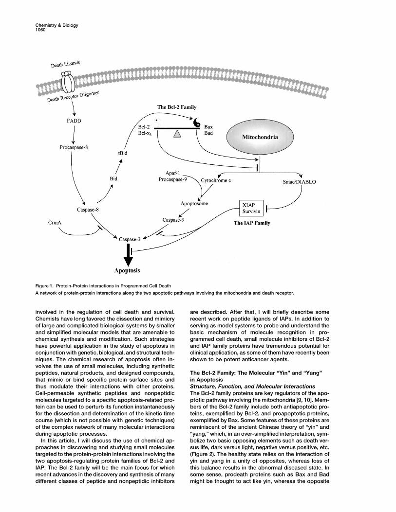
In apoptosis, protein-protein interactions are the un-tein-protein interactions, notable examples of whichderlying theme in both mitochondria and death receptorare the interactions involving Bcl-2 and IAP proteinpathways (Figure 1). Obviously, life and death are mat-families. This article discusses recent advances in theters that cannot be taken lightly. Thus, nature hasuse of chemical approaches in discovering and study-evolved a sophisticated and tightly controlled networking small molecules targeted to proteins of the Bcl-2of protein-protein interactions to ensure the accuracyand IAP families. These small molecules and theirand check-and-balance of the cell-death machinery.complexes with receptors provide the tools and modelThe activation of caspases, which are central for apopto-systems to probe the basic mechanism of moleculesis, depends on various protein-protein interactions. Forrecognition underling the life and death of cells andexample, caspase-8 is activated through a cascade ofdevelop novel strategies for therapeutic interventionsequential oligomerization of death receptors, the adap-of the dysregulated apoptotic process. The review oftor molecules FADD (Fas-associated protein with deaththese studies highlights the opportunity and challengedomain), and procaspase-8, whereas caspase-9 is acti-in this emerging area of chemical and chemical biolog-vated through the association of at least cytochromeical research.c, Apaf-1, and procaspase-9 to form apoptosome. Theaction of these “killer” enzymes before or after activation
Introduction is controlled or safeguarded through protein-protein in-Apoptosis or programmed cell death is the prevalent teractions involving multiple families of proteins, includ-mechanism complementary to proliferation that is criti- ing the Bcl-2 (B cell lymphoma-2) and IAP (inhibitor ofcal for the normal development and function of multicel- apoptosis proteins) families, which have recently re-lular organisms [1]. As approximately 1011–1012 cells are ceived intensive attention.produced every day in healthy adult humans, this rapid The Bcl-2 family is a large group of apoptosis regula-proliferation needs to be balanced by apoptosis to main- tors which, through the diverse interactions amongtain a constant cell number. Changing this balance in themselves and with other proteins, control the releaseeither direction has pathological consequences. Abnor- of apoptogenic factors, such as cytochrome c and Smacmally high rate of cell death is found in neurodegenera- (second mitochondria-derived activator of caspases) ortive diseases including Alzheimer’s and Parkinson’s dis- its murine homolog DIABLO (direct IAP binding proteineases, AIDS and cardiovascular diseases, whereas with low pI), needed for caspase activation [9, 10]. Whileretarded cell death contributes to a wide variety of hu- the Bcl-2 family regulates the integrity of mitochondriaman cancers [2]. and normally has little impact on signals from death
There are two major pathways for apoptosis that have receptors, a crosstalk is found in some cells where cas-been elucidated so far, both of which involve a family pase-8 activated by death receptors cleaves inert Bidof cysteine proteases with aspartate specificity, called to release tBid, which then triggers the mitochondiralcaspases, as the executioners of apoptosis [3, 4]. The apoptotic pathway (Figure 1). The IAP family is anothermitochondria provide one of the apoptotic pathways family of proteins that reprieve caspases’ execution offrom which cytochrome c is released upon the stimula- apoptosis through physically interacting with caspasestion by a variety of cell-death triggers [5]. This leads and thereby directly inhibiting their function. In turn, theto activation of Apaf-1 (apoptosis protease activating function of the IAP family is inhibited by Smac/DIABLO,factor-1), caspase-9, and caspase-3. Another apoptotic which, upon its release from the mitochondria togetherpathway involves the ligation of death receptors such with cytochrome c, binds IAPs and relieves them fromas Fas and activation of caspase-8 and subsequently the complexes with caspases.caspase-3 [6] (Figure 1). Given the fundamental role of apoptosis in both nor-
Protein-protein interactions are important for a wide mal physiology and a wide variety of human diseases,variety of physiological as well as pathological pro- there has been an explosion of research in the pastcesses. The significance of protein-protein interactions decade which has made apoptosis one of the centralhas never been more apparent than in the post-genome areas in biomedical sciences. There are many recentera, in which the functional characterization of every reviews in the biological [11–15], clinical [16–19], andprotein encoded by the human genome will require un- structural [20, 21] aspects of apoptosis. This article fo-derstanding of its molecular interactions with others. cuses on the chemical and chemical biological perspec-
tives of apoptosis in terms of how chemical approachesare used to explore and control the function of proteins1Correspondence: [email protected]
Chemistry & Biology1060
Figure 1. Protein-Protein Interactions in Programmed Cell Death
A network of protein-protein interactions along the two apoptotic pathways involving the mitochondria and death receptor.
involved in the regulation of cell death and survival. are described. After that, I will briefly describe somerecent work on peptide ligands of IAPs. In addition toChemists have long favored the dissection and mimicry
of large and complicated biological systems by smaller serving as model systems to probe and understand thebasic mechanism of molecule recognition in pro-and simplified molecular models that are amenable to
chemical synthesis and modification. Such strategies grammed cell death, small molecule inhibitors of Bcl-2and IAP family proteins have tremendous potential forhave powerful application in the study of apoptosis in
conjunction with genetic, biological, and structural tech- clinical application, as some of them have recently beenshown to be potent anticancer agents.niques. The chemical research of apoptosis often in-
volves the use of small molecules, including syntheticpeptides, natural products, and designed compounds, The Bcl-2 Family: The Molecular “Yin” and “Yang”
in Apoptosisthat mimic or bind specific protein surface sites andthus modulate their interactions with other proteins. Structure, Function, and Molecular Interactions
The Bcl-2 family proteins are key regulators of the apo-Cell-permeable synthetic peptides and nonpeptidicmolecules targeted to a specific apoptosis-related pro- ptotic pathway involving the mitochondria [9, 10]. Mem-
bers of the Bcl-2 family include both antiapoptotic pro-tein can be used to perturb its function instantaneouslyfor the dissection and determination of the kinetic time teins, exemplified by Bcl-2, and proapoptotic proteins,
exemplified by Bax. Some features of these proteins arecourse (which is not possible with genetic techniques)of the complex network of many molecular interactions reminiscent of the ancient Chinese theory of “yin” and
“yang,” which, in an over-simplified interpretation, sym-during apoptotic processes.In this article, I will discuss the use of chemical ap- bolize two basic opposing elements such as death ver-
sus life, dark versus light, negative versus positive, etc.proaches in discovering and studying small moleculestargeted to the protein-protein interactions involving the (Figure 2). The healthy state relies on the interaction of
yin and yang in a unity of opposites, whereas loss oftwo apoptosis-regulating protein families of Bcl-2 andIAP. The Bcl-2 family will be the main focus for which this balance results in the abnormal diseased state. In
some sense, prodeath proteins such as Bax and Badrecent advances in the discovery and synthesis of manydifferent classes of peptide and nonpeptidic inhibitors might be thought to act like yin, whereas the opposite
Review1061
Figure 2. The Molecular “Yin” and “Yang“ inApoptosis
The interaction and relative ratio of Bcl-2 andBax (or their functional homologs), like thatof “yang” and “yin,” is essential for determin-ing the fate of cells. According to ancient Chi-nese theory, yin and yang symbolize two ba-sic opposing elements, such as death versuslife. The healthy state relies on the interactionof yin and yang in a unity of opposites,whereas loss of this balance results in theabnormal diseased state. In many ways, pro-death proteins, such as Bax, act like yin,whereas the opposite prolife proteins, suchas Bcl-2, act like yang. Capable of hetero-dimerization, they neutralize each other’sfunction and retain a delicate balance. How-ever, the excess of either one can tilt the bal-ance toward cell death or survival.
prolife proteins such as Bcl-2 and Bcl-xL act like yang. tional changes [31] triggered by dephosphorylation [32]or proteolytic cleavage by caspases (such as the cleav-Capable of heterodimerization, they neutralize each oth-
er’s function and retain a delicate balance. However, age of Bid to generate tBid; Figure 1) [33–35].the excess of either one can tilt the balance toward celldeath or survival. In terms of sequences, Bcl-2 family The BH3 Domain: Analysis and Mimicry
by Peptidesproteins share at least one of four homologous regionstermed Bcl homology (BH) domains (BH1 to BH4) (Figure BH3 Peptide Binding and Mechanistic Studies
In Vitro or in Cell-free Systems3A). Based on sequence homology, a subclass of pro-apoptotic proteins termed “BH3-only” can be classified Among the four BH domains of the Bcl-2 family, the BH3
domains in proapoptotic Bcl-2 family members (Figurethat share sequence homology only in the BH3 domain.While all of proapoptotic members use the BH3 domain 4A) play a key role in cell death by interacting with the
surface pocket of Bcl-2 or Bcl-xL and neutralizing itsto interact with antiapoptotic proteins, BH3-only pro-teins, including Bad and Bid, appear to act mainly as antiapoptotic activity [36]. The three-dimensional struc-
tures of Bcl-xL and Bcl-2 as determined by X-ray crystal-antagonists of antiapoptotic members such as Bcl-2and Bcl-xL. In contrast to the opposing biological func- lography and/or NMR spectroscopy reveal a hydropho-
bic surface pocket formed by the BH1-3 domains (Figuretions and wide differences in amino-acid sequences,experimentally determined structures of Bcl-2 [22], Bcl- 4B) [22, 23]. Residues at the BH1 and BH2 domains are
essential for the antiapoptotic function of Bcl-2 or Bcl-xL [23, 24], Bax [25], and Bid [26, 27] are surprisinglysimilar (Figure 3B). xL, as studies have shown that mutations at these sites
abolished their biological function [37]. Death agonists,The mechanism by which Bcl-2 family proteins regu-late apoptosis has been a subject of intensive research. such as Bax, Bak and Bad, use their BH3 domains to
bind to the surface pocket and promote apoptosis [38–Currently it remains controversial and several modelshave been proposed. An attractive mode of action is 40]. How these BH3 domains recognize their antiapo-
ptotic partners and the mechanism by which they triggerthe heterodimerization between antiapoptotic and pro-apoptotic Bcl-2 family members (Figure 2) [17, 28, 29]. apoptosis have been the subject of much investigation.
Sattler et al. synthesized peptides containing the BH3Some information about the structural basis of theseinteractions is provided by the three-dimensional struc- domain of several Bcl-2 family proteins, including Bcl-2,
Bcl-xL, Bax, Bik, and Bak [30]. These synthetic BH3ture of Bcl-xL in complex with a peptide derived fromthe BH3 domain of Bak (Figure 4A) [30]. The structure peptides displayed widely different binding affinities to
Bcl-xL, while the peptide derived from Bak BH3 domainreveals a hydrophobic surface pocket on Bcl-xL formedby the BH1-3 domains bound by the Bak BH3 domain (residues 72–87, see Figure 4A) showed relatively high
binding affinity (KD � 0.34 �M). The difference in bindingpeptide in helical conformation. Since the BH3 domainis buried in the structures of proapoptotic proteins Bid affinity of these BH3 peptides derived from different
Bcl-2 family members and Ala-substituted analogs of[26, 27] and Bax [25], this raises the speculation thatconformational changes are necessary for the exposure Bak BH3 peptide was explained by the NMR structure
of Bcl-xL bound by Bak BH3 peptide [30].of the BH3 domain of a proapoptotic protein and itsinhibition of the functional pocket on antiapoptotic part- Compared with other BH3 peptides described above,
the BH3 peptide derived from mouse Bad protein (resi-ners. In the cell environment, proapoptotic Bcl-2 familymembers are suggested to undergo such conforma- dues 140–165 of mBad, Figure 4A) showed higher affinity
Chemistry & Biology1062
Figure 3. The Sequence and Structural Ho-mology of the Bcl-2 Family
(A) One or more of the four homologous re-gions termed BH domains (BH1 to BH4)shared by the Bcl-2 family. A subclass of pro-apoptotic proteins termed “BH3-only” sharesequence homology only in the BH3 domain.(B) The structures of Bcl-2 [22], Bcl-xL [23],Bax [25], and Bid [26, 27] displaying the simi-larity in their overall structure and folding pat-tern, despite their opposing biological func-tions and wide differences in amino-acidsequences. Note that the C-terminal helix ofBax, which is a putative transmembrane do-main and is packed against the surfacepocket on Bax, is not shown in the figure. Alsonote that Bid possesses two helical segments(�A and �B) in BH4 region, whereas Bcl-2 andBcl-xL display a continuous helix combiningthese two segments, as denoted by �(A�B).
for Bcl-xL, with a KD value of 6 nM [41]. The structure 16 amino acids or longer) derived from the BH3 domainsof Bak, Bax, and Bad in blocking the heterodimerizationof Bcl-xL protein complexed with a 25-residue peptide
derived from human Bad BH3 domain (NLWAAQRY of Bcl-xL with death agonists [43, 44]. These BH3 pep-tides were shown to inhibit Bcl-xL-Bax and Bcl-xL-BadGRELRRMSDEFVDSFKK) was determined by using
NMR spectroscopy [42]. The overall structure is similar interactions in a dose-dependent manner. Consistentwith its high binding affinity to Bcl-xL [41], peptides de-to the complex of Bcl-xL bound by a 16-residue Bak
BH3 peptide. The N and C termini of Bad BH3 peptide rived from Bad BH3 (26 amino acids) were more potentthan other BH3-derived peptides in blocking protein-form additional interactions with Bcl-xL protein. How-
ever, such contacts were suggested to play no major protein interactions involving Bcl-xL.Cosulich et al. studied the biological activity of syn-role in the increase of receptor binding, based on results
from mutant peptides containing Ala substitutions at N thetic BH3 peptides in triggering apoptosis in a cell-free system based on extracts of Xenopus eggs [45].and C termini. Rather, it was argued that the high Bcl-xL
binding affinity was contributed by the enhanced helical Peptides of 16 amino acids derived from the BH3 do-mains of Bak, Bax, or Bid were found to induce apopto-stability of the longer Bad BH3 peptide sequences as
compared with other shorter 16-amino-acid BH3 pep- sis by causing rapid activation of caspases, whereas aBak BH3 mutant peptide containing Ala substitution attides. Using an in vitro protein-protein binding assay,
Ottilie, Diaz et al. evaluated the effects of peptides (either Leu-78, which is critical for Bcl-xL binding [30], did not
Review1063
introduced into prostate carcinoma PC-3 and DU145cells by electroporation, these peptides caused apopto-sis in these cells, which could be blocked by a broad-spectrum caspase inhibitor zVAD-fmk.Biological Activity of BH3 Peptides in Intact CellsIn addition to structure and function studies in vitroand in cell-free systems as described above, syntheticpeptides derived from the Bcl-2 family can be used toprobe and modulate Bcl-2-regulated apoptotic path-ways in living cells. To do this, one needs to modifythese peptides to make them cell permeable, becausenormally peptides have little ability to cross the cellmembrane and arrive at their intracellular targets, suchas Bcl-2 family proteins. Several strategies have beenreported in which synthetic peptides derived from Bcl-2family proteins are linked with transporter peptides ornonpeptidic molecules that can deliver functional pep-tides into the cell.
The internalization domain of the Antennapedia (Ant)protein, RQIKIWFQNRRMKWKK, has been used in anumber of studies as the transporter to deliver functionalpeptides into cells [49–54]. Holinger et al. synthesizeda fusion Ant-BH3 peptide combining the 16-amino-acidAnt internalization sequence and the Bak BH3 sequence(residues 71–89, Figure 4A) and found that this fusionpeptide caused the activation of caspases and triggeredapoptosis in intact HeLa cells [55]. While microinjectionof recombinant Bcl-xL into these cells suppressed pro-grammed cell death induced by Fas, Ant-BH3 peptidewas shown to antagonize the function of Bcl-xL by inhib-iting its ability to suppress Fas-induced apoptosis. Itwas suggested that the cell-killing effect of Ant-BH3peptide was due to its binding to Bcl-2-related deathantagonists, because a mutant Ant-BH3 peptide con-
Figure 4. The BH3 Domain: Sequences and Complex Structure withtaining a single Ala substitution at Val-78 lost the Bcl-xLBcl-xL
binding activity and was incapable of inducing apopto-(A) The aligned amino-acid sequences of the BH3 domain in repre-sis. Interestingly, Ant-BH3 appeared to trigger apoptosissentative members of the proapoptotic and BH3-only subfamilies.in a cytochrome c-independent manner. It did not causeThe amino-acid sequence of the BH3 domain of mouse Bad (mBad)
is shown because it has been used for peptide design. All other the early loss of mitochondrial membrane potential orBH3 sequences are from human proteins. The numbering of residues cytochrome c release from the mitochondria.corresponds to that in the native protein. Fatty acids are a class of nonpeptidic molecules that(B) The three-dimensional structure of Bcl-xL in complex with a
can help peptides cross the cell membrane. For exam-peptide derived from the BH3 domain of Bak [30]. This surfaceple, myristic acid and stearic acid have been used forpocket is conserved in other antiapoptotic proteins, such as Bcl-2the intracellular delivery of peptide inhibitors of protein[22], and proapoptotic proteins, such as Bax [25]. The molecular
recognition of this surface pocket and the BH3 domain determines kinase C [56] and protein-tyrosine phosphatase [57],the affinity and specificity of protein-protein interactions of the Bcl-2 respectively. Using a smaller fatty acid, the decanoicfamily. Thus, peptides or nonpeptidic molecules mimicking the bind- acid, as the cell-permeable moiety (CPM), Wang et al.ing of the BH3 domain can modulate the function of the Bcl-2 family
synthesized cell-permeable Bcl-2 binding peptides byand be used as novel therapeutic agents.attaching this CPM to the N terminus of synthetic BH3peptides [58]. One peptide, termed CPM-1285, that con-
show any effect. Since cytochrome c is involved in the tained the BH3 domain of mouse Bad protein (residuesactivation of apoptosis, the activity of synthetic BH3 140–165 of mBad, Figure 4A) was shown to enter humanpeptides in affecting cytochrome c release in isolated myeloid leukemia HL-60 cells by confocal microscopy.rat mitochondria was evaluated. Whereas a 16-amino- The in vitro Bcl-2 binding assay suggested that CPM-acid peptide derived from Bax BH3 domain was shown 1285 strongly competed with the binding of a fluores-to lack the activity in inducing cytochrome c release cein-labeled Bak BH3 peptide with an IC50 of 130 nM.[46], another study observed significant cytochrome c In intact HL-60 cells, CPM-1285 induced the activationrelease after treatment with a 20-amino-acid Bax BH3 of caspase-3 and triggered apoptosis characterized bypeptide (residues 55–74) or a 15-amino-acid Bak BH3 DNA fragmentation and cleavage of PARP. The apopto-peptide (residues 73–87) [47]. Morgan et al. described sis induced by CPM-1285 was dependent on caspasein a conference report how synthetic peptides derived activation, since the addition of a caspase inhibitor,from the BH3 domain of Bak or Bax diminished the zVAD-fmk, completely blocked the effect of CPM-1285.
Furthermore, CPM-1285 was shown to slow human my-association of Bcl-2 with Bak in PC-3 cells [48]. When
Chemistry & Biology1064
eloid leukemia growth in severe combined immunodefi- peptide from its value of the receptor-bound state, theyapplied this FP competitive binding assay to analyze thecient (SCID) mice.
In addition to apoptosis induction, the cell-permeable binding affinity of synthetic peptides and nonpeptidiccompounds. The FP-based assay is relatively easy topeptide approach was applied to inhibit apoptosis.
Shimizu et al. showed that the BH4 domain of antiapo- conduct and highly amenable for high-throughputscreening. This assay and its subsequently modifiedptotic Bcl-2 or Bcl-xL was required for inhibiting VDAC
activity and apoptotic release of cytochrome c [59]. Us- protocols using different labeled BH3 peptide probeshave been commonly used for screening compound li-ing the protein transduction domain of HIV TAT protein
that is known to facilitate the delivery of proteins into braries and determining ligand affinity by a number ofgroups [22, 42, 65, 67, 68].cells [60–62], they synthesized a fusion TAT-BH4 pep-
tide that combined the HIV TAT domain (RKKRRQRRR) Discovery of Nonpeptidic Inhibitorsof Bcl-2 or Bcl-xLand Bcl-xL BH4 sequence [59]. This TAT-BH4 peptide
prevented apoptosis in HeLa cells, whereas the TAT- In general, it has been a difficult challenge to find smallmolecule inhibitors of protein-protein interactions. Thus,only sequence did not show any effect. These results
suggest that peptides mimicking the BH4 domain of even though several assays were available for com-pound screening, the discovery of small molecule inhibi-antiapoptotic Bcl-2 family proteins, when transferred
into cells, act as inhibitors of apoptosis by suppressing tors of Bcl-2 or Bcl-xL remained elusive until the last twoyears, when multiple, different classes of nonpeptidicVDAC activity and subsequently the release of cyto-
chrome c through VDAC. inhibitors were reported. The first inhibitor of Bcl-2 wasreported by Wang et al. in 2000 and was discovered byusing a computer screening strategy [66]. This method
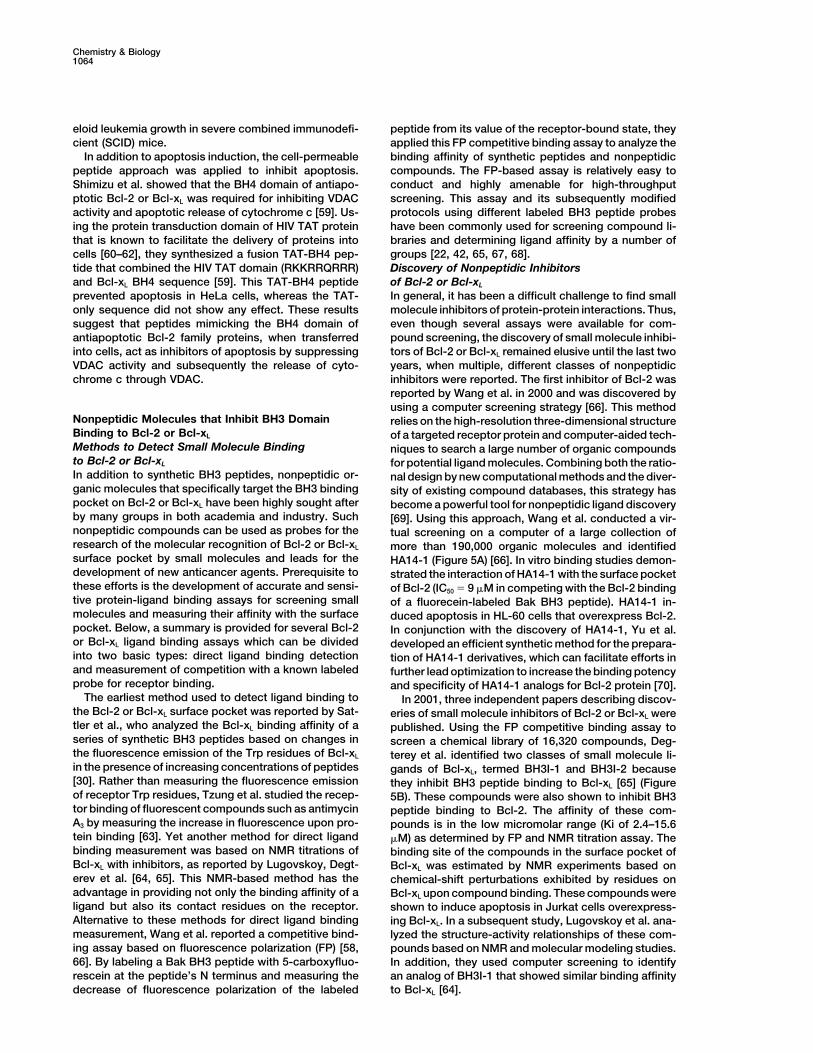
Nonpeptidic Molecules that Inhibit BH3 Domain relies on the high-resolution three-dimensional structureBinding to Bcl-2 or Bcl-xL of a targeted receptor protein and computer-aided tech-Methods to Detect Small Molecule Binding niques to search a large number of organic compoundsto Bcl-2 or Bcl-xL for potential ligand molecules. Combining both the ratio-In addition to synthetic BH3 peptides, nonpeptidic or- nal design by new computational methods and the diver-ganic molecules that specifically target the BH3 binding sity of existing compound databases, this strategy haspocket on Bcl-2 or Bcl-xL have been highly sought after become a powerful tool for nonpeptidic ligand discoveryby many groups in both academia and industry. Such [69]. Using this approach, Wang et al. conducted a vir-nonpeptidic compounds can be used as probes for the tual screening on a computer of a large collection ofresearch of the molecular recognition of Bcl-2 or Bcl-xL more than 190,000 organic molecules and identifiedsurface pocket by small molecules and leads for the HA14-1 (Figure 5A) [66]. In vitro binding studies demon-development of new anticancer agents. Prerequisite to strated the interaction of HA14-1 with the surface pocketthese efforts is the development of accurate and sensi- of Bcl-2 (IC50 � 9 �M in competing with the Bcl-2 bindingtive protein-ligand binding assays for screening small of a fluorecein-labeled Bak BH3 peptide). HA14-1 in-molecules and measuring their affinity with the surface duced apoptosis in HL-60 cells that overexpress Bcl-2.pocket. Below, a summary is provided for several Bcl-2 In conjunction with the discovery of HA14-1, Yu et al.or Bcl-xL ligand binding assays which can be divided developed an efficient synthetic method for the prepara-into two basic types: direct ligand binding detection tion of HA14-1 derivatives, which can facilitate efforts inand measurement of competition with a known labeled further lead optimization to increase the binding potencyprobe for receptor binding. and specificity of HA14-1 analogs for Bcl-2 protein [70].
The earliest method used to detect ligand binding to In 2001, three independent papers describing discov-the Bcl-2 or Bcl-xL surface pocket was reported by Sat- eries of small molecule inhibitors of Bcl-2 or Bcl-xL weretler et al., who analyzed the Bcl-xL binding affinity of a published. Using the FP competitive binding assay toseries of synthetic BH3 peptides based on changes in screen a chemical library of 16,320 compounds, Deg-the fluorescence emission of the Trp residues of Bcl-xL terey et al. identified two classes of small molecule li-in the presence of increasing concentrations of peptides gands of Bcl-xL, termed BH3I-1 and BH3I-2 because[30]. Rather than measuring the fluorescence emission they inhibit BH3 peptide binding to Bcl-xL [65] (Figureof receptor Trp residues, Tzung et al. studied the recep- 5B). These compounds were also shown to inhibit BH3tor binding of fluorescent compounds such as antimycin peptide binding to Bcl-2. The affinity of these com-A3 by measuring the increase in fluorescence upon pro- pounds is in the low micromolar range (Ki of 2.4–15.6tein binding [63]. Yet another method for direct ligand �M) as determined by FP and NMR titration assay. Thebinding measurement was based on NMR titrations of binding site of the compounds in the surface pocket ofBcl-xL with inhibitors, as reported by Lugovskoy, Degt- Bcl-xL was estimated by NMR experiments based onerev et al. [64, 65]. This NMR-based method has the chemical-shift perturbations exhibited by residues onadvantage in providing not only the binding affinity of a Bcl-xL upon compound binding. These compounds wereligand but also its contact residues on the receptor. shown to induce apoptosis in Jurkat cells overexpress-Alternative to these methods for direct ligand binding ing Bcl-xL. In a subsequent study, Lugovskoy et al. ana-measurement, Wang et al. reported a competitive bind- lyzed the structure-activity relationships of these com-ing assay based on fluorescence polarization (FP) [58, pounds based on NMR and molecular modeling studies.66]. By labeling a Bak BH3 peptide with 5-carboxyfluo- In addition, they used computer screening to identifyrescein at the peptide’s N terminus and measuring the an analog of BH3I-1 that showed similar binding affinity
to Bcl-xL [64].decrease of fluorescence polarization of the labeled
Review1065
chemical shifts as determined by NMR experimentsshowed those residues around the BH3 binding pocketof Bcl-xL to be involved in the binding of compound 6.
The most recent addition to the reported list of non-peptidic ligands of Bcl-2/Bcl-xL surface pocket was aseries of terphenyl derivatives published by Kutzki et al.[68]. Previously, this group showed that the terphenylscaffold, in a staggered conformation, closely repro-duces the projection of functionality on the surface ofan � helix [74, 75]. Extending this scaffold for the mimicryof the BH3 helix that binds the Bcl-2/Bcl-xL surfacepocket, they synthesized terphenyl molecules con-taining alkyl or aryl substituents on the three ortho posi-tions which were designed to mimic the key hydropho-bic side chains (i.e., Val-74, Leu-78, Ile-81, and Ile-85)of Bak BH3 peptide. Such compounds were indeedfound to bind to Bcl-xL as determined by the FP competi-tive binding assay, with a KD of 114 nM for the mostpotent derivative (Figure 5E). Further NMR experimentsand computational docking studies showed that thiscompound binds to the same hydrophobic surfacepocket on Bcl-xL as the Bak BH3 peptide.
Clinical Application of Inhibitors of Bcl-2 or Bcl-xL
The Bcl-2 family is implicated in many human diseases.In particular, cancer is linked to both the original discov-eries of many genes belonging to this family and much
Figure 5. Nonpeptidic Inhibitors of Bcl-2 and Bcl-xL of subsequent biological research to understand theirThe chemical structures of recently discovered nonpeptidic ligands function. The antiapoptotic members of the Bcl-2 familyof Bcl-2 and Bcl-xL, reported by several groups independently, in- are known to contribute to neoplastic cell expansion bycluding (A) HA14-1 [66], (B) BH3I-1 and BH3I-2 [65], (C) antimycin
preventing normal cell turnover caused by physiologicalA3 [63], (D) compound 6 [67], and (E) a terphenyl derivative [68].cell-death mechanisms. For example, high levels andaberrant patterns of Bcl-2 gene expression are found ina wide variety of human cancers, including �70% ofA natural product, antimycin A (Figure 5C), which is
a known antibiotic and binds cytochrome b [71, 72], was breast cancers, �30%–60% of prostate cancers, �90%of colorectal cancers, �60% of gastric cancers, 100%reported by Tzung et al. to have biological activity in
mimicking the death-inducing BH3 domain [63]. Based of small-cell lung carcinomas, �20% of non-small-celllung cancers, �30% of neuroblastomas, �80% of B cellon fluorescence emission spectra, antimycin A was
shown to compete with Bak BH3 peptide binding to the lymphomas, and variable percentages of melanomas,renal cell, and thyroid cancers, as well as acute andsurface pocket of Bcl-2 and Bcl-xL. Antimycin A inhibited
the pore-forming activity of Bcl-xL in synthetic liposomes chronic lymphocytic and nonlymphocytic leukemias [17,76]. The expression levels of Bcl-2 proteins also corre-and induced apoptosis in murine hepatocyte cell lines
transfected with Bcl-xL. To address the issue of selectiv- late with relative resistance to a wide spectrum of cur-rent chemotherapeutic drugs and �-irradiation. Sinceity of antimycin A for the Bcl-2 or Bcl-xL targets, an
analog, 2-methoxy-antimycin A3, which was previously Bcl-2 can protect against such a wide variety of drugsthat have very different mechanisms of action, it appearsknown to be inactive as an inhibitor of cytochrome b-c1
[72, 73], was studied and shown to retain binding to that all of these drugs use a common final pathway forthe eventual induction of cell death which is regulatedBcl-2. This suggested the feasibility of using antimycin
A as a template to develop more specific inhibitors of by Bcl-2.The revealed role of the Bcl-2 family in cancer andBcl-2 or Bcl-xL devoid of general mitochondrial toxicity.
Enyedy et al. used the structure-based computer resistance to cytotoxic therapies has opened new ave-nues in the development of novel anticancer strategiesscreening approach to search a collection of 206,876
compounds and identified seven compounds that had [77, 78]. An attractive strategy for inducing apoptosis incancer cells or overcoming their chemoresistance is toBcl-2 binding with IC50 values by the FP assay ranging
from 1.6 to 14.0 �M [67]. Compound 6 was the most inhibit the protective function of Bcl-2 or related anti-apoptotic proteins such as Bcl-xL that are often over-potent compound and had an IC50 value of 4 �M in
reducing the viability of HL-60 cells expressing a high expressed or upregulated in cancer cells. Apoptosis-inducing activity in cancer cell lines was observed inlevel of Bcl-2 (Figure 5D). It was further shown that com-
pound 6 was active in inducing apoptosis in HL-60 and the above-described small molecule inhibitors of Bcl-2or Bcl-xL. HA14-1 was shown to induce apoptosis inMDA-231 cells that express Bcl-2 but had much less or
no activity in T47D and MDA453 cells that express low several types of cancer cell lines, including human my-eloid leukemia (HL-60) [66], breast cancer (MDA-MB-or undetectable levels of Bcl-2. Finally, changes in
Chemistry & Biology1066
468) [79], and prostate cancer (PC3, LNCaP) (unpub- Future Research in Molecule Recognitionand Drug Design of the Bcl-2 Familylished results from laboratory). The apoptotic effect ofFrom chemical and structural perspectives, a centralHA14-1 in breast and prostate cancer cells was foundquestion that needs to be further studied is the mecha-to be dependent on Bcl-2 expression, as the compoundnism by which the BH3 domain recognizes the surfacewas not active in breast and prostate cancer cells thatpocket commonly seen in the Bcl-2 family. The BH3have very low or undetectable levels of Bcl-2. Com-domain seems to be responsible for the affinity of differ-pound 6 also displayed a selective killing effect on Bcl-ent protein-protein interactions within the Bcl-2 family,2-positive cancer cells [67]. Most recently, HA14-1 wassince synthetic peptides derived from the BH3 domainshown to induce the apoptosis of primary acute myelog-of different Bcl-2 family members showed interactionsenous leukemia (AML) and to be synergistic withwith Bcl-xL corresponding to their native proteins [30].PD184352, a MEK inhibitor, in reducing the number ofIt remains to be further elucidated as to the amino-acidCFU-blast of primary AML patients 50% more than thosesequence and structural bases that dictate the selectiv-treated with either alone [80]. The potential benefits ofity and hierarchy of associations between BH3 peptidescombining HA14-1 with other cytotoxic agents in cancer(or native BH3 domains in proapoptotic proteins) withtreatment were also suggested by another study inthe surface pocket of antiapoptotic Bcl-2 or Bcl-xL. Thewhich the use of HA14-1 together with an epothilone Bexperimentally determined structures of proapoptoticanalog was significantly more effective in killing humanBax and Bid reveal that their BH3 domain is buried inbreast cancer cells than the use of either alone [79]. Asthe core of the protein, implying that conformationalto in vivo animal models, although the activity of thischanges are necessary for the BH3 domain to interactand other nonpeptidic inhibitors of Bcl-2 or Bcl-xL havewith its antiapoptotic partners [25–27]. More needs tonot been reported and await further studies, CPM-1285,be learned about the mechanism and kinetics of thea cell-permeable peptide inhibitor of Bcl-2, has beenconformational switch and refolding of BH3 domainsshown to slow human myeloid leukemia growth in SCIDduring their binding to Bcl-2 or Bcl-xL surface pocket.mice [58].To address these questions, synthetic BH3 peptidesAn important question to be considered in the clinicalmay provide a useful model for further investigation em-application of Bcl-2 or Bcl-xL inhibitors is whether theseploying synthetic and combinatorial chemistry and bio-compounds are toxic to normal cells, which is a commonphysical techniques.limitation of current chemotherapeutic drugs. Because
Significant progress in the field was made with thethe function of Bcl-2 is not absolutely necessary in manyrecent discovery by several groups independently ofnormal cell types [81], a systemic inhibition of Bcl-2multiple classes of nonpeptidic inhibitors capable ofmay not affect the normal cellular function. Furthermore,mimicking the binding of BH3 domain to the surfaceoncogenic changes in certain cancer cells render thempocket of Bcl-2 and/or Bcl-xL. Whereas the structuresmore susceptible than normal cells to apoptosis [82].of Bcl-2 or Bcl-xL bound by these inhibitors were pre-This seems to be consistent with the observation thatdicted by using molecular modeling alone or in combina-a higher level of Bcl-2 or Bcl-xL is found in many cancers,tion with NMR experimental data, high-resolution co-which may serve to protect the vulnerability of thesecrystal or NMR structure is needed for the delineationcancers. Therefore, it can be expected that small mole-of detailed binding modes of these molecules with Bcl-2cule inhibitors targeting cancer cells highly expressingor Bcl-xL surface pocket. This will be important for theBcl-2 or Bcl-xL are more selective than conventionaldesign of new analogs with higher affinity for the surfacecytotoxic drugs. In this regard, it was encouraging topocket. Another important question is the mechanismnote that peptide inhibitor CPM-1285 had little effect onunderlying the specificity of small molecules recognizing
the viability of normal human peripheral blood lympho-the surface pocket of different Bcl-2 family members.
cytes or the proliferative response of activated lympho-All of the nonpeptidic inhibitors described above bind
cytes stimulated with PHA mitogen for 48 hr at concen-both Bcl-2 and Bcl-xL, which may not be surprising given
trations where the peptide strongly induced apoptosis that presumably they all recognize the surface pocketin cancer cells [58]. Also, a nonpeptidic inhibitor HA14-1 highly conserved in these two proteins. This may behad no noticeable effect on normal human hematopoi- acceptable or even desirable for clinical application inetic CD34� cells (determined by the average number of certain cancers, as suggested by the study of antisensecolony-forming cells with or without HA14-1 treatment), oligonucletides that are bispecific for Bcl-2 and Bcl-xLeven though at the same treatment condition HA14-1 [84, 85]. Still, inhibitors with monospecificity for eitherstrongly decreased the number of CFU-blast of primary Bcl-2 or Bcl-xL are needed if they are to be used toAML (personal communication, Dr. Michael Andreeff’s probe the function of a specific protein or treat cancergroup at M.D. Anderson Cancer Center). Finally, recent cells where targeting of only one protein is desired.encouraging data from the clinical trial indicating that Toward the development of such specific inhibitors, theantisense oligonucleotides targeted against Bcl-2 gene existing compounds can provide starting templates tocan specifically inhibit non-Hodgkin’s lymphoma in hu- generate new derivatives based on structural designmans provided an important validation of Bcl-2 as a and combinatorial chemistry.therapeutic target [83]. Since the clinical value of anti- So far, all of the reported small molecule (both peptidicsense oligonucleotides is often limited by their lack of and nonpeptidic) inhibitors target the surface pocket ofenzymatic stability, cell permeability, and oral activity, antiapoptotic Bcl-2 or Bcl-xL for triggering apoptosis insmall molecule inhibitors devoid of such limitations are cancer cells. It may also be worthwhile to target proapo-
ptotic Bcl-2 family members with small molecules tomore desired agents for clinical application.
Review1067
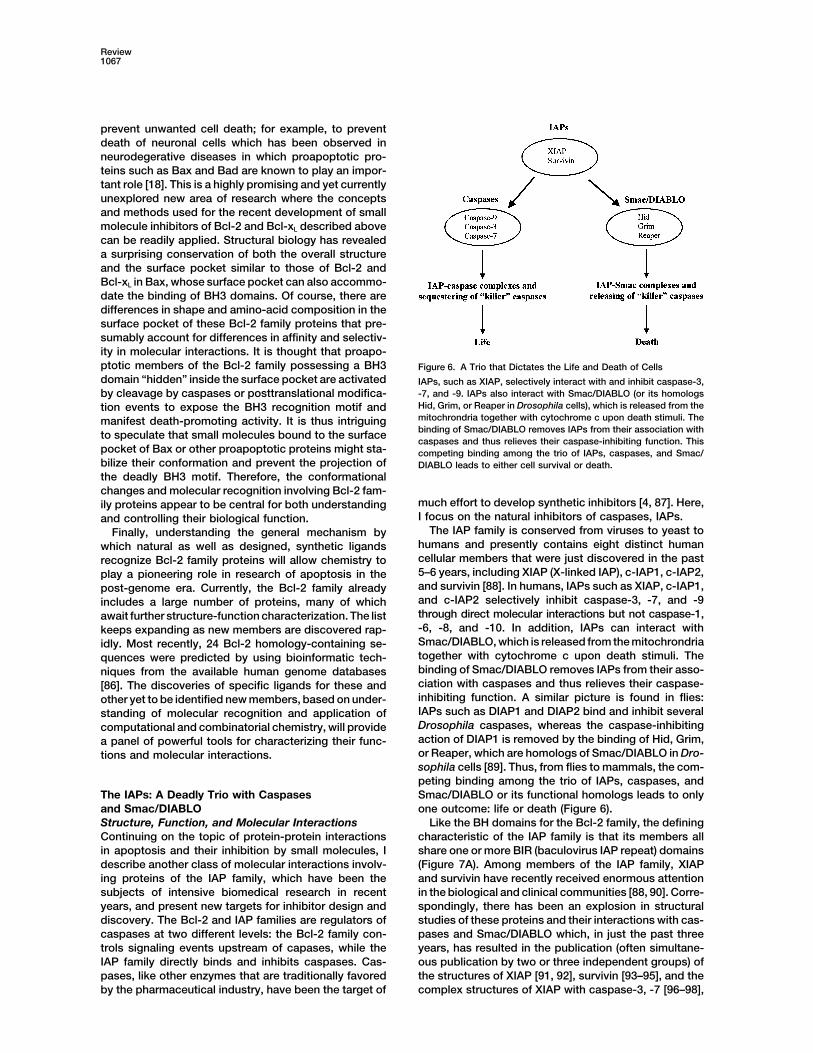
prevent unwanted cell death; for example, to preventdeath of neuronal cells which has been observed inneurodegerative diseases in which proapoptotic pro-teins such as Bax and Bad are known to play an impor-tant role [18]. This is a highly promising and yet currentlyunexplored new area of research where the conceptsand methods used for the recent development of smallmolecule inhibitors of Bcl-2 and Bcl-xL described abovecan be readily applied. Structural biology has revealeda surprising conservation of both the overall structureand the surface pocket similar to those of Bcl-2 andBcl-xL in Bax, whose surface pocket can also accommo-date the binding of BH3 domains. Of course, there aredifferences in shape and amino-acid composition in thesurface pocket of these Bcl-2 family proteins that pre-sumably account for differences in affinity and selectiv-ity in molecular interactions. It is thought that proapo-ptotic members of the Bcl-2 family possessing a BH3 Figure 6. A Trio that Dictates the Life and Death of Cellsdomain “hidden” inside the surface pocket are activated IAPs, such as XIAP, selectively interact with and inhibit caspase-3,
-7, and -9. IAPs also interact with Smac/DIABLO (or its homologsby cleavage by caspases or posttranslational modifica-Hid, Grim, or Reaper in Drosophila cells), which is released from thetion events to expose the BH3 recognition motif andmitochrondria together with cytochrome c upon death stimuli. Themanifest death-promoting activity. It is thus intriguingbinding of Smac/DIABLO removes IAPs from their association withto speculate that small molecules bound to the surfacecaspases and thus relieves their caspase-inhibiting function. This
pocket of Bax or other proapoptotic proteins might sta- competing binding among the trio of IAPs, caspases, and Smac/bilize their conformation and prevent the projection of DIABLO leads to either cell survival or death.the deadly BH3 motif. Therefore, the conformationalchanges and molecular recognition involving Bcl-2 fam-
much effort to develop synthetic inhibitors [4, 87]. Here,ily proteins appear to be central for both understandingI focus on the natural inhibitors of caspases, IAPs.and controlling their biological function.
The IAP family is conserved from viruses to yeast toFinally, understanding the general mechanism byhumans and presently contains eight distinct humanwhich natural as well as designed, synthetic ligandscellular members that were just discovered in the pastrecognize Bcl-2 family proteins will allow chemistry to5–6 years, including XIAP (X-linked IAP), c-IAP1, c-IAP2,play a pioneering role in research of apoptosis in theand survivin [88]. In humans, IAPs such as XIAP, c-IAP1,post-genome era. Currently, the Bcl-2 family alreadyand c-IAP2 selectively inhibit caspase-3, -7, and -9includes a large number of proteins, many of whichthrough direct molecular interactions but not caspase-1,await further structure-function characterization. The list-6, -8, and -10. In addition, IAPs can interact withkeeps expanding as new members are discovered rap-Smac/DIABLO, which is released from the mitochrondriaidly. Most recently, 24 Bcl-2 homology-containing se-together with cytochrome c upon death stimuli. Thequences were predicted by using bioinformatic tech-binding of Smac/DIABLO removes IAPs from their asso-niques from the available human genome databasesciation with caspases and thus relieves their caspase-[86]. The discoveries of specific ligands for these andinhibiting function. A similar picture is found in flies:other yet to be identified new members, based on under-IAPs such as DIAP1 and DIAP2 bind and inhibit severalstanding of molecular recognition and application ofDrosophila caspases, whereas the caspase-inhibitingcomputational and combinatorial chemistry, will provideaction of DIAP1 is removed by the binding of Hid, Grim,a panel of powerful tools for characterizing their func-or Reaper, which are homologs of Smac/DIABLO in Dro-tions and molecular interactions.sophila cells [89]. Thus, from flies to mammals, the com-peting binding among the trio of IAPs, caspases, andSmac/DIABLO or its functional homologs leads to onlyThe IAPs: A Deadly Trio with Caspases
and Smac/DIABLO one outcome: life or death (Figure 6).Like the BH domains for the Bcl-2 family, the definingStructure, Function, and Molecular Interactions
Continuing on the topic of protein-protein interactions characteristic of the IAP family is that its members allshare one or more BIR (baculovirus IAP repeat) domainsin apoptosis and their inhibition by small molecules, I
describe another class of molecular interactions involv- (Figure 7A). Among members of the IAP family, XIAPand survivin have recently received enormous attentioning proteins of the IAP family, which have been the
subjects of intensive biomedical research in recent in the biological and clinical communities [88, 90]. Corre-spondingly, there has been an explosion in structuralyears, and present new targets for inhibitor design and
discovery. The Bcl-2 and IAP families are regulators of studies of these proteins and their interactions with cas-pases and Smac/DIABLO which, in just the past threecaspases at two different levels: the Bcl-2 family con-
trols signaling events upstream of capases, while the years, has resulted in the publication (often simultane-ous publication by two or three independent groups) ofIAP family directly binds and inhibits caspases. Cas-
pases, like other enzymes that are traditionally favored the structures of XIAP [91, 92], survivin [93–95], and thecomplex structures of XIAP with caspase-3, -7 [96–98],by the pharmaceutical industry, have been the target of
Chemistry & Biology1068
found on the BIR2 domain of the DIAP1 protein, a memberof the IAP family which is recognized by the conservedN-terminal tetrapeptide motif in Hid and Grim [103].
Like Bcl-2 or Bcl-xL, elevated levels of IAPs are foundin cancer cells. XIAP is abnormally overexpressed inmany cancer cells [88]. Survivin is expressed in a varietyof cancers but lacks expression in differentiated adulttissues [90]. Inhibition of these protein targets by smallmolecules may allow for selective elimination of cancercells that overexpress IAPs or sensitization of these cells tochemotherapies. Consistent with this notion, an antisenseoligonucleotide that reduces survivin expression wasshown to induce apoptosis and sensitize lung cancer cellsto a chemotherapeutic drug, etoposide, but had no effecton the viability of normal blood leukocytes [104].
The Tetrapeptide Binding Pocket on IAPs: Targetfor Ligand DesignThe surface pocket on XIAP or other IAPs for the bindingof the tetrapeptide motif conserved in both caspase-9and Smac/DIABLO or its Drosophila homologs Hid,Grim, and Reaper presents an attractive target for dis-covering small molecule inhibitors (Figure 7B) [105].Synthetic peptides or nonpeptidic mimics bound to thispocket should disrupt the interaction between IAPs andcaspases, thus releasing active caspases-3, -7, and -9and facilitating apoptosis. In 2000, in vitro studies ofsynthetic peptides containing the tetrapeptide motif ofSmac/DIABLO were reported by several groups inde-pendently. Chai et al. showed that a peptide consistingof the N-terminal seven residues (i.e., the tetrapeptidemotif plus three additional residues) could promote pro-Figure 7. The BIR Domains of the IAP Family and Their Recognitioncaspase-3 activation at 10–300 �M, whereas a mutantby Natural and Synthetic Ligandspeptide containing the Met substitution at the Ala-1 po-(A) One or more BIR domains (BIR1-3) shared by the IAP family.
(B) The surface pocket on XIAP bound by the tetrapeptide motif sition or a reversed peptide sequence was not active[99, 100], which is conserved in both caspase-9 and Smac/DIABLO [106]. Srinivasula et al. synthesized peptides containingor its Drosophila homologs Hid, Grim, and Reaper. This surface the first 7 or 35 residues of Smac/DIABLO and found thatpocket offers an attractive site for discovering small molecule inhib- both were effective in promoting caspase-3 activation initors.
the XIAP-containing extracts at 100–500 �M concentra-tions and that the longer peptide was noticeably moreeffective than the short peptide [107].or Smac/DIABLO [99, 100]. From these studies, a picture
of how XIAP works with caspase-3, -7, and Smac/DIA- A detailed analysis of the structure-activity relation-ship of Smac/DIABLO N-terminal peptides was reportedBLO has emerged [101]. It has been shown that a long
peptide segment preceding the BIR2 domain of XIAP by Liu et al., who determined the NMR structure of theXIAP BIR3 domain complexed with a nine-residue pep-occupies the active site of caspase-3 or -7 (thus blocking
the access of substrates) and adjacent surfaces that tide derived from Smac/DIABLO and measured the bind-ing affinity of a series of Smac/DIABLO N-terminal pep-are specific to these two caspases (thus achieving the
specificity over other caspases) [96–98]. However, the tides of various lengths and mutants using a FPcompetitive binding assay [100]. This nine-residue pep-mechanism by which XIAP inhibits caspase-9 is quite
different. Rather than directly restricting the enzyme ac- tide adopts an extended conformation with a kink atPro3 when bound to the surface pocket on XIAP BIR3tive site for substrate cleavage, the BIR3 domain of
XIAP interacts with the N-terminal tetrapeptide (ATPF) domain (Figure 7B). The main chain of Ala-1, Val-2, andIle-4 forms extensive hydrogen bonds with the pocket,of caspase-9, which becomes available after the cleav-
age of procaspase-9, thus sequestering caspase-9 and while the side chains of Val-2, Pro3, and Ile-4 engage inhydrophobic interactions. By contrast, residues beyondpreventing it from its deadly action [102]. Surprisingly,
a similar tetrapeptide motif (AVPI) exists in the N termi- this tetrapeptide motif do not show any interaction withthe pocket. On the pocket, mutational analysis has sug-nus of Smac/DIABLO that has been shown in X-ray and
NMR structures to bind to a surface pocket on the BIR3 gested the important role in ligand binding of residuesLeu-307, Trp-310, Trp-323, E314, and D296. Consistentdomain of XIAP [99, 100]. Thus, Smac/DIABLO competes
with the same tetrapeptide binding pocket on the XIAP with the NMR and mutational analyses, synthetic pep-tides containing modifications at the N-terminal amineBIR3 domain and relieves XIAP from its complex with
caspase-9. In Drosophila, a similar binding pocket is (Ala-1) or Ala replacement at Val-2, Pro3, or Ile-4 resulted
Review1069
in loss or decrease in XIAP binding. Interestingly, a pep- The rapid progress made recently in the discovery ofnonpeptidic inhibitors of Bcl-2 and Bcl-xL seems to sug-tide containing only the first five residues is almost as
potent as the nine-residue peptide. gest that similar advances in the study of IAPs may notbe far off, given the enormous interest in these proteinsMore recently, combinatorial chemistry has been ap-
plied to the development of novel ligands for the tetra- in both academia and pharmaceutical companies.peptide binding pocket on the BIR3 domain of IAPs.Kipp et al. synthesized libraries of tetrapeptides using
Conclusionthe Smac/DIABLO N-terminal sequence as a starting
A highly regulated network of protein-protein interac-point [108]. With a competitive binding assay based on
tions is the central theme underlying the machinery ofa solvent-sensitive fluorogenic dye molecule, badan, the
programmed cell death. Understanding the chemicalbinding affinities of these tetrapeptides, which contain
basis of these interactions is a key to unlock the mysteryvariations at different residues, were determined in order
of life and death of cells at the atomic level, while control-to dissect the contribution of each residue of the tetra-
ling the outcome of these interactions offers tremendouspeptide to the total binding energy with the surface
benefits in the treatment of many human diseases. Fo-pocket on the BIR3 domain of XIAP. From the analysis
cusing on the Bcl-2 family as a model system, I summa-of a total of six libraries of related tetrapeptides, it was
rize the progress, especially during the last two years,found that Arg and Phe substitutions at positions 2 and
in the discovery of small molecule ligands targeting4, respectively, led to significant increase in binding
Bcl-2 or Bcl-xL and use of these ligands as both probesaffinity. Combining these two substitutions resulted in
and inhibitors of relevant molecular interactions and bio-a tetrapeptide, ARPF, that had the highest affinity, with
logical function. These studies have provided an excitinga KD of 20 nM.
prospect in further developing and using these novelsmall molecules for chemical biological research of mo-lecular interactions involving the Bcl-2 family and inFuture Research in Molecule Recognitionapplying similar strategies to explore and control otherand Drug Design of the IAP Familymolecular interactions that are also important for apo-The interactions of the IAP family with caspases andptosis. The interactions of IAPs with caspases andSmac/DIABLO present new targets for inhibitor design.Smac/DIABLO is one additional example described inAs highlighted by the tetrapeptide binding pocket onthis article for the application of small molecule ap-the IAPs, small synthetic peptides derived from Smac/proaches to study intracellular protein-protein interac-DIABLO have been shown to be active in inhibiting thetions mediating apoptotic signaling. Besides Bcl-2 andfunction of IAPs and promoting caspase activation. De-IAP families, there are many other protein-protein inter-spite their small sizes (the shortest being four residues),actions that can be targeted by structure-based andthese peptides display high affinity for the IAP BIR3chemical biological strategies, such as those involvingdomain, down to the low nM concentrations. These re-FADD adaptor motifs, various procaspases, Apaf-1, andsults strongly suggest the feasibility of discovering smallapoptosome, just to name a few. With the frenetic pacemolecules capable of mimicking the effect of Smac/of biomedical research in apoptosis, the list of knownDIABLO. While some of these Smac/DIABLO peptidesand yet to be known protein-protein interactions of bothwere shown to cause the activation of caspase-3 in vitro,biological and therapeutic significance goes on and sofurther studies are needed to demonstrate whether theydoes the tremendous challenge and opportunity forcan promote caspase activation and apoptosis in livingchemists and biochemists in years to come.cells and animals. Interestingly, short peptides con-
taining the N-terminal four or seven residues of Smac/AcknowledgmentsDIABLO were able to enter Jurkat cells and potentiate
apoptosis triggered by other agents [109]. To improveI apologize to my colleagues if I have missed or failed to cite theirthe cell permeability of IAP targeting peptides, the strat-works and papers here due to the specific focus and space con-
egies described above in the studies of cell-permeable straint of this review and the rapid development of this field. Thepeptide inhibitors of Bcl-2 may be readily applied. In- work conducted in our laboratory was supported by grants from
the National Institutes of Health, the American Cancer Society, anddeed, a recent paper by Fulda et al. showed that a Smacthe Sidney Kimmel Foundation for Cancer Research. I would likeN-terminal seven-residue peptide linked to the proteinto thank Professors Robert Gennis, Paul Hergenrother, and Johntransduction domain of HIV Tat protein sensitized tumorKatzenellenbogen (University of Illinois at Urbana-Champaign) forcells to the effect of apoptosis-inducing agents in vitrohelpful comments and suggestions and Dr. Dongxiang Liu for help
and in vivo [110]. in preparing the figures.Perhaps a more important goal will be the transforma-
tion of the high-affinity tetrapeptide into nonpeptidicReferences
molecules through peptidomimetic modifications and/or discovery of novel organic compounds that bind to 1. Wyllie, A.H., Kerr, J.F., and Currie, A.R. (1980). Cell death: the
significance of apoptosis. Int. Rev. Cytol. 68, 251–306.the same surface pocket. Such nonpeptidic molecules2. Thompson, C.B. (1995). Apoptosis in the pathogenesis andwill be more suitable agents for probing the function of
treatment of disease. Science 267, 1456–1462.IAPs and inhibiting their function in vivo. So far, nonpep-3. Alnemri, E.S., Livingston, D.J., Nicholson, D.W., Salvesen, G.,tidic inhibitors have not been reported for the IAPs,
Thornberry, N.A., Wong, W.W., and Yuan, J. (1996). Human ICE/whose biology and structure are starting to be under- CED-3 protease nomenclature. Cell 87, 171.stood only very recently. Thus, this is a largely unex- 4. Thornberry, N., and Lazebnik, Y. (1998). Caspases: enemies
within. Science 281, 1312–1316.plored field that offers great opportunity to chemists.
Chemistry & Biology1070
5. Green, D.R., and Reed, J.C. (1998). Mitochondria and apoptosis. 31. Desagher, S., Osen-Sand, A., Nichols, A., Eskes, R., Montessuit,S., Lauper, S., Maundrell, K., Antonsson, B., and Martinou, J.C.Science 281, 1309–1312.
6. Ashkenazi, A., and Dixit, V.M. (1998). Death receptors: signaling (1999). Bid-induced conformational change of Bax is responsi-ble for mitochondrial cytochrome c release during apoptosis.and modulation. Science 281, 1305–1308.
7. Peczuh, M.W., and Hamilton, A.D. (2000). Peptide and protein J. Cell Biol. 144, 891–901.32. Zha, J., Harada, H., Yang, E., Jockel, J., and Korsmeyer, S.J.recognition by designed molecules. Chem. Rev. 100, 2479–
2494. (1996). Serine phosphorylation of death agonist BAD in re-sponse to survival factor results in binding to 14-3-3 not Bcl-8. Cochran, A.G. (2001). Protein-protein interfaces: mimics and
inhibitors. Curr. Opin. Chem. Biol. 5, 654–659. xL. Cell 87, 619–628.33. Slee, E.A., Keogh, S.A., and Martin, S.J. (2000). Cleavage of9. Adams, J., and Cory, S. (1998). The Bcl-2 protein family: arbiters
of cell survival. Science 281, 1322–1326. BID during cytotoxic drug and UV radiation-induced apoptosisoccurs downstream of the point of Bcl-2 action and is catalysed10. Chao, D., and Korsmeyer, S. (1998). Bcl-2 family: regulators of
cell death. Annu. Rev. Immunol. 16, 395–419. by caspase-3: a potential feedback loop for amplification ofapoptosis-associated mitochondrial cytochrome c release. Cell11. Vaux, D.L., and Korsmeyer, S.J. (1999). Cell death in develop-
ment. Cell 96, 245–254. Death Differ. 95, 556–565.12. Adams, J.M., and Cory, S. (2001). Life-or-death decisions by 34. Li, H., Zhu, H., Xu, C.J., and Yuan, J. (1998). Cleavage of BID
the Bcl-2 protein family. Trends Biochem. Sci. 25, 61–66. by caspase 8 mediates the mitochondrial damage in the Fas13. Hengartner, M.O. (2000). The biochemistry of apoptosis. Nature pathway of apoptosis. Cell 94, 491–501.
407, 770–776. 35. Luo, X., Budihardjo, I., Zou, H., Slaughter, C., and Wang, X.14. Zimmermann, K.C., Bonzon, C., and Green, D.R. (2001). The (1998). Bid, a Bcl2 interacting protein, mediates cytochrome c
machinery of programmed cell death. Pharmacol. Ther. 92, release from mitochondria in response to activation of cell sur-57–70. face death receptors. Cell 94, 481–490.
15. Budihardjo, I., Oliver, H., Lutter, M., Luo, X., and Wang, X. (1999). 36. Kelekar, A., and Thompson, C.B. (1998). Bcl-2-family proteins:Biochemical pathways of caspase activation during apoptosis. the role of the BH3 domain in apoptosis. Trends Cell Biol. 8,Annu. Rev. Cell Dev. Biol. 15, 269–290. 324–330.
16. Wyllie, A.H. (1997). Apoptosis and carcinogenesis. Eur. J. Cell 37. Yin, X.M., Oltvai, Z.N., and Korsmeyer, S.J. (1994). BH1 and BH2Biol. 73, 189–197. domains of Bcl-2 are required for inhibition of apoptosis and
17. Reed, J.C., Miyashita, T., Takayama, S., Wang, H.G., Sato, T., heterodimerization with Bax. Nature 369, 321–323.Krajewski, S., Aime-Sempe, C., Bodrug, S., Kitada, S., and Ha- 38. Chittenden, T., Flemington, C., Houghton, A.B., Ebb, R.G., Gallo,nada, M. (1996). BCL-2 family proteins: regulators of cell death G.J., Elangovan, B., Chinnadurai, G., and Lutz, R.J. (1995). Ainvolved in the pathogenesis of cancer and resistance to ther- conserved domain in Bak, distinct from BH1 and BH2, mediatesapy. J. Cell. Biochem. 60, 23–32. cell death and protein binding functions. EMBO J. 14, 5589–
19. Andreeff, M., Goodrich, D.W., and Pardee, A.B. (2000). Chapter strom, S., Avery, B.J., Ebb, R.G., Subramanian, T., Chittenden,2: Cell Proliferation, Differentiation and Apoptosis. (Hamilton, T., Lutz, R.J., et al. (1995). Bik, a novel death-inducing proteinOntario, Canada: B.C. Decker, Inc.). shares a distinct sequence motif with Bcl-2 family proteins and
20. Fesik, S.W. (2000). Insights into programmed cell death through interacts with viral and cellular survival-promoting proteins. On-structural biology. Cell 103, 273–282. cogene 11, 1921–1928.
21. Shi, Y. (2001). A structural view of mitochondria-mediated apo- 40. Zha, H., Aime-Sempe, C., Sato, T., and Reed, J.C. (1996). Pro-ptosis. Nat. Struct. Biol. 8, 394–401. apoptotic protein Bax heterodimerizes with Bcl-2 and homodi-
22. Petros, A.M., Medek, A., Nettesheim, D.G., Kim, D.H., Yoon, merizes with Bax via a novel domain (BH3) distinct from BH1H.S., Swift, K., Matayoshi, E.D., Oltersdorf, T., and Fesik, S.W. and BH2. J. Biol. Chem. 271, 7440–7444.(2001). Solution structure of the antiapoptotic protein bcl-2. 41. Kelekar, A., Chang, B., Harlan, J., Fesik, S., and Thompson, C.Proc. Natl. Acad. Sci. USA 98, 3012–3017. (1997). Bad is a BH3 domain-containing protein that forms an
23. Muchmore, S.W., Sattler, M., Liang, H., Meadows, R.P., Harlan, inactivating dimer with Bcl-xL. Mol. Cell. Biol. 17, 7040–7046.J.E., Yoon, H.S., Nettesheim, D., Chang, B.S., Thompson, C.B., 42. Petros, A.M., Nettesheim, D.G., Wang, Y., Olejniczak, E.T.,Wong, S.L., et al. (1996). X-ray and NMR structure of human Bcl- Meadows, R.P., Mack, J., Swift, K., Matayoshi, E.D., Zhang, H.,xL, an inhibitor of programmed cell death. Nature 381, 335–341. Thompson, C.B., et al. (2000). Rationale for Bcl-xL/Bad peptide
24. Aritomi, M., Kunishima, N., Inohara, N., Ishibashi, Y., Ohta, S.,complex formation from structure, mutagenesis, and biophysi-
and Morikawa, K. (1997). Crystal structure of rat Bcl-xL. Implica-cal studies. Protein Sci. 9, 2528–2534.
tions for the function of the Bcl-2 protein family. J. Biol. Chem.43. Ottilie, S., Diaz, J.L., Horne, W., Chang, J., Wang, Y., Wilson,
272, 27886–27892.G., Chang, S., Weeks, S., Fritz, L.C., and Oltersdorf, T. (1997).25. Suzuki, M., Youle, R.J., and Tjandra, N. (2000). Structure of Bax:Dimerization properties of human BAD. Identification of a BH-3coregulation of dimer formation and intracellular localization.domain and analysis of its binding to mutant BCL-2 and BCL-Cell 103, 645–654.xL proteins. J. Biol. Chem. 272, 30866–30872.26. McDonnell, J.M., Fushman, D., Milliman, C.L., Korsmeyer, S.J.,
44. Diaz, J.L., Oltersdorf, T., Horne, W., McConnell, M., Wilson, G.,and Cowburn, D. (1999). Solution structure of the proapoptoticWeeks, S., Garcia, T., and Fritz, L.C. (1997). A common bindingmolecule BID: a structural basis for apoptotic agonists and an-site mediates heterodimerization and homodimerization oftagonists. Cell 96, 625–634.Bcl-2 family members. J. Biol. Chem. 272, 11350–11355.27. Chou, J.J., Li, H., Salvesen, G.S., Yuan, J., and Wagner, G.
45. Cosulich, S., Worrall, V., Hedge, P., Green, S., and Clarke, P.(1999). Solution structure of BID, an intracellular amplifier of(1997). Regulation of apoptosis by BH3 domains in a cell-freeapoptotic signaling. Cell 96, 615–624.system. Curr. Biol. 7, 913–920.28. Yang, E., Zha, J., Jockel, J., Boise, L., Thompson, C., and Kors-
46. Jurgensmeier, J.M., Xie, Z., Deveraux, Q., Ellerby, L., Bredesen,meyer, S. (1995). Bad, a heterodimeric partner for Bcl-xL andD., and Reed, J.C. (1998). Bax directly induces release of cyto-Bcl-2, displaces Bax and promotes cell death. Cell 80, 285–291.chrome c from isolated mitochondria. Proc. Natl. Acad. Sci.29. Oltvai, Z.N., Milliman, C.L., and Korsmeyer, S.J. (1993). Bcl-2USA 95, 4997–5002.heterodimerizes in vivo with a conserved homolog, Bax, that
47. Narita, M., Shimizu, S., Ito, T., Chittenden, T., Lutz, R.J., Mat-accelerates programmed cell death. Cell 74, 609–619.suda, H., and Tsujimoto, Y. (1998). Bax interacts with the perme-30. Sattler, M., Liang, H., Nettesheim, D., Meadows, R.P., Harlan,ability transition pore to induce permeability transition and cyto-J.E., Eberstadt, M., Yoon, H.S., Shuker, S.B., Chang, B.S., Minn,chrome c release in isolated mitochondria. Proc. Natl. Acad.A.J., et al. (1997). Structure of Bcl-xL-Bak peptide complex:Sci. USA 95, 14681–14686.recognition between regulators of apoptosis. Science 275,
(2000). 91st Annual Meeting of the American Association for Croce, C.M., Alnemri, E.S., and Huang, Z. (2000). Structure-based discovery of a novel organic compound that binds Bcl-Cancer Research. 42, 4693.
49. Theodore, L., Derossi, D., Chassaing, G., Llirbat, B., Kubes, M., 2 protein and induces apoptosis of tumor cells. Proc. Natl. Acad.Sci. USA 97, 7124–7129.Jordan, P., Chneiweiss, H., Godement, P., and Prochiantz, A.
(1995). Intraneuronal delivery of protein kinase C pseudosub- 67. Enyedy, I.J., Ling, Y., Nacro, K., Tomita, Y., Wu, X., Cao, Y.,Guo, R., Li, B., Zhu, X., Huang, Y., et al. (2001). Discovery ofstrate leads to growth cone collapse. J. Neurosci. 15, 7158–
7167. small-molecule inhibitors of Bcl-2 through structure-basedcomputer screening. J. Med. Chem. 44, 4313–4324.50. Ball, K.L., Lain, S., Fahraeus, R., Smythe, C., and Lane, D.P.
(1997). Cell-cycle arrest and inhibition of Cdk4 activity by small 68. Kutzki, O., Park, H.S., Ernst, J.T., Orner, B.P., Yin, H., and Hamil-ton, A.D. Development of a potent Bcl-xL antagonist based onpeptides based on the carboxy-terminal domain of p21WAF1.
Curr. Biol. 7, 71–80. a-helix mimicry. J. Am. Chem. Soc., in press.69. Huang, Z. (2000). Structural chemistry and therapeutic interven-51. Bonfanti, M., Taverna, S., Salmona, M., D’Incalci, M., and Brog-
gini, M. (1997). p21WAF1-derived peptides linked to an internal- tion of protein-protein interaction in immune response, HIV entryand apoptosis. Pharmacol. Ther. 86, 201–215.ization peptide inhibit human cancer cell growth. Cancer Res.
57, 1442–1446. 70. Yu, N., Aramini, J.M., Germann, M.W., and Huang, Z. (2000).52. Selivanova, G., Iotsova, V., Okan, I., Fritsche, M., Strom, M., Reactions of salicylaldehydes with alkyl cyanoacetates on the
Groner, B., Grafstrom, R.C., and Wiman, K.G. (1997). Restoration surface of solid catalysts: syntheses of 4H-chromene deriva-of the growth suppression function of mutant p53 by a synthetic tives. Tetrahedron Lett. 41, 6993–6996.peptide derived from the p53 C-terminal domain. Nat. Med. 3, 71. Dunshee, B.R., Leben, C., Keitt, G.W., and Strong, F.M. (1949).632–638. The isolation and properties of antimycin A. J. Am. Chem. Soc.
53. Williams, E.J., Dunican, D.J., Green, P.J., Howell, F.V., Derossi, 71, 2436–2437.D., Walsh, F.S., and Doherty, P. (1997). Selective inhibition of 72. Tokutake, N., Miyoshi, H., Satoh, T., Hatano, T., and Iwamura,growth factor-stimulated mitogenesis by a cell-permeable H. (1994). Structural factors of antimycin A molecule requiredGrb2-binding peptide. J. Biol. Chem. 272, 22349–22354. for inhibitory action. Biochim. Biophys. Acta 1185, 271–278.
54. Hall, H., Williams, E.J., Moore, S.E., Walsh, F.S., Prochiantz, A., 73. Miyoshi, H., Tokutake, N., Imaeda, Y., Akagi, T., and Iwamura,and Doherty, P. (1996). Inhibition of FGF-stimulated phosphati- H. (1995). A model of antimycin A binding based on structure-dylinositol hydrolysis and neurite outgrowth by a cell-membrane activity studies of synthetic antimycin A analogues. Biochim.permeable phosphopeptide. Curr. Biol. 6, 580–587. Biophys. Acta 1229, 149–154.
55. Holinger, E.P., Chittenden, T., and Lutz, R.J. (1999). Bak BH3 74. Orner, B.P., Ernst, J.T., and Hamilton, A.D. (2001). Towards pro-peptides antagonize Bcl-xL function and induce apoptosis teomimetics: terphenyl derivatives as structural and functionalthrough cytochrome c–independent activation of caspases. J. mimics of extended regions of an a-helix. J. Am. Chem. Soc.Biol. Chem. 274, 13298–13304. 123, 5382–5383.
56. Ioannides, C.G., Freedman, R.S., Liskamp, R.M., Ward, N.E., 75. Ernst, J.T., Kutzki, O., Debnath, A.K., Jiang, S., Lu, H., andand O’Brian, C.A. (1990). Inhibition of IL-2 receptor induction Hamilton, A.D. (2002). Design of a protein surface antagonistand IL-2 production in the human leukemic cell line Jurkat by based on a-helix mimicry: inhibition of gp41 assembly and virala novel peptide inhibitor of protein kinase C. Cell. Immunol. 131, fusion. Angew. Chem. Int. Ed. 41, 278–281.242–252. 76. Reed, J.C. (1994). Bcl-2 and the regulation of programmed cell
57. Kole, H.K., Garant, M.J., Kole, S., and Bernier, M. (1996). A death. J. Cell Biol. 124, 1–6.peptide-based protein-tyrosine phosphatase inhibitor specifi- 77. Huang, Z. (2000). The Bcl-2 family of proteins as targets forcally enhances insulin receptor function in intact cells. J. Biol. anticancer drug design. Oncogene 19, 6627–6631.Chem. 271, 14302–14307. 78. Liu, D., and Huang, Z. (2001). Synthetic peptides and non-pep-
58. Wang, J.L., Zhang, Z.J., Choksi, S., Shan, S., Lu, Z., Croce, tidic molecules as probes of structure and function of Bcl-2C.M., Alnemri, E.S., Korngold, R., and Huang, Z. (2000). Cell family proteins and modulators of apoptosis. Apoptosis 6,Permeable Bcl-2 Binding Peptides: A Chemical Approach to 453–462.Apotosis Induction in Tumor Cells. Cancer Res. 60, 1498–1502. 79. Yamaguchi, H., Paranawithana, S.R., Lee, M.W., Huang, Z.W.,
59. Shimizu, S., Konishi, A., Kodama, T., and Tsujimoto, Y. (2000). Bhalla, K.N., and Wang, H.G. (2002). Epothione B analogueBH4 domain of antiapoptotic Bcl-2 family members closes volt- (BMS-247550)-mediated cytotoxicity through induction of Baxage-dependent anion channel and inhibits apoptotic mitochon- conformational change in human breast cancer cells. Cancerdrial changes and cell death. Proc. Natl. Acad. Sci. USA 97, Res. 62, 466–471.3100–3105.
80. Milella, M., Kornblau, S.M., Estrov, Z., Konopleva, M., Tari, A.,60. Green, M., and Loewenstein, P.M. (1988). Autonomous func-
Schober, W.D., Carter, B., Harris, D., Leysath, C.L., Berestein,tional domains of chemically synthesized human immunodefi-
G.L., et al. (2002). Simultaneous disruption of the Bcl-2 andciency virus tat trans-activator protein. Cell 55, 1179–1188.
MEK/MAPK pathways sinergistically induces apoptosis in acute61. Derossi, D., Calvet, S., Trembleau, A., Brunissen, A., Chassaing,myelogenous leukemia. Blood 62, 466–471.G., and Prochiantz, A. (1996). Cell internalization of the third helix
81. Veis, D., Sorenson, C., Shutter, J., and Korsmeyer, S. (1993).of the Antennapedia homeodomain is receptor-independent. J.Bcl-2-deficient mice demonstrate fulminant lymphoid apopto-Biol. Chem. 271, 18188–18193.sis, polycystic kidneys, and hypopigmented hair. Cell 75,62. Schwarze, S.R., Ho, A., Vocero-Akbani, A., and Dowdy, S.F.229–240.(1999). In vivo protein transduction: delivery of a biologically
82. Evan, G., and Littlewood, T. (1998). A matter of life and cellactive protein into the mouse. Science 285, 1569–1572.death. Science 281, 1317–1322.63. Tzung, S.P., Kim, K.M., Basanez, G., Giedt, C.D., Simon, J.,
83. Webb, A., Cunningham, D., Cotter, F., Clarke, P., di Stefano,Zimmerberg, J., Zhang, K.Y., and Hockenbery, D.M. (2001). Anti-F., Ross, P., Corbo, M., and Dziewanowska, Z. (1997). Bcl-2mycin A mimics a cell-death-inducing Bcl-2 homology domainantisense therapy in patients with non-Hodgkin lymphoma. Lan-3. Nat. Cell Biol. 3, 183–191.cet 349, 1137–1141.64. Lugovskoy, A.A., Degterev, A.I., Fahmy, A.F., Zhou, P., Gross,
84. Zangemeister-Wittke, U., Leech, S.H., Olie, R.A., Simoes-Wust,J.D., Yuan, J., and Wagner, G. (2002). A novel approach forA.P., Gautschi, O., Luedke, G.H., Natt, F., Haner, R., Martin, P.,characterizing protein ligand complexes: molecular basis forHall, J., et al. (2000). A novel bispecific antisense oligonucleotidespecificity of small-molecule Bcl-2 inhibitors. J. Am. Chem. Soc.inhibiting both bcl-2 and bcl-xL expression efficiently induces124, 1234–1240.apoptosis in tumor cells. Clin. Cancer Res. 6, 2547–2555.65. Degterev, A., Lugovskoy, A., Cardone, M., Mulley, B., Wagner,
85. Strasberg Rieber, M., Zangemeister-Wittke, U., and Rieber, M.G., Mitchison, T., and Yuan, J. (2001). Identification of small-(2001). p53-Independent induction of apoptosis in human mela-molecule inhibitors of interaction between the BH3 domain andnoma cells by a Bcl-2/Bcl-xL bispecific antisense oligonucleo-Bcl-xL. Nat. Cell Biol. 3, 173–182.
66. Wang, J., Liu, D., Zhang, Z., Shan, S., Han, X., Srinivasula, S.M., tide. Clin. Cancer Res. 7, 1446–1451.
Chemistry & Biology1072
Huang, Z., and Alnemri, E.S. (2000). Molecular determinants86. Gozani, O., Boyce, M., Yoo, L., Karuman, P., and Yuan, J.of the caspase-promoting activity of Smac/DIABLO and its(2002). Life and death in paradise. Nat. Cell Biol. 4, E159–E162.role in the death receptor pathway. J. Biol. Chem. 275, 36152–87. Thornberry, N.A., Rano, T.A., Peterson, E.P., Rasper, D.M.,36157.Timkey, T., Garcia-Calvo, M., Houtzager, V.M., Nordstrom,
108. Kipp, R.A., Case, M.A., Wist, A.D., Cresson, C.M., Carrell, M.,P.A., Roy, S., Vaillancourt, J.P., et al. (1997). A combinatorialGriner, E., Wiita, A., Albiniak, P.A., Chai, J., Shi, Y., et al. (2002).approach defines specificities of members of the caspase fam-Molecular targeting of inhibitor of apoptosis proteins basedily and granzyme B. Functional relationships established foron small molecule mimics of natural binding partners. Bio-key mediators of apoptosis. J. Biol. Chem. 272, 17907–17911.chemistry 41, 7344–7349.88. Deveraux, Q.L., and Reed, J.C. (1999). IAP family proteins–
109. Guo, F., Nimmanapalli, R., Paranawithana, S., Wittman, S.,suppressors of apoptosis. Genes Dev. 13, 239–252.Griffin, D., Bali, P., O’Bryan, E., Fumero, C., Wang, H.G., and89. Hay, B.A. (2000). Understanding IAP function and regulation:Bhalla, K. (2002). Ectopic overexpression of second mitochon-a view from Drosophila. Cell Death Differ. 7, 1045–1056.dria-derived activator of caspases (Smac/DIABLO) or cotreat-90. Altieri, D.C. (2001). The molecular basis and potential role ofment with N-terminus of Smac/DIABLO peptide potentiatessurvivin in cancer diagnosis and therapy. Trends Mol. Med. 7,epothilone B derivative-(BMS 247550) and Apo-2L/TRAIL-in-542–547.duced apoptosis. Blood 99, 3419–3426.91. Sun, C., Cai, M., Gunasekera, A.H., Meadows, R.P., Wang, H.,
110. Fulda, S., Wick, W., Weller, M., and Debatin, K.M. (2002). SmacChen, J., Zhang, H., Wu, W., Xu, N., Ng, S.C., et al. (1999).agonists sensitize for Apo2L/TRAIL- or anticancer drug-in-NMR structure and mutagenesis of the inhibitor-of-apoptosisduced apoptosis and induce regression of malignant gliomaprotein XIAP. Nature 401, 818–822.in vivo. Nat. Med. 8, 808–815.92. Sun, C., Cai, M., Meadows, R.P., Xu, N., Gunasekera, A.H.,
Herrmann, J., Wu, J.C., and Fesik, S.W. (2000). NMR structureand mutagenesis of the third Bir domain of the inhibitor ofapoptosis protein XIAP. J. Biol. Chem. 275, 33777–33781.
93. Chantalat, L., Skoufias, D.A., Kleman, J.P., Jung, B., Dideberg,O., and Margolis, R.L. (2000). Crystal structure of human sur-vivin reveals a bow tie-shaped dimer with two unusual alpha-helical extensions. Mol. Cell 6, 183–189.
94. Muchmore, S.W., Chen, J., Jakob, C., Zakula, D., Matayoshi,E.D., Wu, W., Zhang, H., Li, F., Ng, S.C., and Altieri, D.C. (2000).Crystal structure and mutagenic analysis of the inhibitor-of-apoptosis protein survivin. Mol. Cell 6, 173–182.
95. Verdecia, M.A., Huang, H., Dutil, E., Kaiser, D.A., Hunter, T.,and Noel, J.P. (2000). Structure of the human anti-apoptoticprotein survivin reveals a dimeric arrangement. Nat. Struct.Biol. 7, 602–608.
96. Chai, J., Shiozaki, E., Srinivasula, S.M., Wu, Q., Datta, P.,Alnemri, E.S., Shi, Y., and Dataa, P. (2001). Structural basis ofcaspase-7 inhibition by XIAP. Cell 104, 769–780.
97. Huang, Y., Park, Y.C., Rich, R.L., Segal, D., Myszka, D.G., andWu, H. (2001). Structural basis of caspase inhibition by XIAP:differential roles of the linker versus the BIR domain. Cell 104,781–790.
98. Riedl, S.J., Renatus, M., Schwarzenbacher, R., Zhou, Q., Sun,C., Fesik, S.W., Liddington, R.C., and Salvesen, G.S. (2001).Structural basis for the inhibition of caspase-3 by XIAP. Cell104, 791–800.
99. Wu, G., Chai, J., Suber, T.L., Wu, J.W., Du, C., Wang, X., andShi, Y. (2000). Structural basis of IAP recognition by Smac/DIABLO. Nature 408, 1008–1012.
100. Liu, Z., Sun, C., Olejniczak, E.T., Meadows, R.P., Betz, S.F.,Oost, T., Herrmann, J., Wu, J.C., and Fesik, S.W. (2000). Struc-tural basis for binding of Smac/DIABLO to the XIAP BIR3 do-main. Nature 408, 1004–1008.
101. Fesik, S.W., and Shi, Y. (2001). Controlling the caspase. Sci-ence 294, 1477–1478.
102. Srinivasula, S.M., Hegde, R., Saleh, A., Datta, P., Shiozaki, E.,Chai, J., Lee, R.A., Robbins, P.D., Fernandes-Alnemri, T., Shi,Y., et al. (2001). A conserved XIAP-interaction motif in cas-pase-9 and Smac/DIABLO regulates caspase activity and apo-ptosis. Nature 410, 112–116.
103. Wu, J.W., Cocina, A.E., Chai, J., Hay, B.A., and Shi, Y. (2001).Structural analysis of a functional DIAP1 fragment bound togrim and hid peptides. Mol. Cell 8, 95–104.
104. Olie, R.A., Simoes-Wust, A.P., Baumann, B., Leech, S.H., Fab-bro, D., Stahel, R.A., and Zangemeister-Wittke, U. (2001). Anovel antisense oligonucleotide targeting survivin expressioninduces apoptosis and sensitizes lung cancer cells to chemo-therapy. Cancer Res. 60, 2805–2809.
105. Shi, Y. (2002). A conserved tetrapeptide motif: potentiatingapoptosis through IAP-binding. Cell Death Differ. 9, 93–95.
106. Chai, J., Du, C., Wu, J.W., Kyin, S., Wang, X., and Shi, Y. (2000).Structural and biochemical basis of apoptotic activation bySmac/DIABLO. Nature 406, 855–862.