Chemotaxis of Pseudomonas spp. to the PolyaromaticHydrocarbon Naphthalene
ANN C. GRIMM AND CAROLINE S. HARWOOD*
Department of Microbiology, The University of Iowa, Iowa City, Iowa
Received 15 April 1997/Accepted 24 July 1997
Two naphthalene-degrading bacteria, Pseudomonas putida G7 and Pseudomonas sp. strain NCIB 9816-4, werechemotactically attracted to naphthalene in drop assays and modified capillary assays. Growth on naphthaleneor salicylate induced the chemotactic response. P. putida G7 was also chemotactic to biphenyl; other polyaro-matic hydrocarbons that were tested did not appear to be chemoattractants for either Pseudomonas strain.Strains that were cured of the naphthalene degradation plasmid were not attracted to naphthalene.
The bacterial degradation of a number of toxic organic com-pounds, including toluene, naphthalene, and chlorinated bi-phenyls, has been extensively studied. Many of the enzymesinvolved in the degradation of these compounds have beenpurified, and the genes encoding these proteins have beencloned and sequenced (17, 22, 24). Although the actual path-ways of catabolism are well-known, an aspect of degradationthat has been overlooked is chemotaxis. Chemotaxis enhancesthe ability of motile bacteria to locate and degrade low con-centrations of organic compounds, and it is reasonable to ex-pect that it also directs the movement of motile bacteria totoxic, but metabolizable, compounds present in contaminatedenvironments. Naphthalene is a priority pollutant commonlyfound in industrial effluents and is a constituent of coal tar(23). It is often used as a model compound for studies of in situbiodegradation of polyaromatic hydrocarbons (PAHs) becauseit is relatively easily degraded by bacteria (26). Naphthalene’srelatively high solubility compared to those of other PAHs (15)and the fact that the naphthalene degradation genes are plas-mid encoded (4, 5, 8, 16, 18, 21, 26–28) have contributed to arapid pace of laboratory research on naphthalene degradation.In this study we examined the abilities of two Pseudomonasstrains to respond chemotactically to naphthalene and otherPAHs in two chemotaxis assays.
The naphthalene-degrading strains used, Pseudomonasputida G7 (4) and Pseudomonas sp. strain NCIB 9816-4 (20), aswell as their naphthalene degradation plasmid-cured deriva-tives P. putida G7.C1 (18, 26) and Pseudomonas sp. strain9816-4 C84 (19), were obtained from D. T. Gibson of theUniversity of Iowa. All strains were motile by means of polarflagella. Cells were grown in a basal mineral salt medium (11)at 30°C with shaking at 250 rpm. All compounds used asgrowth substrates or tested as possible attractants in chemo-taxis assays were obtained from Sigma Chemical Co. (St. Louis,Mo.). Salicylate and succinate were used as growth substratesat final concentrations of 5 and 10 mM, respectively. Naphtha-lene was provided as a carbon source by direct addition ofnaphthalene crystals to liquid basal medium.
Chemotaxis was tested with a drop assay (6) and a modifiedcapillary assay. For the drop assay, 40 ml of cells in the loga-rithmic phase of growth were harvested and resuspended in 12ml of chemotaxis buffer (100 mM potassium phosphate [pH
7.0], 20 mM EDTA). For strain 9816-4, 10 mM succinate wasadded to the chemotaxis buffer as an energy source, since thisstrain’s motility decreased rapidly in unamended buffer. A 1%aqueous solution of hydroxypropylmethylcellulose (formulatedto give a viscosity of about 4,000 cP in 2% aqueous solution)(Sigma Chemical Co.) was added to the cell suspension to givea final volume of 15 ml. The viscous cell suspension was thenlayered on the bottoms of 60-mm-diameter petri dishes to adepth of about 3 mm. A small amount of a test attractant wasadded to the center of a dish either as crystals, for the poorlysoluble PAHs, or as 10-ml drops of 500 mM salicylate, a 10%Casamino Acids solution, or chemotaxis buffer. A chemotacticresponse of cells to the added compound resulted in the for-mation of a ring of turbidity near the center of the petri dishafter about an hour. To photograph drop assays, petri disheswere placed on a transparent plastic sheet with supports andblack paper was placed under the supports.
The second chemotaxis assay, developed as a modification ofthe classical capillary assay (1), allowed qualitative assessmentof chemotaxis with a phase-contrast microscope. Cells grownto mid-logarithmic phase were harvested and resuspended inchemotaxis buffer (strain G7) or chemotaxis buffer with 10 mMsuccinate (strain 9816-4) to an A660 of approximately 0.1. Thesuspension of motile cells was placed in a small chamberformed by placing a U-shaped glass tube between a microscopeslide and coverslip. A heat-sealed 1-ml capillary tube was filledwith 100 mM potassium phosphate buffer (pH 7.0), and thenthe open end was packed with finely ground crystals of naph-thalene or another insoluble aromatic compound by pressingthe capillary tube into a mound of crystals. The phosphatebuffer was drawn in first to avoid the formation of air pocketsaround the naphthalene crystals, as this could result in aero-taxis. Phosphate buffer agar capillaries were prepared by draw-ing 1.5% molten Noble agar (Difco, Detroit, Mich.) dissolvedin 100 mM potassium phosphate into a heated capillary asdescribed by Masduki et al. (14). A capillary was then insertedinto a chemotaxis chamber that had been placed on a micro-scope stage. A dark background was achieved by setting theoptical objective at 310 magnification and the phase-contrastring at the setting normally used for 3100 magnification. Thetip of the capillary was brought into focus and photographedat appropriate times. A positive chemotactic response wasvisualized by the accumulation of a cloud of motile cellsaround the mouth of the capillary tube over a period ofabout 10 min.
P. putida G7 and Pseudomonas sp. strain 9816-4 cells weremotile when grown with naphthalene as a sole source of carbon
* Corresponding author. Mailing address: Department of Microbi-ology, University of Iowa, Iowa City, IA 52242. Phone: (319) 335-7783.Fax: (319) 335-7679. E-mail: [email protected].
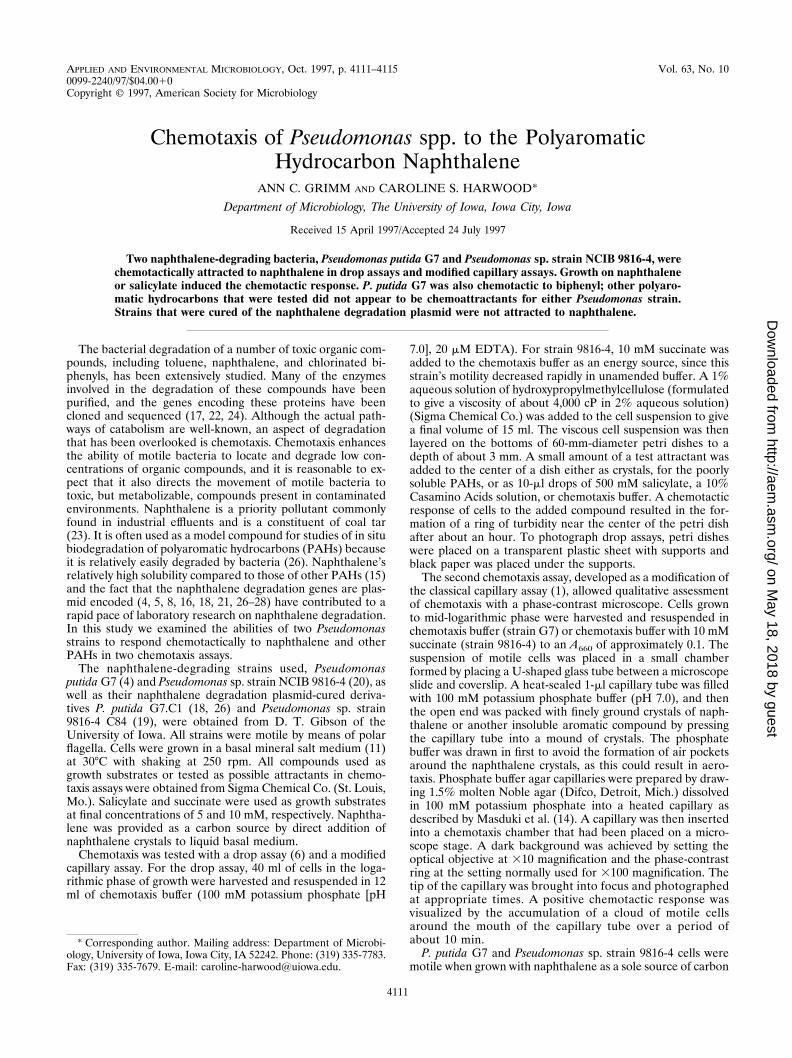
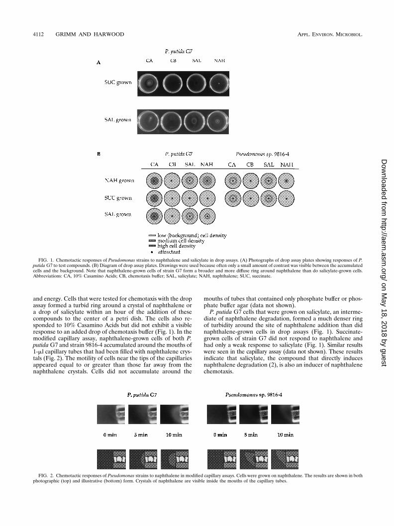
and energy. Cells that were tested for chemotaxis with the dropassay formed a turbid ring around a crystal of naphthalene ora drop of salicylate within an hour of the addition of thesecompounds to the center of a petri dish. The cells also re-sponded to 10% Casamino Acids but did not exhibit a visibleresponse to an added drop of chemotaxis buffer (Fig. 1). In themodified capillary assay, naphthalene-grown cells of both P.putida G7 and strain 9816-4 accumulated around the mouths of1-ml capillary tubes that had been filled with naphthalene crys-tals (Fig. 2). The motility of cells near the tips of the capillariesappeared equal to or greater than those far away from thenaphthalene crystals. Cells did not accumulate around the
mouths of tubes that contained only phosphate buffer or phos-phate buffer agar (data not shown).
P. putida G7 cells that were grown on salicylate, an interme-diate of naphthalene degradation, formed a much denser ringof turbidity around the site of naphthalene addition than didnaphthalene-grown cells in drop assays (Fig. 1). Succinate-grown cells of strain G7 did not respond to naphthalene andhad only a weak response to salicylate (Fig. 1). Similar resultswere seen in the capillary assay (data not shown). These resultsindicate that salicylate, the compound that directly inducesnaphthalene degradation (2), is also an inducer of naphthalenechemotaxis.
FIG. 1. Chemotactic responses of Pseudomonas strains to naphthalene and salicylate in drop assays. (A) Photographs of drop assay plates showing responses of P.putida G7 to test compounds. (B) Diagram of drop assay plates. Drawings were used because often only a small amount of contrast was visible between the accumulatedcells and the background. Note that naphthalene-grown cells of strain G7 form a broader and more diffuse ring around naphthalene than do salicylate-grown cells.Abbreviations: CA, 10% Casamino Acids; CB, chemotaxis buffer; SAL, salicylate; NAH, naphthalene; SUC, succinate.
FIG. 2. Chemotactic responses of Pseudomonas strains to naphthalene in modified capillary assays. Cells were grown on naphthalene. The results are shown in bothphotographic (top) and illustrative (bottom) form. Crystals of naphthalene are visible inside the mouths of the capillary tubes.
Pseudomonas sp. strain 9816-4 cells grown on succinate hadlittle to no response to naphthalene, indicating that this strainalso has an inducible response to this PAH (Fig. 1). Cellsgrown on 5 mM salicylate alone had very poor motility, asviewed microscopically, and so were not tested in chemotaxisassays. However, 9816-4 cells grown with 5 mM benzoate and0.5 mM salicylate were motile and attracted to both naphtha-lene and salicylate (data not shown). In contrast to the situa-tion with naphthalene, succinate-grown cells had a good re-sponse to salicylate (Fig. 1). This suggests that chemotaxis tosalicylate may be a constitutive property of strain 9816-4.
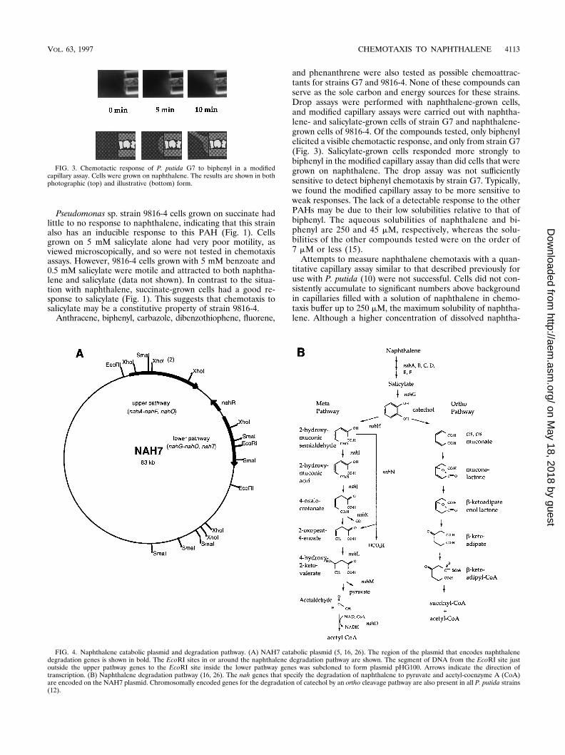
and phenanthrene were also tested as possible chemoattrac-tants for strains G7 and 9816-4. None of these compounds canserve as the sole carbon and energy sources for these strains.Drop assays were performed with naphthalene-grown cells,and modified capillary assays were carried out with naphtha-lene- and salicylate-grown cells of strain G7 and naphthalene-grown cells of 9816-4. Of the compounds tested, only biphenylelicited a visible chemotactic response, and only from strain G7(Fig. 3). Salicylate-grown cells responded more strongly tobiphenyl in the modified capillary assay than did cells that weregrown on naphthalene. The drop assay was not sufficientlysensitive to detect biphenyl chemotaxis by strain G7. Typically,we found the modified capillary assay to be more sensitive toweak responses. The lack of a detectable response to the otherPAHs may be due to their low solubilities relative to that ofbiphenyl. The aqueous solubilities of naphthalene and bi-phenyl are 250 and 45 mM, respectively, whereas the solu-bilities of the other compounds tested were on the order of7 mM or less (15).
Attempts to measure naphthalene chemotaxis with a quan-titative capillary assay similar to that described previously foruse with P. putida (10) were not successful. Cells did not con-sistently accumulate to significant numbers above backgroundin capillaries filled with a solution of naphthalene in chemo-taxis buffer up to 250 mM, the maximum solubility of naphtha-lene. Although a higher concentration of dissolved naphtha-
FIG. 3. Chemotactic response of P. putida G7 to biphenyl in a modifiedcapillary assay. Cells were grown on naphthalene. The results are shown in bothphotographic (top) and illustrative (bottom) form.
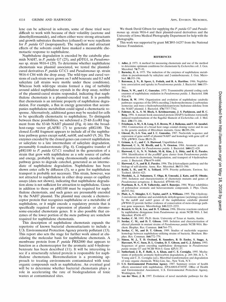
FIG. 4. Naphthalene catabolic plasmid and degradation pathway. (A) NAH7 catabolic plasmid (5, 16, 26). The region of the plasmid that encodes naphthalenedegradation genes is shown in bold. The EcoRI sites in or around the naphthalene degradation pathway are shown. The segment of DNA from the EcoRI site justoutside the upper pathway genes to the EcoRI site inside the lower pathway genes was subcloned to form plasmid pHG100. Arrows indicate the direction oftranscription. (B) Naphthalene degradation pathway (16, 26). The nah genes that specify the degradation of naphthalene to pyruvate and acetyl-coenzyme A (CoA)are encoded on the NAH7 plasmid. Chromosomally encoded genes for the degradation of catechol by an ortho cleavage pathway are also present in all P. putida strains(12).
lene can be achieved in solvents, some of those tried weredifficult to work with because of their volatility (acetone anddimethylformamide), and others either were strong attractantsand growth substrates themselves (ethanol) or were repellentsfor P. putida G7 (isopropanol). The repellent and attractanteffects of the solvents could have masked a measurable che-motactic response to naphthalene.
Naphthalene degradation is encoded by the catabolic plas-mids NAH7, in P. putida G7 (25), and pDTG1, in Pseudomo-nas sp. strain 9816-4 (20). To determine whether naphthalenechemotaxis was plasmid associated, we tested the plasmid-cured derivatives P. putida G7.C1 and Pseudomonas sp. strain9816-4 C84 with the drop assay. The wild-type and cured ver-sions of each strain were grown on 5 mM benzoate and 0.5 mMsalicylate (all strains were motile under these conditions).Whereas both wild-type strains formed a ring of turbidityaround added naphthalene crystals in the drop assay, neitherof the plasmid-cured strains responded, indicating that naph-thalene chemotaxis is a plasmid-encoded trait. It is possiblethat chemotaxis is an intrinsic property of naphthalene degra-dation. For example, a flux in energy generation that accom-panies naphthalene metabolism could signal a chemotactic re-sponse. Alternatively, additional genes may be needed for cellsto be specifically chemotactic to naphthalene. To distinguishbetween these possibilities, we subcloned a 25-kb EcoRI frag-ment from the 83-kb NAH7 plasmid (Fig. 4) into the broad-host-range vector pLAFR1 (7) to generate pHG100. Thecloned EcoRI fragment appears to include all of the naphtha-lene pathway genes except nahK, nahM, and nahO (9, 26). Theenzymes encoded by the cloned genes can convert naphthaleneor salicylate to a late intermediate of salicylate degradation,presumably 4-oxalocrotonate (Fig. 4). Conjugative transfer ofpHG100 to P. putida G7.C1 resulted in the generation of astrain that grew with naphthalene as a sole source of carbonand energy, probably by using chromosomally encoded orthopathway genes to degrade catechol, generated as an interme-diate of naphthalene degradation. Naphthalene has beenshown enter cells by diffusion (3), so a gene for naphthalenetransport is probably not necessary. This strain, however, wasnot attracted to naphthalene in either drop assays or capillaryassays (data not shown), indicating that naphthalene degrada-tion alone is not sufficient for attraction to naphthalene. Genesin addition to those on pHG100 must be required for naph-thalene chemotaxis, and such genes are presumably encodedby the NAH7 plasmid. The plasmid may encode a chemore-ceptor protein that recognizes naphthalene or a metabolite ofnaphthalene, or it might encode a regulatory protein that isspecifically required for expression of plasmid- or chromo-some-encoded chemotaxis genes. It is also possible that en-zymes of the lower portion of the meta pathway are somehowrequired for naphthalene chemotaxis.
This description of naphthalene chemotaxis expands therepertoire of known bacterial chemoattactants to include aU.S. Environmental Protection Agency priority pollutant (13).This report also sets the stage for further work aimed at elu-cidating the molecular basis for naphthalene chemotaxis. Amembrane protein from P. putida PRS2000 that appears tofunction as a chemoreceptor for the aromatic acid 4-hydroxy-benzoate has been described (11). It will be interesting tosee if a structurally related protein is responsible for naph-thalene chemotaxis. Bioremediation is a promising ap-proach to treating environments contaminated with toxicorganic compounds such as naphthalene. An eventual goalwill be to determine whether bacterial chemotaxis plays arole in accelerating the rate of biodegradation of toxicwastes at contaminated sites.
We thank David Gibson for supplying the P. putida G7 and Pseudo-monas sp. strain 9816-4 and their plasmid-cured derivatives and theUniversity of Iowa Medical Photography Department for help with thephotographs.
This work was supported by grant MCB93-16257 from the NationalScience Foundation.
REFERENCES
1. Adler, J. 1973. A method for measuring chemotaxis and use of the methodto determine optimum conditions for chemotaxis by Escherichia coli. J. Gen.Microbiol. 74:77–91.
2. Barnsley, E. A. 1975. The induction of the enzymes of naphthalene metab-olism in pseudomonads by salicylate and 2-aminobenzoate. J. Gen. Micro-biol. 88:193–196.
3. Bateman, J. N., B. Speer, L. Feduik, and R. A. Hartline. 1986. Naphtha-lene association and uptake in Pseudomonas putida. J. Bacteriol. 166:155–161.
4. Dunn, N. W., and I. C. Gunsalus. 1973. Transmissible plasmid coding earlyenzymes of naphthalene oxidation in Pseudomonas putida. J. Bacteriol. 114:974–979.
5. Eaton, R. W. 1994. Organization and evolution of naphthalene catabolicpathways: sequence of the DNA encoding 2-hydroxychromene-2-carboxylateisomerase and trans-o-hydroxybenzylidenepyruvate hydratase-aldolase fromthe NAH7 plasmid. J. Bacteriol. 176:7757–7762.
6. Fahrner, K. A., S. M. Block, S. Krishnaswamy, J. S. Parkinson, and H. C.Berg. 1994. A mutant hook-associated protein (HAP3) facilitates torsionallyinduced transformations of the flagellar filament of Escherichia coli. J. Mol.Biol. 238:173–186.
7. Friedman, A. M., S. R. Long, S. E. Brown, W. J. Buikema, and F. M. Ausubel.1982. Construction of a broad host range cosmid cloning vector and its usein the genetic analysis of Rhizobium mutants. Gene 18:289–296.
8. Ghosal, D., I.-S. You, and I. C. Gunsalus. 1987. Nucleotide sequence andexpression of gene nahH of plasmid NAH7 and homology with gene xylE ofTOL pWWO. Gene 55:19–28.
9. Grimm, A. C., and C. S. Harwood. Unpublished data.10. Harwood, C. S., M. Rivelli, and L. N. Ornston. 1984. Aromatic acids are
chemoattractants for Pseudomonas putida. J. Bacteriol. 160:622–628.11. Harwood, C. S., N. N. Nichols, M.-K. Kim, J. L. Ditty, and R. E. Parales.
1994. Identification of the pcaRKF gene cluster from Pseudomonas putida:involvement in chemotaxis, biodegradation, and transport of 4-hydroxyben-zoate. J. Bacteriol. 176:6479–6488.
12. Harwood, C. S., and R. E. Parales. 1996. The b-ketoadipate pathway and thebiology of self-identity. Annu. Rev. Microbiol. 50:553–590.
13. Keith, L. H., and W. A. Telliard. 1979. Priority pollutants. Environ. Sci.Technol. 13:416–423.
14. Masduki, A., J. Nakamura, T. Ohga, R. Umezaki, J. Kato, and H. Ohtake.1995. Isolation and characterization of chemotaxis mutants and genes ofPseudomonas aeruginosa. J. Bacteriol. 177:948–952.
15. Pearlman, R. S., S. H. Yalkowsky, and S. Banerjee. 1984. Water solubilitiesof polynuclear aromatic and heteroaromatic compounds. J. Phys. Chem.13:555–562.
16. Platt, A., V. Shingler, S. C. Taylor, and P. A. Williams. 1995. The 4-hydroxy-2-oxovalerate aldolase and acetaldehyde dehydrogenase (acylating) encodedby the nahM and nahO genes of the naphthalene catabolic plasmidpWW60-22 provide further evidence of conservation of meta-cleavage path-way gene sequences. Microbiology 141:2223–2233.
17. Resnick, S. M., K. Lee, and D. T. Gibson. 1996. Diverse reactions catalyzedby naphthalene dioxygenase from Pseudomonas sp. strain NCIB 9816. J. Ind.Microbiol. 17:438–457.
18. Serdar, C. M. 1985. Ph.D. thesis. University of Texas at Austin, Austin.19. Serdar, C. M., and D. T. Gibson. 1989. Isolation and characterization of
altered plasmids in mutant strains of Pseudomonas putida NCIB 9816. Bio-chem. Biophys. Res. Commun. 164:764–771.
20. Serdar, C. M., and D. T. Gibson. 1989. Studies of nucleotide sequencehomology between naphthalene-utilizing strains of bacteria. Biochem. Bio-phys. Res. Commun. 164:772–779.
21. Simon, M. J., T. D. Osslund, R. Saunders, B. D. Ensley, S. Suggs, A.Harcourt, W.-C. Suen, D. L. Cruden, D. T. Gibson, and G. J. Zylstra. 1993.Sequences of genes encoding naphthalene dioxygenase in Pseudomonasputida strains G7 and NCIB 9816-4. Gene 127:31–37.
22. Sutherland, J. B., F. Rafii, A. A. Khan, and C. E. Cerniglia. 1995. Mecha-nisms of polycyclic aromatic hydrocarbon degradation, p. 269–306. In L. Y.Young and C. E. Cerniglia (ed.), Microbial transformation and degradationof toxic organic chemicals. Wiley-Liss, Inc., New York, N.Y.
23. U.S. Environmental Protection Agency. 1987. Summary review of healtheffects associated with naphthalene. EPA/600/8-87/055F. Office of Healthand Environmental Assessment, U.S. Environmental Protection Agency,Washington, D.C.
24. van der Meer, J. R. 1997. Evolution of novel metabolic pathways for the
degradation of chloroaromatic compounds. Antonie Leeuwenhoek 71:159–178.
25. Yen, K.-M., and I. C. Gunsalus. 1982. Plasmid gene organization: naphtha-lene/salicylate oxidation. Proc. Natl. Acad. Sci. USA 79:874–878.
26. Yen, K.-M., and C. M. Serdar. 1988. Genetics of naphthalene catabolism inpseudomonads. Crit. Rev. Microbiol. 15:247–268.
27. You, I.-S., D. Ghosal, and I. C. Gunsalus. 1991. Nucleotide sequence analysisof the Pseudomonas putida PpG7 salicylate hydroxylase gene (nahG) and its39-flanking region. Biochemistry 30:1635–1641.
28. You, I.-S., D. Ghosal, and I. C. Gunsalus. 1988. Nucleotide sequence ofplasmid NAH7 gene nahR and DNA binding of the nahR product. J. Bac-teriol. 170:5409–5415.