68
Chest Surface and Pleura Cavity Advanced Anatomy & Physiology Tony Serino, Ph.D. Biology Department Misericordia Univ.

Chest Surface and
Pleura Cavity
Advanced Anatomy & Physiology
Tony Serino, Ph.D.
Biology Department
Misericordia Univ.

Thoracic Vertebrae

Vertebrae and Ribs

Rib Types and Sternum

Rib Anomalies
Bicipital rib(rib fusion)
Cervical ribs
Bifid rib (two heads)
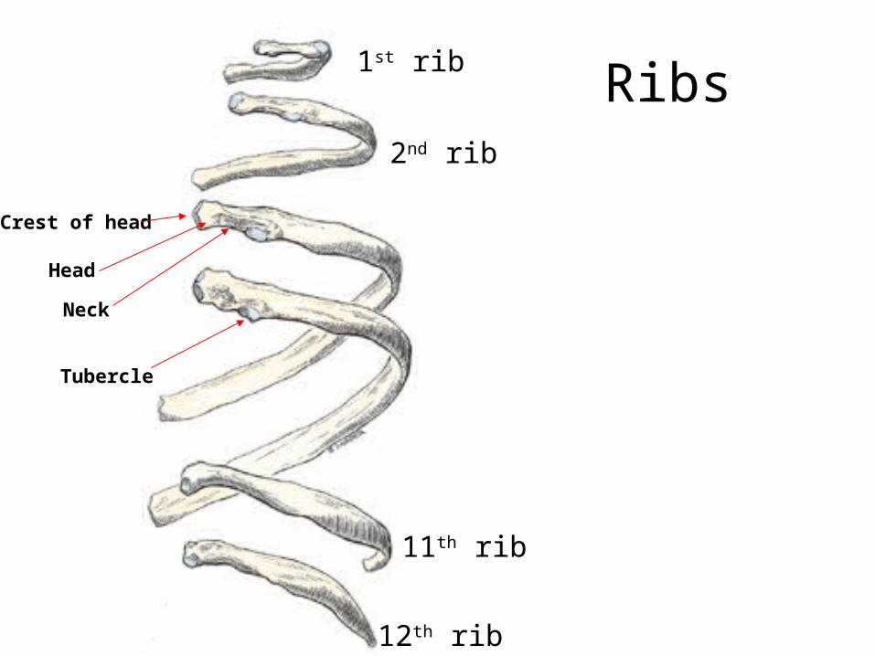
Ribs1st rib
2nd rib
11th rib
12th rib
Crest of head
Head
Neck
Tubercle

Clavicle

Scapula

Scapular Fossa

Superficial Muscles
Deltopectoral triangle (contains Cephalic vein)

Thoracic Apertures
Superior
Inferior

Breast
Male nipple at T4 Dermatome

Female Breast
Tail of breastGlandular tissue and stroma
Suspensory ligaments
Retromammary space

Female BreastRetromammary space

Blood supply to the Breast
Internal thoracic a.(from subclavian)
Lateral thoracic (from axillary a.)
Anterior intercostals
Post. Intercostals(from thoracic aorta)
(Venous drainage mostly to axillary v. and internal thoracic v.)

Lymphatic Drainage of Breast
Subareolar plexus
Axillary nodes
Parasternal nodes
Pectoral nodes
Inferior phrenic nodes
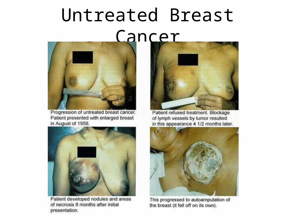
Untreated Breast Cancer
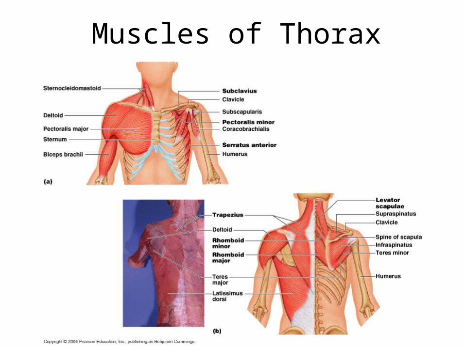
Muscles of Thorax
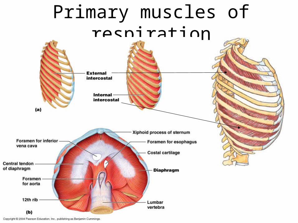
Primary muscles of respiration

Only used during rapid breathing.
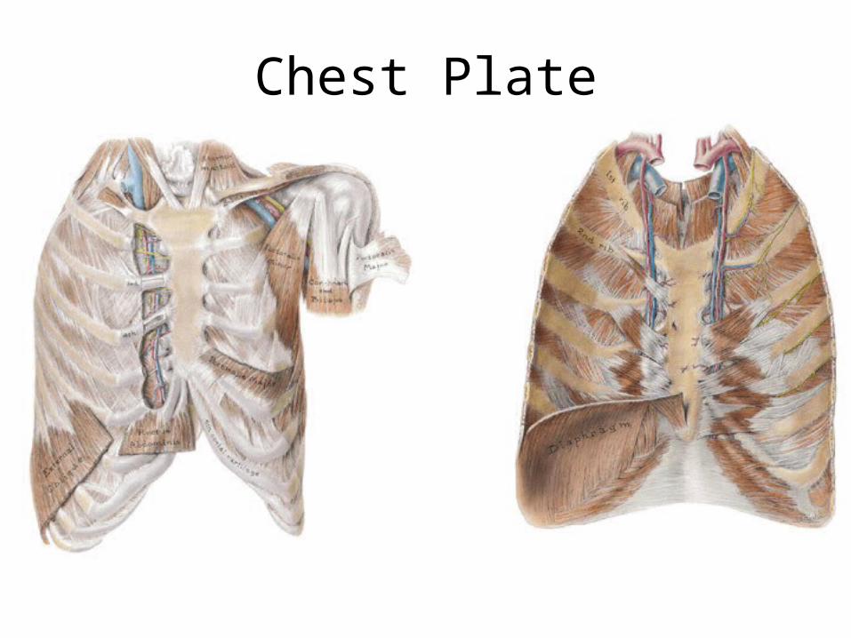
Chest Plate

Pressures affecting Breathing

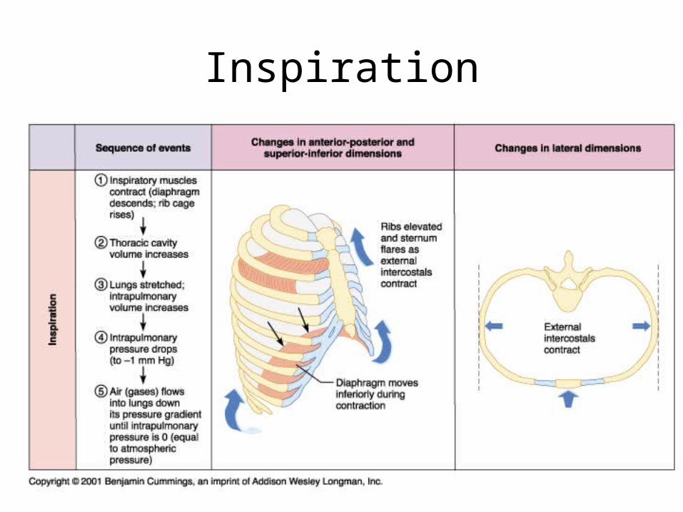
Inspiration

Expiration

Pressure changes around lung

Lung Volumes
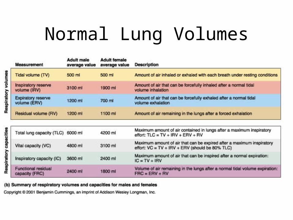
Normal Lung Volumes

Nerves of thoracic wall

Intercostal arteries and nerves

Pleura
Costodiaphragmatic recessCostomediastinal recess
Cardiac notch

Surface to Deep Structure Alignment
Bare Pericardium

Respiration• External Respiration
– The exchange of gas between the blood and external environment (usually includes ventilation)
• Internal Respiration– The exchange of gas between the blood and the tissues
• Cellular Respiration– Burning of fuel to produce energy within cells
• Ventilation (Breathing)– Movement of air in and out of the lungs

Respiratory Organs
– Divided into:• Upper Respiratory Tract
– Includes: nostrils (nares), nasal cavity, and nasopharynx
• Lower Respiratory Tract– Includes: larynx, trachea,
bronchi, and lungs
– Conducting Air passages include: nares to terminal bronchioles
• Move air to respiratory membrane
• Condition the air– Moisten, Warm, Clean

Trachea

Trachea (x.s.)

Mucous Membrane(pseudostratified columnar epithelium)

Bronchi
• Primary bronchi lead to to each lung (left and right)
• Secondary (lobar) bronchi lead to each lung lobe (3 on right and 2 on left)

Cadaver Lungs

Lobes of Right Lung
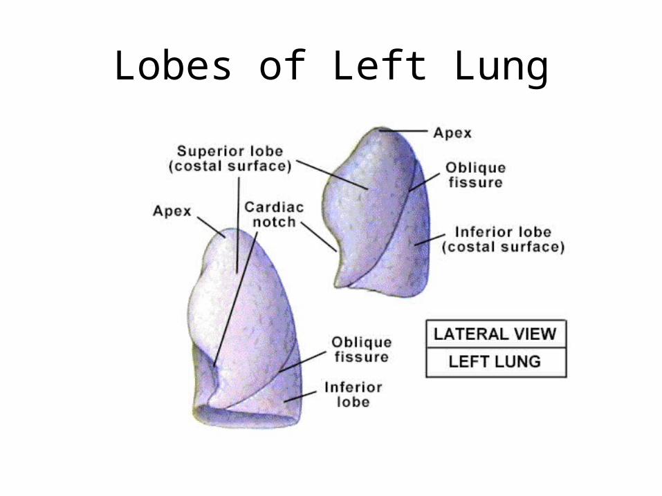
Lobes of Left Lung

Bronchi Branches
Primary BronchiTertiary Bronchi
Secondary BronchiTertiary (segmental) bronchi lead to each lung broncho-pulmonary segment
Bronchi continue to divide at least 20 more times.

Broncho-pulmonary Segments
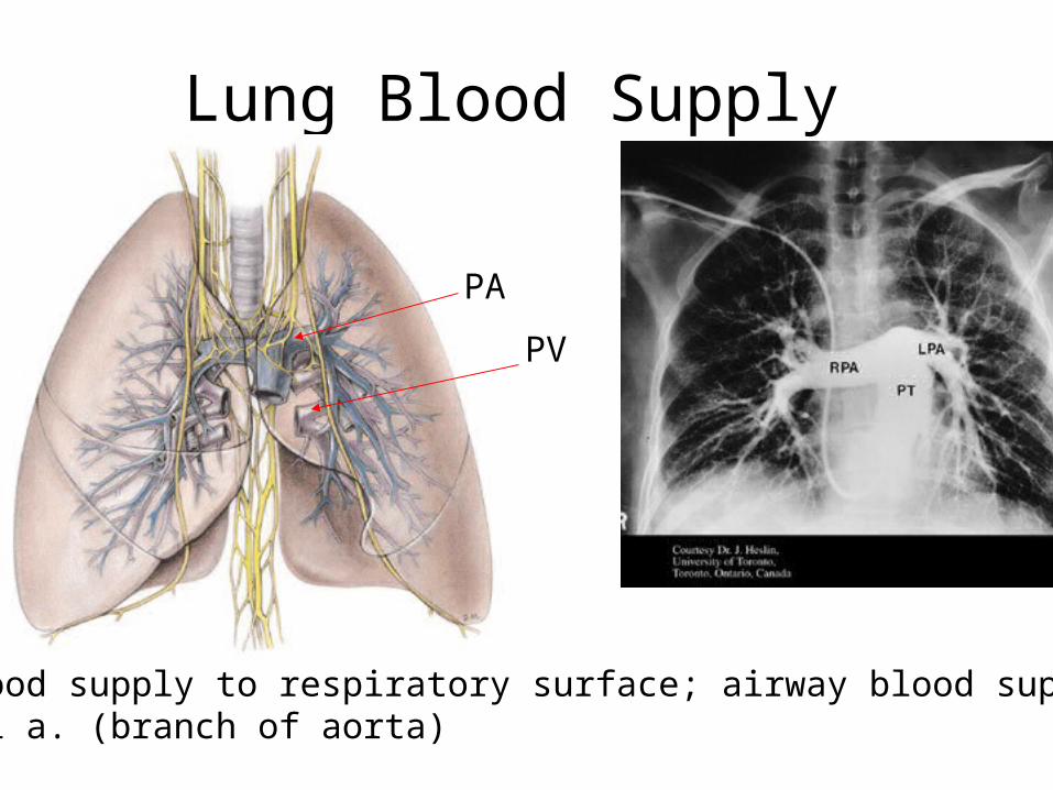
Lung Blood Supply
Note: blood supply to respiratory surface; airway blood supplied bybronchial a. (branch of aorta)
PA
PV

Blood pathways
Bronchi
PA
PV
Bronchioles
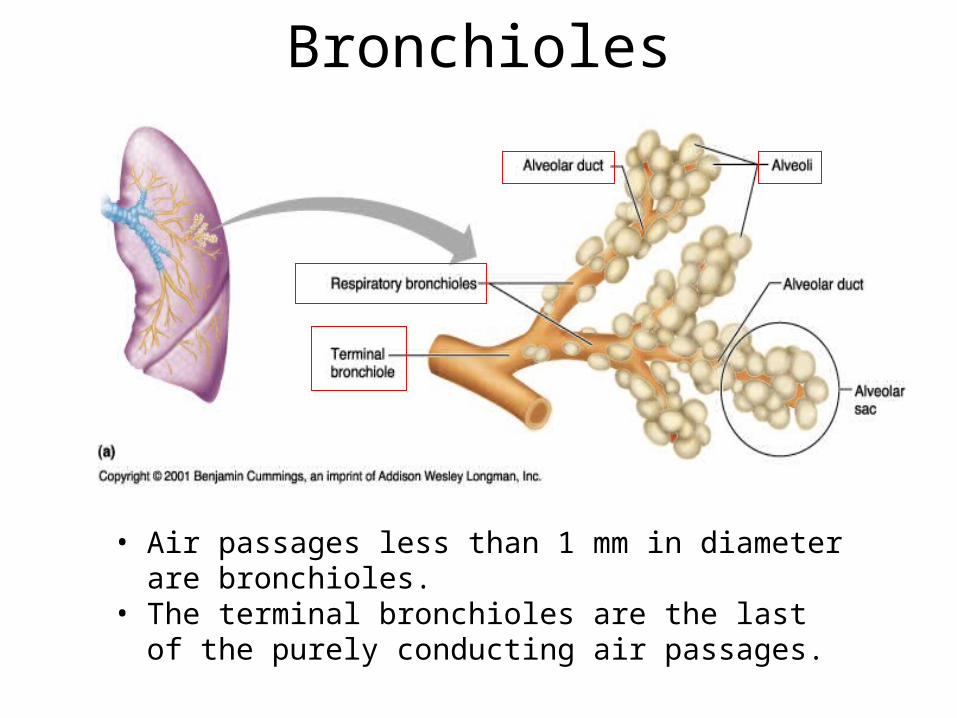
• Air passages less than 1 mm in diameter are bronchioles.
• The terminal bronchioles are the last of the purely conducting air passages.
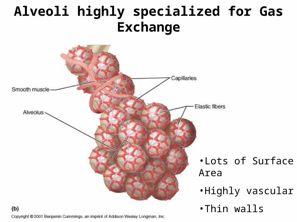
Alveoli highly specialized for Gas Exchange
•Lots of Surface Area
•Highly vascular
•Thin walls

Lung Tissue

Alveolus

Role of surfactant is to decrease surface tension in alveoli.
P = pressure to collapseT = surface tension r = radius

Partial Pressure Favors Resp. Gas Movement

Time to Complete O2 Saturation in Pulmonary Capillaries
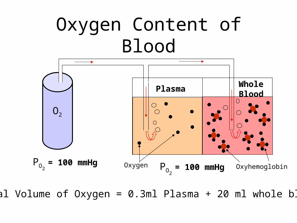
Oxygen Content of Blood
O2
PO2 = 100 mmHg PO2
= 100 mmHg
PlasmaWhole Blood
Oxygen Oxyhemoglobin
Total Volume of Oxygen = 0.3ml Plasma + 20 ml whole blood

Hemoglobin

Oxyhemoglobin Dissociation Curve

Hemoglobin Affinity for Oxygen:Effect of Temperature
Affinity decreases with increasing Temperature

Hemoglobin Affinity for Oxygen:Effect of pH
Affinity decreases with increasing acidity ( pH)
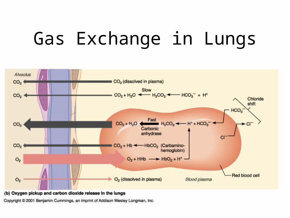
Gas Exchange in Lungs

Gas Exchange in Tissues
70%
10%
20%
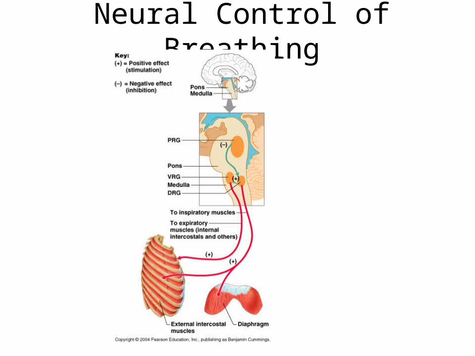
Neural Control of Breathing

Neural Control of Breathing
PRG –pontine resp. group (formerly the apneustic and pneumotaxic centers) –play role in smoothing between insp. and exp., especially during sleep, vocalization and exercise.VRG and DRG – ventral and dorsal resp. groupof the medulla. DRG primarily responsible forinspiration; VRG mixture of I and E neurons contains Pre-Botzinger complex which may bepacemaker cells for respiration
Voluntary control located in cerebral cortex and acts through the corticospinal tract.Involuntary located in pons and medulla acting through the spinal cord in the roots of the phrenicnerve (C3-C5) and thoracic cord roots of theexternal (inspriation(I)) and internal (expiration(E)) intercostal nerves
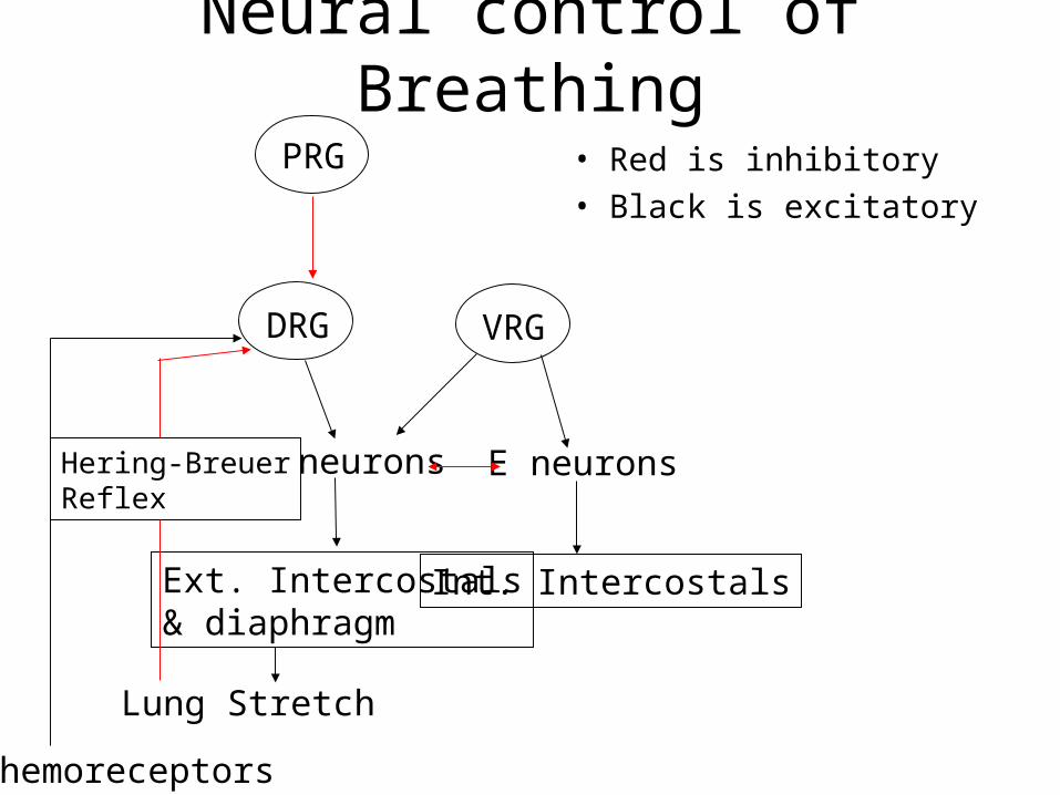
Neural control of Breathing
• Red is inhibitory
• Black is excitatory
PRG
DRG VRG
E neuronsI neurons
Ext. Intercostals& diaphragm
Int. Intercostals
Chemoreceptors
Lung Stretch
Hering-BreuerReflex


Factors Effecting Respiratory Centers
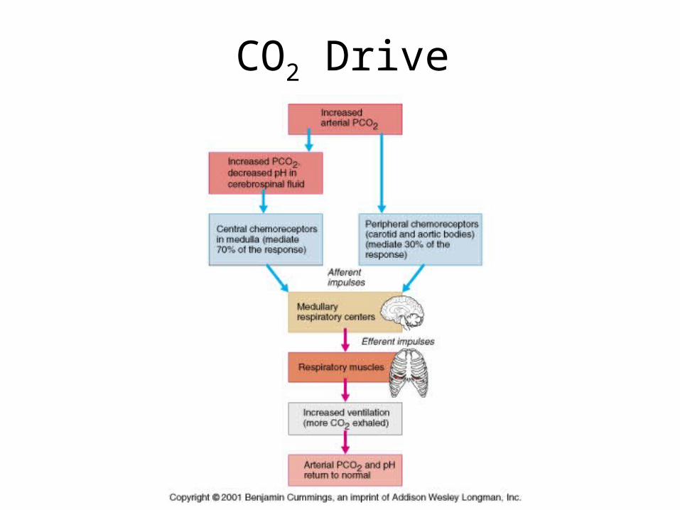
CO2 Drive

COPD


