Profilin is a major regulator of actin assembly in all eukaryoticcells and profilin isoforms from angiosperm plants, vertebrates,yeasts and Vacciniavirus have been characterized extensively(Gibbon and Staiger, 2000; Schlüter et al., 1997). Profilin hasthree main cellular ligands: monomeric or G-actin, proline-richproteins and polyphosphoinositide lipids. The formation of a1:1 complex between profilin and G-actin can have complexeffects on actin assembly. In the presence of capped filamentends, profilin functions as a simple sequestering protein,preventing actin polymerization. However, when the barbedend of filaments are not capped and a large pool of actinmonomers exists, the profilin-actin complex can assemble ontoF-actin. Furthermore, most profilins are able to function asnucleotide exchange factors for monomeric actin. This ledto models that profilin contributes to actin assembly by‘recharging’ subunits with ATP. Interestingly, plant profilinsdo not stimulate nucleotide exchange (Eads et al., 1998;Perelroizen et al., 1996), even with plant actin (Kovar et al.,2000a), yet still promote assembly under appropriateconditions (Perelroizen et al., 1996; Ballweber et al., 1998).Biochemical and genetic studies further underscore the conceptthat profilin might have distinct functions depending upon theorganism in which it is found, the presence of other actin-binding proteins and other unique cellular conditions. Manyquestions about profilin remain, including the significance ofprofilin’s effect on nucleotide exchange, whether profilinisoforms within the same cell perform distinct roles, whetherprofilin promotes polymerization or acts as a sequestering
molecule during specific cellular processes and how profilin isprecisely regulated by its association with other cellularligands. Further progress on cellular roles will requirecontinued analysis of profilin isoforms in model geneticsystems (Balasubramanian et al., 1994; Cooley et al., 1992;Haarer et al., 1993; Haugwitz et al., 1994; Lu and Pollard,2001; Wolven et al., 2000).
Chlamydomonas reinhardtiiis a unicellular alga thatpotentially provides many experimental advantages for thestudy of the actin cytoskeleton and profilin. Like yeast,Chlamydomonashas well understood haploid genetics (Harris,2001) and offers several molecular tools for analysis ofgenes and protein function (Lefebvre and Silfow, 1999).Chlamydomonascontains only a single conventional actin,IDA5 (Kato-Minoura et al., 1997; Sugase et al., 1996), whichis predicted to be 90% identical to mammalian skeletal muscleactin, and contains an unusual, novel actin protein (Kato-Minoura et al., 1997; Lee et al., 1997). Interestingly, this novelactin can, in some instances, replace the functions ofconventional actin (Kato-Minoura et al., 1997; Ohara et al.,1998). Although the localization of actin in Chlamydomonashas been studied (Detmers et al., 1983; Detmers et al., 1985;Harper et al., 1992), the role of the actin cytoskeleton inChlamydomonasis just beginning to be understood. Forexample, actin has been identified as a subunit of flagellardyneins (Kagami and Kamiya, 1992; Muto et al., 1994;Piperno and Luck, 1979; Piperno et al., 1990; Sugase et al.,1996). The role of the actin subunits in dynein is not known,but mutations in actin can lead to a failure of dynein assembly(Kato-Minoura et al., 1998). The best-understood F-actin-
4293
We report the characterization of a profilin orthologuefrom Chlamydomonas reinhardtii. CrPRF, probably theonly profilin isoform, is present in both the cell body andflagella. Examination of vegetative and gametic cells byimmunofluorescence microscopy using multiple fixationprocedures also revealed enrichment of CrPRF at theanterior of the cell near the base of flagella and near thebase of the fertilization tubule in mating type plus gametes.Purified, recombinant CrPRF binds to actin with a Kdvalue ~10–7 and displaces nuclei in a live cell ‘nucleardisplacement’ assay, consistent with profilin’s ability tobind G-actin in vivo. However, when compared with other
profilin isoforms, CrPRF has a relatively low affinityfor poly-L-proline and for phosphatidylinositol (4,5)bisphosphate micelles. Furthermore, and surprisingly,CrPRF inhibits exchange of adenine nucleotide on G-actinin a manner similar to human ADF or DNase I. Thus, wepostulate that a primary role for CrPRF is to sequesteractin in Chlamydomonas. The unusual biochemicalproperties of CrPRF offer a new opportunity to distinguishspecific functions for profilin isoforms.
Chlamydomonas reinhardtii produces a profilin withunusual biochemical propertiesDavid R. Kovar 1, Pinfen Yang 2, Winfield S. Sale 2, Bjørn K. Drobak 3 and Christopher J. Staiger 1,*1Department of Biological Sciences, Purdue University, West Lafayette, IN 47907-1392, USA2Department of Cell Biology, Emory University School of Medicine, Atlanta, GA 30322, USA3Department of Disease and Stress Biology, John Innes Centre, Norwich, NR4 7UH, UK*Author for correspondence (e-mail: [email protected])
containing organelle in Chlamydomonasis the fertilizationtubule of mating type plus (mt+) gametes (Detmers et al., 1983;Detmers et al., 1985; Goodenough and Weiss, 1975; Martinand Goodenough, 1975; Wilson et al., 1997a). Defects in eitherthe actin gene (Kato-Minoura et al., 1998) or the signaltransduction events of fertilization (Pan and Snell, 2000) canresult in failure to form F-actin and the fertilization tubule,thereby blocking fertilization. Finally, mutations have beenidentified that alter the cleavage furrow during cytokinesis,possibly by affecting the actin cytoskeleton (Ehler and Dutcher,1998).
In interphase Chlamydomonascells, little F-actin isobserved (Detmers et al., 1983; Detmers et al., 1985; Harperet al., 1992), suggesting an unusual control of the actincytoskeleton. However, actin-binding proteins that mightregulate filament assembly and organization have not beenreported. Here, we describe the molecular, cellular andbiochemical characteristics of Chlamydomonas profilin,CrPRF. CrPRF is the only profilin gene in Chlamydomonas.Localization and western analysis reveals that profilin islocated throughout the cell, including the flagellum, but isenriched at the anterior of the cell near the base of the flagellain vegetative and gametic cells. Biochemical characterizationshowed that CrPRF has a high affinity for G-actin butan extremely low affinity for both poly-L-proline andphosphatidylinositol (4,5) bisphosphate (PtdIns(4,5)P2).Surprisingly, and in contrast to all other profilins examined,CrPRF significantly inhibits nucleotide exchange on actin.CrPRF is the first actin-binding protein characterized fromChlamydomonasand, based on the biochemical analysis, itsprimary role might be to sequester G-actin.
MATERIALS AND METHODS
Cell strainsWild-type strains (CC-124, and CC-620 mt+) were provided by theChlamydomonas Genetics Center (E. H. Harris, Duke University).CC-124, used for molecular and biochemical studies, was grown inL-medium with aeration over a 14/10 light/dark cycle (Witman, 1986).CC-620 mt+, used for immunofluorescence microscopy, was grownon L-medium agar plates.
Molecular characterizationGenomic DNA was prepared as described (Wilkerson et al., 1995).Total and poly(A)+ mRNA were prepared from wild-type cells asdescribed previously (Yang and Sale, 1998). Probes for northern- andSouthern-blot analysis, and a bacterial expression construct ofChlamydomonasprofilin, CrPRF, were produced with 30 cycles of thepolymerase chain reaction (PCR) using Pfu DNA polymerase(Stratagene, La Jolla, CA) as described (Yang and Sale, 1998).Genomic DNA was used as a template because the predicted openreading frame did not contain introns. The sense primer (5′-ACCATGGCCTGGGAAGCCTAC-3′) introduced an NcoI site(underlined) into the start codon (ATG) at the 5′ end for subsequentcloning. The antisense primer (5′-CAAGTTTAGTACCCCTGGTCC-3′) included the stop codon (underlined). The resulting 400 bp productwas used as a probe for northern and Southern analysis as describedpreviously (Yang and Sale, 1998). The PCR product was cloned intothe SmaI site in pPCRSCRIPT SK (Stratagene) following themanufacturer’s instructions. The insert was then digested with NcoIand SalI and cloned into the same sites of the pET-28a expressionvector (Novagen, Madison, WI, USA). The pET-28a-CrPRF constructwas transformed into strain BL21 (DE3) of Escherichia coli.
Protein purificationExpression of the pET-28a-CrPRF construct was induced by theaddition of 0.4 mM isopropyl β-D-thiogalactopyranoside to a log-phase culture for 4 hours at 37°C. Recombinant CrPRF protein waspurified by poly-L-proline (PLP)-Sepharose chromatography,according to methods described previously (Karakesisoglou et al.,1996), with modifications. Unlike other profilins we have purified,substantial amounts of CrPRF eluted from PLP-Sepharose with 1 Murea. Therefore, following washes with buffer I (20 mM Tris-HCl, 150mM KCl, 0.2 mM DTT, pH 7.5), CrPRF was eluted with consecutive1 M and 3 M urea washes in buffer I. The initial 1 M urea fractionswere found to contain additional proteins by SDS-PAGE (not shown)and were discarded. The remaining 1 M urea eluant and entire 3 Murea eluant were pooled, yielding a 12 kDa protein. No proteincontaminants were visible when 10 µg of purified protein wasseparated by SDS-PAGE and stained with Coomassie Blue, and yieldsof purified CrPRF were ~9.2 mg l–1 bacteria. Recombinant Zea maysprofilin 5 (ZmPRO5) and human profilin I (HPRO1) proteins, andmaize pollen actin were purified as described previously (Kovar et al.,2000a; Ren et al., 1997). Rabbit skeletal muscle actin (99% pure) waspurchased from Cytoskeleton (Denver, CO, USA) and prepared withone cycle of polymerization and depolymerization as describedpreviously (Kovar et al., 2000a). Recombinant human actindepolymerizing factor (ADF) was purified according to Hawkins etal. (Hawkins et al., 1993). Three independent batches of each profilinwere used for microinjection and for the biochemical experimentsdescribed below.
Protein concentrations were determined with extinctioncoefficients. For ZmPRO5, A280=16,000 M–1 cm–1 (Kovar et al.,2000a). For maize pollen actin and rabbit skeletal muscle actin,A290=0.63 for a 1 mg ml–1 solution (Houk and Ue, 1974; Kovar et al.,2000a). For human profilin I, A280=0.015 µM–1 cm–1. For humanADF, A280=11,210 M–1 cm–1 (Hawkins et al., 1993). An extinctioncoefficient (A280) of 19,190 M–1cm–1 for CrPRF was determined (Gilland von Hippel, 1989) and gave calculated protein concentrationswithin 5% of the concentration determined by the Bradford assay(BioRad, Hercules, CA, USA) using BSA as a standard.
Urea denaturationThe stability of the purified recombinant profilins was analysed bydetermining the concentration of urea required for their half-maximaldenaturation, according to methods published previously (Eads et al.,1998). 1 µM profilin was incubated for 1 hour at room temperaturein buffer I with increasing concentrations of urea (0-8 M). Theintrinsic tryptophan fluorescence of each sample was measured withexcitation at 292 nm and emission at 370 nm. Normalized relativefluorescence was then plotted versus urea concentration and fitted toa sigmoid curve.
Antisera production and analysisRabbit polyclonal antisera were raised (Spring Valley Laboratories,Sykesville, MD, USA) against purified recombinant CrPRF. Forwestern analysis, a 1:5000 dilution of serum was used and purifiedrecombinant CrPRF was used for calibration of profilin on the blots.Cell body extracts were produced by vortexing wild-type cells withglass beads and collecting the supernatant as described (Fowkes andMitchell, 1998). Flagella, axonemes and a 0.5% Nonidet-P40-solublefraction in Buffer A (30 mM NaCl, 10 mM Hepes, pH 7.4, 5 mMMgSO4, 1 mM DTT, 0.5 mM EDTA, PMSF and aprotinin) wereprepared as described previously (Yang et al., 2000). Flagellar puritywas monitored by phase-contrast microscopy and isolated flagellawere washed twice in buffer to avoid contamination from the cellbody. To compare profilin in flagellar fractions (Fig. 3C), aliquots offlagella, axonemes and membrane matrix (detergent extract) werediluted proportionally with buffer A so that each sample was derivedfrom equal amounts of flagella. The resulting fractions were separatedon 12.5% SDS-PAGE gels. Blots were visualized by enhanced
JOURNAL OF CELL SCIENCE 114 (23)
4295Chlamydomonas profilin
chemiluminescence (Pharmacia Biotech, Piscataway, NJ, USA) asdescribed previously (Yang and Sale, 1998).
For immunofluorescence, cell-wall-less mt+ vegetative cells (cw92)were grown in nitrogen-containing L-medium for three days to a finaldensity ~2×10⁶ ml–1. For gametes, mt+ (cc620) and mt– (cc621) cellsgrown on L-plates for >6 days were resuspended in nitrogen-free M-N medium for 5 hours to induce differentiation. Gametes wereactivated by treatment with 15 mM dibutyryl cAMP and 0.15 mMpapaverine/fresh DMSO as described (Wilson et al., 1997b) for ~40minutes. Cells, attached to cover glasses, were processed forimmunofluorescent microscopy as described (Sanders and Salisbury,1994) with modified fixation procedures, as noted below and in thefigure legend. To ensure that the localization of profilin or actin wasnot simply a consequence of a single method of fixation, variousfixation methods were used, including 4% formaldehyde (Ted Pella,Redding, CA) in the growth medium for 10 minutes followed by 80%acetone/PBS and 100% acetone at –20°C for 6 minutes each (Fig.4A,E) or followed by 4% formaldehyde in 0.25% Nonidet-P40 for 15minutes (Fig. 4B). Alternatively, cells were fixed directly with 80%acetone and then with 100% acetone (Fig. 4C), or cells in suspensionwere fixed in 2% paraformaldehyde (Sigma, St Louis, MO, USA) for30 minutes followed by 80% and 100% acetone fixation (Fig. 4D).Anti-CrPRF was affinity purified using recombinant CrPRF and wasrevealed by FITC-conjugated goat anti-rabbit antibody (ICN, Aurora,OH, USA). Actin was revealed by anti-actin monoclonal antibodyC4 (ICN) and Cy5-conjugated goat anti-mouse antibody (JacksonImmunoResearch, West Grove). Negative controls included theaddition of an irrelevant primary rabbit antibody. Double labelling wasrecorded using confocal microscopy (Axiovert 100 M; Zeiss). FITClabelling alone was observed by inverted fluorescent light microscopy(Axiovert 35; Zeiss).
Nuclear displacement assayFreshly opened Tradescantia virginianastamen hair cells werecollected and microinjected with profilin, following proceduresdescribed previously (Gibbon et al., 1997; Gibbon et al., 1998;Karakesisoglou et al., 1996; Ren et al., 1997). ~5-6 pl of proteinsolution was delivered into each hair cell. At least 30 cells wereinjected for each profilin and average times required for nucleardisplacement were determined. In order to rule out the possibility thatmicroinjection alone, or microinjection of any protein, affects theplacement of the nucleus, we have previously injected equivalentconcentrations of both bovine serum albumin and bovine gammaglobulin (BGG) (Gibbon et al., 1997; Gibbon et al., 1998; Kovar etal., 2000a). These control injections do not cause a significant nucleardisplacement during the 20 minute assay.
PLP and G-actin bindingThe affinity (Kd value) of profilin for PLP was determined bymeasuring the increase of intrinsic tryptophan fluorescence uponcomplex formation (Perelroizen et al., 1994; Petrella et al., 1996), asdescribed in detail previously (Gibbon et al., 1997; Gibbon et al.,1998). Because of the low affinity of CrPRF for PLP, solutions of 5µM CrPRF were titrated with PLP (12 mg ml–1) to a finalconcentration of ~6000 µM proline residues.
The ability of profilin to reduce the concentration of filamentousactin in the presence of 1 µM calcium, determined by monitoring ashift in the critical concentration (Cc) at steady state, was used tomeasure profilin’s apparent affinity for monomeric actin, as describedpreviously (Kovar et al., 2000a).
PtdIns(4,5)P 2-bindingPtdIns(4,5)P2-binding was assayed by microfiltration as describedpreviously (Haarer et al., 1993; Lambrechts et al., 1997).PtdIns(4,5)P2 (Sigma) micelles (1 mg ml–1 in H2O) were prepared bysonication for 5 minutes at room temperature. In a 150 µl reactionvolume, increasing concentrations of PtdIns(4,5)P2 (0-250 µM)
micelles were incubated with 2.5 µM profilin on ice for 2 hours in 10mM Tris-HCl, pH 7.5, 75 mM KCl, 0.5 mM DTT. The samples werethen loaded onto low binding regenerated cellulose Ultrafree-MCmembranes (Fisher, Pittsburgh, PA, USA) with a molecular weightcut-off of 30,000 and centrifuged for ~1 minute at 2000 g. The flow-through from each reaction was separated by 15% SDS-PAGE, stainedwith Coomassie Brilliant Blue R (Sigma), scanned and the intensityof the profilin bands were determined with IMAGEQUANT software(Molecular Dynamics, Sunnyvale, CA, USA).
The inhibition of bean (Vicia faba) plasma membranephosphoinositide phospholipase C activity by profilin was measuredas described previously (Drøbak et al., 1994). Briefly,phosphoinositidase activity was assayed by incubating bean plasmamembranes at 25°C in 50 µl buffer E (50 mM Tris/malate, pH 6.0, 10µM CaCl2) with 50 µM PtdIns(4,5)P2 and 0.86 kBq 3H-PtdIns(4,5)P2,in the presence of 5 µM profilin. Reactions were stopped by theaddition of 1 ml of chloroform-methanol (2:1 [v/v]). After a 5-minuteincubation on ice and the addition of 250 µl of 0.6 M HCl, tubes werevortexed and centrifuged at 14,000 g for 2 minutes. 400 µl of the topphase was removed and radioactivity was determined by liquidscintillation spectrometry (Wallac 1410) after addition of scintillationfluid (Hionic-Fluor, Hewlett-Packard, UK).
Nucleotide exchange analysisThe rate of nucleotide exchange on G-actin in the absence or presenceof the indicated concentrations of CrPRF, ZmPRO5, HPRO1, humanADF or DNase I (Sigma) was determined by measuring the increasein fluorescence upon incorporation of 1,N6-ethenoadenosine 5′-triphosphate (ε-ATP; Sigma) (Goldschmidt-Clermont et al., 1992).The ε-ATP (50 µM) and profilin, ADF or DNase I (in a constantvolume of 220 µl) were mixed with either 2× low salt buffer (4 mMTris-HCl, pH 6.5, 1.0 mM DTT) or 2× physiological salt buffer (4mM Tris-HCl, pH 7.5, 1.0 mM DTT, 200 mM KCl, 10 mM MgCl2)and brought to a final reaction volume of 1.485 ml with water. Theinitial fluorescence was determined in a spectrofluorimeter withexcitation at 360 nm and emission at 410 nm. The reaction wasinitiated by addition of 0.5 µM G-actin from a 50 µM stock solutionin buffer G (Ren et al., 1997) and monitored for 400 seconds. The rateof ε-ATP incorporation (∆fluorescence (arbitrary units per second))was determined by fitting the data for the first 240 seconds to a singleexponential function.
The effect of a range of concentrations of CrPRF (0.1-20 µM) onthe rate of nucleotide exchange of 2.0 µM pollen G-actin was carriedout in low salt buffer in a similar manner. The determined rates wereplotted against the concentration of CrPRF. A dissociation equilibriumconstant (Kd) was calculated with MacCurveFit (Raner Software, MtWaverly, Australia) using the equation
kobs= ka + (kap – ka) {[P + A + Kd] – [(P + A + Kd)2 – 4PA]}0.5 ÷ 4,
where ka is the nucleotide exchange rate of free actin, kap is thenucleotide exchange rate of actin bound to profilin, P is theconcentration of profilin and A is the concentration of actin.
RESULTS
Identification of a profilin-like gene fromChlamydomonas reinhardtiiWhile cloning the PF24 gene from Chlamydomonas, wediscovered an open reading frame (ORF) 860 bp upstream ofthe first ATG of PF24, a gene for an axonemal protein RSP2(Yang and Sale, 1999). The ORF was preceded by an in-framestop codon (TGA) 9 bp 5′ of the first ATG and a predictedTGTAA polyadenylation signal was located 330 bp 3′ of thepredicted stop codon. The full nucleotide sequence informationis available under GenBank accession number AF335423. A
4296 JOURNAL OF CELL SCIENCE 114 (23)
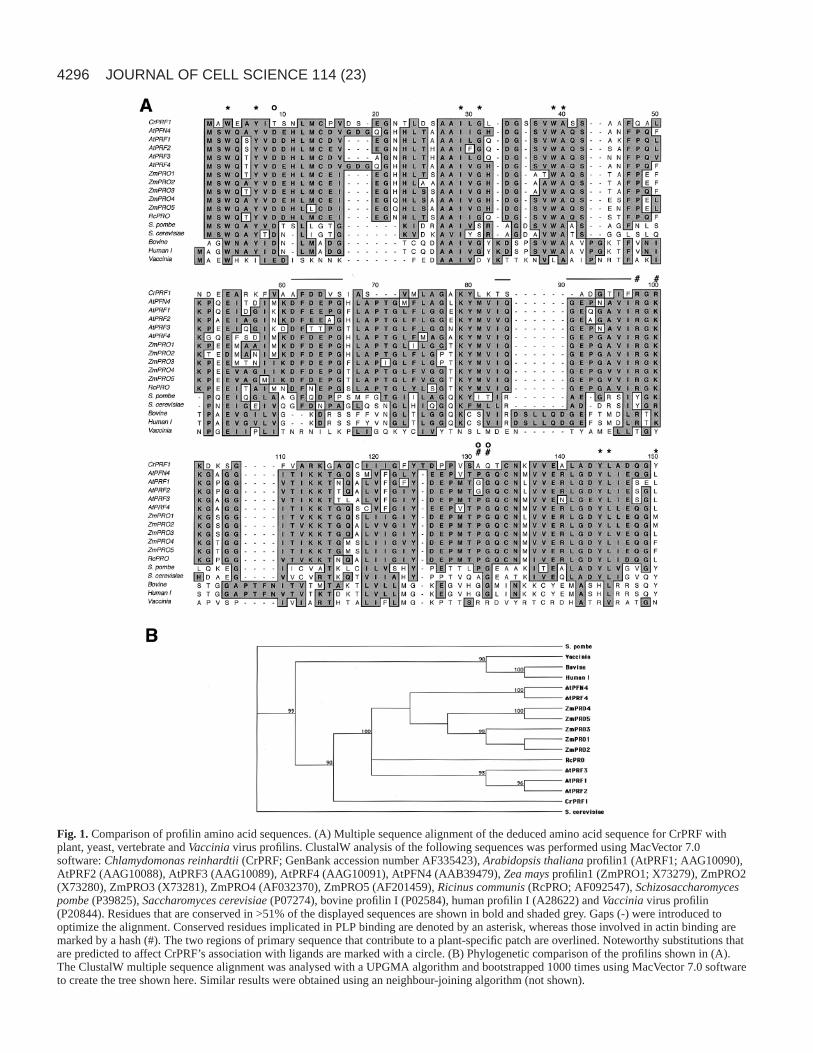
Fig. 1. Comparison of profilin amino acid sequences. (A) Multiple sequence alignment of the deduced amino acid sequence for CrPRF withplant, yeast, vertebrate and Vacciniavirus profilins. ClustalW analysis of the following sequences was performed using MacVector 7.0software: Chlamydomonas reinhardtii(CrPRF; GenBank accession number AF335423), Arabidopsis thalianaprofilin1 (AtPRF1; AAG10090),AtPRF2 (AAG10088), AtPRF3 (AAG10089), AtPRF4 (AAG10091), AtPFN4 (AAB39479), Zea maysprofilin1 (ZmPRO1; X73279), ZmPRO2(X73280), ZmPRO3 (X73281), ZmPRO4 (AF032370), ZmPRO5 (AF201459), Ricinus communis (RcPRO; AF092547), Schizosaccharomycespombe(P39825), Saccharomyces cerevisiae(P07274), bovine profilin I (P02584), human profilin I (A28622) and Vacciniavirus profilin(P20844). Residues that are conserved in >51% of the displayed sequences are shown in bold and shaded grey. Gaps (-) were introduced tooptimize the alignment. Conserved residues implicated in PLP binding are denoted by an asterisk, whereas those involved in actin binding aremarked by a hash (#). The two regions of primary sequence that contribute to a plant-specific patch are overlined. Noteworthy substitutions thatare predicted to affect CrPRF’s association with ligands are marked with a circle. (B) Phylogenetic comparison of the profilins shown in (A).The ClustalW multiple sequence alignment was analysed with a UPGMA algorithm and bootstrapped 1000 times using MacVector 7.0 softwareto create the tree shown here. Similar results were obtained using an neighbour-joining algorithm (not shown).
4297Chlamydomonas profilin
BLAST search of the expressed sequence tag (EST) databases(NCBI; http://www.ncbi.nlm.nih.gov/) recovered severalcDNA sequences that contained the entire ORF (BF866678and AV624542) or part of the ORF. The EST nucleotidesequences were identical to the ORF and the flanking sequenceexcept for single base pair changes in a few cases, which mightbe a consequence of sequencing errors or strain differences.The presence of these EST clones indicated that this ORF isan active gene.
Conceptual translation of the ORF produced a 131 aminoacid long protein (Fig. 1A), with a predicted weight of 13.9kDa and pI of 4.43. BLAST searches revealed that thepredicted protein was orthologous to the small actin-bindingprotein profilin and so it was subsequently named CrPRF.CrPRF shared ~39% identity with plant profilins, ~32%identity with yeast and fungal profilins, ~23% identity withvertebrate profilins and 14% identity with Vaccinia virusprofilin. Phylogenetic analyses placed CrPRF in a branchsomewhat closer to angiosperm than to fungal profilins (Fig.1B). Further BLAST searches of the ChlamydomonasESTdatabase with the predicted amino acid sequence of CrPRF orthe amino acid sequences of profilins from other organismsrevealed no additional Chlamydomonasprofilin-like isoforms.Therefore, it was predicted that this ORF encodes a bona fideprofilin orthologue in Chlamydomonasand was likely to be theonly profilin isoform.
The residues that are most highly conserved in profilins fromdifferent species are those implicated in PLP binding (Fig. 1A;Table 1); these form a hydrophobic patch positioned betweenthe N- and C-terminal α-helices. Of 20 amino acids that areconserved in >80% of eukaryotic profilins, nine are implicatedin binding to PLP and six of these make direct contact with theproline resides (Mahoney et al., 1997). All nine residues are
absolutely conserved in CrPRF (Table 1). Residues that arethought to be involved in actin binding (Fig. 1A; Table 1) areless well conserved among profilins from different species(Thorn et al., 1997). Of three conserved residues, each of whichmakes direct contact with actin in the bovine profilin-β-actincrystal (Schutt et al., 1993), only one is conserved in CrPRF(Table 1). The phospholipid-binding site on the overall fold ofeukaryotic profilins remains a matter for debate (Gibbon andStaiger, 2000; Schlüter et al., 1997) but, when a highly-conserved aspartic acid (D) on the N-terminal α-helix ischanged to alanine in human (Sohn et al., 1995) or plant (Kovaret al., 2001) profilin, the resulting mutant profilins haveenhanced PtdIns(4,5)P2-binding properties. Interestingly,CrPRF contains an uncharged threonine residue at theequivalent position. Finally, examination of several plantprofilin sequences and comparison of the overall fold ofArabidopsisprofilin I and birch pollen profilin with non-plantprofilin structures reveals a plant-specific patch adjacent to theactin-binding site (Fedorov et al., 1997; Thorn et al., 1997).The primary sequence of CrPRF in the equivalent region ispoorly conserved, with several non-conservative substitutionsand deletions. Therefore, based upon amino-acid sequence,CrPRF was predicted to have normal affinities for PLP andPtdIns(4,5)P2, but possibly a reduced affinity for G-actin.
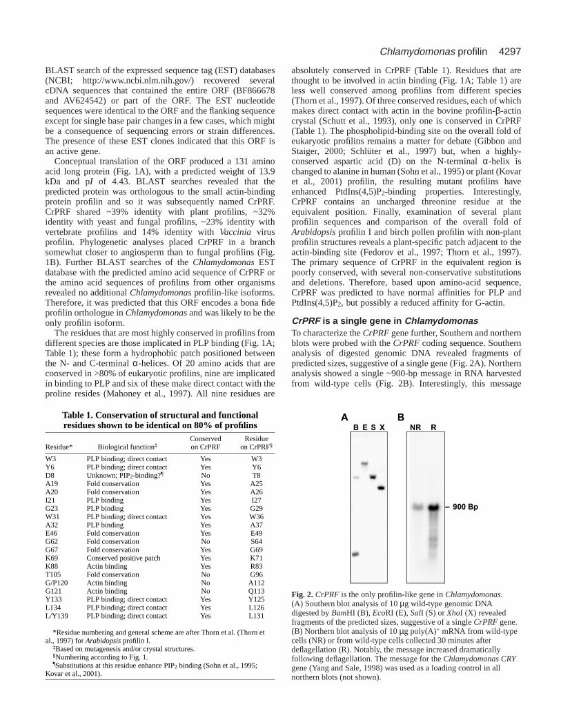
CrPRF is a single gene in ChlamydomonasTo characterize the CrPRFgene further, Southern and northernblots were probed with the CrPRFcoding sequence. Southernanalysis of digested genomic DNA revealed fragments ofpredicted sizes, suggestive of a single gene (Fig. 2A). Northernanalysis showed a single ~900-bp message in RNA harvestedfrom wild-type cells (Fig. 2B). Interestingly, this message
Table 1. Conservation of structural and functionalresidues shown to be identical on 80% of profilins
Conserved Residue Residue* Biological function‡ on CrPRF on CrPRF§
W3 PLP binding; direct contact Yes W3Y6 PLP binding; direct contact Yes Y6D8 Unknown; PIP2-binding?¶ No T8A19 Fold conservation Yes A25A20 Fold conservation Yes A26I21 PLP binding Yes I27G23 PLP binding Yes G29W31 PLP binding; direct contact Yes W36A32 PLP binding Yes A37E46 Fold conservation Yes E49G62 Fold conservation No S64G67 Fold conservation Yes G69K69 Conserved positive patch Yes K71K88 Actin binding Yes R83T105 Fold conservation No G96G/P120 Actin binding No A112G121 Actin binding No Q113Y133 PLP binding; direct contact Yes Y125L134 PLP binding; direct contact Yes L126L/Y139 PLP binding; direct contact Yes L131
*Residue numbering and general scheme are after Thorn et al. (Thorn etal., 1997) for Arabidopsisprofilin I.
‡Based on mutagenesis and/or crystal structures.§Numbering according to Fig. 1.¶Substitutions at this residue enhance PIP2 binding (Sohn et al., 1995;
Kovar et al., 2001).
Fig. 2.CrPRF is the only profilin-like gene in Chlamydomonas.(A) Southern blot analysis of 10 µg wild-type genomic DNAdigested by BamHI (B), EcoRI (E), SalI (S) or XhoI (X) revealedfragments of the predicted sizes, suggestive of a single CrPRFgene.(B) Northern blot analysis of 10 µg poly(A)+ mRNA from wild-typecells (NR) or from wild-type cells collected 30 minutes afterdeflagellation (R). Notably, the message increased dramaticallyfollowing deflagellation. The message for the Chlamydomonas CRYgene (Yang and Sale, 1998) was used as a loading control in allnorthern blots (not shown).
increased dramatically when wild-type cells undergo flagellarregeneration (Fig. 2B). Upregulation of transcript expressionfollowing deflagellation is a phenomenon common to manyflagellar components and suggests that CrPRF is present in theflagellar compartment. These data, along with evidence fromEST database searches (see above), indicated that CrPRF is theonly Chlamydomonasprofilin isoform.
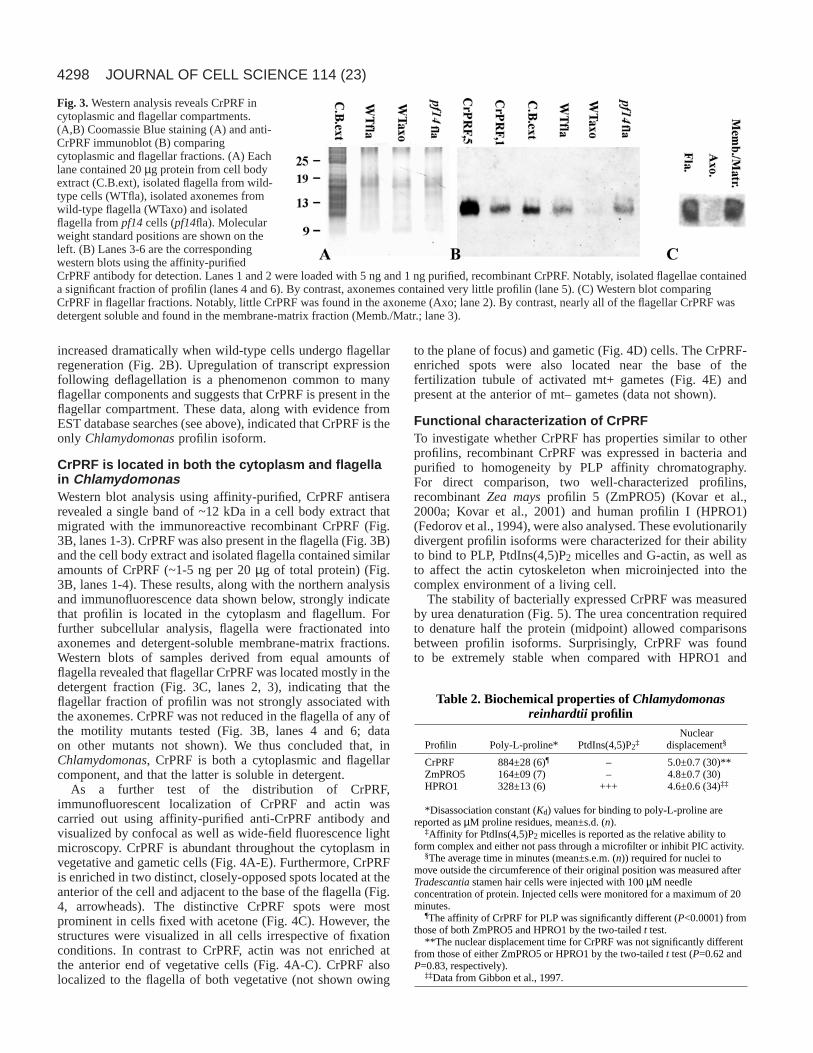
CrPRF is located in both the cytoplasm and flagellain ChlamydomonasWestern blot analysis using affinity-purified, CrPRF antiserarevealed a single band of ~12 kDa in a cell body extract thatmigrated with the immunoreactive recombinant CrPRF (Fig.3B, lanes 1-3). CrPRF was also present in the flagella (Fig. 3B)and the cell body extract and isolated flagella contained similaramounts of CrPRF (~1-5 ng per 20 µg of total protein) (Fig.3B, lanes 1-4). These results, along with the northern analysisand immunofluorescence data shown below, strongly indicatethat profilin is located in the cytoplasm and flagellum. Forfurther subcellular analysis, flagella were fractionated intoaxonemes and detergent-soluble membrane-matrix fractions.Western blots of samples derived from equal amounts offlagella revealed that flagellar CrPRF was located mostly in thedetergent fraction (Fig. 3C, lanes 2, 3), indicating that theflagellar fraction of profilin was not strongly associated withthe axonemes. CrPRF was not reduced in the flagella of any ofthe motility mutants tested (Fig. 3B, lanes 4 and 6; dataon other mutants not shown). We thus concluded that, inChlamydomonas, CrPRF is both a cytoplasmic and flagellarcomponent, and that the latter is soluble in detergent.
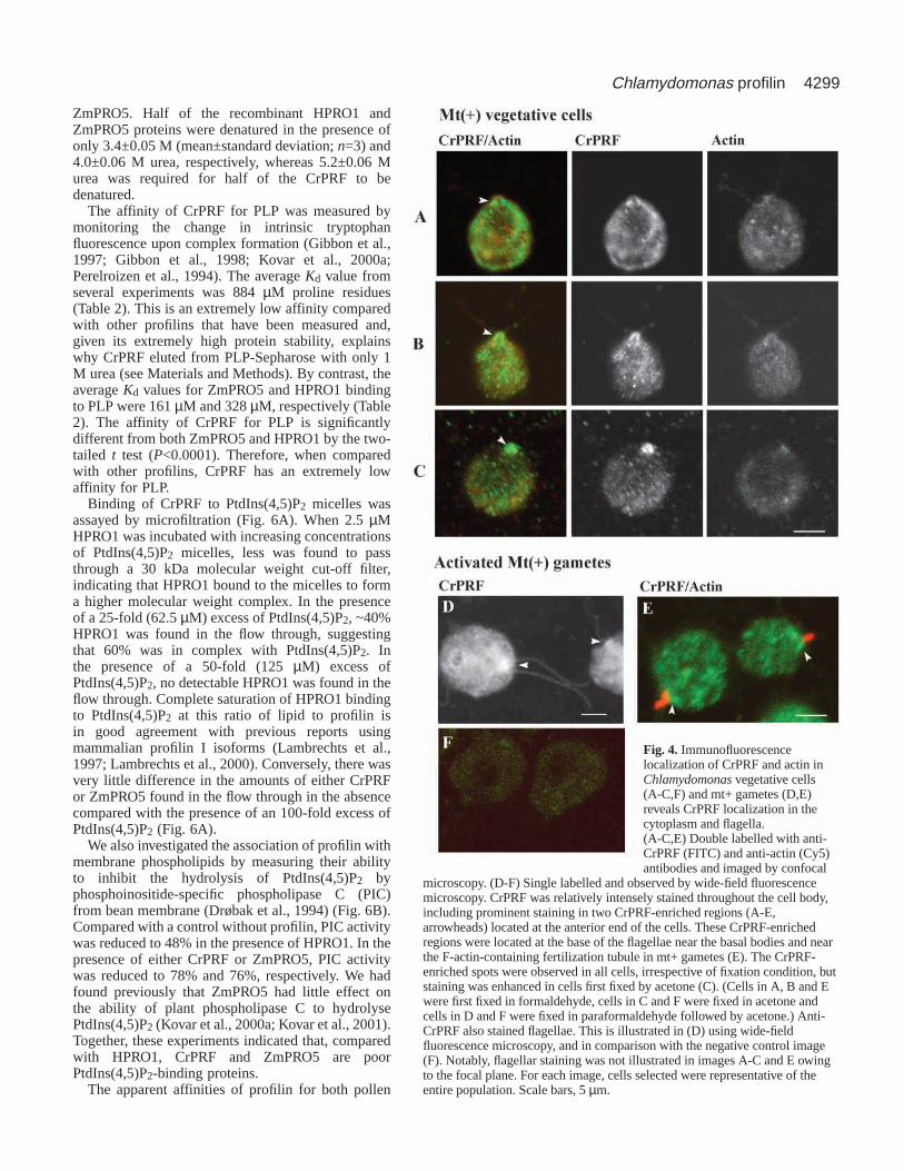
As a further test of the distribution of CrPRF,immunofluorescent localization of CrPRF and actin wascarried out using affinity-purified anti-CrPRF antibody andvisualized by confocal as well as wide-field fluorescence lightmicroscopy. CrPRF is abundant throughout the cytoplasm invegetative and gametic cells (Fig. 4A-E). Furthermore, CrPRFis enriched in two distinct, closely-opposed spots located at theanterior of the cell and adjacent to the base of the flagella (Fig.4, arrowheads). The distinctive CrPRF spots were mostprominent in cells fixed with acetone (Fig. 4C). However, thestructures were visualized in all cells irrespective of fixationconditions. In contrast to CrPRF, actin was not enriched atthe anterior end of vegetative cells (Fig. 4A-C). CrPRF alsolocalized to the flagella of both vegetative (not shown owing
to the plane of focus) and gametic (Fig. 4D) cells. The CrPRF-enriched spots were also located near the base of thefertilization tubule of activated mt+ gametes (Fig. 4E) andpresent at the anterior of mt– gametes (data not shown).
Functional characterization of CrPRFTo investigate whether CrPRF has properties similar to otherprofilins, recombinant CrPRF was expressed in bacteria andpurified to homogeneity by PLP affinity chromatography.For direct comparison, two well-characterized profilins,recombinant Zea maysprofilin 5 (ZmPRO5) (Kovar et al.,2000a; Kovar et al., 2001) and human profilin I (HPRO1)(Fedorov et al., 1994), were also analysed. These evolutionarilydivergent profilin isoforms were characterized for their abilityto bind to PLP, PtdIns(4,5)P2 micelles and G-actin, as well asto affect the actin cytoskeleton when microinjected into thecomplex environment of a living cell.
The stability of bacterially expressed CrPRF was measuredby urea denaturation (Fig. 5). The urea concentration requiredto denature half the protein (midpoint) allowed comparisonsbetween profilin isoforms. Surprisingly, CrPRF was foundto be extremely stable when compared with HPRO1 and
JOURNAL OF CELL SCIENCE 114 (23)
Fig. 3.Western analysis reveals CrPRF incytoplasmic and flagellar compartments.(A,B) Coomassie Blue staining (A) and anti-CrPRF immunoblot (B) comparingcytoplasmic and flagellar fractions. (A) Eachlane contained 20 µg protein from cell bodyextract (C.B.ext), isolated flagella from wild-type cells (WTfla), isolated axonemes fromwild-type flagella (WTaxo) and isolatedflagella from pf14cells (pf14fla). Molecularweight standard positions are shown on theleft. (B) Lanes 3-6 are the correspondingwestern blots using the affinity-purifiedCrPRF antibody for detection. Lanes 1 and 2 were loaded with 5 ng and 1 ng purified, recombinant CrPRF. Notably, isolated flagellae containeda significant fraction of profilin (lanes 4 and 6). By contrast, axonemes contained very little profilin (lane 5). (C) Western blot comparingCrPRF in flagellar fractions. Notably, little CrPRF was found in the axoneme (Axo; lane 2). By contrast, nearly all of the flagellar CrPRF wasdetergent soluble and found in the membrane-matrix fraction (Memb./Matr.; lane 3).
Table 2. Biochemical properties of Chlamydomonasreinhardtii profilin
*Disassociation constant (Kd) values for binding to poly-L-proline arereported as µM proline residues, mean±s.d. (n).
‡Affinity for PtdIns(4,5)P2 micelles is reported as the relative ability toform complex and either not pass through a microfilter or inhibit PIC activity.
§The average time in minutes (mean±s.e.m. (n)) required for nuclei tomove outside the circumference of their original position was measured afterTradescantiastamen hair cells were injected with 100 µM needleconcentration of protein. Injected cells were monitored for a maximum of 20minutes.
¶The affinity of CrPRF for PLP was significantly different (P<0.0001) fromthose of both ZmPRO5 and HPRO1 by the two-tailed t test.
**The nuclear displacement time for CrPRF was not significantly differentfrom those of either ZmPRO5 or HPRO1 by the two-tailed t test (P=0.62 andP=0.83, respectively).
‡‡Data from Gibbon et al., 1997.
4299Chlamydomonas profilin
ZmPRO5. Half of the recombinant HPRO1 andZmPRO5 proteins were denatured in the presence ofonly 3.4±0.05 M (mean±standard deviation; n=3) and4.0±0.06 M urea, respectively, whereas 5.2±0.06 Murea was required for half of the CrPRF to bedenatured.
The affinity of CrPRF for PLP was measured bymonitoring the change in intrinsic tryptophanfluorescence upon complex formation (Gibbon et al.,1997; Gibbon et al., 1998; Kovar et al., 2000a;Perelroizen et al., 1994). The average Kd value fromseveral experiments was 884 µM proline residues(Table 2). This is an extremely low affinity comparedwith other profilins that have been measured and,given its extremely high protein stability, explainswhy CrPRF eluted from PLP-Sepharose with only 1M urea (see Materials and Methods). By contrast, theaverage Kd values for ZmPRO5 and HPRO1 bindingto PLP were 161 µM and 328 µM, respectively (Table2). The affinity of CrPRF for PLP is significantlydifferent from both ZmPRO5 and HPRO1 by the two-tailed t test (P<0.0001). Therefore, when comparedwith other profilins, CrPRF has an extremely lowaffinity for PLP.
Binding of CrPRF to PtdIns(4,5)P2 micelles wasassayed by microfiltration (Fig. 6A). When 2.5 µMHPRO1 was incubated with increasing concentrationsof PtdIns(4,5)P2 micelles, less was found to passthrough a 30 kDa molecular weight cut-off filter,indicating that HPRO1 bound to the micelles to forma higher molecular weight complex. In the presenceof a 25-fold (62.5 µM) excess of PtdIns(4,5)P2, ~40%HPRO1 was found in the flow through, suggestingthat 60% was in complex with PtdIns(4,5)P2. Inthe presence of a 50-fold (125 µM) excess ofPtdIns(4,5)P2, no detectable HPRO1 was found in theflow through. Complete saturation of HPRO1 bindingto PtdIns(4,5)P2 at this ratio of lipid to profilin isin good agreement with previous reports usingmammalian profilin I isoforms (Lambrechts et al.,1997; Lambrechts et al., 2000). Conversely, there wasvery little difference in the amounts of either CrPRFor ZmPRO5 found in the flow through in the absencecompared with the presence of an 100-fold excess ofPtdIns(4,5)P2 (Fig. 6A).
We also investigated the association of profilin withmembrane phospholipids by measuring their abilityto inhibit the hydrolysis of PtdIns(4,5)P2 byphosphoinositide-specific phospholipase C (PIC)from bean membrane (Drøbak et al., 1994) (Fig. 6B).Compared with a control without profilin, PIC activitywas reduced to 48% in the presence of HPRO1. In thepresence of either CrPRF or ZmPRO5, PIC activitywas reduced to 78% and 76%, respectively. We hadfound previously that ZmPRO5 had little effect onthe ability of plant phospholipase C to hydrolysePtdIns(4,5)P2 (Kovar et al., 2000a; Kovar et al., 2001).Together, these experiments indicated that, comparedwith HPRO1, CrPRF and ZmPRO5 are poorPtdIns(4,5)P2-binding proteins.
The apparent affinities of profilin for both pollen
Fig. 4. Immunofluorescencelocalization of CrPRF and actin inChlamydomonasvegetative cells(A-C,F) and mt+ gametes (D,E)reveals CrPRF localization in thecytoplasm and flagella.(A-C,E) Double labelled with anti-CrPRF (FITC) and anti-actin (Cy5)antibodies and imaged by confocal
microscopy. (D-F) Single labelled and observed by wide-field fluorescencemicroscopy. CrPRF was relatively intensely stained throughout the cell body,including prominent staining in two CrPRF-enriched regions (A-E,arrowheads) located at the anterior end of the cells. These CrPRF-enrichedregions were located at the base of the flagellae near the basal bodies and nearthe F-actin-containing fertilization tubule in mt+ gametes (E). The CrPRF-enriched spots were observed in all cells, irrespective of fixation condition, butstaining was enhanced in cells first fixed by acetone (C). (Cells in A, B and Ewere first fixed in formaldehyde, cells in C and F were fixed in acetone andcells in D and F were fixed in paraformaldehyde followed by acetone.) Anti-CrPRF also stained flagellae. This is illustrated in (D) using wide-fieldfluorescence microscopy, and in comparison with the negative control image(F). Notably, flagellar staining was not illustrated in images A-C and E owingto the focal plane. For each image, cells selected were representative of theentire population. Scale bars, 5 µm.
4300
actin and rabbit skeletal muscle actin (RSMA) weredetermined indirectly, by measuring the difference in theamount of filamentous actin (F-actin) in the absence, comparedwith the presence, of profilin. As shown in Fig. 7, increasingconcentrations of G-actin alone or G-actin in the presence of1 µM CrPRF, ZmPRO5 or HPRO1 were allowed to polymerizeuntil steady state was reached. Subsequently, the relativeamount of F-actin was determined by 90° light scattering andplotted against the starting concentration of G-actin. Usingthese plots and the assumptions and calculations statedpreviously (Kovar et al., 2000a), the apparent affinity ofprofilin for actin was determined. From several independentexperiments, the average apparent affinities of CrPRF,ZmPRO5 and HPRO1 binding to pollen G-actin were 0.41 µM,0.46 µM and 0.14 µM, respectively (Table 3). For ZmPRO5,these results were similar to our previous findings (Kovar etal., 2000a). The average apparent affinities of CrPRF,ZmPRO5 and HPRO1 for binding to RSMA G-actin were 0.30µM, 0.28 µM and 0.20 µM, respectively (Table 3). For bothsources of actin, CrPRF and ZmPRO5 were not significantlydifferent from each other (P>0.25), whereas HPRO1 wassignificantly different from both CrPRF and ZmPRO5 forpollen actin (P<0.001) but not RSMA (P>0.2). ThereforeCrPRF, ZmPRO5 and HPRO1 have similar affinities for G-actin.
To assess the interaction between CrPRF and actin further,we took advantage of an in vivo ‘nuclear displacement’ assaydescribed in detail previously (Gibbon et al., 1997; Gibbon etal., 1998; Kovar et al., 2000a; Kovar et al., 2000b). The assayindirectly measures the sequestering activity of profilin bymonitoring the time required for the destruction of actinfilaments, indicated by the degradation of transvacuolar strandsand the movement of the nucleus from a central position. Asillustrated in Table 2, when CrPRF was injected, the averagetime for nuclear displacement was 5.0 minutes, whereasZmPRO5 displaced the nucleus in 4.8 minutes. Previously, wefound that HPRO1 displaces the nucleus in 4.6 minutes, whichis similar to both ZmPRO5 and CrPRF (Gibbon et al., 1997).By contrast, Zea maysprofilin I (ZmPRO1) displaces the
nucleus significantly more slowly than these other profilins,with an average of 7.0 minutes (Gibbon et al., 1998; Kovar et
JOURNAL OF CELL SCIENCE 114 (23)
0 1 2 3 4 5 6 7 8
0
0.2
0.4
0.6
0.8
1
[Urea] M
Rel
ativ
e flu
ore
scen
ce
Fig. 5.CrPRF is an extremely stable profilin. The stability of CrPRF(circles), HPRO1 (triangles) and ZmPRO5 (diamonds) proteins wasdetermined by incubating them with increasing concentrations ofurea and measuring their intrinsic tryptophan fluorescences. Therelative fluorescence was plotted against the urea concentration andfitted to a sigmoid curve. For this representative experiment, thedenaturation midpoint for HPRO1 was 3.3 M, for ZmPRO5 4.0 Mand for CrPRF 5.2 M.
Fig. 6.CrPRF has a low affinity for PtdIns(4,5)P2.(A) Microfiltration of profilin-PtdIns(4,5)P2 complexes shows thatlittle CrPRF bound to lipid micelles. The indicated concentrations ofPtdIns(4,5)P2 in micelles were incubated with 2.5 µM profilin andspun through a 30,000 molecular weight cut-off filter. The flowthrough was analysed by SDS-PAGE. The 14-kDa region of gelsfrom a representative experiment are shown for CrPRF, ZmPRO5and HPRO1 (top). The intensity of each Coomassie-stained band wasdetermined with a densitometer and normalized against the intensityof the profilin band found in the flow through in the absence ofPtdIns(4,5)P2. Bars (bottom) represent the percentage of CrPRF(black), ZmPRO5 (grey) or HPRO1 (white) present in the flowthrough from two independent experiments. (B) The hydrolysis ofPtdIns(4,5)P2 by phospholipase C (PIC) was measured in the absenceor presence of 5 µM profilin. Each bar represents the average (±standard deviation) of at least four independent determinations. PICactivity in the absence of profilin (ø profilin) was set to 100%.
Table 3. Apparent affinities (Kd values) for rabbit skeletalmuscle actin (RSMA) and maize pollen actin (MPA)
*The apparent Kd values for profilin binding to actin under polymerizingconditions, in the presence of 1 µM Ca2+, were determined by measuring theshift in Cc values at steady state. All values, in µM, are reported as mean±s.d.(n).
‡CrPRF was not significantly different from ZmPRO5 by the two-tailed ttest for either MPA or RSMA (P=0.25 and P=0.31, respectively).
§HPRO1 was significantly different from CrPRF and ZmPRO5 for MPA(P<0.001) but not for RSMA (P>0.19) by the two-tailed t test.
4301Chlamydomonas profilin
al., 2000a). Therefore, CrPRF appeared to be more similar toZmPRO5 and HPRO1 than to ZmPRO1. The biochemical basisfor differences in the live cell appears to be due primarily todifferences in a profilin’s apparent affinity for G-actin, becauseZmPRO5, HPRO1 and CrPRF have a significantly higheraffinity for G-actin than does ZmPRO1 (Kovar et al., 2000a;Kovar et al., 2001).
CrPRF inhibits the rate of nucleotide exchange onactinMost profilins, including those from vertebrates (Goldschmidt-Clermont et al., 1992; Perelroizen et al., 1996), Acanthamoeba(Mockrin and Korn, 1980; Nishida, 1985), Vaccinia virus(Machesky et al., 1994) and yeasts (Eads et al., 1998; Lu andPollard, 2001), have been shown to increase the exchange rateof the nucleotide bound to G-actin. However, plant profilinshave no effect on nucleotide exchange (Eads et al., 1998;Perelroizen et al., 1996), even when tested with plant actin(Kovar et al., 2000a). We examined whether CrPRF behavesmore similarly to plant or to non-plant profilins with respect toits effect on nucleotide exchange.
The rates of nucleotide exchange for 0.5 µM RSMA alone(Fig. 8A) and in the presence of profilin were determined bymeasuring the increase in fluorescence emission when G-actinincorporates the ATP analogue ε-ATP. As expected, evena substoichiometric concentration of HPRO1 (0.1 µM)significantly enhanced the rate of nucleotide exchange,whereas nearly saturating amounts of ZmPRO5 had littleeffect. Surprisingly, an equimolar concentration of CrPRF (0.5µM) substantially decreased the rate of nucleotide exchange,and nearly saturating amounts of CrPRF (2.5 µM) furtherinhibited nucleotide exchange. To our knowledge, this is thefirst report of any profilin that significantly inhibits nucleotideexchange on actin.
The experiments shown in Fig. 8A were extended to includeboth RSMA and maize pollen actin under both low ionic and
more physiological conditions. The rates of nucleotideexchange (kobs, measured per second) were determined byfitting the first 240 seconds of each reaction with a singleexponential function (Table 4). In the presence of either saltcondition, an equal concentration of CrPRF (0.5 µM) inhibitedthe intrinsic rate of nucleotide exchange of both RSMA andmaize pollen actin by about two times. Nearly saturatingamounts of CrPRF (2.5 µM) inhibited nucleotide exchange ofboth actins under both conditions by four to eight times. Underall conditions tested, substoichiometric amounts of HPRO1significantly enhanced nucleotide exchange of both actins(three to 15 times). ZmPRO5 had little effect on either sourceof actin under low ionic conditions, but slightly inhibited(approximately two times with nearly saturating amounts ofZmPRO5) under more physiological conditions.
For comparison with a known inhibitor of nucleotide
Actin ( µM)
Lig
ht
scat
teri
ng
(A
U)
0
250
500
750
1000
1250
0 1 2 3
Fig. 7.CrPRF has a high apparent affinity for maize pollen G-actin.A representative experiment shows that CrPRF shifts the steady stateCc (see below) for actin assembly. Increasing concentrations ofpollen G-actin were polymerized alone (squares) or in the presenceof 1 µM CrPRF (circles), 1 µM ZmPRO5 (diamonds) or 1 µMHPRO1 (triangles). The x-axis intercepts of each regression line (Ccvalues) were 0.29 µM in the absence of profilin and 0.74 µM, 0.71µM and 0.91 µM in the presence of CrPRF, ZmPRO5 and HPRO1,respectively. The calculated apparent Kd values were 0.35 µM forCrPRF1, 0.40 µM for ZmPRO5 and 0.18 µM for HPRO1.Abbreviation: A.U., arbitrary light-scattering units.
A
B
[CrPRF] ( µM)k o
bs (
s-1 )
0
0.02
0.04
0.06
0.08
0 5 10 15 20
500
400
300
200
100
0
5004003002001000
Flu
ores
cenc
e (A
U)
Seconds
1
23
4
5
Fig. 8.CrPRF inhibits nucleotide exchange. (A) Representativeexperiments show that profilins from diverse organisms havesubstantially different effects on the initial rate of nucleotideexchange for G-actin. The incorporation of ε-ATP by 0.5 µM G-actin(RSMA) in low salt buffer alone (curve 3) or in the presence of 0.1µM HPRO1 (curve 1), 2.5 µM ZmPRO5 (curve 2), 0.5 µM CrPRF(curve 4) or 2.5 µM CrPRF (curve 5) was monitored over time. Thecurves shown are fits of raw data (not shown) with a singleexponential function. Human profilin I dramatically enhanced theinitial rate of nucleotide exchange, whereas ZmPRO5 had little effectand CrPRF significantly decreased the initial rate. (B) The effect of arange of CrPRF concentrations (0.1-20 µM) on the initial rate ofnucleotide exchange of 2.0 µM G-actin (maize pollen) in low ionicstrength buffer is shown. Initial rates were determined by fitting thefirst 240 seconds of curves similar to those shown in (A) to a singleexponential function. The initial rates were plotted against theconcentration of CrPRF and fitted to the equation described inMethods. The calculated dissociation constant was 0.11 µM.
4302
exchange, an equal concentration of human ADF (0.5 µM)inhibited nucleotide exchange of both actins by about twotimes under low ionic conditions (Table 4). Therefore, CrPRFand human ADF have similar effects on nucleotide exchange.Interestingly, in the presence of salt, human ADF did notappear to inhibit nucleotide exchange. An equal concentrationof DNase I (0.5 µM) greatly inhibited nucleotide exchange(approximately four times) for both actins under bothconditions (Table 4).
The rate of nucleotide exchange for 2 µM maize pollen actinwas measured in the presence of a range of CrPRFconcentrations (0.1-20 µM). Nucleotide exchange rates wereplotted against the concentration of CrPRF and the data fittedto the equation described in Methods (Fig. 8B). Theequilibrium dissociation constant (Kd) was determined to be0.11 µM. This is lower than the apparent affinity derived fromsteady-state experiments (0.41 µM; see above).
DISCUSSION
CrPRF has an extremely low affinity for PLP andPtdIns(4,5)P 2, but a high affinity for G-actinProfilins are ubiquitous, cytosolic proteins that form a 1:1complex with monomeric (G-) actin and have complex effectson the actin cytoskeleton (Gibbon and Staiger, 2000; Schlüter etal., 1997). Profilins also interact with poly-L-proline and proline-rich proteins (Frazier and Field, 1997; Holt and Koffer, 2001;Wasserman, 1998), and membrane polyphosphoinositides(Lassing and Lindberg, 1985). We have described theidentification and characterization of a profilin from C.reinhardtii (CrPRF). CrPRF protein was found in both theflagella and the cell body. Within the cell body, CrPRF isenriched in an unusual structure at the anterior end of vegetativeand gametic cells at the base of the flagella. Based upon aminoacid sequence comparisons with other profilins, CrPRF waspredicted to have normal affinities for PLP and PtdIns(4,5)P2 butpossibly a reduced affinity for G-actin. Using two independentmethods for each ligand, we discovered that CrPRF has anextremely low affinity for both PLP and PtdIns(4,5)P2 but a highaffinity for G-actin. Surprisingly, when complexed with G-actin,CrPRF inhibits nucleotide exchange strongly. To date, no otherprofilin has been reported to inhibit nucleotide exchange. CrPRFappears to be a unique profilin that might help to elucidate thediverse roles of profilins from evolutionarily distant species.
Numerous native and recombinant profilin isoforms havebeen isolated from diverse eukaryotic cells and tested for theability to interact with the three general profilin ligands: G-actin, PLP and proline-rich peptides and phosphoinositidelipids. Direct comparison of results from these studies iscomplicated by differences in the approaches used by variouslaboratories. In general, profilins from different organisms, aswell as individual profilin isoforms from the same organism,do not have identical properties. For example, threemammalian and five maize profilin isoforms are quite divergentin amino acid sequence, expression patterns and biochemicalproperties (Di Nardo et al., 2000; Gibbon et al., 1997; Gibbonet al., 1998; Jonckheere et al., 1999; Kovar et al., 2000a;Lambrechts et al., 1995; Lambrechts et al., 1997; Lambrechtset al., 2000; Perelroizen et al., 1996; Suetsugu et al., 1998;Witke et al., 1998). A picture emerges that, despite grosssimilarities in biochemical properties and overall fold, profilinisoforms might have unique functions adapted for therequirements of specific cell types.
Proline-rich binding partners are proposed to link profilin tosignal transduction cascades that result in actin cytoskeletonreorganization, either through regulating the subcellulardistribution or the activity of profilin (Frazier and Field, 1997;Wasserman, 1998; Holt and Koffer, 2001). The residues thatare most highly conserved in profilins from different speciesare those implicated in PLP binding, which contribute to ahydrophobic patch positioned between the N- and C-terminalα-helices (Fedorov et al., 1997; Thorn et al., 1997). Becauseall nine conserved PLP-binding residues are present, wepredicted that CrPRF would have a comparatively normalaffinity for PLP. Affinities of profilin for PLP range from 55µM for fission yeast profilin (Lu and Pollard, 2001) to 360 µMfor human profilin (Petrella et al., 1996). Surprisingly, CrPRFhas a Kd of 884 µM, which is at least a 2.5 times lower affinityfor PLP than any other eukaryotic profilin measured. Theseresults suggest that a high affinity interaction with proline-richproteins is probably not important for the in vivo function ofCrPRF. However, mammalian profilin I binds more strongly tothe proline-rich protein N-WASP than does mammalianprofilin II, even though profilin II has a higher affinity for PLP,demonstrating the importance of characterizing the affinity ofprofilins for their actual protein binding partners (Suetsugu etal., 1998).
Interaction with membrane polyphosphoinositides has alsobeen suggested to link profilin with intracellular signalling
JOURNAL OF CELL SCIENCE 114 (23)
Table 4. Nucleotide exchange rates for rabbit skeletal muscle actin (RSMA) and maize pollen actin (MPA)Low ionic strength‡ Physiological§
*Nucleotide exchange rates are reported as the change in fluorescence (arbitrary units) per second±s.d. (n), as determined by fitting experimental data, such asthose shown in Fig. 8, to a single exponential function.
‡Low ionic strength buffer: 2 mM Tris, pH 7.0, 0.5 mM DTT.§Physiological buffer: 2 mM Tris, pH 7.5, 0.5 mM DTT, 100 mM KCl, 5 mM MgCl2.
4303Chlamydomonas profilin
events (Goldschmidt-Clermont et al., 1991; Lassing andLindberg, 1985). The phospholipid-binding site on the overallfold of profilin is still a matter of debate (Schlüter et al., 1997)but, when a highly-conserved aspartic acid on the N-terminalα-helix is changed to alanine in HPRO1 (Sohn et al., 1995) orZmPRO5 (Kovar et al., 2001), the mutants have an increasedaffinity for PtdIns(4,5)P2. CrPRF has an uncharged threonineresidue at the equivalent position, which we thought mightmimic the aspartic acid to alanine substitution. However,binding of CrPRF to PtdIns(4,5)P2 micelles was barelydetectable, even in the presence of a 100-fold excess of lipid.By comparison, ZmPRO5 had a similarly low affinity, whereasHPRO1 had a high affinity for PtdIns(4,5)P2 micelles.Therefore, CrPRF does not appear to associate with membranelipids. The possibility that CrPRF binds to D-3phosphoinositides with higher affinity than to PtdIns(4,5)P2, asis the case for mammalian profilin (Lu et al., 1996), should beexamined.
Residues that are implicated in actin binding are poorlyconserved among different profilins. Of three conservedresidues that make direct contact with actin in the bovineprofilin-β-actin co-crystal (Schutt et al., 1993), only one isconserved in CrPRF. However, CrPRF was found to have ahigh affinity for both plant G-actin (0.41 µM) and rabbitskeletal muscle G-actin (0.33 µM). Although they have beenmeasured with different sources of actin and by a variety ofmethods, affinities of profilins for G-actin range from 0.1 µMto >10 µM.
The biochemical properties of CrPRF are exactly oppositefrom the predictions that were made based on the amino acidsequence and are not currently explainable. One possibility isthat the overall fold of CrPRF is unlike those of other profilins.CrPRF is extremely stable (midpoint for denaturation is at 5.2M urea) compared with ZmPRO5 (4.0 M), HPRO1 (3.4 M),budding yeast profilin (3.4 M) (Eads et al., 1998) and fissionyeast profilin (4.5 M) (Lu and Pollard, 2001). To determinewhy CrPRF has unique biochemical properties, a crystalstructure for CrPRF with its different ligands would be useful.
CrPRF is the only profilin known to inhibitnucleotide exchangeBecause profilins inhibit the addition of monomers to the slow-growing (pointed) end but not the fast-growing (barbed) end ofactin filaments (Pollard and Cooper, 1984), they can haveopposite effects on the assembly of actin in vitro (Kang et al.,1999). When the barbed ends of actin filaments are capped,profilins cause depolymerization of actin filaments by bindingand sequestering G-actin. Conversely, when the barbed endsare uncapped and a large pool of actin monomers is available,profilin-actin complexes can add to the barbed end andpromote polymerization.
It has been suggested that profilin-enhanced polymerizationinvolves ‘recharging’ ADP-loaded actin subunits with ATP,through stimulation of nucleotide exchange, because ATP-loaded G-actin adds onto filaments more readily (Goldschmidt-Clermont et al., 1992; Mockrin and Korn, 1980). Vertebrateprofilin also interacts synergistically with ADF/cofilin toincrease the rate of filament treadmilling 125-fold over the rateof actin alone (Didry et al., 1998). The importance ofenhancing nucleotide exchange has been challenged by the factthat plant profilins do not enhance nucleotide exchange (Eads
et al., 1998; Kovar et al., 2000a; Perelroizen et al., 1996), yetare still able to promote polymerization in vitro (Perelroizen etal., 1996; Ballweber et al., 1998) and interact synergisticallywith ADF/cofilin to increase the rate of treadmilling 75-foldover actin alone (Didry et al., 1998). The extent to whichvertebrate profilin, compared with plant profilin, interactssynergistically with ADF/cofilin might be explained by itsability to enhance nucleotide exchange. Evidence for the invivo importance of enhanced nucleotide exchange is providedfor fission yeast profilin by Lu and Pollard (Lu and Pollard,2001). A fission yeast mutant profilin with a single amino acidsubstitution, which does not affect actin binding but is nolonger able to enhance nucleotide exchange, does notcomplement either profilin-null or temperature-sensitivefission yeast strains.
Therefore, profilin’s ability to enhance nucleotide exchangemight be important in some species (yeast), but not all(plants). Surprisingly, we found that, with near-saturatingconcentrations, CrPRF decreased the rate of nucleotideexchange up to eight times. The significance of this finding isnot entirely clear but it certainly adds to the complexity ofdifferences in biochemical properties between evolutionarilydiverse profilins.
The intrinsic nucleotide exchange rate of actin may also bean important factor. A budding yeast actin mutation with anincreased rate of nucleotide exchange suppresses defects inprofilin (Wolven et al., 2000). Additionally, maize pollen actinhas a 10- to 20-fold higher intrinsic rate of nucleotide exchangethan rabbit skeletal muscle actin (Table 4). Perhaps plantprofilins do not enhance nucleotide exchange because this isnot the rate-limiting step for treadmilling in plant cells. Testingwhether CrPRF complements null mutants for yeast profilinand investigating the effect of ADF/cofilin and CrPRF on therate of filament treadmilling would be useful.
CrPRF probably functions as a G-actin-sequesteringprotein in ChlamydomonasActin exists predominantly in a diffuse subunit pool within thecytoplasm of Chlamydomonas, because interphase cells arelargely devoid of phalloidin-stainable F-actin (Detmers et al.,1983; Detmers, 1985). Actin is enriched in a presumptivecontractile ring structure during cytokinesis (Harper et al.,1992) and F-actin to the fertilization tubule of mt+ gametes(Detmers, 1983; Detmers, 1985). Cytochalasin treatmentsdecrease mating efficiency by inhibiting the appearance ofactin filaments in fertilization tubules, but have no obviouseffect on other processes including cell division (Detmers et al.,1983; Harper et al., 1992). A mutant (ida5) that has completeloss of expression of the conventional Chlamydomonasactingene also has no defects in cell growth or division, which mightbe due to compensation by a non-conventional actin (Kato-Minoura et al., 1997).
Therefore, in Chlamydomonas, polymeric actin appears tobe required sparingly. With the exception of the CrPRF-enriched structures adjacent to the base of the flagella andunderneath the fertilization tubule of mt+ gametes, CrPRF isalso distributed throughout the cytosol and flagella. Althoughwe do not know the concentration of G-actin in the cytoplasmof Chlamydomonas, we expect it to be higher than the criticalconcentration for assembly. CrPRF might be the primary actin-binding protein responsible for sequestering this pool of G-
4304
actin. Presumably CrPRF-actin complexes are capable ofassembling onto uncapped barbed ends like other profilins(Kang et al., 1999). In support of this, a direct measurement ofCrPRF’s affinity for G-actin gave a lower Kd value (0.11 µM)than did an indirect measurement at steady state underpolymerizing conditions (0.41 µM). These differences could bedue to the different ionic conditions between the two assays,or could reflect the assembly of profilin-actin complexes atsteady state. Even allowing for addition of complexes ontouncapped filament ends, the apparent affinity of CrPRF foractin is high enough to account for a large amount ofunpolymerized actin in the cytoplasm of interphaseChlamydomonascells. To provide further evidence for thissimple model, measurements of total actin, F-actin, cappingprotein, profilin and profilin-actin levels in Chlamydomonasshould be made. The localization of CrPRF in flagella suggestsa role in Chlamydomonasmotility or flagellar biogenesis,perhaps by preventing undesired polymerization of actin inflagella (Kato-Minoura et al., 1997; Kato-Minoura et al., 1998;Ohara et al., 1998). CrPRF was almost entirely in thedetergent-soluble fraction of flagella, suggesting that it mightnot interact directly with the actin subunit of the inner dyneinor be involved in flagellar motility. Given the CrPRF-enrichedstructure at the base of the fertilization tubule of mt+ gametes,it is possible that CrPRF plays a role in actin dynamics duringmating. A possible role for CrPRF in functions such as mating,cytokinesis and intraflagellar transport (Rosenbaum et al.,1999) remains to be tested.
We thank S. Almo (Albert Einstein College of Medicine) and J.Bamburg (Colorado State University) for supplying human profilin Iand human ADF expression plasmids, respectively, L. Blanchoin(CNRS, Grenoble) for help fitting the nucleotide exchange data overa range of CrPRF concentrations, A. Lambrechts (Ghent University)for advice about PtdIns(4,5)P2 microfiltration, and U. Goodenough(Washington University), N. Wilson (University of Minnesota) and S.Ono (Emory University) for advice on immunofluorescence staining.This work was supported by a US Department of Agriculture -National Research Initiative grant (99-35304-8640) to C.J.S., a grantfrom the National Institute of Health to P.Y. and W.S.S. and a USDepartment of Education GAANN training grant to D.R.K. Theresults reported here are in partial fulfilment of the requirements fora PhD degree by D.R.K.
REFERENCES
Balasubramanian, M. K., Hirani, B. R., Burke, J. D. and Gould, K. L.(1994). TheSchizosaccharomyces pombe cdc3+ gene encodes a profilinessential for cytokinesis. J. Cell Biol. 125, 1289-1301.
Ballweber, E., Giehl, K., Hannapel, E., Huff, T., Jockusch, B. M. andMannherz, H. G. (1998). Plant profilin induces actin polymerization fromactin:β-thymosin complexes and competes directly with β-thymosins andwith negative co-operativity with DNase I for binding to actin. FEBS Lett.425, 251-255.
Cooley, L., Verheyen, E. and Ayers, K.(1992). chickadeeencodes a profilinrequired for intercellular cytoplasm transport during Drosophilaoogenesis.Cell 69, 173-184.
Detmers, P. A.(1985). Elongation of cytoplasmic processes during gameticmating: models for actin-based motility. Can. J. Biochem. Cell Biol. 63, 599-607.
Detmers, P. A., Goodenough, U. W. and Condeelis, J.(1983). Elongation ofthe fertilization tubule in Chlamydomonas: new observations on the coremicrofilaments and the effect of transient intracellular signals in theirstructural integrity. J. Cell Biol. 97, 522-532.
Detmers, P. A., Carboni, J. M. and Condeelis, J.(1985). Localization of
actin in Chlamydomonasusing antiactin and NBD-phallacidin. Cell Motil.5, 415-430.
Di Nardo, A., Gareus, R., Kwiatkowski, D. and Witke, W. (2000).Alternative splicing of the mouse profilin II gene generates functionallydifferent profilin isoforms. J. Cell Sci. 113, 3795-3803.
Didry, D., Carlier, M.-F. and Pantaloni, D. (1998). Synergy between actindepolymerizing factor/cofilin and profilin in increasing actin filamentturnover. J. Biol. Chem. 273, 25602-25611.
Drøbak, B. K., Watkins, P. A. C., Valenta, R., Dove, S. K., Lloyd, C. W.and Staiger, C. J. (1994). Inhibition of plant plasma membranephosphoinositide phospholipase C by the actin-binding protein, profilin.Plant J. 6, 389-400.
Eads, J. C., Mahoney, N. M., Vorobiev, S., Bresnick, A. R., Wen, K. K.,Rubenstein, P. A., Haarer, B. K. and Almo, S. C.(1998). Structuredetermination and characterization of Saccharomyces cerevisiaeprofilin.Biochemistry37, 11171-11181.
Ehler, L. L. and Dutcher, S. K.(1998). Pharmacological and genetic evidencefor a role of rootlet and phycoplast microtubules in the positioning andassembly of cleavage furrows in Chlamydomonas reinhardtii. Cell Motil.Cytoskeleton40, 193-207.
Fedorov, A. A., Magnus, K. A., Graupe, M. H., Lattman, E. E., Pollard,T. D. and Almo, S. C.(1994). X-ray structures of isoforms of the actin-binding protein profilin that differ in their affinity for phosphatidylinositolphosphates. Proc. Natl. Acad. Sci. USA 91, 8636-8640.
Fedorov, A. A., Ball, T., Mahoney, N. M., Valenta, R. and Almo, S. C.(1997). The molecular basis for allergen cross-reactivity: crystal structureand IgE-epitope mapping of birch pollen profilin. Structure5, 33-45.
Fowkes, M. E. and Mitchell, D. R. (1998). The role of preassembledcytoplasmic complexes in assembly of flagellar dynein subunits. Mol. Biol.Cell 9, 2337-2347.
Frazier, J. A. and Field, C. M. (1997). Actin cytoskeleton: are FH proteinslocal organizers? Curr. Biol. 7, R414-R417.
Gibbon, B. G. and Staiger, C. J.(2000). Profilin. In Actin: A DynamicFramework For Multiple Plant Cell Functions(ed. C. J. Staiger, F. Baluska,D. Volkmann and P. Barlow), pp. 45-65. Dordrecht, The Netherlands:Kluwer Academic Publishers.
Gibbon, B. C., Ren, H. and Staiger, C. J.(1997). Characterization of maize(Zea mays) pollen profilin function in vitro and in live cells. Biochem. J.327, 909-915.
Gibbon, B. C., Zonia, L. E., Kovar, D. R., Hussey, P. J. and Staiger, C. J.(1998). Pollen profilin function depends on interaction with proline-richmotifs. Plant Cell10, 981-994 [erratum: Plant Cell11, 1603].
Gill, C. G. and von Hippel, P. H.(1989). Calculation of protein extinctioncoefficients from amino acid sequence data. Anal. Biochem. 182, 319-326.
Goldschmidt-Clermont, P. J., Kim, J. W., Machesky, L. M., Rhee, S. G.and Pollard, T. D. (1991). Regulation of phospholipase C-γ1 by profilinand tyrosine phosphorylation. Science251, 1231-1233.
Goldschmidt-Clermont, P. J., Furman, M. I., Wachsstock, D., Safer, D.,Nachmias, V. T. and Pollard, T. D.(1992). The control of actin nucleotideexchange by thymosin β4 and profilin. A potential regulatory mechanismfor actin polymerization in cells. Mol. Biol. Cell3, 1015-1024.
Goodenough, U. W. and Weiss, R. L.(1975). Gametic differentiation inChlamydomonas reinhardtii. J. Cell Biol. 67, 623-637.
Haarer, B. K., Petzold, A. S. and Brown, S. S.(1993). Mutational analysisof yeast profilin. Mol. Cell Biol. 13, 7864-7873.
Harper, J. D. I., McCurdy, D. W., Sanders, M. A., Salisbury, J. L. andJohn, P. C. L. (1992). Actin dynamics during the cell cycle inChlamydomonas reinhardtii. Cell Motil. Cytoskeleton22, 117-126.
Harris, E. H. (2001). Chlamydomonasas a model organism. Annu. Rev. PlantPhysiol. Plant Mol. Biol. 52, 363-406.
Haugwitz, M., Noegel, A. A., Karakesisoglou, J. and Schleicher, M.(1994).Dictyosteliumamoeba that lack G-actin-sequestering profilins show defectsin F-actin content, cytokinesis, and development. Cell 79, 303-314.
Hawkins, M., Pope, B., Maciver, S. K. and Weeds, A. G.(1993). Humanactin depolymerizing factor mediates a pH-sensitive destruction of actinfilaments. Biochemistry32, 9985-9993.
Holt, M. R. and Koffer, A. (2001). Cell motility: proline-rich proteinspromote protrusions. Trends Cell Biol. 11, 38-46.
Houk, T. W., Jr and Ue, K. (1974). The measurement of actin concentrationin solution: a comparison of methods. Anal. Biochem. 62, 66-74.
Jonckheere, V., Lambrechts, A., Vandekerckhove, J. and Ampe, C.(1999).Dimerization of profilin II upon binding the (GP5)3 peptide from VASPovercomes the inhibition of actin nucleation by profilin II and thymosin β4.FEBS Lett. 447, 257-263.
JOURNAL OF CELL SCIENCE 114 (23)
4305Chlamydomonas profilin
Kagami, O. and Kamiya, R. (1992). Translocation and rotation ofmicrotubules caused by multiple species of Chlamydomonasinner-armdynein. J. Cell Biol. 103, 653-664.
Kang, F., Purich, D. L. and Southwick, F. S.(1999). Profilin promotesbarbed-end actin filament assembly without lowering the criticalconcentration. J. Biol. Chem. 274, 36963-36972.
Karakesisoglou, I., Schleicher, M., Gibbon, B. C. and Staiger, C. J.(1996).Plant profilins rescue the aberrant phenotype of profilin-deficientDictyosteliumcells. Cell Motil. Cytoskeleton34, 36-47.
Kato-Minoura, T., Hirono, M. and Kamiya, R. (1997). Chlamydomonasinner-arm dynein mutant, ida5, has a mutation in an actin-encoding gene. J.Cell Biol. 137, 649-656.
Kato-Minoura, T., Uryu, S., Hirono, M. and Kamiya, R. (1998). Highlydivergent actin expressed in a Chlamydomonasmutant lacking theconventional actin gene. Biochem. Biophys. Res. Commun. 251, 71-76.
Kovar, D. R., Drøbak, B. K. and Staiger, C. J.(2000a). Maize profilinisoforms are functionally distinct. Plant Cell12, 583-598.
Kovar, D. R., Staiger, C. J., Weaver, E. A. and McCurdy, D. W.(2000b).AtFim1 is an actin filament-crosslinking protein from Arabidopsis thaliana.Plant J. 24, 625-636.
Kovar, D. R., Drøbak, B. K., Collings, D. A. and Staiger, C. J.(2001). Thecharacterization of ligand-specific maize profilin mutants. Biochem. J. 358,49-57.
Lambrechts, A., Vandamme, J., Goethals, M., Vandekerckhove, J. andAmpe, C. (1995). Purification and characterization of bovine profilin II.Actin, poly(L-proline) and inositolphospholipid binding. Eur. J. Biochem.230, 281-286.
Lambrechts, A., Verschelde, J. L., Jonckheere, V., Goethals, M.,Vandekerckhove, J. and Ampe, C.(1997). The mammalian profilinisoforms display complementary affinities for PIP2 and proline-richsequences. EMBO J. 16, 484-494.
Lambrechts, A., Braun, A., Jonckheere, V., Aszodi, A., Lanier, L.,Robbens, J., Van Colen, I., Vandekerckhove, J., Fässler, R. and Ampe,C. (2000). Profilin II is alternatively spliced, resulting in profilin isoformsthat are differentially expressed and have distinct biochemical properties.Mol. Cell Biol. 20, 8209-8219.
Lassing, I. and Lindberg, U. (1985). Specific interaction betweenphosphatidylinositol 4,5-bisphosphate and profilactin. Nature314, 472-474.
Lee, V. D., Finstad, S. L. and Huang, B.(1997). Cloning and characterizationof a gene encoding an actin-related protein in Chlamydomonas. Gene197,153-159.
Lefebvre, P. A. and Silfow, C. D.(1999). Chlamydomonas: the cell and itsgenomes. Genetics151, 9-14.
Lu, J. and Pollard, T. D. (2001). Profilin binding to poly-L-proline and actinmonomers along with ability to catalyze actin nucleotide exchange isrequired for viability of fission yeast. Mol. Biol. Cell12, 1161-1175.
Lu, P.-J., Shieh, W.-R., Rhee, S. G., Yin, H. L. and Chen, C.-S.(1996).Lipid products of phosphoinositide 3-kinase bind human profilin with highaffinity. Biochemistry35, 14027-14034.
Machesky, L. M., Cole, N. B., Moss, B. and Pollard, T. D.(1994). Vacciniavirus expressed a novel profilin with a higher affinity forpolyphosphoinositides than actin. Biochemistry33, 10815-10824.
Mahoney, N. M., Janmey, P. A. and Almo, S. C. (1997). Structure of theprofilin-poly-L-proline complex involved in morphogenesis and cytoskeletalregulation. Nat. Struct. Biol. 4, 953-960.
Martin, N. C. and Goodenough, U. W.(1975). Gametic differentiation inChlamydomonas reinhardtii. I. Production of gametes and their finestructure. J. Cell Biol. 67, 587-605.
Mockrin, S. C. and Korn, E. D. (1980). Acanthamoebaprofilin interacts withG-actin to increase the rate of exchange of actin-bound adenosine 5′-triphosphate. Biochemistry19, 5359-5362.
Muto, E., Edamatsu, M., Hirono, M. and Kamiya, R. (1994).Immunological detection of actin in the 14S ciliary dynein of Tetrahymena.FEBS Lett. 343, 173-177.
Nishida, E. (1985). Opposite effects of cofilin and profilin from porcine brainon rate of exchange of actin-bound adenosine 5′-triphosphate. Biochemistry24, 1160-1164.
Ohara, A., Kato-Minoura, T., Kamiya, R. and Hirono, M. (1998). Recoveryof flagellar inner-arm dynein and the fertilization tubule in Chlamydomonasida5mutant by transformation with actin genes. Cell Struct. Funct. 23, 273-281.
Pan, J. and Snell, W. J.(2000). Signal transduction during fertilization in theunicellular green alga, Chlamydomonas. Curr. Opin. Microbiol. 3, 596-602.
Perelroizen, I., Marchand, J.-B., Blanchoin, L., Didry, D. and Carlier, M.-F. (1994). Interaction of profilin with G-actin and poly(L-proline).Biochemistry33, 8472-8478.
Perelroizen, I., Didry, D., Christensen, H., Chua, N.-H. and Carlier, M.-F.(1996). Role of nucleotide exchange and hydrolysis in the function ofprofilin in actin assembly. J. Biol. Chem. 271, 12302-12309.
Petrella, E. C., Machesky, L. M., Kaiser, D. A. and Pollard, T. D.(1996).Structural requirements and thermodynamics of the interaction of prolinepeptides with profilin. Biochemistry35, 16535-16543.
Piperno, G. and Luck, D. J.(1979). An actin-like protein is a componentof axonemes from Chlamydomonasflagella. J. Biol. Chem. 254, 2187-2190.
Piperno, G., Ramanis, Z., Smith, E. F. and Sale, W. S.(1990). Three distinctinner dynein arms in Chlamydomonasflagella: molecular composition andlocation in the axoneme. J. Cell Biol. 110, 379-389.
Pollard, T. D. and Cooper, J. A.(1984). Quantitative analysis of the effectof Acanthamoebaprofilin on actin filament nucleation and elongation.Biochemistry23, 6631-6641.
Ren, H., Gibbon, B. C., Ashworth, S. L., Sherman, D. M., Yuan, M. andStaiger, C. J.(1997). Actin purified from maize pollen functions in livingplant cells. Plant Cell9, 1445-1457.
Rosenbaum, J. L., Cole, D. G. and Diener, D. R.(1999). Intraflagellartransport: the eyes have it. J. Cell Biol. 144, 385-388.
Sanders, M. A. and Salisbury, J. L.(1994). Immunofluorescence microscopyof cilia and flagella. Methods Cell Biol. 47, 163-169.
Schlüter, K., Jockusch, B. M. and Rothkegel, M.(1997). Profilins asregulators of actin dynamics. Biochim. Biophys. Acta1359, 97-109.
Schutt, C. E., Myslik, J. C., Rozycki, M. D., Goonesekere, N. C. W. andLindberg, U. (1993). The structure of crystalline profilin-β-actin. Nature365, 810-816.
Sohn, R. H., Chen, J., Koblan, K. S., Bray, P. F. and Goldschmidt-Clermont, P. J. (1995). Localization of a binding site forphosphatidylinositol-4,5-bisphosphate on human profilin. J. Biol. Chem.270, 21114-21120.
Suetsugu, S., Miki, H. and Takenawa, T.(1998). The essential role of profilinin the assembly of actin for microspike formation. EMBO J. 17, 6516-6526.
Sugase, Y., Hirono, M., Kindle, K. L. and Kamiya, R.(1996). Cloning andcharacterization of the actin-encoding gene of Chlamydomonas reinhardtii.Gene168, 117-121.
Thorn, K. S., Christensen, H. E. M., Shigeta, R., Huddler, D., Shalaby, L.,Lindberg, U., Chua, N. H. and Schutt, C. E.(1997). The crystal structureof a major allergen from plants. Structure5, 19-32.
Wilkerson, C. G., King, S. M., Koutoulis, A., Pazour, G. J. and Witman,G. B. (1995). The 78,000 Mr intermediate chain of Chlamydomonasouterarm dynein is a WD-repeat protein required for arm assembly. J. Cell Biol.129, 169-178.
Wilson, N. F., Figkesib, M. J. and Snell, W. J.(1997a). The Chlamydomonasmating type plus fertilization tubule, a prototypic cell fusion organelle:isolation, characterization, and in vitro adhesion to mating type minusgametes. J. Cell Biol. 137, 1537-1553.
Wilson, N. F., Foglesong, M. J. and Snell, W. J.(1997b). TheChlamydomonasmating type plus fertilization tubule, a prototypic cellfusion organelle: isolation, characterization, and in vitro adhesion to matingtype minus gametes. J. Cell Biol. 137, 1537-1553.
Witke, W., Podtelejnikov, A. V., Di Nardo, A., Sutherland, J. D., Gurniak,C. B., Dotti, C. and Mann, M.(1998). In mouse brain profilin I and profilinII associate with regulators of the endocytic pathway and actin assembly.EMBO J. 17, 967-976.
Witman, G. B. (1986). Isolation of Chlamydomonasflagella and flagellaraxonemes. Methods Enzymol. 134, 280-290.
Wolven, A. K., Belmont, L. D., Mahoney, N. M., Almo, S. C. and Drubin,D. G. (2000). In vivo importance of actin nucleotide exchange catalyzed byprofilin. J. Cell Biol. 150, 895-903.
Yang, P. and Sale, W. S.(1998). The Mr 140,000 intermediate chain ofChlamydomonasflagellar inner arm dynein is a WD-repeat proteinimplicated in dynein arm anchoring. Mol. Biol. Cell9, 3335-3349.
Yang, P. and Sale, W. S.(1999). Flagellar radial spoke protein 2 (RSP2) is aprotein kinase. Mol. Biol. Cell10, 338a.
Yang, P., Fox, L., Colbran, R. J. and Sale, W. S.(2000). Protein phosphatasesPP1 and PP2A are located in distinct positions in the Chlamydomonasflagellar axoneme. J. Cell Sci. 113, 91-102.