Integrated Risk Information System (IRIS) U.S. Environmental Protection Agency Chemical Assessment Summary National Center for Environmental Assessment 1 Chloral hydrate; CASRN 302-17-0 Human health assessment information on a chemical substance is included in the IRIS database only after a comprehensive review of toxicity data, as outlined in the IRIS assessment development process. Sections I (Health Hazard Assessments for Noncarcinogenic Effects) and II (Carcinogenicity Assessment for Lifetime Exposure) present the conclusions that were reached during the assessment development process. Supporting information and explanations of the methods used to derive the values given in IRIS are provided in the guidance documents located on the IRIS website. STATUS OF DATA FOR Chloral hydrate File First On-Line 08/22/1988 Category (section) Assessment Available? Last Revised Oral RfD (I.A.) yes 09/15/2000 Inhalation RfC (I.B.) qualitative discussion 09/15/2000 Carcinogenicity Assessment (II.) yes 09/15/2000 I. Chronic Health Hazard Assessments for Noncarcinogenic Effects I.A. Reference Dose for Chronic Oral Exposure (RfD) Substance Name — Chloral hydrate CASRN — 302-17-0 Last Revised — 09/15/2000 The oral Reference Dose (RfD) is based on the assumption that thresholds exist for certain toxic effects such as cellular necrosis. It is expressed in units of mg/kg-day. In general, the RfD is an estimate (with uncertainty spanning perhaps an order of magnitude) of a daily exposure to the human population (including sensitive subgroups) that is likely to be without an appreciable risk of deleterious effects during a lifetime. Please refer to the Background Document for an elaboration of these concepts. RfDs can also be derived for the noncarcinogenic health effects of substances that are also carcinogens. Therefore, it is
Transcript
Integrated Risk Information System (IRIS) U.S. Environmental Protection Agency Chemical Assessment Summary National Center for Environmental Assessment
1
Chloral hydrate; CASRN 302-17-0
Human health assessment information on a chemical substance is included in the IRIS database only after a comprehensive review of toxicity data, as outlined in the IRIS assessment development process. Sections I (Health Hazard Assessments for Noncarcinogenic Effects) and II (Carcinogenicity Assessment for Lifetime Exposure) present the conclusions that were reached during the assessment development process. Supporting information and explanations of the methods used to derive the values given in IRIS are provided in the guidance documents located on the IRIS website.
STATUS OF DATA FOR Chloral hydrate
File First On-Line 08/22/1988
Category (section) Assessment Available? Last Revised
I. Chronic Health Hazard Assessments for Noncarcinogenic Effects
I.A. Reference Dose for Chronic Oral Exposure (RfD)
Substance Name — Chloral hydrate CASRN — 302-17-0 Last Revised — 09/15/2000
The oral Reference Dose (RfD) is based on the assumption that thresholds exist for certain toxic effects such as cellular necrosis. It is expressed in units of mg/kg-day. In general, the RfD is an estimate (with uncertainty spanning perhaps an order of magnitude) of a daily exposure to the human population (including sensitive subgroups) that is likely to be without an appreciable risk of deleterious effects during a lifetime. Please refer to the Background Document for an elaboration of these concepts. RfDs can also be derived for the noncarcinogenic health effects of substances that are also carcinogens. Therefore, it is
Integrated Risk Information System (IRIS) U.S. Environmental Protection Agency Chemical Assessment Summary National Center for Environmental Assessment
2
essential to refer to other sources of information concerning the carcinogenicity of this substance. If the U.S. EPA has evaluated this substance for potential human carcinogenicity, a summary of that evaluation will be contained in Section II of this file.
I.A.1. Oral RfD Summary
Critical Effect Experimental Doses* UF MF RfD
CNS depression and GI irritation in humans
Goodman and Gilman, 1985
NOAEL: None
LOAEL: 10.7 mg/kg-day
100 1 0.1 mg/kg-day
*Conversion Factors and Assumptions — The LOAEL is based on an exposure of 250 mg, three times a day, causing CNS depression (sedation) and GI irritation (nausea, vomiting), and on an average body weight of 70 kg.
I.A.2. Principal and Supporting Studies (Oral RfD)
Although the reference value of 0.1 mg/kg-day derived from the pharmacologically active dose in humans is an acute RfD, keeping the exposure below this level will also be protective for any noncancer health effect from chronic exposure. For example, chronic exposure to chloral hydrate does not cause adverse effects in the liver of rats or mice until the exposure approaches 135 or 160 mg/kg-day, respectively. Similarly, there are no reproductive, developmental, neurobehavioral, or immunological effects following long-term treatment of laboratory animals until the exposure approaches 160 mg/kg-day. Therefore, it is appropriate to use the acute RfD also as the chronic RfD.
Chloral hydrate has been widely used as a sedative/hypnotic drug in humans. The recommended dose for an adult as a sedative is 250 mg, three times a day (equivalent to 10.7 mg/kg-day); the recommended dose as an hypnotic is 500-1,000 mg (equivalent to 7-14 mg/kg) (Goodman and Gilman, 1985). The recommended dose for a child as a sedative is 9 mg/kg, three times a day, to 25 mg/kg in single dose (Hindmarsh et al., 1991). The recommended dose for a child undergoing a medical or dental procedure is 50 to 100 mg/kg (Badalaty et al., 1990; Fox et al., 1990). A child is typically given a higher dose than an adult because a deeper level of sedation is desired to obtain better cooperation from the child during the medical or dental procedure. There is no evidence that a child is less sensitive than an adult to the sedative effects of chloral hydrate. Because of the rapid metabolism of chloral hydrate,
Integrated Risk Information System (IRIS) U.S. Environmental Protection Agency Chemical Assessment Summary National Center for Environmental Assessment
3
trichloroethanol is responsible for the majority of the pharmacological activity (Marshall and Owens, 1954; Breimer, 1977; Goodman and Gilman, 1985). The concentration of trichloroethanol in the plasma in the pharmacologically active range is approximately 5 mg/L and above and in the toxic range is 100 mg/L and above.
Chloral hydrate is irritating to the skin and mucous membranes and often causes gastric distress (nausea and vomiting) at recommended doses. There are no reports of sensitization in humans. Overdoses produce (in order of progression) ataxia, lethargy, deep coma, respiratory depression, hypotension, and cardiac arrhythmias. The life-threatening effects are from severe respiratory depression, hypotension, and cardiac arrhythmias. For some representative case reports, see Anyebuno and Rosenfeld (1991), Ludwigs et al. (1996), Marshall (1977), and Sing et al. (1996). A potentially life-threatening oral dose for humans is approximately 10 g (143 mg/kg), although death has been reported from as little as 4 g, and some individuals have survived ingesting 30 g or more. Extended abuse of chloral hydrate may result in development of paranoid behavior, in tolerance to the pharmacological effect, and in physical dependence or addiction. Sudden withdrawal after habituation can precipitate seizure, delirium, and death in untreated individuals.
Shapiro et al. (1969) reviewed the medical records of 1,618 patients who had received chloral hydrate at 1 g (213 patients, 13%), 0.5 g (1345 patients, 83%), or various other doses (60 patients, 4%). Adverse reactions were reported in 38 patients (2.3%). Of these patients, four received 1 g, one received 0.75 g, and 33 received 0.5 g. Reported adverse reactions included gastrointestinal symptoms in 10 patients, depression of the central nervous system (CNS) in 20 patients, skin rash in 5 patients, prolonged prothrombin time in 1 patient, and bradycardia in 1 patient. In all patients the side effects disappeared when chloral hydrate therapy was stopped. There was no evidence of association between adverse side effects and age, weight, or sex.
Miller and Greenblatt (1979) reviewed medical records of 5,435 hospital patients who received chloral hydrate at a dose of either 0.5 g (about 7 to 8 mg/kg) or 1 g (about 14 to 16 mg/kg). Adverse reactions were noted in 119 cases (2.2%). CNS depression was most common (58 patients, or 1.1%), with minor sensitivity reactions, including rash, pruritus, fever, and eosinophilia, second most common (19 patients, or 0.35%). Other adverse reactions included gastrointestinal disturbances (0.28%) and CNS excitement (0.22%). Three individuals (0.05%) were judged to have life-threatening reactions involving CNS depression, asterixis (flapping tremor characterized by an intermittent lapse of assumed posture due to involuntary sustained contractions of groups of muscles), or hypotension. The data show that adverse reactions involving the CNS became more frequent with increasing dosage in patients older than 50 years, in patients who died during hospitalization, in patients who received concurrently benzodiazepine antianxiety drugs, and in patients with elevated levels of blood urea nitrogen.
Integrated Risk Information System (IRIS) U.S. Environmental Protection Agency Chemical Assessment Summary National Center for Environmental Assessment
4
Greenberg et al. (1991) reported various side effects experienced by children receiving chloral hydrate sedation in preparation for computer tomography (CT) procedures. In a "high-dose" group, composed of 295 children (average age 2.18 years) that received a single dose of 80 to 100 mg/kg and a maximum total dose of 2 g, adverse reactions occurred in 23 of the patients (7%) and included vomiting (14 patients), hyperactivity (5 patients), and respiratory symptoms such as wheezing and secretion aspiration (4 patients). Cardiac monitoring did not reveal any abnormalities or arrhythmias in any of the children. A second "lower-dose" cohort of 111 children (average age 1.9 years) received 40 to 75 mg/kg chloral hydrate. These patients received the lower dose because of existing liver or renal impairment, respiratory insufficiency, or CNS depression. There were no adverse side effects or complications reported in this group. Children with severe liver or renal disease or affected by severe CNS depression were not treated with chloral hydrate.
Lambert et al. (1990) conducted a retrospective analysis of hospital medical records to investigate a possible link between chloral hydrate administration and direct hyperbilirubinemia (DHB), an increase in the concentration of unconjugated bilirubin in the serum, in neonates following prolonged administration of chloral hydrate (25 to 50 mg/kg administered for up to 20 days). In the first study, the DHB was of unknown etiology in 10 of the 14 newborns with DHB; all 10 of these DHB patients had received chloral hydrate. In the second study, among 44 newborns who had received chloral hydrate, 10 patients that developed DHB had received a mean cumulative dose of 1,035 mg/kg. In contrast, 34 patients whose direct bilirubin levels were within normal ranges received a mean cumulative dose of 183 mg/kg. As the total bilirubin levels (free plus conjugated bilirubin) were the same in both groups and within the normal range, the increased direct bilirubin could result from competition between trichloroethanol and bilirubin in the glucuronidation pathway, known to function suboptimally in neonates.
Kaplan et al. (1967) investigated whether ethanol ingestion altered the metabolism of chloral hydrate or increased subjective symptoms. Five male volunteers weighing 70 to 107 kg consumed ethanol (880 mg/kg), chloral hydrate (1 g, 9 to 14 mg/kg), or both. Blood pressure and cardiac rate did not vary significantly among treatments. In the presence of ethanol, the concentration of trichloroethanol in the blood rose more rapidly and reached a higher concentration, but the rate of depletion was not significantly changed. The increase in the concentration of trichloroethanol was not sufficient to produce a marked enhancement of the hypnotic effect. The volunteers reported symptoms (drowsiness, dizziness, blurred vision) and their severity during the 6-hour observation period. At all time points, the rank order of effects was: ethanol plus chloral hydrate > ethanol > chloral hydrate.
No long-term studies of chloral hydrate in humans were located in the published literature. Chloral hydrate is addictive and is a controlled substance (Schedule IV) in the United States.
Integrated Risk Information System (IRIS) U.S. Environmental Protection Agency Chemical Assessment Summary National Center for Environmental Assessment
5
The effect that occurs at the lowest exposure is CNS depression and gastrointestinal irritation in humans. As these effects would not be intended or desirable in the general population, EPA considers these responses as adverse effects and are used to derive the reference dose.
Acute gavage exposure in mice shows neurological effects (ataxia) at about the same exposure for the comparable effect in humans. A subchronic study in mice using sensitive tests for neurobehavioral changes found none. Chronic studies in rats and mice show no evidence of neurobehavioral changes and no evidence of histopathological changes in nervous tissue. As with other chlorinated chemicals, there is some evidence of hepatotoxicity in rodent liver following chronic oral exposure. These effects are of minimal severity, may be related to precancerous lesions, and occur at an exposure greater than that required for CNS depression and gastrointestinal irritation following an acute bolus dose.
No data are available to determine a NOAEL in humans. The recommended clinical dose for sedation in adults is 250 mg, taken 3 times a day (Goodman and Gilman, 1985). The LOAEL is 10.7 mg/kg-day (assuming a 70 kg body weight). The pharmacokinetic information shows that chloral hydrate and the pharmacologically active metabolite, trichloroethanol, will not bioaccumulate.
I.A.3. Uncertainty and Modifying Factors (Oral RfD)
UF = 100.
An uncertainty factor of 10 was used to extrapolate from a LOAEL to NOAEL. An uncertainty factor of 10 was used for intraspecies variability. An uncertainty factor for chronic duration is not used. Chloral hydrate and the active metabolite, trichloroethanol, do not bioaccumulate. Therefore, continuous daily exposure to chloral hydrate at the reference dose will not result in a concentration of trichloroethanol in the blood required for the pharmacological effect. Developmental toxicity, including developmental neurotoxicity, and immunotoxicity are not critical effects. Although there is no two-generation reproduction study, an uncertainty factor for database limitations is not needed, as there is evidence from several studies that reproductive toxicity is not likely to be a critical effect.
Although the reference value derived from the pharmacologically active dose in humans is an acute RfD, keeping the exposure below this level will also be protective for any noncancer health effect from chronic exposure. For example, chronic exposure to chloral hydrate does not cause adverse effects in the liver of rats or mice until the exposure approaches 135 or 160 mg/kg-day, respectively. Similarly, there are no reproductive, developmental, neurobehavioral, or immunological effects following long-term treatment of laboratory
Integrated Risk Information System (IRIS) U.S. Environmental Protection Agency Chemical Assessment Summary National Center for Environmental Assessment
6
animals until exposure approaches 160 mg/kg-day. Therefore, it is appropriate to use the acute RfD also as the chronic RfD.
Simultaneous ingestion of ethanol and chloral hydrate increases the sedative and side-effects of chloral hydrate. The mechanism is the increase in the concentration of the pharmacologically active metabolite, trichloroethanol, in the presence of ethanol. Chronic abusers of ethanol are, therefore, somewhat more sensitive to the adverse effects of chloral hydrate.
Because of the immaturity of hepatic metabolism, particularly the glucuronidation pathway, and decreased glomerular filtration, the half-life of trichloroethanol is longer in infants (pre-term and full term) than in adults. The half-life of trichloroethanol in toddlers and adults is similar. Because of the longer half-life of trichloroethanol, pre-term and full term infants will experience prolonged effects when chloral hydrate is administered. However, at the reference dose for chloral hydrate, the steady-state concentration of trichloroethanol in these groups is far below the concentration required for the pharmacological effect.
Although male laboratory rodents seem to be more sensitive than female laboratory rodents to hepatic effects, there is no evidence of a gender effect in humans to the sedative or side-effects of chloral hydrate at the recommended clinical dose.
MF = 1
I.A.4. Additional Studies/Comments (Oral RfD)
Metabolism and Toxicokinetics
Chloral hydrate is completely absorbed following oral administration. Qualitatively similar metabolism occurs in mice, rats, dogs, Japanese Medaka, and humans (Abbas et al., 1996; Abbas and Fisher, 1997; Beland et al., 1998; Breimer, 1977; Elfarra et al., 1998; Fisher et al., 1998; Goodman and Gilman, 1985; Gorecki et al., 1990; Gosselin et al., 1981; Greenberg et al., 1999; Henderson et al., 1997; Hindmarsh et al., 1991; Hobara et al., 1986, 1987a,b, 1988a,b; Lipscomb et al., 1996, 1998; Marshall and Owens, 1954; Mayers et al., 1991; Merdink et al., 1998, 1999; Owens and Marshall, 1955; Reimche et al., 1989; Stenner et al., 1997, 1998).
Chloral hydrate is rapidly metabolized in both hepatic and extrahepatic tissues to trichloroethanol and trichloroacetic acid. The alcohol dehydrogenase responsible for reducing it to trichloroethanol is located in both liver and erythrocytes. A portion of the trichloroethanol produced is conjugated with glucuronic acid to form trichloroethanol-ß-glucuronide, which is
Integrated Risk Information System (IRIS) U.S. Environmental Protection Agency Chemical Assessment Summary National Center for Environmental Assessment
7
excreted in the urine. A portion of the trichloroethanol-glucuronide is secreted into the bile and is subject to enterohepatic circulation. Oxidation of chloral hydrate to trichloracetic acid occurs primarily in the liver and kidney via an aldehyde dehydrogenase using nicotinamide adenine dinucleotide (NAD) as a cofactor. The major route of excretion of the metabolites of chloral hydrate is the urine.
Chloral hydrate and its metabolites have been found in milk (Bernstine et al., 1956). As soon as lactation started, mothers (n=50) were treated with a 1.33 g rectal suppository of chloral hydrate. Samples of maternal blood and breast milk were taken for analysis from 15 minutes and at varying intervals up to 24 hours following administration of the drug. The maximum concentration of the sum of chloral hydrate, trichloroethanol, and trichloroethanol-glucuronide (the potential pharmacologically active species) in milk occurred within 1 hour after administration of the drug and averaged 53 mg/L (n=11). The amount of chloral hydrate required for sedation in infants is 10 mg in a single feeding of 100 mL of milk.
In mice and rats, 8% of the administered dose of chloral hydrate is directly eliminated in urine, 15% is converted to trichloroacetic acid (including the contribution from enterohepatic circulation), and 77% is converted to trichloroethanol (Beland et al., 1998). In humans 92% of the administered dose of chloral hydrate is converted to trichloroethanol and 8% is converted directly to trichloroacetic acid; additional trichloroacetic acid is formed during enterohepatic circulation of trichloroethanol such that 35% of the initial dose of chloral hydrate is converted to trichloroacetic acid (Allen and Fisher, 1993).
Although earlier reports claimed the detection of substantial quantities of dichloroacetic acid in blood from studies in rodents (Abbas et al., 1996), data show that the dichloroacetic acid is most likely formed by an acid-catalyzed dechlorination of trichloroacetic acid in the presence of reduced hemoglobin (Ketcha et al., 1996). Recent experimental data and pharmacokinetic model simulations in rodents suggest that dichloroacetic acid occurs only as a short-lived metabolite in the liver and is rapidly converted to two-carbon, nonchlorinated metabolites and carbon dioxide (Merdink et al., 1998). Using a different extraction procedure less likely to induce the artifactual formation of dichloroacetic acid, Henderson et al. (1997) showed the presence of dichloroacetic acid in children treated with chloral hydrate in a clinic.
Breimer (1977) administered an aqueous solution of chloral hydrate to five human volunteers. Each volunteer received a single oral dose of 15 mg/kg. Chloral hydrate could not be detected in the plasma even at the first sampling time of 10 minutes. A method with a limit of detection of 0.5 mg/L was used. Trichloroethanol and trichloroethanol-glucuronide reached peak concentrations 20 to 60 minutes after administration of chloral hydrate. The maximum concentration of trichloroethanol in the plasma was about 5 mg/L. The average half-lives of
Integrated Risk Information System (IRIS) U.S. Environmental Protection Agency Chemical Assessment Summary National Center for Environmental Assessment
8
trichloroethanol and trichloroethanol-glucuronide were 8 hours (range 7-9.5 hours) and 6.7 hours (range 6-8 hours), respectively. The half-life of trichloroacetic acid was about 4 days.
Zimmermann et al. (1998) administered a single dose of 250 mg chloral hydrate in 150 mL of drinking water to 18 healthy male volunteers (20 to 28 years of age). Chloral hydrate, trichloroethanol, and trichloroacetic acid were measured in plasma. Chloral hydrate could only be detected 8 to 60 minutes after dosing in 15 of 18 plasma samples. The measured concentration of chloral hydrate in plasma ranged from 0.1 mg/L (the limit of detection) to 1 mg/L. The mean maximum plasma concentration of trichloroethanol of 3 mg/L was achieved 0.67 hours after dosing. The mean maximum plasma concentration of trichloroacetic acid of 8 mg/L was achieved 32 hours after dosing. The terminal half-life for trichloroethanol was 9.3 to 10.2 hours and for trichloroacetic acid was 89 to 94 hours.
Two toxicokinetic models for chloral hydrate in rats and mice are available (Abbas et al., 1996; Beland et al., 1998). Beland et al. (1998) treated rats and mice with chloral hydrate by gavage with one or 12 doses using 50 or 200 mg/kg per dose. The maximum concentrations of chloral hydrate, trichloroethanol, and trichloroethanol-glucuronide in the plasma were observed at the initial sampling time of 0.25 hour. The half-life of chloral hydrate in the plasma was approximately 3 minutes. The half-lives of trichloroethanol and trichloroethanol-glucuronide in the mouse plasma were approximately 5 and 7 minutes, respectively. Trichloroacetic acid was the major metabolite found in the mouse plasma, with the maximum concentration being reached 1-6 hours after dosing. The half-life of trichloroacetic acid in the mouse plasma was approximately 8-11 hours. Comparable values were obtained for rats.
Several studies have investigated the age-dependence of the metabolism of chloral hydrate (Gorecki et al., 1990; Hindmarsh et al., 1991; Mayers et al., 1991; Reimche et al., 1989). These studies were conducted in critically ill patients in neonatal and pediatric intensive care units and may not be representative of a population of healthy infants. The half-lives for trichloroethanol and its glucuronide were increased fourfold in preterm and threefold in full-term infants. The half-life for trichloroethanol in toddlers was similar to that reported for adults. The reported half-lives for elimination of trichloroethanol were 39.8 hours, 27.8 hours, and 9.67 hours for pre-term infants, full-term infants, and toddlers, respectively (Mayers et al., 1991), compared with 7-9.5 hours reported by Breimer (1977) and 9.3-10.2 hours reported by Zimmermann et al. (1998). These age-related differences likely are the result of the immaturity of hepatic metabolism, particularly glucuronidation, and decreased glomerular filtration.
Kaplan et al. (1967) investigated the effect of ethanol consumption on the metabolism of chloral hydrate in adults. Subjects ingested doses of ethanol (880 mg/kg), chloral hydrate (9 to 14 mg/kg), or both. In subjects consuming both ethanol and chloral hydrate, the concentration
Integrated Risk Information System (IRIS) U.S. Environmental Protection Agency Chemical Assessment Summary National Center for Environmental Assessment
9
of trichloroethanol in blood rose more rapidly and reached a higher concentration than in subjects consuming chloral hydrate only. Ethanol promotes the formation of trichloroethanol because the oxidation of ethanol provides NADH used for the reduction of chloral hydrate (Watanabe et al., 1998).
Chronic Bioassays
Daniel et al. (1992a) exposed 40 male B6C3F1 mice for 104 weeks to drinking water containing chloral hydrate at 1 g/L (equivalent to 166 mg/kg-day). Untreated control animals (23 in one group and 10 in a second group) received distilled water. Interim sacrifices were conducted at 30 and 60 weeks of exposure (5 animals per group at each sacrifice interval). Complete necropsy and microscopic examination were performed. There were no significant treatment-related effects on survival or body weight. With the exception of changes in the liver, there were no changes in organ weight (spleen, kidneys, or testes) or histopathological changes in any tissues. The toxicity in the liver was characterized by increased absolute liver weight and liver-to-body weight ratio at all three sacrifice intervals. At week 104, liver weight was 37% higher than controls, and liver-to-body weight ratio was 42% higher than controls. Hepatocellular necrosis was noted in 10/24 (42%) treated animals; other pathological changes of mild severity reported in the livers of treated animals included cytoplasmic vacuolization, cytomegaly, and cytoplasmic alteration. This study shows a LOAEL at 166 mg/kg-day (the only exposure tested).
George et al. (2000) conducted a chronic bioassay for carcinogenicity in male B6C3F1 mice. Mice were administered chloral hydrate in drinking water for 104 weeks. Mice (72 in each group) had a mean exposure of 0, 13.5, 65, or 146.6 mg/kg-day. At the termination of the study, a complete necropsy and histopathological examination of liver, kidney, spleen, and testes from all animals was conducted. In addition a complete histopathological examination was conducted on five animals from the high-dose group. There was no change in water consumption, survival, behavior, body weight, or organ weights (liver, kidney, spleen, and testes) at any exposure. There was no evidence of hepatocellular necrosis at any exposure and only minimal changes in the levels of serum enzymes. This study identifies a NOAEL for noncancer effects in male mice of 146.6 mg/kg-day (the highest exposure tested).
NTP (2000a) conducted a chronic bioassay for carcinogenicity in female B6C3F1 mice. Mice were administered chloral hydrate by gavage in distilled water at 0, 25, 50, or 100 mg/kg 5 days a week for up to 2 years. The calculated exposures are 0, 17.9, 35.7, or 71.4 mg/kg-day. Additional groups were administered chloral hydrate by gavage for 3, 6, or 12 months and held without further dosing for the duration of the study (stop-exposure studies). There was no significant effect on survival, body weight, or organ weights at any exposure. The NOAEL in this study is 71.4 mg/kg-day (the highest exposure tested).
Integrated Risk Information System (IRIS) U.S. Environmental Protection Agency Chemical Assessment Summary National Center for Environmental Assessment
10
NTP (2000b) conducted a chronic bioassay for carcinogenicity in male B6C3F1 mice. Groups of 120 male mice received chloral hydrate by gavage in distilled water at 0, 25, 50, or 100 mg/kg for up to 2 years. The calculated exposures are 0, 17.9, 35.7, or 71.4 mg/kg-day. At each exposure 60 mice received feed ad libitum; the other 60 mice received feed in measured daily amount calculated to maintain body weight on a previously computed idealized body weight curve. Twelve mice from each diet and dose group were evaluated after 15 months of exposure. The remaining 48 animals from each diet and dose group were evaluated at 2 years. Survival, body weight, organ weights, and serum enzymes in the dosed groups were comparable to the respective vehicle control. Following complete necropsy and histopathological examination, no non-neoplastic changes were found in any organ when compared to the respective vehicle control. The NOAEL for non-neoplastic effects in this study is 71.4 mg/kg-day (the highest exposure tested).
Leuschner and Beuscher (1998) conducted a chronic bioassay in Sprague-Dawley rats. Chloral hydrate was administered in drinking water for 124 weeks (males) and 128 weeks (females). The rats (50 males and 50 females in each group) had an exposure of 15, 45, or 135 mg/kg-day. There was no effect on survival, appearance, behavior, body weight, food and water consumption, and organ weights. There was no evidence of increased incidence of tumors in any organ. Histopathological examination revealed an increased incidence of hepatocellular hypertrophy at the highest exposure in males only (11% in controls versus 28% at the highest exposure, p< 0.01 ). This finding, graded as minimal to slight in severity, was characterized by a diffuse liver cell enlargement with slightly eosinophilic cytoplasm and was considered by the authors as a first sign of toxicity. The type, incidence, and severity or other non-neoplastic lesions were not increased in treated animals compared to controls. Based on the evidence of minimal toxicity in the liver, which is of doubtful biological significance, this study establishes a NOAEL of 45 mg/kg-day and a LOAEL of 135 mg/kg-day.
George et al. (2000) conducted a chronic bioassay for carcinogenicity in male F344 rats. Rats were administered chloral hydrate in drinking water for 104 weeks. Rats (78 in each group) had a mean daily exposure of 0, 7.4, 37.4, or 162.6 mg/kg-day. At the termination of the study, a complete necropsy and histopathological examination of liver, kidney, spleen, and testes from all animals was conducted. In addition a complete histopathological examination was conducted on five animals from the high-dose group. There was no change in water consumption, survival, behavior, body weight, or organ weights (liver, kidney, spleen, and testes) at any exposure. There was no indication of liver toxicity at any exposure as shown by the lack of liver necrosis, hyperplasia, increased mitotic index, and only minimal changes in the levels of serum enzymes. The NOAEL in this study is 162.6 mg/kg-day (the highest exposure tested).
Subchronic Bioassays
Integrated Risk Information System (IRIS) U.S. Environmental Protection Agency Chemical Assessment Summary National Center for Environmental Assessment
11
Sanders et al. (1982) administered chloral hydrate in drinking water to CD-1 mice at 70 or 700 mg/L (equivalent to 16 mg/kg-day or 160 mg/kg-day) for 90 days. In males, hepatomegaly (an increase in weight of 20% and 34% at the low and high exposure, respectively) and microsome proliferation (increase in cytochrome b5 of 26% and 40%, increase in aminopyrine N-demethylase of 28% and 20%, and increase in aniline hydroxylase of 24% and 30% at the low and high exposure, respectively). There were no biologically significant changes in serum enzymes. Hepatomegaly was not seen in females, but there were changes in hepatic microsomal parameters (increase in total microsomal protein of 10%, increase in aniline hydroxylase of 23%, and decrease in cytochrome b5 of 12%) but only at the high exposure. No other significant toxicological changes were observed. Based on hepatomegaly and changes in microsomal parameters in males at the high exposure, this study identifies a LOAEL of 160 mg/kg-day and a NOAEL of 16 mg/kg-day.
Daniel et al. (1992b) exposed male and female Sprague-Dawley rats (10/sex/dose) for 90 days to chloral hydrate in drinking water at a concentration of 300, 600, 1,200, or 2,400 mg/L (equivalent to 24, 48, 96, or 168 mg/kg-day in males and 33, 72, 132, or 288 mg/kg-day in females). The tissues of animals from the high-exposure group and liver sections from all treated males were examined histopathologically. No mortality occurred in any groups prior to sacrifice. Organ weights, including liver weight, and clinical chemistry values in treated animals were only sporadically or inconsistently different from control animal values. Focal hepatocellular necrosis was observed in 2 of 10 males in each of the groups exposed to 96 and 168 mg/kg-day. The necrotic lesion was minimal at 96 mg/kg-day and was significantly more severe at 168 mg/kg-day. Necrotic lesions were not reported in any treated females or in any control animals. While serum enzymes were generally increased in treated animals, dramatic increases were reported in males in the 168 mg/kg-day group; mean aspartate aminotransferase, alanine aminotransferase, and lactate dehydrogenase levels in this group were elevated 89%, 54% and 127% above the corresponding control values, respectively. Based on the focal hepatocellular necrosis and accompanying serum enzyme changes, the study identifies a LOAEL of 168 mg/kg-day and a NOAEL of 96 mg/kg-day. The 96 mg/kg-day exposure is not considered a LOAEL because the authors reported only minimal microscopic necrosis, there was not a corresponding increase in serum enzymes, and because chronic exposure in Sprague-Dawley rats showed no necrosis at higher exposure (Leuschner and Beuscher, 1998; George et al., 2000).
Integrated Risk Information System (IRIS) U.S. Environmental Protection Agency Chemical Assessment Summary National Center for Environmental Assessment
12
Reproductive/Developmental Studies
Klinefelter et al. (1995) evaluated sperm morphology and motility in F344 rats administered chloral hydrate in drinking water for 52 weeks at 0, 55, or 188 mg/kg-day. The researchers examined cauda epididymal sperm motion parameters and testicular and epididymal histopathology. Chloral hydrate did not cause any visible systemic toxicity, and had no effects on epididymal or testicular histopathology. However, the percentage of motile sperm was significantly decreased (p<0.01) from 68% in controls to 58% in rats exposed to 188 mg/kg-day. The percentage of progressively motile sperm was also significantly decreased (p<0.01) from 63% in controls to 53% in this group. In addition, the frequency distribution of the average straight-line velocities of sperm at this exposure was significantly shifted (p<0.01) to the lower ranges when compared to controls. In this study the NOAEL is 55 mg/kg-day; the LOAEL is 188 mg/kg-day.
Kallman et al. (1984) exposed male and female CD-1 mice to chloral hydrate in drinking water at 21.3 or 204.8 mg/kg-day. Animals were exposed for 3 weeks prior to breeding. Exposure of females (5 per group) continued during gestation and until pups were weaned at 21 days of age. There was no change in drinking water consumption or weight gain in the dams. No gross malformations were noted in pups, and no significant effects were observed in duration of gestation, number of pups delivered, pup weight, or number of stillborn pups. All pups (15 per group) showed the same rate of development and level of performance on several neurobehavioral tests, except that pups exposed to 204.8 mg/kg-day when tested at 23 days of age showed impaired retention of passive avoidance learning on both the 1-hour and 24-hour retention tests (p<0.05). This study identified a NOAEL for neurodevelopmental toxicity of 21.3 mg/kg-day and a LOAEL of 204.8 mg/kg-day based on the impairment in passive avoidance learning. This study also identifies a NOAEL for reproductive and other developmental effects of 204.8 mg/kg-day (the highest exposure tested).
Johnson et al. (1998) tested the potential for chloral hydrate to cause developmental toxicity in Sprague-Dawley rats. Chloral hydrate was administered in drinking water to 20 rats from gestational day 1 to 22 at an average exposure of 151 mg/kg-day. Control animals were given distilled water. There was no evidence of maternal toxicity, no change in the number of implantation or resorption sites, no change in the number of live or dead fetuses, no change in placental or fetal weight, no change in crown-rump length, and no increase in the incidence of morphological changes. At necropsy there was no evidence of cardiac anomalies. Based on this study, the NOAEL for developmental toxicity is 151 mg/kg-day (the highest exposure tested).
Johnson et al. (1998) also tested the potential for trichloroethanol and trichloroacetic acid to cause developmental toxicity in Sprague-Dawley rats. The protocol was identical to the study
Integrated Risk Information System (IRIS) U.S. Environmental Protection Agency Chemical Assessment Summary National Center for Environmental Assessment
13
with chloral hydrate. Trichloroethanol was administered to 10 rats at an average exposure of 153 mg/kg-day. No evidence of developmental toxicity was found. In contrast, when trichloroacetic acid was administered to 11 rats at an average exposure of 291 mg/kg-day, developmental toxicity was observed. The effects included a statistically significant (p<0.05) increase in total cardiac defects per litter and an increased number of implantation and resorption sites. The results with trichloroacetic acid are generally consistent with those reported by Smith et al. (1989), who reported adverse developmental effects (levocardia) from trichloroacetic acid at an exposure of 330 mg/kg-day and above.
Saillenfait et al. (1995) tested the potential of chloral hydrate to cause developmental toxicity in vitro using a rat whole-embryo culture system. Embryos from Sprague-Dawley rats were explanted on gestational day 10 and exposed to chloral hydrate at a concentration of 0, 0.5, 1, 1.5, 2, or 2.5 mM (20 embryos/dose) for 46 hours. At 2.5 mM all embryos died. No lethality was seen at lower exposures. Chloral hydrate caused concentration-dependent decreases in growth and differentiation and increases in the incidence of morphologically abnormal embryos. No effects were observed in any parameter at 0.5 mM. Decreases in crown-rump length, somite (embryonic segment) number, and the protein or DNA content of embryos were seen at 1 mM and above. At 1, 1.5, and 2 mM chloral hydrate, respectively, 18%, 68%, and 100% of embryos were malformed. Brain, eye, and ear malformations were the most prominent effects at these concentrations. Abnormalities in the trunk and pericardial dilation also occurred at 2 mM. In this in vitro test system, chloral hydrate was a slightly more potent teratogen than trichloroacetic acid or dichloroacetic acid.
Although chloral hydrate did not cause meiotic delay in the oocytes of adult mice when administered at the time of resumption of maturation induced by hormones (Mailhes et al., 1994), it did cause adverse effects in vitro when a synchronized population of oocytes was exposed prior to resumption of maturation (Eichenlaub-Ritter and Betzendahl, 1995; Eichenlaub-Ritter et al., 1996). In this test system, chloral hydrate induced lagging of chromosomes during telophase I, inhibited spindle elongation in anaphase B, and caused chromosome displacement from the spindle equator in metaphase I and II. Oocytes became irreversibly arrested in maturation when exposed to chloral hydrate prior to resumption of maturation, or when chloral hydrate was present during the first or second 8 hours of maturation. Spindle aberrations (lagging chromosomes and a short interpolar space) were observed when oocytes were treated with trichloroethanol (Eichenlaub-Ritter et al., 1996).
Neurological Study
Kallman et al. (1984) exposed groups of 12 male 5-week-old CD-1 mice to drinking water containing chloral hydrate at 70 or 700 mg/L (equivalent to 15.7 or 160 mg/kg-day) for 90 days. When measured 24 hours after the 90-day exposure was terminated, no treatment-related
Integrated Risk Information System (IRIS) U.S. Environmental Protection Agency Chemical Assessment Summary National Center for Environmental Assessment
14
effects were observed on mortality, body weight, physical appearance, behavior, locomotor activity, learning in repetitive tests of coordination, response to painful stimuli, strength, endurance, or passive avoidance learning. Both exposures resulted in a decrease of about 1° C in mean body temperature (p<0.05). Because of the lack of increased effect with a tenfold increase in exposure and because hypothermia has not been reported as a side effect of chloral hydrate in humans, the decrease in body temperature is not considered an adverse effect. This study identifies a NOAEL for neurobehavioral toxicity of 160 mg/kg-day (the highest exposure tested).
Immunological Study
Kauffmann et al. (1982) administered chloral hydrate to male and female 4-week-old CD-1 mice in drinking water at 70 or 700 mg/L (equivalent to 16 or 160 mg/kg-day) for 90 days. Humoral immunity was assessed by the number of splenic antibody-forming cells produced against sheep red blood cells (12 mice in the control group and 8 mice in the exposed groups) and hemagglutination titers (20-21 mice in the control group and 13-16 mice in the exposed groups). Cell-mediated immunity was assessed by delayed type hypersensitivity to sheep red blood cells (17-20 mice in the control group and 15-16 mice in the exposed groups). Lymphocyte response was assessed using a T-cell mitogen (Con A) and a B-cell mitogen (LPS) (17-22 animals in the control group and 13-16 mice in the exposed groups). In males, no effects were detected in either humoral or cell-mediated immunity at either exposure. No effects on cell-mediated immunity were noted in females at either exposure. In females, both exposures resulted in a statistically significant decrease (p<0.05) in humoral immune function (36% and 40% at the low and high exposure, respectively) when expressed as antibody-forming cells per spleen. The decrease, however, was statistically significant only at the higher exposure when expressed as antibody-forming cells per million spleen cells (a 32% decrease). There was no effect on hemagglutination titers or on spleen cell response to the B-cell mitogen at either exposure.
The antibody-forming cell response is considered an excellent indicator of the status of humoral immunity because of the complex cellular cooperation required to produce antibody and because the number of cells that produce antibody can be quantified. A depression in the number of these cells is considered an adverse response because the production of antibodies is important to the defense strategy of the organism. However, the quantitative relationship between the depression in antibody-forming cells in the spleen and the concentration of circulating antibody is unknown. In this study, because there was no depression in circulating antibodies measured by the hemagglutination titer, there might be no significant depression in the ability of the host to mount a protective antibody response. EPA, however, considers the decrease in antibody-forming cells per million spleen cells at the higher exposure in female
Integrated Risk Information System (IRIS) U.S. Environmental Protection Agency Chemical Assessment Summary National Center for Environmental Assessment
15
mice an adverse response. Accordingly, the NOAEL for immunotoxicity is 16 mg/kg-day; the LOAEL is 160 mg/kg-day.
For more detail on Synthesis and Evaluation of Major Noncancer Effects and Mode of Action, exit to the toxicological review, Section 4.5 (PDF).
I.A.5. Confidence in the Oral RfD
Study — medium Database — high RfD -- high
The overall confidence in this RfD assessment is high. Chloral hydrate has been extensively used as a sedative and hypnotic drug in human and veterinary medicine. The metabolite, trichloroethanol, is responsible for the pharmacological effect. Chloral hydrate is irritating to the skin and mucous membranes and often causes gastric distress, nausea, and vomiting at recommended doses. Acute overdoses produce (in order of progression) ataxia, lethargy, deep coma, respiratory depression, hypotension, and cardiac arrhythmias. There is some evidence of hepatic injury in people surviving near-lethal acute overdoses, but no convincing evidence that hepatic injury results from the recommended clinical dose. Despite chloral hydrate's long use in human medicine, there is no published information on toxicity in controlled studies in humans following extended exposure, and no study clearly establishing a NOAEL in humans. Therefore, confidence in the principal study is medium-to-high.
Acute administration of chloral hydrate to mice causes loss of coordination (ataxia) at about the same exposure as in humans for the same effect. A 90-day study in mice shows no evidence of behavioral changes or other neurotoxicity. Chronic studies in rats and mice show no evidence of behavioral changes and no evidence of histopathological changes in nervous tissue. There is some evidence of mild liver toxicity in rodents. These effects are generally observed in males at lower exposures than in females. The effects in the liver in male mice may be associated with enzyme induction and precancerous lesions. A slight decrement in humoral immunity was observed in female mice following exposure for 90 days. Chloral hydrate has been tested for developmental effects in rats and mice. No structural abnormalities were observed. A slight effect was observed in mice in passive avoidance learning when dams were exposed prior to breeding, during gestation and nursing, and pups were tested at 23 days of age. Although chloral hydrate has not been tested in a two-generation reproduction study, the data on reproductive performance and on effects on sperm and oocytes do not suggest that reproductive toxicity is likely to be a critical effect. In addition, no histopathological effects are observed in reproductive organs of rodents in subchronic or chronic studies. All of the studies in laboratory animals show noncancer health effects at an exposure far in excess of the
Integrated Risk Information System (IRIS) U.S. Environmental Protection Agency Chemical Assessment Summary National Center for Environmental Assessment
16
exposure that is effective for sedation in humans. Therefore, confidence in the database is high.
For more detail on Characterization of Hazard and Dose Response, exit to the toxicological review, Section 6 (PDF).
I.A.6. EPA Documentation and Review of the Oral RfD
Source Document — U.S. EPA, 2000.
This assessment was peer reviewed by external scientists. Their comments have been evaluated carefully and incorporated in finalization of this IRIS Summary. A record of these comments is included as an appendix to U.S. EPA (2000). To review this appendix, exit to the toxicological review, Appendix A, Summary of and Response to External Peer Review Comments (PDF).
Other EPA Documentation - Final draft for the drinking water criteria document on chlorinated acids/aldehydes/ketone/alcohols. U.S. EPA, Office of Water, March 1994.
Agency Consensus Date - 9/6/2000
Screening-Level Literature Review Findings — A screening-level review conducted by an EPA contractor of the more recent toxicology literature pertinent to the RfD for Chloral hydrate conducted in November 2001 did not identify any critical new studies. IRIS users who know of important new studies may provide that information to the IRIS Hotline at [email protected] or (202)566-1676.
I.A.7. EPA Contacts (Oral RfD)
Please contact the IRIS Hotline for all questions concerning this assessment or IRIS, in general, at (202)566-1676 (phone), (202)566-1749 (fax), or [email protected] (Internet address).
Integrated Risk Information System (IRIS) U.S. Environmental Protection Agency Chemical Assessment Summary National Center for Environmental Assessment
17
I.B. Reference Concentration for Chronic Inhalation Exposure (RfC)
Substance Name — Chloral hydrate CASRN — 302-17-0 Last Revised — 09/15/2000
The inhalation Reference Concentration (RfC) is analogous to the oral RfD and is likewise based on the assumption that thresholds exist for certain toxic effects such as cellular necrosis. The inhalation RfC considers toxic effects for both the respiratory system (portal-of-entry) and for effects peripheral to the respiratory system (extrarespiratory effects). It is generally expressed in units of mg/cu.m. In general, the RfC is an estimate (with uncertainty spanning perhaps an order of magnitude) of a daily inhalation exposure of the human population (including sensitive subgroups) that is likely to be without an appreciable risk of deleterious effects during a lifetime. Inhalation RfCs were derived according to the Interim Methods for Development of Inhalation Reference Doses (EPA/600/8-88/066F August 1989) and subsequently, according to Methods for Derivation of Inhalation Reference Concentrations and Application of Inhalation Dosimetry (EPA/600/8-90/066F October 1994). RfCs can also be derived for the noncarcinogenic health effects of substances that are carcinogens. Therefore, it is essential to refer to other sources of information concerning the carcinogenicity of this substance. If the U.S. EPA has evaluated this substance for potential human carcinogenicity, a summary of that evaluation will be contained in Section II of this file.
There are no adequate data to derive the RfC.
Screening-Level Literature Review Findings — A screening-level review conducted by an EPA contractor of the more recent toxicology literature pertinent to the RfC for Chloral hydrate conducted in November 2001 did not identify any critical new studies. IRIS users who know of important new studies may provide that information to the IRIS Hotline at [email protected] or (202)566-1676.
Integrated Risk Information System (IRIS) U.S. Environmental Protection Agency Chemical Assessment Summary National Center for Environmental Assessment
18
II. Carcinogenicity Assessment for Lifetime Exposure
Substance Name — Chloral hydrate CASRN —302-17-0 Last Revised — 09/15/2000
Section II provides information on three aspects of the carcinogenic assessment for the substance in question, the weight-of-evidence judgment of the likelihood that the substance is a human carcinogen, and quantitative estimates of risk from oral exposure and from inhalation exposure. The quantitative risk estimates are presented in three ways. The slope factor is the result of application of a low-dose extrapolation procedure and is presented as the risk per (mg/kg)/day. The unit risk is the quantitative estimate in terms of either risk per µg/L drinking water or risk per µg/cu.m air breathed. The third form in which risk is presented is a concentration of the chemical in drinking water or air associated with cancer risks of 1 in 10,000, 1 in 100,000, or 1 in 1,000,000. The rationale and methods used to develop the carcinogenicity information in IRIS are described in The Risk Assessment Guidelines of 1986 (EPA/600/8-87/045) and in the IRIS Background Document. IRIS summaries developed since the publication of EPA's more recent Proposed Guidelines for Carcinogen Risk Assessment also utilize those Guidelines where indicated (Federal Register 61(79):17960-18011, April 23, 1996). Users are referred to Section I of this IRIS file for information on long-term toxic effects other than carcinogenicity.
II.A. Evidence for Human Carcinogenicity
II.A.1. Weight-of-Evidence Characterization
Under the 1986 cancer guidelines (U.S. EPA, 1986), chloral hydrate is assigned to Group C, possible human carcinogen. Under the 1996 proposed guidelines for carcinogen risk assessment (U.S. EPA, 1996), chloral hydrate shows suggestive evidence of human carcinogenicity by the oral route of exposure. There are no carcinogenicity data from humans. Two bioassays in rats in which chloral hydrate was administered by drinking water show no increase in tumors at any site. Because only minimal toxicity was observed in the livers of the rats in these bioassays, the tests were not conducted at the maximum tolerated dose. A chronic bioassay in female mice showed a slight increase in the severity grade of hyperplasia and a slight increase in the incidence of adenoma in the pituitary gland pars distalis at the highest exposure tested. There is some evidence that chloral hydrate causes hepatocellular tumors in male mice. An earlier study showing an increase in hepatic adenomas or trabecular carcinomas following a single bolus exposure could not be confirmed in a study using more animals and higher exposures. Three separate 2-year bioassays in male mice show an increased incidence of hepatocellular adenoma or carcinoma. There are no data identifying a lesion that is a
Integrated Risk Information System (IRIS) U.S. Environmental Protection Agency Chemical Assessment Summary National Center for Environmental Assessment
19
precursor to the hepatocellular tumors. The strain of mice used has a very high spontaneous incidence of hepatocellular tumors. Two of the metabolites of chloral hydrate, trichloroacetic acid and dichloroacetic acid, have been shown to cause hepatocellular tumors in rodents. Trichloroacetic acid causes hepatocellular tumors only in mice. Dichloroacetic acid causes hepatocellular tumors in both rats and mice.
There is an extensive database on genetic toxicity. A variety of results show that chloral hydrate is a weak gene mutagen and clastogen. Chloral hydrate induces aneuploidy in a wide variety of cell types. These latter effects are thought to arise by disruption of the spindle apparatus. A high concentration of chloral hydrate is required to cause observable effects. Although these data suggest that genotoxicity may play a role in the toxicity of chloral hydrate, the data indicate that these effects require concentrations that are unlikely to occur under physiological conditions at the exposures typically encountered from the environment. Collectively, these data provide suggestive evidence of carcinogenicity, but the weight-of-evidence is not sufficient to conduct a risk assessment assuming a linear response at low exposure.
For more detail on Characterization of Hazard and Dose Response, exit to the toxicological review, Section 6 (PDF).
For more detail on Susceptible Populations, exit to the toxicological review, Section 4.6.1 (PDF).
II.A.2. Human Carcinogenicity Data
None.
II.A.3. Animal Carcinogenicity Data
Limited.
NTP (2000a) conducted a chronic bioassay for carcinogenicity in female B6C3F1 mice. Mice were administered chloral hydrate by gavage in distilled water at 0, 25, 50, or 100 mg/kg 5 days a week for up to 2 years. The calculated exposures are 0, 17.9, 35.7, or 71.4 mg/kg-day. Additional groups were administered chloral hydrate by gavage for 3, 6, or 12 months and held without further dosing for the duration of the study (stop-exposure studies). There was no significant effect on survival, body weight, or organ weights at any exposure. Following complete necropsy and histopathological examination, the only significant findings were in the pituitary gland pars distalis. There were no significant effects in the pituitary in the stop-exposure studies. Following the full exposure regime, the incidence of hyperplasia in the
Integrated Risk Information System (IRIS) U.S. Environmental Protection Agency Chemical Assessment Summary National Center for Environmental Assessment
20
pituitary gland pars distalis was 4/45, 6/44, 4/50, and 9/50 in the control, 25, 50, and 100 mg/kg group, respectively. The average severity grade for hyperplasia was 1.5, 1.0, 1.0, and 2.2 in the control, 25, 50, and 100 mg/kg group, respectively. Only the average severity grade at 100 mg/kg was statistically different from the control (p<0.05). The incidence of adenoma in the pituitary gland pars distalis was 0/45, 2/44, 0/47, and 5/41 in the control, 25, 50, and 100 mg/kg group, respectively. Only at 100 mg/kg was the incidence significantly greater than the control (p=0.0237). For non-neoplastic effects, the NOAEL in this study is 71.4 mg/kg-day (the highest exposure tested). NTP concluded that this study provided equivocal evidence of carcinogenic activity for chloral hydrate in female mice.
NTP (2000b) conducted a chronic bioassay for carcinogenicity in male B6C3F1 mice. Groups of 120 male mice received chloral hydrate by gavage in distilled water at 0, 25, 50, or 100 mg/kg for up to 2 years. The calculated exposures are 0, 17.9, 35.7, or 71.4 mg/kg-day. At each exposure 60 mice received feed ad libitum; the other 60 mice received feed in measured daily amounts calculated to maintain body weight on a previously computed idealized body weight curve. Twelve mice from each diet and dose group were evaluated after 15 months of exposure. The remaining 48 animals from each diet and dose group were evaluated at 2 years. Survival, body weight, organ weights, and serum enzymes in the dosed groups were comparable to the respective vehicle control. Following complete necropsy and histopathological examination, no changes were found in any organ except the liver when compared to the respective vehicle control. The incidence of hepatocellular adenoma or carcinoma in the ad libitum study was 16/48, 25/48, 23/47, and 22/48 in the control, 25, 50, or 100 mg/kg groups, respectively. Only in the 25 mg/kg group was the incidence significantly greater than control (p= 0.0437). In the dietary-controlled study, the incidence of hepatocellular adenoma or carcinoma was 11/48, 11/48, 14/48, and 18/48 and the incidence of hepatocellular carcinoma was 2/48, 5/48, 4/48, and 8/48 in the control, 25, 50, or 100 mg/kg group, respectively. The only statistically significant increase in incidence was for hepatocellular carcinoma in the 100 mg/kg group (p=0.042). The NOAEL for non-neoplastic effects in this study is 71.4 mg/kg-day (the highest exposure tested). NTP concluded that this study provided some evidence of carcinogenic activity for chloral hydrate in male mice.
George et al. (2000) conducted a chronic bioassay for carcinogenicity in male B6C3F1 mice. Mice were administered chloral hydrate in drinking water for 104 weeks. Mice (72 in each group) had a mean exposure of 0, 13.5, 65, or 146.6 mg/kg-day. At the termination of the study, a complete necropsy and histopathological examination of liver, kidney, spleen, and testes from all animals was conducted. In addition, a complete histopathological examination was conducted on five animals from the high-dose group. There was no change in water consumption, survival, behavior, body weight, or organ weights (liver, kidney, spleen, and testes) at any exposure. There was no increase in the prevalence of neoplasia at sites other than the liver. The male mice showed an increase of proliferative lesions in the liver (hyperplasia,
Integrated Risk Information System (IRIS) U.S. Environmental Protection Agency Chemical Assessment Summary National Center for Environmental Assessment
21
adenoma, carcinoma, and combined adenoma and carcinoma) at all exposures. The prevalence of proliferative lesions in the control, 13.5, 65, or 146.6 mg/kg-day groups was as follows: hyperplasia, 3/42, 15/46, 13/39, 12/32; adenoma, 9/42, 20/46, 20/39, 16/32; carcinoma, 23/42, 25/46, 23/39, 27/32; adenoma or carcinoma, 27/42, 36/46, 31/39, 29/32. All of the changes were statistically significant (p<0.05) except for carcinoma at the two lower exposures.
Daniel et al. (1992) exposed 40 male B6C3F1 mice for 104 weeks to drinking water containing chloral hydrate at 1 g/L (equivalent to 166 mg/kg-day). Untreated control animals (23 in one group and 10 in a second group) received distilled water. Interim sacrifices were conducted at 30 and 60 weeks of exposure (5 animals per group at each sacrifice interval). Complete necropsy and microscopic examination were performed. There were no significant treatment-related effects on survival or body weight. The prevalence of liver tumors at terminal sacrifice was statistically significantly (p<0.05) increased over controls, with hepatocellular carcinomas in 11/24 and hepatocellular adenomas in 7/24; for carcinomas and adenomas combined, the prevalence was 17/24. In control animals, carcinomas, adenomas, and carcinomas and adenomas (combined) occurred in 2/20, 1/20, and 3/20, respectively. At the 60-week sacrifice, there were 2/5 treated animals with hepatocellular carcinomas, compared to 0/5 controls. No carcinomas, adenomas, or hyperplastic nodules were reported in animals sacrificed at week 30.
NTP (2000a) investigated the ability of a single exposure to chloral hydrate to induce tumors in female and male B6C3F1 mice. Groups of 15-day-old or 28-day-old female mice (48 animals per dose group) received a single gavage dose of chloral hydrate in distilled water at 0, 10, 25, or 50 mg/kg. An identical study was conducted in 15-day-old male mice. All animals were sacrificed at 105 weeks of age. No neoplastic or non-neoplastic effects were found in any organ at any exposure.
Rijhsinghani et al. (1986) evaluated carcinogenic potential in male mice (C57BL x C3HF1). Groups of 15-day-old mice received a single dose of chloral hydrate by gavage in distilled water at 0, 5, or 10 mg/kg (26, 15, and 14 mice per group, respectively). Animals were sacrificed when moribund or at week 78, week 88, or between weeks 89 and 92. Livers were examined histopathologically using light and electron microscopy. In mice sacrificed 48 to 92 weeks after treatment, the incidence of hepatic nodules (adenomas or trabecular carcinomas) was 3/9 and 6/8 for animals from the 5 and 10 mg/kg-day dose groups, respectively, compared to 2/19 in controls. The increase in hepatic nodules was statistically significant (p<0.05) only in the 10 mg/kg group.
Integrated Risk Information System (IRIS) U.S. Environmental Protection Agency Chemical Assessment Summary National Center for Environmental Assessment
22
II.A.4. Supporting Data for Carcinogenicity
Leuschner and Beuscher (1998) conducted a chronic bioassay for carcinogenicity in Sprague-Dawley rats. Chloral hydrate was administered in drinking water for 124 weeks (males) and 128 weeks (females). The rats (50 males and 50 females in each group) had an exposure of 15, 45, or 135 mg/kg-day. There was no effect on survival, appearance, behavior, body weight, food and water consumption, and organ weights. There was no evidence of increased incidence of tumors in any organ.
George et al. (2000) conducted a chronic bioassay for carcinogenicity in male F344 rats. Rats were administered chloral hydrate in drinking water for 104 weeks. Rats (78 in each group) had a mean daily exposure of 0, 7.4, 37.4, or 162.6 mg/kg-day. At the termination of the study, a complete necropsy and histopathological examination of liver, kidney, spleen, and testes from all animals was conducted. In addition a complete histopathological examination was conducted on five animals from the high-dose group. There was no change in water consumption, survival, behavior, body weight, or organ weights (liver, kidney, spleen, and testes) at any exposure. There was no indication of liver toxicity at any exposure, as shown by the lack of liver necrosis, hyperplasia, increased mitotic index, and only minimal changes in the levels of serum enzymes. There was no increase at any exposure in the prevalence or multiplicity of hepatocellular neoplasia or neoplasia at any other site.
Two of the metabolites of chloral hydrate, trichloroacetic acid and dichloroacetic acid, have been associated with increased hepatocellular adenomas or carcinomas in rodents. For example, trichloroacetic acid in drinking water induced hepatocellular adenomas or carcinomas in male and female mice when the exposure exceeded 200 mg/kg-day (Bull et al., 1990; Herren-Freund et al., 1987; Pereira, 1996). There was no evidence of increased carcinogenicity, however, when male rats were exposed to trichloroacetic acid at 360 mg/kg-day (DeAngelo et al., 1997). Dichloroacetic acid in drinking water induced hepatocellular adenomas or carcinomas in male and female mice when the exposure exceeded 160 mg/kg-day (Bull et al., 1990; Daniel et al., 1992a; DeAngelo et al., 1991; Ferreira-Gonzalez et al., 1995; Herren-Freund et al., 1987; Pereira, 1996). Dichloroacetic acid also induced hepatocellular adenomas or carcinomas in male rats when the exposure exceeded 40 mg/kg-day (DeAngelo et al., 1996; Richmond et al., 1995).
Genetic Toxicity
There is an extensive database on the genotoxicity of chloral hydrate and its metabolites. The European Union included chloral hydrate in a collaborative study on aneuploidy (Adler, 1993; Natarajan et al., 1993; Parry, 1993; Parry and Sors, 1993). These data are summarized in U. S. EPA (2000).
Integrated Risk Information System (IRIS) U.S. Environmental Protection Agency Chemical Assessment Summary National Center for Environmental Assessment
23
Chloral hydrate did not induce mutation in most strains of Salmonella typhimurium, but did in some studies with Salmonella typhimurium TA 100 and in a single study with Salmonella typhimurium TA 104. The latter response was inhibited by free-radical scavengers a-tocopherol and menadione (Ni et al., 1994).
Chloral hydrate did not induce mitotic crossing over in Aspergillus nidulans in the absence of metabolic activation. Chloral hydrate caused weak induction of meiotic recombination in the presence of metabolic activation and gene conversion in the absence of metabolic activation in Saccharomyces cerevisiae. It did not induce reverse mutation in Saccharomyces cerevisiae. Chloral hydrate clearly induced aneuploidy in various fungi in the absence of metabolic activation.
Chloral hydrate induced somatic and germ cell mutations in Drosophila melanogaster.
Choral hydrate did not produce DNA-protein crosslinks in rat liver nuclei, DNA single-strand breaks/alkaline-labile sites in primary hepatocytes in vitro, or DNA repair in Escherichia coli. One study showed induction of single-strand breaks in liver DNA of both rats and mice treated in vivo; another study in both species using higher concentrations of chloral hydrate found no such effect.
Chloral hydrate was weakly mutagenic, but did not induce micronuclei in mouse lymphoma cells in vitro. Chloral hydrate increased the frequency of micronuclei in Chinese hamster cell lines. Although a single study suggested that chloral hydrate induces chromosomal aberrations in Chinese hamster CHED cells in vitro, the micronuclei produced probably contained whole chromosomes and not chromosome fragments, as the micronuclei could all be labeled with antikinetochore antibodies.
In kangaroo rat kidney epithelial cells, choral hydrate inhibited spindle elongation and broke down mitotic microtubuli, although it did not inhibit pole-to-pole movement of chromosomes. It produced multipolar spindles, chromosomal dislocation from the mitotic spindle, and a total lack of mitotic spindles in Chinese hamster DON:Wg3h cells.
Chloral hydrate weakly induced sister chromatid exchange in cultures of human lymphocytes. It induced micronuclei, aneuploidy, C-mitosis, and polyploidy in human lymphocytes in vitro. Micronuclei were induced in studies with human whole blood cultures but not in one study with isolated lymphocytes. The differences seen in the micronucleus test have been attributed to differences between whole blood and purified lymphocytes cultures (Vian et al., 1995), but this hypothesis has not been tested.
Integrated Risk Information System (IRIS) U.S. Environmental Protection Agency Chemical Assessment Summary National Center for Environmental Assessment
24
Chloral hydrate increased the frequency of chromosomal aberrations in mouse bone marrow, spermatogonia, and primary and secondary spermatocytes, but not in oocytes, after in vivo treatment. Chloral hydrate induced chromosomal aberrations in mouse bone-marrow erythrocytes after treatment in vivo. Chloral hydrate induced micronuclei in the spermatids of mice treated in vivo in some studies. Chloral hydrate induced aneuploidy in the bone marrow of mice treated in vivo. It increased the rate of aneuploidy in mouse secondary spermatocytes. It did not produce polyploidy in bone marrow, oocytes, or gonosomal or autosomal univalents in primary spermatocytes of mice treated in vivo. Chloral hydrate, however, induced polyploidy and meiotic delay when a synchronized population of mouse oocytes were exposed in vitro prior to the resumption of maturation.
Trichloroethanol, a reduction product of chloral hydrate, did not induce l prophage in Escherichia coli or mutation in Salmonella typhimurium TA 100. Trichloroethanol caused spindle aberrations when mouse oocytes were treated in vitro.
Trichloroacetic acid did not induce l prophage in Escherichia coli and was not mutagenic to Salmonella typhimurium in the presence or absence of metabolic activation. Trichloroacetic acid was weakly positive in the mouse lymphoma assay with metabolic activation. Trichloroacetic acid also did not induce chromosomal damage in human lymphocytes or micronuclei in bone marrow in vitro. It is unclear whether trichloroacetic acid can induce chromosomal damage in vivo because some studies have been positive and others negative.
Dichloroacetic acid did not induce differential toxicity in DNA-repair-deficient strains of Salmonella typhimurium but did induce l prophage in Escherichia coli. Dichloroacetic acid gave equivocal results for gene mutation in Salmonella typhimurium TA100 and TA98. Dichloroacetic acid was weakly mutagenic in the in vitro mouse lymphoma assay and induced chromosomal aberrations but not micronuclei or aneuploidy in that test system. Dichloroacetic acid induced micronuclei in mouse polychromatic erythrocytes in vivo and mutations at the LacI locus in the transgenic B6C3F1 mouse (Big Blue® mouse) in vivo at an exposure that induces liver tumors in male mice. It is unclear whether dichloroacetic acid can induce primary DNA damage, as some assays are positive and others negative.
Cell Proliferation
The acute effects of chloral hydrate on liver cell proliferation were evaluated by Rijhsinghani et al. (1986) in 15-day-old mice (C57BL x C3HF1). Mice were given 0, 5, or 10 mg/kg chloral hydrate by gavage in distilled water (9, 10, and 6 mice per group, respectively) and sacrificed after 24 hours. Cell proliferation was evaluated by calculating the mitotic index (number of mitoses/100 nuclei) from liver sections. The mitotic index in liver cells was significantly increased (0.9235) in mice receiving 5 mg/kg when compared to the control value (0.3382),
Integrated Risk Information System (IRIS) U.S. Environmental Protection Agency Chemical Assessment Summary National Center for Environmental Assessment
25
and elevated (0.7433) (although not statistically significantly) in mice receiving 10 mg/kg. Hepatic necrosis was not observed in mice from either treatment group at autopsy.
As part of the chronic bioassay for carcinogenicity, George et al. (2000) evaluated hepatocyte proliferation in F344 rats and B6C3F1 mice. Exposures are given in Sections II.A.3 and II.A.4. Five days prior to sacrifice at 13, 26, 52, or 72 weeks in rats and 26, 52, or 78 weeks in mice, animals were given bromodeoxyuridine. Labeled nuclei were identified by chromogen pigment over the nuclei and the labeling index was calculated. Outside of the areas with tumors in the livers of male mice, there was no significant evidence of increased hepatocyte proliferation in rats or mice.
Oncogene Activation
The induction of the H-ras proto-oncogene in rodents by chloral hydrate was investigated by Velazquez (1994). DNA from normal liver and tumor tissue was obtained from male B6C3F1 mice administered 1 g/L (166 mg/kg-day) chloral hydrate in drinking water for 2 years. H-ras mutations were present in one out of seven (14%) tumors. The spectrum of mutations was the same as that of spontaneous liver tumors. Based on these data, it is unlikely that H-ras activation is a mechanism of carcinogenicity relevant to chloral hydrate.
Free Radicals and DNA Adduct Formation
Ni et al. (1994, 1995, 1996) studied the metabolism of chloral hydrate in an in vitro system using microsomes from male B6C3F1 mice. The metabolism of chloral hydrate generated free radicals as detected by electron spin resonance spectroscopy and caused endogenous lipid peroxidation, resulting in the production of malondialdehyde, formaldehyde, and acetaldehyde, all of which are known to produce liver tumors in rodents. Trichloroacetic acid and trichloroethanol also produced free radicals and induced lipid peroxidation when tested in this system. The authors speculated that the free radicals were Cl3CCO2· and/or Cl3C·. Incubation of chloral hydrate, trichloroethanol, or trichloroacetic acid in the presence of microsomes and calf thymus DNA resulted in the formation of a malondialdehyde-modified DNA adduct. This research group further showed that chloral hydrate induced an increase in mutations at the hprt and tk loci in transgenic human lymphoblastoid cells containing CYP2E1. In contrast, when the parental cell line lacking CYP2E1 was treated with the same concentration of chloral hydrate, no mutations were found at either loci. These data implicate CYP2E1 as the primary cytochrome subfamily involved in the metabolism of chloral hydrate to reactive intermediates.
Integrated Risk Information System (IRIS) U.S. Environmental Protection Agency Chemical Assessment Summary National Center for Environmental Assessment
26
Cell Communication
The effects of 1-, 4-, 6-, 24-, 48-, and 168-hour exposures to chloral hydrate (0, 1, 5, or 10 mM) on gap junction intercellular communication in Clone 9 cell cultures (normal rat hepatocytes) were reported by Benane et al. (1996). No differences in intercellular communication were seen between the groups treated with 1 mM at 1, 4, and 6 hours of exposure and controls, as measured by a dye transfer protocol. There were significant differences between all other groups and the controls. The shortest exposure time and lowest exposure concentration that reduced dye transfer significantly was in the group treated with 1 mM for 24 hours.
Peroxisome Proliferation
As part of the chronic bioassay for carcinogenicity in mice, George et al. (2000) found no evidence of peroxisome proliferation using cyanide-insensitive palmitoyl CoA oxidase in the livers of mice treated with chloral hydrate for 26 weeks. As part of the chronic bioassay for carcinogenicity in male mice, NTP (2000b) found that chloral hydrate in the 100 mg/kg group significantly induced (p<0.05) both lauric acid w-hydroxylase activity and CYP4A immunoreactive protein in the dietary-controlled study, but not in the ad libitum study.
II.B. Quantitative Estimate of Carcinogenic Risk from Oral Exposure
II.B.1. Summary of Risk Estimates
Insufficient data are available to calculate an oral slope factor.
II.B.2. Dose-Response Data (Carcinogenicity, Oral Exposure)
Integrated Risk Information System (IRIS) U.S. Environmental Protection Agency Chemical Assessment Summary National Center for Environmental Assessment
27
II.B.4. Discussion of Confidence (Carcinogenicity, Oral Exposure)
There are no carcinogenicity data from humans. Two bioassays in rats show no increase in tumors at any site. Because only minimal toxicity was observed in the livers of the rats in these bioassays, the tests were not conducted at the maximum tolerated dose. A chronic bioassay in female mice showed a slight increase in the severity grade of hyperplasia and a slight increase in the incidence of adenoma in the pituitary gland pars distalis at the highest exposure tested. There is some evidence that chloral hydrate causes hepatocellular tumors in male mice. An earlier study showing an increase in hepatic adenomas or trabecular carcinomas following a single bolus exposure could not be confirmed in a study using more animals and higher exposures. Three separate 2-year bioassays in male mice show an increased incidence of hepatocellular adenoma or carcinoma. There are no data identifying a lesion that is a precursor to the hepatocellular tumors. The strain of mice used has a very high spontaneous incidence of hepatocellular tumors. Two of the metabolites of chloral hydrate, trichloroacetic acid and dichloroacetic acid, have been shown to cause hepatocellular tumors in rodents. Trichloroacetic acid causes hepatocellular tumors only in mice. Dichloroacetic acid causes hepatocellular tumors in both rats and mice.
Chloral hydrate has been extensively studied as a genotoxic agent. Chloral hydrate was positive in some bacterial mutation tests, indicating that it may be capable of inducing point mutations. It was also positive in the mouse lymphoma assay for mutations at the TK locus. Chloral hydrate also induced somatic and germ cell mutations in Drosophila melanogaster. Some data also show chloral hydrate to be a very weak clastogen in mammalian cells.
Chloral hydrate has been shown to induce aneuploidy in a variety of cells, including Saccharomyces cerevisiae, Aspergillus nidulans, Chinese hamster embryonic fibroblasts, Chinese hamster primary cell lines LUC2 and DON:Wg3h, human peripheral blood lymphocytes, mouse spermatocytes, and mouse spermatids. Because there is a mixture of positive and negative in vivo data, with no reason to weigh some studies more than others, it is not clear whether chloral hydrate is capable of inducing genetic damage in vivo. Additional in vivo studies using standard protocols would help clarify the relevance of genetic damage to a human health risk assessment.
The aneugenic effects of chloral hydrate are exposure-dependent and thought to arise via disruption of the mitotic spindle structure and/or function by inhibition of tubulin and/or microtubule-associated proteins; both substances are components of the spindle apparatus (Brunner et al., 1991; Lee at al., 1987; Wallin and Hartley-Asp, 1993). Some data also suggest that chloral hydrate may act on the spindle apparatus through an increase in the concentration of intracellular free calcium (Lee et al; 1987).
Integrated Risk Information System (IRIS) U.S. Environmental Protection Agency Chemical Assessment Summary National Center for Environmental Assessment
28
Although chloral hydrate and its metabolites, trichloroacetic acid and dichloroacetic acid, can induce a variety of mutational events, they do so with very low potency. Owing to the high concentration of chloral hydrate and its metabolites required to induce an observable effect in these assays, it is not likely that a genotoxic mode of action can be held responsible for the pituitary adenomas found in female mice or the hepatocellular tumors found in male mice.
Several other mechanisms may play a role in the induction of tumors in the liver of male mice. There is no convincing evidence that chloral hydrate causes direct damage to DNA. In vitro studies with chloral hydrate, trichloroethanol, and trichloroacetic acid and mouse microsomes, however, show lipid peroxidation and the formation of covalently bound DNA adducts. These effects appear to be mediated by the formation of free radicals by CYP2E1. Another possibility concerns exposure-dependent cytotoxicity leading to compensatory hyperplasia. A single treatment of mice with chloral hydrate caused an increase in the mitotic index in liver cells. The increased cell division is hypothesized to either provide additional opportunities for errors in DNA replication or allow initiated cells to progress to a tumor. Some data suggest a role for peroxisomal proliferation in the liver of male mice. Another potentially contributing mechanism of carcinogenesis is disruption of intercellular communication, which has been shown in one experiment to be influenced by chloral hydrate.
Although the mechanism of chloral hydrate-induced carcinogenicity in mice is unclear, one mechanisms that appears less likely to be responsible is H-ras proto-oncogene activation.
II.C. Quantitative Estimate of Carcinogenic Risk from Inhalation Exposure
No data are available to calculate an inhalation unit risk.
II.D. EPA Documentation, Review, and Contacts (Carcinogenicity Assessment)
II.D.1. EPA Documentation
Source Document — U.S. EPA, 2000.
This assessment was peer reviewed by external scientists. Their comments have been evaluated carefully and incorporated in finalization of this IRIS Summary. A record of these
Integrated Risk Information System (IRIS) U.S. Environmental Protection Agency Chemical Assessment Summary National Center for Environmental Assessment
29
comments is included as an appendix to U.S. EPA (2000). To review this appendix, exit to the toxicological review, Appendix A, Summary of and Response to External Peer Review Comments (PDF).
II.D.2. EPA Review (Carcinogenicity Assessment)
Agency Consensus Date — 09/06/2000
Screening-Level Literature Review Findings — A screening-level review conducted by an EPA contractor of the more recent toxicology literature pertinent to the cancer assessment for Chloral hydrate conducted in November 2001 did not identify any critical new studies. IRIS users who know of important new studies may provide that information to the IRIS Hotline at [email protected] or (202)566-1676.
II.D.3. EPA Contacts (Carcinogenicity Assessment)
Please contact the IRIS Hotline for all questions concerning this assessment or IRIS, in general, at (202)566-1676 (phone), (202)566-1749 (fax), or [email protected] (Internet address).
III. [reserved] IV. [reserved] V. [reserved]
VI. Bibliography
Substance Name — Chloral hydrate CASRN — 302-17-0
VI.A. Oral RfD References
Abbas, R; Fisher, JW. (1997) A physiologically based pharmacokinetic model for trichloroethylene, and its metabolites, chloral hydrate, trichloroacetate, dichloroacetate,
Integrated Risk Information System (IRIS) U.S. Environmental Protection Agency Chemical Assessment Summary National Center for Environmental Assessment
30
trichloroethanol, and trichloroethanol glucuronide in B6C3F1 mice. Toxicol Appl Pharmacol 137:15-30.
Abbas, R; Seckel, CS; Kidney, JK; et al. (1996) Pharmacokinetic analysis of chloral hydrate and its metabolism in B6C3F1 mice. Drug Metab Dispos 24:1340-1346. See also Erratum. Drug Metal Dispos 25:1449 (1997).
Allen, BC; Fisher, JW. (1993) Pharmacokinetic modeling of trichloroethylene and trichloroacetic acid in humans. Risk Anal 13:71-86.
Anyebuno, MA; Rosenfeld, CR. (1991) Chloral hydrate toxicity in a term infant. Dev Pharmacol Ther 17:116-120.
Badalaty, MM; Houpt, MI; Koenigsberg, SR; et al. (1990) A comparison of chloral hydrate and diazepam sedation in young children. Pediatr Dent 12:33-37.
Beland, FA; Schmitt, TC; Fullerton, NF; et al. (1998) Metabolism of chloral hydrate in mice and rats after single and multiple doses. J Toxicol Environ Health 54:209-226.
Bernstine, JB; Meyer, AE; Bernstine, RL. (1954) Maternal blood and breast milk estimation following the administration of chloral hydrate during the puerperium. J Obster Gynaec Br Emp 63:228-231.
Breimer, DD. (1977) Clinical pharmacokinetics of hypnotics. Clin Pharmacokinet 2:93-109.
Daniel, FB; DeAngelo, AB; Stober, JA; et al. (1992a) Hepatocarcinogenicity of chloral hydrate, 2-chloroacetaldehyde, and dichloroacetic acid in the male B6C3F1 mouse. Fundam Appl Toxicol 19:159-168.
Daniel, FB; Robinson, M; Stober, JA; et al. (1992b) Ninety-day toxicity study of chloral hydrate in the Sprague-Dawley rat. Drug Chem Toxicol 15:217-232.
Eichenlaub-Ritter, U; Betzendahl, I. (1995) Chloral hydrate induced spindle aberrations, metaphase I arrest and aneuploidy in mouse oocytes. Mutagenesis 10:477-486.
Eichenlaub-Ritter, U; Baart, E; Yin, H; et al. (1996) Mechanisms of spontaneous and chemically-induced aneuploidy in mammalian oogenesis: basis of sex specific differences in response to aneugens and the necessity for further tests. Mutat Res 372:274-294.
Integrated Risk Information System (IRIS) U.S. Environmental Protection Agency Chemical Assessment Summary National Center for Environmental Assessment
31
Elfarra, AA; Krause, RJ; Last, AR; et al. (1998) Species- and sex-related differences in metabolism of trichloroethylene to yield chloral and trichloroethanol in mouse, rat, and human liver microsomes. Drug Metab Dispos 26:779-785.
Fisher, JW; Mahle, D; Abbas, R. (1998) A human physiologically based pharmacokinetic model for trichloroethylene and its metabolites: trichloroacetic acid and free trichloroethanol. Toxicol Appl Pharm 152:339-359.
Fox, BE; O'Brien, CO; Kangas, KJ; et al. (1990) Use of high-dose chloral hydrate for ophthalmic exams in children: a retrospective review of 302 cases. J Pediatr Ophthalmol Strabismus 27:242-244.
George, MH; Kilburn, S; Moore, T; et al. (2000) The carcinogenicity of chloral hydrate administered in the drinking water to the male B6C3F1 mouse and F344/N rat. Toxicol Pathol, in press.
Goodman, LS; Gilman, A. (1985) The pharmacological basis of therapeutics, 7th ed. New York: The Macmillan Co.
Gorecki, DKJ; Hindmarsh, KW; Hall, CA; et al. (1990) Determination of chloral hydrate metabolism in adult and neonate biological fluids after single-dose administration. J Chromatogr 528:333-341.
Gosselin, RE; Smith, RP; Hodge, HC. (1981) Clinical toxicology of commercial products. Baltimore: Williams & Wilkins, p. II-365.
Greenberg, MS; Burton, GA, Jr; Fisher, FW. (1999) Physiologically based pharmacokinetic modeling of inhaled trichloroethylene and its oxidative metabolites in B6C3F1 mice. Toxicol Appl Pharmacol 154:264-278.
Greenberg, SB; Faerber, EN; Aspinall, CL. (1991) High-dose chloral hydrate sedation for children undergoing CT. J Comput Assist Tomogr 15:467-469.
Henderson, GN; Yan, Z; James, MO; et al. (1997) Kinetics and metabolism of chloral hydrate in children: identification of dichloroacetate as a metabolites. Biochem Biophys Res Commun 235:695-698.
Hindmarsh, KW; Gorecki, DKJ; Sankaran, K; et al. (1991) Chloral hydrate administration to neonates: potential toxicological implications. Can Soc Forensic Sci 24:239-245.
Integrated Risk Information System (IRIS) U.S. Environmental Protection Agency Chemical Assessment Summary National Center for Environmental Assessment
32
Hobara, T; Kobayashi, H; Kawamoto, T; et al. (1986) Biliary excretion of trichloroethylene and its metabolites in dogs. Toxicol Lett 32:119-122.
Hobara, T; Kobayashi, H; Kawamoto, T; et al. (1987a) The cholecystohepatic circulation of trichloroethylene and its metabolites in dogs. Toxicology 44:283-295.
Hobara, T; Kobayashi, H; Kawamoto, T; et al. (1987b) Extrahepatic metabolism of chloral hydrate, trichloroethanol, and trichloroacetic acid in dogs. Pharmacol Toxicol 61:58-62.
Hobara, T; Kobayashi, H; Kawamoto, T; et al. (1988a) Intestinal absorption of chloral hydrate, free trichloroethanol, and trichloroacetic acid in dogs. Pharmacol Toxicol 62:250-258.
Hobara, T; Kobayashi, H; Kawamoto, T; et al. (1988b) The absorption of trichloroethylene and its metabolites from the urinary bladder of anesthetized dogs. Toxicology 48:141-153.
Johnson, PD; Dawson, BV; Goldberg, SJ. (1998) Cardiac teratogenicity of trichloroethylene metabolites. J Am Coll Cardiol 32:540-545.
Kallman, MJ; Kaempf, GL; Balster, RL. (1984) Behavioral toxicity of chloral in mice: an approach to evaluation. Neurobehav Toxicol Teratol 6:137-146.
Kaplan, HL; Forney, RB; Hughes, FW; et al. (1967) Chloral hydrate and alcohol metabolism in human subjects. J Forensic Sci 12:295-304.
Kauffmann, BM; White, KL; Sanders, VM; et al. (1982) Humoral and cell-mediated immune status in mice exposed to chloral hydrate. Environ Health Perspect 44:147-151.
Ketcha, MM; Stevens, DK; Warren, DA; et al. (1996) Conversion of trichloroacetic acid to dichloroacetic acid in biological samples. J Anal Toxicol 20:236-241.
Klinefelter, GR; Suarez, JD; Roberts, NL. (1995) Preliminary screening test for the potential of drinking water disinfectant by-products to alter male reproduction. Reprod Toxicol 9:571-578.
Lambert, GH; Muraskas, J; Anderson, CL; et al. (1990) Direct hyperbilirubinemia associated with chloral hydrate administration in the newborn. Pediatrics 86:277-281.
Leuschner, J; Beuscher, N. (1998) Studies on the mutagenic and carcinogenic potential of chloral hydrate. Arzneim-Forsch/Drug Res 48:961-968.
Integrated Risk Information System (IRIS) U.S. Environmental Protection Agency Chemical Assessment Summary National Center for Environmental Assessment
33
Lipscomb, JC; Mahle, DA; Brashear, WT; et al. (1996) A species comparison of chloral hydrate metabolism in blood and liver. Biochem Biophys Res Commun 227:340-350.
Lipscomb, JC; Confer, PD; Miller, MR; et al. (1998) Metabolism of trichloroethylene and chloral hydrate by the Japanese Medaka (Oryzias latipes) in vitro. Environ Toxicol Chem 17:325-332.
Mailhes, JB; Marchette, F. (1994) Chemically induced aneuploidy in mammalian oocytes. Mutat Res 320:87-111.
Marshall, AJ. (1977) Cardiac arrhythmias caused by chloral hydrate. Br Med J 2:994.
Marshall, EK; Owens, AH. (1954) Absorption, excretion and metabolic fate of chloral hydrate and trichloroethanol. Bull Johns Hopkins Hosp 95:1-18.
Mayers, DJ; Hindmarsh, KW; Sankaran, K; et al. (1991) Chloral hydrate disposition following single-dose administration to critically ill neonates and children. Dev Pharmacol Ther 16:71-77.
Merdink, JL; Conzalez-Leon, A; Bull, RJ; et al. (1998) The extent of dichloroacetate formation from trichloroethylene, chloral hydrate, trichloroacetate, and trichloroethanol in B6C3F1 mice. Toxicol Sci 45:33-41.
Merdink, JL; Stenner, RD; Stevens, DK; et al. (1999) Effect of enterohepatic circulation on the pharmacokinetics of chloral hydrate and its metabolites in F344 rats. J Toxicol Environ Health 56:357-368.
Miller, RR; Greenblatt, DJ. (1979) Clinical effects of chloral hydrate in hospitalized medical patients. J Clin Pharmacol 19:669-674.
National Toxicology Program (NTP). (2000a) Toxicology and carcinogenesis studies of chloral hydrate in B6C3F1 mice (gavage studies). NTP TR 502.
NTP. (2000b) Toxicology and carcinogenesis studies of chloral hydrate (ad libitum and dietary controlled) in male B6C3F1 mice (gavage study). NTP TR 503.
Integrated Risk Information System (IRIS) U.S. Environmental Protection Agency Chemical Assessment Summary National Center for Environmental Assessment
34
Owens, AH; Marshall, EK. (1955) Further studies on the metabolic fate of chloral hydrate and trichloroethanol. Bull Johns Hopkins Hosp 97:320-326.
Reimche, LD; Sankaran, K; Hindmarsh, KW; et al. (1989) Chloral hydrate sedation in neonates and infants - clinical and pharmacologic considerations. Dev Pharmacol Ther 12:57-64.
Saillenfait, AM; Langonne, I; Abate, JP. (1995) Developmental toxicity of trichloroethylene, tetrachloroethylene and four of their metabolites in rat whole embryo culture. Arch Toxicol 70:71-82.
Sanders, VM; Kauffman, BM; White, KL; et al. (1982) Toxicology of chloral hydrate in the mouse. Environ Health Perspect 44:137-146.
Shapiro, S; Stone, D; Lewis, GP; et al. (1969) Clinical effects of hypnotics. II. An epidemiological study. J Am Med Assoc 209:2016-2020.
Sing, K; Erickson, T; Amitai, Y; et al. (1996) Chloral hydrate toxicity from oral and intravenous administration. J Toxicol Clin Toxicol 34:101-106.
Stenner, RD; Merdink, JL; Stevens, DK; et al. (1997) Enterohepatic recirculation of trichloroethanol glucuronide as a significant source of trichloroacetic acid. Drug Metab Dispos 25:529-535.
Stenner, RD; Merdink, JL; Fisher, JW; et al. (1998) Physiologically-based pharmacokinetic model for trichloroethylene considering enterohepatic recirculation of major metabolites. Risk Anal 18:261-269.
U.S. EPA. (2000) Toxicological review of chloral hydrate. Available at http://www.epa.gov/iris.
Zimmermann, T; Wehling, M; Schultz, HU. (1998) Untersuchungen zur relativen Bioverfugbarkeit und Pharmakokinetik von Chloralhydrat und seinen Metaboliten [The relative bioavailability and pharmacokinetics of chloral hydrate and its metabolites]. Arzneimittelforschung 48:5-12.
Integrated Risk Information System (IRIS) U.S. Environmental Protection Agency Chemical Assessment Summary National Center for Environmental Assessment
35
VI.B. Inhalation RfC References
None.
VI.C. Carcinogenicity Assessment References
Adler, ID. (1993) Synopsis of the in vivo results obtained with 10 known or suspected aneugens tested in the CEC collaborative study. Mutat Res 287:131-137.
Benane, SG; Blackman, CF; House, DE. (1996) Effect of perchloroethylene and its metabolites on intercellular communication in clone 9 rat liver cells. J Toxicol Environ Health 48:427-437.
Brunner, M; Albertini, S; Würgler, FE. (1991) Effects of 10 known or suspected spindle poisons in the in vitro porcine brain tubulin assembly assay. Mutagenesis 6:65-70.
Bull, RJ; Sanchez, IM; Nelson, MA; et al. (1990) Liver tumor induction in B6C3F1 mice by dichloroacetate and trichloroacetate. Toxicology 63:341-359.
Daniel, FB; DeAngelo, AB; Stober, JA; et al. (1992) Hepatocarcinogenicity of chloral hydrate, 2-chloroacetaldehyde, and dichloroacetic acid in the male B6C3F1 mouse. Fundam Appl Toxicol 19:159-168.
DeAngelo, AB; Daniel, FB; Stober, JA; et al. (1991) The carcinogenicity of dichloroacetic acid in the male B6C3F1 mouse. Fundam Appl Toxicol 16:337-347.
DeAngelo, AB; Daniel, FB; Most, BM; et al. (1996) The carcinogenicity of dichloroacetic acid in the male Fischer 344 rat. Toxicology 114:207-221.
DeAngelo, AB; Daniel, FB; Most, BM; et al. (1997) The failure of monochloroacetic acid and trichloroacetic acid administered in the drinking water to produce liver cancer in male F344/N rats. J Toxicol Environ Health 52:425-445.
Ferreira-Gonzalez, A; DeAngelo, AB; Nasim, S; et al. (1995) Ras oncogene activation during hepatocarcinogenesis in B6C3F1 male mice by dichloroacetic and trichloroacetic acid. Carcinogenesis 16:495-500.
Integrated Risk Information System (IRIS) U.S. Environmental Protection Agency Chemical Assessment Summary National Center for Environmental Assessment
36
George, MH; Kilburn, S; Moore, T; et al. (2000) The carcinogenicity of chloral hydrate administered in the drinking water to the male B6C3F1 mouse and F344/N rat. Toxicol Pathol, in press.
Herren-Freund, SL; Pereira, MA; Khoury, MD; et al. (1987) The carcinogenicity of trichloroethylene and its metabolites, trichloroacetic acid and dichloroacetic acid, in mouse liver. Toxicol Appl Pharmacol 90:183-189.
Leuschner, J; Beuscher, N. (1998) Studies on the mutagenic and carcinogenic potential of chloral hydrate. Arzneim-Forsch/Drug Res 48:961-968.
Natarajan, AT. (1993) An overview of the results of testing of known or suspected aneugens using mammalian cells in vitro. Mutat Res 287:113-118.
National Toxicology Program (NTP). (2000a) Toxicology and carcinogenesis studies of chloral hydrate in B6C3F1 mice (gavage studies). NTP TR 502.
NTP. (2000b) Toxicology and carcinogenesis studies of chloral hydrate (ad libitum and dietary controlled) in male B6C3F1 mice (gavage study). NTP TR 503.
Ni, Y-C; Wong, T-Y; Kadlubar, FF; et al. (1994) Hepatic metabolism of chloral hydrate to free-radical(s) and induction of lipid peroxidation. Biochem Biophys Res Commun 204:937-943.
Ni, Y-C; Kadlubar, FF; Fu, FF. (1995) Formation of malondialdehyde-modified 2'-deoxyguanosinyl adduct from metabolism of chloral hydrate by mouse liver microsomes. Biochem Biophys Res Commun 205:1110-1117.
Ni, Y-C; Wong, T-Y; Lloyd, RV; et al. (1996) Mouse liver microsomal metabolism of chloral hydrate, trichloroacetic acid, and trichloroethanol leading to induction of lipid peroxidation via a free radical mechanism. Drug Metab Dispos 24:81-90.
Parry, JM. (1993) An evaluation of the use of in vitro tubulin polymerization, fungal and wheat assays to detect the activity of potential chemical aneugens. Mutat Res 287:23-28.
Parry, JM; Sors, A. (1993) The detection and assessment of the aneuploidogenic potential of environmental chemicals: the European Community Aneuploidy Project Mutat Res 287:3-16.
Integrated Risk Information System (IRIS) U.S. Environmental Protection Agency Chemical Assessment Summary National Center for Environmental Assessment
37
Pereira, MA. (1996) Carcinogenic activity of dichloroacetic acid and trichloroacetic acid in the liver of female B6C3F1 mice. Fundam Appl Toxicol 31:192-199.
Richmond, RE; Carter, JH; Carter, HW; et al. (1995) Immunohistochemical analysis of dichloroacetic acid (DCA)-induced hepatocarcinogenesis in male Fischer (F344) rats. Cancer Lett 92:67-76.
Rijhsinghani, KS; Abrahams, C; Swerdlow, MA; et al. (1986) Induction of neoplastic lesions in the livers of C57BL x C3HF1 mice by chloral hydrate. Cancer Detect Prev 9:279-288.
U.S. Environmental Protection Agency (EPA). (1986) Guidelines for carcinogen risk assessment. Federal Register 51(185):33992-34002.
U.S. EPA. (1996) Proposed guidelines for carcinogen risk assessment. Federal Register 61(79):17960-18011.
U.S. EPA. (2000) Toxicological review of chloral hydrate. Available at http://www.epa.gov/iris.
Velazquez, SF. (1994) Activation of the H-ras oncogene by drinking water disinfection by-products. NTIS/PB95-200515.
Vian, L; Van Hummelen, P; Bichet, N; et al. (1995) Evaluation of hydroquinone and chloral hydrate on the in vitro micronucleus test on isolated lymphocytes. Mutat Res 334:1-7.
Wallin, M; Hartley-Asp, B. (1993) Effects of potential aneuploidy inducing agents on microtubule assembly in vitro. Mutat Res 287:17-22.
Integrated Risk Information System (IRIS) U.S. Environmental Protection Agency Chemical Assessment Summary National Center for Environmental Assessment
38
VII. Revision History
Substance Name — Chloral hydrate CASRN — 302-17-0
Date Section Description
08/22/1988 I.A. Oral RfD Summary on-line
09/15/2000 I-VIII New RfD and cancer assessment; name changed from chloral to chloral hydrate
12/03/2002 I.A.6., I.B., II.D.2.
Screening-Level Literature Review Findings message has been added.
VIII. Synonyms
Substance Name — Chloral hydrate CASRN — 302-17-0 Last Revised — 09/15/2000
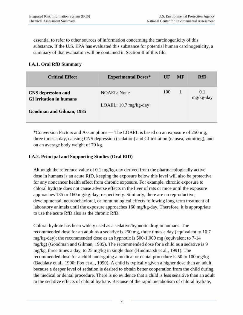
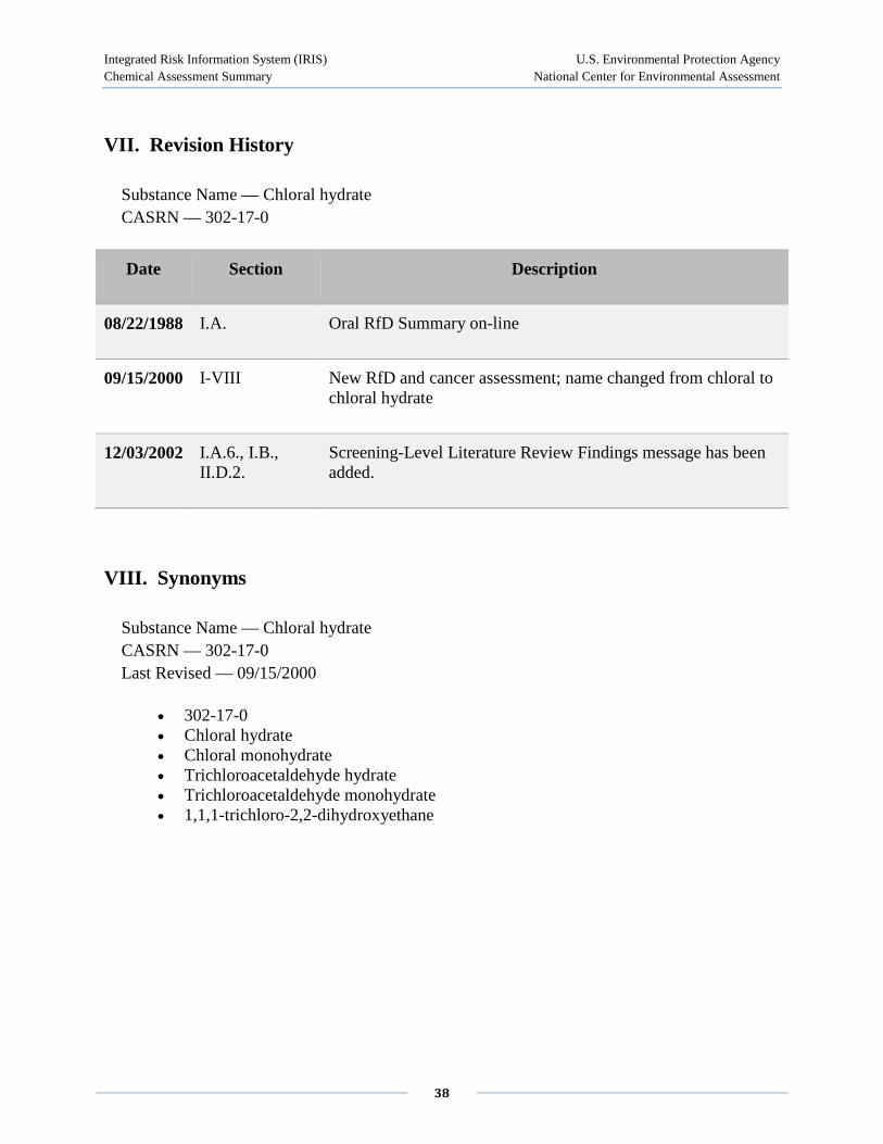


![Journal of Radiation Research and Applied Sciences Use of ... · 10 kGy. Fig. (2) represents the absorption spectra of PVB-co-PVA/PVAC ([CPR]= 0.64 phr) of CPR without chloral hydrate.](https://static.documents.pub/doc/80x56/5f0bddd77e708231d4329a17/journal-of-radiation-research-and-applied-sciences-use-of-10-kgy-fig-2-represents.jpg)


