90
Cholangiocarcinoma: Radiologic evaluation and interventions Colin Nevins, Harvard Medical School Year III Gillian Lieberman, MD November 2014 Colin Nevins, MSIII Gillian Lieberman, MD

Cholangiocarcinoma:
Radiologic evaluation and interventions
Colin Nevins, Harvard Medical School Year III
Gillian Lieberman, MD
November 2014
Colin Nevins, MSIII
Gillian Lieberman, MD

Agenda
• Initial course and work-up
• Endoscopic retrograde cholangiopancreatography (ERCP)
• Initial intervention
• Cholangiocarcinoma
• Post-operative course and imaging
• Magnetic resonance cholangiopancreatography (MRCP)
• ACR appropriateness criteria
• Percutaneous transhepatic cholangiography (PTC)
• Cholangiocarcinoma prognosis
• Outcome
• Recap
2
Colin Nevins, MSIII
Gillian Lieberman, MD

Agenda
• Initial course and work-up
• Endoscopic retrograde cholangiopancreatography (ERCP)
• Initial intervention
• Cholangiocarcinoma
• Post-operative course and imaging
• Magnetic resonance cholangiopancreatography (MRCP)
• ACR appropriateness criteria
• Percutaneous transhepatic cholangiography (PTC)
• Cholangiocarcinoma prognosis
• Outcome
• Recap
3
Colin Nevins, MSIII
Gillian Lieberman, MD
Our patient: Initial presentation
• 84 year old Asian male presenting to his PCP
• CC/HPI: fatigue, fever, and weight loss for several weeks
4
Colin Nevins, MSIII
Gillian Lieberman, MD

Our patient: Initial presentation
• 84 year old Asian male presenting to his PCP
• CC/HPI: fatigue, fever, and weight loss for several weeks
• ROS: negative for RUQ pain
• PMH: HTN, BPH, GERD
• PSH: remote cholecystectomy
• Medications: hydrochlorothiazide, omeprazole
• Allergies: none
• Family Hx: none
• Social Hx: rare EtOH, no tobacco, no illicit substances
5
Colin Nevins, MSIII
Gillian Lieberman, MD
Our patient: Physical Exam
• Vitals: T 100.4, HR 60, BP 132/72, RR 16, SaO2 98% RA
• Gen: elderly male, appears tired
• HEENT: mild scleral icterus
• CV: RRR, no murmurs/rubs/gallops
• RESP: CTAB, no rales, rhonchi, or wheezes
• ABD: soft, NT, ND, + bowel sounds, no hepatomegaly or
splenomegaly, no caput medusae
• EXT: WWP, no palmar erythema
• NEURO: A&O x3, moving all extremities, no asterixis
• SKIN: no generalized jaundice, no spider angioma
6
Colin Nevins, MSIII
Gillian Lieberman, MD

Our patient: Selected Labs
• Basic labs including CBC and chem-10 were within
normal limits
• However, the liver panel was suggestive of biliary
obstruction due to the combination of:
– Elevated transaminases
• AST: 142, ALT: 172
– Elevated alkaline phosphatase: 458
– Direct hyperbilirubinemia
• total bilirubin: 2.9, direct bilirubin: 2.2
• A radiologic work-up was then begun for biliary
obstruction
7
Colin Nevins, MSIII
Gillian Lieberman, MD

Our patient: Radiologic work-up for biliary obstruction
8
Colin Nevins, MSIII
Gillian Lieberman, MD

Our patient: Radiologic work-up for biliary obstruction
1. RUQ U/S
9
Colin Nevins, MSIII
Gillian Lieberman, MD

Our patient: Radiologic work-up for biliary obstruction
1. RUQ U/S
– Renal cysts and s/p cholecystectomy
– otherwise unremarkable
10
Colin Nevins, MSIII
Gillian Lieberman, MD

Our patient: Radiologic work-up for biliary obstruction
1. RUQ U/S
– Renal cysts and s/p cholecystectomy
– otherwise unremarkable
2. Follow-up abdominal CT
11
Colin Nevins, MSIII
Gillian Lieberman, MD
Our patient: Radiologic work-up for biliary obstruction
1. RUQ U/S
– Renal cysts and s/p cholecystectomy
– otherwise unremarkable
2. Follow-up abdominal CT
– “Benign”
12
Colin Nevins, MSIII
Gillian Lieberman, MD

Our patient: Radiologic work-up for biliary obstruction
1. RUQ U/S
– Renal cysts and s/p cholecystectomy
– otherwise unremarkable
2. Follow-up abdominal CT
– “Benign”
RUQ U/S and follow-up abdominal CT were unrevealing
An ERCP was then recommended
13
Colin Nevins, MSIII
Gillian Lieberman, MD

Agenda
• Initial course and work-up
• Endoscopic retrograde cholangiopancreatography (ERCP)
• Initial intervention
• Cholangiocarcinoma
• Post-operative course and imaging
• Magnetic resonance cholangiopancreatography (MRCP)
• ACR appropriateness criteria
• Percutaneous transhepatic cholangiography (PTC)
• Cholangiocarcinoma prognosis
• Outcome
• Recap
14
Colin Nevins, MSIII
Gillian Lieberman, MD

Endoscopic Retrograde Cholangiopancreatography (ERCP):
some facts
15
Colin Nevins, MSIII
Gillian Lieberman, MD
From: Gillian Lieberman, MD. Lieberman’s eRadiology: Primary Care Radiology: Menu of radiologic tests

Endoscopic Retrograde Cholangiopancreatography (ERCP):
some facts
16
Colin Nevins, MSIII
Gillian Lieberman, MD
Ultrasound and abdominal CT are usually used in
the initial evaluation of suspected biliary or
pancreatic disease.
From: Gillian Lieberman, MD. Lieberman’s eRadiology: Primary Care Radiology: Menu of radiologic tests

Endoscopic Retrograde Cholangiopancreatography (ERCP):
some facts
17
Colin Nevins, MSIII
Gillian Lieberman, MD
Ultrasound and abdominal CT are usually used in
the initial evaluation of suspected biliary or
pancreatic disease.
ERCP provides a more detailed evaluation of the
biliary tree and pancreatic duct and allows for
possible therapeutic intervention.
From: Gillian Lieberman, MD. Lieberman’s eRadiology: Primary Care Radiology: Menu of radiologic tests

Endoscopic Retrograde Cholangiopancreatography (ERCP):
some facts
18
Colin Nevins, MSIII
Gillian Lieberman, MD
Ultrasound and abdominal CT are usually used in
the initial evaluation of suspected biliary or
pancreatic disease.
ERCP provides a more detailed evaluation of the
biliary tree and pancreatic duct and allows for
possible therapeutic intervention.
Indications
1. Evaluation of biliary obstruction in patients
with persistent jaundice
2. Evaluation of recurrent pancreatitis
3. Suspected pancreatic cancer, when other
studies, e.g. CT, are equivocal
4. Evaluation of pancreatic and biliary anatomy
prior to surgery
5. Bile duct stent placement to relieve obstruction
6. Biopsy of identified lesions
7. Stone removal
Contraindications
1. Fluoroscopy utilizes x-rays, i.e. ionizing
radiation and should not be performed in pregnant
women unless absolutely necessary.
2. Coagulopathy is a relative contraindication.
3. There is a low risk of allergic reaction to
contrast material, so the exam is not
contraindicated in patients with a history of
contrast allergy.
From: Gillian Lieberman, MD. Lieberman’s eRadiology: Primary Care Radiology: Menu of radiologic tests

Endoscopic Retrograde Cholangiopancreatography (ERCP):
some facts
19
Colin Nevins, MSIII
Gillian Lieberman, MD
Ultrasound and abdominal CT are usually used in
the initial evaluation of suspected biliary or
pancreatic disease.
ERCP provides a more detailed evaluation of the
biliary tree and pancreatic duct and allows for
possible therapeutic intervention.
Indications
1. Evaluation of biliary obstruction in patients
with persistent jaundice
2. Evaluation of recurrent pancreatitis
3. Suspected pancreatic cancer, when other
studies, e.g. CT, are equivocal
4. Evaluation of pancreatic and biliary anatomy
prior to surgery
5. Bile duct stent placement to relieve obstruction
6. Biopsy of identified lesions
7. Stone removal
Contraindications
1. Fluoroscopy utilizes x-rays, i.e. ionizing
radiation and should not be performed in pregnant
women unless absolutely necessary.
2. Coagulopathy is a relative contraindication.
3. There is a low risk of allergic reaction to
contrast material, so the exam is not
contraindicated in patients with a history of
contrast allergy.
Limitations
1. Invasive procedure with attendant risks.
2. There are many less expensive, noninvasive
procedures that visualize the pancreaticobiliary
systems, such as ultrasound, CT, and MRCP.
ERCP should only be used when results of other
imaging modalities are equivocal or intervention is
anticipated
From: Gillian Lieberman, MD. Lieberman’s eRadiology: Primary Care Radiology: Menu of radiologic tests

ERCP: technique
20
Colin Nevins, MSIII
Gillian Lieberman, MD
From: Johns Hopkins Medicine. Bile Duct Cancer (Cholangiocarcinoma),
http://www.hopkinsmedicine.org/liver_tumor_center/conditions/bile_duct_cancer.html
1. Endoscope
positioned in the
second portion of
the duodenum
2. Cannulation of the
Ampulla of Vater
3. Fluoroscopic
imaging with
contrast medium *

Our patient: ERCP findings
• The left intrahepatic ducts were not
filled with contrast.
• 1.5 cm stricture in the proximal CHD.
• The CBD was 7mm in maximum
diameter. No filling defects identified.
• No evidence of Primary Sclerosing
Cholangitis (PSC).
• Sphincterotomy with brushings was
performed and a biliary stent was
placed into the right main hepatic duct.
21
Colin Nevins, MSIII
Gillian Lieberman, MD
From: Hansen, J. T., & Netter, F. H. (2010). Netter's clinical anatomy. Philadelphia:
Saunders/Elsevier.

Our patient: pathology and decision making
• Common bile duct stricture, brushings:
– “Highly atypical but degenerated glandular cells are
present; cannot rule out reactive atypia.”
22
Colin Nevins, MSIII
Gillian Lieberman, MD

Our patient: pathology and decision making
• Common bile duct stricture, brushings:
– “Highly atypical but degenerated glandular cells are
present; cannot rule out reactive atypia.”
• Patient also had mildly elevated CA-19-9
23
Colin Nevins, MSIII
Gillian Lieberman, MD

Our patient: pathology and decision making
• Common bile duct stricture, brushings:
– “Highly atypical but degenerated glandular cells are
present; cannot rule out reactive atypia.”
• Patient also had mildly elevated CA-19-9
• High concern for cholangiocarcinoma, warranting surgical
intervention
24
Colin Nevins, MSIII
Gillian Lieberman, MD

Agenda
• Initial course and work-up
• Endoscopic retrograde cholangiopancreatography (ERCP)
• Initial intervention
• Cholangiocarcinoma
• Post-operative course and imaging
• Magnetic resonance cholangiopancreatography (MRCP)
• ACR appropriateness criteria
• Percutaneous transhepatic cholangiography (PTC)
• Cholangiocarcinoma prognosis
• Outcome
• Recap
25
Colin Nevins, MSIII
Gillian Lieberman, MD
Our patient: surgical plan
• High concern for cholangiocarcinoma, warranting surgical
intervention
– Underwent a Roux-en-Y hepatojejunostomy to the
extrahepatic common hepatic duct (resected the
extrahepatic biliary tree and performed a biliary bypass)
– Family and surgeon opted not to perform an extensive
liver or pancreatic resection
26
Colin Nevins, MSIII
Gillian Lieberman, MD

Roux-en-Y hepatojejunostomy to the extrahepatic common hepatic duct
27
Colin Nevins, MSIII
Gillian Lieberman, MD
From: Johns Hopkins Medicine. Bile Duct Cancer (Cholangiocarcinoma),
http://www.hopkinsmedicine.org/liver_tumor_center/conditions/bile_duct_cancer.html
(note: this patient did not undergo a hepatic lobectomy)
Our patient: post-operative course
• The resected tissue was sent to pathology for diagnosis
28
Colin Nevins, MSIII
Gillian Lieberman, MD

Our patient: post-operative course
• The resected tissue was sent to pathology for diagnosis
• Post- operative diagnosis: invasive cholangiocarcinoma
29
Colin Nevins, MSIII
Gillian Lieberman, MD

Agenda
• Initial course and work-up
• Endoscopic retrograde cholangiopancreatography (ERCP)
• Initial intervention
• Cholangiocarcinoma
• Post-operative course and imaging
• Magnetic resonance cholangiopancreatography (MRCP)
• ACR appropriateness criteria
• Percutaneous transhepatic cholangiography (PTC)
• Cholangiocarcinoma prognosis
• Outcome
• Recap
30
Colin Nevins, MSIII
Gillian Lieberman, MD

Cholangiocarcinoma: some facts
• Rare, but often highly lethal as it is often locally advanced at presentation
31
Colin Nevins, MSIII
Gillian Lieberman, MD
From: Cholangiocarcinoma, UpToDate:
http://www.uptodate.com/contents/image?imageKey=GAST/52489&topicKey=ONC/2500&source=outlin
e_link&search=cholangiocarcinoma&utdPopup=true

Cholangiocarcinoma: some facts
• Rare, but often highly lethal as it is often locally advanced at presentation
• Main risk factors:
32
Colin Nevins, MSIII
Gillian Lieberman, MD
From: Cholangiocarcinoma, UpToDate:
http://www.uptodate.com/contents/image?imageKey=GAST/52489&topicKey=ONC/2500&source=outlin
e_link&search=cholangiocarcinoma&utdPopup=true
Cholangiocarcinoma: some facts
• Rare, but often highly lethal as it is often locally advanced at presentation
• Main risk factors:
– Primary Sclerosing Cholangitis (PSC)
– Fibropolycystic liver disease (e.g., choledochal cysts)
33
Colin Nevins, MSIII
Gillian Lieberman, MD
From: Cholangiocarcinoma, UpToDate:
http://www.uptodate.com/contents/image?imageKey=GAST/52489&topicKey=ONC/2500&source=outlin
e_link&search=cholangiocarcinoma&utdPopup=true
Cholangiocarcinoma: some facts
• Rare, but often highly lethal as it is often locally advanced at presentation
• Main risk factors:
– Primary Sclerosing Cholangitis (PSC)
– Fibropolycystic liver disease (e.g., choledochal cysts)
• Classified according to location along the biliary tree
34
Colin Nevins, MSIII
Gillian Lieberman, MD
From: Cholangiocarcinoma, UpToDate:
http://www.uptodate.com/contents/image?imageKey=GAST/52489&topicKey=ONC/2500&source=outlin
e_link&search=cholangiocarcinoma&utdPopup=true

Cholangiocarcinoma: some facts
• Rare, but often highly lethal as it is often locally advanced at presentation
• Main risk factors:
– Primary Sclerosing Cholangitis (PSC)
– Fibropolycystic liver disease (e.g., choledochal cysts)
• Classified according to location along the biliary tree
– Two-thirds involve the bifurcation of the common hepatic duct
35
Colin Nevins, MSIII
Gillian Lieberman, MD
From: Cholangiocarcinoma, UpToDate:
http://www.uptodate.com/contents/image?imageKey=GAST/52489&topicKey=ONC/2500&source=outlin
e_link&search=cholangiocarcinoma&utdPopup=true

Cholangiocarcinoma: locations
36
Colin Nevins, MSIII
Gillian Lieberman, MD
From: Johns Hopkins Medicine. Bile Duct Cancer (Cholangiocarcinoma),
http://www.hopkinsmedicine.org/liver_tumor_center/conditions/bile_duct_cancer.html
2/3 involve the
bifurcation of the
common hepatic duct
(“Klatskin tumors”)
May also involve
peripheral ducts
Ductal dilation may
occur proximally to an
obstruction
* *
*
*

Comparison patients #1 and #2: examples of the
radiologic manifestations of cholangiocarcinoma
37
Colin Nevins, MSIII
Gillian Lieberman, MD
From: Johns Hopkins Medicine. Bile Duct Cancer (Cholangiocarcinoma),
http://www.hopkinsmedicine.org/liver_tumor_center/conditions/bile_duct_cancer.html
Cholangiocarcinoma: some facts
• Rare, but often highly lethal as it is often locally advanced at presentation
• Main risk factors:
– Primary Sclerosing Cholangitis (PSC)
– Fibropolycystic liver disease (e.g., choledochal cysts)
• Classified according to location along the biliary tree
– Two-thirds involve the bifurcation of the common hepatic duct (termed
“Klatskin tumors”)
• Histology:
38
Colin Nevins, MSIII
Gillian Lieberman, MD
From: Cholangiocarcinoma, UpToDate:
http://www.uptodate.com/contents/image?imageKey=GAST/52489&topicKey=ONC/2500&source=outlin
e_link&search=cholangiocarcinoma&utdPopup=true

Cholangiocarcinoma: some facts
• Rare, but often highly lethal as it is often locally advanced at presentation
• Main risk factors:
– Primary Sclerosing Cholangitis (PSC)
– Fibropolycystic liver disease (e.g., choledochal cysts)
• Classified according to location along the biliary tree
– Two-thirds involve the bifurcation of the common hepatic duct (termed
“Klatskin tumors”)
• Histology:
– Greater than 90% are adenocarcinomas, most others are squamous cell
carcinomas
39
Colin Nevins, MSIII
Gillian Lieberman, MD
From: Cholangiocarcinoma, UpToDate:
http://www.uptodate.com/contents/image?imageKey=GAST/52489&topicKey=ONC/2500&source=outlin
e_link&search=cholangiocarcinoma&utdPopup=true

Cholangiocarcinoma: some facts
• Rare, but often highly lethal as it is often locally advanced at presentation
• Main risk factors:
– Primary Sclerosing Cholangitis (PSC)
– Fibropolycystic liver disease (e.g., choledochal cysts)
• Classified according to location along the biliary tree
– Two-thirds involve the bifurcation of the common hepatic duct (termed
“Klatskin tumors”)
• Histology:
– Greater than 90% are adenocarcinomas, most others are squamous cell
carcinomas
• Grading:
40
Colin Nevins, MSIII
Gillian Lieberman, MD
From: Cholangiocarcinoma, UpToDate:
http://www.uptodate.com/contents/image?imageKey=GAST/52489&topicKey=ONC/2500&source=outlin
e_link&search=cholangiocarcinoma&utdPopup=true
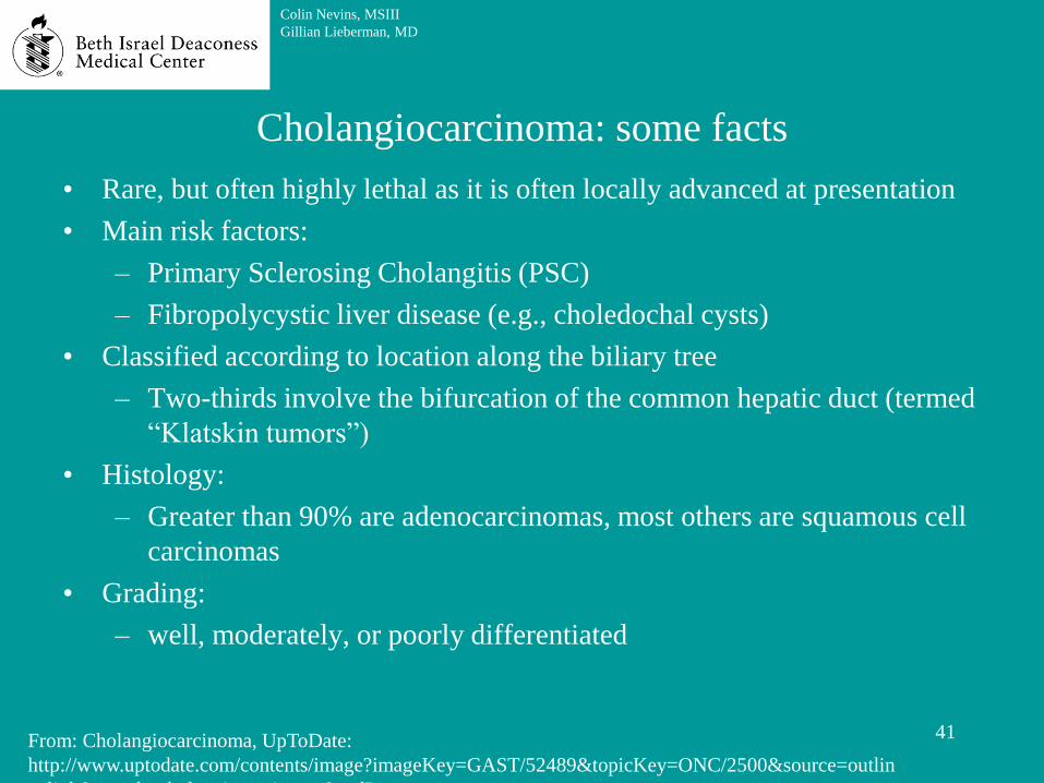
Cholangiocarcinoma: some facts
• Rare, but often highly lethal as it is often locally advanced at presentation
• Main risk factors:
– Primary Sclerosing Cholangitis (PSC)
– Fibropolycystic liver disease (e.g., choledochal cysts)
• Classified according to location along the biliary tree
– Two-thirds involve the bifurcation of the common hepatic duct (termed
“Klatskin tumors”)
• Histology:
– Greater than 90% are adenocarcinomas, most others are squamous cell
carcinomas
• Grading:
– well, moderately, or poorly differentiated
41
Colin Nevins, MSIII
Gillian Lieberman, MD
From: Cholangiocarcinoma, UpToDate:
http://www.uptodate.com/contents/image?imageKey=GAST/52489&topicKey=ONC/2500&source=outlin
e_link&search=cholangiocarcinoma&utdPopup=true
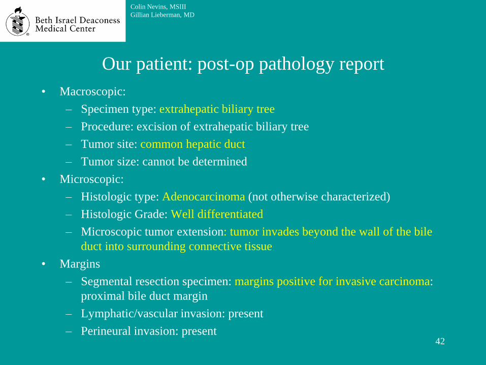
Our patient: post-op pathology report
• Macroscopic:
– Specimen type: extrahepatic biliary tree
– Procedure: excision of extrahepatic biliary tree
– Tumor site: common hepatic duct
– Tumor size: cannot be determined
• Microscopic:
– Histologic type: Adenocarcinoma (not otherwise characterized)
– Histologic Grade: Well differentiated
– Microscopic tumor extension: tumor invades beyond the wall of the bile
duct into surrounding connective tissue
• Margins
– Segmental resection specimen: margins positive for invasive carcinoma:
proximal bile duct margin
– Lymphatic/vascular invasion: present
– Perineural invasion: present 42
Colin Nevins, MSIII
Gillian Lieberman, MD

Agenda
• Initial course and work-up
• Endoscopic retrograde cholangiopancreatography (ERCP)
• Initial intervention
• Cholangiocarcinoma
• Post-operative course and imaging
• Magnetic resonance cholangiopancreatography (MRCP)
• ACR appropriateness criteria
• Percutaneous transhepatic cholangiography (PTC)
• Cholangiocarcinoma prognosis
• Outcome
• Recap
43
Colin Nevins, MSIII
Gillian Lieberman, MD

Our patient: POD-5 gravity cholangiogram
Routine study to evaluate patency of Roux-En-Y hepatojejunostomy for biliary stricture
44
Colin Nevins, MSIII
Gillian Lieberman, MD
Pause to evaluate the images, then continue to reveal the findings

Our patient: POD-5 gravity cholangiogram
Routine study to evaluate patency of Roux-En-Y hepatojejunostomy for biliary stricture
45
Colin Nevins, MSIII
Gillian Lieberman, MD
FINDINGS:
1. Abdominal surgical drain.
2. Right upper quadrant
surgical clips and T-tube
3. Dilated left hepatic ducts
centrally with no peripheral
dilation.
4. Right hepatic ducts were
not seen.
5. No evidence of leak.
6. No evidence of
anastomotic stricture.
IMPRESSION:
No evidence of anastomotic
stricture or leak.

POD-10: elevated transaminases were found on standard follow-up labs.
Patient mildly febrile and team was concerned about a possible liver
abscess
46
Colin Nevins, MSIII
Gillian Lieberman, MD
Our patient: post-operative course

POD-10: elevated transaminases were found on standard follow-up labs.
Patient mildly febrile and team was concerned about a possible liver
abscess
CTA was obtained to asses for liver abscess
47
Colin Nevins, MSIII
Gillian Lieberman, MD
Our patient: post-operative course
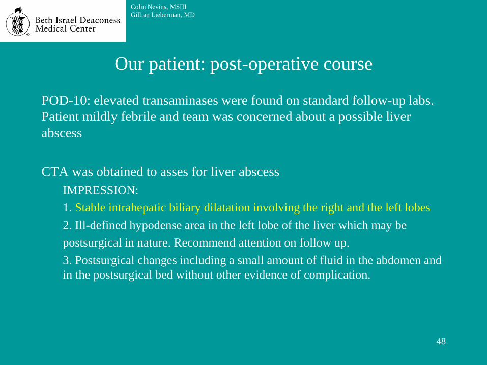
POD-10: elevated transaminases were found on standard follow-up labs.
Patient mildly febrile and team was concerned about a possible liver
abscess
CTA was obtained to asses for liver abscess
IMPRESSION:
1. Stable intrahepatic biliary dilatation involving the right and the left lobes
2. Ill-defined hypodense area in the left lobe of the liver which may be
postsurgical in nature. Recommend attention on follow up.
3. Postsurgical changes including a small amount of fluid in the abdomen and
in the postsurgical bed without other evidence of complication.
48
Colin Nevins, MSIII
Gillian Lieberman, MD
Our patient: post-operative course

POD-10: elevated transaminases were found on standard follow-up labs.
Patient mildly febrile and team was concerned about a possible liver
abscess
CTA was obtained to asses for liver abscess
IMPRESSION:
1. Stable intrahepatic biliary dilatation involving the right and the left lobes
2. Ill-defined hypodense area in the left lobe of the liver which may be
postsurgical in nature. Recommend attention on follow up.
3. Postsurgical changes including a small amount of fluid in the abdomen and
in the postsurgical bed without other evidence of complication.
Five days later, LFT’s were still rising.
MRCP then obtained to asses biliary system for obstructions or leaks
49
Colin Nevins, MSIII
Gillian Lieberman, MD
Our patient: post-operative course

Agenda
• Initial course and work-up
• Endoscopic retrograde cholangiopancreatography (ERCP)
• Initial intervention
• Cholangiocarcinoma
• Post-operative course and imaging
• Magnetic resonance cholangiopancreatography (MRCP)
• ACR appropriateness criteria
• Percutaneous transhepatic cholangiography (PTC)
• Cholangiocarcinoma prognosis
• Outcome
• Recap
50
Colin Nevins, MSIII
Gillian Lieberman, MD

Non-invasive imaging of the bile and pancreatic ducts without the need for
contrast.
51
Colin Nevins, MSIII
Gillian Lieberman, MD
From: Gillian Lieberman, MD. Lieberman’s eRadiology: Primary Care Radiology: Menu of radiologic tests
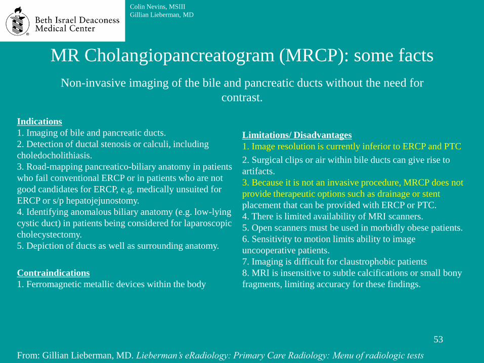
MR Cholangiopancreatogram (MRCP): some facts

Non-invasive imaging of the bile and pancreatic ducts without the need for
contrast.
52
Colin Nevins, MSIII
Gillian Lieberman, MD
Indications
1. Imaging of bile and pancreatic ducts.
2. Detection of ductal stenosis or calculi, including
choledocholithiasis.
3. Road-mapping pancreatico-biliary anatomy in patients
who fail conventional ERCP or in patients who are not
good candidates for ERCP, e.g. medically unsuited for
ERCP or s/p hepatojejunostomy.
4. Identifying anomalous biliary anatomy (e.g. low-lying
cystic duct) in patients being considered for laparoscopic
cholecystectomy.
5. Depiction of ducts as well as surrounding anatomy.
Contraindications
1. Ferromagnetic metallic devices within the body
From: Gillian Lieberman, MD. Lieberman’s eRadiology: Primary Care Radiology: Menu of radiologic tests
MR Cholangiopancreatogram (MRCP): some facts
Non-invasive imaging of the bile and pancreatic ducts without the need for
contrast.
53
Colin Nevins, MSIII
Gillian Lieberman, MD
Indications
1. Imaging of bile and pancreatic ducts.
2. Detection of ductal stenosis or calculi, including
choledocholithiasis.
3. Road-mapping pancreatico-biliary anatomy in patients
who fail conventional ERCP or in patients who are not
good candidates for ERCP, e.g. medically unsuited for
ERCP or s/p hepatojejunostomy.
4. Identifying anomalous biliary anatomy (e.g. low-lying
cystic duct) in patients being considered for laparoscopic
cholecystectomy.
5. Depiction of ducts as well as surrounding anatomy.
Contraindications
1. Ferromagnetic metallic devices within the body
Limitations/ Disadvantages
1. Image resolution is currently inferior to ERCP and PTC
2. Surgical clips or air within bile ducts can give rise to
artifacts.
3. Because it is not an invasive procedure, MRCP does not
provide therapeutic options such as drainage or stent
placement that can be provided with ERCP or PTC.
4. There is limited availability of MRI scanners.
5. Open scanners must be used in morbidly obese patients.
6. Sensitivity to motion limits ability to image
uncooperative patients.
7. Imaging is difficult for claustrophobic patients
8. MRI is insensitive to subtle calcifications or small bony
fragments, limiting accuracy for these findings.
From: Gillian Lieberman, MD. Lieberman’s eRadiology: Primary Care Radiology: Menu of radiologic tests
MR Cholangiopancreatogram (MRCP): some facts

54
Colin Nevins, MSIII
Gillian Lieberman, MD
Comparison patient #3: normal MRCP
Normal MRCP image
showing the common bile
duct (curved arrow) and the
pancreatic duct (arrow). Note
the fluid filled duodenum (*)
*
From: Janet Cochrane Miller, D. Phil., Susanna I. Lee, M.D., Ph.D. Radiology Rounds: MRCP
http://www.mghradrounds.org/index.php?src=gendocs&link=june_2004
55
Colin Nevins, MSIII
Gillian Lieberman, MD
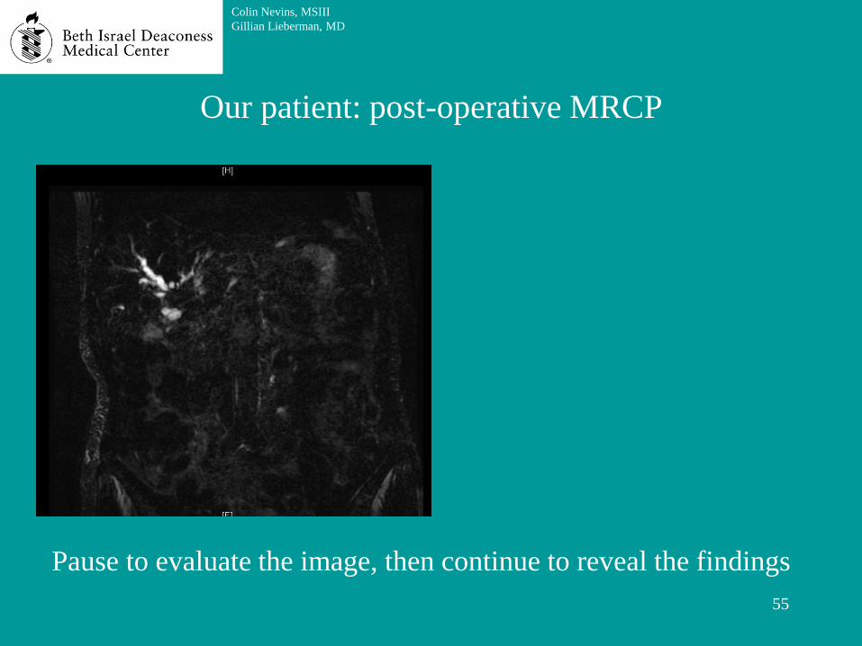
Our patient: post-operative MRCP
Pause to evaluate the image, then continue to reveal the findings

56
Colin Nevins, MSIII
Gillian Lieberman, MD
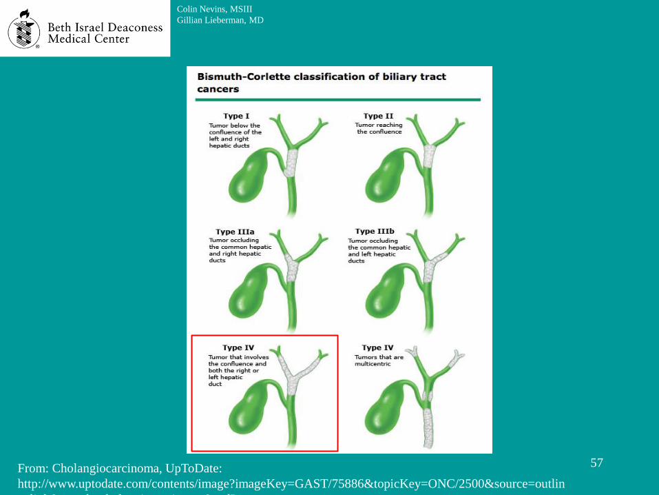
Persistent
intrahepatic biliary tree dilatation with
discontinuation and
amputation at the
level of biliary confluence at the hilum.
The findings are
concerning for residual Klatskin tumor in
volving the secondary biliary
confluences bilaterally (Bismuth Corlette
type IV).
No concerning lymph nodes.
Our patient: post-operative MRCP
57
Colin Nevins, MSIII
Gillian Lieberman, MD
From: Cholangiocarcinoma, UpToDate:
http://www.uptodate.com/contents/image?imageKey=GAST/75886&topicKey=ONC/2500&source=outlin
e_link&search=cholangiocarcinoma&utdPopup=true

Our patient: medical decision making
58
Colin Nevins, MSIII
Gillian Lieberman, MD
Due to residual tumor, our patient was recommended to undergo
stenting to relieve the obstruction
The ACR Appropriateness Criteria was used to aid in the
decision of how to insert a stent to treat this obstruction

Agenda
• Initial course and work-up
• Endoscopic retrograde cholangiopancreatography (ERCP)
• Initial intervention
• Cholangiocarcinoma
• Post-operative course and imaging
• Magnetic resonance cholangiopancreatography (MRCP)
• ACR appropriateness criteria
• Percutaneous transhepatic cholangiography (PTC)
• Cholangiocarcinoma prognosis
• Outcome
• Recap
59
Colin Nevins, MSIII
Gillian Lieberman, MD

ACR Appropriateness Criteria
60
Colin Nevins, MSIII
Gillian Lieberman, MD
From: American College of Radiology. Appropriatness Criteria: Radiologic Management of Benign and
Malignant Biliary Obstruction. http://www.acr.org/Quality-Safety/Appropriateness-Criteria

ACR Appropriateness Criteria
61
Colin Nevins, MSIII
Gillian Lieberman, MD
From: American College of Radiology. Appropriatness Criteria: Radiologic Management of Benign and
Malignant Biliary Obstruction. http://www.acr.org/Quality-Safety/Appropriateness-Criteria

Our patient: medical decision making
62
Colin Nevins, MSIII
Gillian Lieberman, MD
As our patient was s/p an operation that complicated his GI
anatomy (Roux-en-Y hepatojejunostomy), Percutaneous
Transhepatic Cholangiography (PTC) with permanent stent
placement was deemed to be preferable to an ERCP which
would require navigating an endoscope through the roux-en-y.
PTC has an appropriateness rating of 8 in this clinical setting
and permanent stent placement has a rating of 6, so these were
chosen as the next steps in our patient’s care.

ACR Appropriateness Criteria
63
Colin Nevins, MSIII
Gillian Lieberman, MD
From: American College of Radiology. Appropriatness Criteria: Radiologic Management of Benign and
Malignant Biliary Obstruction. http://www.acr.org/Quality-Safety/Appropriateness-Criteria

Agenda
• Initial course and work-up
• Endoscopic retrograde cholangiopancreatography (ERCP)
• Initial intervention
• Cholangiocarcinoma
• Post-operative course and imaging
• Magnetic resonance cholangiopancreatography (MRCP)
• ACR appropriateness criteria
• Percutaneous transhepatic cholangiography (PTC)
• Cholangiocarcinoma prognosis
• Outcome
• Recap
64
Colin Nevins, MSIII
Gillian Lieberman, MD

Comparison patient #4: Percutaneous Transhepatic
Cholangiography (PTC) technique
65
Colin Nevins, MSIII
Gillian Lieberman, MD
From: Johns Hopkins Medicine. Bile Duct Cancer (Cholangiocarcinoma),
http://www.hopkinsmedicine.org/liver_tumor_center/conditions/bile_duct_cancer.html
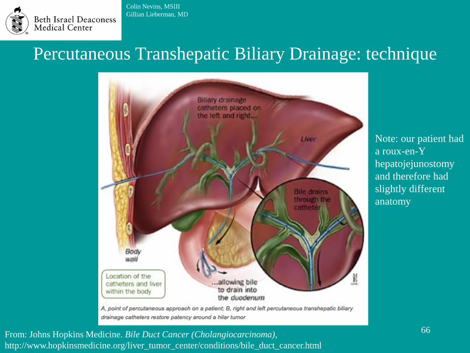
Percutaneous Transhepatic Biliary Drainage: technique
66
Colin Nevins, MSIII
Gillian Lieberman, MD
Note: our patient had
a roux-en-Y
hepatojejunostomy
and therefore had
slightly different
anatomy
From: Johns Hopkins Medicine. Bile Duct Cancer (Cholangiocarcinoma),
http://www.hopkinsmedicine.org/liver_tumor_center/conditions/bile_duct_cancer.html

67
Colin Nevins, MSIII
Gillian Lieberman, MD
Our patient: PTC imaging before stent placement
Pause to evaluate the image, then continue to reveal the findings
1

1. Intrahepatic biliary stricture at the confluence of the right ducts.
68
Colin Nevins, MSIII
Gillian Lieberman, MD
Our patient: PTC imaging before stent placement
1

69
Colin Nevins, MSIII
Gillian Lieberman, MD
Our patient: PTC imaging series pre (1) and post (2,3) stent
placement
Pause to evaluate the image, then continue to reveal the findings
1 2 3

1. Intrahepatic biliary stricture at the confluence of the right ducts.
2. Appropriate position and patency of the internal external drainage catheter
placed into the right anterior biliary system. (contrast is seen passing through the stent)
70
Colin Nevins, MSIII
Gillian Lieberman, MD
Our patient: PTC imaging series pre (1) and post (2,3) stent
placement
1 2 3

Agenda
• Initial course and work-up
• Endoscopic retrograde cholangiopancreatography (ERCP)
• Initial intervention
• Cholangiocarcinoma
• Post-operative course and imaging
• Magnetic resonance cholangiopancreatography (MRCP)
• ACR appropriateness criteria
• Percutaneous transhepatic cholangiography (PTC)
• Cholangiocarcinoma prognosis
• Outcome
• Recap
71
Colin Nevins, MSIII
Gillian Lieberman, MD

Cholangiocarcinoma: prognosis
• Bile can be drained externally or internally
72
Colin Nevins, MSIII
Gillian Lieberman, MD
Reference: Nakeeb A, Tran KQ, Black MJ, et al. Improved survival in resected biliary malignancies.
Surgery. 2002;132(4):555.

Cholangiocarcinoma: prognosis
• Bile can be drained externally or internally
• Relapse for cholangiocarcinoma can occur even after complete surgical
resection
73
Colin Nevins, MSIII
Gillian Lieberman, MD
Reference: Nakeeb A, Tran KQ, Black MJ, et al. Improved survival in resected biliary malignancies.
Surgery. 2002;132(4):555.

Cholangiocarcinoma: prognosis
• Bile can be drained externally or internally
• Relapse for cholangiocarcinoma can occur even after complete surgical
resection
– Most common relapse pattern is local
– Main risk factors for recurrence
• Histologically positive margins
• Lymph node involvement
74
Colin Nevins, MSIII
Gillian Lieberman, MD
Reference: Nakeeb A, Tran KQ, Black MJ, et al. Improved survival in resected biliary malignancies.
Surgery. 2002;132(4):555.
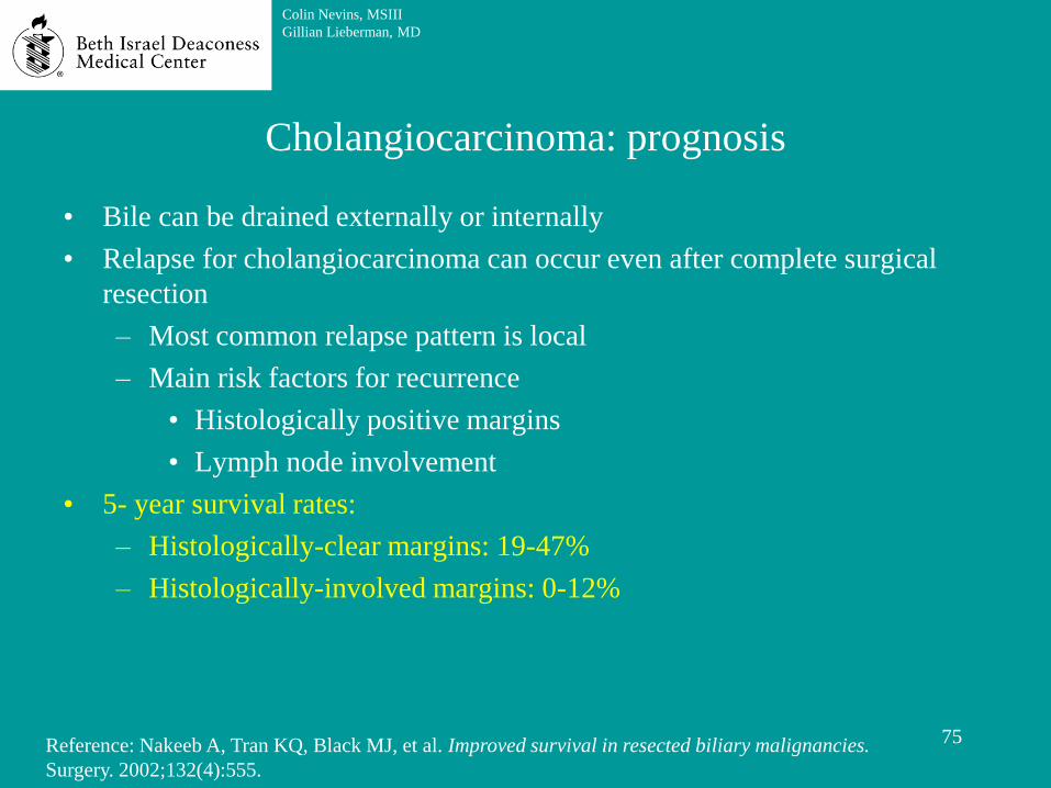
Cholangiocarcinoma: prognosis
• Bile can be drained externally or internally
• Relapse for cholangiocarcinoma can occur even after complete surgical
resection
– Most common relapse pattern is local
– Main risk factors for recurrence
• Histologically positive margins
• Lymph node involvement
• 5- year survival rates:
– Histologically-clear margins: 19-47%
– Histologically-involved margins: 0-12%
75
Colin Nevins, MSIII
Gillian Lieberman, MD
Reference: Nakeeb A, Tran KQ, Black MJ, et al. Improved survival in resected biliary malignancies.
Surgery. 2002;132(4):555.

Cholangiocarcinoma: prognosis
• Bile can be drained externally or internally
• Relapse for cholangiocarcinoma can occur even after complete surgical
resection
– Most common relapse pattern is local
– Main risk factors for recurrence
• Histologically positive margins
• Lymph node involvement
• 5- year survival rates:
– Histologically-clear margins: 19-47%
– Histologically-involved margins: 0-12%
• If margins are histologically involved, additional surgery to remove cancer
is not recommended (however, stent placement may be indicated)
76
Colin Nevins, MSIII
Gillian Lieberman, MD
Reference: Nakeeb A, Tran KQ, Black MJ, et al. Improved survival in resected biliary malignancies.
Surgery. 2002;132(4):555.

Agenda
• Initial course and work-up
• Endoscopic retrograde cholangiopancreatography (ERCP)
• Initial intervention
• Cholangiocarcinoma
• Post-operative course and imaging
• Magnetic resonance cholangiopancreatography (MRCP)
• ACR appropriateness criteria
• Percutaneous transhepatic cholangiography (PTC)
• Cholangiocarcinoma prognosis
• Outcome
• Recap
77
Colin Nevins, MSIII
Gillian Lieberman, MD

Our patient: outcome
• Our patient was discharged one week after his PTC
• His lab abnormalities were improving and he was doing well clinically
• An external drain was in place and capped. The bile was successfully
draining internally
• The patient was seen in clinic three weeks after discharge and was
recovering well.
78
Colin Nevins, MSIII
Gillian Lieberman, MD

Agenda
• Initial course and work-up
• Endoscopic retrograde cholangiopancreatography (ERCP)
• Initial intervention
• Cholangiocarcinoma
• Post-operative course and imaging
• Magnetic resonance cholangiopancreatography (MRCP)
• ACR appropriateness criteria
• Percutaneous transhepatic cholangiography (PTC)
• Cholangiocarcinoma prognosis
• Outcome
• Recap
79
Colin Nevins, MSIII
Gillian Lieberman, MD

Recap: Initial Course
• Cholangiocarcinoma may initially present with:
– Jaundice, clay-colored stools, bilirubinuria, pruritus, weight loss,
abdominal pain, fever
• Physical exam may reveal jaundice or hepatomegaly
• Labs may be suggestive of biliary obstruction
– elevated transaminases
– elevated alkaline phosphatase
– direct hyperbilirubinemia
80
Colin Nevins, MSIII
Gillian Lieberman, MD
Reference: Cholangiocarcinoma, UpToDate:
http://www.uptodate.com/contents/image?imageKey=GAST/52489&topicKey=ONC/2500&source=outli
ne_link&search=cholangiocarcinoma&utdPopup=true

Recap: Radiologic Workup for biliary obstruction
• Began with a RUQ ultrasound
• This was unrevealing, so an abdominal CT was obtained
• The CT did not reveal a cause of the biliary obstruction, so an ERCP was
performed
81
Colin Nevins, MSIII
Gillian Lieberman, MD

Recap: ERCP
• ERCP allows for a more
detailed evaluation of the
biliary tree and
pancreatic duct than
ultrasound or CT.
• It also allows for
biopsies to be obtained
and therapeutic
interventions, such as
stent placement
• However, it is an
invasive procedure and
also requires the use of
contrast
82
Colin Nevins, MSIII
Gillian Lieberman, MD

Recap: Cholangiocarcinoma
• Rare, but often highly lethal as it is often locally advanced at presentation
• Main risk factors:
– Primary Sclerosing Cholangitis (PSC)
– Fibropolycystic liver disease (e.g., choledochal cysts)
• Classified according to location along the biliary tree
– Two-thirds involve the bifurcation of the common hepatic duct (termed
“Klatskin tumors”)
• Histology:
– Greater than 90% are adenocarcinomas, most others are squamous cell
carcinomas
• Grading:
– well, moderately, or poorly differentiated
83
Colin Nevins, MSIII
Gillian Lieberman, MD

Recap: Post-operative Complications
• A post-operative cholangiogram is performed routinely to asses for strictures
or bile leaks
• After a normal cholangiogram our patient developed elevated transaminases
which led to a follow-up CTA
• CTA was then followed by an MRCP 84
Colin Nevins, MSIII
Gillian Lieberman, MD

Recap: MRCP
• MRCP offers non-invasive
imaging of the bile and
pancreatic ducts without the need
for contrast.
• Image resolution is currently
inferior to ERCP and PTC
• Because it is not an invasive
procedure, MRCP does not
provide therapeutic options such
as drainage or stenting
85
Colin Nevins, MSIII
Gillian Lieberman, MD
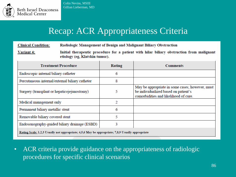
Recap: ACR Appropriateness Criteria
• ACR criteria provide guidance on the appropriateness of radiologic
procedures for specific clinical scenarios
86
Colin Nevins, MSIII
Gillian Lieberman, MD

Recap: PTC
• PTC allows for imaging
of and interventional
access to the biliary
system
• PTC can be helpful when
the patient’s anatomy
makes ERCP difficult or
the patient is medically
unfit for ERCP
87
Colin Nevins, MSIII
Gillian Lieberman, MD

Recap: Cholangiocarcinoma prognosis
• Bile can be drained externally or internally
• Relapse for cholangiocarcinoma can occur even after complete surgical
resection
– Most common relapse pattern is local
– Main risk factors for recurrence
• Histologically positive margins
• Lymph node involvement
• 5- year survival rates:
– Histologically-clear margins: 19-47%
– Histologically-involved margins: 0-12%
• If margins are histologically involved, additional surgery to remove cancer
is not recommended (however, stent placement may be indicated)
88
Colin Nevins, MSIII
Gillian Lieberman, MD

89
References
• American College of Radiology. Appropriatness Criteria: Radiologic Management of Benign and
Malignant Biliary Obstruction. http://www.acr.org/Quality-Safety/Appropriateness-Criteria
• Anatomic classification of cancers of the human biliary tract,
http://www.uptodate.com/contents/image?imageKey=GAST/52489&topicKey=ONC/2500&sou
rce=outline_link&search=cholangiocarcinoma&utdPopup=true
• Bismuth-Corlette classification of biliary tract cancers:
http://www.uptodate.com/contents/image?imageKey=GAST/75886&topicKey=ONC/2467&sou
rce=outline_link&search=cholangiocarcinoma&utdPopup=true
• Gillian Lierberman, MD. Lieberman’s eRadiology: Primary Care Radiology: Menu of radiologic
tests
• Janet Cochrane Miller, D. Phil., Susanna I. Lee, M.D., Ph.D. Radiology Rounds: MRCP http://
www.mghradrounds.org/index.php?src=gendocs&link=june_2004
• Johns Hopkins Medicine. Bile Duct Cancer (Cholangiocarcinoma),
http://www.hopkinsmedicine.org/liver_tumor_center/conditions/bile_duct_cancer.html
• Hansen, J. T., & Netter, F. H. (2010). Netter's clinical anatomy. Philadelphia: Saunders/Elsevier.
Colin Nevins, MSIII
Gillian Lieberman, MD

90
Acknowledgements
Thank you to Dr. Lieberman for her guidance with this case
presentation and the BIDMC radiology archives department
for their assistance collecting images.
Colin Nevins, MSIII
Gillian Lieberman, MD


