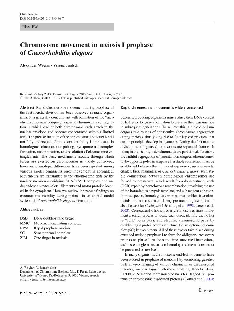
REVIEW Chromosome movement in meiosis I prophase of Caenorhabditis elegans Alexander Woglar & Verena Jantsch Received: 27 July 2013 /Revised: 29 August 2013 /Accepted: 30 August 2013 # The Author(s) 2013. This article is published with open access at Springerlink.com Abstract Rapid chromosome movement during prophase of the first meiotic division has been observed in many organ- isms. It is generally concomitant with formation of the “mei- otic chromosome bouquet,” a special chromosome configura- tion in which one or both chromosome ends attach to the nuclear envelope and become concentrated within a limited area. The precise function of the chromosomal bouquet is still not fully understood. Chromosome mobility is implicated in homologous chromosome pairing, synaptonemal complex formation, recombination, and resolution of chromosome en- tanglements. The basic mechanistic module through which forces are exerted on chromosomes is widely conserved; however, phenotypic differences have been reported among various model organisms once movement is abrogated. Movements are transmitted to the chromosome ends by the nuclear membrane-bridging SUN/KASH complex and are dependent on cytoskeletal filaments and motor proteins locat- ed in the cytoplasm. Here we review the recent findings on chromosome mobility during meiosis in an animal model system: the Caenorhabditis elegans nematode. Abbreviations DSB DNA double-strand break MMC Movement-mediating complex RPM Rapid prophase motion SC Synaptonemal complex ZIM Zinc finger in meiosis Rapid chromosome movement is widely conserved Sexual reproducing organisms must reduce their DNA content by half prior to gamete formation to preserve their genome size in subsequent generations. To achieve this, a diploid cell un- dergoes two rounds of consecutive chromosome segregation during meiosis, thus giving rise to four haploid products that can, in principle, develop into gametes. During the first meiotic division, homologous chromosomes are separated from each other; in the second, sister chromatids are partitioned. To enable the faithful segregation of parental homologous chromosomes to the opposite poles in anaphase I, a stable connection must be established between them. In most organisms, such as yeasts, ciliates, flies, mammals, or Caenorhabditis elegans , such sta- ble connections between homologous chromosomes are formed by crossovers, which result from double-strand break (DSB) repair by homologous recombination, involving the use of the homolog as a repair template, and subsequent cohesion. In most species, homologous chromosomes, unlike sister chro- matids, are not associated during pre-meiotic growth; this is also the case for C. elegans (Dernburg et al. 1998; Lorenz et al. 2003). Consequently, homologous chromosomes must imple- ment a search process to locate each other, identify each other as “self,” form pairs, and stabilize chromosome pairs by establishing a proteinaceous structure, the synaptonemal com- plex (SC) between them. All of these events take place during extended meiotic prophase I to form the obligatory crossovers prior to anaphase I. At the same time, unwanted interactions, such as entanglements or non-homologous interactions, must be prevented or resolved. In many organisms, chromosome end-led movements have been studied in prophase of meiosis I by combining genetics with in vivo imaging of various chromatin or chromosomal markers, such as tagged telomere proteins, Hoechst dyes, LacO/LacR-inserted repressor-binding sites, tagged SC pro- teins or chromosome associated proteins (Conrad et al. 2008; A. Woglar : V. Jantsch (*) Department of Chromosome Biology, Max F. Perutz Laboratories, University of Vienna, Dr.-Bohrgasse 9, 1030 Vienna, Austria e-mail: [email protected]Chromosoma DOI 10.1007/s00412-013-0436-7
Transcript
REVIEW
Chromosome movement in meiosis I prophaseof Caenorhabditis elegans
Alexander Woglar & Verena Jantsch
Received: 27 July 2013 /Revised: 29 August 2013 /Accepted: 30 August 2013# The Author(s) 2013. This article is published with open access at Springerlink.com
Abstract Rapid chromosome movement during prophase ofthe first meiotic division has been observed in many organ-isms. It is generally concomitant with formation of the “mei-otic chromosome bouquet,” a special chromosome configura-tion in which one or both chromosome ends attach to thenuclear envelope and become concentrated within a limitedarea. The precise function of the chromosomal bouquet is stillnot fully understood. Chromosome mobility is implicated inhomologous chromosome pairing, synaptonemal complexformation, recombination, and resolution of chromosome en-tanglements. The basic mechanistic module through whichforces are exerted on chromosomes is widely conserved;however, phenotypic differences have been reported amongvarious model organisms once movement is abrogated.Movements are transmitted to the chromosome ends by thenuclear membrane-bridging SUN/KASH complex and aredependent on cytoskeletal filaments and motor proteins locat-ed in the cytoplasm. Here we review the recent findings onchromosome mobility during meiosis in an animal modelsystem: the Caenorhabditis elegans nematode.
Abbreviations
DSB DNA double-strand breakMMC Movement-mediating complexRPM Rapid prophase motionSC Synaptonemal complexZIM Zinc finger in meiosis
Rapid chromosome movement is widely conserved
Sexual reproducing organisms must reduce their DNA contentby half prior to gamete formation to preserve their genome sizein subsequent generations. To achieve this, a diploid cell un-dergoes two rounds of consecutive chromosome segregationduring meiosis, thus giving rise to four haploid products thatcan, in principle, develop into gametes. During the first meioticdivision, homologous chromosomes are separated from eachother; in the second, sister chromatids are partitioned. To enablethe faithful segregation of parental homologous chromosomesto the opposite poles in anaphase I, a stable connection must beestablished between them. In most organisms, such as yeasts,ciliates, flies, mammals, or Caenorhabditis elegans , such sta-ble connections between homologous chromosomes areformed by crossovers, which result from double-strand break(DSB) repair by homologous recombination, involving the useof the homolog as a repair template, and subsequent cohesion.In most species, homologous chromosomes, unlike sister chro-matids, are not associated during pre-meiotic growth; this isalso the case forC. elegans (Dernburg et al. 1998; Lorenz et al.2003). Consequently, homologous chromosomes must imple-ment a search process to locate each other, identify each otheras “self,” form pairs, and stabilize chromosome pairs byestablishing a proteinaceous structure, the synaptonemal com-plex (SC) between them. All of these events take place duringextended meiotic prophase I to form the obligatory crossoversprior to anaphase I. At the same time, unwanted interactions,such as entanglements or non-homologous interactions, mustbe prevented or resolved.
In many organisms, chromosome end-led movements havebeen studied in prophase of meiosis I by combining geneticswith in vivo imaging of various chromatin or chromosomalmarkers, such as tagged telomere proteins, Hoechst dyes,LacO/LacR-inserted repressor-binding sites, tagged SC pro-teins or chromosome associated proteins (Conrad et al. 2008;
A. Woglar :V. Jantsch (*)Department of Chromosome Biology, Max F. Perutz Laboratories,University of Vienna, Dr.-Bohrgasse 9, 1030 Vienna, Austriae-mail: [email protected]
ChromosomaDOI 10.1007/s00412-013-0436-7
Ding et al. 2004; Koszul et al. 2008; Morimoto et al. 2012;Parvinen and Soderstrom 1976; Scherthan et al. 2007; Sheehanand Pawlowski 2009). Characterization of both wild-typemovement and impaired movement in mutants led to proposedroles for chromosome movement in the homology search pro-cess, in synapsis, in minimizing undesirable chromosomalinteractions, and even in actively promoting recombination(Koszul and Kleckner 2009; Wanat et al. 2008). C. elegans isone of the most intensively studied model organisms to date.Many of the requirements and functions of meiotic chromo-some mobility are understood in this 1-mm-long roundworm.Here we attempt to summarize recent insights into the proper-ties, biological significance, regulation, and mechanism ofchromosome movement during meiotic prophase in this well-established meiotic model system.
Why use C. elegans as a model to study chromosomemobility?
In the two gonad tubes of C. elegans hermaphrodites (or thesingle one in males), complete meiotic prophase I is bothtemporally and spatially organized (Fig. 1). In the adult worm,meiocytes progress through the meiotic cell cycle as they movealong the gonad tube from the distal tip to the spermatheca.This means that nuclei at any given disto-proximal position inthe syncytial gonad have spent the same time in meiosis(Crittenden et al. 1994; Hirsh et al. 1976); therefore, the gonadrepresents a highly synchronous meiotic time course (Fig. 2).The gonads of an adult hermaphrodite contain half the totalnumber of nuclei of the entire animal (Hirsh et al. 1976); as aresult, sufficient meiocytes are available for biochemical anal-ysis. In addition, because the worm is transparent, the gonadcan be visualized in whole mounted animals. Furthermore, itcan easily be extruded from the adult hermaphrodite (Figs. 1and 2). These properties make C. elegans a highly suitablemodel for both high-resolution immunofluorescence analysisof fixed tissues and live imaging of meiocytes progressing
through the different stages of meiotic prophase. The availabil-ity of several GFP- and mCherry-tagged transgenes has sub-stantially contributed to our improved understanding of meioticchromosomemotion inC. elegans . Elegant methods for single-copy transgene insertion (Frokjaer-Jensen et al. 2008) andtargeted genomic deletions (Friedland et al. 2013; Frokjaer-Jensen et al. 2010; Wood et al. 2011) have enabled the efficientgeneration of transgenic animals as powerful tools for analyz-ing meiotic chromosome motion.
Meiosis in C. elegans
After pre-meiotic DNA replication and passage through themeiotic entry zone, meiocytes with unpaired homologouschromosomes enter the leptotene/zygotene stage, located atapproximately 20 cell rows from the distal tip cell (see Fig. 2)(Crittenden et al. 2006; Dernburg et al. 1998). Leptotene andzygotene cannot be readily distinguished by cytology. For thisreason, the leptotene/zygotene stage is referred to as the transi-tion zone in worms. Chromatin in nuclei within the transitionzone is concentrated to one side and thus appears crescent-shaped (Hirsh et al. 1976). It is at this stage that chromosomepairing is achieved (Dernburg et al. 1998). Concomitantly, theSC progressively stabilizes the association between individualhomologous chromosome pairs at a distance of roughly 100 nmapart along the length of the chromosomal axes (Smolikov et al.2008). SPO-11-generated DSBs are first introduced within thetransition zone (Alpi et al. 2003; Colaiacovo et al. 2003; Metsand Meyer 2009). However, unlike in other organisms, pairingand SC formation inC. elegans are independent of both meioticDSBs and recombination (Dernburg et al. 1998). In this organ-ism, homolog recognition can be genetically separated fromsynapsis (Colaiacovo et al. 2003); however, DSB repair musttake place in the context of the SC to form the obligate cross-overs between homologous chromosome pairs (Alpi et al. 2003;Colaiacovo et al. 2003; MacQueen et al. 2002). So-calledhomology recognition regions or pairing centers are responsible
Fig. 1 The majority of cells in an adult hermaphrodite are undergoing meiosis. In the C. elegans hermaphrodite, a sun-1::gfp transgene highlights thenuclear envelope of germ cells and embryonic cells (co-stained with DAPI). Meiotic and embryonic divisions are indicated
Chromosoma
for DSB-independent pairing in C. elegans (Herman and Kari1989; Herman et al. 1982; McKim et al. 1988; Rose et al. 1984;Rosenbluth and Baillie 1981; Villeneuve 1994). These pairingcenters are localized to a single subtelomeric region of eachchromosome (McKim et al. 1988). C. elegans expresses fourC2H2 zinc finger proteins [HIM-8 and ZIM-1, ZIM-2, ZIM-3,referred to as ZIMs (i.e., zinc finger in meiosis)] that bindspecifically to the highly repetitive sequences of the pairingcenter in one or two of the six chromosomes (ZIM-1, ZIM-2,and ZIM-3 bind to the autosomes and HIM-8 binds to the Xchromosome) (Phillips and Dernburg 2006; Phillips et al. 2009,2005; Sanford and Perry 2001). Pairing center repeats aresufficient to trigger ZIM loading. Furthermore, ZIMs are re-quired for aligning homologs and synapsis of the specific chro-mosomes with which they are associated (Phillips andDernburg2006; Phillips et al. 2009, 2005).
Meiocytesmove along the gonadwith an approximate speedof one cell row per hour (Crittenden et al. 2006). Pairing andsynapsis between homologous chromosomes is completedwithin the 7–12 cell rows of the transition zone, and the cellsthen enter early pachytene stage. In the early pachytene, chro-matin starts to be redistributed but remains loosely clusteredtoward one pole. Later in pachytene, chromatin is found dis-tributed throughout the entire nuclear periphery.Most DSBs arerepaired in early pachytene, and sites of the maturing crossoverbecome visible in late pachytene (see Fig. 3) (Alpi et al. 2003;Bhalla et al. 2008; Colaiacovo et al. 2003; Jantsch et al. 2004;Yokoo et al. 2012). In C. elegans , recombination using thehomologous chromosome as a repair template depends on SC
formation (Alpi et al. 2003; Colaiacovo et al. 2003; MacQueenet al. 2002).
The diplotene stage begins with the disassembly of the SC(Nabeshima et al. 2005), and consequently, chiasmata, thecytological manifestations of crossovers, become apparent dur-ing this process. During diakinesis, meiocytes leave the syncy-tium and cellularize. Following further chromosome conden-sation, six DAPI-positive structures become visible, corre-sponding to each of the six bivalents. These pairs of homologsare usually held together by only one chiasma per chromosomepair (Villeneuve 1994), reflecting the presence of robust cross-over interference in this organism.
Core components of the C. elegans meiotic chromosomemovement machinery
Pairing centers and pairing center-binding proteins drive andregulate the highly conserved meiotic prophase chromosomemovements that occur inC. elegans (Fig. 3). HIM-8 is expressedand localized as foci to X chromosome pairing centers through-out the gonad (Phillips et al. 2005). Autosomal pairing centersbehave differently; ZIM-1, ZIM-2 and ZIM-3 foci are only foundat the autosomal pairing centers during leptotene/zygotene (andto a reduced extent in early pachytene) stage. Loading of ZIMsonto autosomal pairing centers depends on the chk-2 kinase(Phillips and Dernburg 2006; Phillips et al. 2005), a key masterregulator of C. elegans meiosis (MacQueen and Villeneuve2001). The pairing center bearing chromosome ends are found
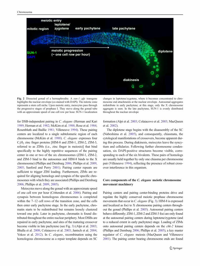
Fig. 2 Dissected gonad of a hermaphrodite. A sun-1::gfp transgenehighlights the nuclear envelope (co-stained with DAPI). The mitotic zonerepresents a stem cell niche. Upon meiotic entry, meiocytes pass throughthe progressive stages of prophase I. They move along the gonad tubewith an approximate speed of one cell row per hour. SUN-1 localization
changes in leptotene/zygotene, where it becomes concentrated to chro-mosome end attachments at the nuclear envelope. Autosomal aggregatesredistribute in early pachytene; at this stage, only the X chromosomeaggregate is seen. In the late pachytene, SUN-1 is evenly distributedthroughout the nuclear envelope
Chromosoma
at the nuclear envelope while loading pairing center proteins tothe appropriate chromosome region, (Phillips et al. 2009).Subsequent polo-like kinase 2 (PLK-2) recruitment to these sitesis dependent on the presence of pairing center proteins. PLK-2kinase activity induces dramatic changes at the nuclear envelope(Harper et al. 2011; Labella et al. 2011): The inner and outernuclear envelope proteins SUN-1 and ZYG-12 form distinctaggregates at pairing center localization sites (Penkner et al.2009; Sato et al. 2009). SUN-1 and ZYG-12 form a functionalSUN/KASH domain protein–protein interaction module (Fig. 4)(Fridkin et al. 2004; Malone et al. 2003; Minn et al. 2009;Penkner et al. 2007), which bridges the nuclear membranes andconnects the cytoskeleton to chromatin [for review, see Burke(2012)]. For simplicity, we term the multiprotein complexes atthe nuclear envelope, consisting of pairing centers, ZIMs, PLK-2, and the SUN-1/ZYG-12 nuclear envelope bridging complex,
the “movement-mediating complex” (MMC). In the absence ofSUN/KASH interaction, ZYG-12 retention at the nuclear enve-lope is defective (Malone et al. 2003; Penkner et al. 2007),despite the assembly of SUN-1 aggregates following the loadingof pairing center proteins to the one chromosome end at thenuclear envelope (Penkner et al. 2009). Therefore, the trigger foraggregation is most likely to be transmitted from the nucleus tothe cytoplasm.MMC formation is independent of DSBs, recom-bination, pairing, and synapsis, but requires chk-2 (Penkner et al.2009; Sato et al. 2009).
MMCs persist on all autosomal pairing centers until theend of leptotene/zygotene (Fig. 3). In early pachytene, SUN-1and ZYG-12 start to redistribute throughout the nuclear enve-lope, and the autosomal ZIM foci diminish (Penkner et al.2009; Sato et al. 2009). Only the MMC formed around the Xchromosome pairing center persists throughout early
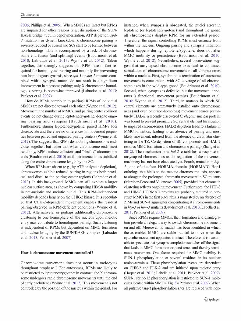
Fig. 3 Representative nuclei at pre-meiotic and meiotic (leptotene/zygo-tene, early pachytene, and late pachytene) stages. Images of SUN-1::GFP,SUN-1 phosphoserine-12 (S12), and HIM-8 (marks the X chromosomepairing center) labeling, and DAPI staining. The following structures andmarkers were hand-drawn: chromosome axes, synaptonemal complex,tubulin, and dynein. RAD-51, MSH-5, ZHP-3, and COSA-1 indicate theprecursors of crossover recombination. In leptotene/zygotene, SUN-1 isconcentrated at chromosome ends, where it becomes phosphorylated atserine-12 (S12). SUN-1 aggregation is mirrored on the cytoplasmic sideof the membrane by the aggregation of SUN-1’s KASH partner ZYG-12and components of the dynein motor. Chromosome axes load the lateralelement SC components upon meiotic entry. In leptotene/zygotene, chro-mosomes vigorously move and install the central elements of the SC. At
the same time, DSBs are induced and the RAD-51 strand invasionprotein, and other markers of maturing crossovers allow us to followongoing recombination. COSA-1 labels the six crossover sites in latepachytene. In early pachytene, synapsis is complete and SUN-1 which isconcentrated around autosomal chromosome end attachments isdephosphorylated and probably relocalizes; only X chromosome endattachment persists and is dissolved in the late pachytene. Pairing centerregions at chromosome end attachments at the nuclear envelope recruitpairing center proteins (HIM-8 marks the X chromosome). These act asrecruitment sites for PLK-2. SUN-1 serine-12 is a substrate for PLK-2.PLK-2 colocalizes with SUN-1 aggregates at chromosome ends. PLK-2also decorates the developing SC
Chromosoma
pachytene. In middle and late pachytene stages, most nucleilack SUN-1/ZYG-12 aggregates, despite the presence of aHIM-8 focus at the nuclear envelope (Penkner et al. 2009;Phillips et al. 2005; Sato et al. 2009). PLK-2 diffuses fromMMCs to synapsed chromosome axes throughout prophaseand is found on the synapsed chromosomes during pachytenestage (Harper et al. 2011; Labella et al. 2011).
Features of chromosome mobility in wild-type C. elegans
Several fluorescent markers have been used to study chromo-some movement in vivo. GFP-tagged transgenes of SUN-1(Penkner et al. 2009) and ZYG-12 (Malone et al. 2003; Satoet al. 2009) mark all chromosome ends (the sex chromosomeand autosomes). GFP-tagged HIM-8 can specifically visualizethe single X chromosome pairing center and was used toassess pre-meiotic movement (Wynne et al. 2012). In addition,delayed pre-meiotic replication of the X chromosome enablesspecific X chromosome labeling for live imaging (Jaramillo-Lambert et al. 2007). Hoechst dye or mCherry-tagged histoneH2B (McNally et al. 2006) has been employed to assess themobility of the entire chromatin mass.
It is clear that MMCs display highly mobile behavior (seeFig. 5). During leptotene/zygotene, an average of four to sixMMCs can be observed moving within a nucleus, but the num-ber can fluctuate between 1 and 12 (in C. elegans , 2n=12)(Baudrimont et al. 2010; Penkner et al. 2009; Sato et al. 2009;Wynne et al. 2012). In contrast to the highlymobile chromosomeends, other chromosome regions are relatively static. A high
degree of chromosome elasticity enables the pairing center toexplore a larger area of the nucleus than the distal ends of thechromosomes (Wynne et al. 2012). In addition, pairing centerchromosome ends have been observed to move independently.Over time, MMCs fuse into local clusters; from there they eithercontinue to move together or “bud out” of those clusters andresume moving individually. Thus, chromosomes interact withboth homologous and heterologous partners during the period ofhomology search and SC formation. Most interactions exist foraround 1 min, a smaller fraction for up to 3 min, and an evensmaller fraction for more than 3 min (Baudrimont et al. 2010).
High-resolution 4D in vivo analysis of MMC mobilitysuggests that two types of movement alternate. When analyz-ing the motion of a single-MMC, processive chromosomemovements, in the sense that an MMC moves continuouslyin the same direction for several seconds, are superimposed onundirected movements, in which an MMC oscillates close toits origin. MMCs only rarely engage in processive move-ments; thus, fast trajectories only contribute to 15 % of dis-placement tracks. Nevertheless, this processive movementaccounts for most surface area exploration by a singleMMC. These chromosome-end trajectories have an averagelength of 0.5 μm (with an average speed of 0.19μm/s) but canbe as long as 2 μm (Wynne et al. 2012).
It is important to note that limitations to the time resolution ofchromosome movement (minimum recording intervals used inthe most technically advance study were 0.4 s in a single planeand 2 s in Z stacks) currently impedes the accurate measurementof MMC mobility (Wynne et al. 2012). It also complicates thedetermination of background movements in the complex tissueof the gonad. Recordings of euthanized animals suggest that thelower speed ranges (which are especially overrepresented in non-processive movement) may represent background motion(Baudrimont et al. 2010). Residual muscle activity and gonadpumping in anesthetized animals may even increase backgroundmovement during live imaging. Therefore, we would like torefrain from making a distinction between processive and non-processive motions and refer to “rapid prophase movements”(RPMs) to specifically describe movements above the back-ground level of diffusion and Brownian motion.
Chromosome endmovements during leptotene/zygotene aremainly restricted to one hemisphere of the nucleus (Baudrimontet al. 2010; Labrador et al. 2013; Penkner et al. 2009). Aftercompletion of pairing and synapsis, meiocytes enter earlypachytene and chromatin clustering is loosened but notcompletely abrogated (Fig. 3). RPMs of the single remainingMMC (induced by the pairing center of the X chromosome) arereduced and the explored area of the nuclear envelope is smallerthan during leptotene/zygotene. Nevertheless, the X chromo-some remains mobile and continues to undergo rapid prophasemotion (RPM) in early pachytene (Wynne et al. 2012).Although it is obvious that chromosomes continue to pair andsynapse while they progress through leptotene/zygotene, the
Fig. 4 Schematic diagram of the SUN-1/KASH bridge spanning thenuclear membranes. The SUN-1/KASH bridge spans the outer and innernuclear membranes (ONM and INM). ZYG-12 connects to the move-ment apparatus (comprising dynein and cytoplasmic microtubules).Chromosomes connect to the SUN-1/KASH bridge via the pairing cen-ters, which are bound by pairing center proteins (ZIMs) and PLK-2
Chromosoma
characteristics of chromosome movement remain essentiallythe same with respect to the number of MMCs, the distancetraveled on the nuclear envelope, and the rate of MMC fusionand splitting events (Baudrimont et al. 2010; Wynne et al.2012), indicating that paired/synapsed chromosome pairs con-tinue to move throughout this stage.
How are rapid chromosome movements generated?
In the gonad syncytium of C. elegans, nuclei are embedded in adense meshwork of microtubule bundles (Minn et al. 2009).Components of the dynein motor complex are enriched at sitesof SUN-1/ZYG-12 aggregation (Sato et al. 2009), and ZYG-12 isrequired for dynein localization to the nuclear envelope (Maloneet al. 2003; Sato et al. 2009). Furthermore, an analysis of RPMcharacteristics found that they fitted well with known parametersfor motor protein-driven movement. Consistent with this, actinwas found to be dispensable for RPMs (Wynne et al. 2012).
RPMs are absent following dynein knockdown or tubulindepolymerization, despite correct MMC assembly (Labradoret al. 2013; Wynne et al. 2012). Since dynein localizes to
MMCs that are not engaged in RPM, it is apparent that dyneinlocalization to MMCs is insufficient to engage them in RPMs(Labrador et al. 2013; Wynne et al. 2012). Dynein motor func-tion requires ATP. Consequently, ATP depletion by cytochromeC oxidase inhibition caused by the administration of low dosesof sodium azide severely abrogates RPMs. Furthermore, exper-iments using a specific allele of the mitochondria-localizedSPD-3 protein showed that mitochondria are likely to producethe energy required for RPMs, but not for MMC formation ordynein complex localization to MMCs (Labrador et al. 2013).
Taken together, these observations suggest that RPMs arethe result of dynein–ATP-driven MMC mobilization alongcytoplasmic microtubules.
Are RPMs required for chromosome pairing?
Under conditions that prevent the formation of functionalMMCs (such as mutations in sun-1 , chk-2, or plk-2 or ZIMknockdown), both homologous pairing and RPMs are absent(Harper et al. 2011; Labella et al. 2011; MacQueen andVilleneuve 2001; Penkner et al. 2007; Phillips and Dernburg
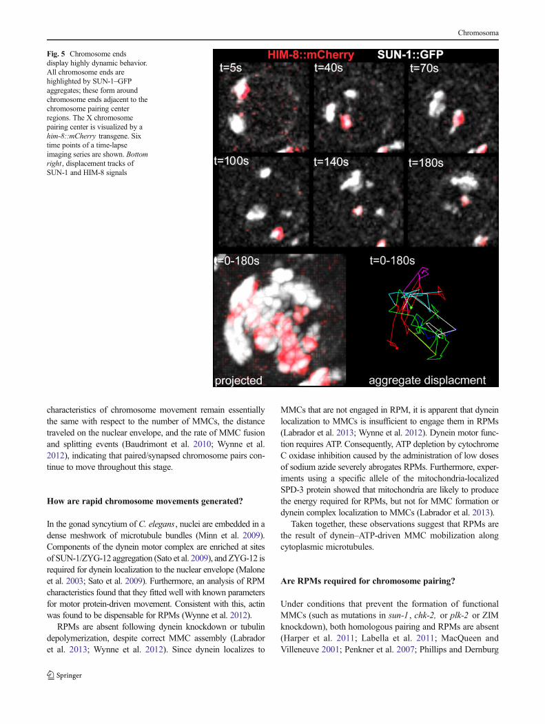
Fig. 5 Chromosome endsdisplay highly dynamic behavior.All chromosome ends arehighlighted by SUN-1–GFPaggregates; these form aroundchromosome ends adjacent to thechromosome pairing centerregions. The X chromosomepairing center is visualized by ahim-8::mCherry transgene. Sixtime points of a time-lapseimaging series are shown. Bottomright, displacement tracks ofSUN-1 and HIM-8 signals
Chromosoma
2006; Phillips et al. 2005). When MMCs are intact but RPMsare impaired for other reasons (e.g., disruption of the SUN/KASH bridge, tubulin depolymerization, ATP depletion, spd-3 mutation, or dynein knockdown), chromosome pairing isseverely reduced or absent and SCs start to be formed betweennon-homologs. This is accompanied by a lack of chromo-some end fusion (and splitting) events (Baudrimont et al.2010; Labrador et al. 2013; Wynne et al. 2012). Takentogether, this strongly suggests that RPMs are in fact re-quired for homologous pairing and not only for preventingnon-homologous synapsis, since spd-3 or sun-1 mutants com-bined with a synapsis mutant do not result in a significantimprovement in autosome pairing; only X chromosome homol-ogous pairing is somewhat improved (Labrador et al. 2013;Penkner et al. 2007).
How do RPMs contribute to pairing? RPMs of individualMMCs are not directed toward each other (Wynne et al. 2012).Movement, the number of MMCs, and pairing center collisionevents do not change during leptotene/zygotene, despite ongo-ing pairing and synapsis (Baudrimont et al. 2010).Furthermore, during leptotene/zygotene, paired HIM-8 focidisassociate and there are no differences in movement proper-ties between paired and unpaired pairing centers (Wynne et al.2012). This suggests that RPMs do not bring chromosome endscloser together, but rather that when chromosome ends meetrandomly, RPMs induce collisions and “shuffle” chromosomeends (Baudrimont et al. 2010) until their interaction is stabilizedalong the entire chromosome length by the SC.
When RPMs are absent (e.g., by ATP or dynein depletion),chromosomes exhibit reduced pairing in regions both proxi-mal and distal to the pairing center regions (Labrador et al.2013). In this background, MMCs can still explore a largernuclear surface area, as shown by comparing HIM-8 mobilityin pre-meiotic and meiotic nuclei. This RPM-independentmobility depends largely on the CHK-2 kinase. It is speculat-ed that CHK-2-dependent movement enables the residualpairing observed in RPM-deficient conditions (Wynne et al.2012). Alternatively, or perhaps additionally, chromosomeclustering to one hemisphere of the nucleus upon meioticentry may contribute to homologous pairing. Such clusteringis independent of RPMs but dependent on MMC formationand nuclear bridging by the SUN/KASH complex (Labradoret al. 2013; Penkner et al. 2007).
How is chromosome movement controlled?
Chromosome movement does not occur in meiocytesthroughout prophase I. For autosomes, RPMs are likely tobe restricted to leptotene/zygotene; in contrast, the X chromo-some undergoes rapid chromosome movements until the endof early pachytene (Wynne et al. 2012). This movement is notcontrolled by the position of the nucleus within the gonad. For
instance, when synapsis is abrogated, the nuclei arrest inleptotene (or leptotene/zygotene) and throughout the gonadall chromosomes display RPM for an extended period.Therefore, the signal controlling RPMs must emanate fromwithin the nucleus. Ongoing pairing and synapsis initiation,which happens during leptotene/zygotene, does not alterMMC mobility or persistence (Baudrimont et al. 2010;Wynne et al. 2012). Nevertheless, several observations sug-gest that unsynapsed chromosome axes lead to continuedstimulation of chromosome movement of all chromosomeswithin a nucleus. First, synchronous termination of autosomemovement is concomitant with SC coverage of all chromo-some axes in the wild-type gonad (Baudrimont et al. 2010).Second, when synapsis is defective but the movement appa-ratus is functional, movement persists (Baudrimont et al.2010; Wynne et al. 2012). Third, in mutants in which SCcentral elements are prematurely installed onto chromosomeaxes (and even onto non-homologs) movement ceases prema-turely. HAL-2, a recently discoveredC. elegans nuclear protein,was found to prevent premature SC central element localizationto unpaired chromosomes. HAL-2 depletion leads to a failure inMMC formation, leading to an absence of pairing and mostlikely movement, inferred from the absence of chromatin clus-tering in the TZ. Co-depletion of SC components and HAL-2restores MMC formation and chromosome pairing (Zhang et al.2012). The mechanism how hal-2 establishes a response ofunsynapsed chromosomes to the regulation of the movementmachinery has not been elucidated yet. Fourth, mutation in htp-1 , one of the four HORMA-domain (HORMAD) Hop1orthologs that binds to the meiotic chromosome axis, appearsto abrogate the prolonged chromatin movement in SC mutants(Martinez-Perez and Villeneuve 2005), provided that chromatinclustering reflects ongoing movement. Furthermore, the HTP-3and HIM-3 HORMAD proteins are probably required to con-structMMCs in the first place; this is suggested by an absence ofZIMs and SUN-1 aggregates concentrating at chromosome endsin htp-3 or him-3 mutants (Baudrimont et al. 2010; Labella et al.2011; Penkner et al. 2009).
Since RPMs require MMCs, their formation and disintegra-tion provide an elegant way to switch chromosome movementon and off. Moreover, no mutant has been identified in whichthe assembled MMCs are stable but fail to move when thecytosolic movement apparatus is intact. Therefore, it is reason-able to speculate that synapsis completion switches off the signalthat leads to MMC formation or persistence and thereby termi-nates movement. One factor required for MMC stability isSUN-1 phosphorylation at several residues in its nuclearamino-terminus. These phosphorylation events are dependenton CHK-2 and PLK-2 and are initiated upon meiotic entry(Harper et al. 2011; Labella et al. 2011; Penkner et al. 2009).SUN-1 serine-12 phosphorylation is restricted to SUN-1 mole-cules locatedwithinMMCs (Fig. 3) (Penkner et al. 2009).Whenall putative target phosphorylation sites are replaced with non-
Chromosoma
phosphorylatable amino acids, smaller, less distinct MMCs areobserved; however, these can still undergo RPM and chromo-some pairing follows wild-type kinetics, albeit with a decreasedrate of SC polymerization. MMCs disassemble prematurely inthese SUN-1 phosphorylation-deficient mutants when synapsisis compromised (Penkner et al. 2009; Woglar et al. 2013).Therefore, SUN-1 phosphorylations are required to sustainPLK-2 recruitment to MMCs under a challenged conditionand thus they are able to prolong the time window of chromo-some movement. By analogy, PLK-2 deletion is somewhatcompensated for by the PLK-1 homolog, andMMCs form uponmeiotic entry but fail to be sustained in synapsis-deficient mu-tants (Harper et al. 2011; Labella et al. 2011). PLK-2 recruitmentto chromosome ends and SUN-1 phosphorylation are thereforeinterdependent. Since PLKs have an essential role in chromo-some movement [(Harper et al. 2011; Labella et al. 2011);Monique Zetka, personal communication], SUN-1 phosphory-lation and PLK-2 localization to the ends of unsynapsed chro-mosomes could represent a positive feedback loop to sustainchromosome end-led mobility for as long as necessary to com-plete synapsis.
Chromosome mobility during early pachytene
RPMs of all chromosomes are required for chromosome pairingand homolog synapsis during leptotene/zygotene (Baudrimontet al. 2010; Penkner et al. 2007; Penkner et al. 2009; Sato et al.2009; Wynne et al. 2012). Although RPMs seem to persist inearly pachytene (Wynne et al. 2012), to date we have informa-tion only onX chromosomemobility during early pachytene. Atthis stage, the pairing is complete and SC installation hasprogressed to a large extent (Dernburg et al. 1998; Tang et al.2010). Recombinational repair of spo-11-inducedmeiotic DSBsthat can result in crossover formation takes place during thismeiotic stage (Fig. 3). Most RAD-51 foci start to appear in thetransition zone, peak in early to mid-pachytene, and then disap-pear before late pachytene (Alpi et al. 2003; Colaiacovo et al.2003); sites of future crossovers are marked by ZHP-3 andMSH-5 proteins during pachytene (Fig. 3) (Bhalla et al. 2008;Jantsch et al. 2004; Yokoo et al. 2012). During early pachytene,the period of chromosome mobility is prolonged when repair orrecombination are prevented (Woglar et al. 2013), similar to theextension of leptotene/zygotene mobility observed when synap-sis is abrogated (Baudrimont et al. 2010; MacQueen et al. 2002;Wynne et al. 2012).
In other organisms in which movement plays a minor rolein the actual chromosome pairing process, ectopic recombi-nation and chromosome entanglements have been observedwhen movement is limited, suggesting that movement canovercome non-homologous (i.e., weaker) chromosomal inter-actions (Conrad et al. 2008; Davis and Smith 2006; Goldman
and Lichten 2000; Golubovskaya et al. 2002; Koszul et al.2008; Niwa et al. 2000; Schlecht et al. 2004).
Furthermore, the movement apparatus plays a role in cross-over interference in budding yeast (Chua and Roeder 1997). Itwas previously speculated that chromosome oscillations couldaccount for negative crossover interference (Hulten 2011).Also, the enlargement of chromosomal territories caused bythe pull on chromosomes (Wynne et al. 2012) may aid axiselongation and SC extension in early pachytene stage. Thiswould improve the efficiency of crossover interference, sincecomplete SC localization to chromosome axes is required forthe execution of crossover interference (Hayashi et al. 2010;Hillers and Villeneuve 2003; Sym and Roeder 1994).
To date, there is no experimental evidence in C. elegans todirectly link the residual early pachytene movement with anyof these tasks, since movement is required for the precedinghomologous chromosome pairing. However, the easy-to-access C. elegans gonad offers the possibility of transientmovement inhibition in meiotic nuclei in which chromosomepairing and synapse have already taken place. This wouldfacilitate the use of the worm as an animal model to studyall aspects of chromosome mobility.
Synopsis
The power of C. elegans genetics has enabled the genera-tion of particularly informative alleles of genes required todrive the movement machinery of chromosomes in meioticprophase I. Furthermore, drug inhibition of cytoskeletalforces and the establishment of live imaging in wholemounted animals, together with the insight provided bystudying these key mutants, have enabled the constructionof a basic framework to understand the major role of pro-phase chromosome movement in an important animal mod-el system (also see Fig. 3).
& Following meiotic entry, one end of each chromosome istethered to the nuclear envelope, where cytoskeletal forcesare transmitted to chromosomes via a SUN/KASH bridgespanning the nuclear membranes.
& Chromosome tethering to the nuclear envelope is accom-panied by the binding of pairing center-binding zinc fingerproteins to pairing centers/homology recognition regionson one end of each chromosome.
& The pairing centers/homology recognition regions recruitPLK(s) that couple chromosomes to the movementapparatus.
& Chromosome movement depends on microtubules, thedyneinmotor complex, and ATP supplied bymitochondria.
& Rapid chromosome movements are essential for homologsearching through random chromosome encounters.
Chromosoma
& Reduced chromosome end-led motion has an equallynegative effect on pairing for chromosome regions distantfrom pairing centers.
& Although frequently seen, chromosome end-led move-ments correlate with the presence of clustered chromatinin the transition zone; however, this is not obligatory.
& Meiotic chromosome axis proteins and PLK-2 and CHK-2 kinases control movement.
& It is possible that chromosome movement has a positiveeffect on the processivity of SC polymerization. Delayedsynapsis correlates with reduced plk-2 activity.
& A surveillance mechanism couples events leading to theestablishment of the obligate crossover to chromosomeend mobilization in prophase I. In this mechanism, revers-ible phospho-modifications of SUN-1 establish a positivefeedback loop for recruiting PLK-2 to chromosome ends.
Acknowledgments We would like to thank Anahita Daryabeigi,Monique Zetka and EnriqueMartinez-Perez for sharing unpublished data.We are indebted to Monique Zetka and Dimitra Paouneskou for criticallyreading this manuscript. This work was funded by FWF grants P-23638-B12 and SFB-F34.
Open Access This article is distributed under the terms of the CreativeCommons Attribution License which permits any use, distribution, andreproduction in any medium, provided the original author(s) and thesource are credited.
References
Alpi A, Pasierbek P, Gartner A, Loidl J (2003) Genetic and cytologicalcharacterization of the recombination protein RAD-51 inCaenorhabditis elegans. Chromosoma 112:6–16
Baudrimont A, Penkner A, Woglar A, Machacek T, Wegrostek C,Gloggnitzer J, Fridkin A, Klein F, Gruenbaum Y, Pasierbek P et al(2010) Leptotene/zygotene chromosome movement via the SUN/KASH protein bridge in Caenorhabditis elegans. PLoS Genet 6:e1001219
Bhalla N, Wynne DJ, Jantsch V, Dernburg AF (2008) ZHP-3 acts atcrossovers to couple meiotic recombination with synaptonemalcomplex disassembly and bivalent formation in C. elegans . PLoSGenet 4:e1000235
Burke B (2012) It takes KASH to hitch to the SUN. Cell 149:961–963Chua PR, Roeder GS (1997) Tam1, a telomere-associated meiotic pro-
tein, functions in chromosome synapsis and crossover interference.Genes Dev 11:1786–1800
Colaiacovo MP, MacQueen AJ, Martinez-Perez E, McDonald K, AdamoA, La Volpe A, Villeneuve AM (2003) Synaptonemal complexassembly in C. elegans is dispensable for loading strand-exchangeproteins but critical for proper completion of recombination. DevCell 5:463–474
Conrad MN, Lee CY, Chao G, Shinohara M, Kosaka H, Shinohara A,Conchello JA, Dresser ME (2008) Rapid telomere movement inmeiotic prophase is promoted by NDJ1, MPS3, and CSM4 and ismodulated by recombination. Cell 133:1175–1187
Crittenden SL, Troemel ER, Evans TC, Kimble J (1994) GLP-1 islocalized to the mitotic region of the C. elegans germ line. Devel-opment 120:2901–2911
Crittenden SL, Leonhard KA, Byrd DT, Kimble J (2006) Cellular anal-yses of the mitotic region in the Caenorhabditis elegans adult germline. Mol Biol Cell 17:3051–3061
Davis L, Smith GR (2006) The meiotic bouquet promotes homolog inter-actions and restricts ectopic recombination in Schizosaccharomycespombe. Genetics 174:167–177
Dernburg AF, McDonald K, Moulder G, Barstead R, Dresser M, Ville-neuve AM (1998)Meiotic recombination inC. elegans initiates by aconserved mechanism and is dispensable for homologous chromo-some synapsis. Cell 94:387–398
Ding DQ, Yamamoto A, Haraguchi T, Hiraoka Y (2004) Dynamics ofhomologous chromosome pairing duringmeiotic prophase in fissionyeast. Dev Cell 6:329–341
Fridkin A, Mills E, Margalit A, Neufeld E, Lee KK, Feinstein N, CohenM, Wilson KL, Gruenbaum Y (2004) Matefin, a Caenorhabditiselegans germ line-specific SUN-domain nuclear membrane protein,is essential for early embryonic and germ cell development. ProcNatl Acad Sci U S A 101:6987–6992
Friedland AE, Tzur YB, Esvelt KM, Colaiacovo MP, Church GM,Calarco JA (2013) Heritable genome editing in C. elegans via aCRISPR-Cas9 system. Nat Methods 10:741–743
Frokjaer-Jensen C, Davis MW, Hopkins CE, Newman BJ, ThummelJM, Olesen SP, Grunnet M, Jorgensen EM (2008) Single-copyinsertion of transgenes in Caenorhabditis elegans . Nat Genet40:1375–1383
Frokjaer-Jensen C, Davis MW, Hollopeter G, Taylor J, Harris TW, Nix P,Lofgren R, Prestgard-Duke M, Bastiani M, Moerman DG et al(2010) Targeted gene deletions in C. elegans using transposonexcision. Nat Methods 7:451–453
Goldman AS, Lichten M (2000) Restriction of ectopic recombination byinterhomolog interactions during Saccharomyces cerevisiae meio-sis. Proc Natl Acad Sci U S A 97:9537–9542
Golubovskaya IN, Harper LC, Pawlowski WP, Schichnes D, Cande WZ(2002) The pam1 gene is required for meiotic bouquet formationand efficient homologous synapsis in maize (Zea mays L.). Genetics162:1979–1993
Harper NC, Rillo R, Jover-Gil S, Assaf ZJ, Bhalla N, Dernburg AF (2011)Pairing centers recruit a Polo-like kinase to orchestrate meioticchromosome dynamics in C. elegans. Dev Cell 21:934–947
Hayashi M, Mlynarczyk-Evans S, Villeneuve AM (2010) Thesynaptonemal complex shapes the crossover landscape throughcooperative assembly, crossover promotion and crossover inhibitionduring Caenorhabditis elegans meiosis. Genetics 186:45–58
Herman RK, Kari CK (1989) Recombination between small X chromo-some duplications and the X chromosome in Caenorhabditiselegans . Genetics 121:723–737
Herman RK, Kari CK, Hartman PS (1982) Dominant X-chromosome non-disjunction mutants of Caenorhabditis elegans. Genetics 102:379–400
Hillers KJ, Villeneuve AM (2003) Chromosome-wide control of meioticcrossing over in C. elegans. Curr Biol: CB 13:1641–1647
Hirsh D, OppenheimD,KlassM (1976) Development of the reproductivesystem of Caenorhabditis elegans . Dev Biol 49:200–219
HultenMA (2011)On the origin of crossover interference: a chromosomeoscillatory movement (COM) model. Mol Cytogenet 4:10
Jantsch V, Pasierbek P, Mueller MM, Schweizer D, Jantsch M, Loidl J(2004) Targeted gene knockout reveals a role in meiotic recombi-nation for ZHP-3, a Zip3-related protein in Caenorhabditis elegans .Mol Cell Biol 24:7998–8006
Jaramillo-Lambert A, Ellefson M, Villeneuve AM, Engebrecht J(2007) Differential timing of S phases, X chromosome replication,and meiotic prophase in the C. elegans germ line. Dev Biol 308:206–221
Chromosoma
Koszul R, Kleckner N (2009) Dynamic chromosome movements duringmeiosis: a way to eliminate unwanted connections? Trends Cell Biol19:716–724
Koszul R, Kim KP, Prentiss M, Kleckner N, Kameoka S (2008) Meioticchromosomes move by linkage to dynamic actin cables with trans-duction of force through the nuclear envelope. Cell 133:1188–1201
Labella S, Woglar A, Jantsch V, Zetka M (2011) Polo kinases establishlinks between meiotic chromosomes and cytoskeletal forces essen-tial for homolog pairing. Dev Cell 21:948–958
Labrador L, Barroso C, Lightfoot J, Muller-Reichert T, Flibotte S, TaylorJ, Moerman DG, Villeneuve AM, Martinez-Perez E (2013) Chro-mosome movements promoted by the mitochondrial protein SPD-3are required for homology search during Caenorhabditis elegansmeiosis. PLoS Genet 9:e1003497
Lorenz A, Fuchs J, Burger R, Loidl J (2003) Chromosome pairing doesnot contribute to nuclear architecture in vegetative yeast cells.Eukaryot Cell 2:856–866
MacQueen AJ, Villeneuve AM (2001) Nuclear reorganization and ho-mologous chromosome pairing during meiotic prophase require C.elegans chk-2. Genes Dev 15:1674–1687
MacQueen AJ, Colaiacovo MP, McDonald K, Villeneuve AM (2002)Synapsis-dependent and -independent mechanisms stabilize homo-log pairing during meiotic prophase in C. elegans. Genes Dev 16:2428–2442
Malone CJ, Misner L, Le Bot N, Tsai MC, Campbell JM, Ahringer J,White JG (2003) The C. elegans hook protein, ZYG-12, mediatesthe essential attachment between the centrosome and nucleus. Cell115:825–836
Martinez-Perez E, Villeneuve AM (2005) HTP-1-dependent constraintscoordinate homolog pairing and synapsis and promote chiasmaformation during C. elegans meiosis. Genes Dev 19:2727–2743
McKimKS, Howell AM, Rose AM (1988) The effects of translocations onrecombination frequency in Caenorhabditis elegans. Genetics 120:987–1001
McNally K, Audhya A, Oegema K, McNally FJ (2006) Katanin controlsmitotic and meiotic spindle length. J Cell Biol 175:881–891
Mets DG, Meyer BJ (2009) Condensins regulate meiotic DNA breakdistribution, thus crossover frequency, by controlling chromosomestructure. Cell 139:73–86
Minn IL, Rolls MM, Hanna-Rose W, Malone CJ (2009) SUN-1 andZYG-12, mediators of centrosome-nucleus attachment, are a func-tional SUN/KASH pair in Caenorhabditis elegans . Mol Biol Cell20:4586–4595
Morimoto A, Shibuya H, Zhu X, Kim J, Ishiguro K, Han M,Watanabe Y(2012) A conserved KASH domain protein associates with telo-meres, SUN1, and dynactin during mammalian meiosis. J Cell Biol198(2):165–172. doi:10.1083/jcb.201204085
Nabeshima K, Villeneuve AM, Colaiácovo MP (2005) Crossing over iscoupled to late meiotic prophase bivalent differentiation throughasymmetric disassembly of the SC. J Cell Biol 168(5):683–689
Niwa O, Shimanuki M, Miki F (2000) Telomere-led bouquet formationfacilitates homologous chromosome pairing and restricts ectopicinteraction in fission yeast meiosis. EMBO J 19:3831–3840
Parvinen M, SoderstromKO (1976) Chromosome rotation and formationof synapsis. Nature 260:534–535
Penkner A, Tang L, NovatchkovaM, LadurnerM, Fridkin A, GruenbaumY, Schweizer D, Loidl J, Jantsch V (2007) The nuclear envelopeprotein Matefin/SUN-1 is required for homologous pairing in C.elegans meiosis. Dev Cell 12:873–885
Penkner AM, Fridkin A, Gloggnitzer J, Baudrimont A, Machacek T,Woglar A, Csaszar E, Pasierbek P, Ammerer G, Gruenbaum Y et al(2009)Meiotic chromosome homology search involves modificationsof the nuclear envelope protein Matefin/SUN-1. Cell 139:920–933
Phillips CM, Dernburg AF (2006) A family of zinc-finger proteins isrequired for chromosome-specific pairing and synapsis during mei-osis in C. elegans . Dev Cell 11:817–829
Phillips CM, Wong C, Bhalla N, Carlton PM, Weiser P, Meneely PM,Dernburg AF (2005) HIM-8 binds to the X chromosome pairingcenter and mediates chromosome-specific meiotic synapsis. Cell123:1051–1063
Phillips CM, Meng X, Zhang L, Chretien JH, Urnov FD, Dernburg AF(2009) Identification of chromosome sequence motifs that mediatemeiotic pairing and synapsis inC. elegans . Nat Cell Biol 11:934–942
Rose AM, Baillie DL, Curran J (1984) Meiotic pairing behavior of twofree duplications of linkage group I in Caenorhabditis elegans. MolGen Genet 195:52–56
Rosenbluth RE, Baillie DL (1981) The genetic analysis of a reciprocaltranslocation, eT1(III; V), in Caenorhabditis elegans . Genetics 99:415–428
Sanford C, Perry MD (2001) Asymmetrically distributed oligonucleotiderepeats in theCaenorhabditis elegans genome sequence that map toregions important for meiotic chromosome segregation. NucleicAcids Res 29:2920–2926
Sato A, Isaac B, Phillips CM, Rillo R, Carlton PM, Wynne DJ, KasadRA, Dernburg AF (2009) Cytoskeletal forces span the nuclearenvelope to coordinate meiotic chromosome pairing and synapsis.Cell 139:907–919
ScherthanH,WangH,Adelfalk C,White EJ, CowanC, CandeWZ,KabackDB (2007) Chromosome mobility during meiotic prophase in Saccha-romyces cerevisiae. Proc Natl Acad Sci U S A 104:16934–16939
Schlecht HB, Lichten M, Goldman AS (2004) Compartmentalization ofthe yeast meiotic nucleus revealed by analysis of ectopic recombi-nation. Genetics 168:1189–1203
Sheehan MJ, Pawlowski WP (2009) Live imaging of rapid chromosomemovements in meiotic prophase I in maize. Proc Natl Acad Sci U SA 106:20989–20994
Smolikov S, Schild-Prufert K, Colaiacovo MP (2008) CRA-1 uncovers adouble-strand break-dependent pathway promoting the assembly ofcentral region proteins on chromosome axes during C. elegansmeiosis. PLoS Genet 4:e1000088
Sym M, Roeder GS (1994) Crossover interference is abolished in theabsence of a synaptonemal complex protein. Cell 79:283–292
Tang L, Machacek T, Mamnun YM, Penkner A, Gloggnitzer J,Wegrostek C, Konrat R, Jantsch MF, Loidl J, Jantsch V (2010)Mutations in Caenorhabditis elegans him-19 show meiotic defectsthat worsen with age. Mol Biol Cell 21:885–896
Villeneuve AM (1994) A cis-acting locus that promotes crossing overbetween X chromosomes in Caenorhabditis elegans . Genetics 136:887–902
Wanat JJ, Kim KP, Koszul R, Zanders S, Weiner B, Kleckner N, Alani E(2008) Csm4, in collaboration with Ndj1, mediates telomere-ledchromosome dynamics and recombination during yeast meiosis.PLoS Genet 4:e1000188
Woglar A, Daryabeigi A, Adamo A, Habacher C, Machacek T, La VolpeA, Jantsch V (2013) Matefin/SUN-1 phosphorylation is part of asurveillance mechanism to coordinate chromosome synapsis andrecombination with meiotic progression and chromosome move-ment. PLoS Genet 9:e1003335
Wood AJ, Lo TW, Zeitler B, Pickle CS, Ralston EJ, Lee AH, Amora R,Miller JC, Leung E, Meng X et al (2011) Targeted genome editingacross species using ZFNs and TALENs. Science 333:307
Wynne DJ, Rog O, Carlton PM, Dernburg AF (2012) Dynein-dependentprocessive chromosome motions promote homologous pairing in C.elegans meiosis. J Cell Biol 196:47–64
Yokoo R, Zawadzki KA, Nabeshima K, Drake M, Arur S, Villeneuve AM(2012) COSA-1 reveals robust homeostasis and separable licensingand reinforcement steps governing meiotic crossovers. Cell 149:75–87
Zhang W, Miley N, Zastrow MS, MacQueen AJ, Sato A, Nabeshima K,Martinez-Perez E, Mlynarczyk-Evans S, Carlton PM, VilleneuveAM (2012) HAL-2 promotes homologous pairing duringCaenorhabditis elegans meiosis by antagonizing inhibitory effectsof synaptonemal complex precursors. PLoS Genet 8:e1002880