Copyright 0 1991 by the Genetics Society of America Chromosome Rearrangement by Ectopic Recombination in Drosophila melanogaster: Genome Structure and Evolution E. A. Montgomery, S.-M. Huang, C. H. Langley' and B. H. Judd' Laboratory of Genetics, National Institute of Environmental Health Sciences, Research Triangle Park, North Carolina 27709 Manuscript received May 13, 199 1 Accepted for publication August 13, 1991 ABSTRACT Ectopic recombination between interspersed repeat sequences generates chromosomal re- arrangements that have a major impact on genome structure. A survey of ectopic recombination in the region flanking the white locus of Drosophila melanogaster identified 25 transposon-mediated rearrangements from four parallel experiments. Eighteen of the 25 were generated from females carrying X chromosomes heterozygous for interspersed repeat sequences. The cytogenetic and molecular analyses of the rearrangements and the parental chromosomes show: (1) interchromosomal and intrachromosomal recombinants are generated in about equal numbers; (2) ectopic recombination appears to be a meiotic process that is stimulated by the interchromosomal effect to about the same degree as regular crossing over; (3) copies of the retrotransposon roo were involved in all of the interchromosomal exchanges; some copies were involved much more frequently than others in the target region; (4) homozygosis for interspersed repeat sequences and other sequence variations significantly reduced ectopic recombination. C HROMOSOMAL rearrangement by ectopic pairing and recombination between interspersed repeat sequences presents a mechanism for dramatic reorganizationofeukaryoticgenomes.Reorganiza- tion by gene duplications or deletions, or by inversions or transpositions of various kinds offers raw material for evolutionary modifications and at the same time poses a threat to the stability of the genome. The insertion or excision of transposons at various sites in the genome account for a significant portion of mu- tations in Drosophila and other eukaryotes. However, possibly of equal or greater consequence are the changes in chromosome structure that result from the involvement of interspersed repeat sequences in un- equal crossing over. Meiotic and mitotic exchange between ectopically paired repeat sequences that cre- ate chromosomal rearrangements have been studied in yeast (ROEDER 1983; PETES and HILL 1988), and in Drosophila (GOLDBERG et al. 1983; DAVIS, SHEN and JuDD 1987; ENGELS and PRESTON 1984; and LIM 1988). Rearrangements associated with some recessive mutations cause human diseases. These include hy- percholesterolemia (locus: low density lipoprotein recep- tor, LEHRMAN et al. 1987), hereditary angioedema (locus: Cl inhibitor; STOPPA-LYONNET et al. 1990) and human steroid sulfatase deletions (locus: STS, YEN et al. 1990), indicating that such ectopic exchanges be- tween dispersed repeat sequences also occur in the human germline. California 95616. I Present address: Department of Genetics, University of California, Davis, ' To whom correspondence should be addressed. Genetics 149: 1085-1098 (December, 1991) Little is known about the quantitative contribution of ectopic exchanges between repeat sequences to the origin of chromosomal rearrangements. In Drosophila melanogaster there aresome 40 families of transposa- ble elements with copies of the elements of each family closely conserved. The average copy numbers of these families rangefrom only a few toover 50. These elements comprise 10-1 2% of the genome. Surveys of the distributions of transposable elements in the euchromatic regions of the genome in populations of D. melanogaster (CHARLESWORTH and LANGLEY 1989) have shown that while the presence ofan insert at any particular site is rare, the number of possible sites is so large that the average individual has multiple rep- resentatives of each family. Considering the number of interspersed repetitive elements, it is evident that, depending on the rate of ectopic recombination, the resulting chromosome aberrations could be a major source of mutations in germinal and somatic cells. The impact of these mutations on development and fitness of zygotes is for themost part unknown. It has been proposed, however, that the ectopic exchange process with its associated mutational effects is an important population genetic mechanism influencing the average copy numbers of these DNA parasites in natural populations of D. melanogaster (LANGLEY et al. 1988). To learn more about the role of ectopic exchange in the origin of chromosomal rearrangements, we have carried out a series of experiments designed to measure the amount and types of ectopic exchanges in a region of the X chromosome surrounding the Downloaded from https://academic.oup.com/genetics/article/129/4/1085/6059201 by guest on 21 December 2021
Transcript
Copyright 0 1991 by the Genetics Society of America
Chromosome Rearrangement by Ectopic Recombination in Drosophila melanogaster: Genome Structure and Evolution
E. A. Montgomery, S.-M. Huang, C. H. Langley' and B. H. Judd' Laboratory of Genetics, National Institute of Environmental Health Sciences, Research Triangle Park, North Carolina 27709
Manuscript received May 13, 199 1 Accepted for publication August 13, 1991
ABSTRACT Ectopic recombination between interspersed repeat sequences generates chromosomal re-
arrangements that have a major impact on genome structure. A survey of ectopic recombination in the region flanking the white locus of Drosophila melanogaster identified 25 transposon-mediated rearrangements from four parallel experiments. Eighteen of the 25 were generated from females carrying X chromosomes heterozygous for interspersed repeat sequences. The cytogenetic and molecular analyses of the rearrangements and the parental chromosomes show: (1) interchromosomal and intrachromosomal recombinants are generated in about equal numbers; (2) ectopic recombination appears to be a meiotic process that is stimulated by the interchromosomal effect to about the same degree as regular crossing over; (3) copies of the retrotransposon roo were involved in all of the interchromosomal exchanges; some copies were involved much more frequently than others in the target region; (4) homozygosis for interspersed repeat sequences and other sequence variations significantly reduced ectopic recombination.
C HROMOSOMAL rearrangement by ectopic pairing and recombination between interspersed
repeat sequences presents a mechanism for dramatic reorganization of eukaryotic genomes. Reorganiza- tion by gene duplications or deletions, or by inversions or transpositions of various kinds offers raw material for evolutionary modifications and at the same time poses a threat to the stability of the genome. The insertion or excision of transposons at various sites in the genome account for a significant portion of mu- tations in Drosophila and other eukaryotes. However, possibly of equal or greater consequence are the changes in chromosome structure that result from the involvement of interspersed repeat sequences in un- equal crossing over. Meiotic and mitotic exchange between ectopically paired repeat sequences that cre- ate chromosomal rearrangements have been studied in yeast (ROEDER 1983; PETES and HILL 1988), and in Drosophila (GOLDBERG et al. 1983; DAVIS, SHEN and JuDD 1987; ENGELS and PRESTON 1984; and LIM 1988). Rearrangements associated with some recessive mutations cause human diseases. These include hy- percholesterolemia (locus: low density lipoprotein recep- tor, LEHRMAN et al. 1987), hereditary angioedema (locus: C l inhibitor; STOPPA-LYONNET et al. 1990) and human steroid sulfatase deletions (locus: STS, YEN et al. 1990), indicating that such ectopic exchanges be- tween dispersed repeat sequences also occur in the human germline.
California 95616. I Present address: Department of Genetics, University of California, Davis,
' To whom correspondence should be addressed.
Genetics 149: 1085-1098 (December, 1991)
Little is known about the quantitative contribution of ectopic exchanges between repeat sequences to the origin of chromosomal rearrangements. In Drosophila melanogaster there are some 40 families of transposa- ble elements with copies of the elements of each family closely conserved. The average copy numbers of these families range from only a few to over 50. These elements comprise 10-1 2% of the genome. Surveys of the distributions of transposable elements in the euchromatic regions of the genome in populations of D. melanogaster (CHARLESWORTH and LANGLEY 1989) have shown that while the presence of an insert at any particular site is rare, the number of possible sites is so large that the average individual has multiple rep- resentatives of each family. Considering the number of interspersed repetitive elements, it is evident that, depending on the rate of ectopic recombination, the resulting chromosome aberrations could be a major source of mutations in germinal and somatic cells. The impact of these mutations on development and fitness of zygotes is for the most part unknown. It has been proposed, however, that the ectopic exchange process with its associated mutational effects is an important population genetic mechanism influencing the average copy numbers of these DNA parasites in natural populations of D. melanogaster (LANGLEY et al. 1988).
T o learn more about the role of ectopic exchange in the origin of chromosomal rearrangements, we have carried out a series of experiments designed to measure the amount and types of ectopic exchanges in a region of the X chromosome surrounding the
Dow
nloaded from https://academ
ic.oup.com/genetics/article/129/4/1085/6059201 by guest on 21 D
ecember 2021
1086 E. A. Montgomery et al.
TABLE 1
Sites of roo and copia in parental X chromosomes
Element Oregon-R N yz wm spf ec N
roo lE, 2B, 2F-3A (4 sites), 3D, 4D, 5A, and 17 additional 26 lA, 2B, 2D-E, 3C (2 sites), 4A-B, 6B-C, 6D-E, and 21 euchromatic sites proximal to 5A 13 additional euchromatic sites proximal to 6, in-
cluding 18F
of sites.
white locus of D. melanogaster. Specifically we were interested in discovering (1) which repeated elements are involved and how frequently; (2) whether ex- changes are meiotic or mitotic, interchromosomal or intrachromosomal; (3) whether homozygosis of ge- netic polymorphisms in a region influences ectopic exchanges in that region; and (4) how recombination frequencies are influenced by the interchromosomal effect. The interchromosomal effect is seen where heterozygous rearrangements are present in one or more of the major chromosome arms in D. melano- gaster. Their presence results in an increase in crossing over of the other pair(s) of chromosomes, with the increase most marked in regions bordering centric heterochromatin and at the distal ends of chromo- some arms. T h e mechanistic basis for the increased recombination is not well understood [see LUCCHESI (1976) for review]. The answers to these questions will help in understanding the ectopic exchange proc- ess, and will provide avenues of investigation into its role in mutation processes and the containment of transposable element copy number.
MATERIALS AND METHODS
Drosophila strains and crosses: Two X chromosomes were selected for this study. One, carrying the mutant markers yellow-2 (y2) , white-ric (w"'), split (spl) and echinus (ec ) , was selected because of the useful markers flanking the white locus and because considerable molecular characteriza- tion of its white region had already been done [see JUDD (1987) for a review]. The wrir mutation was determined to be a derivative of white-apricot (w") in which a copy of the transposon roo had inserted into a copy of copia occupying a site at coordinate 0.0 of the white locus map (DAVIS, SHEN andJuDD 1987). This copia insert originally caused the white- apricot mutation. The other X chromosome was extracted from the wild type Oregon-R strain carried in this labora- tory. It was of unknown transposon content in the region of interest.
Polytene chromosome squashes of both parental strains were subsequently probed in situ to determine the copy numbers and relative positions of roo and copia in the X chromosomes (Table 1). We chose these two probes because both transposons were positioned within the white locus of w'" and because roo is generally present in high copy number (Figure 1).
Stocks of each parental type were established from a single male of each type, by crossing to FM7 females, then back-
crossing FI daughters to the parental male to establish cultures that were essentially isogenic for the X chromo- somes.
Heterozy ous autosomal rearrangements, SMI, Cy/+ and TM3, Ubx /+ of the second and third chromosomes re- spectively, were introduced into the 3 W~ spl ec strain and maintained by selection. When males of this strain were mated to Ore-R females (Figure 2), two of the four resulting classes of FI females were selected as virgins. Females of both classes were heterozygous for the two X chromosomes; one class carried rearranged autosomes (cross A), while the other had autosomes of standard configuration (cross B). The autosomal rearrangements are known to be effective in increasing crossing over in the region of the white locus by a factor of 3 to 5 UWDD 1959). Both classes of females were crossed to males of genotype z1 w6jaZ5 spl sn3, and offspring were examined for individuals exhibiting excep- tional eye color, i.e., other than the wild type of Ore-R and the orange-brown of w ~ / w ~ ~ ~ ~ ~ .
Females of the genotype yz wric spl ec; SMI/+; TM3/+ (cross C ) and Ore-R; SMI/+; TM3/+ (cross D), both of which were homoz ous for the X chromosomes, were also crossed to z1 w65a2ygspl sn3 males and the offspring were scored for exceptional eye-color phenotypes. These crosses may be viewed as controls for the heterozygous genotypes in crosses A and B however, they were included to test for a possible effect of homozygosity per se on ectopic pairing and exchange. The X chromosomes of the females in crosses C and D were homozygous for both the interspersed repeat sequence differences and any nucleotide sequence differ- ences between them. Either type of genetic variant may have different consequences in heterozygous us. homozy- gous condition. Unfortunately, exchange of flanking mark- ers cannot be scored in crosses C and D and the construction of new marker arrangements would likely have upset the patterns of sequence variation.
The design of the four crosses allows deletions or break- points within the white locus to be easily recognized by their bleached white phenotype. Duplications for the wild-type white locus should produce an orange mottle phenotype in females because of the interaction of the white duplication with the heterozygous z1 mutation. However, that pheno- type is similar to the w ~ / w ~ ~ ~ ~ ~ phenotype and possibly would not be recognized. Deletions or breakpoints within the zeste locus would be expected to produce an orange colored eye that would be easily recognized as an exception in cross D but recognized with diffkulty in other crosses because of its similarity to the w ~ ' / w ~ ~ ~ ~ ~ phenotype.
Molecular analysis: DNA for analysis was isolated from 0.5 to 2 g of adult flies and purified on a cesium chloride gradient as described in BINGHAM, LEVIS and RUBIN (1 98 1). Genomic libraries were constructed from DNA from paren- tal and deficiency strains as described previously (SHRIMP-
I 3 f
Dow
nloaded from https://academ
ic.oup.com/genetics/article/129/4/1085/6059201 by guest on 21 D
ecember 2021
P
Ectopic Recombina
. . .
FIGURE 1.-Sites of hybridiration of the transposable element, roo, to Oregon-R and f w"' spl ec X chromosomes. A biotinylated rootontaining plasmid was hybridized in situ to polytene chromo- somes from each o f the parental strains used in the study. (A) Ore- R by YOO; (B) f w"' spl cc by YOO. Arrows indicate the 3C region of the X chromosomes (magnification 500X).
TON, MONTGOMERY and LANGLEY 1986), with the following change: ligations were packaged using Gigapack Gold (Stra- tagene) using the manufacturers recommendations.
Library screening for positive phages, and the isolation and purification of those phages were performed using standard techniques (MANIATIS, FRITSCH and SAMBROOK 1982). Probe DNA was labeled with ["PIdCTP either by nick translation (RIGBY et al. 1977), or by the random- primed labeling technique using the reagents and recom- mended protocols provided by Boehringer-Mannheim in the Nick Translation or Random-Primed DNA Labeling Kits, respectively.
Restriction digests of DNA were performed using the conditions and buffers provided with the enzymes. Electro- phoretic separation of fragments was performed in 0.8% agarose gels. DNA was transferred to either nitrocellulose or to nylon membranes using standard transfer conditions for each. Transfer was either by the capillary technique (MANIATIS, FRITSCH and SAMBROOK 1982), or by using the LKB VacuGene Vacuum Blotting System and the manufac- turer's recommended conditions. Hybridizations were per- formed using buffers and conditions as described in MAN- IATIS, FRITSCH and SAMBROOK (1 982) either at 42' with the addition of formamide to the hybridization buffer, or at 65 O
degrees without formamide. Clones used in genomic screening and analysis: A num-
ber of cloned sequences from the white and reste regions were used in the preliminary screening for rearrangement breakpoints in this study. Most had been identified and isolated in the chromosomal walks (M. COLDBERG, personal communication; PIRROTTA, HADFIELD and PRETORIUS 1983) for the white region, and (MARIANI, PIRROTTA and
ition in Drosophila 1087
MANET 1985) for the reste region. Several clones from the Notch region (M. YOUNG, personal communication) were also used in the preliminary screening for breakpoints.
The cloned DNA sequences that allowed us to localize breakpoints and that were used extensively in the analysis of those breakpoints were, for the white region: X28P2, X16- 13-4, 1070R1, 1070XR20, and M751 (M. GOLDBERG, per- sonal communication, and XM1.2 (LEVIS, BINGHAM and RUBIN 1982); and for the zeste region: COST-4, T17-8-53, XTl-1 (MARIANI, PIRROTTA and MANET 1985). 751RIB, a 3-kb EcoRI subclone of M751, was cloned by the authors into the BlueScript vector using the manufacturer's recom- mended conditions, and used in the analysis of the +41 region on the white map. Fragment 6, a 3-kb BarnHI sub- clone from the left end of COST-4, was provided by L. AMBROSIO (personal communication). See Figure 3 for mo- lecular map locations of these clones.
Clones of repetitive elements used in the analysis were pACYB104 (SCHERER et al. 1982) which contains the B104 transposable element, herein and elsewhere also referred to as the roo element earlier described by MEYEROWITZ and HWNESS (1982). and cDm5002 (FINNEGAN et al. 1978) which contains the copia element.
Cytological analysis and in situ hybridizations: Salivary gland dissection and squashes for polytene chromosome cytology were performed essentially as described by PARDUE AND GALL (1 975).
DNA used for in situ hybridizations to polytene chromo- somes was labelled with Biotin-l6dUTP from Boehringer- Mannheim (LANCER, WALDROP and WARD 1981) by the random-primed technique, using conditions and reagents included with the Boehringer-Mannheim Random-Primed DNA Labeling Kit. Slide preparation of chromosomes was as described in PARDUE and GALL (1975) with modifications by J. LIM (JOHNSON-SCHLITZ and LIM 1987). The conditions for the hybridization, washes and detection were previously described (MONTGOMERY, CHARLESWORTH and LANGLEY 1987).
RESULTS
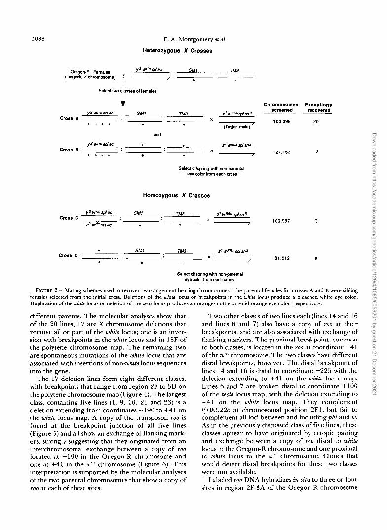
A summary of the results from four experiments designed to detect rearrangements in the region of the white locus is given in Figure 2. Mutations that survive as heterozygotes and that modify the expres- sion of the white or zeste loci will usually be recognized and recovered by this screen. The viabilities of defi- ciencies are such that usually only those with both breakpoints in sections 2 through 5 on the polytene chromosome map (flanking the white locus at 3C2) are likely to survive and produce offspring with excep- tional eye color phenotypes. Duplications for the re- gion, through generally having good viability, are not easily recognized by this screen because they have phenotypes identical or similar to parental pheno- types.
Analysis of exceptional offspring from cross A: A total of 23 individuals having nonparental eye colors were recovered from cross A. One of these proved to be the result of X chromosome nondisjunction (line 11) and two others (lines 5 and 8) failed to produce offspring. The 20 exceptional progeny that were ana- lyzed from cross A arose independently, i.e., from
Dow
nloaded from https://academ
ic.oup.com/genetics/article/129/4/1085/6059201 by guest on 21 D
ecember 2021
1088 E. A. Montgomery et al.
Heterozygous X Crosses
Oregon-R Females (isogenic Xchromosome)
2 wric @e SM1 TM3
+ +
Select two classes of females I + Chromosomes ExceDtiona
y 2 wric @e SM1 TM3 z'w65a@?Jl3 screened recovered
+ + + + Cross A X 100,398 20 + + Fester male) '
and
y2 wric YJ E +
+ + + + + F
+ Crora B
z1w65a@sn3 X 127,153 3
Select offspring with non-parental eye color from each cross
Homozygous X Crosses
+ Cross D
SM1 Th43 zf w65a Splw3 X
+ + F 81,512 6
Select offspring with non-parental eye c o l ~ from each cross
FIGURE 2.-Mating schemes used to recover rearrangement-bearing chromosomes. The parental females for crosses A and B were sibling females selected from the initial cross. Deletions of the white locus or breakpoints in the white locus produce a bleached white eye color. Duplication of the white locus or deletion of the reste locus produces an orange-mottle or solid orange eye color, respectively.
different parents. The molecular analyses show that of the 20 lines, 17 are X chromosome deletions that remove all or part of the white locus; one is an inver- sion with breakpoints in the white locus and in 18F of the polytene chromosome map. The remaining two are spontaneous mutations of the white locus that are associated with insertions of non-white locus sequences into the gene.
The 17 deletion lines form eight different classes, with breakpoints that range from region 2F to 3D on the polytene chromosome map (Figure 4). The largest class, containing five lines ( 1 , 9, 10, 21 and 23) is a deletion extending from coordinates - 190 to +4 1 on the white locus map. A copy of the transposon roo is found at the breakpoint junction of all five lines (Figure 5 ) and all show an exchange of flanking mark- ers, strongly suggesting that they originated from an interchromosomal exchange between a copy of roo located at -190 in the Oregon-R chromosome and one at +41 in the w" chromosome (Figure 6). This interpretation is supported by the molecular analyses of the two parental chromosomes that show a copy of roo at each of these sites.
Two other classes of two lines each (lines 14 and 16 and lines 6 and 7) also have a copy of roo at their breakpoints, and are also associated with exchange of flanking markers. The proximal breakpoint, common to both classes, is located in the roo at coordinate +41 of the wyic chromosome. The two classes have different distal breakpoints, however. The distal breakpoint of lines 14 and 16 is distal to coordinate -225 with the deletion extending to +41 on the white locus map. Lines 6 and 7 are broken distal to coordinate +lo0 of the zeste locus map, with the deletion extending to +41 on the white locus map. They complement Z(l)EC226 at chromosomal position 2F 1, but fail to complement all loci between and including phZ and w . As in the previously discussed class of five lines, these classes appear to have originated by ectopic pairing and exchange between a copy of roo distal to white locus in the Oregon-R chromosome and one proximal to white locus in the wr" chromosome. Clones that would detect distal breakpoints for these two classes were not available.
Labeled roo DNA hybridizes in situ to three or four sites in region 2F-3A of the Oregon-R chromosome
Dow
nloaded from https://academ
ic.oup.com/genetics/article/129/4/1085/6059201 by guest on 21 D
ecember 2021
Ectopic Recombination in Drosophila 1089
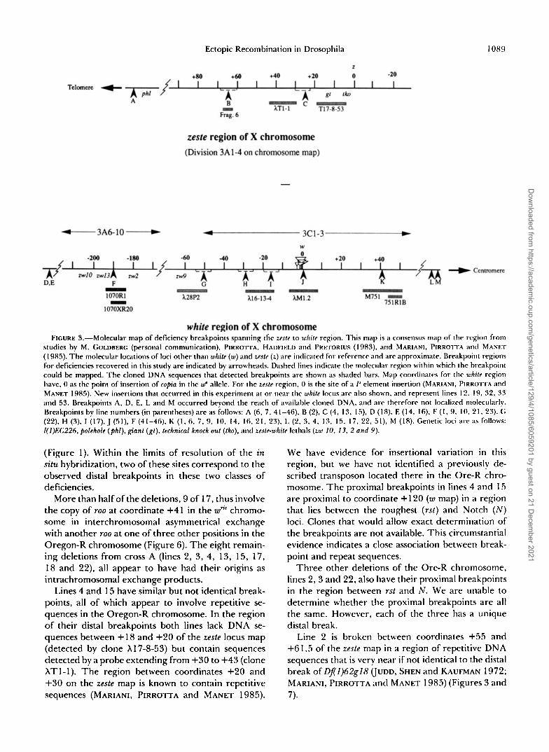
- FIGURE 3.-Molecular map of deficiency breakpoints spanning the zeste to white region. This map is a consensus m;~p of the region from
studies by M. GOLDRERG (personal communication), I'IRROTTA. HADFIELD and I'REIORIUS (1983). and MARIANI. P I R R O I I A and MANET
( I 985). The molecular locations of loci other than white (W) and zesfe (L) are indicated for reference and are approximate. Breakpoint regions for deficiencies recovered in this study are indicated by arrowheads. Dished lines indicate the molecular region within which the breakpoint could be mapped. The cloned DNA sequences that detected breakpoints are shown as shaded bars. Map coordinates for the uhite region have, 0 as the point of insertion of copia in the W" allele. For the reste region. 0 is the site of a P element insertion (MARIANI, PIRROTTA ;~nd MANET 1985). New insertions that occurred in this experiment at or near the while locus are also shown, and represent lines 1 2, 19, 32, 33 ;uld 53. Breakpoints A, D, E, L and M occurred beyond the reach of available cloned DNA, and are therefore not localized molecularly. Breakpoints by line numbers (in parentheses) are as follows: A (6, 7, 4 1-46), B (2). C (4. 13, 15). D ( 1 8). E ( 1 4. 16). F ( I , 9. I O . 2 I . 23). G (22). H (S), I ( l 7 ) , J (51), F (41-46). K (1, 6. 7. 9. 10, 14. 16, 21, 23). 1, (2. 3 , 4, 13. 15, 17, 22. 51). M (18). Genetic loci are a s follows: 1(1)EC226, polehole ( p h l ) , gianf (gt) , technical knock out (tko), and zeste-white lethals (LW IO, 13, 2 and 9 ) .
(Figure 1). Within the limits of resolution of the in situ hybridization, two of these sites correspond to the observed distal breakpoints in these two classes of deficiencies.
More than half of the deletions, 9 of 17, thus involve the copy of roo at coordinate +41 in the wnc chromo- some in interchromosomal asymmetrical exchange with another roo at one of three other positions in the Oregon-R chromosome (Figure 6). The eight remain- ing deletions from cross A (lines 2, 3, 4, 13, 15, 17, 18 and 22), all appear to have had their origins as intrachromosomal exchange products.
Lines 4 and 15 have similar but not identical break- points, all of which appear to involve repetitive se- quences in the Oregon-R chromosome. In the region of their distal breakpoints both lines lack DNA se- quences between + l 8 and +20 of the zeste locus map (detected by clone X17-8-53) but contain sequences detected by a probe extending from +30 to +43 (clone XTl-1). The region between coordinates +20 and +30 on the zeste map is known to contain repetitive sequences (MARIANI, PIRROTTA and MANET 1985).
We have evidence for insertional variation in this region, but we have not identified a previously de- scribed transposon located there in the Ore-R chro- mosome. The proximal breakpoints in lines 4 and 15 are proximal to coordinate + 120 (W map) in a region that lies between the roughest (rst) and Notch ( N ) loci. Clones that would allow exact determination of the breakpoints are not available. This circumstantial evidence indicates a close association between break- point and repeat sequences.
Three other deletions of the Ore-R chromosome, lines 2, 3 and 22, also have their proximal breakpoints in the region between rst and N . We are unable to determine whether the proximal breakpoints are all the same. However, each of the three has a unique distal break.
Line 2 is broken between coordinates +55 and +61.5 of the zeste map in a region of repetitive DNA sequences that is very near if not identical to the distal break of Df(1)62gl8 (JUDD, SHEN and KAUFMAN 1972; MARIANI, PIRROTTA and MANET 1985) (Figures 3 and 7).
Dow
nloaded from https://academ
ic.oup.com/genetics/article/129/4/1085/6059201 by guest on 21 D
ecember 2021
1090 E. A. Montgomery et al.
213 0 Lines 1. 9. 10, 21, 23
0 Lines I4 I 16 - Lines 6 & 7
I Line 17
m Line 3
a Llne 22
I 3 Line 51 - Line 2 - LInn 4, 13, IS
0- Line I8
0 Line1 41-46
FIGURE 4.-The cytological limits of the deficiencies recovered in these experiments are shown by open bars. Stippled areas indicate that the breakpoint has not been located molecularly.
Line 3 has a distal break at -30 ( w map) and a proximal break in the rst to N region, creating a small deletion that is hemizygous viable with a white, roughest phenotype. The molecular analysis shows that the Ore-R chromosome has a 10 kb insertion between EcoRI sites at -25 and -30, which is not present in the w* chromosome. Line 3 appears to have lost this insertion, though there is no evidence to suggest that it was directly involved in the formation of the re- arrangement. PIRROTTA, HADFIELD and PRETORIUS (1 983) also noted repetitive sequences in this region of the white locus map of Ore-R.
Line 22, like line 3, has a viable white, roughest phenotype, although its distal break is between coor- dinates -50 and -65 ( w map), making it a deletion of at least 20 kb larger than in line 3. Restriction maps of this region of several wild-type strains show consid- erable insertional polymorphism and a cluster of EcoRI restriction sites that may indicate tandem re- peats (PIRROTTA, HADFIELD and PRETORIUS 1983). The line 22 deletion does not extend distally to re- move 1(Z)zw9, which means that this locus, most prox- imate to w, must lie distal to coordinate -50.
T w o lines, 13 and 17, are deletions that occurred in the y' writ spl ec chromosome. Line 13 is a large deletion extending from between +20 and +30 on the z map to between rst and N proximally. Note that the distal breakpoint is in the same region as in lines 4 and 15, and the proximal breakpoint is indistin- guishable from those seen in lines 2, 3, 4, 15, 18 and 22. Line 17 has its distal break at about coordinate -15 of the white locus map, in the same region as but
different from the breakpoint in line 3 discussed above.
The one remaining deletion from cross A, line 18, has breakpoints distal to -225 of the w map and proximal to the N locus. I t is an intrachromosomal deletion of the y' wric spl ec chromosome, having a yellow-2, white, Notch, echinus, recessive lethal pheno- type. Cloned DNA that would allow precise location of breakpoints was not available. Complementation mapping locates the distal break between l(Z)zwZ and l(Z)zw8, cytological position 3A4,5 on the polytene chromosome map. Line 18 is not rescued by w+Y (2D1,2; 3D3,4), but it is rescued by Dp(Z;3)w49a (3A9,B2; 3E2,3) and it complements diminutive (dm, approximate location 3333-6). These cytogenetic re- sults suggest that the proximal breakpoint is between 3D3,4 and 3E2,3, which agrees with our cytological observations that place the break at 3D4,5. .
Line 20 is an inversion that occurred in the y' wric spl ec chromosome. It has a white eye-color phenotype in addition to those of the flanking markers. Cytolog- ical examination of the polytene chromosomes shows breakpoints at 3C and 18F. In the writ chromosome, copia, with a copy of roo inserted 2.95 kb from its 3' end, is inserted at coordinate 0.0 of white. In situ analysis shows that there is also a copy of roo at 18F in that chromosome. Molecular analysis of the white locus of Line 20 shows a restriction pattern like that of w* for endonucleases that cut in white locus and in either of the transposons. This is consistent with the inversion being formed by pairing and exchange be- tween two copies of roo in reverse orientation at 3C and 18F. The BamHI restriction pattern for Line 20 white locus definitively rules out the involvement of copia as the site of exchange in the formation of the inversion. BamHI cuts in roo but not in copia, thus such a digest of line 20 DNA would differ from the writ pattern if copia were the site of the breakpoint (Figure 8).
The two remaining white-eyed offspring from cross A, lines 12 and 19, show restriction patterns consistent with the insertion of non-white locus, presumably tran- sposon, sequences into the white locus of the wric chro- mosome. Line 12 has approximately 2 kb inserted between coordinate -5 and the EcoRI site in the 3' end of copia. Line 19 has about 5 kb inserted between the 5' Sal1 site in roo and coordinate +2 of white (data not shown).
Analysis of exceptional offspring from cross B Three exceptional individuals of independent origin were recovered from cross B. Line 51 is a deletion of the 5' region of the white locus in the writ chromosome from +O.O to proximal of +120. In addition to the flanking mutant markers, this line shows a white, roughest phenotype. The restriction map of this line is best interpreted as having the 3' portion of copia and
Dow
nloaded from https://academ
ic.oup.com/genetics/article/129/4/1085/6059201 by guest on 21 D
ecember 2021
Ectopic Recombination in Drosophila 1091
3 s $ . 1 0-0 B. w a A - w a "
1070AI A.
= a.
kll -200 -1 90 -180
I I 1 w - 1
1 Y W 2 r i c w m - -8.5
Oregon-R 1 -5.8
-3.5
1 2 1 2
Probe Probe roo 1070RI
A S SE
-6.5 -3.5
A s SE
FIGURE 5.-Analysis of DNA from the region -190 kb distal to the white locus in parental strains Oregon-R and $ w* spl ec, and in two classes of deficiencies. (A) SalI and EcoRl restriction map of the region for the two parental strains. A roo insertion (hatched bar) at -1 90 kb in the Oregon-R DNA alters the map relative to 9 w* spl ec. Subcloned DNA sequences that were used as probes are shown as solid black bars above the diagram. (B) Autoradiograph of cloned DNA from Oregon-R and 9 w* spl ec digested with EcoRI. Genomic libraries of parental strains Oregon-R and 9 w* spl ec were screened with 1070R1, and positive clones Ore-R3.1 and wk6. 1, respectively, were identified and isolated. A Southern blot of EcoRI digests of these clones was first hybridized to S2P-labeled pACYB104 (roo), then washed and reprobed with "P-labeled 1070RI. Digests of both clones hybridize to 1070RI unique sequence DNA, but only Ore-R3.1 shows a signal when probed with roo. (C) and (D) Determination of the breakpoints in two classes of deficiencies. (C) Sal1 digests of genomic DNA from Oregon-R, ywm spl ec, and recombinant deficiency lines 1, 9, and 10 probed with szP-labeled plasmid subclone 1070XR20 (unique sequence DNA from -187.5 to -191.5, white map). (Data not shown for lines 21 and 23, which have the same restriction pattern.) The 6.5-kb Sal1 fragment which spans the junction between distal unique sequence DNA and roo DNA is present in DNA from deficiency lines 1, 9, 10 and Oregon- R. The IO-kb SalI fragment in the Oregon-R digest that spans the junction between roo and proximal unique sequence DNA has been deleted by the rearrangements. The 13-kb SalI fragment is from the balancer chromosome FM7, over which the lethal deficiencies are carried. (D) EcoRIdigested DNA from two of six identical deficiencies that occurred in the Oregon-R chromosome (lines 41-46 from cross D), along with DNAs from the parental chromosome Oregon-R and the balancer chromosome FM7, were probed with "P-labeled plasmid subclone 1070RI containing only unique sequence DNA from the same region. The presence of the 3.5-kb EcoRI fragment, but not the 14.5-kb fragment, which has been deleted, indicates that the roo insert in Oregon-R at -190 was also responsible for this class of deficiencies.
at least 1 kb of roo at the breakpoint but with all sequences of white locus proximal to 0.0 deleted (data not shown). The most logical explanation is that the deletion resulted from ectopic pairing and exchange between roo elements at coordinates 0.0 and proximal to +120. It should be noted that the proximal break- point is in the same region as seven of the eight intrachromosomal deletions found in cross A.
Line 52 is derived from the y2 wfic spl ec chromo- some. It has a white phenotype in addition to the
flanking markers, and is viable as a hemizygote. The restriction patterns from five different endonucleases showed no changes from the wfic pattern. This strongly suggests that line 52 carries a mutation created by a change in one or a few nucleotides in the white locus (data not shown).
Line 53, which has a white eyed, hemizygous viable phenotype, appears to be a mutation in the Ore-R chromosome. Molecular analysis shows that there is an insertion of about 6 kb of DNA into the white locus
Dow
nloaded from https://academ
ic.oup.com/genetics/article/129/4/1085/6059201 by guest on 21 D
ecember 2021
1092 E. A. Montgomery et al.
Oregon-R - S SE
" S SE
.?
Probe: 751 RIB Probe: 100 b B. " ".-
- -8.0
-4.9
-4.0
-2.5
-1 .o
-0.7 -0.5
B / s B / E B B / S R E B
1 2 3 4 5 6 7 sal I Digests
FIGURE 6.-Analysis or a regon proximal to the white locus containing breakpoints of several deficiency lines. (A) Sal1 restriction maps for parental chronrosomes Oregon-R and f wn' spl cc, and for a deficiency chromosome representing the proximal breakpoint for lines 1, 6, 7, 9, IO, 14. 16, 21 and 23. A roo insertion (hatched bar) at +41 creates two Sal1 fragments of 2.9 kb and 4.9 kb in the f wN spl cc restriction pattern relative to the Oregon R Sal1 fragment of 5.5 kb. (B) Southern blot of Xw"' 5.1, which contains both roo and unique sequence DNA. Xw"' 5.1 is a clone isolated from a genomic f w"' spl cc library by screening with 751 RIB, an EcoRl subclone of M751 (see diagram in A). A filter with BamHl (B), BnmHI/EcoRI (B/E), and BamHI/SalI (B/S) digests of hw"'5.1 was probed first with 751RIB. washed, and reprobed with pACYB104 (roo-containing plasmid). Xwric 5.1 contains 6.8 kb of roo DNA (hatched bar), as well as unique sequence DNA from the 751 region, as shown in the restriction map of the cloned region. (C) Southern blot of Sal1 digests of genomic DNA isolated from deficiency lines 4, 6, 7, 9 and I O , and from parental strains Oregon-R and f w~ spl cc. The filter was probed with XM751, indicated by a solid bar in the diagram in (A). Four of the deficiency lines (lanes 3.4, 5 and 6) have the distal 4.9-kb fragment that has been created by the insertion of roo, but are missing the 2.9-kb fragment. Deficiencies with more proximal breakpoints (example line 4, lane 7) show only the FM7 restriction pattern. Restriction fragments unaffected by the roo insertion, but detected by the probe, are identical for the f w* spl cc and FM7 chromosomes.
between coordinates -0.67 and + 1.6 (data not for y' wk spl ec X chromosomes and heterozygous for shown). Quite likely the mutation was caused by the the autosomal inversions SMZ and TM3. All three insertion of a transposon into that region of the gene. lines are hemizygous viable. Line 31 shows restriction
Analysis of exceptional offspring from cross C patterns like those of w ~ ' , suggesting that it is a spon- Three white eyed individuals of independent origin taneous mutation involving one or a few nucleotides appeared from cross C, involving females homozygous in the white locus. Lines 32 and 33 are both associated
Dow
nloaded from https://academ
ic.oup.com/genetics/article/129/4/1085/6059201 by guest on 21 D
ecember 2021
Ectopic Recombination in Drosophila 1093
FIGURE 7.-X chromosomes from female heterozygous for line 2 deficiency and a normal X . The deletion of the zeste-white region from 3A1 to 3C2-3 in the deficiency chromosome results in an ;)synapsed region (at arrow) as a result of its inability to pair conlpletely with the normal X chromosome.
with the insertion of non-white locus sequences into the locus. Line 32 has about 4 kb of DNA inserted between coordinate -0.67 and the most 3' Sal1 site in roo. Similarly, line 33 has an insert of about 6.5 kb between the BamHI site in roo and the BamHI site at +1.38 (data for lines 32 and 33 not shown). It is entirely possible that in both lines 32 and 33 the insertion sites were in the copia-roo complex at 0.0 in the white locus. It is of significance that none of the exceptions recovered from cross C resulted from ec- topic pairing and exchange between transposons.
Analysis of exceptional offspring from cross D: Six individuals with zeste eye color were recovered from cross D. All six (lines 41,42, 43, 44,45 and 46) are deletions that extend from 2F4-5 to 3A8-9. They fail to complement phl, gt, tho, z, l( l)zwl, l( l)zw8, l(l)zw4 and l(l)zwlO, but complement l(l)zw2. Molec- ular analysis shows that the distal breakpoint is distal to +lo0 on the z map. The proximal breakpoint is at - 190 on the w map, identical to the distal breakpoints found in lines 1 , 9, 10, 21, and 23 from cross A (Figure 5). These deletions have the transposon roo at their breakpoints. Unlike the cross A deletions, which were generated via ectopic interchromosomal ex- changes between the roo element at -190 in the Oregon-R chromosome and a roo in the wm chromo- some at coordinate +41, the deletions from cross D extend distally from the roo at coordinate -190 to another copy in Ore-R distal to reste. Because the females used in cross D were homozygous for the X, we are unable to determine whether the exchange was inter- or intrachromosomal.
Two of the cross D lines arose from subcultures of one mating group and two from another; the remain- ing two occurred in different mating groups. Each
"R
a &/ 8A-
- mow -"I
5
b. ;=?I>
c. +/ 46 lBF 4 0 4s I 1 I - \ I I
8 0 8 8 B 8
FIGURE 8.-A model for the creation of line 20 inversion. (a) The wnr chromosome showing roo inserted into copia at coordinate 0.0 in white locus. (b) Pairing and exchange between this copy of roo and another in the opposite orientation at 18F. (c) Inversion chronlosome with diagnostic restriction sites illustrating why a white locus restriction map of the inversion is the same as the map for w"' when roo is involved in the ectopic recombination.
mating group consisted of four to six parental females. It is possible that the deficiencies in all six cross D lines have a common origin as a preexisting change in the Ore-R strain. Thus, it is unclear whether these deletions should be scored as a single event or as having arisen during the experiment as six separate events.
Interchromosomal effect: Map distances were de- termined for the y-w and the w-spl intervals for crosses A and B to measure the effect of the autosomal rearrangements on crossover frequencies in the X chromosomes. For the y-w interval the values were 5.4 (95% confidence interval: 4.5-6.3) and 0.7 (95% con- fidence interval: 0.5-1 .O) map units for crosses A and B, respectively. The standard map distance for this interval is 1.5 m.u. The w-spl interval values from cross A and B were 2.4 (95% confidence interval: 1.8- 3.1) and 1.1 (95% confidence interval: 0.8-1.5) m.u., respectively, with the standard being 1.5 m.u. For the whole interval, y to spl, the observed map distances were 7.8 (95% confidence interval: 6.7-8.9) us. 1.8 (95% confidence iriterval: 1.4-2.3) in crosses A and B, respectively. Thus there was at least a 2.9-fold increase (6.7/2.3) associated with inversion heterozy- gosity in the autosomes.
In cross A, twenty individuals among a total of 100,398 offspring had nonparental eye colors; eight- een of these appeared to be transposon-associated rearrangements. (18/100, 398 = 1.8 X 95% confidence interval: 1 . 1 x to 2.2 X Cross B produced only three nonparental eye color flies among 127,153 offspring. One of them, line 5 1 , ap- pears to have been generated as an ectopic exchange involving the copia-roo transposon complex within white (1/127, 153 = 1 X 95% confidence inter-
Dow
nloaded from https://academ
ic.oup.com/genetics/article/129/4/1085/6059201 by guest on 21 D
ecember 2021
1094 E. A. Montgomery et al.
Val: 2 X lo” to 4 X 1 O-5). Thus there is at least a 2.75-fold increase in the frequency of transposon- associated rearrangements when comparing crosses A and B, which differ only in the heterozygosity for autosomal inversions. This is comparable to the mag- nitude of the increase in normal crossing over. There was no increase observed in the other classes of excep- tions (insertions or point mutations) in crosses A and B.
Effect of homozygosity of the X chromosome: In crosses C and D, the interchromosomal effect on classical crossing over cannot be measured because X chromosomes were homozygous. However, the num- bers of exceptional eye-color offspring generated by crosses C and D were clearly lower than from cross A, yet parental females used in all three cases were heterozygous for autosomal inversions. None of the exceptions from cross C involve exchanges between repeated sequences. The exception(s) from cross D do involve repeat sequences, but they all may have been derived from a single preexisting mutation. The re- sults of these two crosses indicate that homozygosity of the two X chromosomes significantly reduces the yield of exceptions (especially those associated with repeat sequences).
DISCUSSION
We have surveyed about four of the numbered sections of the polytene chromosome map or about 0.5% of the euchromatic genome sequences for ec- topic exchange events. In relating the observed fre- quencies of ectopic recombination to the entire ge- nome of natural or laboratory populations of D. mel- anogaster, it should be noted that the screen employed is sensitive primarily to deletion events. Most dupli- cations and a large majority of inversions will not be detected. We have no evidence concerning ectopic exchanges between heterologs that would produce translocations. If such occur our screen would not detect them unless one break was within the white locus. Clearly the frequencies of ectopic exchanges we observed are likely to represent less than half the real values.
It might appear that the choice of wTtC, which is associated with the insertion of copia and roo into the white locus, as one of the chromosomes in this study may have biased the screen toward a higher number of exchanges in the target area. However, the copia- roo complex was involved in only two cases, the inver- sion in line 21 from cross A and the deletion in line 5 1 of cross B. The copia-roo complex did appear to be the target of new transposon insertions in lines 12, 19, 52 and 53.
Of more significance is the exceptionally frequent involvement of roo in the ectopic exchanges observed. The numbers of roo elements on the two X chromo-
somes used in the study (26 in Oregon-R and 21 in writ) are approximately twice the average number ob- served in natural populations (MONTGOMERY and LANGLEY 1983; MONTGOMERY, CHARLESWORTH and LANGLEY 1987; CHARLESWORTH and LAPID 1989). It is possible that the level of ectopic exchange in natural populations is somewhat lower and that the increase in copy number in these laboratory stocks reflects relaxation of various selective processes including those associated with ectopic exchange.
Interchromosomal effect: Except for the six copies of the deficiencies from cross D (lines 41-46), 18 of the 19 transposon-associated rearrangements re- covered in the four experiments reported here were from cross A females that were heterozygous for the X chromosomes and for the autosomal re- arrangements S M l and TM3. One was generated from cross B, utilizing female sibs that had standard auto- somal configurations, which indicates that the inter- chromosomal effect enhances the ectopic pairing and exchange involving transposons. In view of the overall increase in crossing over in the y-spl region seen in cross A compared to that in cross B, a case can be made for transposon involvement being stimulated to about the same degree as crossing over in general. This strongly supports the concept that the process generating the rearrangements recovered here is meiotic and responsive to the interchromosomal ef- fect. Only in cross D, which yielded the remaining six (or one) transposon-associated rearrangements, is there any suggestion of recovery of a cluster of excep- tional offspring that would indicate a premeiotic ori- gin. Because the six exceptional individuals from cross D, though apparently identical deletions, were re- covered from four separate lines, we consider it un- likely that the deletions were premeiotic in origin. However, it is possible that all descended from a single ectopic exchange that occurred in the Ore-R strain prior to the selection of the parental females used in cross D. If this is the case, the fertility and/or viability of females heterozygous for this deletion would have to be quite low to account for the small numbers recovered (2, 2, 1 and 1) from the four separate mating groups with four to six females each.
The involvement of roo: Sixteen of the 25 transpo- son-associated rearrangements recovered from the four experiments involved copies of roo. The data in Table 1 show that the Oregon-R X chromosome con- tained 26 copies of roo and the y2 uric spl ec X chro- mosome had 21. The region flanking the white locus over which deletions extending into that gene would be viable as heterozygotes is about four numbered sections, 2 to 5 , on the polytene chromosome map. The Ore-R chromosome had eight copies of roo in that region and the y2 writ spl ec chromosome had five, including the copy inserted into copia at coordinate
Dow
nloaded from https://academ
ic.oup.com/genetics/article/129/4/1085/6059201 by guest on 21 D
ecember 2021
Ectopic Recombination in Drosophila 1095
0.0 within the white locus. Clearly this rather large number of copies in the target region offered consid- erable opportunity for roo to be involved in re- arrangements that could be recognized by our genetic screen. It is important to note that the copy of roo at +4 1 was involved in rearrangements much more often than other roo elements in the same region. A similar finding was reported (LIM 1988) in studies of intrach- romosomal exchanges involving the hobo element. One copy of hobo was a participant in every hobo- mediated rearrangement scored.
Interchromosomal exchanges: Another notable as- pect is that every one of the interchromosomal ex- changes recovered from cross A involved copies of roo, one of which was at +41 in the wric chromosome. These exchanges total nine in number; five of one class and two each in two other classes. Another roo site at -190 of the white locus map in the Ore-R chromosome was the corresponding copy involved in the formation of the class of five from cross A above and in all six of the exceptional offspring recovered in cross D. Because females homozygous for the Ore- R X chromosome were used in cross D, it is not possible to determine whether these 6 exceptions were from inter- or intrachromosomal exchanges.
Intrachromosomal exchanges: The observation that approximately half of the ectopic recombinants were intrachromosomal is of particular interest. This is likely to be an underestimate of this fraction because inversions can be generated by ectopic exchange only as intramolecular events, and only those inversions that disrupt the white locus would be recognized by the genetic screen employed.
The intrachromosomal exchanges utilized a diverse array of repetitive sequence sites. Each line appears to have a unique combination of breakpoints, al- though lines 2, 3, 4, 13, 15, 17 and 22 could possibly share the same breakpoint in the region proximal to coordinate +120 on the white locus map. This region lies beyond any available cloned sequences, therefore, we have not been able to map breakpoints with pre- cision.
We have been unsuccessful in attempts to clone the breakpoints of most of the intrachromosomal dele- tions. This strongly suggests that those regions contain repetitive sequences that are unstable in cloned con- structs. Available restriction maps of the regions of breakpoints show insertional variation, and the avail- able clones covering some of these regions contain repetitive DNA (M. GOLDBERG, personal communi- cation; PIRROTTA, HADFIELD and PRETORIUS 1983; MARIANI, PIRROTTA and MANET 1985). This supports our evidence that all of these breakpoints are in or proximate to repetitive sequences. We, therefore, fa- vor the view that ectopic pairing and exchange be- tween interspersed repeat sequences is the mechanism
of their origins. We interpret these results as evidence that both inter- and intrachromosomal ectopic ex- change respond to the interchromosomal effect, in- dicating that they are primarily meiotic in origin.
The diversity of types of intrachromosomal dele- tions and the complete lack of evidence of clusters lend additional weight to the view that the intrachro- mosomal rearrangements are meiotic in origin. It is interesting to compare these results with those re- ported by ENGELS and PRESTON (1984) and LIM (1 988), who found predominantly (if not exclusively) premeiotic ectopic intrachromosomal exchange asso- ciated with P and hobo elements, respectively. The premeiotic nature and transposon-specificity of the events in their experiments may reflect the dysgenic properties of the stocks used and the elements in- volved.
Heterozygosity per se: Possibly the most important point to be drawn from the four experiments comes from comparisons of the frequencies and types of rearrangements from heterozygous and homozygous X chromosomes. Crosses A, C and D are equivalent in autosomal heterozygosity for S M l and TM3. The expectation was that crossing over in the target region would be stimulated equally by the interchromosomal effect in all three crosses, and that transposon-associ- ated exchanges would be influenced accordingly. We consider it significant that neither the array nor fre- quencies of rearrangements expected from crosses C and D, based on what was generated from cross A, were realized. Although the interchromosomal ex- change classes generated in cross A would not be duplicated in either cross C or D, it was expected that rearrangements resulting from pairing and exchange between other combinations of transposons would appear. Frequencies might be expected to be some- what different, depending on the numbers and ori- entations of transposon copies. However, the numbers of exceptional offspring from both cross C and D were significantly below those recovered in cross A.
A significant point is that the intrachromosomal exchange classes seen in cross A were completely absent from crosses C and D. They would be expected to occur as they did in cross A unless homozygosity, per se, reduces the ectopic pairing frequency. These results focus attention on how interspersed repeat sequences become involved in ectopic pairing. It is clear that repeat sequences can search over consider- able distances for a pairing partner. How this search is initiated is not clear. However, homozygosity for interspersed repeat sequences, and/or other types of sequence variation, reduces the frequency of ectopic recombination. It is probable that homozygous ele- ments will pair with their allelic counterpart and thus be unavailable for involvement in ectopic pairing. Exchange between homozygous elements would go
Dow
nloaded from https://academ
ic.oup.com/genetics/article/129/4/1085/6059201 by guest on 21 D
ecember 2021
1096 E:. A. Montgonlery et al.
undetected. The result would be that fewer excep- tions would be generated i n crosses C and I).
Presumably ectopic pairing involves the establish- ment of a heteroduplex region, an early step prereq- uisite to recombination. However, our results show that ectopic exchange is not limited to interchromo- so~nal pairing partners. The formation of intrachro- ~l~osomal heteroduplexes raises interesting questions about the regulation of the recombination process, 1)articularly with regard to sister chromatid ex- changes, which go undetected genetically. Our obser- vations are consistent with the idea that heterozygous sequence variation has the potential to modify the way that heteroduplex regions form during the recombi- Iution process.
Comparison with yeast: The ectopic exchange process has been studied extensively in microorga- 11isms (PETES and HILL 1988). In Saccharomyces cere- visiae, exchange with engineered ectopic copies occurs at rates comparable to those with the homologous allele. Those results indicate that heterozygosity has little effect on ectopic exchange. Studies with artificial constructs show that heterozygous insertions are no no re likely to be involved in ectopic exchange than when they are homozygous. We see no obvious reason for this discrepancy with our results.
Recent results indicate that the number or positions of available ectopic copies of a gene have little influ- ence on the proportion of ectopic conversions or on the overall frequency of exchange (HABER et a l . 1991). These authors suggest that the limiting step in recom- bination is the activation of a locus to be involved in exchange. The recombinationally activated allele can then pair with any one of the several copies through- o u t the genome. They also note that there is some Ilinderance to the extension of this simple model of meiotic pairing and exchange to higher eukaryotes having abundant middle repetitive dispersed DNA sequences.
In Drosophila, the region over which a sequence may search for an exchange partner appears to be quite wide within a chromosome as evidenced by recovery of a long inversion. It is possible, however, that the search may be restricted by mechanisms such ;IS pairing site positions (HAWLEY 1980). Our results show considerable variation in the propensity of ele- ments at different sites (even in the same family) to participate in ectopic exchange. This variation could reflect differences in chromosomal positions relative to pairing sites or inherent differences in levels of involvement in recombination among transposon in- sertions.
Potential impact of translocations and other un- detected types of rearrangements: Our data indicate that inter- and intrachromosomal ectopic pairing and exchange can occur between widely separated sites in
chro~nsomes. I t should be noted, however, that these experiments were not designed to detect a large frac- tion of exchanges between repeat sequences in hom- ologs. Also, recombination between nonhomologous chromosomes, an event that would have produced translocations, is not detected. Ectopic exchange has been shown to give rise to translocations in various fungi (PETES and HILL 1988). Our data do not allow assessment of the role transposons might have in the formation of translocations in Drosophila. If, how- ever, transposons are important in the generation of translocations (as well as deletions, duplications and inversions), any estimate of the the impact of transpo- sons on fitness based on the results reported here, would have to be revised upward.
Population genetics: Ectopic exchange and its sub- sequent chromosomal rearrangements have been pro- posed as a general mechanism that contains copy numbers of transposable elements (LANGLEY et a l . 1988). Unlike the situation with other parasites, the mechanism(s) that limits the growth of genomic par- asites are obscure (CHARLESWORTH and LANGLEY 1986; CHARLESWORTH and LANGLEY 1989).
The results of our study here have three important implications for evaluating the possible role of ectopic exchange as a population genetic mechanism in the containment of copy number. (1) Different sites may have different propensities for involvement in ectopic exchange. Such a possibility has not been incorporated into the modelling. (2) The significance of the inter- chromosomal effect on ectopic exchange, normal ex- change and disjunction is not clear. The fact that inversion heterozygosity is common in some species, while absent in others, should be considered in the interpretation of the differences in the distribution of transposable elements in various species. (3) Our ob- servations of little or no ectopic exchange in the homozygous X chromosomes suggest that once an insertion reaches high frequency, the selection asso- ciated with ectopic exchange would favor the inser- tion-bearing chromosome (because of the high pro- portion as homozygotes). Thus the dynamics of such insertions would be quite different in small popula- tions (in which they might drift into high frequency) compared with large populations (in which they would remain rare and thus heterozygous).
The allelic frequencies of middle repetitive inser- tions in natural populations are not known for many species. It is tempting, however, to view the contrast between D. melanogaster (insertions usually very rare) and humans (insertions usually fixed) in light of the differences in their population sizes, genetic drift, and the effect of homozygosity on ectopic exchange.
One curious result from the surveys in natural populations of the densities of transposable elements in the D. melanogaster euchromatic genome, was their
Dow
nloaded from https://academ
ic.oup.com/genetics/article/129/4/1085/6059201 by guest on 21 D
ecember 2021
Ectopic Recombination in Drosophila 1097
accumulation in the centromere-proximal regions where regular exchange is reduced (CHARLESWORTH and LANGLEY 1989; CHARLESWORTH and LAPID 1989). However, no increase in density of elements was observed near the telomeres where regular cross- ing over is also reduced. It will be important to deter- mine if ectopic exchange is not reduced in the telom- eric region. The available evidence indicates that the interchromosomal effect is greatest near the telomere (LUCCHESI 1976) which would suggest, in light of our results, that ectopic exchange may be quite high throughout the telomeric region when there is inver- sion heterozygosity in the other chromosomes, a com- mon state in nature.
Our results demonstrate that ectopic exchange must be considered as a major source of spontaneous mu- tation in Drosophila. The underlying mechanisms may be investigated utilizing meiotic mutants. The role of heterozygosity and the interchromosomal effect de- serve further attention. Finally these results raise new questions about the impact of ectopic exchange and naturally occurring polymorphisms (base-pair, inser- tional, and karyotypic) on the stability and evolution of genomes.
We thank MIKE GOLDBERG, VINCE PIRROTTA, MIKE YOUNG and LINDA AMBROSIO for providing cloned DNA used in the analyses of rearrangements; WALTER EANES and CEDRIC WESLEY for iden- tifying a repetitive sequence by in situ hybridization; MIKE GOLD- BERG, MIKE RESNICK and BOB VOELKER for helpful comments and discussion; and JAN CAPPS, CINDY BAKER and DELORES HOVEY for excellent technical assistance.
LITERATURE CITED
BINGHAM, P. M., R. LEVIS and G. M. RUBIN, 1981 Cloning of DNA sequences from the white locus of D. melanogaster by a novel and general method. Cell 25: 693-704.
CHARLESWORTH, B., and C. H. LANGLEY, 1986 The evolution of self-regulated transposition of transposable elements. Genetics 112: 359-383.
CHARLESWORTH, B., and C. H. LANGLEY, 1989 The population genetics of Drosophila transposable elements. Annu. Rev. Ge- net. 23: 251-287.
CHARLESWORTH, B., and A. LAPID, 1989 A study of ten families of transposable elements on X chromosomes from a population of Drosophila melanogaster. Genet. Res. 54: 1 13-1 25.
DAVIS, P. S . , M. W. SHEN and B. H. JUDD, 1987 Asymmetrical pairings of transposons in and proximal to the white locus of Drosophila account for four classes of regularly occurring exchange products. Proc. Natl. Acad. Sci. USA 8 4 174-178.
ENGELS, W. R., and C. R. PRESTON, 1984 Formation of chromo- some rearrangements by P factors. Genetics 107: 657-678.
FINNEGAN, D. J,, G. M. RUBIN, M. W. YOUNG and D. S. HOGNESS, 1978 Repeated gene families in Drosophila melanogaster. Cold Spring Harbor Symp. Quant. Biol. 42: 1053-1063.
GOLDBERG, M. L., J.-Y. SHEEN, W. J. GEHRING and M. M. GREEN, 1983 Unequal crossing-over associated with asymmetrical synapsis between nomadic elements in the Drosophila melano- gaster genome. Proc. Natl. Acad. Sci. USA 8 0 50 17-502 1 .
HABER, J. E., W.-Y. LEUNG, R. H. BORTS and M. LICHTEN, 1991 The frequency of meiotic recombination in yeast is independent of the number and position of homologous donor
sequences: implication for chromosome pairing. Proc. Natl. Acad. Sci. USA 88: 1120-1 124.
HAWLEY, R. S., 1980 Chromosomal sites necessary for normal levels of meiotic recombination in Drosophila melanogaster. I. Evidence for and mapping of the sites. Genetics 94: 625-646.
JOHNSON-SCHLITZ, D., and J. K. LIM, 1987 Cytogenetics of Notch mutations arising in the unstable X chromosome Uc of Drosoph- ila melanogaster. Genetics 115: 701-709.
JUDD, B. H., 1959 Studies on some position pseudoalleles at the white locus region in Drosophila melanogaster. Genetics 4 4 34- 42.
JUDD, B. H., 1987 The white locus of Drosophila melanogaster, pp. 8 1-94, in Structure and Function of Eukaryotic Chromosomes, Results and Problems in Cell Daflerentiation 14, edited by W. HENNIG. Springer-Verlag, Berlin.
JUDD, B. H., M. W. SHEN and T. C. KAUFMAN, 1972 The anatomy and function of a segment of the X chromosome of Drosophila melanogaster. Genetics 71: 139-1 56.
LANGER, P. R., A. A. WALDROPand D. C. WARD, 1981 Enzymatic synthesis of biotin-labeled polynucleotides. Proc. Natl. Acad. Sci. USA 78: 6633-6637.
LANGLEY, C. H., E. A. MONTGOMERY, R. H. HUDSON, N. L. KAPLAN and B. CHARLESWORTH, 1988 On the role of unequal ex- change in the containment of transposable element copy num- ber. Genet. Res. 52: 223-235.
LEHRMAN, M. A,, J. L. GOLDSTEIN, D. W. RUSSELL and M. S. BROWN, 1987 Duplication of seven exons in LDL receptor gene caused by alu-alu recombination in a subject with familial hypercholesterolemia. Cell 48: 827-835.
LEVIS, R., P. M. BINGHAM and G. M. RUBIN, 1982 Physical map of the white locus of Drosophila melanogaster. Proc. Natl. Acad. Sci. USA 79: 564-568.
LIM, J. K., 1988 Intrachromosomal rearrangements mediated by hobo transposons in Drosophila melanogaster. Proc. Natl. Acad. Sci. USA 85: 9153-9157.
LUCCHESI, J. C., 1976 Inter-chromosomal effects, pp. 315-330, in The Genetics and Biology of Drosofihila, edited by M. ASHBUR- NER and E. NOVITSKI. Academic Press, New York.
MANIATIS, T., E. F. FRITSCH and J. SAMBROOK, 1982 Molecular Cloning: A Laboratory Manual. Cold Spring Harbor Laboratory, Cold Spring Harbor, N.Y.
MARIANI, C., V. PIROTTA and E. MANET, 1985 Isolation and characterization of the zeste locus of Drosophila. EMBO J. 4:
MEYEROWITZ, E. M., and D. S. HOGNESS, 1982 Molecular orga- nization of a Drosophila puff site that responds to ecdysone. Cell 28: 165-176.
MONTGOMERY, E. A., B. CHARLESWORTH and C. H. LANGLEY, 1987 A test for the role of natural selection in the stabilization of transposable element copy number in a population of Dro- sophila melanogaster. Genet. Res. 49: 3 1-4 1.
MONTGOMERY, E. A., and C. H. LANGLEY, 1983 Transposable elements in Mendelian populations. 11. Distribution of three copia-like elements in a natural population. Genetics 104: 473- 483.
PARDUE, M. L., and J. G. GALL, 1975 Nucleic acid hybridization to the DNA of cytological preparations. Methods Cell Biol. 10: 1-17.
PETES, T. D., and C. W. HILL, 1988 Recombination between repeated genes in microorganisms. Annu. Rev. Genet. 22: 147- 168.
PIRROTTA, V., C. HADFIELD and G. H. J. PRETORIUS, 1983 Microdissection and cloning of the white locus and the 3B1-3C2 region of the Drosophila X chromosome. EMBO J.
RIGBY, P. W. J., J. M. DIECKMANN, C. RHODES and P. BERG, 1977 Labelling deoxyribonucleic acid to high specific activity
2045-2052.
2: 927-934.
Dow
nloaded from https://academ
ic.oup.com/genetics/article/129/4/1085/6059201 by guest on 21 D
ecember 2021
1098 E. A. Montgomery et al.
in vitro by nick translation with DNA polymerase. J. Mol. Biol.
KOEDER, G. S., 1983 Unequal crossing over between yeast trans- posable elements. Mol. Gen. Genet. 190: 117-121.
SCHERER, G. , C. TSCHUDI, J. PERERA, H. DELIAS and J. PIRROTTA, 1982 B104, a new dispersed repeated gene family in Drosoph- ila melanogaster and its analogies with retroviruses. J. Mol. Biol.
SHRIMPTON, A. E., E. A. MONTGOMERY and C . H. LANGLEY, 1986 Om mutations in Drosophila ananassue are linked to
113: 237-25 1 .
157: 435-451.
insertions of a transposable element. Genetics 114: 125-135. STOPPA-LYONNET, D., P. E. CARTER, T. MEO and M. TOSI,
1990 Clusters of intragenic Alu repeats predispose the human C1 inhibitor locus to deleterious rearrangements. Proc. Natl. Acad. Sci. USA 87: 1551-1555.
YEN, P. H., X.". LI, S.-P. TSAI, C. JOHNSON, T. MOHANDAS and L. J. SHAPIRO, 1990 Frequent deletions of the human X chromosome distal short arm result from recombination be- tween low copy repetitive elements. Cell 61: 603-610.
Communicating editor: M. J. SIMMONS
Dow
nloaded from https://academ
ic.oup.com/genetics/article/129/4/1085/6059201 by guest on 21 D