Journal of Pre-Clinical and Clinical Research, 2010, Vol 4, No 2, 168-170 www.jpccr.eu CASE REPORT Chronic abdominal pain caused by stenosis of celiac trunk – case report Patrycja Lachowska-Kotowska 1 , Andrzej Prystupa 1 , Ewa Kurys-Denis 2 , Jan Jakub Kęsik 3 , Grzegorz Opielak 4 , Zbigniew Kupis 3 , Witold Krupski 2 , Tomasz Zubilewicz 3 , Ryszard Maciejewski 4 , Jerzy Mosiewicz 1 1 Department of Internal Medicine, Medical Universiy, Lublin, Poland 2 II Department of Radiology, Medical University, Lublin, Poland 3 Department of Vascular Surgery and Angiology, Medical University, Lublin, Poland 4 Human Anatomy Department, Medical University, Lublin, Poland Abstract: An 63-year-old male patient was admitted to the Department of Internal Medicine due to the presence of intra- abdominal and epigastric pain lasting one year. During hospitalization the following examination were performed: X-ray of the chest, cervical, thoracic and lumbar regions of the spinal column, panendoscopy and ultrasonography of the abdomen, none of which showed any abnormalities. The angio-CT of visceral arteries revealed isolated, critical stenosis of the celiac trunk in its proximal part. The symptoms regressed after procedure of stent implantation to the celiac trunk. Key words: abdominal pain, stenosis of celiac trunk INTRODUCTION Chronic abdominal pain is defined as continuous or intermittent abdominal discomfort lasting for at least 6 months. Pain may arise from any system, including the genitourinary, gastrointestinal, and gynaecological tracts. A clear relationship with an anatomical structure or underlying process may not always be present [1]. Stenosis of the celiac trunk should be suspected in elderly patients with chronic abdominal pain, and in those with other high risk factors of a cardiovascular disease. Chronic mesenteric ischemia is a medical condition connected with stenosis of the mesenteric arteries and inadequate blood supply to the small intestine wall. The main cause of this condition is atherosclerosis, other less frequent causes are: fibromuscular dysplasia, Buerger disease, periarteritis nodosa, and aortic aneurysm. It is considered that symptoms of chronic mesenteric ischemia occur if two of the three major intestinal vessels are stenotic or occluded [2]. Most patients have symptoms of atherosclerotic changes in other vessels, especially the coronary arteries. Typical symptoms of chronic mesenteric ischemia are dull, cramping abdominal pain felt 10-30 minutes after eating, and reaching its peak 1-3 hours after a meal, persistent diarrhea, and weight loss. In some patients, the presence of epigastric diastolic bruit is highly suggestive [3]. We report a patient with chronic abdominal pain caused by stenosis of the celiac trunk. CASE REPORT The 63-year-old patient was admitted to the Department of Internal Medicine due to the presence of severe intra-abdominal and epigastric pain which was not connected with ingestion, but occurred during the changes of body position and after physical exertion, releasing at rest. These symptoms appeared approximately 1 year before admission. Due to a positive cardiovascular history of the patient (hypertension, diabetes mellitus type 2, three myocardial infarctions in patient history, PTCA of right coronary artery with stent implantation) a coronarography was performed in order to exclude a cardiac origin of the mentioned complaints. However, the examination did not reveal any significant abnormalities in the coronary arteries. The ambulatory computer tomography of the abdominal cavity and colonoscopy additionally performed in this patient also did not reveal any abnormalities. The patient was under the supervision of the Cardiologic and Diabetologic Ambulatory, where he was treated with clopidogrel, aspirin, ramipril, metoprolol, spironolacton, doxazosin, simvastatin, omeprazol and insulin. During the hospital admission, the patient was in a quite good general condition, blood pressure 140/90 mmHg. Physical examination did not reveal any significant abnormalities except severe intra-abdominal and epigastric pain during profound palpation. Laboratory test did not show any important abnormalities in blood morphology, biochemistry and urinalysis. Troponins were tested twice, and both samples were negative at the time of admission. Although the levels of D-dimers and TSH were normal, the level of NT-pro BNP was elevated. Serial ECG examinations revealed sinus rhythm, tachycardia approximately 100/min, sinistrogram and LBBB. During hospitalization, the following examinations were performed: X-ray of the chest, cervical, thoracic and lumbar regions of the spinal column, panendoscopy and ultrasonography of the abdomen, none of which showed any abnormalities. Regarding to recurrent abdominal pain during hospitalization which increased after even the slight physical exertion, and positive anamnesis of a cardiovascular disease, an angio-CT of the visceral arteries was performed. This examination finally revealed an isolated, critical stenosis of the celiac trunk in its proximal part, just 1 cm from the origin of the artery. The superior and inferior mesenteric arteries were Corresponding author: Dr. Andrzej Prystupa, Department of Internal Medicine, Medical University, Lublin, Staszica 16, 20-081 Lublin, Poland. E-mail: [email protected]Received: 28 November 2010; accepted: 29 December 2010
Transcript
Journal of Pre-Clinical and Clinical Research, 2010, Vol 4, No 2, 168-170www.jpccr.eu CASE REPORT
Chronic abdominal pain caused by stenosis of celiac trunk – case reportPatrycja Lachowska-Kotowska1, Andrzej Prystupa1, Ewa Kurys-Denis2, Jan Jakub Kęsik3, Grzegorz Opielak4, Zbigniew Kupis3, Witold Krupski2, Tomasz Zubilewicz3, Ryszard Maciejewski4, Jerzy Mosiewicz1
1 Department of Internal Medicine, Medical Universiy, Lublin, Poland2 II Department of Radiology, Medical University, Lublin, Poland3 Department of Vascular Surgery and Angiology, Medical University, Lublin, Poland4 Human Anatomy Department, Medical University, Lublin, Poland
Abstract: An 63-year-old male patient was admitted to the Department of Internal Medicine due to the presence of intra-abdominal and epigastric pain lasting one year. During hospitalization the following examination were performed: X-ray of the chest, cervical, thoracic and lumbar regions of the spinal column, panendoscopy and ultrasonography of the abdomen, none of which showed any abnormalities. The angio-CT of visceral arteries revealed isolated, critical stenosis of the celiac trunk in its proximal part. The symptoms regressed after procedure of stent implantation to the celiac trunk.
Keywords: abdominal pain, stenosis of celiac trunk
INTRODUCTION
Chronic abdominal pain is defined as continuous or intermittent abdominal discomfort lasting for at least 6 months. Pain may arise from any system, including the genitourinary, gastrointestinal, and gynaecological tracts. A clear relationship with an anatomical structure or underlying process may not always be present [1]. Stenosis of the celiac trunk should be suspected in elderly patients with chronic abdominal pain, and in those with other high risk factors of a cardiovascular disease.
Chronic mesenteric ischemia is a medical condition connected with stenosis of the mesenteric arteries and inadequate blood supply to the small intestine wall. The main cause of this condition is atherosclerosis, other less frequent causes are: fibromuscular dysplasia, Buerger disease, periarteritis nodosa, and aortic aneurysm. It is considered that symptoms of chronic mesenteric ischemia occur if two of the three major intestinal vessels are stenotic or occluded [2]. Most patients have symptoms of atherosclerotic changes in other vessels, especially the coronary arteries. Typical symptoms of chronic mesenteric ischemia are dull, cramping abdominal pain felt 10-30 minutes after eating, and reaching its peak 1-3 hours after a meal, persistent diarrhea, and weight loss. In some patients, the presence of epigastric diastolic bruit is highly suggestive [3]. We report a patient with chronic abdominal pain caused by stenosis of the celiac trunk.
CASEREPORT
The 63-year-old patient was admitted to the Department of Internal Medicine due to the presence of severe intra-abdominal and epigastric pain which was not connected with ingestion,
but occurred during the changes of body position and after physical exertion, releasing at rest. These symptoms appeared approximately 1 year before admission. Due to a positive cardiovascular history of the patient (hypertension, diabetes mellitus type 2, three myocardial infarctions in patient history, PTCA of right coronary artery with stent implantation) a coronarography was performed in order to exclude a cardiac origin of the mentioned complaints. However, the examination did not reveal any significant abnormalities in the coronary arteries. The ambulatory computer tomography of the abdominal cavity and colonoscopy additionally performed in this patient also did not reveal any abnormalities. The patient was under the supervision of the Cardiologic and Diabetologic Ambulatory, where he was treated with clopidogrel, aspirin, ramipril, metoprolol, spironolacton, doxazosin, simvastatin, omeprazol and insulin.
During the hospital admission, the patient was in a quite good general condition, blood pressure 140/90 mmHg. Physical examination did not reveal any significant abnormalities except severe intra-abdominal and epigastric pain during profound palpation. Laboratory test did not show any important abnormalities in blood morphology, biochemistry and urinalysis. Troponins were tested twice, and both samples were negative at the time of admission. Although the levels of D-dimers and TSH were normal, the level of NT-pro BNP was elevated. Serial ECG examinations revealed sinus rhythm, tachycardia approximately 100/min, sinistrogram and LBBB.
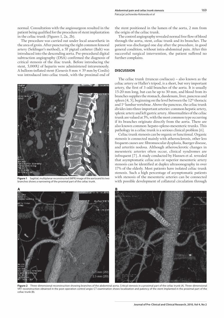
During hospitalization, the following examinations were performed: X-ray of the chest, cervical, thoracic and lumbar regions of the spinal column, panendoscopy and ultrasonography of the abdomen, none of which showed any abnormalities. Regarding to recurrent abdominal pain during hospitalization which increased after even the slight physical exertion, and positive anamnesis of a cardiovascular disease, an angio-CT of the visceral arteries was performed. This examination finally revealed an isolated, critical stenosis of the celiac trunk in its proximal part, just 1 cm from the origin of the artery. The superior and inferior mesenteric arteries were
Corresponding author: Dr. Andrzej Prystupa, Department of Internal Medicine, Medical University, Lublin, Staszica 16, 20-081 Lublin, Poland.E-mail: [email protected]
Received: 28 November 2010; accepted: 29 December 2010
169Abdominal pain and celiac trunk stenosisPatrycja Lachowska-Kotowska et al
Journal of Pre-Clinical and Clinical Research, 2010, Vol 4, No 2
the stent positioned in the lumen of the aorta, 2 mm from the origin of the celiac trunk.
The control angiography revealed normal free flow of blood through the aorta, stent, celiac trunk and its branches. The patient was discharged one day after the procedure, in good general condition, without intra-abdominal pain. After this successful surgical intervention, the patient suffered no further complains.
DISCUSSION
The celiac trunk (truncus coeliacus) – also known as the celiac artery or Haller’s tripod, is a short, but very important artery, the first of 3 odd branches of the aorta. It is usually 15-20 mm long, but can be up to 30 mm, and blood from its branches supplies the stomach, duodenum, liver, pancreas and spleen [4, 5], beginning on the level between the 12th thoracic and 1st lumbar vertebrae. Above the pancreas, the celiac trunk divides into three important arteries: common hepatic artery, splenic artery and left gastric artery. Abnormalities of the celiac trunk are valued at 5%, with the most common type occurring if its branches originate directly from the aorta. There are also known common hepato-spleno-mesenteric trunks. This pathology in a celiac trunk is a serious clinical problem [6].
Celiac trunk stenosis can be organic or functional. Organic stenosis is connected mainly with atherosclerosis, other less frequent causes are: fibromuscular dysplasia, Buerger disease, and arteritis nodosa. Although atherosclerotic changes in mesenteric arteries often occur, clinical syndromes are infrequent [7]. A study conducted by Hansen et al. revealed that asymptomatic celiac axis or superior mesenteric artery stenosis can be identified at duplex ultrasonography in over 17% of the elderly. Most patients have isolated celiac trunk stenosis. Such a high percentage of asymptomatic patients with stenosis of the mesenteric arteries can be connected with possible development of collateral circulation through
normal. Consultation with the angiosurgeon resulted in the patient being qualified for the procedure of stent implantation in the celiac trunk (Figures 1, 2a, 2b).
The procedure was carried out under local anaesthetic in the area of groin. After puncturing the right common femoral artery (Seldinger’s method), a 5F pigtail catheter (Balt) was introduced into the descending aorta. Pre-procedural digital subtraction angiography (DSA) confirmed the diagnosis of critical stenosis of the iliac trunk. Before introducing the stent, 5,000U of heparin were administered intravenously. A balloon-inflated stent (Genesis 8 mm × 39 mm by Cordis) was introduced into celiac trunk, with the proximal end of
Figure1 Sagittal, multiplanar reconstructed (MPR) image of the aorta and its two branches shows a narrowing of the proximal part of the celiac trunk.
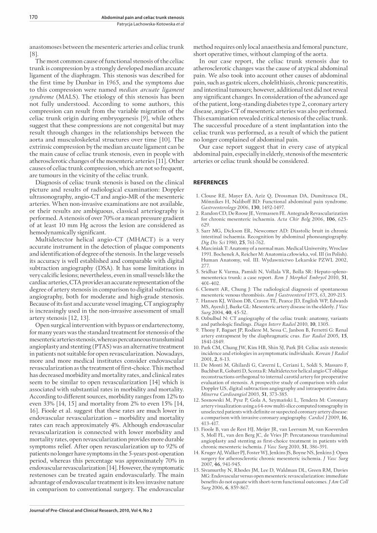
Figure2 Three-dimensional reconstruction showing branches of the abdominal aorta. Critical stenosis in a proximal part of the celiac trunk (A). Three-dimensional VRT reconstruction obtained in the post-operation control angio-CT examination shows localization and patency of the stent implanted in the proximal part of the celiac trunk (B).
A B
170 Abdominal pain and celiac trunk stenosisPatrycja Lachowska-Kotowska et al
Journal of Pre-Clinical and Clinical Research, 2010, Vol 4, No 2
method requires only local anaesthesia and femoral puncture, short operative times, without clamping of the aorta.
In our case report, the celiac trunk stenosis due to atherosclerotic changes was the cause of atypical abdominal pain. We also took into account other causes of abdominal pain, such as gastric ulcers, cholelithiasis, chronic pancreatitis, and intestinal tumours; however, additional test did not reveal any significant changes. In consideration of the advanced age of the patient, long-standing diabetes type 2, coronary artery disease, angio-CT of mesenteric arteries was also performed. This examination revealed critical stenosis of the celiac trunk. The successful procedure of a stent implantation into the celiac trunk was performed, as a result of which the patient no longer complained of abdominal pain.
Our case report suggest that in every case of atypical abdominal pain, especially in elderly, stenosis of the mesenteric arteries or celiac trunk should be considered.
REFERENCES
1. Clouse RE, Mayer EA, Aziz Q, Drossman DA, Dumitrascu DL, Mönnikes H, Naliboff BD: Functional abdominal pain syndrome. Gastroenterology 2006, 130, 1492-1497.
2. Randon CD, De Roose JE, Vermassen FE. Antegrade Revascularization for chronic mesenteric ischaemia. Acta Chir Belg 2006, 106, 625-629.
3. Sarr MG, Dickson ER, Newcomer AD: Diastolic bruit in chronic intestinal ischaemia. Recognition by abdominal phonoangiography. Dig Dis Sci 1980, 25, 761-762.
4. Marciniak T: Anatomy of a normal man. Medical University, Wroclaw 1991. Bochenek A, Reicher M: Anatomia człowieka, vol. III (in Polish). Human Anatomy, vol. III. Wydawnictwo Lekarskie PZWL 2002, 277.
5. Sridhar K Varma, Pamidi N, Vollala VR, Bolla SR: Hepato-spleno-mesenterica trunk: a case report. Rom J Morphol Embryol 2010, 51, 401-402.
6. Clemett AR, Chung J: The radiological diagnosis of spontaneous mesenteric venous thrombisis. Am J Gastroenterol 1975, 63, 209-215.
7. Hansen KJ, Wilson DB, Craven TE, Pearce JD, English WP, Edwards MS, Ayerdi J, Burke GL: Mesenteric artery disease in the elderly. J Vasc Surg 2004, 40, 45-52.
8. Ozbulbul N: CT angiography of the celiac trunk: anatomy, variants and pathologic findings. Diagn Interv Radiol 2010, 10, 1305.
9. Thony F, Baguet JP, Rodiere M, Sessa C, Janbon B, Ferretti G: Renal artery entrapment by the diaphragmatic crus. Eur Radiol 2005, 15, 1841-1849.
10. Park CM, Chung JW, Kim HB, Shin SJ, Park JH: Celiac axis stenosis: incidence and etiologies in asymptomatic individuals. Korean J Radiol 2001, 2, 8-13.
11. De Monti M, Ghilardi G, Caverni L, Ceriani L, Soldi S, Massaro F, Buchbut R, Gobatti D, Scorza R: Multidetector helical angio CT oblique reconstructions orthogonal to internal carotid artery for preoperative evaluation of stenosis. A prospective study of comparison with color Doppler US, digital subtraction angiography and intraoperative data. Minerva Cardioangiol 2003, 51, 373-385.
12. Sosnowski M, Pysz P, Gola A, Szymański L, Tendera M: Coronary artery visualization using a 64-row multi-slice computed tomography in unselected patients with definite or suspected coronary artery disease: a comparison with invasive coronary angiography. Cardiol J 2009, 16, 413-417.
13. Fioole B, van de Rest HJ, Meijer JR, van Leersum M, van Koeverden S, Moll FL, van den Berg JC, de Vries JP: Percutaneous transluminal angioplasty and stenting as first-choice treatment in patients with chronic mesenteric ischemia. J Vasc Surg 2010, 51, 386-391.
14. Kruger AJ, Walker PJ, Foster WJ, Jenkins JS, Boyne NS, Jenkins J: Open surgery for atherosclerotic chronic mesenteric ischemia. J Vasc Surg 2007, 46, 941-945.
15. Sivamurthy N, Rhodes JM, Lee D, Waldman DL, Green RM, Davies MG: Endovascular versus open mesenteric revascularization: immediate benefits do not equate with short-term functional outcomes. J Am Coll Surg 2006, 6, 859-867.
anastomoses between the mesenteric arteries and celiac trunk [8].
The most common cause of functional stenosis of the celiac trunk is compression by a strongly developed median arcuate ligament of the diaphragm. This stenosis was described for the first time by Dunbar in 1965, and the symptoms due to this compression were named median arcuate ligament syndrome (MALS). The etiology of this stenosis has been not fully understood. According to some authors, this compression can result from the variable migration of the celiac trunk origin during embryogenesis [9], while others suggest that these compressions are not congenital but may result through changes in the relationships between the aorta and musculoskeletal structures over time [10]. The extrinsic compression by the median arcuate ligament can be the main cause of celiac trunk stenosis, even in people with atherosclerotic changes of the mesenteric arteries [11]. Other causes of celiac trunk compression, which are not so frequent, are tumours in the vicinity of the celiac trunk.
Diagnosis of celiac trunk stenosis is based on the clinical picture and results of radiological examination: Doppler ultrasonography, angio-CT and angio-MR of the mesenteric arteries. When non-invasive examinations are not available, or their results are ambiguous, classical arteriography is performed. A stenosis of over 70% or a mean pressure gradient of at least 10 mm Hg across the lesion are considered as hemodynamically significant.
Multidetector helical angio-CT (MHACT) is a very accurate instrument in the detection of plaque components and identification of degree of the stenosis. In the large vessels its accuracy is well established and comparable with digital subtraction angiography (DSA). It has some limitations in very calcific lesions; nevertheless, even in small vessels like the cardiac arteries, CTA provides an accurate representation of the degree of artery stenosis in comparison to digital subtraction angiography, both for moderate and high-grade stenosis. Because of its fast and accurate vessel imaging, CT angiography is increasingly used in the non-invasive assessment of small artery stenosis [12, 13].
Open surgical intervention with bypass or endarterectomy, for many years was the standard treatment for stenossis of the mesenteric arteries stenosis, whereas percutaneous transluminal angioplasty and stenting (PTAS) was an alternative treatment in patients not suitable for open revascularization. Nowadays, more and more medical institutes consider endovascular revascularization as the treatment of first-choice. This method has decreased morbidity and mortality rates, and clinical rates seem to be similar to open revascularization [14] which is associated with substantial rates in morbidity and mortality. According to different sources, morbidity ranges from 12% to even 33% [14, 15] and mortality from 2% to even 15% [14, 16]. Fioole et al. suggest that these rates are much lower in endovascular revascularization – morbidity and mortality rates can reach approximately 4%. Although endovascular revascularization is connected with lower morbidity and mortality rates, open revascularization provides more durable symptoms relief. After open revascularization up to 92% of patients no longer have symptoms in the 5-years post-operation period, whereas this percentage was approximately 70% in endovascular revascularization [14]. However, the symptomatic restenoses can be treated again endovascularly. The main advantage of endovascular treatment is its less invasive nature in comparison to conventional surgery. The endovascular