39
| Date post: | 15-Mar-2018 |
| Category: |
Documents |
| Upload: | truongkhue |
| View: | 226 times |
| Download: | 1 times |
Chronic meningitis and
encephalitis
John Ferguson
UPNG 2013
CHRONIC MENINGITIS
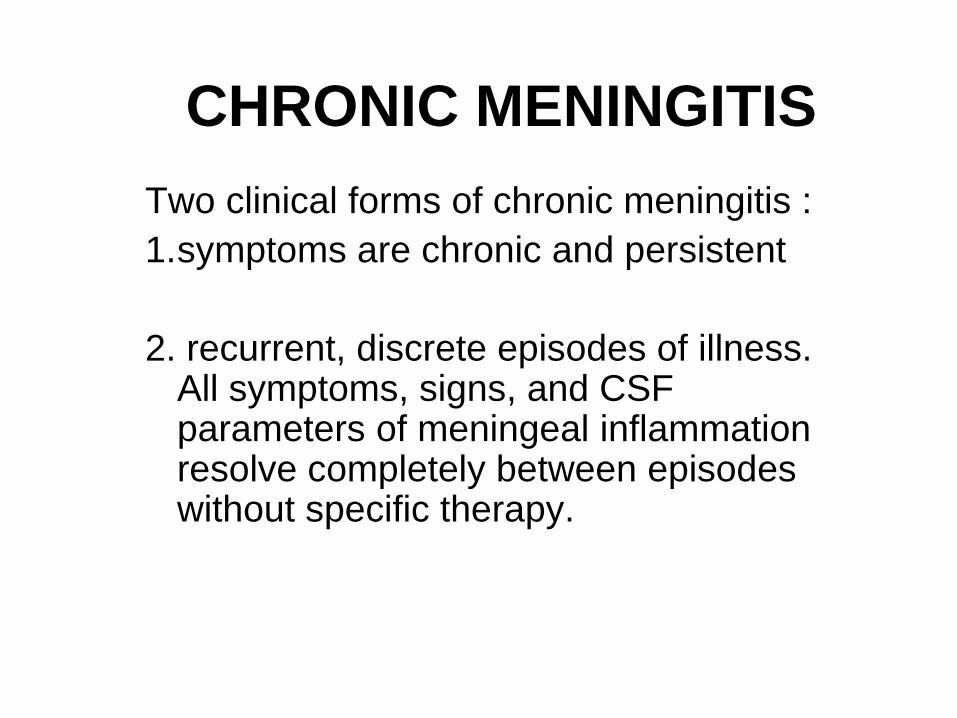
Two clinical forms of chronic meningitis :
1.symptoms are chronic and persistent
2. recurrent, discrete episodes of illness. All symptoms, signs, and CSF parameters of meningeal inflammation resolve completely between episodes without specific therapy.
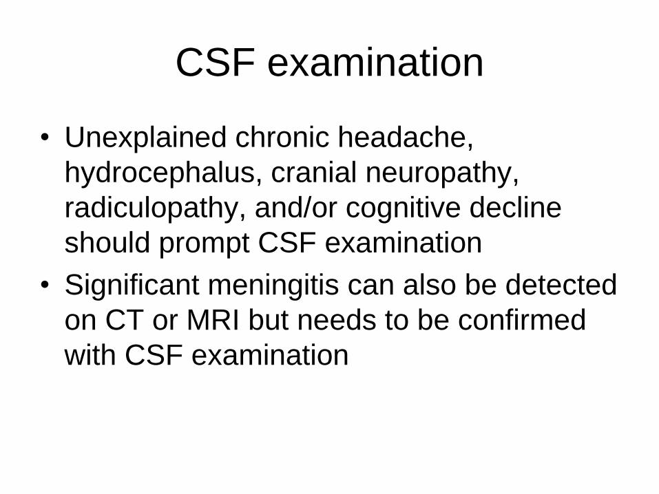
CSF examination
• Unexplained chronic headache,
hydrocephalus, cranial neuropathy,
radiculopathy, and/or cognitive decline
should prompt CSF examination
• Significant meningitis can also be detected
on CT or MRI but needs to be confirmed
with CSF examination
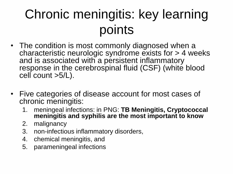
Chronic meningitis: key learning
points • The condition is most commonly diagnosed when a
characteristic neurologic syndrome exists for > 4 weeks and is associated with a persistent inflammatory response in the cerebrospinal fluid (CSF) (white blood cell count >5/L).
• Five categories of disease account for most cases of chronic meningitis: 1. meningeal infections: in PNG: TB Meningitis, Cryptococcal
meningitis and syphilis are the most important to know
2. malignancy
3. non-infectious inflammatory disorders,
4. chemical meningitis, and
5. parameningeal infections
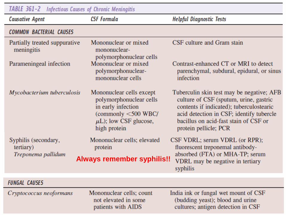
Always remember syphilis!!
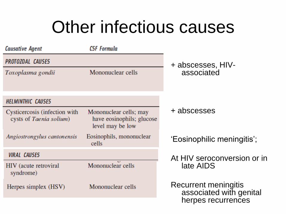
Other infectious causes
+ abscesses, HIV-associated
+ abscesses
‘Eosinophilic meningitis’;
At HIV seroconversion or in late AIDS
Recurrent meningitis associated with genital herpes recurrences
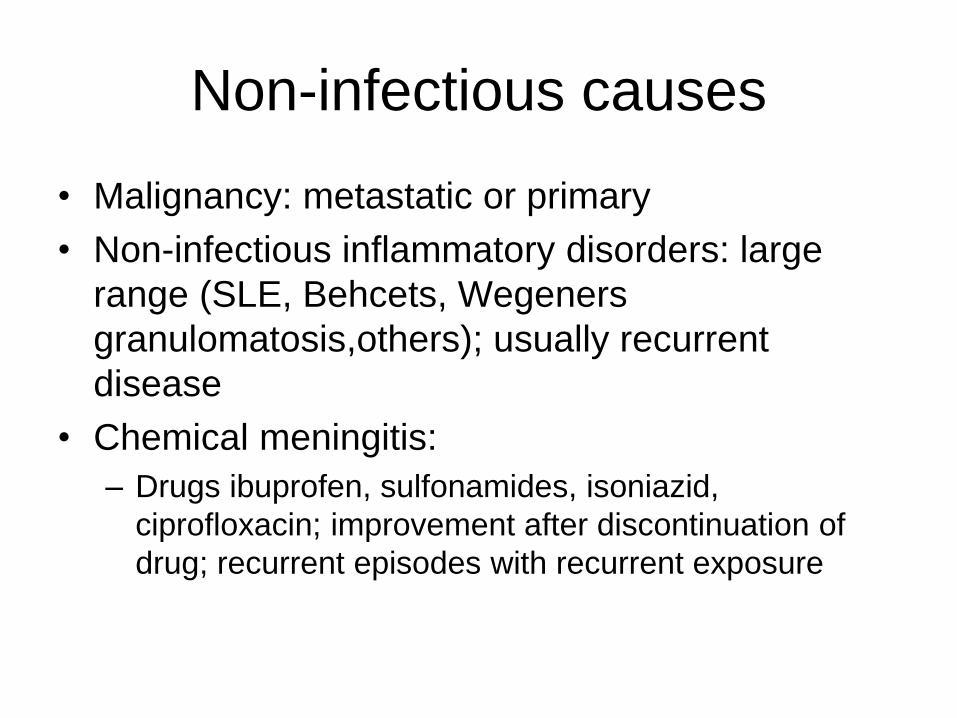
Non-infectious causes
• Malignancy: metastatic or primary
• Non-infectious inflammatory disorders: large
range (SLE, Behcets, Wegeners
granulomatosis,others); usually recurrent
disease
• Chemical meningitis:
– Drugs ibuprofen, sulfonamides, isoniazid,
ciprofloxacin; improvement after discontinuation of
drug; recurrent episodes with recurrent exposure
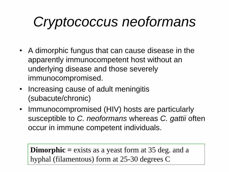
Cryptococcus neoformans
• A dimorphic fungus that can cause disease in the
apparently immunocompetent host without an
underlying disease and those severely
immunocompromised.
• Increasing cause of adult meningitis
(subacute/chronic)
• Immunocompromised (HIV) hosts are particularly
susceptible to C. neoformans whereas C. gattii often
occur in immune competent individuals.
Dimorphic = exists as a yeast form at 35 deg. and a
hyphal (filamentous) form at 25-30 degrees C
Cryptococcus neoformans
Presentations:
- subacute meningitis: headache, fever,
raised ICP
- pneumonia
- disseminated disease
- localised disease (cryptococcoma) -
brain, lung
Cryptococcosis
Diagnosis:
Microscopy (India Ink) and culture
- CSF
- bronchial secretions or lung biopsy
Polysaccharide antigen detection is the
best test:
- serum or CSF latex agglutination
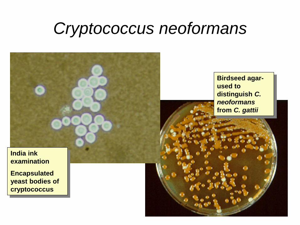
Cryptococcus neoformans
India ink
examination
Encapsulated
yeast bodies of
cryptococcus
Birdseed agar-
used to
distinguish C.
neoformans
from C. gattii
Cryptococcal meningitis
• Most patients with cryptococcosis of the CNS present with signs and symptoms of subacute meningitis or meningoencephalitis, such as headache, fever, cranial nerve palsies, lethargy, coma, or memory loss over several weeks.
• Symptoms may not be typical, and patients may present with acute (several days) symptoms of severe headaches, with intermittent headaches, or even with no headache but with altered mental status.
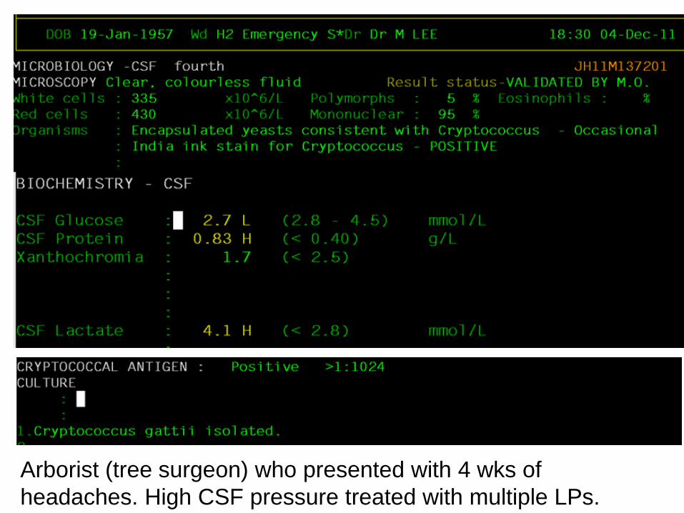
Arborist (tree surgeon) who presented with 4 wks of
headaches. High CSF pressure treated with multiple LPs.
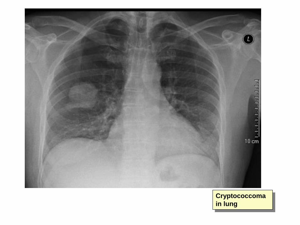
Cryptococcoma
in lung
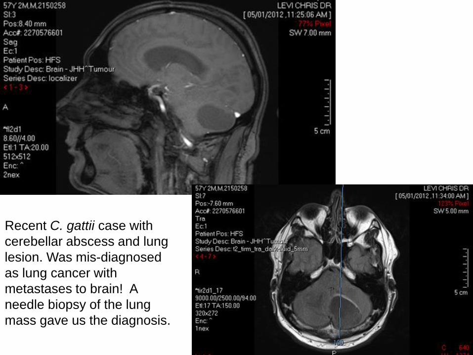
Recent C. gattii case with
cerebellar abscess and lung
lesion. Was mis-diagnosed
as lung cancer with
metastases to brain! A
needle biopsy of the lung
mass gave us the diagnosis.
HIV-infected Patients
• The burden of yeast is generally higher, and this
may be reflected slower conversion of CSF to
culture negativity during treatment, and a
tendency toward a higher incidence of increased
intracranial pressure.
• Extracranial disease sites more likely to be
found
• Important recent work that supports screening of
new HIV patients for cryptococcus with blood
antigen test at the time they start ARV treatment
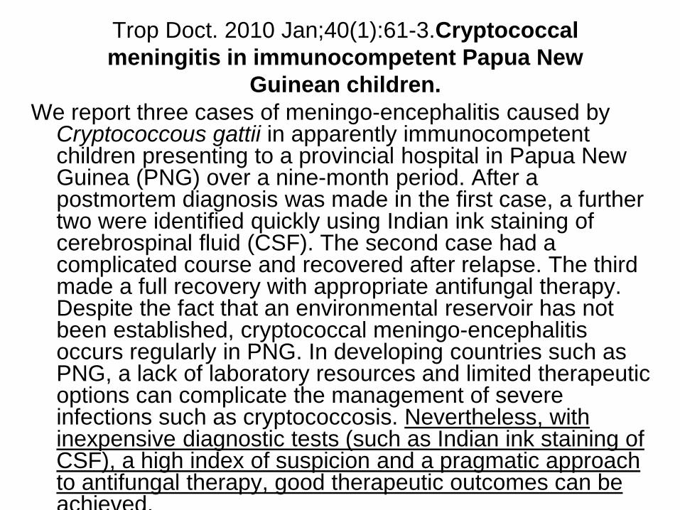
Trop Doct. 2010 Jan;40(1):61-3.Cryptococcal
meningitis in immunocompetent Papua New
Guinean children.
We report three cases of meningo-encephalitis caused by Cryptococcous gattii in apparently immunocompetent children presenting to a provincial hospital in Papua New Guinea (PNG) over a nine-month period. After a postmortem diagnosis was made in the first case, a further two were identified quickly using Indian ink staining of cerebrospinal fluid (CSF). The second case had a complicated course and recovered after relapse. The third made a full recovery with appropriate antifungal therapy. Despite the fact that an environmental reservoir has not been established, cryptococcal meningo-encephalitis occurs regularly in PNG. In developing countries such as PNG, a lack of laboratory resources and limited therapeutic options can complicate the management of severe infections such as cryptococcosis. Nevertheless, with inexpensive diagnostic tests (such as Indian ink staining of CSF), a high index of suspicion and a pragmatic approach to antifungal therapy, good therapeutic outcomes can be achieved.
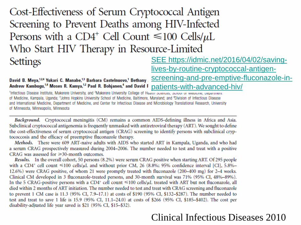
Clinical Infectious Diseases 2010
SEE https://idmic.net/2016/04/02/saving-
lives-by-routine-cryptococcal-antigen-
screening-and-pre-emptive-fluconazole-in-
patients-with-advanced-hiv/
Cryptococcus: learning points
1. Presentation and diagnosis
2. Treatment to sterilise CSF: induction and
maintenance; culture CSF after 2 wks of
treatment to check – should be no growth
3. Treatment to aggressively manage raised
intracranial pressure: improves outcomes,
reduces visual complications
4. Primary and secondary prophylaxis with
fluconazole for HIV patients with low CD4
count (<250)
TB meningitis: pathogenesis
• Miliary tubercles form in the parenchyma of the brain during hematogenous dissemination of tubercle bacilli in the course of primary infection.
• These tubercles enlarge and are usually caseating. The propensity for a caseous lesion to produce meningitis is determined by its proximity to the subarachnoid space and the rate at which fibrous encapsulation develops.
• Caseous foci cause meningitis via discharge of bacilli and tuberculous antigens into the subarachnoid space.
• Mycobacterial antigens produce intense inflammatory reaction in the CSF - thick exudate formed that surrounds the cranial nerves and major blood vessels at the base of the brain.
TB meningitis : presentation
• Subacute : unrelenting headache, neck stiffness, fatigue, night sweats, eventually altered level of consiousness/coma
• The classic CSF abnormalities (NB. atypical findings may occur): – elevated opening pressure,
– Lymphocytic pleocytosis (10 to 500 cells/L),
– elevated protein concentration in the range of 1 to 5 g/L
– Decreased glucose concentration (moderate)
• CSF AFB examination 10-40% sensitive and culture 50 % sensitive and slow! PCR more sensitive
TBM: learning points
• Presentation and diagnosis: must suspect
early
• Early presumptive treatment: delay
associated with poor outcomes
• Combination tuberculous chemotherapy
with addition of corticosteroids
• Active management of raised intracranial
pressure – repeated LPs, CSF shunt
sometimes
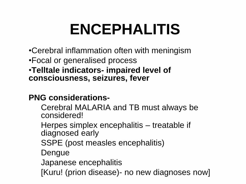
ENCEPHALITIS
•Cerebral inflammation often with meningism
•Focal or generalised process
•Telltale indicators- impaired level of consciousness, seizures, fever
PNG considerations-
Cerebral MALARIA and TB must always be considered!
Herpes simplex encephalitis – treatable if diagnosed early
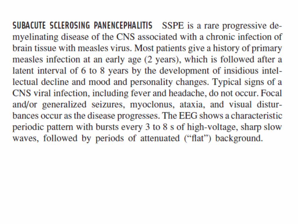
SSPE (post measles encephalitis)
Dengue
Japanese encephalitis
[Kuru! (prion disease)- no new diagnoses now]
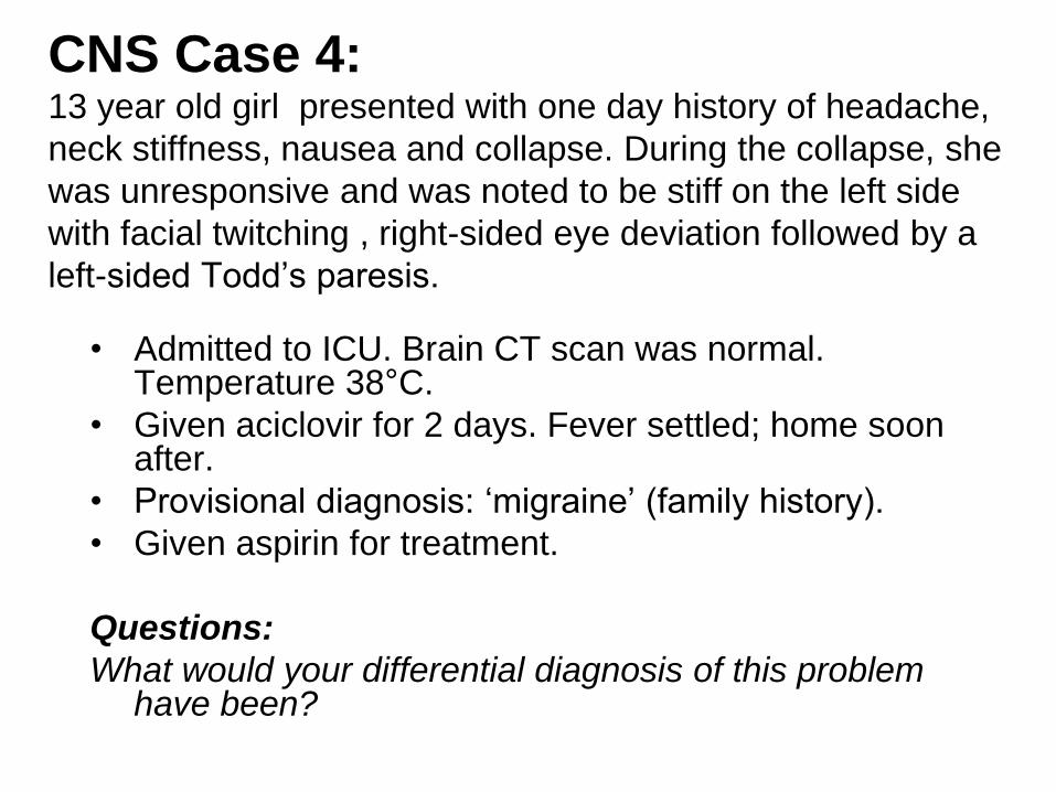
CNS Case 4:
13 year old girl presented with one day history of headache,
neck stiffness, nausea and collapse. During the collapse, she
was unresponsive and was noted to be stiff on the left side
with facial twitching , right-sided eye deviation followed by a
left-sided Todd’s paresis.
• Admitted to ICU. Brain CT scan was normal.
Temperature 38°C.
• Given aciclovir for 2 days. Fever settled; home soon after.
• Provisional diagnosis: ‘migraine’ (family history).
• Given aspirin for treatment.
Questions:
What would your differential diagnosis of this problem have been?
CNS case 4: Differential diagnosis
• The history is of an acute febrile neurological disorder with evidence of focal seizure activity and some meningism. An infective process should be top most in mind. The normal CT scan largely rules out a focal process such as brain abscess.
• The clinical picture suggests meningo-encephalitis. Herpes simplex encephalitis must be considered strongly.
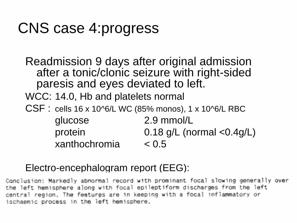
CNS case 4:progress
Readmission 9 days after original admission after a tonic/clonic seizure with right-sided paresis and eyes deviated to left.
WCC: 14.0, Hb and platelets normal
CSF : cells 16 x 10^6/L WC (85% monos), 1 x 10^6/L RBC
glucose 2.9 mmol/L
protein 0.18 g/L (normal <0.4g/L)
xanthochromia < 0.5
Electro-encephalogram report (EEG):
CNS Case 4- Diagnosis and
management
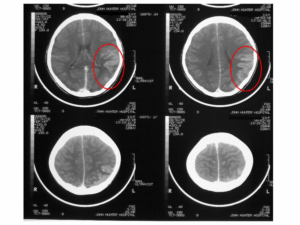
• The CT scan findings were compatible with either a left fronto-parietal infarct or cerebritis.
• The findings of CSF pleocytosis, focal EEG and CT abnormalities are strongly suggestive of herpes simplex encephalitis.
• Early treatment with high dose intravenous aciclovir continued for 14 days at least whenever the diagnosis is suspected.
• The patient received a full course of aciclovir and progressed well (see temperature chart). She was given phenytoin and had no further seizures.
• Herpes simplex PCR on CSFwas positive, confirming the diagnosis.
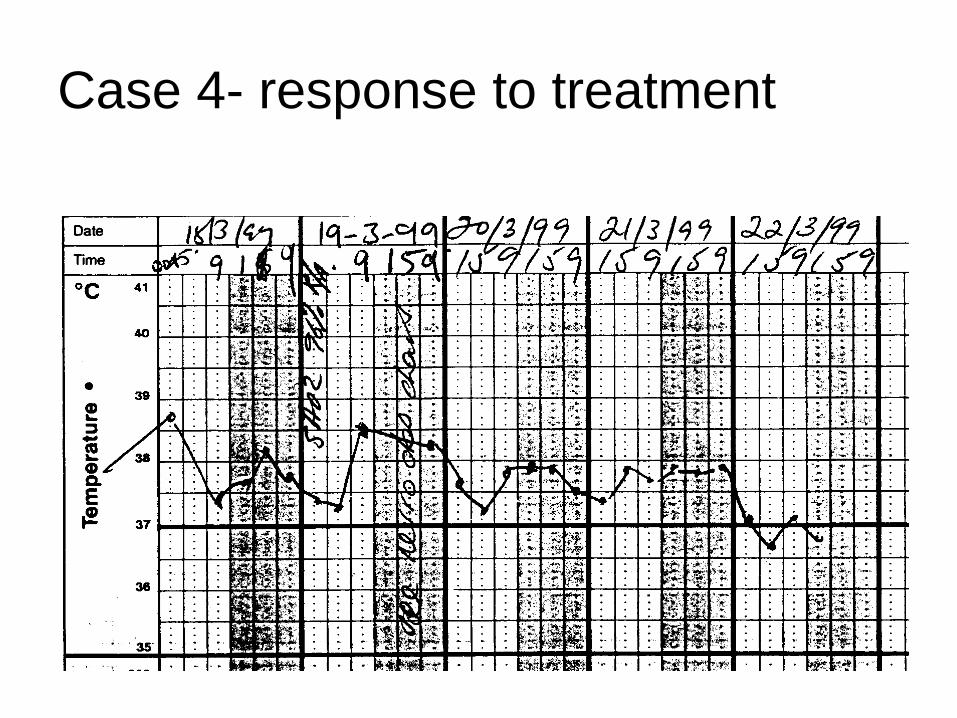
Case 4- response to treatment
Differential diagnosis of encephalitis
• Infective causes (next slide)
• Meningitis (PNG: TB, or Cryptococcus)
• Parameningeal focus
• Causes of encephalopathy- – Systemic infections, severe sepsis
– metabolic disturbances (hypoglycaemia, hyponatraemia and hypocalcaemia);
– thiamine deficiency (Wernicke’s encephalopathy);
– drugs (including neuroleptics [neuroleptic malignant syndrome], trimethoprim–sulfamethoxazole, isoniazid, nonsteroidal anti-inflammatory drugs, intoxications); and
– Various auto-immune inflammatory disorders
• Status epilepticus and malignancy (paraneoplastic syndrome) may also sometimes be confused with encephalitis.
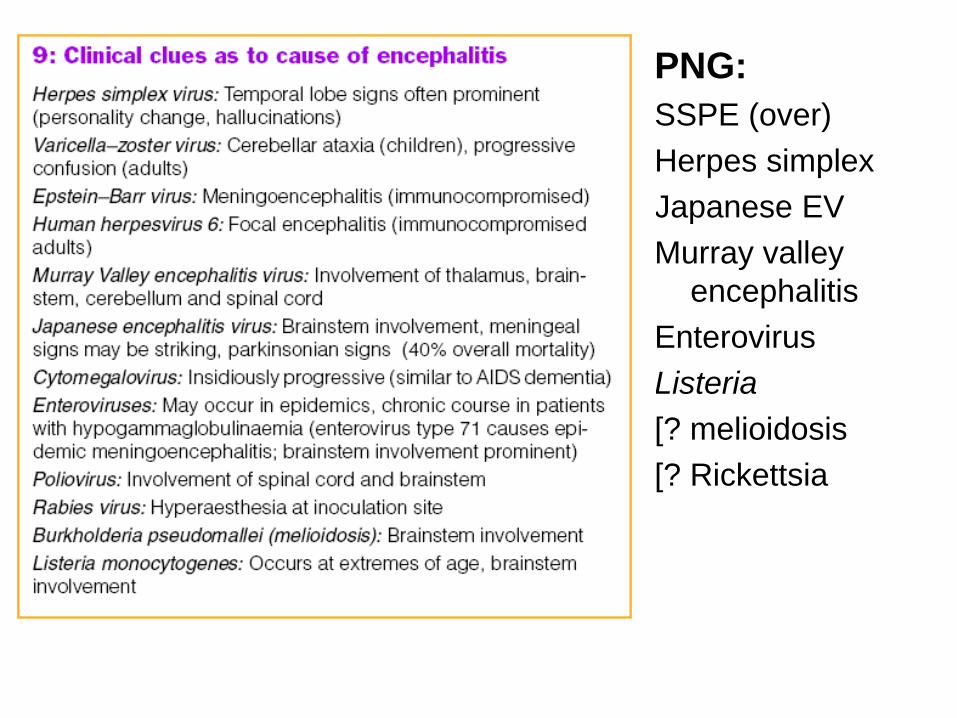
PNG:
SSPE (over)
Herpes simplex
Japanese EV
Murray valley
encephalitis
Enterovirus
Listeria
[? melioidosis
[? Rickettsia
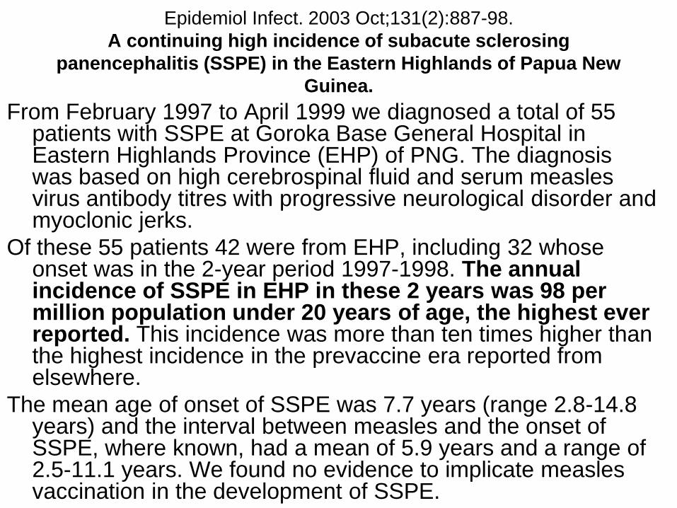
Epidemiol Infect. 2003 Oct;131(2):887-98.
A continuing high incidence of subacute sclerosing
panencephalitis (SSPE) in the Eastern Highlands of Papua New
Guinea.
From February 1997 to April 1999 we diagnosed a total of 55 patients with SSPE at Goroka Base General Hospital in Eastern Highlands Province (EHP) of PNG. The diagnosis was based on high cerebrospinal fluid and serum measles virus antibody titres with progressive neurological disorder and myoclonic jerks.
Of these 55 patients 42 were from EHP, including 32 whose onset was in the 2-year period 1997-1998. The annual incidence of SSPE in EHP in these 2 years was 98 per million population under 20 years of age, the highest ever reported. This incidence was more than ten times higher than the highest incidence in the prevaccine era reported from elsewhere.
The mean age of onset of SSPE was 7.7 years (range 2.8-14.8 years) and the interval between measles and the onset of SSPE, where known, had a mean of 5.9 years and a range of 2.5-11.1 years. We found no evidence to implicate measles vaccination in the development of SSPE.
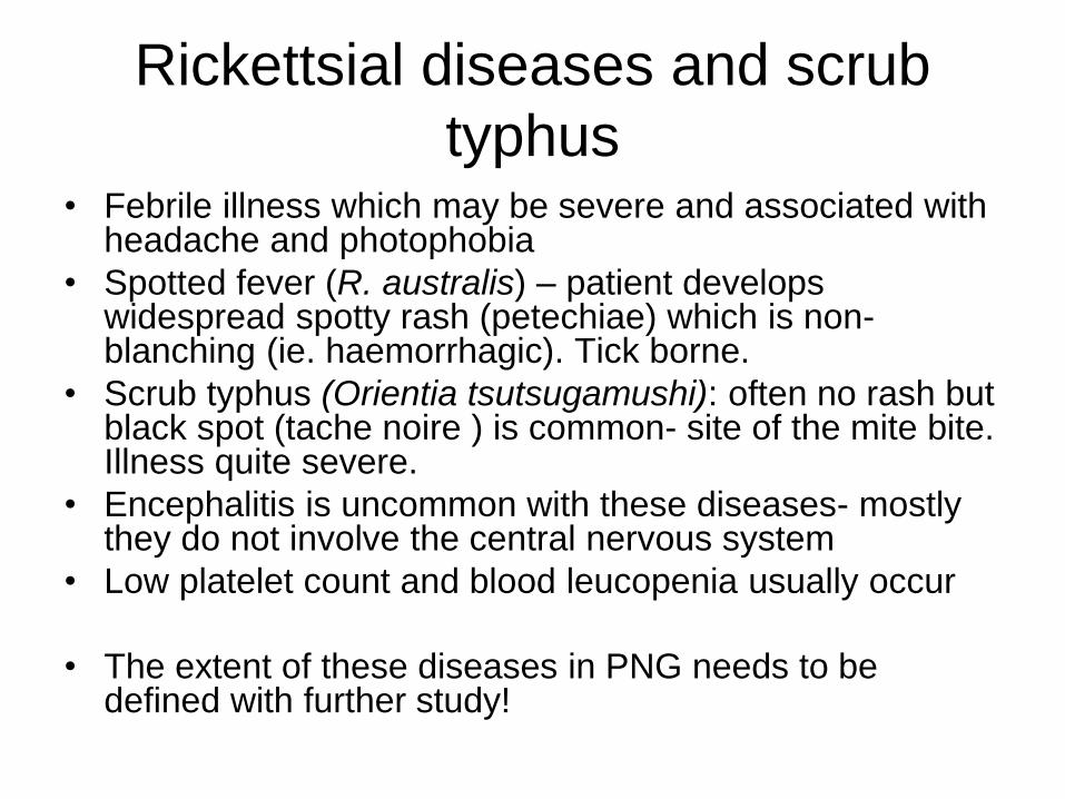
Rickettsial diseases and scrub
typhus • Febrile illness which may be severe and associated with
headache and photophobia
• Spotted fever (R. australis) – patient develops widespread spotty rash (petechiae) which is non-blanching (ie. haemorrhagic). Tick borne.
• Scrub typhus (Orientia tsutsugamushi): often no rash but black spot (tache noire ) is common- site of the mite bite. Illness quite severe.
• Encephalitis is uncommon with these diseases- mostly they do not involve the central nervous system
• Low platelet count and blood leucopenia usually occur
• The extent of these diseases in PNG needs to be defined with further study!
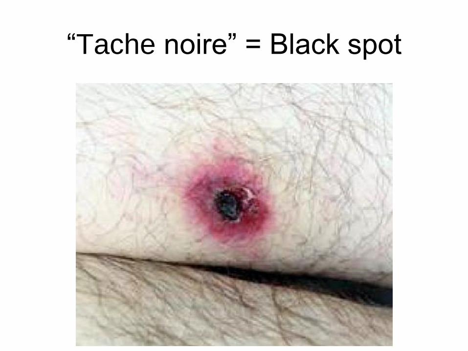
“Tache noire” = Black spot
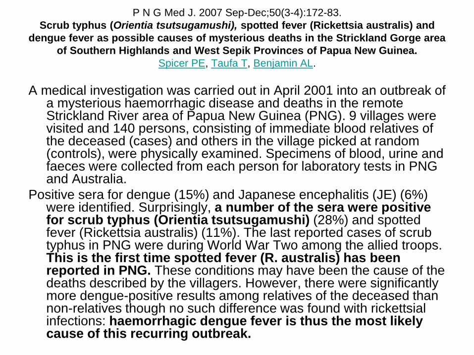
P N G Med J. 2007 Sep-Dec;50(3-4):172-83.
Scrub typhus (Orientia tsutsugamushi), spotted fever (Rickettsia australis) and
dengue fever as possible causes of mysterious deaths in the Strickland Gorge area
of Southern Highlands and West Sepik Provinces of Papua New Guinea.
Spicer PE, Taufa T, Benjamin AL.
A medical investigation was carried out in April 2001 into an outbreak of a mysterious haemorrhagic disease and deaths in the remote Strickland River area of Papua New Guinea (PNG). 9 villages were visited and 140 persons, consisting of immediate blood relatives of the deceased (cases) and others in the village picked at random (controls), were physically examined. Specimens of blood, urine and faeces were collected from each person for laboratory tests in PNG and Australia.
Positive sera for dengue (15%) and Japanese encephalitis (JE) (6%) were identified. Surprisingly, a number of the sera were positive for scrub typhus (Orientia tsutsugamushi) (28%) and spotted fever (Rickettsia australis) (11%). The last reported cases of scrub typhus in PNG were during World War Two among the allied troops. This is the first time spotted fever (R. australis) has been reported in PNG. These conditions may have been the cause of the deaths described by the villagers. However, there were significantly more dengue-positive results among relatives of the deceased than non-relatives though no such difference was found with rickettsial infections: haemorrhagic dengue fever is thus the most likely cause of this recurring outbreak.
Encephalitis: key learning
• Presentation and diagnosis
• Herpes simplex encephalitis
• PNG : what is the infectious differential
diagnosis (many unknowns!!)
• Other non-infectious causes of
encephalopathy


