CHRONIC OBSTRUCTIVE PULMONARY DISEASE CLINICAL PRACTICE GUIDELINES REVIEW WEEK 1: DIAGNOSIS AMBULATORY INTERNAL MEDICINE GROUP PRACTICE UNIVERSITY HEALTH NETWORK / MSH SEPTEMBER 2007 Prepared by: Dr. D. Panisko
Transcript
CHRONIC OBSTRUCTIVE PULMONARY DISEASE
CLINICAL PRACTICE GUIDELINES REVIEW
WEEK 1: DIAGNOSIS
AMBULATORY INTERNAL MEDICINE
GROUP PRACTICE
UNIVERSITY HEALTH NETWORK / MSH
SEPTEMBER 2007
Prepared by: Dr. D. Panisko
COPD: Guidelines for this Seminar
Standards for the diagnosis and treatment of patients with COPD: a summary of the ATS/ERS position paper. Celli BR et al. Eur Respir J 2004; 23: 932-46. Full document, with updates, available at: www.thoracic.org, accessed Sept 2007
Canadian Thoracic Society recommendations for the management of chronic obstructive pulmonary disease - 2003. O’Donnell DE et al. Can Respir J 2003; 10(SupplA): 11A-33A
Global Initiative for Chronic Obstructive Lung Disease. (GOLD). A collaborative of the NIH and WHO. Updated Nov 2006, accessed Sept 2007. Available at www.goldcopd.com
COPD Diagnosis: Objectives After this seminar you should:
be aware of diagnostic clinical practice guidelines for stable chronic COPD
be able to define COPD and asthma and outline a differential diagnosis
be able list important historical and laboratory diagnostic features of COPD
be able to describe the evidence-based physical examination for COPD and airflow limitation
COPD I: DIAGNOSISCASE:A 61 year old man comes to your clinic as a new patient. He had just been admitted to hospital for his first exacerbation of COPD. He has completed a 10 day antibiotic course and 10 days of oral Prednisone. He is now only on an ipratropium puffer, 2 puffs qid.
How is COPD defined ? What is emphysema ? What is asthma ?
Why is it important to make a diagnosis of COPD (as opposed to asthma) in this patient ?
COPD I: DIAGNOSIS COPD Definition:
A preventable and treatable disease state characterized by airflow limitation that is not fully reversible. The airflow limitation is usually progressive and associated with an abnormal inflammatory response of the lungs to noxious particles or gases, primarily caused by cigarette smoking. Although COPD affects the lungs, it also produces significant systemic consequences. Implies post bronchodilator FEV1/FVC<0.7
ATS/ERS position paper
COPD I: DIAGNOSIS
COPD: traditionally understood as a spectrum - components of chronic bronchitis or emphysema. The latter may take on revitalized significance because of a new approach that considers the importance of different phenotypes of COPD.
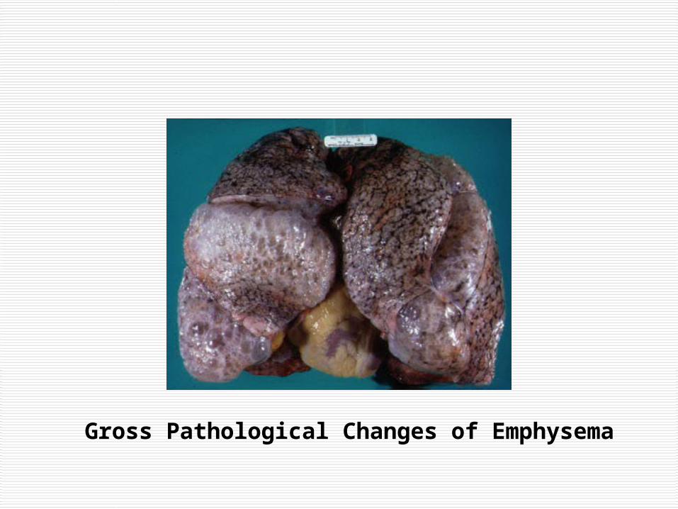
EMPHYSEMA: Abnormal permanent enlargement of the airspaces distal to the terminal bronchioles, accompanied by destruction (lack of uniformity in the pattern of airspace enlargement; the orderly appearance of the acinus and its components is disturbed and may be lost) of their walls and without obvious fibrosis.
Emphysema = Pink Puffer !
Gross Pathological Changes of Emphysema
Microscopic Changes of Emphysema
COPD I: DIAGNOSIS
ASTHMA: A chronic inflammatory disorder of the tracheobronchial tree, many cells and cellular elements play a role, leading to airway hyperreactivity and reversible airflow limitation. IMPLICATION: airway can return to normal between attacks or with treatment BUT in chronic asthma a condition similar to COPD can develop with irreversibility and progression of the airflow limitation.
It is also important to make a diagnosis of asthma as there are differences in therapy for asthma and COPD.
COPD I: DIAGNOSIS
CASE (cont.):
Can the severity of COPD be staged ? What is the relevance and importance of staging a patient with COPD ?
Stage IV: Very Severe FEV1/FVC < 0.70 FEV1 < 30% predicted or
FEV1 < 50% predicted plus chronic respiratory failure
COPD I: DIAGNOSIS
What is the relevance and importance of staging a patient with COPD ?
Stages (GOLD) are currently mainly for educational and research purposes
Not extensively validated by trials Represent expert consensus opinion Some treatment recommendations exist based
on patient stage… presumably will be further validated by clinical trials
Canadian guidelines list another severity scale but do not recommend treatment on that basis
COPD I: DIAGNOSIS
CASE (cont.):
What historical features contribute to the diagnosis of COPD ?
What are other important features of the hx ?
COPD I: DIAGNOSIS Important historical information:
age of onset of symptoms quantify exposure to risk factors i.e.:
tobacco smoke occupational exposures exposure to outdoor and indoor air pollution
presence of liver disease family history childhood respiratory illnesses information that allows a diagnosis of chronic
bronchitis
COPD I: DIAGNOSIS
Perform respiratory functional inquiry to determine current symptom status and to classify COPD:
asymptomatic intermittent symptoms (on exertion,
nocturnal/sleep) regularly symptomatic severely symptomatic frequency and course of exacerbations
COPD I: DIAGNOSIS
CASE (cont.):
What is the differential diagnosis of COPD ?
COPD I: DIAGNOSISDifferential Diagnosis of COPD:
Cystic fibrosis, asthma, bronchiectasis, and bronchiolitis obliterans (all specific causes of airflow limitation) have been conventionally excluded from the diagnosis definition of COPD and therefore are part of the dx dx. Interstitial lung disease (fibrosis, TB, hypersensitivity pneumonitis, sarcoidosis, pneumoconioses, etc.) may also present in a patient with recurrent shortness of breath, exacerbations, and cough. Consider a variety of non-pulmonary causes of breathlessness (i.e. CHF)
COPD I: DIAGNOSISCASE (cont.):This patient indicates a three year history of productive cough, at least on 50% of days, and an audible wheeze with SOBOE. His symptoms have been progressing over the entire year and he now gets SOB with 1 flight of stairs or 3 level blocks. He has a 45 pack year smoking history, has worked in an office all of his life, and has no relevant past medical, childhood, or family history.
What are indications for screening for alpha-1 antitrypsin deficiency ? Should this patient be screened? How can screening be performed ?
COPD I: DIAGNOSIS Screen for alpha-1 antitrypsin deficiency if patient
is under the age of 45, has a predominance of basilar emphysema, has a minimal smoking history, has a family history of early onset COPD, has a known family history of alpha-1 antitrypsin
deficiency, or associated liver disease.
Screening therefore not indicated in the case. Screening: serum assay for alpha-1 antitrypsin level – 10cc of clotted blood in red top tube (on a misc. req. at UHN). For update on genetics of COPD see Rabe et al. 2007
COPD I: DIAGNOSIS
CASE: What physical exam maneuvers are helpful to diagnose airflow limitation ?
Not this
one !!!
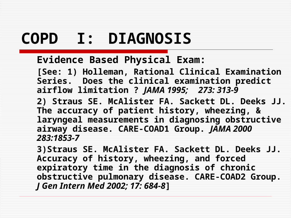
COPD I: DIAGNOSISEvidence Based Physical Exam:[See: 1) Holleman, Rational Clinical Examination Series. Does the clinical examination predict airflow limitation ? JAMA 1995; 273: 313-9 2) Straus SE. McAlister FA. Sackett DL. Deeks JJ. The accuracy of patient history, wheezing, & laryngeal measurements in diagnosing obstructive airway disease. CARE-COAD1 Group. JAMA 2000 283:1853-7 3)Straus SE. McAlister FA. Sackett DL. Deeks JJ. Accuracy of history, wheezing, and forced expiratory time in the diagnosis of chronic obstructive pulmonary disease. CARE-COAD2 Group. J Gen Intern Med 2002; 17: 684-8]
COPD I: DIAGNOSISWheezing: Grade: A Positive likelihood ratio: 36Barrel Chest: B, 10Decreased Cardiac Dullness: B, 10Match Test: B, 7.1Hyperresonance: B, 4.8Forced Expiratory Time >9 seconds: A, 4.8Subxiphoid Apical Impulse: B, 4.6Pulsus Paradoxus > 15mmHg: C, 3.7Decreased Breath Sounds: B, 3.7Forced Expiratory Time 6 - 9 seconds: A, 2.7 * Many other signs not systematically evaluated (diaphragmatic levels, pursed lip breathing, use of accessory muscles, indrawing)
COPD I: DIAGNOSIS Straus et al’s important contributions to the literature have shown that a single physical sign is not as useful as a combination of historical and physical findings to make a diagnosis of COPD
They have published two models
What maneuvre isbeing performed ?
COPD I: DIAGNOSIS
Combined history/physical exam Model I:
Smoking > 40 P.Y. (LR 8.3)Self reported history of COPD (LR 7.3)Maximum laryngeal height (LR 2.8)Age > 45 years (LR 1.3)
Combined all 4: +LR 220 Combined patients with none: -LR 0.13
COPD I: DIAGNOSIS
Combined history/physical exam Model II:
*Forced Exp Time > 9 sec (LR 6.7) Multivariate: (LR 4.6)*Self reported history of COPD (LR 5.6) (LR 4.4)*Wheezing (LR 4.0) (LR 2.9)Smoked longer than 40 pack years (LR 3.3)Male gender (LR 1.6)Age over 65 years (LR 1.6)
*Combined all 3: +LR 59.0 *Combined patients with none: -LR 0.3
COPD I: DIAGNOSIS
CASE (cont.):
Physical examination of our patient was only relevant for a barrel chest, diffuse occasional audible wheezes, and a forced expiratory time of 7 seconds. Laryngeal height was 5 cm.
There were no signs of cor pulmonale.
Otherwise, the exam was unremarkable.
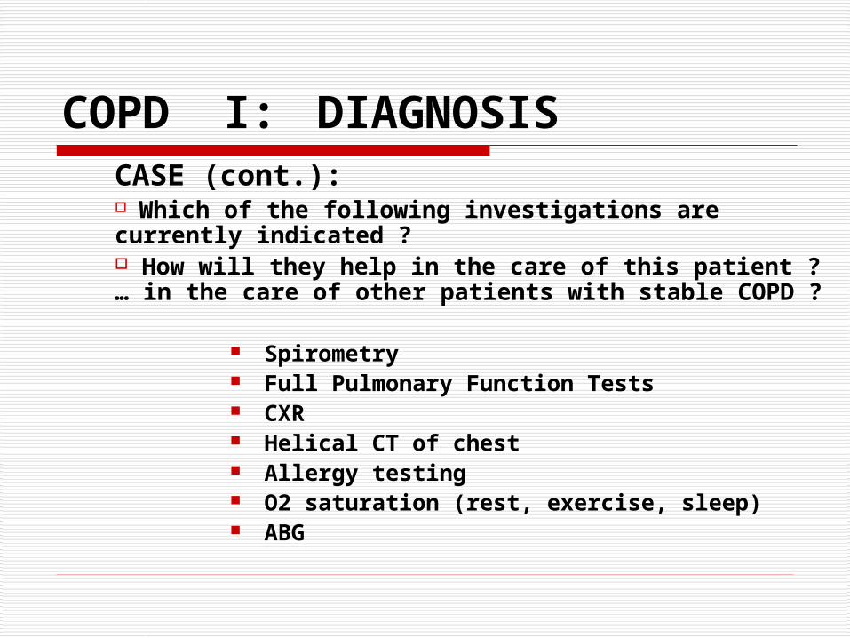
COPD I: DIAGNOSISCASE (cont.): Which of the following investigations are currently indicated ? How will they help in the care of this patient ? … in the care of other patients with stable COPD ?
Spirometry Full Pulmonary Function Tests CXR Helical CT of chest Allergy testing O2 saturation (rest, exercise, sleep) ABG
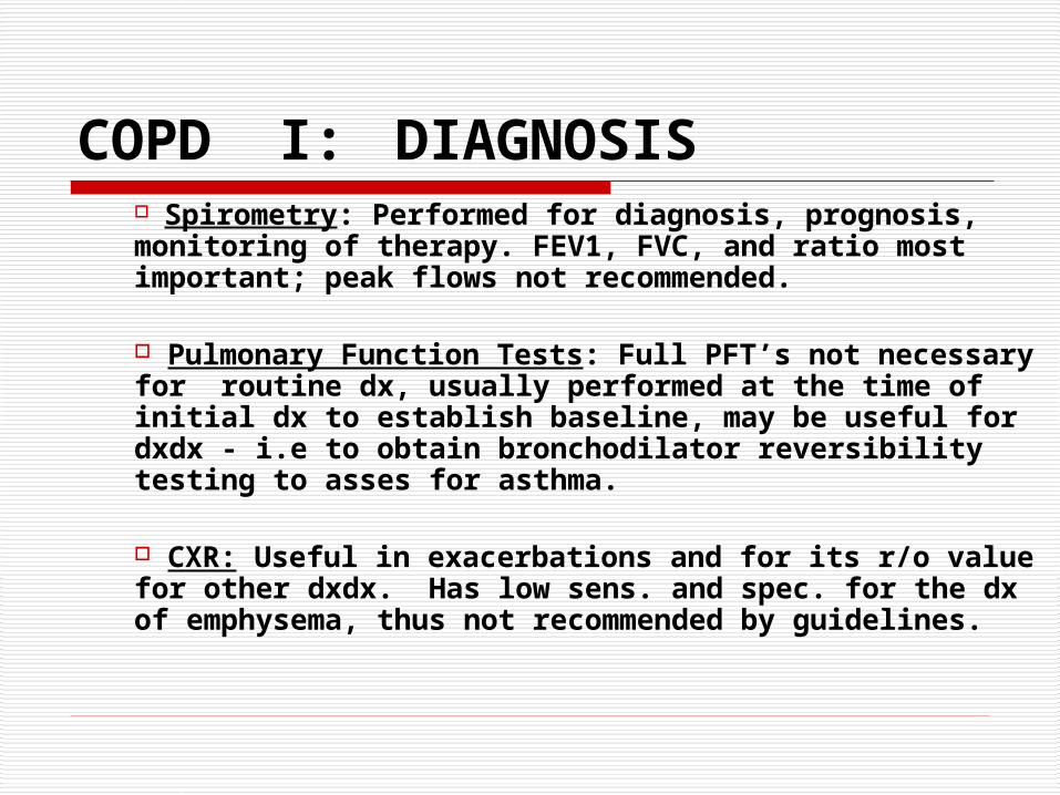
COPD I: DIAGNOSIS Spirometry: Performed for diagnosis, prognosis, monitoring of therapy. FEV1, FVC, and ratio most important; peak flows not recommended.
Pulmonary Function Tests: Full PFT’s not necessary for routine dx, usually performed at the time of initial dx to establish baseline, may be useful for dxdx - i.e to obtain bronchodilator reversibility testing to asses for asthma.
CXR: Useful in exacerbations and for its r/o value for other dxdx. Has low sens. and spec. for the dx of emphysema, thus not recommended by guidelines.
COPD I: DIAGNOSIS Helical CT of Chest: Not necessary for routine diagnosis, may be useful for dxdx or for lung volume reduction OR.
Allergy Testing: May have use in asthma, not COPD.
O2 Sat: In severe COPD (stage 2b or 3) useful to guide O2 therapy. Nocturnal desaturations are probably under diagnosed.
ABG: Needed to guide long term oxygen therapy and to obtain government funding for same. (See guidelines for actual criteria for initiation of treatment… will be discussed next week).
COPD I: DIAGNOSIS
CASE (cont.):
The current Canadian guidelines: do not emphasize evidence based diagnosis for
patients with COPD put more emphasis on evaluation of
impairment, disability with exercise testing, dyspnea assessment scales, and quality of life assessment scales
do not give specific recommendations on how or at what point in the patient’s course these evaluations should be used
COPD: other useful references:
2 recent review series on COPD: 5 article series on exacerbations:
Thorax Feb – June, 2006 12 article series:
BMJ May 13th to July 22nd, 2006
Excellent recent update: Update in Chronic Obstructive Pulmonary
Disease 2006: Rabe KF, et al. Am J Resp Crit Care 2007; 175: 1222-1232