LONG-TERM FOLLOW-UP OF EXERCISE REHABILITATION OUTCOMES IN PATIENTS WITH CHRONIC OBSTRUCTIVE PULMONARY DISEASE By TAMARA MARIE ARROWOOD A Thesis Submitted to the Graduate Faculty of WAKE FOREST UNIVERSITY in Partial Fulfillment of the Requirements for the Degree of MASTER OF SCIENCE in the Department of Health and Exercise Science May 2002 Winston-Salem, North Carolina Approved By: Michael J. Berry, Ph.D., Advisor ___________________________ Examining Committee: Gary D. Miller, Ph.D. ___________________________ Patricia A. Nixon, Ph.D. ___________________________
Transcript
LONG-TERM FOLLOW-UP OF EXERCISE REHABILITATION OUTCOMES IN PATIENTS WITH CHRONIC OBSTRUCTIVE PULMONARY DISEASE
By
TAMARA MARIE ARROWOOD
A Thesis Submitted to the Graduate Faculty of
WAKE FOREST UNIVERSITY
in Partial Fulfillment of the Requirements
for the Degree of
MASTER OF SCIENCE
in the Department of Health and Exercise Science
May 2002
Winston-Salem, North Carolina
Approved By: Michael J. Berry, Ph.D., Advisor ___________________________ Examining Committee: Gary D. Miller, Ph.D. ___________________________ Patricia A. Nixon, Ph.D. ___________________________
DEDICATION
This thesis is dedicated to my mom and dad, A.K.A. Gongo and Bubba, who have always believed in me, supported me in every possible way, showed me the value of determination and hard work, inspired me to dream big and go after whatever I wanted, and encouraged me to be the best person that I could possibly be. To my sister, Cindy, and Nader, Nathan, and Megan, who instill in me the true meaning of joy by letting me experience the happiness of childhood all over again and by being such a loving family. To Rick, who over the years has helped me grow, and learn more about myself than I ever thought I wanted to know.
ii
ACKNOWLEDGEMENTS
I wish to thank many people who have made graduate school and the last 2 years such an enjoyable experience for me:
My family, who without their continual encouragement, love, and support, I would be unable to accomplish all that I have in my life.
Bill, for his love, support, and confidence in me to always do my best.
My classmates: Gretchen, Jamie, Stacey, Leigh Anne, and Theresa; I couldn’t ask for a more supportive, caring, and entertaining group. Thanks, I love you guys! Jim, for his friendship, guidance, and never-ending encouragement throughout the past year.
Dr. Berry, for sharing his expertise and outrageous sense of humor with me over the past 2 years, as well as for his ability to somehow make “square wheels roll”.
Dr. Miller and Dr. Nixon, for their valuable input to this project and for serving on my thesis examining committee. Dr. Ribisl and the HES Department for giving me the incredible opportunity to study, learn, teach, lead, and become a part of the Wake Forest family. The Cardiac Rehab Program staff and participants, for making my job not seem like “work”.
The REACT participants who graciously volunteered their time and effort to make this study possible, and also Dave and Eve-Marie for their support. The HES Classes of 2001 and 2003, for all of their help and for reminding me how to have fun!
Ann, Dorothy, Sherry, and Teresa, all very good friends who have stuck by me over the years, through good times and bad.
iii
TABLE OF CONTENTS
DEDICATION................................................................................................................... ii
ACKNOWLEDGEMENTS ............................................................................................ iii
TABLE OF CONTENTS ................................................................................................ iv
LIST OF ABBREVIATIONS ......................................................................................... vi
LIST OF TABLES AND FIGURES.............................................................................. vii
ABSTRACT.................................................................................................................... viii
Outcome Measures........................................................................................................ 43 Pulmonary Function Tests ........................................................................................ 43 Physical Activity Scale for the Elderly..................................................................... 43 Physical Function Questionnaire .............................................................................. 43 Chronic Respiratory Disease Index Questionnaire ................................................... 44 6-Minute Walk Test .................................................................................................. 45
SCHOLASTIC VITA ..................................................................................................... 78
v
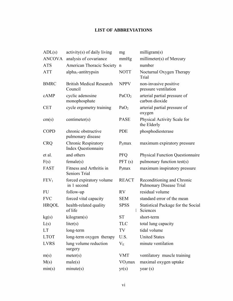
LIST OF ABBREVIATIONS
ADL(s) activity(s) of daily living mg milligram(s) ANCOVA analysis of covariance mmHg millimeter(s) of Mercury ATS American Thoracic Society n number ATT alpha1-antitrypsin NOTT Nocturnal Oxygen Therapy
Trial BMRC British Medical Research
Council NPPV non-invasive positive
pressure ventilation cAMP cyclic adenosine
monophosphate PaCO2 arterial partial pressure of
carbon dioxide CET cycle ergometry training PaO2 arterial partial pressure of
oxygen cm(s) centimeter(s)
PASE Physical Activity Scale for
the Elderly COPD chronic obstructive
pulmonary disease PDE phosphodiesterase
CRQ Chronic Respiratory Index Questionnaire
PEmax maximum expiratory pressure
et al. and others PFQ Physical Function Questionnaire F(s) female(s) PFT (s) pulmonary function test(s) FAST Fitness and Arthritis in
Seniors Trial PImax maximum inspiratory pressure
FEV1 forced expiratory volume in 1 second
REACT Reconditioning and Chronic Pulmonary Disease Trial
FU follow-up RV residual volume FVC forced vital capacity SEM standard error of the mean HRQOL health-related quality
of life SPSS Statistical Package for the Social
l Sciences kg(s) kilogram(s) ST short-term L(s) liter(s) TLC total lung capacity LT long-term TV tidal volume LTOT long-term oxygen therapy U.S. United States LVRS lung volume reduction
surgery VE minute ventilation
m(s) meter(s) VMT ventilatory muscle training M(s) male(s) VO2max maximal oxygen uptake min(s) minute(s) yr(s) year (s)
vi
LIST OF TABLES AND FIGURES
TABLE PAGE I Participant Demographics/Clinical Information ……………………………….48
FIGURES
1 ST vs. LT Mean FEV1 % Predicted…………………………………………….49
2 ST vs. LT Mean FEV1/FVC Ratio …………………………………………….50
3 ST vs. LT Mean RV/TLC Ratio………………………………………………..50
4 ST vs. LT Mean CRQ Dyspnea Score………………………………………….51
5 ST vs. LT Mean CRQ Mastery Score…………………………………………..52
6 ST vs. LT Mean CRQ Emotion Score…………………………………………..53
7 ST vs. LT Mean CRQ Fatigue Score…………………………………………....53
8 ST vs. LT Mean PASE Score…………………………………………………...54
9 ST vs. LT Mean PFQ Score…………………………………………………….55
10 ST vs. LT Mean 6-Minute Walk Test Distance ………….…………………….56
vii
Tamara Marie Arrowood ABSTRACT
LONG-TERM FOLLOW-UP OF EXERCISE REHABILITATION OUTCOMES IN PATIENTS WITH CHRONIC OBSTRUCTIVE PULMONARY DISEASE
Thesis under the direction of Michael J. Berry, Ph.D., Professor Health and Exercise Science The purpose of this study was to compare the long-term outcomes in pulmonary
function, self-reported health–related quality of life, physical activity, and disability,
along with functional exercise capacity in COPD patients either completing a 3-month
(short-term, ST) or an 18-month (long-term, LT) exercise rehabilitation program at 58
months. Thirty-nine patients completed the follow-up study, including 12 from the ST
and 27 from the LT groups. There were no significant differences between the ST and the
LT groups in the adjusted means of FEV1 % predicted, (57.6 ± 3.1 versus 56.6 ± 2.1%),
FEV1/FVC ratio, (51.5 ± 2.1 versus 52.1 ± 1.4%), RV/TLC ratio, (53.7 ± 4.2 versus 58.4
± 2.8%), CRQ: dyspnea (4.9 ± 0.4 versus 5.1 ± 0.2 units), mastery (6.3 ± 0.2 versus 6.4 ±
0.1 units), emotion (5.6 ± 0.2 versus 5.7 ± 0.1 units), fatigue (4.3 ± 0.3 versus 4.7 ± 0.2
units), PASE (103.0 ± 12.2 versus 92.2 ± 8.8 units), PFQ (35.0 ± 3.8 versus 39.4 ± 2.5
units), or 6-minute walk distance, (500.7 ± 26.6 versus 517.9 ± 18.8 m) respectively,
measured at the 58-months. These results showed that at 58 months there are no
significant differences between the ST and the LT exercise rehabilitation groups in any of
the outcome variables. Therefore, an additional 15 months of participation in an exercise
rehabilitation program did not result in a difference in the level of benefits maintained at
58 months.
viii
INTRODUCTION
Chronic obstructive pulmonary disease (COPD), a condition of progressive
deterioration of the respiratory system characterized by the obstruction of pulmonary
airways and decreased airflow, is a major health problem worldwide. COPD is currently
the fourth leading cause of death in the United States (U.S.), following heart disease,
cancer, and stroke, (95) and is the only leading cause of death which has increased in
prevalence (by 71%) over the last several years (68). COPD also has a major influence on
morbidity, accounting for an estimated 668,362 hospital discharges at a rate of 24.5 per
10,000 individuals in 1998 (95).
Individuals with COPD experience functional capacity limitations that adversely
impact the physical activity level and the performance of activities of daily living (ADL’s)
in these patients. A recent national survey of 573 individuals with COPD given by
Schulman, Ronca, and Bucuvalas, Incorporated, and supported by the American Lung
Association (100) revealed that 70% of respondents experienced limitations in activities
requiring physical exertion, 51% had limitations in their occupational abilities, and 56%
were limited in performing household chores. The survey also showed that social
activities and family activities were limited in 53% and 46% respectively, while normal
sleeping patterns were disturbed in 50% of respondents.
As a result of these physical limitations, the quality of life for many COPD
patients and their families is significantly compromised. Twenty-three percent of
respondents described themselves as “invalid”, while 8% remained homebound due to
dyspnea (100), not only because of its physical effects, but also due to the fear and the
1
anxiety associated with the thought of experiencing the sensation of breathlessness
outside of the home.
The medical treatment of individuals with COPD centers on the management of
the current symptoms and the prevention of future exacerbations. Various drug therapies,
supplemental oxygen, pulmonary rehabilitation programs, and surgical interventions all
are currently available treatment options. Smoking cessation is paramount to any
successful treatment plan in COPD patients who continue to smoke, and many alternative
strategies are available to assist with this difficult process (29, 91, 94).
Multidisciplinary pulmonary rehabilitation programs have been successful in
enhancing the physical function and the health related quality of life (HRQOL) in
individuals with COPD. A meta-analysis of 14 randomized controlled clinical trials
looking at the effectiveness of pulmonary rehabilitation revealed that significant
improvements occurred in maximal exercise capacity, functional exercise capacity, and
HRQOL in pulmonary rehabilitation participants as compared to individuals in the
control groups (72).
The long-term (greater than one year post intervention) benefits of participation in
pulmonary rehabilitation programs have also been analyzed (13, 21, 61, 81, 102, 123, 146,
147). The outcomes typically measured in these long-term follow-up studies include
physical performance parameters, HRQOL, morbidity as shown by the number of
hospitalizations or by the total number of days of hospitalization, mortality, as well as the
total utilization of healthcare dollars. In a randomized control trial (128) the long-term
benefits achieved by 50 COPD patients who underwent a 6-month outpatient exercise
therapy only regimen were compared to 50 COPD patients who received “usual care”
2
treatment. The significant improvements in the 6-minute walk test distance, maximal
oxygen uptake (VO2max), quadriceps strength, and HRQOL made by the exercise
therapy group (n=37), as compared to the control group (n=33), were still present and
clinically relevant at the LT follow-up visit.
Strijbos et al. (123) examined the long-term outcomes of COPD patients
participating in a 12-week home-based or a hospital-based outpatient pulmonary
rehabilitation program, as compared to that of a control group that did not receive an
exercise intervention. Their findings showed that both the hospital-based and the home-
based rehabilitation groups had similar significant improvements in exercise capacity,
perceived dyspnea, and well-being at the 6-month follow-up as compared to the control
group. At 18 months, the improvement in exercise capacity began to diminish in the
hospital-based rehabilitation group, as compared to that of the home-based rehabilitation
group. The benefits achieved in both rehabilitation groups were still statistically
significant when compared to the control group at 18 months.
A randomized control trial by Ries et al. (102) found that many of the significant
physical and psychological benefits achieved from participating in an 8-week outpatient
comprehensive pulmonary rehabilitation program diminished within one year of follow-
up. Results from the Reconditioning and Chronic Pulmonary Disease Trial (REACT)
study by Berry et al. (21) also showed that the improvements in self-reported disability,
physical function, and HRQOL made by COPD patients following the completion of a
3-month exercise therapy program had greatly diminished by 18 months, while the
18-month exercise therapy group maintained and improved upon the benefits achieved at
the end of the initial 3-month program.
3
Maintenance exercise programs initiated after the completion of a structured
pulmonary rehabilitation program have been successful in postponing the decline in
benefits achieved following the initial intervention. A study by Guell and colleagues (61)
compared the long-term effects of a 3-month exercise training program followed by a
6-month maintenance program in 30 COPD patients (exercise group) to that of 30 COPD
patients who received standard care only (control group). The results revealed that a
6-month supervised one-time-a-week maintenance therapy program consisting of
breathing and arm/leg coordination exercises helped maintain the benefits achieved in
6-minute walk distance, dyspnea, and HRQOL in the exercise group at 24 months. This
study also showed that the exercise group experienced significantly fewer COPD
exacerbations (p<0.0001) and a trend for fewer hospitalizations than the control group.
Groisbois et al. (59) investigated the effects that a maintenance exercise regimen had on
58 COPD patients who had completed an initial 7-week outpatient pulmonary
rehabilitation program. The patients were self-selected into 1 of 4 groups who had
continued supervised exercise training either once or twice a week, continued an
independent home exercise program, or did not continue any type of exercise program.
Improvements in maximum workload and dyspnea ratings occurred in all patients
following completion of the initial program, but remained significant only in the once-a-
week supervised exercise training group and the independent home exercise group at the
LT follow-up. The non-exercisers showed diminished benefits approaching baseline
measures at 18 months.
The duration of an exercise intervention may also influence the magnitude of
benefits achieved as well as the length of time that they are maintained in COPD patients.
4
The REACT study, a recent unpublished investigation by Berry and colleagues (21)
compared the effects of a 3-month (short-term, ST) versus an 18-month (long-term, LT)
exercise rehabilitation program on self-reported disability, physical function, and
HRQOL in 140 COPD patients. The results showed that both the ST and the LT groups
had similar improvements in these outcomes at 3 months. At 18 months, those patients
randomized into the LT exercise group (n=62) maintained the improvements in self-
reported disability, physical function, and HRQOL obtained at 3 months. Those patients
in the ST exercise group (n=56), showed declining benefits at 18 months, which
approached baseline values. The results of this study present a case for prolonged
exercise rehabilitation programs (greater than 3 months), but also showed that the
benefits achieved from short-term exercise rehabilitation programs are not permanent.
While the results of the REACT study showed diminished benefits in COPD
patients who completed a ST exercise rehabilitation program at 18 months, it is not
known whether this decline in benefits persisted beyond 18 months. It is also not known
whether the improvements made by those patients participating in the LT exercise
rehabilitation program were maintained. Therefore, it was the primary purpose of this
thesis to describe and compare the outcomes of COPD patients who participated in either
a ST or a LT exercise rehabilitation program at 58 months.
5
LITERATURE REVIEW
Definition of Chronic Obstructive Pulmonary Disease
Chronic obstructive pulmonary disease (COPD) is a progressive lung disease
characterized primarily by airway obstruction and decreased airflow. Chronic bronchitis
and emphysema are 2 distinct components of COPD, but may occur simultaneously in the
same individual. Chronic bronchitis is defined as “the presence of a chronic cough for 3
months in each of 2 successive years in a patient in whom other causes of chronic cough
have been excluded” (3). Chronic bronchitis is characterized by chronic inflammation
and edema of the peripheral airways, excessive mucus production and accumulation,
bronchospasm, bronchial airway obstruction hyperinflation of the alveoli distal to the
obstructed airways.
Emphysema is defined as “the abnormal permanent enlargement of the airspaces
distal to the terminal bronchioles, accompanied by the destruction of their walls and
without obvious fibrosis” (3). Emphysema is characterized by alveolar deterioration and
hyperinflation, destruction of pulmonary capillaries, weakened respiratory bronchioles,
and air trapping.
Depending upon the degree of pulmonary system damage and airway
hyperresponsiveness, the following may all occur in an individual with COPD: obstructed
airflow, lung hyperinflation, mismatched ventilation/perfusion ratio, decreased maximum
inspiratory pressure (PImax), increased work of breathing, and a decreased gas diffusion
capacity (33). The primary physical manifestations that result from these
pathophysiologic changes include dyspnea (the sensation of breathlessness), persistent
and functional exercise capacity in COPD patients either completing a ST (3-month) or a
LT (18-month) exercise rehabilitation program at 58 months.
HYPOTHESIS
There will be no significant differences in the long-term outcomes between the ST
and the LT exercise rehabilitation groups at the 58-month follow-up.
40
METHODS
Reconditioning Exercise and Chronic Obstructive Pulmonary Disease Trial
The Reconditioning Exercise and Chronic Obstructive Pulmonary Disease Trial
(REACT) (21) served as the original research study from which COPD patients were
recruited for the current follow-up study. The REACT study was conducted from August
1995 through July 1999 and involved 140 patients with COPD (non-reversible airflow
limitation with a FEV1 >/= 20% of predicted and a FEV1/FVC ratio of </= 70%). All
patients participated in a supervised, 3-time per week, facility-based aerobic and upper
extremity resistive group exercise intervention lasting 3 months. Following the 3-month
exercise intervention, the patients were randomly assigned to either one of 2 groups,
those who received an additional 15 months of supervised exercise training (long-term,
LT) or those who did not (short-term, ST). Outcome measures were taken at baseline, 3,
9, 15, and 18 months and included self-reported disability, physical function (as
determined by 6-minute walk test, stair climb time, and overhead task time), pulmonary
function tests (PFT’s), self-reported physical activity, and HRQOL.
Participants
All participants were volunteers recruited from COPD patients who had
completed the initial REACT study. Of the 140 original REACT participants, 39 agreed
to participate in the follow-up study, 5 were known to have expired, while the remainder
of subjects either declined to participate or were unable to be contacted. Informed consent
41
for the follow-up study was obtained from all participants and the Wake Forest
University Institutional Review Board approved the study protocol.
Procedures
All follow-up testing was completed during a 1-time visit to the Wake Forest
University Human Performance Laboratory. Subjects were asked to refrain from using
bronchodilator inhalers within 4 hours prior to testing, but were instructed to take all
other medications as usual. Subjects completed a comprehensive medical history
questionnaire, physical activity readiness questionnaire (9), and a current medication
form.
Each subject’s height and weight (in shoes) were determined using a sliding
balance scale. Pulmonary function tests were then performed using a Medical Graphics
Corporation (St. Paul, MN) 1085D plethysmograph according to the guidelines set by the
ATS (2). Following the PFT’s, subjects completed the physical activity scale for the
elderly (PASE) (137) and the physical function questionnaire (PFQ) (78). A trained
examiner then administered the CRQ (63). Performance of the 6-minute walk test to
assess the functional capacity of each subject completed the follow-up testing protocol.
The 6-minute walk test was not performed in those subjects who had a history of chest
pain while walking or at rest, or who currently had musculoskeletal conditions that
compromised the safe completion of the test.
42
Outcome Measures
Pulmonary Function Tests
Pulmonary function tests determined each subject’s forced expiratory volume in
1 second (FEV1), forced vital capacity (FVC), residual volume (RV), and total lung
capacity (TLC). Forced expiratory volume was expressed as a percent of predicted, based
on the equations of Knudson and colleagues (71) and also as a percent of FVC. All
subjects met reproducibility and acceptable requirements for testing as described by the
ATS (2).
Physical Activity Scale for the Elderly
The PASE (137) is a self-administered questionnaire that is based on a 7-day
recall of occupational, household, and leisure activities. The frequency and duration of
occupational and leisure activities were assessed. The total PASE score is based on
summing the totals of each activity’s empirically derived weight times the duration of
performance, or participation (136). The PASE has been validated and shown to be
reliable as an indicator of physical activity in older adults (136, 137).
Physical Function Questionnaire
The PFQ is a self-administered tool used to measure the level of physical
disability experienced by subjects due to their health over the past month. It is a modified
version of an original questionnaire used in the Fitness and Arthritis in Seniors Trial
(FAST) (99) and is composed of 23 items describing common daily activities. Each item
of the PFQ is scored on a 1-6 scale, with 1 representing “usually did with no difficulty”,
and 6 representing “usually did not do for other reasons”. The scores were categorized
into five activity subscales including basic, transfer, ambulation/climbing, upper
43
extremity, and complex. An overall score was determined by a composite average of
responses for all 23 items.
Chronic Respiratory Disease Index Questionnaire
The CRQ (63) is a disease specific, examiner administered tool that measures the
change in HRQOL over time. The HRQOL is categorized into four categories including
dyspnea, fatigue, emotional function, and mastery of the disease. With respect to the
dyspnea category, subjects listed five important activities in which they experienced
shortness of breath or dyspnea to individualize the baseline CRQ. The same five activities
were used on each subsequent administration of the CRQ to assess the change in dyspnea
over time. Subjects were informed of the answer given for each item during the previous
administration of the CRQ in order to decrease the randomness of their current responses.
The answers were scored on a 1-7 scale, with 1 representing “extremely short of breath”
and 7 representing “not at all short of breath”. The remainder of the questions were not
individualized and dealt with fatigue, emotional function, and mastery of the disease.
These questions were scored on a 1-7 scale, with choices ranging from “all of the time”
to “none of the time”. Scoring is based on the mean responses of each of the 4 categories.
The minimal clinically important difference has been shown to be 0.5 units per question
per dimension (62, 72). Torres and colleagues (35) have determined that the CRQ is one
of the best practical tools for measuring the efficacy of pulmonary rehabilitation due to its
simplicity to administer and sensitivity to detect clinically important differences in COPD
patients.
44
6-Minute Walk Test
The 6-minute walk test was performed to assess the functional capacity of each
subject. Subjects were instructed to cover the greatest distance possible by walking for
6 minutes around the perimeter of a gym that measured 70’x 88’. Subjects’ walking pace
was self-determined and rest periods were allowed if needed. Verbal encouragement was
not provided and subjects were not permitted to time or pace themselves by using a watch.
Performance was based on the measured total distance walked. The minimal clinically
important distance has been established to be 54 m in patients with COPD (98). The
6-minute walk test has been deemed a reliable and valid measure in evaluating the
physical response of COPD patients to pulmonary rehabilitation programs (35). Although
the 6-minute walk test is considered a submaximal exercise test, positive correlations of
between 0.51 and 0.90 have been demonstrated between total walk distance and VO2max
(121), thus making the 6-minute walk test a good measure of functional exercise capacity
when properly performed.
Statistical Analysis
Descriptive statistics were determined for all variables measured in the ST and LT
groups at baseline and 58 months. Independent t-tests were utilized to compare the means
for the ST and LT groups’ descriptive statistics. Independent t-tests were also performed
on selected variables of physical function and disease severity using the original baseline
REACT data. Comparisons of these variables were made between the group of
participants who returned for the 58-month follow-up study and the group of participants
who did not return. Analysis of covariance (ANCOVA), using baseline scores as the
45
covariate, was performed on the PFQ, CRQ, and PASE scores, as well as the PFT values
and the 6-minute walk test distance. All statistical analyses were performed using the
Statistical Package for Social Sciences (SPSS) software for Windows 98 (version 10.1,
SPSS, Incorporated, Chicago, IL). The significance level was set at p = 0.05.
46
RESULTS
A total of 39 (23 M/16 F) participants completed the REACT 58-month follow-up
study, with an average age of 72.4 ± 6.2 yrs (range = 62-85 yrs) and average follow-up
time of 58.5 ± 6.2 months, (range = 48-72 months). Table I shows participant
demographic and clinical characteristics. Overall, there were no significant differences in
the demographic characteristics between the ST and the LT groups at 58 months. The
most common associated comorbidities included hypertension, arthritis, and coronary
artery disease respectively, which is very similar to a sample of 290 COPD patients
studied by Van Manen (134). Using the original REACT data set, a comparison of the
functional exercise capacity between those participants who returned (returnees) for the
58-month follow-up study and those who did not return (non-returnees) revealed that the
returnees demonstrated significantly higher (p=0.01) functional exercise capacity as
determined by the 6-minute walk test distance at 18 months than the non-returnees (518 ±
13 m versus 474 ± 10 m respectively).
47
TABLE I*
PARTICIPANT DEMOGRAPHICS/CLINICAL INFORMATION
PARTICIPANTS
ST
(3-MONTH)
LT
(18-MONTH)
Completed REACT, n 56 62 Completed 58 mos FU, n (%) 12 (21.4) 27 (43.5) Unable to contact 11 (19.6) 8 (12.9) Contacted but refused FU 17 (30.4) 12 (19.4) Expired 5 (8.9) 0 (0) CHARACTERISTICS
Weight, kg 79.4 ± 4.5 79.1 ± 3.7 Gender, M, n (%)/ F, n (%) 6 (50)/6 (50) 17 (63)/10 (37) Age, yrs 71.3 ± 1.8 72.9 ± 1.2 Height, cm 168.3 ± 3.4 171.7 ± 1.6 FU time, mos 59.3 ± 1.8 58.2 ± 1.2 COMORBIDITY
Arthritis, n (%) 5 (41.7) 14 (51.9) Cancer, n (%) 2 (16.7) 3 (11.1) Circulatory problems, n (%) 1 (8.3) 3 (11.1) Coronary artery disease, n (%) 4 (33.3) 6 (22.2) Diabetes mellitus, n (%) 1 (8.3) 2 (7.4) Hypertension, n (%) 6 (50.0) 13 (48.1) Transient ischemic attack, n (%) 2 (16.7) 2 (7.4) SMOKING STATUS
Current, n (%) 0 (0) 3 (11.1) Past, n (%) 8 (75) 25 (92.6)
*values for FU time, age, height, weight, and pack yrs are means ± SEM; ST= short-term, LT= long-term, mos=months, FU= follow-up, n= number, kg= kilograms, M= male, F= female, yrs= years, cm= centimeters, SEM= standard error of the mean.
48
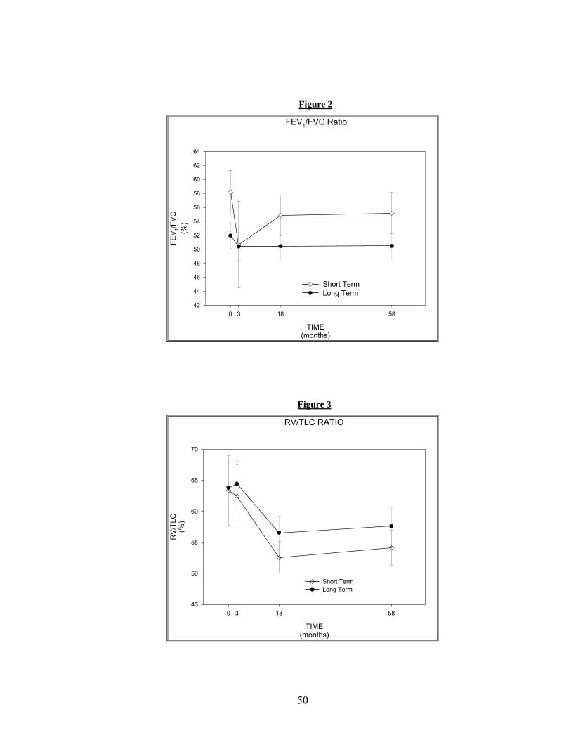
Pulmonary Function Tests
Mean FEV1 % predicted scores for both the ST and the LT groups at 0, 3, 18, and
58 months are shown in Figure 1. No significant differences were found when comparing
adjusted mean scores between the ST and the LT groups at 58 months, (57.6 ± 3.1 vs.
56.6 ± 2.1%, respectively). Mean FEV1/FVC scores for both the ST and LT groups at 0, 3,
18, and 58 months are shown in Figure 2. No significant differences were found when
comparing adjusted mean scores between the ST and LT groups at 58 months, (51.5 ± 2.1
vs. 52.1 ± 1.4 %, respectively). Mean RV/TLC ratio scores for both the ST and the LT
groups at 0, 3, 18, and 58 months are shown in Figure 3. No significant differences were
found when comparing adjusted mean scores between the ST and the LT groups at 58
months, (53.7 ± 4.2 versus 58.4 ± 2.8%, respectively).
Figure 1
FEV1 Predicted
TIME(months)
0 3 18 58
FEV 1 P
RED
ICTE
D(%
)
40
45
50
55
60
65
70
Short Term Long Term
49
Figure 2
FEV1/FVC Ratio
TIME(months)
0 3 18 58
FEV 1/F
VC(%
)
42
44
46
48
50
52
54
56
58
60
62
64
Short TermLong Term
Figure 3
RV/TLC RATIO
TIME(months)
0 3 18 58
RV/
TLC
(%)
45
50
55
60
65
70
Short TermLong Term
50
Chronic Respiratory Index Questionnaire
With respect to the CRQ, no significant differences were observed between the
ST and the LT groups in any of the individual domain scores (dyspnea, mastery, emotion,
or fatigue) at 58 months. Mean dyspnea scores for both the ST and the LT groups at time
0, 3, 18, and 58 months are shown in Figure 4. No significant differences were found
when comparing adjusted mean scores between the ST and the LT groups at 58 months,
(4.9 ± 0.4 versus 5.1 ± 0.2 units, respectively)
Figure 4
CRQ
TIME(months)
0 3 18 58
DYS
PNEA
SC
OR
E
3
4
5
6
Short TermLong Term
51
Mean mastery scores for both the ST and the LT groups at time 0, 3, 18, and 58 months
are shown in Figure 5. No significant differences were found when comparing adjusted
mean scores between the ST and the LT groups at 58 months, (6.3 ± 0.2 versus 6.4 ± 0.1
units, respectively).
Figure 5
CRQ
TIME(months)
0 3 18 58
MAS
TER
Y SC
OR
E
5
6
7
8
Short TermLong Term
Mean emotion scores for both the ST and the LT groups at time 0, 3, 18, and 58 months
are shown in Figure 6. No significant differences were found when comparing adjusted
mean scores between the ST and the LT groups at 58 months, (5.6 ± 0.2 versus 5.7 ± 0.1
units, respectively). Mean fatigue scores for both the ST and the LT groups at time 0, 3,
18, and 58 months are shown in Figure 7. No significant differences were found when
comparing adjusted mean scores between the ST and the LT groups at 58 months, (4.3 ±
0.3 versus 4.7 ± 0.2 units, respectively).
52
Figure 6
CRQ
TIME(months)
0 3 18 58
EMO
TIO
N S
CO
RE
5.0
5.2
5.4
5.6
5.8
6.0
Short TermLong Term
Figure 7
CRQ
TIME(months)
0 3 18 58
FATI
GU
E SC
OR
E
3
4
5
6
Short Term Long Term
53
Physical Activity Scale for the Elderly
Mean PASE scores for both the ST and the LT groups at time 0, 3, 18, and 58
months are shown in Figure 8. No significant differences were found when comparing
adjusted mean scores between the ST and the LT groups at 58 months, (103.0 ± 12.2
versus 92.2 ± 8.8 units, respectively).
Figure 8
PASE
TIME(months)
0 3 18 58
PA
SE
SC
OR
E
70
80
90
100
110
120
130
Short TermLong Term
54
Physical Function Questionnaire
Mean PFQ scores for both the ST and the LT groups at time 0, 3, 18, and 58
months are shown in Figure 9. No significant differences were found when comparing
adjusted mean scores between the ST and the LT groups at 58 months, (35.0 ± 3.8 versus
39.4 ± 2.5 units, respectively). No significant differences were observed when comparing
adjusted mean scores between the ST and the LT groups in any of the activity subscales
at 58 months: (basic: 1.1 ± 0.1 versus 1.3 ± 0.1, transfer: 1.3 ± 0.2 versus 1.3 ± 0.1,
ambulation/climbing: 2.1 ± 0.3 versus 2.3 ± 0.2, upper extremity: 1.7 ± 0.2 versus 1.9 ±
0.1, or complex: 1.7 ± 0.3 versus 1.8 ± 0.2 units, respectively).
Figure 9
PFQ
TIME(months)
0 3 18 58
PFQ
SC
OR
E
1.2
1.4
1.6
1.8
2.0
Short TermLong Term
55
6-Minute Walk Test
Mean 6-minute walk test distance values for both the ST and the LT groups at
time 0, 3, 18, and 58 months are shown in Figure 10. No significant differences were
found when comparing adjusted mean scores between the ST and the LT groups at 58
months, (500.7 ± 26.6 versus 517.9 ± 18.8 m, respectively).
Figure 10
6-Minute Walk Test
TIME(months)
0 3 18 58
6-M
inut
e W
alk
Dis
tanc
e(m
eter
s)
400
450
500
550
600
650
Short TermLong Term
56
DISCUSSION
The original REACT study was a clinical trial in which patients with COPD
participated in a 3-month facility-based exercise therapy program and then were either
randomized to continue the supervised program for an extra 15 months or were
encouraged to continue to exercise on their own, independent of the study. At 18 months,
study participants returned for follow-up assessments. In general, the results of the
REACT study revealed that both the ST and the LT exercise therapy groups showed
improvements in the HRQOL, self-reported disability, and functional exercise capacity at
the completion of the initial ST exercise therapy program. At 18 months, these benefits
were maintained in the LT group, but were not maintained in the ST group whose
outcome variables approached the initial baseline measures. The primary purpose of this
follow-up study was to describe and compare the long-term outcomes of COPD patients
who had participated in either a ST or an LT exercise rehabilitation program (REACT) at
58 months. The outcomes assessed included: lung function (PFT’s- FEV1 % predicted,
FEV1/FVC ratio, and RV/TLC ratio); HRQOL (CRQ); self-reported physical activity
(PASE); self-reported disability (PFQ); and functional capacity (6-minute walk test).
Overall, the results of this study showed no significant differences between the ST and
the LT groups in any of these variables measured at 58 months. These findings suggest
that even patients who participated in a long-term formal exercise rehabilitation program
will not maintain the benefits achieved once the program ends.
Research has demonstrated that the benefits gained from participation in
pulmonary rehabilitation programs that include exercise therapy, can be maintained for
57
up to 2 years (48)and even 3 years in select COPD patients (122). Other studies have
shown that the benefits gained from participation in pulmonary rehabilitation programs
diminish by 18 months following completion of these programs (21;102;123). To our
knowledge, this is the first study to look at the long-term (58 months) outcomes in COPD
patients following completion of an 18-month exercise rehabilitation therapy program.
Lung function (FEV1% predicted, FEV1/FVC ratio, and RV/TLC ratio), as
measured by PFT’s, was not significantly different between the ST and LT groups at 58
months. Many studies investigating the efficacy of pulmonary rehabilitation programs
demonstrate that lung function is typically not affected by exercise therapy interventions
(51;102).
Results from the present investigation show that HRQOL, as measured by the
four domains of the CRQ (dyspnea, mastery, emotion, and fatigue) was not significantly
different between patients from the ST and the LT exercise therapy groups at 58 months.
More specifically, the results of this follow-up study showed that the dyspnea, mastery,
and emotion scores were similar between the 2 groups at 58 months. Results from the
original REACT study showed a significant difference in all domains of the CRQ when
comparing the ST and the LT exercise therapy groups at the 18-month follow-up (49).
Given the fact that the differences seen at 18 months between the 2 groups were not seen
at 58 months, it does not appear that those patients participating in an 18-month exercise
therapy program will maintain long-term benefits to any greater degree than those
participating in a short-term exercise therapy program.
Self-reported activity, as measured by the PASE, was not significantly different
between the ST and the LT groups at 58 months. Older adult volunteers were recruited
58
from community centers and retirement homes in order to assess the value of the PASE
in measuring physical activity in a study conducted by Harada et al. (65). The mean
PASE score was 50 ± 44 units in 36 adults recruited from retirement homes, while the
mean PASE score for 51 adults recruited from community centers was 158 ± 65 units,
respectively. The adjusted mean PASE scores from the ST and the LT groups this follow-
up study were 103 ± 12 and 92 ± 9 units, which demonstrates that our participants were
more active than those individuals residing in retirement homes and somewhat less active
than those individuals recruited from community centers. The subjects in each study were
all of similar age. These results support the idea that individuals with COPD are less
active on a regular basis than independent-living older adults without COPD.
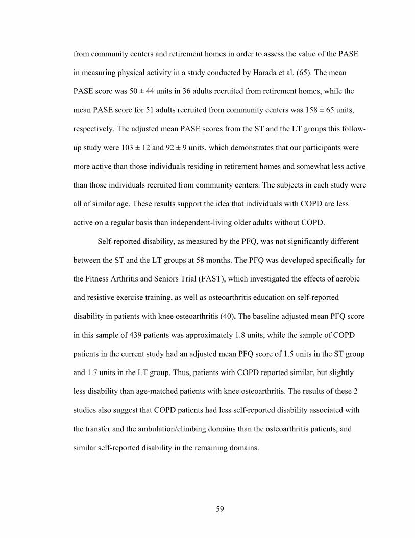
Self-reported disability, as measured by the PFQ, was not significantly different
between the ST and the LT groups at 58 months. The PFQ was developed specifically for
the Fitness Arthritis and Seniors Trial (FAST), which investigated the effects of aerobic
and resistive exercise training, as well as osteoarthritis education on self-reported
disability in patients with knee osteoarthritis (40). The baseline adjusted mean PFQ score
in this sample of 439 patients was approximately 1.8 units, while the sample of COPD
patients in the current study had an adjusted mean PFQ score of 1.5 units in the ST group
and 1.7 units in the LT group. Thus, patients with COPD reported similar, but slightly
less disability than age-matched patients with knee osteoarthritis. The results of these 2
studies also suggest that COPD patients had less self-reported disability associated with
the transfer and the ambulation/climbing domains than the osteoarthritis patients, and
similar self-reported disability in the remaining domains.
59
Functional capacity, as measured by the 6-minute walk test, was not significantly
different between the ST and the LT groups at 58 months. The participants in this follow-
up study had a mean 6-minute walk test distance of 511 ± 45 m in the ST group, and
455 ± 39 m in the LT group. In healthy age-matched individuals the mean 6-minute walk
distance has been reported to be 631 ± 93 m (129), though these subjects had
standardized verbal encouragement every 30 seconds, which may have positively
influenced the total distance walked (64). In the present study, the participants had no
such verbal encouragement. Our participants were also older which may have also
limited the total distance walked given that age has been shown to be an independent
predictor of the 6-minute walk test distance (129). Gibbons et al. (55)also established
reference 6-minute walk test distance values for 20 healthy older adults aged 61-80 years.
The mean distance covered was 635 ± 71 m, which again is higher than that of our study
participants.
The limitations of this study include: small sample size with a disproportionate
number of subjects representing the LT versus the ST group. This discrepancy somewhat
biases the sample and can be possibly explained by idea that the LT group felt more
dedicated to the study because of participation in the longer initial exercise intervention.
Another potential source of bias involves the inclusion criteria for the original REACT
study. Subjects had to be able to complete the 6-minute walk test and must have not been
consistently exercising prior to entry into the study. These criteria exclude low level
functioning COPD patients as well as those COPD patients who were motivated and
already active, which may have influenced adherence to exercise behavior following the
study intervention resulting in differences in the current follow-up study. The follow-up
60
study participants were probably also higher functioning than those individuals who did
not return for follow-up testing. The original REACT data analysis at 18 months showed
that the 58 month follow-up participants had significantly higher 6-minute walk test
distances as compared to the participants which did not return for the 58-month follow-up
study. This finding lends support to the possibility that similar differences between the
returnees and the non-returnees may have also been present at 58 months as well
resulting in a somewhat biased sample. Also, of the original REACT participants that
were contacted, several were no longer able to live independently at home and/or
required assistance for routine activities. Many did not feel it was appropriate to
participate in the follow-up testing and/or were unable to arrange for assistance and
transportation to the campus. An additional limitation of this study, as well as the original
REACT study, is that there was no control group of healthy individuals involved in order
to track the overall age-associated decline in the different outcome variables.
Further research is indicated to identify the ideal parameters of pulmonary
rehabilitation programs in order to maximize benefits and minimize patient healthcare
costs. Long-term follow-up studies (greater than 2 years) looking at morbidity and
mortality in COPD patients need to be completed in order to provide further evidence for
the efficacy of pulmonary rehabilitation programs. Alternative strategies for developing
more cost effective exercise interventions and social support that could be more
accessible to a larger number of COPD patients need to be developed. Programs that
offer a behavioral component that would promote exercise adherence and individual
accountability along with exercise therapy may be more successful in maintaining
benefits gained from short-term participation in pulmonary rehabilitation programs. The
61
Reconditioning and Chronic Disease Trial 2 (REACT 2) a new study that will incorporate
behavioral change strategies into an exercise therapy program for COPD patients will
attempt to discover the answers to these questions.
Conclusion
Overall, the results of this 58-month follow-up study involving a group of 39
COPD patients who had completed either a 3-month or an 18-month exercise therapy
program demonstrated that there were no significant differences in lung function, self-
reported physical activity level, self-reported disability, self-reported HRQOL, or
functional exercise capacity between the 2 groups. These results indicate that the benefits
gained from participation in either a ST or LT exercise rehabilitation program are not
permanent and diminish considerably over time. Future research must continue to assess
the cost effectiveness of pulmonary rehabilitation programs in order to make the most
efficient use of limited healthcare resources.
62
REFERENCES
1. Continuous or nocturnal oxygen therapy in hypoxemic chronic obstructive lung
2. Standardization of spirometry--1987 update. Statement of the American Thoracic Society. Am.Rev.Respir.Dis. 136: 1285-1298, 1987.
3. Standards for the diagnosis and care of patients with chronic obstructive pulmonary disease. American Thoracic Society. Am.J.Respir.Crit Care Med. 152: S77-121, 1995.
4. American College of Sports Medicine Position Stand. Exercise and physical activity for older adults. Med.Sci.Sports Exerc. 30: 992-1008, 1998.
5. International guidelines for the selection of lung transplant candidates. The American Society for Transplant Physicians (ASTP)/American Thoracic Society (ATS)/European Respiratory Society(ERS)/International Society for Heart and Lung Transplantation(ISHLT). Am.J.Respir.Crit Care Med. 158: 335-339, 1998.
6. Dyspnea. Mechanisms, assessment, and management: a consensus statement. American Thoracic Society. Am.J.Respir.Crit Care Med. 159: 321-340, 1999.
7. Rationale and design of The National Emphysema Treatment Trial: a prospective randomized trial of lung volume reduction surgery. The National Emphysema Treatment Trial Research Group. Chest 116: 1750-1761, 1999.
8. Skeletal muscle dysfunction in chronic obstructive pulmonary disease. A statement of the American Thoracic Society and European Respiratory Society. Am.J.Respir.Crit Care Med. 159: S1-40, 1999.
9. Health Screening and Risk Stratification. In Franklin, B.A., M.H. Whaley, and E.T. Howley, eds., ACSM's Guidelines for Exercise Testing and Prescription. Philadelphia, Lippincott Williams and Wilkins. 2000, 22-32.
63
10. The International Registry of the Society for Heart and Lung Transplantation Eighteenth Annual Report. 2001.
11. American Lung Association. American Lung Association Fact Sheet Secondhand Smoke. http://www.lungusa.org/tobacco/secondhand_factsheet99.html . 2000.
12. American Lung Association. American Lung Association Fact Sheet: Chronic Obstructive Pulmonary Disease.ttp://www.lungusa.org/diseases/copd_factsheet.html. 2001.
13. Anthonisen, N.R., J. Manfreda, C.P. Warren, E.S. Hershfield, G.K. Harding, and N.A. Nelson. Antibiotic therapy in exacerbations of chronic obstructive pulmonary disease. Ann.Intern.Med. 106: 196-204, 1987.
14. Anthonisen, N.R., E.C. Wright, and J.E. Hodgkin. Prognosis in chronic obstructive pulmonary disease. Am.Rev.Respir.Dis. 133: 14-20, 1986.
16. Bavaria, J.E., R. Kotloff, H. Palevsky, B. Rosengard, J.R. Roberts, P.M. Wahl, N. Blumenthal, C. Archer, and L.R. Kaiser. Bilateral versus single lung transplantation for chronic obstructive pulmonary disease. J.Thorac.Cardiovasc.Surg. 113: 520-527, 1997.
17. Benayoun, S., P. Ernst, and S. Suissa. The impact of combined inhaled bronchodilator therapy in the treatment of COPD. Chest 119: 85-92, 2001.
18. Bernard, S., P. LeBlanc, F. Whittom, G. Carrier, J. Jobin, R. Belleau, and F. Maltais. Peripheral muscle weakness in patients with chronic obstructive pulmonary disease. Am.J.Respir.Crit Care Med. 158: 629-634, 1998.
19. Bernard, S., F. Whittom, P. LeBlanc, J. Jobin, R. Belleau, C. Berube, G. Carrier, and F. Maltais. Aerobic and strength training in patients with chronic obstructive pulmonary disease. Am.J.Respir.Crit Care Med. 159: 896-901, 1999.
64
20. Berry, M.J., N.E. Adair, K.S. Sevensky, A. Quinby, and H.M. Lever. Inspiratory muscle training and whole-body reconditioning in chronic obstructive pulmonary disease. Am.J.Respir.Crit Care Med. 153: 1812-1816, 1996.
21. Berry, M.J., Rejeski, W.J., Adair, N.E., Ettinger, W.H., Zacarro, D.J., and Sevick, M.A. A Randomized Controlled Trial Comparing Long- and Short-Term Exercise in COPD Patients. 2002.
22. Bourjeily, G. and C.L. Rochester. Exercise training in chronic obstructive pulmonary disease. Clin.Chest Med. 21: 763-781, 2000.
23. Bowen, J.B., J.J. Votto, R.S. Thrall, M.C. Haggerty, R. Stockdale-Woolley, T. Bandyopadhyay, and R.L. ZuWallack. Functional status and survival following pulmonary rehabilitation. Chest 118: 697-703, 2000.
24. Burge, P.S., P.M. Calverley, P.W. Jones, S. Spencer, J.A. Anderson, and T.K. Maslen. Randomised, double blind, placebo-controlled study of fluticasone propionate in patients with moderate to severe chronic obstructive pulmonary disease: the ISOLDE trial. BMJ 320: 1297-1303, 2000.
25. Casaburi, R. Skeletal muscle function in COPD. Chest 117: 267S-271S, 2000.
26. Casaburi, R. Skeletal muscle dysfunction in chronic obstructive pulmonary disease. Med.Sci.Sports Exerc. 33: S662-S670, 2001.
27. Casaburi, R., J. Porszasz, M.R. Burns, E.R. Carithers, R.S. Chang, and C.B. Cooper. Physiologic benefits of exercise training in rehabilitation of patients with severe chronic obstructive pulmonary disease. Am.J.Respir.Crit Care Med. 155: 1541-1551, 1997.
28. Celli, B., G. Criner, and J. Rassulo. Ventilatory muscle recruitment during unsupported arm exercise in normal subjects. J.Appl.Physiol 64: 1936-1941, 1988.
29. Celli, B.R. Standards for the optimal management of COPD: a summary. Chest 113: 283S-287S, 1998.
30.Centers for Disease Control. 1990. Surgeon General's Report.
65
31. Clark, C.J., L.M. Cochrane, E. Mackay, and B. Paton. Skeletal muscle strength and endurance in patients with mild COPD and the effects of weight training. Eur.Respir.J. 15: 92-97, 2000.
32. Colt, H.G., A.L. Ries, N. Brewer, and K. Moser. Analysis of chronic obstructive pulmonary disease referrals for lung volume reduction surgery. The University of California San Diego Emphysema Treatment Group. J.Cardiopulm.Rehabil. 17: 248-252, 1997.
33. Corbridge, T. and C. Irvin. Pathophysiology of Chronic Obstructive Pulmonary Disease. In Casaburi, R. and T. Petty, eds., Principles and Practice of Pulmonary Rehabilitation. Philadelphia, W.B. Saunders. 1993, 18-32.
34. Criner, G.J., F.C. Cordova, S. Furukawa, A.M. Kuzma, J.M. Travaline, V. Leyenson, and G.M. O'Brien. Prospective randomized trial comparing bilateral lung volume reduction surgery to pulmonary rehabilitation in severe chronic obstructive pulmonary disease. Am.J.Respir.Crit Care Med. 160: 2018-2027, 1999.
35. De Torres, J.P., V. Pinto-Plata, E. Ingenito, P. Bagley, A. Gray, R. Berger, and B. Celli. Power of outcome measurements to detect clinically significant changes in pulmonary rehabilitation of patients with COPD. Chest 121: 1092-1098, 2002.
36. Dudley, G.A. and L.L. Ploutz-Snyder. Deconditioning and Bed Rest: Musculoskeletal Response. In Roitman, J.L. et al., ed., ACSM's Resource Manual for Guidelines for Exercise Testing and Prescription. Philadelphia, Lippincott Williams and Wilkins. 2001, 203-208.
37. El Manshawi, A., K.J. Killian, E. Summers, and N.L. Jones. Breathlessness during exercise with and without resistive loading. J.Appl.Physiol 61: 896-905, 1986.
38. Epidemiology and Statistics Unit. Trends in Tobacco Use. 2001.
39. Estenne, M., J.R. Maurer, A. Boehler, J.J. Egan, A. Frost, M. Hertz, G.B. Mallory, G.I. Snell, and S. Yousem. Bronchiolitis obliterans syndrome 2001: an update of the diagnostic criteria. J.Heart Lung Transplant. 21: 297-310, 2002.
66
40. Ettinger, W.H., Jr., R. Burns, S.P. Messier, W. Applegate, W.J. Rejeski, T. Morgan, S. Shumaker, M.J. Berry, M. O'Toole, J. Monu, and T. Craven. A randomized trial comparing aerobic exercise and resistance exercise with a health education program in older adults with knee osteoarthritis. The Fitness Arthritis and Seniors Trial (FAST). JAMA 277: 25-31, 1997.
41. Ezzell, L. and G.L. Jensen. Malnutrition in chronic obstructive pulmonary disease. Am.J.Clin.Nutr. 72: 1415-1416, 2000.
42. Ferguson, G.T. Recommendations for the management of COPD. Chest 117: 23S-28S, 2000.
43. Ferguson, G.T. Update on pharmacologic therapy for chronic obstructive pulmonary disease. Clin.Chest Med. 21: 723-738, 2000.
44. Finnerty, J.P., I. Keeping, I. Bullough, and J. Jones. The effectiveness of outpatient pulmonary rehabilitation in chronic lung disease: a randomized controlled trial. Chest 119: 1705-1710, 2001.
45. Fiore, M.C., D.E. Jorenby, and T.B. Baker. Smoking cessation: principles and practice based upon the AHCPR Guideline, 1996. Agency for Health Care Policy and Research. Ann.Behav.Med. 19: 213-219, 1997.
47. Flaherty, K.R. and F.J. Martinez. Lung volume reduction surgery for emphysema. Clin.Chest Med. 21: 819-848, 2000.
48. Foglio, K., L. Bianchi, and N. Ambrosino. Is it really useful to repeat outpatient pulmonary rehabilitation programs in patients with chronic airway obstruction? A 2-year controlled study. Chest 119: 1696-1704, 2001.
49. Foy, C.G., W.J. Rejeski, M.J. Berry, D. Zaccaro, and C.M. Woodard. Gender moderates the effects of exercise therapy on health-related quality of life among COPD patients. Chest 119: 70-76, 2001.
67
50. Garrod, R., E.A. Paul, and J.A. Wedzicha. Supplemental oxygen during pulmonary rehabilitation in patients with COPD with exercise hypoxaemia. Thorax 55: 539-543, 2000.
51.Garvey, C. Pulmonary Rehabilitation for the Elderly Client. http://www.medscape.com/Medscape/Nurses/journal/2001/v01.n02/mns0817.01.garv/mns0817.01.garv-01.html. 2001.
52. Gelb, A.F., M. Brenner, R.J. McKenna, Jr., N. Zamel, R. Fischel, and J.D. Epstein. Lung function 12 months following emphysema resection. Chest 110: 1407-1415, 1996.
53. Gelb, A.F., R.J. McKenna, Jr., M. Brenner, J.D. Epstein, and N. Zamel. Lung function 5 yr after lung volume reduction surgery for emphysema. Am.J.Respir.Crit Care Med. 163: 1562-1566, 2001.
54. Gerald, L.B., B. Sanderson, D. Redden, and W.C. Bailey. Chronic obstructive pulmonary disease stage and 6-minute walk outcome. J.Cardiopulm.Rehabil. 21: 296-299, 2001.
55. Gibbons, W.J., N. Fruchter, S. Sloan, and R.D. Levy. Reference values for a multiple repetition 6-minute walk test in healthy adults older than 20 years. J.Cardiopulm.Rehabil. 21: 87-93, 2001.
56. Goldstein, R.S., E.H. Gort, D. Stubbing, M.A. Avendano, and G.H. Guyatt. Randomised controlled trial of respiratory rehabilitation. Lancet 344: 1394-1397, 1994.
57.Goldstein, R.S., Redelmeier, D.A., Baksh, L., and Guyatt, G.H. Subjective comparison ratings of walking ability in patients with COPD. Proceedings of the 5th International Conference on Pulmonary rehabilitation and Home Ventilation. 1995.
58. Gosselink, R., T. Troosters, and M. Decramer. Peripheral muscle weakness contributes to exercise limitation in COPD. Am.J.Respir.Crit Care Med. 153: 976-980, 1996.
68
59. Grosbois, J.M., C. Lamblin, B. Lemaire, H. Chekroud, J.M. Dernis, B. Douay, and F. Fortin. Long-term benefits of exercise maintenance after outpatient rehabilitation program in patients with chronic obstructive pulmonary disease. J.Cardiopulm.Rehabil. 19: 216-225, 1999.
60. Gross, C.R., K. Savik, R.M. Bolman, III, and M.I. Hertz. Long-term health status and quality of life outcomes of lung transplant recipients. Chest 108: 1587-1593, 1995.
61. Guell, R., P. Casan, J. Belda, M. Sangenis, F. Morante, G.H. Guyatt, and J. Sanchis. Long-term effects of outpatient rehabilitation of COPD: A randomized trial. Chest 117: 976-983, 2000.
62. Guyatt, G.H., L.B. Berman, and M. Townsend. Long-term outcome after respiratory rehabilitation. CMAJ. 137: 1089-1095, 1987.
63. Guyatt, G.H., C. Bombardier, and P.X. Tugwell. Measuring disease-specific quality of life in clinical trials. CMAJ. 134: 889-895, 1986.
64. Guyatt, G.H., S.O. Pugsley, M.J. Sullivan, P.J. Thompson, L. Berman, N.L. Jones, E.L. Fallen, and D.W. Taylor. Effect of encouragement on walking test performance. Thorax 39: 818-822, 1984.
65. Harada, N.D., V. Chiu, A.C. King, and A.L. Stewart. An evaluation of three self-report physical activity instruments for older adults. Med.Sci.Sports Exerc. 33: 962-970, 2001.
66. Hernandez, M.T., T.M. Rubio, F.O. Ruiz, H.S. Riera, R.S. Gil, and J.C. Gomez. Results of a home-based training program for patients with COPD. Chest 118: 106-114, 2000.
67. Hudson, L.D., M.L. Tyler, and T.L. Petty. Hospitalization needs during an outpatient rehabilitation program for severe chronic airway obstruction. Chest 70: 606-610, 1976.
68. Hurd, S. The impact of COPD on lung health worldwide: Epidemiology and incidence. Chest 117: 1S-4S, 2000.
69
69. Jensen, P.S. Risk, protective factors, and supportive interventions in chronic airway obstruction. Arch.Gen.Psychiatry 40: 1203-1207, 1983.
70. Jolly, E.C., B. Di, V.L. Aguirre, C.M. Luna, S. Berensztein, and R.J. Gene. Effects of supplemental oxygen during activity in patients with advanced COPD without severe resting hypoxemia. Chest 120: 437-443, 2001.
71. Knudson, R.J., Slatin R.C., M.D. Lebowitz, and B. Burrows. The maximal expiratory flow-volume curve: normal standards, variability, and effects of age. Am.Rev.Respir.Dis. 113: 587, 1976.
72. Lacasse, Y., E. Wong, G.H. Guyatt, D. King, D.J. Cook, and R.S. Goldstein. Meta-analysis of respiratory rehabilitation in chronic obstructive pulmonary disease. Lancet 348: 1115-1119, 1996.
73. Larson, J.L., M.K. Covey, S. E. Wirtz, J.K. Berry, C. G. Alex, W.E. Langbein, and L. Edwards. Cycle ergometer and inspiratory muscle training in chronic obstructive pulmonary disease. Am.J.Respir.Crit Care Med. 160: 500-507, 1999.
74. Lertzman, M.M. and R.M. Cherniack. Rehabilitation of patients with chronic obstructive pulmonary disease. Am.Rev.Respir.Dis. 114: 1145-1165, 1976.
76. Maltais, F., P. LeBlanc, J. Jobin, and R. Casaburi. Peripheral muscle dysfunction in chronic obstructive pulmonary disease. Clin.Chest Med. 21: 665-677, 2000.
77. Marchand, E. and M. Decramer. Respiratory muscle function and drive in chronic obstructive pulmonary disease. Clin.Chest Med. 21: 679-692, 2000.
78. Martin, K.A., W.J. Rejeski, M.E. Miller, M.K. James, W.H. Ettinger, Jr., and S.P. Messier. Validation of the PASE in older adults with knee pain and physical disability. Med.Sci.Sports Exerc. 31: 627-633, 1999.
79. Martin, R.J., B.L. Bartelson, P. Smith, D.W. Hudgel, D. Lewis, G. Pohl, P. Koker, and J. F. Souhrada. Effect of ipratropium bromide treatment on oxygen saturation and sleep quality in COPD. Chest 115: 1338-1345, 1999.
70
80. Martinez, F.J., P.D. Vogel, D.N. Dupont, I. Stanopoulos, A. Gray, and J.F. Beamis. Supported arm exercise versus unsupported arm exercise in the rehabilitation of patients with severe chronic airflow obstruction. Chest 103: 1397-1402, 1993.
81. McEvoy, C.E. and D.E. Niewoehner. Corticosteroids in chronic obstructive pulmonary disease. Clinical benefits and risks. Clin.Chest Med. 21: 739-752, 2000.
82. Miravitlles, M., C. Mayordomo, M. Artes, L. Sanchez-Agudo, F. Nicolau, and J.L. Segu. Treatment of chronic obstructive pulmonary disease and its exacerbations in general practice. EOLO Group. Estudio Observacional de la Limitacion Obstructiva al Flujo aEreo. Respir.Med. 93: 173-179, 1999.
83. National Center for Health Statistics. National Ambulatory Medical Care Survey. 1996.
84. National Institutes of Health. Morbidity and Mortality: 2000 Chart Book on Cardiovascular, Lung, and Blood Diseases. 2000.
85. Niederman, M.S. Introduction: mechanisms and management of COPD: we can do better--it's time for a re-evaluation. Chest 113: 233S-234S, 1998.
86. Niewoehner, D.E., M.L. Erbland, R.H. Deupree, D. Collins, N.J. Gross, R.W. Light, P. Anderson, and N.A. Morgan. Effect of systemic glucocorticoids on exacerbations of chronic obstructive pulmonary disease. Department of Veterans Affairs Cooperative Study Group. N.Engl.J.Med. 340: 1941-1947, 1999.
87. Nishimura, K., H. Koyama, A. Ikeda, N. Sugiura, K. Kawakatsu, and T. Izumi. The additive effect of theophylline on a high-dose combination of inhaled salbutamol and ipratropium bromide in stable COPD. Chest 107: 718-723, 1995.
88. O'Donnell, D.E., M. McGuire, L. Samis, and K.A. Webb. The impact of exercise reconditioning on breathlessness in severe chronic airflow limitation. Am.J.Respir.Crit Care Med. 152: 2005-2013, 1995.
89. Owens, M.W., W.M. Anderson, and R.B. George. Pharmacologic Therapy. In Casaburi, R. and T. Petty, eds., Principles and Practice of Pulmonary Rehabilitation. Philadelphia, W. B. Saunders. 1993, 152-166.
71
90. Owens, M.W., B.A. Markewitz, and D.K. Payne. Outpatient management of chronic obstructive pulmonary disease. Am.J.Med.Sci. 318: 79-83, 1999.
91. Pauwels, R.A. National and international guidelines for COPD: the need for evidence. Chest 117: 20S-22S, 2000.
93. Petty, T.L. and R. Casaburi. Recommendations of the Fifth Oxygen Consensus Conference. Writing and Organizing Committees. Respir.Care 45: 957-961, 2000.
94. Petty, T.L. and S.I. Rennard. Introduction: mechanisms of COPD. Chest 117: 219S, 2000.
95. Popovic, J.R. 1999 National Hospital Discharge Survey: annual summary with detailed diagnosis and procedure data. Vital Health Stat.13 i-206, 2001.
96. Prefaunt, C., A. Varray, and G. Vallet. Pathophysiological basis of exercise training in patients with chronic obstructive pulmonary disease. European Respiratory Review 5: 27-32, 1995.
97. Puente-Maestu, L., M.L. Sanz, P. Sanz, J.M. Cubillo, J. Mayol, and R. Casaburi. Comparison of effects of supervised versus self-monitored training programmes in patients with chronic obstructive pulmonary disease. Eur.Respir.J. 15: 517-525, 2000.
98. Redelmeier, D.A., A.M. Bayoumi, R.S. Goldstein, and G.H. Guyatt. Interpreting small differences in functional status: the Six Minute Walk test in chronic lung disease patients. Am.J.Respir.Crit Care Med. 155: 1278-1282, 1997.
99. Rejeski, W.J., W.H. Ettinger, Jr., S. Schumaker, P. James, R. Burns, and J.T. Elam. Assessing performance-related disability in patients with knee osteoarthritis. Osteoarthritis.Cartilage. 3: 157-167, 1995
100. Rennard, S, American Lung Association, and Schulman, Ronca and Bucuvalas Inc. Confronting COPD in America: Executive Summary. 2000.
72
101. Ries, A.L., B. Ellis, and R.W. Hawkins. Upper extremity exercise training in chronic obstructive pulmonary disease. Chest 93: 688-692, 1988.
102. Ries, A.L., R.M. Kaplan, T.M. Limberg, and L.M. Prewitt. Effects of pulmonary rehabilitation on physiologic and psychosocial outcomes in patients with chronic obstructive pulmonary disease. Ann.Intern.Med. 122: 823-832, 1995.
103. Robertson, C. and H. Levison. Bronchodilators in asthma. Chest 87: 64S-68S, 1985.
104. Sala, E., J. Roca, R.M. Marrades, J. Alonso, J.M. Gonzalez De Suso, A. Moreno, J.A. Barbera, J. Nadal, L. de Jover, R. Rodriguez-Roisin, and P.D. Wagner. Effects of endurance training on skeletal muscle bioenergetics in chronic obstructive pulmonary disease. Am.J.Respir.Crit Care Med. 159: 1726-1734, 1999.
105. Sanchez, R.H., R.T. Montemayor, R.F. Ortega, R.P. Cejudo, O.D. Del Castillo, H.T. Elias, and G.J. Castillo. Inspiratory muscle training in patients with COPD: effect on dyspnea, exercise performance, and quality of life. Chest 120: 748-756, 2001.
106. Sandford, A.J. and P.D. Pare. Genetic risk factors for chronic obstructive pulmonary disease. Clin.Chest Med. 21: 633-643, 2000.
107. Scanlon, P.D., J.E. Connett, L.A. Waller, M.D. Altose, W.C. Bailey, and A.S. Buist. Smoking cessation and lung function in mild-to-moderate chronic obstructive pulmonary disease. The Lung Health Study. Am.J.Respir.Crit Care Med. 161: 381-390, 2000.
108. Scherer, T.A., C.M. Spengler, D. Owassapian, E. Imhof, and U. Boutellier. Respiratory muscle endurance training in chronic obstructive pulmonary disease: impact on exercise capacity, dyspnea, and quality of life. Am.J.Respir.Crit Care Med. 162: 1709-1714, 2000.
110. Selinger, S.R., T.P. Kennedy, P. Buescher, P. Terry, W. Parham, D. Gofreed, A. Medinger, S.V. Spagnolo, and J.R. Michael. Effects of removing oxygen from patients with chronic obstructive pulmonary disease. Am.Rev.Respir.Dis. 136: 85-91, 1987.
73
111. Seneff, M.G., D.P. Wagner, R.P. Wagner, J.E. Zimmerman, and W.A. Knaus. Hospital and 1-year survival of patients admitted to intensive care units with acute exacerbation of chronic obstructive pulmonary disease. JAMA 274: 1852-1857, 1995.
112. Sethi, J.M. and M.D. Siegel. Mechanical ventilation in chronic obstructive lung disease. Clin.Chest Med. 21: 799-818, 2000.
113. Sethi, S. Bacterial infection and the pathogenesis of COPD. Chest 117: 286S-291S, 2000.
114. Shaheen, S.O., J.A. Sterne, J.S. Tucker, and C.D. Florey. Birth weight, childhood lower respiratory tract infection, and adult lung function. Thorax 53: 549-553, 1998.
115. Shannon, M. Life-threatening events after theophylline overdose: a 10-year prospective analysis. Arch.Intern.Med. 159: 989-994, 1999.
116. Shapiro, S.D. Evolving concepts in the pathogenesis of chronic obstructive pulmonary disease. Clin.Chest Med. 21: 621-632, 2000.
117. Sherk, P.A. and R.F. Grossman. The chronic obstructive pulmonary disease exacerbation. Clin.Chest Med. 21: 705-721, 2000.
118. Simpson, K., K. Killian, N. McCartney, D.G. Stubbing, and N.L. Jones. Randomised controlled trial of weightlifting exercise in patients with chronic airflow limitation. Thorax 47: 70-75, 1992.
119. Smith, K., D. Cook, G.H. Guyatt, J. Madhavan, and A.D. Oxman. Respiratory muscle training in chronic airflow limitation: a meta- analysis. Am.Rev.Respir.Dis. 145: 533-539, 1992.
120. Snow, V., S. Lascher, and C. Mottur-Pilson. The evidence base for management of acute exacerbations of COPD: clinical practice guideline, part 1. Chest 119: 1185-1189, 2001.
121. Solway, S., D. Brooks, Y. Lacasse, and S. Thomas. A qualitative systematic overview of the measurement properties of functional walk tests used in the cardiorespiratory domain. Chest 119: 256-270, 2001.
123. Strijbos, J.H., D.S. Postma, R. van Altena, F. Gimeno, and G.H. Koeter. A comparison between an outpatient hospital-based pulmonary rehabilitation program and a home-care pulmonary rehabilitation program in patients with COPD. A follow-up of 18 months. Chest 109: 366-372, 1996.
124. Sullivan, S.D., S.D. Ramsey, and T.A. Lee. The economic burden of COPD. Chest 117: 5S-9S, 2000.
125. Tiep, B.L. Disease management of COPD with pulmonary rehabilitation. Chest 112: 1630-1656, 1997.
126. Torphy, T.J., M.S. Barnette, D.C. Underwood, D.E. Griswold, S.B. Christensen, R.D. Murdoch, R.B. Nieman, and C.H. Compton. Ariflo (SB 207499), a second-generation phosphodiesterase 4 inhibitor for the treatment of asthma and COPD: from concept to clinic. Pulm.Pharmacol.Ther. 12: 131-135, 1999.
127. Traver, G.A., M.G. Cline, and B. Burrows. Predictors of mortality in chronic obstructive pulmonary disease. A 15- year follow-up study. Am.Rev.Respir.Dis. 119: 895-902, 1979.
128. Troosters, T., R. Gosselink, and M. Decramer. Short- and long-term effects of outpatient rehabilitation in patients with chronic obstructive pulmonary disease: a randomized trial. Am.J.Med. 109: 207-212, 2000.
129. Troosters, T., R. Gosselink, and M. Decramer. Exercise training in COPD: how to distinguish responders from nonresponders. J.Cardiopulm.Rehabil. 21: 10-17, 2001.
130. Trulock, E.P., III. Lung Transplantation for COPD. Chest 113: 269S-276S, 1998.
131. Tschernko, E.M., W. Wisser, T. Wanke, M.A. Rajek, M. Kritzinger, H. Lahrmann, M. Kontrus, H. Benditte, and W. Klepetko. Changes in ventilatory mechanics and diaphragmatic function after lung volume reduction surgery in patients with COPD. Thorax 52: 545-550, 1997.
75
132. Van Andel, A.E., C. Reisner, S.S. Menjoge, and T.J. Witek. Analysis of inhaled corticosteroid and oral theophylline use among patients with stable COPD from 1987 to 1995. Chest 115: 703-707, 1999.
133. Van Grunsven, P.M., C.P. van Schayck, J.P. Derenne, H.A. Kerstjens, T.E. Renkema, D.S. Postma, T. Similowski, R.P. Akkermans, P.C. Pasker-de Jong, P.N. Dekhuijzen, C.L. van Herwaarden, and C. van Weel. Long term effects of inhaled corticosteroids in chronic obstructive pulmonary disease: a meta-analysis. Thorax 54: 7-14, 1999.
134. Van Manen, J.G., P.J. Bindels, C.J. IJzermans, J.S. van der Zee, B.J. Bottema, and E. Schade. Prevalence of comorbidity in patients with a chronic airway obstruction and controls over the age of 40. J.Clin.Epidemiol. 54: 287-293, 2001.
135. Vassallo, R. and J.J. Lipsky. Theophylline: recent advances in the understanding of its mode of action and uses in clinical practice. Mayo Clin.Proc. 73: 346-354, 1998.
136. Washburn, R.A., E. McAuley, J. Katula, S.L. Mihalko, and R.A. Boileau. The physical activity scale for the elderly (PASE): evidence for validity. J.Clin.Epidemiol. 52: 643-651, 1999.
137. Washburn, R.A., K.W. Smith, A.M. Jette, and C.A. Janney. The Physical Activity Scale for the Elderly (PASE): development and evaluation. J.Clin.Epidemiol. 46: 153-162, 1993.
138. Wasserman, K. Exercise Tolerance in the Pulmonary Patient. In Casaburi, R. and T. Petty, eds., Principles and Practice of Pulmonary Rehabilitation. Philadelphia, W.B. Saunders. 1993, 115-123.
139. Wedzicha, J.A., J.C. Bestall, R. Garrod, R. Garnham, E.A. Paul, and P.W. Jones. Randomized controlled trial of pulmonary rehabilitation in severe chronic obstructive pulmonary disease patients, stratified with the MRC dyspnoea scale. Eur.Respir.J. 12: 363-369, 1998.
140. Weiner, P., Y. Azgad, and R. Ganam. Inspiratory muscle training combined with general exercise reconditioning in patients with COPD. Chest 102: 1351-1356, 1992.
76
141. Weiner, P., R. Magadle, N. Berar-Yanay, A. Davidovich, and M. Weiner. The cumulative effect of long-acting bronchodilators, exercise, and inspiratory muscle training on the perception of dyspnea in patients with advanced COPD. Chest 118: 672-678, 2000.
142. Weiss, K.B. and S.D. Sullivan. Understanding the costs of asthma: the next step. CMAJ. 154: 841-843, 1996.
143. Wells, C.L. Physiological Response to Upper Extremity Exercise and the Clinical Implications. Cardiopulmonary Physical Therapy 9: 7-9, 1998.
144. West, J.B. Other Tests. In West, J.B., ed., Pulmonary Pathophysiology. Philadelphia, Williams and Wilkins. 1998, 35-45.
145. Wijkstra, P.J., E.M. TenVergert, T.W. van der Mark, D.S. Postma, R. van Altena, J. Kraan, and G.H. Koeter. Relation of lung function, maximal inspiratory pressure, dyspnoea, and quality of life with exercise capacity in patients with chronic obstructive pulmonary disease. Thorax 49: 468-472, 1994.
146. Wijkstra, P.J., R. van Altena, J. Kraan, V. Otten, D.S. Postma, and G.H. Koeter. Quality of life in patients with chronic obstructive pulmonary disease improves after rehabilitation at home. Eur.Respir.J. 7: 269-273, 1994.
147. Wijkstra, P.J., T.W. van der Mark, J. Kraan, R. van Altena, G.H. Koeter, and D.S. Postma. Long-term effects of home rehabilitation on physical performance in chronic obstructive pulmonary disease. Am.J.Respir.Crit Care Med. 153: 1234-1241, 1996.
148. World Health Organization. World Health Statistics Annual. 1999.
77
SCHOLASTIC AND PROFESSIONAL EXPERIENCE: 2000-2001 Exercise Leader Wake Forest University Cardiac Rehab Program
1999-2000 Physical Therapist
Mercy Hospital Charlotte, NC 1997-1999 Physical Therapist Plantation Estates/Genesis Eldercare Matthews, NC 1993-1997 Physical Therapist MeadowHaven Rehab and Specialty Care Center Beverly Enterprises Rock Hill, SC 1988-1993 Physical Therapist II, Cardiac Specialty Carolinas Medical Center Charlotte, NC 1987-1988 Research Assistant University of Maryland at Baltimore Baltimore, MD
1985 Exercise Leader West Virginia University Cardiac Rehabilitation and Adult Fitness Program Morgantown, WV
CERTIFICATIONS: American Heart Association Basic Life Support American Heart Association Advanced Cardiac Life Support American College of Sports Medicine Exercise Specialist North Carolina Physical Therapy Board Licensed Physical
Therapist South Carolina Physical Therapy Board Licensed Physical
Therapist
79
PROFESSIONAL ORGANIZATIONS: American Physical Therapy Association- Cardiopulmonary
Section American College of Sports Medicine American Association of Cardiovascular and Pulmonary Rehabilitation LECTURES GIVEN:
2002 Long-Term Follow-Up of Exercise Rehabilitation Outcomes in Patients with Chronic Obstructive Pulmonary Disease
2002 Peripheral Arterial Disease Rehabilitation: A Summary of the Premier Scientific Symposium
2002 The Acute and Chronic Medical Complications of Diabetes Mellitus
2001 Chronic Obstructive Pulmonary Disease 2001 Epidemiology of Physical Activity and Sudden Cardiac Death 2000 The Effect of Phrenic Nerve Injury and
Diaphragmatic Dysfunction on the Mechanics of Breathing Following Coronary Artery Bypass Surgery
2000 The Effects of Orientation Interventions on State- Anxiety in New Phase II Cardiac Rehab Participants
ABSTRACTS: Oxygen Desaturation During Symptom-Limited Maximal Graded Exercise Tests in Cardiac Patients. T.M. Arrowood, J.H. Ross, M.J. Berry, FACSM, P.H. Brubaker, FACSM. (To be presented at the American College of Sports Medicine Annual Meeting, St. Louis, MO, May/June, 2002).
A Comparison of Fasting Blood Glucose Values Measured with Cholestech And One Touch Glucometer. J.H. Ross, P.H. Brubaker, FACSM, T.M. Arrowood. (To be presented at the American College of Sports Medicine Annual Meeting, St. Louis, MO, May/June, 2002).


![Chronic Obstructive Pulmonary Diseaseopenaccessebooks.com/chronic-obstructive-pulmonary...Chronic Obstructive Pulmonary Disease 5 a-MCI is made [32]. COPD patients without significant](https://static.documents.pub/doc/80x56/5f853ccf82a2412fd65b9e28/chronic-obstructive-pulmonary-dis-chronic-obstructive-pulmonary-disease-5-a-mci.jpg)


