Published by: Pancreapedia: Exocrine Pancreas Knowledge Base
In partnership with the University of Michigan Library Ann Arbor, MI 48109
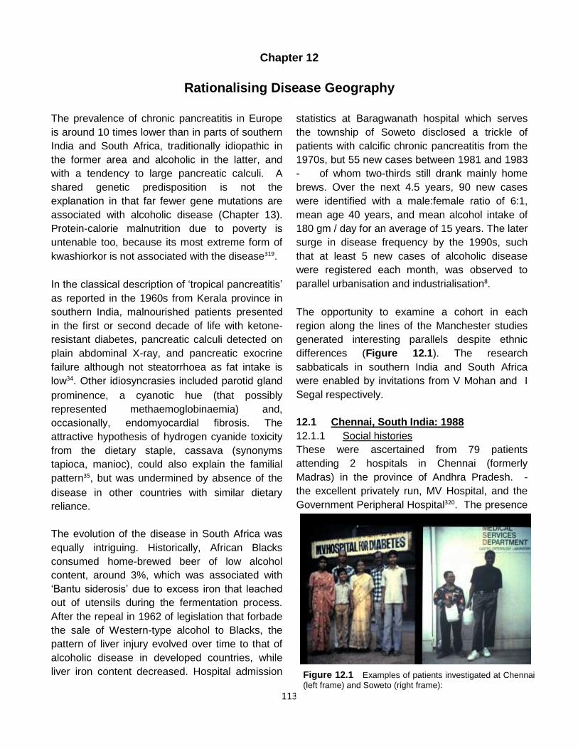
and
The American Pancreatic Association
ISBN 978-1-60785-412-8 (e-book)
This work is licensed under the Creative Commons Attribution-NonCommercial 4.0 International License. To view a copy of this license, visit https://creativecommons.org/licenses/by-
nc/4.0/ or send a letter to Creative Commons, PO Box 1866, Mountain View, California, 94042, USA.
This monograph presents a comprehensive review of the work of Professor Joan Braganza into the
pathogenesis and treatment of chronic pancreatitis as a disease caused by oxidative and particularly
electrophilic stress with an emphasis on environmental toxicants as one factor which is part of the
balance of damaging and protective cellular forces in the pancreas, liver and other participating organs.
This is the second monograph of this type and follows “The Hamster Pancreas” written by Parviz Pour.
Both books are published in open access eBook form under a Creative Commons license and are
available on the Pancreapedia site. Pancreapedia will from time to time publish eBooks that are more
substantive than a review article and where the author prepares the text and secures copyright
approval for figures or tables published previously. Publishing can be done at no or low cost to the
author. Authors should obtain approval from the Editor before preparing detailed content. As was
carried out for this book, the Pancreapedia Editor and staff carry out light copy editing and format the
book with final approval by the author. Our books are assigned an ISBN number and can be cited as
any other book. We thank the American Pancreatic Association for their support and especially thank
Melissa Wu and Juliana Lam, two students from the University of Michigan School of Information who
served as Content/Community Manager for the Pancreapedia and were responsible for taking the
edited content and preparing the book you see here.
John A. Williams
Editor-in-Chief, Pancreapedia
iv
Table of Contents
Glossary and Conversion factors vii Preface ix
1. Introduction 1 2. Chronic pancreatitis: 1980s 3 2.1. Frame of reference 3 2.2 The disease 5 2.3 Diagnosis 10 2.4 Treatment 13 2.5 Summary 14 3. Serendipity! 15 3.1 Boots versus GIH secretin 15 3.2 Studies on copper 17 3.3 Bilirubin hypersecretion 23 3.4 Introduction to free radical pathology 26 3.5 Overview and summary 34 4. Pancreatic Disease: Casualty of Hepatic ‘Detoxification’ Reactions? 35 4.1 Clues 35 4.2 Cytochromes P450 36 4.3 Hypothesis: Pancreatic disease is a casualty of liver ‘detoxification reactions’ 36 4.4 Testing the concept 37 5. Drug Metabolism and Ancillary Studies 38 5.1 Routine tests 39 5.2 Antipyrine, theophylline, debrisoquine 43 5.3 D-glucaric acid 51 5.4 Biliary fatty acids 53 5.5 Overview and summary 58 6. Hepatobiliary Aberrations: Reflux Link to Pancreatitis? 59 6.1 Liver histology 59 6.2 Endoscopic cholangiograms 63 6.3 Chronic pancreatitis-type artificial bile and experimental pancreatitis 66 6.4 Overview and summary 68 7. Probing the Defence Arc: Dietary Antioxidants 69 7.1 Versus intake in healthy controls 69 7.2 Versus intake in controls with epilepsy 72 7.3 Epilepsy plus chronic pancreatitis 75 7.4 Overview and summary 76 8. Occupational Volatile Chemicals 78 8.1 Pilot study 78 8.2 Case-referent study: chronic pancreatitis 81 8.3 Overview and summary 87
v
9. Casualty of Pancreatic ‘Detoxificaton’ Reactions! 88 9.1 Futility of clinical bile diversion 88 9.2 Investigation of pancreatic CYP 91 9.3 Overview and summary 95 10. Taking Stock 97 10.1 Premises of 1983 confirmed 97 10.2 Modification of 1986 confirmed 97 10.3 Electrophilic stress: component clauses 98 10.4 Hepatisation of the pancreas 98 10.5 More questions 98 11. Free Radical Pathology of a Pancreatitis Attack 99 11.1 On secretory polarity 100 11.2 Free radicals as detonator 101 11.3 Methyl and thiol homeostasis 103 11.4 What role for trypsin? 107 11.5 What initiates inflammation? 109 11.6 Acute / RAP link to chronic pancreatitis 110 11.7 Overview and summary 112 12. Rationalising Disease Geography 113 12.1 Chennai, South India: 1988 113 12.2 Soweto, South Africa: 1993 119 12.3 Overview and summary 126 13. Accommodating gene mutations 129 13.1 Received wisdom 129 13.2 Against a central role for trypsin in HP 130 13.3 On CFTR mutations 131 13.4 Miscellaneous 135 13.5 Tweak to template 136 13.6 Conclusion 136 14. Towards an Animal Model Based on CYP Induction 137 14.1 Drug metabolism studies 138 14.2 Secretory studies 139 14.3 Histology studies 142 14.4 Overview and summary 144 15. Antioxidant Therapy for Relapsing Pancreatitis: Exploratory 147 15.1 Idiopathic relapsing pancreatitis 147 15.2 Metabolic predisposition 151 15.3 Overview and summary 154 16. Clinical Trials of AOT: Manchester and Beyond 157 16.1 RCTS: Manchester late 1980s 157 16.2 Subsequent trials 166 16.3 Pancreatic extracts: micronutrient therapy by proxy 170 16.4 Value of long-term treatment 171 16.5 Why does the prescription work? 175 16.6 Overview and summary 176
vi
17. Towards First-Line Treatment for Acute Pancreatitis 177 17.1 Anecdotal clinical experience: positive impact of N-acetylcysteine 177 17.2 Allograft pancreatitis: protection by S-adenosylmethionine 178 17.3 Clinical trial of SAM plus NAC for first 24 hours 181 17.4 Why treatment failure: micronutrient lack / persisting oxidative stress? 183 17.5 Why treatment failure: fibrinolysis precedes hypercoagulability 190 17.6 Overview and summary 197 18. More on Micronutrient Lack and Pancreatitis Risk 198 18.1 Gallstones 198 18.2 Ischaemic heart disease 207 18.3 Raynaud’s phenomenon 209 18.4 Overview and summary 211 19. Electrophilic Stress and Pancreatitis: 2016 216 19.1 Stresses and stressors 216 19.2 Electrophilic stress template for chronic pancreatitis 217 19.3 Electrophilic stress and fatal acute pancreatitis 224 19.4 Summary 228 20. Coda 229 References 231
vii
Glossary & Conversion factors
Abbreviation Form represented α1PI alpha 1 proteinase inhibitor
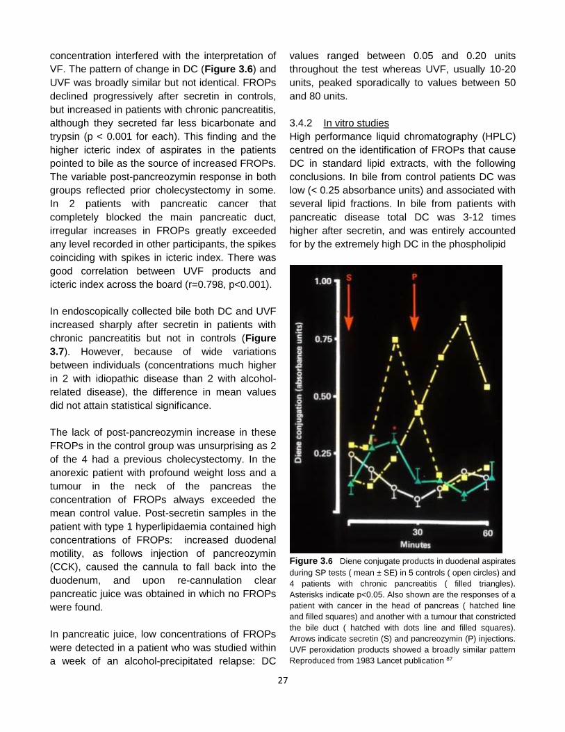
in The Lancet of my hypothesis that the disease is
a casualty of xenobiotic detoxification reactions;
five years of testing confirmed the concept; and,
most importantly, the therapeutic corollary was
realised by a trial of micronutrient antioxidant
supplements in patients with chronic pancreatitis
and relapsing acute pancreatitis.
When elected President of the Pancreatic Society
of Great Britain and Ireland in 1990, I organised a
symposium on ‘The pathogenesis of pancreatitis’,
the proceedings of which, along with additional
Chapters to round off the text, were published the
following year by Manchester University Press.
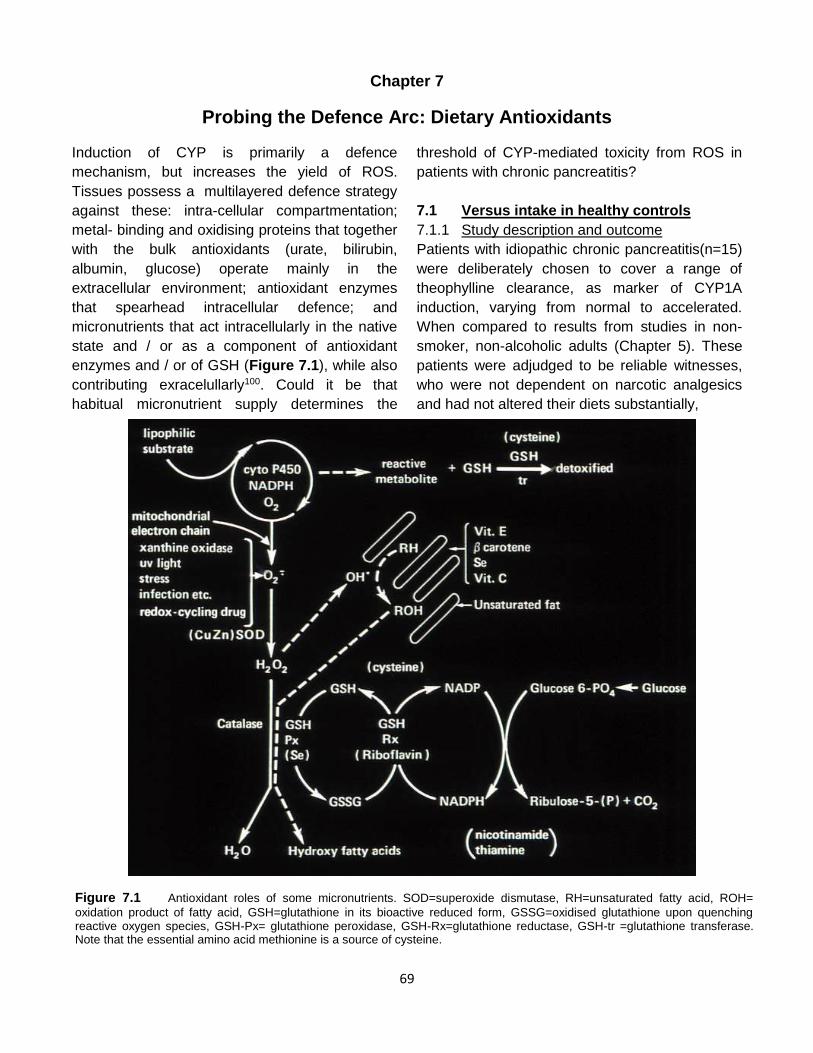
The contents page (shown in the big box)
illustrates the range of topics covered. During the
meeting I met Bent Henriksen OBE, managing
director of ‘Pharmanord UK’, who advised me that
his company could provide antioxidant tablets of
far higher potency than I was using to treat
patients, and also that methionine could be
incorporated so as to lower the daily number of
tablets to 4 (instead of 16). He was true to his
word.
So successful was the new treatment for chronic
pancreatitis, that by 1992 interventional
endoscopy and pancreatic surgery were virtually
redundant. The Manchester Royal Infirmary thus
became the first NHS hospital to provide
monitoring for oxidative stress and antioxidant
status as part-and-parcel of clinical care, the
initiative resourced by one-off payment from the
referrer. In 1994 I was awarded the Doctor of
Science degree by the University’s faculty of
science and engineering in recognition of a new
template for pathogenesis of the disease; and my
collected papers - including on diagnostic
aspects - earned Fellowship of the Royal
College of Pathologists in 1995.
The Pathogenesis of Pancreatitis symposium , 1990 Preface .....................................................................................HT Howat Section A Definition and concepts Chapter 1 Pathology of pancreatitis.............................................DS Longnecker Chapter 2 Evolution of pancreatitis..............................................JM Braganza Chapter 3 Secretory polarity........................................................ OA Musa & RM Case Chapter 4 Biological free radicals: a personal approach..............TL Dormandy Section B: Initiators of acute pancreatic injury Chapter 5 Oxygen free radicals in experimental pancreatitis....... H Sanfey Chapter 6 Toxicology of the pancreas .........................................JM Braganza Chapter 7 Acute necrotising pancreatitis and its complications:...H Rinderknecht an excessive reaction of natural defence mechanisms? Section C: Effectors of chronic pancreatic damage Chapter 8 Cytochromes P450 in chronic pancreatitis..................JB Houston Chapter 9 Xenobiotics in tropical chronic pancreatitis....... V Mohan & JM Braganza Chapter 10 Pancreatitis in cystic fibrosis.........................................JA Dodge Chapter 11 The exocrine pancreas in severe malnutrition.............MHN Golden Section D: Potential therapy for pancreatic disease Chapter 12 Antioxidants in vitro and in vivo..................................G Scott Chapter 13 Antioxidant therapy for chronic pancreatitis: ............. JM Braganza clinical experience Chapter 14 Role of enzyme inhibitors in acute pancreatitis and.....K Ohlsson & S Genell rationale for their therapeutic use Chapter 15 Overview and horizons.................................................JM Braganza
xii
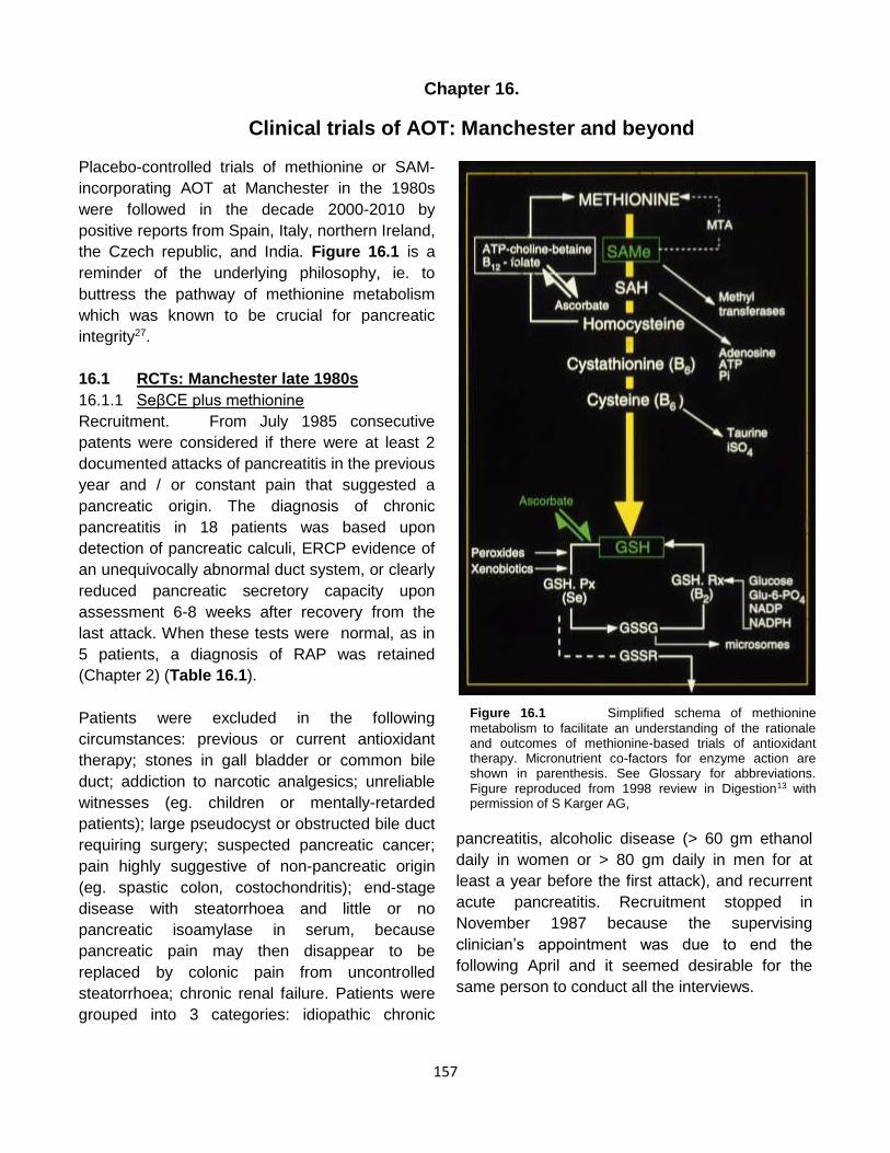
In 1998 I was invited to organise a teaching
session on chronic pancreatitis at the World
Congress of Gastroenterology in Vienna. By now
there were substantial advances in the field:
laboratory studies had shown that whereas
oxidative stress is associated with acute
pancreatitis, electrophilic stress is prominent in
chronic pancreatitis; clinical studies at Soweto
were revelatory; a surgical audit verified the
efficacy of micronutrient therapy at Manchester;
and there was clear evidence for the involvement
of oxidative stress in cystic fibrosis.
Papers based on the symposium proceedings
(shown in the small box) were published under
the title ‘New developments on the aetiogenesis
of chronic pancreatitis: implications for treatment
and disease prophylaxis’ (Digestion 1998; 59
supplement 4: 1-60).
Howat accepted the ‘radical’ path that I was
pursuing, but warned of trouble ahead. Dormandy
advised that it was important to share ideas
because creative minds would always generate
more: thus, I persuaded G Mann in London to
investigate the effect of oxidative stress in the
isolated perfused rat pancreas; A Borgstrom in
Malmo to determine whether mast cell tryptase
could activate trypsinogen, and gifted him
ampoules of the enzyme; and, most recently, F
Uboh in Nigeria to investigate the effect on the rat
pancreas of inhalation exposure to kerosene
fumes.
This monograph charts the 20-year road towards
developing the electrophilic stress template, the
only framework that accommodates all
observations on chronic pancreatitis while offering
specific medical therapy by way of micronutrient
‘antioxidant’ supplements. The exploration of
many other ramifications - not least for first-line
medical treatment of acute pancreatitis and the
prospect for disease prevention in communities at
risk of chronic pancreatitis - was cut short by my
enforced resignation due to daily haemoptysis
from aspergillus bilateral lung cavitation,
consequent upon earlier tuberculosis. My
thwarted priority was to investigate whether the
rapid downward spiral to death in patients with
acute pancreatitis might be aborted if the first
medical respondent administers adrenaline by
‘Epipen’ to curb mast cells, as is standard
treatment for anaphylaxis.
I admit being behind non-participation by the
Manchester Royal Infirmary in national trials of an
antagonist of the receptor for platelet activating
factor in patients with acute pancreatitis, because
it seemed unlikely that this agent was somehow
unique within the toxic circulating brew; and in
refusing to donate patients’ samples for study of
trypsin-control genes, because insufficiency of the
cystic fibrosis transmembrane conductance
regulator seemed far more relevant to the
pathogenesis of chronic pancreatitis as was noted
in our 1998 paper in the New England Journal of
Medicine. This resistance did not gain friends.
Of course, the studies chronicled could not have
materialised without input by experts from
Manchester and London University, NHS
hospitals, and the pharmaceutical industry: these
were cited individually in a ‘Meet the champions’
interview by Martin Fernandez-Zapico, which is
now available under the Pancreapedia enterprise.
I am grateful to surgeons for allowing me into their
theatres when my patients went under the knife;
and I applaud the courage and generosity of Rory
Teaching Symposium at World Congress of Gastroenterology, 1998
Foreward: ........................................................................................... H Rinderknecht A framework for the aetiogenesis of chronic pancreatitis..................... JM Braganza Xenobiotic metabolism, oxidative stress and chronic pancreatitis........ MA Wallig Pancreatitis in Soweto, south Africa: focus on alcohol-related disease...I Segal Chronic pancreatitis at Manchester, UK: focus on antioxidant therapy.....RF McCloy Paediatric and hereditary aspects of chronic pancreatitis........................JA Dodge
xiii
McCloy who arrived at the hospital as a top
pancreato-biliary surgeon in 1983 only to see that
a decade later his skills were barely needed for
chronic pancreatitis. I appreciate the expertise of
Linda Hunt, superb statistitian who steered
through the quagmire of data in various projects.
It remains for me to thank the patients, many
postgraduate students, secretaries and typists,
above all to Jenny Parr without whose voluntary
and painstaking input this monograph would not
see the light of day.
Almoth Wright said that “a new idea in medicine
has to pass 3 stages: when it is regarded as
ridiculous; when doctors say ‘OK it is possible but
where is the proof?’; and when everyone
dismisses it as obvious”. Alan Read, professor of
hepatology at Bristol, warned me after my lecture
in 1989 that at least 20 years would elapse before
my concept of cytochrome P450-mediated
pancreatic injury would even be given a hearing.
He was right, but the challenge has been
exhilarating!
1
Chapter 1
Introduction
Although chronic pancreatitis (CP) is uncommon
in the developed world, it exacts an inordinate toll
in terms of morbidity, mortality and health
economics. This predicament reflects the lack of
agreement on specific medical treatment to
control disabling pain and avoid pancreatitis
relapses that will require hospitalization, leaving
patients to face the further threats of narcotic
addiction, job loss, dislocation of family, and
social isolation. The therapeutic vacuum, in turn,
points to the absence of consensus as to the
pathogenesis of the disease and of pain, an irony
that is highlighted by today’s embarrassment of
riches in relation to diagnostic tools1. It is harsh
testimony that total pancreatectomy, the earliest
operation for chronic pancreatitis in 19462, should
still be on the treatment menu for a non-malignant
disease that was identified at autopsy more than
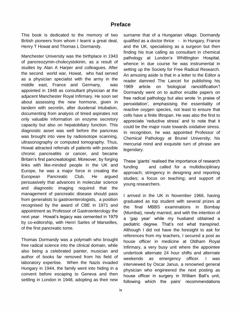
200 years ago3. Clinicians meddle while the
gland smoulders on (Figure 1.1), rather like the
emperor who fiddled while Rome burnt down.
As in any field of science, myths that are
promoted to dogma by public appeal stifle original
thought: heretics run the risk of being lampooned,
vilified, ignored or ousted4. In the context of
chronic pancreatitis, the myth is that recurrent
autodigestion episodes via ‘prematurely activated
trypsin’ in acinar cells is the underlying problem.
This has been elevated to doctrine by the
discovery of trypsin-favoring gene mutations in
patients with heredo-familial disease,
notwithstanding experimental evidence against
the gory interpretation5, 6.
Any alternate philosophy must accommodate the
predisposition conveyed by those mutations, as
also by mutation(s) in the cystic fibrosis gene7.
Moreover, it must rationalise demography, notably
the following facets: the highest recorded
prevalence of CP in Kerala province south India,
despite a curious 6-fold decline between 1962
and 1987 in annual hospital admissions; the
vulnerability of African Americans; the current
endemicity in Soweto, South Africa1, 8.
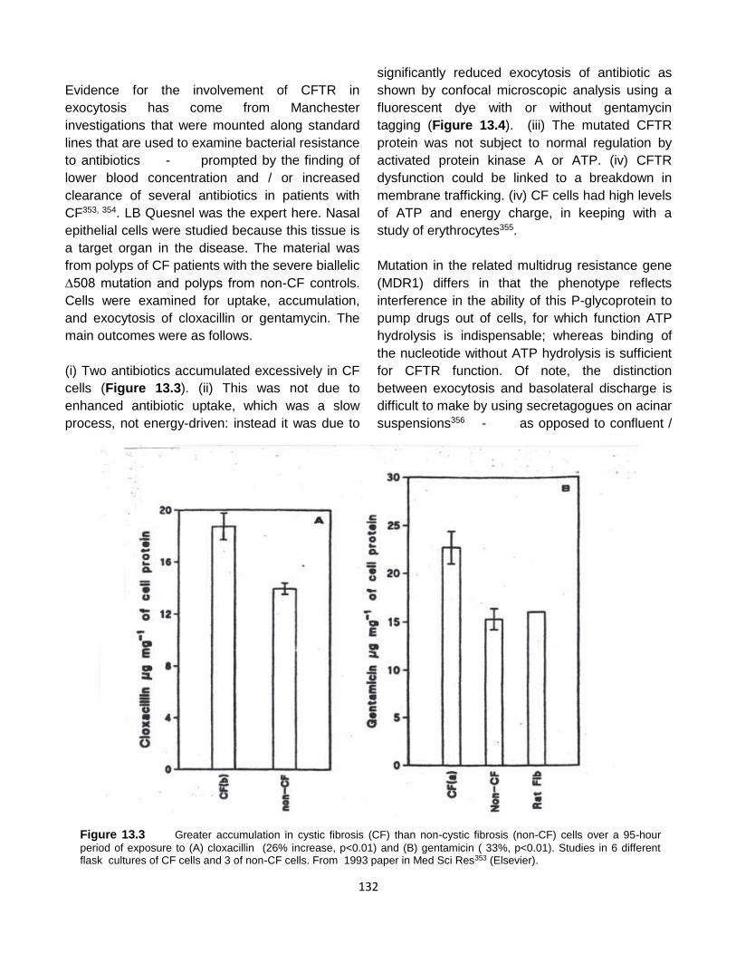
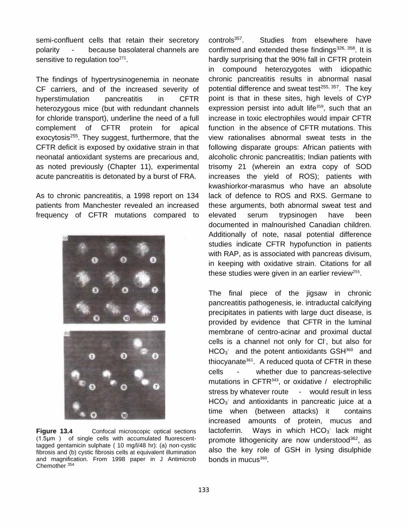
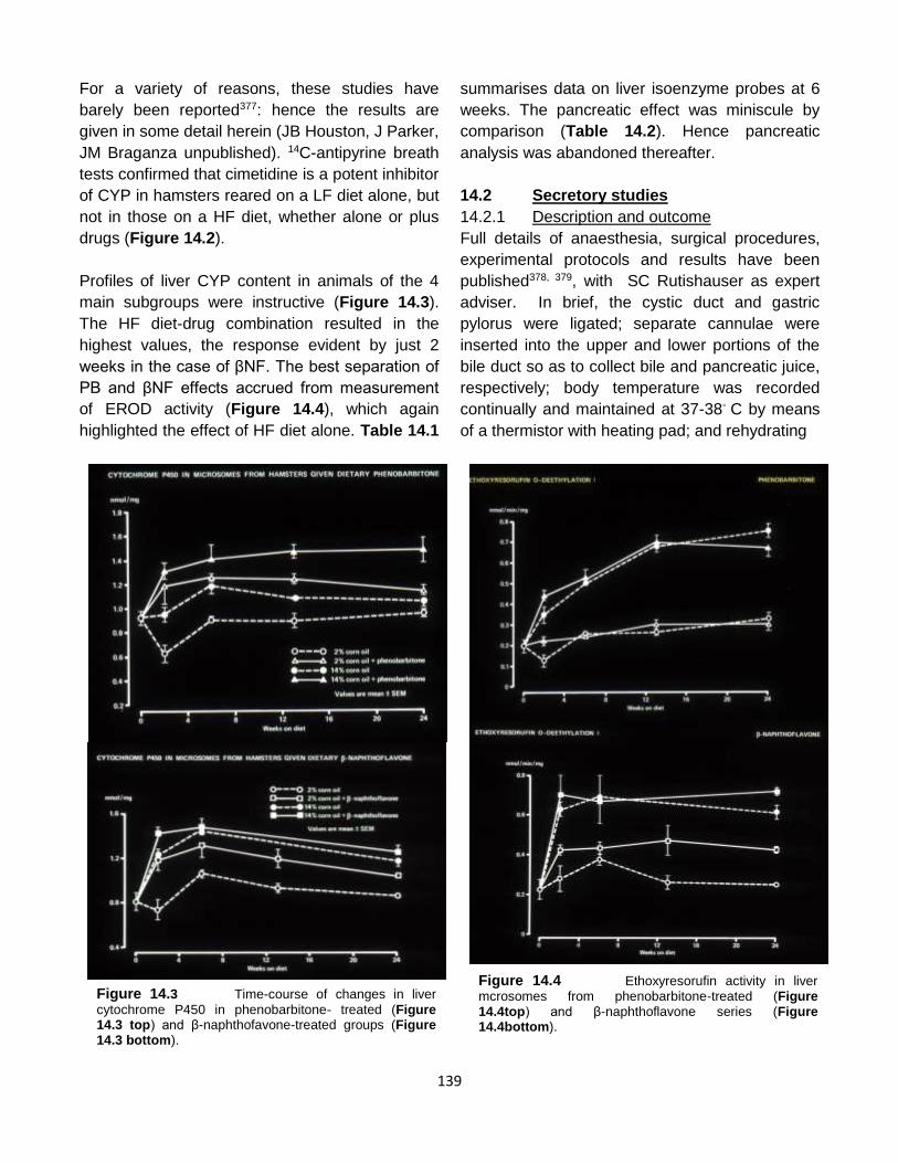
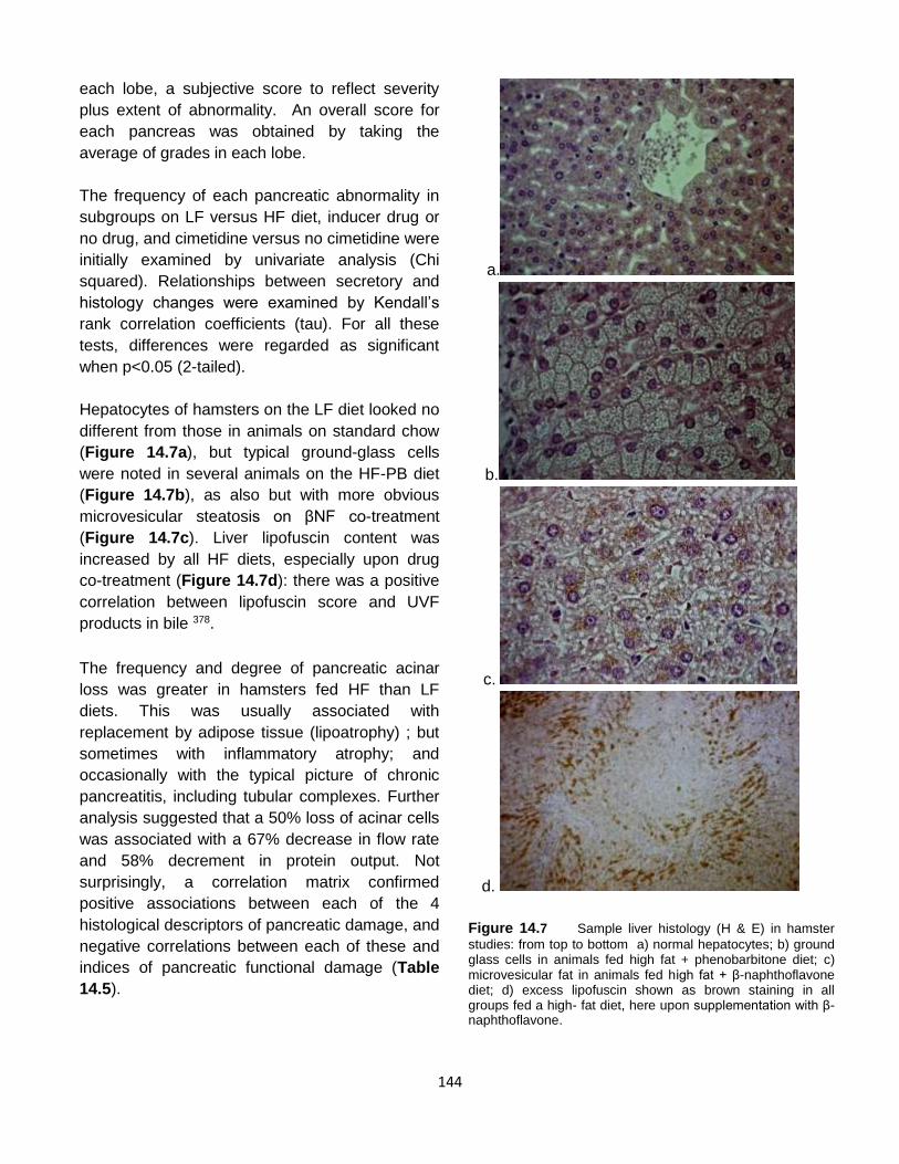
Figure 1.1 ‘Battle-scarred’ abdomen of a woman with
familial lipoprotein lipase deficiency and small-duct chronic
pancreatitis. Surgical procedures included diagnostic
laparotomy; cholecystectomy (no gallstones); distal
pancreatectomy with splenectomy; gastroenterostomy; and
finally, an attempt at total pancreatectomy which was
abandoned after 9 hours. Other useless measures included
pancreatic extracts; 2 coeliac plexus blocks; and
splanchnicectomy. She was referred in 1995 on opiates, and
with jaundice from secondary sclerosing cholangitis. Her
subsequent progress is described in Chapter 15.
2
The monograph begins by describing chronic
pancreatitis and relevant aspects of the normal
gland as were known in the 1980s when, initially
by chance, peculiarities were found in duodenal
bile collected during routine secretin-
pancreozymin (SP) tests. Many avenues for
investigation were simultaneously opened up by
the ensuing hypothesis that the disease is a
casualty of ‘detoxification’ reactions that are
generated via induced cytochrome P450 mono-
oxygenases (CYP)9,10. Hence, the developments
recorded herein are not strictly chronological.
They culminated in the electrophilic stress
template, ie. that injury is generally due to reactive
xenobiotic species (RXS), over-and-above
reactive oxygen species (ROS). This is the only
philosophy that accommodates all aspects of the
disease - from genetics and ‘alcoholic’ or
‘tropical’ aetiology through to pathophysiology.
More importantly, the concept enables first-line
micronutrient ‘antioxidant’ therapy11.
Relapsing acute pancreatitis (RAP) is brought into
the frame because of the overlap in aetiological
factors and clinical identity of attacks. The likely
role of electrophilic stress in lethal acute
pancreatitis, by evoking the wholesale
degranulation of mast cells, is examined too
because of the potential for specific medical
treatment. Finally, the scope for micronutrient
prophylaxis against both chronic pancreatitis and
acute pancreatitis is considered.
[Postscripts. (1) Unorthodox investigations had
the approval of the ethical committee of the
Manchester Royal Infirmary and, if germane, of
institutions housing collaborating researchers,
whereupon prior informed consent was obtained
from participants - and parents when children
were involved. Studies of a purely clinical nature
were guided by judgement and experience. (2)
The investigations were initiated by the author
except when specified otherwise. (3) For the
convenience of readers, a glossary of
abbreviations, together with factors to enable
conversion from metric to Systeme Internationale
units for micronutrient antioxidants follows the
contents pages in the preamble.]
3
Chapter 2
Chronic Pancreatitis: 1980s
The 1980s was a period of excitement for
pancreatologists as the gland was brought into
view by radio-isotope scanning, endoscopic
retrograde cholangio-pancreatography (ERCP),
ultrasonography and computed tomography (CT)
raising the hope that tedious duodenal intubation
tests might no longer be needed to diagnose pre-
calcific chronic pancreatitis. It was also a time of
frustration because trainees were force-fed the
credo that the disease is synonymous with
alcoholism and is due to primary ductal lithiasis12.
How, one asked, could this theory rationalise the
disease in non-alcoholics? It was also difficult to
explain the co-existence of pancreatic, liver and
kidney lesions with one or other of the triad
showing the greatest damage, as was notable in
the first documented case, the chequered medical
history of the musical genius Beethoven, the
earliest report to make the alcohol connection,
and experimental injury?13 There was a need,
therefore, to re-visit the basics, as detailed in
pancreatic tomes14, 15, supplemented by individual
papers.
2.1 Frame of reference
2.1.1 Embryology
Three points are potentially relevant. (i) The liver
and pancreas originate in contiguous buds from
the evolving foregut. (ii) Duct systems of the 2
pancreatic anlages then diverge. The smaller
ventral channel (duct of Wirsung) opens into the
origin of the hepatic duct, and this common
channel will later be sheathed by Oddi’s sphincter.
The larger dorsal channel (duct of Santorini)
opens into the gut directly, just cephalad to the
common channel, and without a sphincter. Both
pancreatic anlages fuse in the 7th week, the
ventral duct now becoming the main pancreatic
duct. In 5-10% of cases, fusion of the ducts is
incomplete, such that the Santorini system
remains dominant, so-called ‘pancreas divisum’.
(iii) Primitive pancreatic ductules are the source of
cell clumps that are forerunners of acinar units as
well as islets of Langerhans.
2.1.2 Anatomy and microanatomy
The following facets are notable in context. (i) A
common ampullary channel could allow toxins in
bile to reflux into the pancreatic duct, either
directly or by way of the duodenum: in this event,
as also when excess pressure is generated during
ERCP, the single-layered epithelium of the
smallest intralobular ducts would yield. However,
the construction of many acini as anastomosing
networks mitigates against intralobular
obstruction. (ii) The rich blood supply to the gland
is arranged in such a way that a portion initially
perfuses islets, constituting a limited ‘portal’
circulation. Also of note13, the pattern of branching
leaves the periphery of acinar lobules vulnerable
in ischemic states16, in the same way as threatens
zone 3 hepatocytes17 (Figure 2.1). Electron
microscopy shows that many of the capillaries are
fenestrated, with pores around 100 nm diameter,
facilitating the diffusion of substances such as
hormones.
(iii) In an autopsy pancreatography study, ductal
irregularities that would be classed as ‘minimal-
change-pancreatitis’ (see below) were recorded in
37% of cases, without any histological evidence of
disease18. (iv) The tight junctions between
pyramidal acinar cells lie on the lateral aspect
adjacent to the lumen, so that there is a wide
surface area for communication with the interstitial
space. A thin lamina at the basal aspect of each
cell abuts on underlying connective tissue. (v) The
pancreatic interstitium contains numerous
adipocytes, including retinol-rich cells that would,
many years later, be identified as precursors of
fibrosis-promoting stellate cells19. (vi) Also herein
are numerous mast cells, between basement
membrane of capillaries and plasma membrane of
acinar cells20 - which, in time, would be shown
4
to be juxtaposed to membrane receptors for a
tissue-type plasminogen activator21, and also
urokinase-plasminogen activator22. (vii) An
extensive lymphatic drainage originates in the
perilobular connective tissue and gains access to
the gland’s surface by channels that run alongside
blood vessels. (ix) Ultratructural studies reveal
nor-adrenergic nerve terminals in close
association with perivascular smooth muscle
cells; whereas cholinergic fibres and their terminal
regions exist between secretory acini.
2.1.3 Physiology
A great deal was known about the regulation of
pancreatic secretion by interaction between food-
evoked neural and hormonal stimuli, the latter
principally via secretin, cholecystokinin-
pancreozymin (CCK) and gastrin, but information
on stimulus-secretion coupling in acinar cells was
scant. It was recognised that the pyramidal acinar
cell is the epitome of a polarised unit, wherein
synthesis of hydrolases occurs within the
abundant rough endoplasmic reticulum (RER) at
the basal pole and the bulk discharge of enzymes,
exocytosis, at the apical pole. It was established
that this is effected via a series of intracellular
messengers. Occupation of the CCK-type
receptor on the plasma membrane leads via a
guanine nucleotide- binding protein (G-protein) to
the activation of phospholipase C, producing
inositol 1,4,5-trisphosphate and diacylglycerols.
Such agents mobilize Ca2+, probably from the
RER and activate protein kinase C. The rise in
cytosolic Ca2+ activates calmodulin and thereby
protein kinase and phosphatase. A second low-
affinity CCK receptor inhibits secretion. Signal
transduction after occupation of the secretin-type
receptor involves an increase in cyclic AMP
which, in turn, activates protein kinase A. Both
pathways alter the level of stimulatory and
inhibitory G-proteins ( Gs, Gi ) and thus modulate
protein phosphorylation23, 24.
It was also appreciated that the actual exocytosis
event involves interaction between Gs / Gi
balance, actin, microtubules, and apical
Figure 2.1 Microcirculatory arrangements in the liver with 3
functional zones determined by location of the central vein (CV) and portal tract (PT), contrasted with that in the pancreas. Composite figure generated for 1998 review in Digestion13 from images redrawn with permission of authors 16,17; now reproduced with permission of publisher, S Karger AG, Basel.
membrane fluidity25. Studies in a variety of cells
pointed to the particular importance of the last
factor by way of membrane phospholipid
methylation26, but were dismissed23. This was
hasty, in that methionine lack had long been
known to paralyse apical enzyme secretion27. In
1987 the presence of constitutive pathways for
enzyme discharge from the acinar cell were
documented, 2 at the apical pole and 1 at the
basolateral pole28.
Meanwhile, the multi-layered defence strategy
against premature detonation of digestive enzyme
5
grenades during transit was being unravelled. (i)
The potent proteases and phospholipase A2 are
produced as zymogens that are unleashed in
cascade fashion when trypsinogen is activated to
trypsin upon contact with enteropeptidase in the
presence of bile salts within the duodenum. (ii)
From the time of their production to discharge
from the acinar cell, digestive hydrolases are held
separate from lysosomal enzymes, in the later
stages being packaged within zymogen granules
(ZG) and stored at the apical pole. (iii) The serine
protease inhibitor Kazal type 1 (SPINK 1),
formerly called pancreatic secretory trypsin
inhibitor, is co-packaged within ZG and can curb
20% of potential tryptic activity. However, perhaps
because the enzyme-inhibitor complex tends to
dissociate, a back-stop is in place, in that trypsin
is soon destroyed by trace amounts of activated
mesotrypsin and ‘peptide Y’15,29, the latter now
identified as a form of chymotrypsin7. (iii) Once
discharged by exocytosis, the cargo of released
pro-enzymes, amylase and lipase in a small
volume of chloride-rich juice is propelled
downstream by secretin-stimulated flow of
bicarbonate and water from centro-acinar and
ductal cells.
2.2 The Disease
2.2.1 Definition
Chronic pancreatitis is a histological entity,
characterised by continuing inflammation with
progressive loss of acinar tissue and fibrosis.
These are dynamic processes that generally
manifest as attack-upon-attack of pancreatitis;
their tempo is unpredictable; and the lesions are
typically patchy. Each burst of inflammation may
lead to foci of interstitial or peripancreatic fat
necrosis20, that are nowadays implicated in the
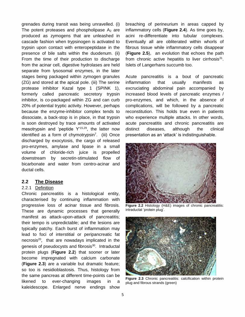
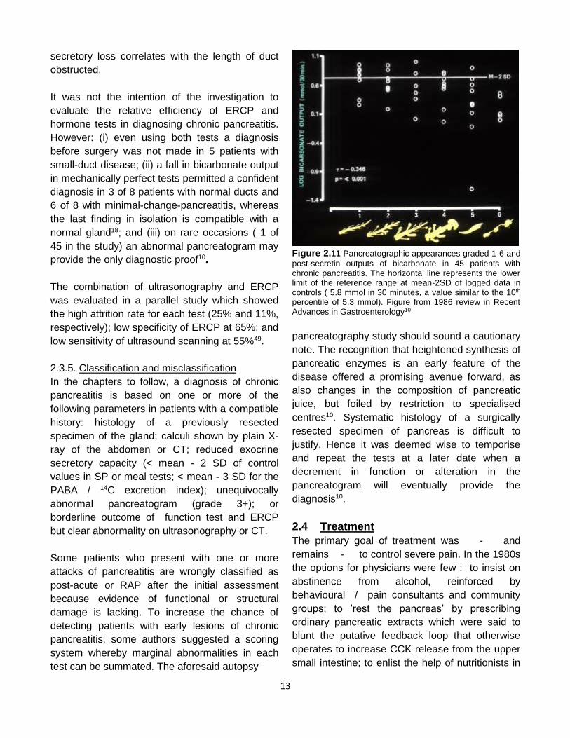
genesis of pseudocysts and fibrosis30. Intraductal
protein plugs (Figure 2.2) that sooner or later
become impregnated with calcium carbonate
(Figure 2.3) are a variable but dramatic feature;
so too is nesidioblastosis. Thus, histology from
the same pancreas at different time-points can be
likened to ever-changing images in a
kaleidescope. Enlarged nerve endings show
breaching of perineurium in areas capped by
inflammatory cells (Figure 2.4). As time goes by,
acini re-differentiate into tubular complexes.
Eventually all are obliterated within whorls of
fibrous tissue while inflammatory cells disappear
(Figure 2.5), an evolution that echoes the path
from chronic active hepatitis to liver cirrhosis31.
Islets of Langerhans succumb too.
Acute pancreatitis is a bout of pancreatic
inflammation that usually manifests as
excruciating abdominal pain accompanied by
increased blood levels of pancreatic enzymes /
pro-enzymes, and which, in the absence of
complications, will be followed by a pancreatic
reconstitution. This holds true even in patients
who experience multiple attacks. In other words,
acute pancreatitis and chronic pancreatitis are
distinct diseases, although the clinical
presentation as an ‘attack’ is indistinguishable.
Figure 2.2 Histology (H&E) images of chronic pancreatitis:
intraductal ‘protein plug’.
Figure 2.3 Chronic pancreatitis: calcification within protein
plug and fibrous strands (green)
6
Figure 2.4 Chronic pancreatitis: inflammatory capping of
nerve endings
Figure 2.5 Chronic pancreatitis: histology of more advanced
disease
Alongside this declaration from a symposium in
198432 was the recommendation that clinicians
discard the useful adjective ‘relapsing’ which was
adopted at the 1963 Marseilles symposium. Given
the impracticality of pancreatic biopsy for the sole
purpose of retrospective assignment, and that
even in patients with chronic pancreatitis random
needle biopsy is like searching for a needle in a
haystack because the early lesions are patchy,
clinicians were stymied unless pancreatic calculi
were identified by admission plain X-ray of the
abdomen or latterly by CT. The advice, although
not foolproof, was to assess residual secretory
capacity 6 weeks or more after the last attack,
when a subnormal value(s) points to chronic
pancreatitis33.
2.2.2 Etiology
Alcoholism was regarded as the predominant
cause of chronic pancreatitis in western societies,
Brazil and South Africa12. However, studies from
France showed that ethanol on its own is a weak
agent, an average of 18 years elapsing before the
heralding attack in men who drink > 80 gm
ethanol daily ( a decade shorter than presentation
with liver problems), and that < 10% of alcoholics
develop the disease. The paradox of no lower
threshold for ethanol toxicity was attributed to the
potentiating effect of diets that contain excessive
amounts of fat and / or protein, unidentified
environmental co-factors, and unknown disease-
susceptibility genes.
Meanwhile studies in Kerala province in southern
India, where residents did not consume alcohol,
showed that patients with chronic pancreatitis
accounted for around 1.5% of hospital
admissions34. The disease typically presented as
non-ketotic diabetes in young lean patients with
extensive pancreatic calculi, parotid hyperplasia
and a cyanotic hue of lips and mucous
membranes. An attractive proposal was that
hydrogen cyanide in dietary cassava (synonyms
manioc, tapioca) might be responsible35. The
theory was, however, discredited by many
observations including that the same disease
pattern was observed in countries such as Nigeria
where cassava is not a staple food. Moreover, the
hypothesis could not rationalise the 6-fold decline
in hospital admissions by the mid 1980s34.
As to other potential factors, gall stones were
regarded as incidental. It was agreed that
impedence to drainage of pancreatic juice, as by
Vaterian stenosis, leads to uniform lesions of
chronic pancreatitis upstream with smooth
dilatation of the pancreatic duct; and also that in
patients with pancreas divisum the ventral
pancreas might be spared. A few cases could be
ascribed to hyperparathyroidism, familial disease
or drug treatment. Autoimmune pancreatitis was
unknown at the time but ‘primary inflammatory
pancreatitis’ was noted anecdotally14.
7
Axiomatic in the 1984 guidelines was that
pancreatic lesions of hemochromatosis or cystic
fibrosis do not equate to chronic pancreatitis32.
Yet, cystic fibrosis is the surest antecedent of
non-calcific chronic pancreatitis: the diffuse
lesions develop in utero and might display
histological features of severe acute pancreatitis;
they manifest at birth by hypertrypsinogenemia,
and follow an accelerated course to exocrine
pancreatic failure33. In 1989 the genetic basis for
its transmission as an autosomal recessive trait
was pinpointed to mutations in both alleles of the
cystic fibrosis transmemrane conductance
regulator gene (CFTR)36. The genetic basis for
the rare hereditary form of chronic pancreatitis
remained elusive, although known to be
transmitted in autosomal dominant fashion with
variable penetrance.
2.2.3 Pathogenesis / Pathophysiology
The party-line was that increased secretion of
lactoferrin and calcium from acinar cells coupled
with reduced secretion of a ‘stone protein’
(synonym lithostatin) within a milieu of falling
bicarbonate production by ductal cells leads to
protein precipitates in pancreatic ducts: these
serve as niduses for calcium carbonate stones,
which then ulcerate ductal mucosa to provoke
periductal fibrosis12. The following were among
other contemporary proposals: reflux of noxious
bile; stagnation of pancreatic juice; recurrent fat
necrosis perpetuating fibrosis; direct toxicity of
ethanol; and slow cyanide poisoning37.
All these hypotheses failed to explain why acinar
cells appeared to be whipped into a state of
hyperactivity10. As evidence, in chronic alcoholics
without manifest disease, electron microscopy of
pancreatic tissue showed expanded RER,
microvesicular fat, areas of cytoplasmic
sequestration and increased numbers of
lysosomes. In symptomatic patients with early
chronic pancreatitis, reports described large
acinar cells, osmiophilic bodies resembling
lipofuscin, and mitochondrial aberrations - in
conjunction with dilated RER, and prominent
lysosomes in ductal cells. Secretory patterns
reflected these morphological changes. Thus,
analysis of pure pancreatic juice from
asymptomatic alcoholics disclosed increased
amounts of protein and calcium, and an increased
ratio of lysosomal to digestive hydrolases.
Symptomatic patients with early disease
displayed these abnormalities and many others:
increased rate of protein synthesis;
hyperscecretion of bicarbonate; and altered
composition of pancreatic secretions, containing
more anionic trypsinogen but less trypsin inhibitor
than normal10.
2.2.4 Experimental models
Whereas alcohol administration on its own did not
produce the disease, the use of modified
protocols that induce acute pancreatitis did so.
Examples include repetitive hyperstimulating
doses of caerulein ( an analogue of gastrin and
CCK), partial obstruction of the main pancreatic
duct, or a choline-deficient DL-ethionine
supplemented (CDE) diet - although damage
is inflicted by utterly different routes, respectively
microvasular, ductal, or intra-acinar38.
Confusingly, a single subcutaneous dose of
carbon tetrachloride (CCl4) in rodents was shown
to cause lesions of chronic pancreatitis in
advance of liver lesions, and which could be
“altered at will” to yield a spectrum from the
patchy lesions of chronic pancreatitis with or
without concretions, through to ‘pancreatic
cirrhosis’ or ‘cystic fibrosis’31.
Moreover, an interesting parallel was emerging
between the effects of certain toxins on the
hepatocyte and acinar cell39. Thus, many of the
indirect hepatotoxins which produce injury by
interfering with a specific pathway or process -
eg. DL ethionine, tetracycline, puromycin -
are also associated with pancreatitis.
As in the case of liver damage by these
substances (Figure 2.6), so is pancreatic damage
by hyperstimulation and certain insecticides
(Figure 2.7), products that are normally extruded
8
Figure 2.6 Schematic representation of macromolecular traffic in the normal hepatocyte (left) and consequences of
interference to traffic routes by hepatotoxins (right). Abbreviations: dIgA, dimeric immunoglobulin A; ASGP, asialoglycoprotein; VLDL, very low density lipoprotein; PL, phospholipid; TG, triglycerides; L, lysosome. Reproduced from 1988 review in Int J Pancreatol39
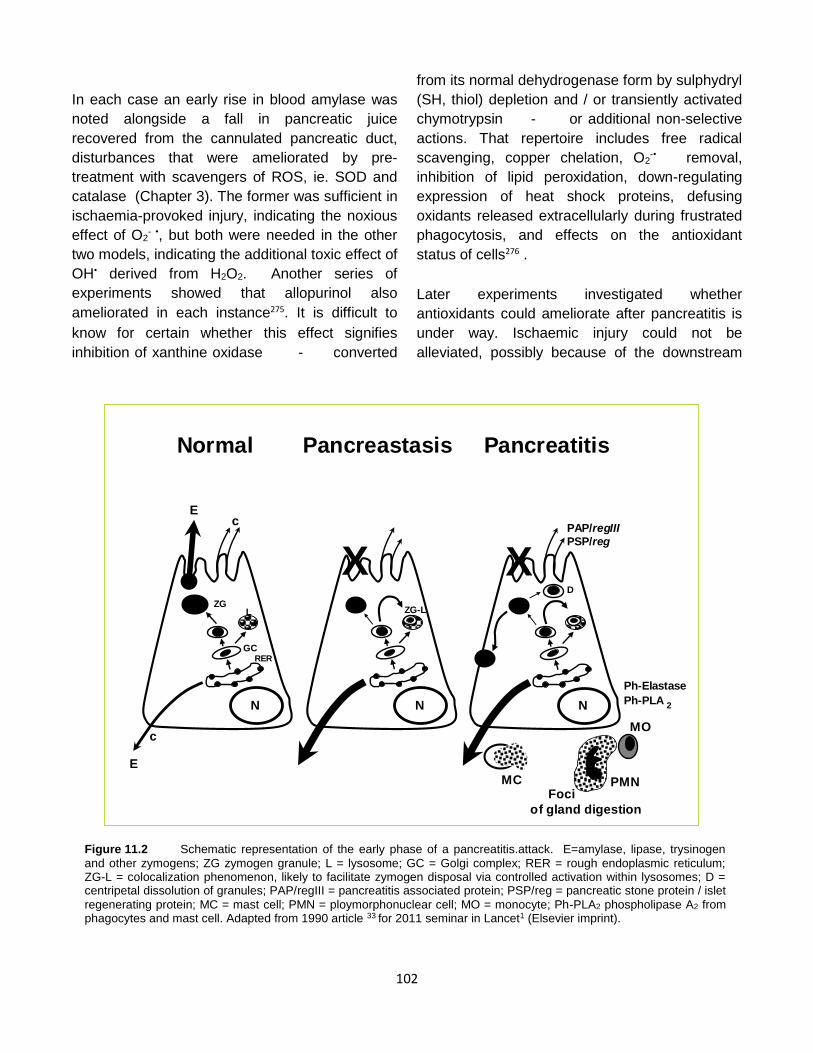
Figure 2.7 Schematic representation of the traffic of food digestive hydrolases in the normal acinar cell (left) and abnormal
partitioning during experimental pancreatic injury as suggested in the 1980s (right); ZG, zymogen granules. Source as for Figure 2.6.
9
at the plasma membrane of the hepatocyte or
luminal pole of acinar cell are seemingly forced to
find egress through alternate routes, namely, the
canalicular pole and basolateral membrane,
respectively. In each instance the cytoplasm of
affected cells contains vacuoles, representing
secretory products that have accumulated
because of microtubular damage (microvesicular
fat in hepatocytes, nascent zymogens in acinar
cells) and / or engulfment of redundant secretory
material by lysosomes. By 1990, the term
‘pancreastasis’ was coined to describe the altered
secretory polarity in the acinar cell, in line with
intrahepatic ‘cholestasis’38.
2.2.5 Clinical features
Pain is the cardinal symptom. In many patients, it
initially accompanies clear-cut attacks of
pancreatitis and moves on to constant pain; in
others it is constant from the start. It may be
primarily in the epigastrium or dorsal spine, or
bore through from front to back. A stooping
posture might alleviate. The agonising intensity of
pain may be evidenced by erythema ab igne
(Figure 2.8) and / or improvised counter-irritant
devices (Figure 2.9). The eventual loss of nearly
all secretory parenchyma, as is recognised by
steatorrhoea, often brings pain relief -
supporting the notion that viable acini are a pre-
requisite for pancreatic pain, provided that by then
patients are not already addicted to narcotic
analgesics. The progression to parenchymal
eradication is more rapid in tropical pancreatitis,
such that pain is less of a problem, but this path is
not marked by steatorrhoea because fat intake is
traditionally low. Irrespective of geography, end-
stage disease is often accompanied by diabetes
and pancreatic calculi. This classical pattern
glosses over small-duct disease, which is as
painful but difficult to detect40. The notion that pain
is generally due to a plumbing problem (ie duct
obstruction) now needed a re-think. Other modes
of presentation include jaundice due to intra-
pancreatic constriction of the common bile duct;
symptoms due to space-occupying cysts /
pseudocysts; effusions (pleural, ascites); and
Figure 2.8 ‘Erythema ab igne’ over the abdomen of a
patient with unremitting pain.
Figure 2.9 Wooden blocks with inward-facing nails
improvised by a patient with intractable back pain.
10
alimentary haemorrhage due to portal
hypertension. A cautionary note was issued in
regard to pancreatic cancer, ie. that proximal
obstruction by a tumour might masquerade as
chronic pancreatitis, which itself increases cancer
risk.
2.3 Diagnosis
2.3.1. Secretin ± Pancreozymin test
The patient, fasted overnight, is intubated with a
double-lumen tube which is advanced under
fluoroscopic guidance into the duodenal loop until
the furthermost ports are positioned adjacent to
the assumed position of the ligament of Treitz.
After a control period wherein gastric and
duodenal secretions are aspirated separately with
a mechanical intermittent-suction pump, secretin
is given intravenously to stimulate the secretion of
fluid and bicarbonate. Thereafter, timed duodenal
aspirates are collected on ice, volumes and pH
recorded, and aliquots retained for analysis. The
results are expressed as peak bicarbonate
concentration and bicarbonate output, and
interpreted by reference to data in controls.
Many variations of the test have been described,
involving impure secretin from the pig intestine
prepared commercially by the Boots Company at
Nottingham, UK, and marketed as Crick Harper
Raper Units (CHRU); highly purified secretin from
the Gastrointestinal Hormone Laboratory (GIH) at
Karolinska, Sweden; or synthetic secretin (eg
Schwarz-Mann), both marketed as Clinical Units
(CU) - whether given as bolus intravenous
injection, or by continuous infusion for variable
periods.
In a further modification, the hormone
pancreozymin (CCK) or an analogue such as
CCK-octapeptide is administered to evoke
enzyme secretion. It may follow or precede
secretin or be given simultaneously by injection or
infusion. Aliquots of aspirated fluid are frozen at -
4ºC. Amylase and / or lipase and / or trypsin
activity are measured and data expressed as
peak enzyme concentration and output over a
defined period. The aim is to stimulate the
pancreas sub-maximally. The Manchester version
of the test involves an intravenous injection of 2
CHRU of Boots secretin / kg body weight followed
30 minutes later by an intravenous injection of 2
CHRU of Boots pancreozymin / kg body weight.
Duodenal aspirates are collected on ice every 10
minutes for 2 basal periods, and 3 periods after
each hormone. Volumes of aspirates are recorded
and aliquots analysed for bicarbonate and
pancreatic enzymes. Secretory profiles in controls
and patients with chronic pancreatitis or
pancreatic cancer have been described - noting
in particular the fall in post-secretin volume of
aspirates in the latter but fall in peak bicarbonate
concentration and especially bicarbonate output
as being characteristic of chronic pancreatitis41.
The fall in bicarbonate is best seen in the third
period after secretin, by which time the hormone’s
choleretic effect is over and aspirates are water-
clear.
The predictive value and efficiency of the
hormone test varies widely, depending on
whether the traditional mean-2SD cut-off value(s)
or percentile estimate(s) is used to define
normality; the number of parameters included in a
logistic regression analysis; and, above all,
whether the test is invalidated by spill-over of
gastric juice or incomplete recovery of fluid42. The
high cost of the SP test in terms of man-power is
a further disincentive.
2.3.2 Meal test
The meal test is far simpler43. The patient, fasted
overnight, is intubated with a single-lumen radio-
opaque tube which is advanced under
fluoroscopic guidance into the proximal jejunum,
following which 500 ml of a standardised liquid
test meal is given, fluid aspirated for 2 hours, and
trypsin activity measured in an aliquot of the
pooled aspirate. It seemed implausible that this
single crude measure could be as sensitive as the
multifaceted SP test in diagnosing chronic
pancreatitis, as claimed.
11
A comparative study was carried out in 39
individuals, including 22 controls and 17 with
chronic pancreatitis. Diagnosis of the disease was
principally by histology of surgically resected
specimens in 2 cases; radiological detection of
pancreatic calculi in 8 cases; the combination of
diabetes, steatorrhoea and previous pancreatitis
attacks in 4 cases; or relapses of pancreatitis from
an early age plus impaired glucose tolerance with
abnormal ultrasonography and CT in 3 cases.
The peak concentration of bicarbonate after
secretin was lower than the mean-2SD lower limit
of normal in 14 patients (82%), as was post-
secretin bicarbonate output in 13 (76%) and peak
tryptic activity (PTA) after pancreozymin in 10
(59%). Mean tryptic activity (MTA) after the test
meal was subnormal in 16 patients (94%). There
was good correlation between the post-secretin
bicarbonate output and MTA, especially in the
patients ( r=0.85, p < 0.001).
In the control group PTA was close to MTA (51.6
and 53.2 IU / ml, respectively), but in the chronic
pancreatitis group MTA in response to the meal
was less than PTA after pancreozymin (p <
0.001).Correlation and regression coefficients
relating PTA and MTA were significant in both
groups (Figure 2.10), and the slopes of the 2
regression lines were not significantly different (F
test p > 0.05). Thus, for an assigned value of
PTA, say 25 IU / ml, the corresponding MTA was
similar at 30 IU / ml when the pancreas was
normal but only a third of the expected result at
9.6 IU / ml in patients with chronic pancreatitis.
The best approximation of the ratio of trypsin
response to the test meal versus hormones in the
patients was 30%, with 95% confidence intervals
of 20-50%. This disproportionate reduction in
tryptic response to endogenous stimulation
accounts for the success of the meal test44.
MTA in duodenal juice after a test meal depends
not only on the amount of CCK released from the
intestinal mucosa but also on dilution of
pancreatic enzymes by gastric, biliary and
intestinal secretions. In an attempt to unravel the
factors, validated double-marker techniques were
used to quantitate trypsin secretion in a separate
study of 5 controls and 7 patients with chronic
pancreatitis. In both groups the mean 10-minute
output of trypsin (logged) over the 2-hour period
after the test meal was very similar to the peak
output (logged) in 10 minutes after pancreozymin:
in fact, the components of the linear regression
equation were nearly identical in controls and
patients45. However, cumulative volumes of
gastric contents entering the duodenum were
twice as high in the chronic pancreatitis group, eg.
at 120 minutes 710 ± 77 ml ( mean ± SE) versus
353±48 ml in controls (2p < 0.05).
Figure 2.10 Extrapolated values for mean trypsin activity
after a test meal in normal (N) and chronic pancreatitis (CP)
groups for an assigned value of 25 for peak trypsin activity
after pancreozymin. Reproduced from 1978 paper in Brit Med
because of differing selection criteria, ie. patients
with end stage disease and steatorrhoea,
alcoholics, or patients with diabetes. Although the
12
contribution of bile and intestinal secretions was
not assessed, the inescapable conclusion is that
the high sensitivity of the meal test is, in a sense,
fortuitous: more surprising was its high
specificity46.
2.3.3 Non-invasive function tests
These were extensively researched in the 1980s,
attempting to overcome the impracticality of
methods involving duodenal intubation, the non-
specificity of radio-isotope scanning, the operator-
dependence of ultrasound scanning, and expense
of CT. Among potential non-invasive avenues, a
test involving the chymotyrpsin-hydrolysable
substrate N-benzoyl L-tyrosyl para-aminobenzoic
acid (BT-PABA) and a marker (14C-PABA) to
correct for problems of intermediate metabolism
was the best. These substances were delivered in
500 ml of flavoured water along with 25 gm casein
as a competitive substrate for chymotrypsin,
followed by analysis of PABA and 14C in a 6-hour
collection of urine. The test could thus be done on
an out-patient basis. Test sensitivity was just as
good as for duodenal intubation tests and
specificity > 90% when the mean-3SD value was
used as cut-off, yielding a screening test with high
efficiency and excellent predictive value of
negatives46. Drawbacks were the imperative to
exclude patients with compromised renal function
or pregnant women; and invalidation of up to 15%
of tests due to interference in the PABA assay by
a variety of drugs and foodstuffs.
A persistently subnormal blood level of a
pancreatic enzyme(s), eg. trypsinogen, indicates
exocrine pancreatic failure as in cystic fibrosis, but
lesser degrees of parenchymal destruction are not
detected. The usefulness of chymotrypsin in a
random sample of faeces was undermined by
false positives when faecal pH is low. The value
of faecal elastase measurement was not yet
known.
2.3.4 ERCP.
The advent of ERCP afforded the opportunity to
analyse the secretory abnormalities detected by
SP tests in relation to alterations in ductal
morphology. Accordingly, both tests were done
within a month of each other in 45 patients with a
compatible history. Diagnosis was by
permutations and combinations among the
following criteria: pancreatic histology; pancreatic
calculi; persisting pancreatic damage ≥ 3 months
after the last attack (ie. the combination of
impaired glucose tolerance, abnormal meal test
and abnormal ultrasound scan); and near-
absence of trypsin in meal test aspirates of
patients with painless steatorrhoea.
Pancreatograms were numbered upwards from
least to most abnormal, broadly conforming with
the Cambridge criteria15 : 1=normal in 8 cases; 2=
‘minimal-change pancreatitis’ in 8 cases (ie.
normal main duct but abnormality in at least 3
side ducts); 3=’moderate-change pancreatitis’ in 6
(ie. main duct dilated too); or ‘advanced-change
pancreatitis’ in 23 cases, arbitrarily sub-grouped
as grade 4= gross alteration of the main duct in 9,
grade 5= chain-of-lakes appearance due to
multiple strictures in 5, and grade 6= truncated /
obstructed main duct in 9 cases.
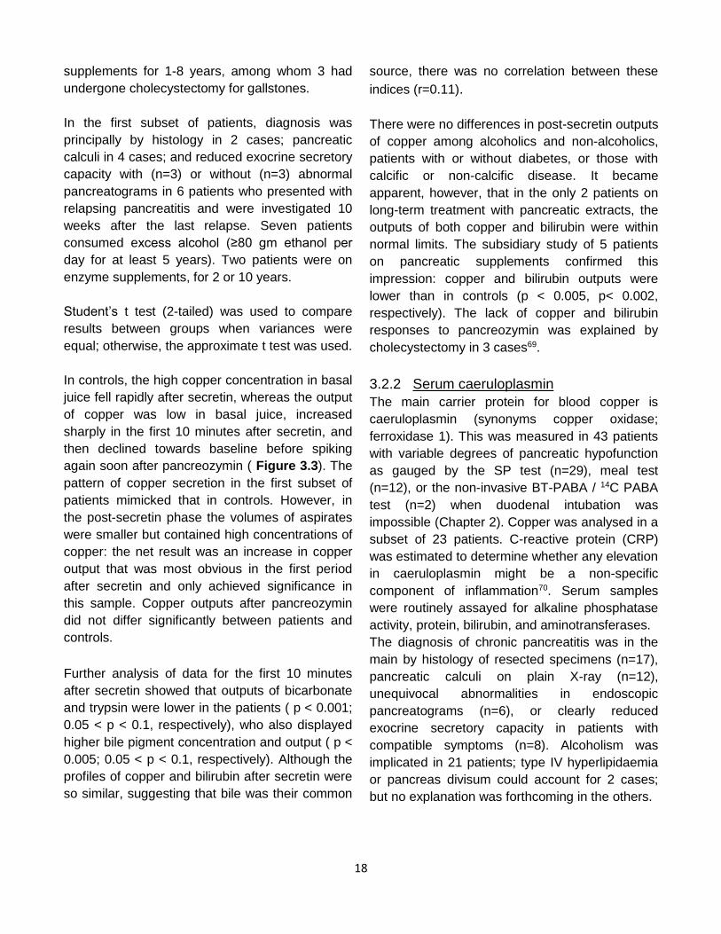
Correlation coefficients calculated on each of the
secretory parameters showed significant trends of
decreasing parameters with increased
pancreatogram scores, - eg. bicarbonate output
after secretin (Figure 2.11) . However, secretory
parameters could not predict pancreatogram
grade in an individual patient. Thus, the % correct
allocation based on bicarbonate output (logged),
peak bicarbonate concentration, volume of
secretion and trypsin output were, respectively,
only 24, 34, 24 and 33%. These data indicated
that duct obstruction and parenchymal destruction
are not separable factors in reducing secretion in
chronic pancreatitis. The study showed,
furthermore, that it is impossible in patients with
chronic pancreatitis to predict accurately exocrine
functional status on the basis of ductal structure
and vice versa47: the same applies to correlation
between functional or pancreatographic
abnormalities and histology48. This position
contrasts with that in pancreatic cancer where
13
secretory loss correlates with the length of duct
obstructed.
It was not the intention of the investigation to
evaluate the relative efficiency of ERCP and
hormone tests in diagnosing chronic pancreatitis.
However: (i) even using both tests a diagnosis
before surgery was not made in 5 patients with
small-duct disease; (ii) a fall in bicarbonate output
in mechanically perfect tests permitted a confident
diagnosis in 3 of 8 patients with normal ducts and
6 of 8 with minimal-change-pancreatitis, whereas
the last finding in isolation is compatible with a
normal gland18; and (iii) on rare occasions ( 1 of
45 in the study) an abnormal pancreatogram may
provide the only diagnostic proof10.
The combination of ultrasonography and ERCP
was evaluated in a parallel study which showed
the high attrition rate for each test (25% and 11%,
respectively); low specificity of ERCP at 65%; and
low sensitivity of ultrasound scanning at 55%49.
2.3.5. Classification and misclassification
In the chapters to follow, a diagnosis of chronic
pancreatitis is based on one or more of the
following parameters in patients with a compatible
history: histology of a previously resected
specimen of the gland; calculi shown by plain X-
ray of the abdomen or CT; reduced exocrine
secretory capacity (< mean - 2 SD of control
values in SP or meal tests; < mean - 3 SD for the
PABA / 14C excretion index); unequivocally
abnormal pancreatogram (grade 3+); or
borderline outcome of function test and ERCP
but clear abnormality on ultrasonography or CT.
Some patients who present with one or more
attacks of pancreatitis are wrongly classified as
post-acute or RAP after the initial assessment
because evidence of functional or structural
damage is lacking. To increase the chance of
detecting patients with early lesions of chronic
pancreatitis, some authors suggested a scoring
system whereby marginal abnormalities in each
test can be summated. The aforesaid autopsy
Figure 2.11 Pancreatographic appearances graded 1-6 and
post-secretin outputs of bicarbonate in 45 patients with chronic pancreatitis. The horizontal line represents the lower limit of the reference range at mean-2SD of logged data in controls ( 5.8 mmol in 30 minutes, a value similar to the 10th percentile of 5.3 mmol). Figure from 1986 review in Recent Advances in Gastroenterology10
pancreatography study should sound a cautionary
note. The recognition that heightened synthesis of
pancreatic enzymes is an early feature of the
disease offered a promising avenue forward, as
also changes in the composition of pancreatic
juice, but foiled by restriction to specialised
centres10. Systematic histology of a surgically
resected specimen of pancreas is difficult to
justify. Hence it was deemed wise to temporise
and repeat the tests at a later date when a
decrement in function or alteration in the
pancreatogram will eventually provide the
diagnosis10.
2.4 Treatment
The primary goal of treatment was - and
remains - to control severe pain. In the 1980s
the options for physicians were few : to insist on
abstinence from alcohol, reinforced by
behavioural / pain consultants and community
groups; to ’rest the pancreas’ by prescribing
ordinary pancreatic extracts which were said to
blunt the putative feedback loop that otherwise
operates to increase CCK release from the upper
small intestine; to enlist the help of nutritionists in
14
patients with malnutrition; to stent the common
bile duct endoscopically in patients with an
obstructive profile of liver function tests; and,
above all, to desist from prescribing narcotic
analgesics. When the last goal was threatened,
surgical opinion was sought, while anaesthetists
facilitated nerve block or ablation procedures -
the efficacy of which was generally short-lived.
Pancreatic surgery was the order of the day.
2.5 Summary
Whereas in the 1980s Bayesian philosophy
enabled evaluation of tools wherewith to diagnose
chronic pancreatitis46, there was no rational
philosophy for its causation. Hence, treatment
options for relentless pain were grim, namely,
narcotic analgesics or piece-meal ablation of the
gland that not infrequently was preceded by duct
drainage and / or nerve block procedures (Figure
1.1). Any advance clearly required a radical
overhaul of thinking on disease pathogenesis.
15
Chapter 3
Serendipity!
In 1970, when reviewing the results of SP tests in
Howat’s laboratory, it became apparent that the
volume of duodenal contents after secretin had
fallen. Since the technique had varied little over
the years, the decline suggested deterioration in
the Boots secretin standard. This was confirmed
by the company’s Head of Bioassay, with
assurance that batches from 119 onwards had
been re-standardized. It was thus necessary to
compare afresh the relative potency of the two
brands of secretin that were in common use
(Chapter 2). This was the start of a fortuitous
series of events.
3.1 Boots versus GIH secretin
3.1.1 On feline pancreas
Like man, the cat secretes pancreatic juice only in
response to meals, and is therefore the preferred
species for the study of responses to secretin.
Cats were fasted for 24 hours, anaesthetised,
splanchnic nerves divided extraperitoneally,
pancreatic duct cannulated, bile duct obliterated
by the ligature retaining the pancreatic cannula,
and gastric pylorus occluded by a tape ligature.
Subsequently, vials from batch numbers 142 and
17421 of Boots and GIH products, respectively,
were used to evaluate pancreatic fluid and
bicarbonate responses. Preliminary experiments
suggested that 1 CHRU of Boots secretin was
around 4 times less potent than 1 CU of the GIH
product (Figure 3.1).
This was borne out by a 4-point assay following 1
and 2 CHRU Boots secretin / kg and 0.25 and 0.5
CU / kg GIH secretin in each of 4 cats. It could
thus be calculated that 1 CU was 3.8 times more
potent than 1 re-standardised CHRU in increasing
the flow rate of pancreatic juice and 4 times more
Figure 3.1 The volume of pancreatic juice and bicarbonate output per 15 minutes in response to intravenous injections of
Boots and GIH secretin.in the anaesthetised cat. Reproduced from 1975 paper in Gut50 with permission of BMJ publishing
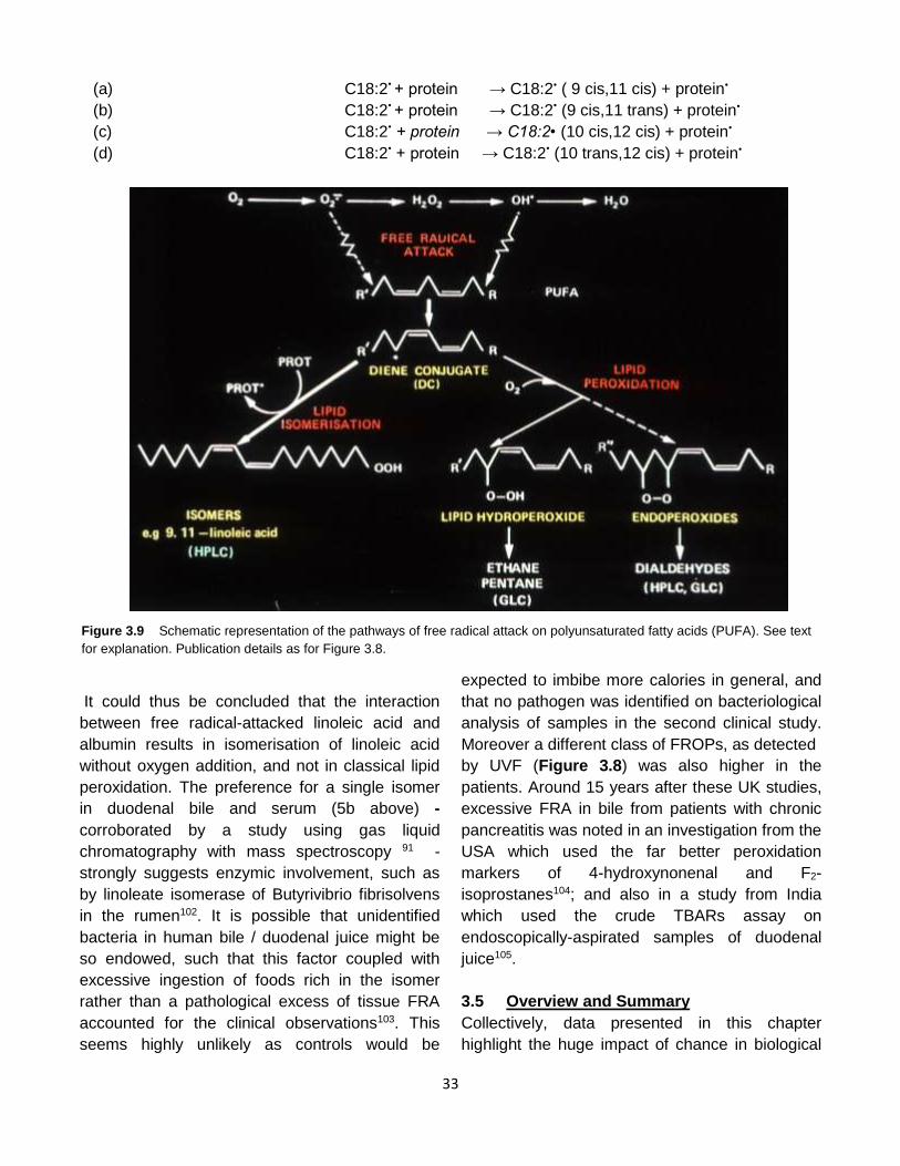
peroxides, GSH glutathione. Reproduced from 1987 paper in Clin Chim Acta 126
39
.
5.1 Routine tests
5.1.1 Bromosulphthalein
Organic anions such as bromosulphthalein (BSP)
follow the same general path as outlined above.
Hence disappearance kinetics following an
intravenous dose of BSP have long been used as
a surrogate to probe hepatic dysfunction: loss of
hepatocytes as in advanced cirrhosis delays the
early-phase clearance of BSP, whereas
intrahepatic cholestasis impedes late-phase
clearance.
Thirty patients with exocrine pancreatic disease
were studied, but the results of 4 tests were later
omitted, because of interference in the BSP assay
(n=1) or erratic data (n=3). The diagnosis of
chronic pancreatitis in 14 patients was mainly
based on the presence of pancreatic calculi (n=4),
unequivocally abnormal endoscopic
pancreatograms (n=9), or grossly reduced
bicarbonate output after secretin (n=1). When
ductal morphology and exocrine secretory
capacity were normal, patients who had
experienced ≥ 1 attack of pancreatitis were
classified as post-acute / RAP (n= 9 ). Fifteen of
the 23 patients with either type of pancreatitis
were on normal diets; 8 adhered to low fat diets
for at least 3 months. Three patients had
pancreatic cancer. Of these, one was fit and
asymptomatic 18 months after a Whipple
operation and was prescribed a supplement of 30
gm medium-chain triglycerides daily with
reduction in long -chain fat. Two patients with
cancer were anorexic for 3 months.
Comprehensive information is given in the study
report127.
The liver was palpable in several patients (Table
5.1), but none had the stigmata of chronic liver
disease, 1 was icteric, and 3 had undergone
biliary bypass surgery to relieve constriction of the
distal bile duct. Endoscopic cholangiography
revealed subtle abnormalities in 11 cases
(Chapter 6). Medical conditions that may be linked
to pancreatitis were found in a few patients;
several were heavy smokers; and some
consumed excessive amounts of alcohol and/or
caffeine -containing beverages.
The test was done exactly as described by its
pioneers and interpreted by reference to their
control data, as was routine hospital practice128.
After an overnight fast, the recumbent patient was
given an intravenous injection of 5 mg BSP / kg
body weight over the course of 30 seconds. Blood
samples were collected from the opposite cubital
vein via an in-dwelling catheter at 3, 5, 7, 10, 15,
20, 25, 30, 35, 40 and 45 minutes after start of the
injection which constituted time zero. Plasma BSP
was measured by a standard spectrophotometric
method for which, as was long recognised,
measurements were adequately reproducible at
plasma values > 3 mg BSP / ml plasma, but
unreliable at lower concentration.
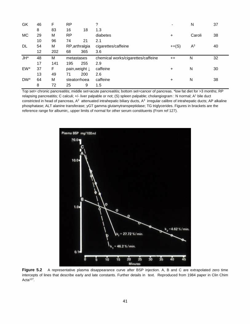
BSP disappearance curves were analysed by the
peeling-off technique (Figure 5.2), wherein
measured BSP concentrations in each test were
plotted against time on a semi-logarithmic scale.
The uncorrected initial disappearance constant,
Ki, was derived by fitting a straight line through at
least 3 of the 4 points between 3 and 10 minutes
after the injection. The second exponential, K2,
was obtained in the same way, using
measurements between the 30 and 45- minute
time-points. If a nearly perfect fit of at least 3
points was unobtainable, the test was omitted
(n=1).The corrected initial disappearance rate
constant, K1, was derived by plotting the
numerical differences between the early plasma
concentrations and the extrapolated straight line
used to determine K2. The corresponding points
were re-fitted by a straight line through at least 3
of the first 4 points. The half-life (T1/2) for
uncorrected and corrected disappearance rates
and for K2 were read off the graph and the
respective constants calculated by the formula K
(% / min) = (0.693 x 100) ÷T1/2. Student’s t test (2-
tailed) was used to compare results with those of
26 controls in the reference publication128.
40
Table 5.1 BSP study: clinical information ___________________________________________________________________________ ID Age Sex Presentation Possibly relevant factors Liver Bile duct Albumin
Bil AP ALT yGT TG
(years) g/l
μmol/l u/l u/l u/l mmol/l
(38-48)
(22) (100) ( 40) (65) (1.8)
PB* 20 F RP ? - N 38
10 71 17 7 0.7
AB 35 M RP (C) alcohol/cigarettes + A1 42
11 128 59 135 4.9
MB 54 F RP( C) cigarettes/tea + N 36
10 48 38 29 1.6
AC 57 M RP (C) alcohol/cigarettes + A1 33
10 29 59 316 1.4
NE 64 M steatorrhoea cigarettes /caffeine + N 32
10 116 35 450 2.0
HG* 78 M RP printing works/cigarettes/tea - N 40
11 50 22 30 1.6
BH 21 M RP ? - N 39
15 70 21 19 1.7
LM 31 F RP Crohn’s/steroids/azathioprine + A2 32
10 50 86 91 2.0
MP* 15 M RP ? - N 38
14 105 127 8 1.5
MR* 54 F pain renal transplant/steroids/azathioprine + A3 31
13 110 20 32 3.7
ER* 47 M pain (C) alcohol/cigarettes/caffeine/pethidine +(S) A2 35
8 165 88 71 1.1
PT 35 F diarrhoea ulcerative colitis/steroids ++(S A3 34
20 485 111 180 2.1
JH 49 M steatorrhoea cigarettes/phenobarbitone/caffeine ++ A2 29
19 297 400 1290 2.7
GH* 30 M RP/jaundice alcohol/pethidine + A1 31
40 159 137 523 1.4
AH 72 F RP gallstones/caffeine - N 42
19 76 14 110 1.9
AF 19 F pseudocyst alcohol + N 40
17 94 28 30 1.8
EH* 42 F RP ? - A2 38
10 74 21 6 2.0
AN 59 M pancreatitis diabetes ++ N 41
16 34 39 26 4.6
MB* 51 F RP ? - N 38
13 62 23 10 1.5
GJ 26 M RP ? - N 38
15 50 58 23 3.5
41
GK 46 F RP ? - N 37
8 83 16 18 1.3
MC 29 M RP diabetes + Caroli 38
10 96 74 21 2.1
DL 54 M RP,arthralgia cigarettes/caffeine ++(S) A3 40
12 202 68 365 3.6
JH* 48 M metastases chemical works/cigarettes/caffeine ++ N 32
17 141 195 255 2.9
EW* 37 F pain,weight ↓ caffeine + N 30
13 49 71 200 2.6
DW* 64 M steatorrhoea caffeine + N 38
8 72 25 9 1.5
Top set= chronic pancreatitis; middle set=acute pancreatitis; bottom set=cancer of pancreas. *low fat diet for >3 months; RP
relapsing pancreatitis; C calculi; +/- liver palpable or not; (S) spleen palpable; cholangiogram : N normal; A1 bile duct
constricted in head of pancreas, A2 attenuated intrahepatic biliary ducts, A3 irregular calibre of intrahepatic ducts; AP alkaline
phosphatase; ALT alanine transferase; yGT gamma glutamytranspeptidase; TG triglycerides. Figures in brackets are the
reference range for albumin;, upper limits of normal for other serum constituents (From ref 127).
Figure 5.2 A representative plasma disappearance curve after BSP injection. A, B and C are extrapolated zero time
intercepts of lines that describe early and late constants. Further details in text. Reproduced from 1984 paper in Clin Chim
Acta127.
42
Reference values (mean ± SD) with number of
observations in their derivation were as follows:
Ki, 12.6 ± 1.6 % / min (n= 26); K1, 14.3 ± 1.5 % /
min (n=16); K2, 5.3 ± 1.9 % / min (n=16). By
comparison, respective values in patients with
pancreatic disease were 15.48 ± 1.7% (0.05 < p <
0.10), 26.06 ± 12.78 (p<0.001), and 3.12 ± 2.43
% / min (p < 0.005), ie. the net early- phase
disappearance of BSP from plasma was higher
but late-phase disappearance lower in patients
than controls. There was no difference in
subgroups with chronic or post-acute / RAP.
The corrected initial disappearance rate constant
K1 was above the 95% upper limit of the reference
range, 117.3% / min, in 19 of the 26 patients
studied (Figure 5.3). Among 7 patients with
normal values, 6 were on low fat diets and a
patient with cancer was profoundly anorexic
(Table 5.1). The uncorrected constant Ki was
below normal in 5 patients (Figure 5.3): there was
evidence of liver disease in 3 of them ( tender
hepatomegaly in a chronic alcoholic,
hepatomegaly plus jaundice, multiple liver
metastases) but not in the other 2 cases. K2 was
reduced in 8 patients (Figure 5.3) including 2 with
questionable data (ie. BSP concentration between
30 and 45 minutes < 2 mg / l). Primary biliary
cirrhosis was identified in a patient with elevated
serum IgM and antimitochondrial antibody. Liver
biopsy was done in 3 of the other 5 cases:
changes of biliary cirrhosis were found in 2, and
sclerosing cholangitis-like changes in the third.
The 2 patients who did not have a liver biopsy
were chronic alcoholics with disturbed liver
function tests (Table 5.1).
5.1.2 Serum triglycerides
This ancillary study was prompted by findings of
hypertriglyceridaemia and / or elevated gamma-
glutamyl transpeptidase (γGT) activity in several
patients (Table 5.1) - given that both changes
have been reported in association with hepatic
‘enzyme induction’129.
Figure 5.3 BSP clearance constants in patients with
exocrine pancreatic disease. Dotted areas represent the
published reference ranges (2SD on either side of the mean).
Publication details as for Figure 5.2.
Accordingly in a separate investigation, 33
consecutive patients who were referred after an
attack or relapse of pancreatitis ≥ 6 weeks earlier
(chronic 23, acute 10) had an intravenous fat
tolerance test to determine capacity to metabolise
triglycerides130. Fasting hypertriglyceridaemia was
present in 14 patients (42 %), including 9 with
chronic and 5 with acute pancreatitis; the highest
value was 6.2 mmol / l; and no patient had
hyperchylomicronemia at the time of testing. In
this subgroup, 4 displayed impaired triglyceride
clearance (29 % of those with hyper-
triglyceridaemia, representing 12 % of the whole
cohort), and hence might be vulnerable to a
further attack of pancreatitis due to massive
hypertriglyceridaemia under certain conditions131.
Although average triglyceride clearance in the
other 10 patients was less than in patients with
normal serum triglycerides (p < 0.05), values were
within the reference range and did not correlate
with serum triglyceride levels.
43
5.1.3 Comments
BSP in plasma, tightly bound to albumin,
traverses the space of Disse and is internalised
by a carrier mechanism that probably involves
albumin receptors on the plasma membrane of
hepatocytes. Once in the cytosol, it binds
preferentially to GST-B, which also binds
hormones, drugs and azo-dye carcinogens132.
The concentration of GST-B within hepatocytes
increases in response to chemicals that cause
SER proliferation. Binding of an anion reduces its
reflux into plasma, which in the case of bilirubin
amounts to around 30 % of that initially taken up
into hepatocytes. Hence, although the amount of
ligandin seems to have no direct effect on influx
rate, the higher its concentration the lower the
rate of efflux, such that the net uptake of an anion
is enhanced84.
After GSH conjugation, BSP is actively excreted
across canalicular membranes, so that about 60-
100% of an injected dose is recoverable in bile
within 2 hours133, 134. Extrahepatic elimination is
negligible when the liver is functioning normally,
but increasingly important in cholestatic states or
when large doses of the dye are given. Reduced
hepatic clearance of BSP has been recorded in
obese, pregnant or elderly people, those with
pyrexia, or when serum albumin is very low135, 136.
With 1 exception (HG in Table 5.1), the study
patients did not fall into those categories.
It is agreed that K2 in the BSP disappearance
curve (Figure 5.2) reflects maximal canalicular
transport capacity, and is thus reduced in patients
with primary biliary cirrhosis128. Judging by K2
data, canalicular transport mechanisms were
intact in the majority of patients with pancreatic
disease (Figure 5.3), which accords with
increased concentrations in bile of substances
generated in hepatocytes (Chapter 3).
The finding of increased K 1 in patients with
pancreatic disease was both new and not easily
explained. There was no clinical feature to
suggest increased hepatic blood flow. Induction of
ligandin, and hence reduced efflux of BSP
seemed the best explanation, based on
experimental evidence137 and also on clinical
studies using another anion, indocyanine green,
in patients on enzyme-inducing drugs138.
An increase in hepatic microsomal mass in
response to enzyme induction could also
rationalise hypertriglyceridaemia and increased
serum γGT (over-and-above that due to
hepatocyte dysfunction) in both studies. Thus, in
the first study potential enzyme inducers included
cigarette smoke, alcohol, theophylline-containing
beverages, steroids, azathioprine,
phenobarbitone, and occupational chemicals
(Table 5.1): in a few patients an inducer was not
identified, but an excess of dietary PUFA might
have played a part, in that 4 patients with normal
K 1 values were on low fat diets for 6 months or
more, and also because PUFA-enriched diets
facilitate enzyme induction in experimental
studies117.
There is thus a 2-way relationship between
hypertriglyceridaemia and pancreatitis, ie. as a
consequence, or potentially causal. In regard to
the latter, the ancillary study of lipid clearance
suggested that a connection is indirect - an
epiphenomenon of some other process130.
Hepatic enzyme induction fits the bill.
5.2 Antipyrine, theophylline, debrisoquine
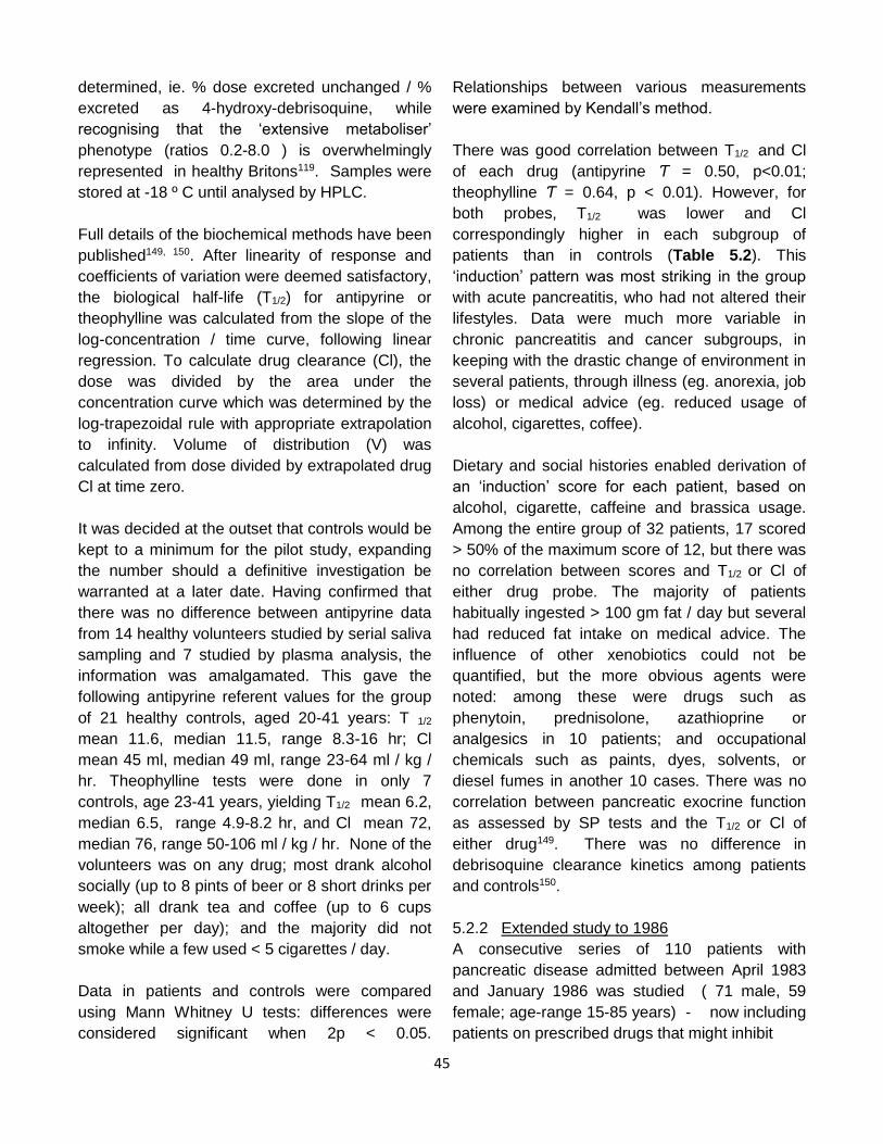
5.2.1 Pilot study 1983 / 1984
Analysis of the microsomal fraction in liver biopsy
material from animals provides a simple way to
detect CYP induction and characterise the sub-
type involved, but this approach is clearly
inapplicable as a routine measure in man. The
disposal of the drug antipyrine was known to
afford a broad gauge of CYP induction in
humans139; moreover, it was accepted that
antipyrine pharmacokinetics derived from blood or
salivary sampling are effectively identical140, and
also that a genetic influence could be exposed, as
in relation to CYP inducibility by phenobarbital141.
The usefulness of theophylline to detect induction
induction, whether across all isoforms (antipyrine
test) or the specific isoform induced by PAH
(theophylline test ).
The diagnosis of chronic pancreatitis in 22 cases
was mainly based on pancreatic calculi (n=7),
unequivocally abnormal pancreatogram ( n=12) or
reduced exocrine function ( n=3 ) ( Chapter 2).
Most of these patients presented with relapsing
pancreatitis, and were assessed between 6
weeks and 14 months after the last episode: 4
were dependent on narcotic analgesics at the
time of referral, and 1 had painless steatorrhoea.
Distal bile duct constriction was excluded by
endoscopic cholangiography in all patients. Post-
acute / RAP was diagnosed in 6 patients with
normal pancreatogram and pancreatic function
test at least 6 weeks after the most recent attack:
none of these patients had gallstones. Four
patients had pancreatic cancer that did not
obstruct bile drainage.
A questionnaire was devised to facilitate
recognition of known or suspected risk factors for
acute pancreatitis, chronic pancreatitis and
pancreatic cancer (Chapter 2). Often, the patient’s
social circumstances had changed on medical
advice, redundancy or redeployment. In order to
facilitate interpretation of pharmacokinetic data,
an a priori search was made for recognised CYP
inducers in each patient, to cover the 6-month
period preceding testing, because it was known
that the clearance of theophylline may remain
elevated for some time after cessation of
exposure to inducers in cigarette smoke148. To
this end, detailed dietary histories were taken with
special emphasis on intake of fat, charcoal-broiled
beef, caffeine, and Brassica vegetables: the
information was supplemented by a 7-day
weighed dietary record (completed by the patient
or immediate relative) at home in the week
following the tests. An arbitrary scoring system
was implemented to quantify these factors, as
also alcohol and cigarettes. Patients were
questioned about exposure to workplace
chemicals, and all drugs taken on a regular basis
were recorded.
Patients were admitted to the hospital’s
programmed investigation unit for antipyrine,
theophylline and debrisoquine tests, together with
any pancreatic investigation that was deemed
necessary, and dietary assessment. These
investigations required 2 consecutive weeks, the
drug studies done in the same week and
commencing around 6 am. For the antipyrine test,
the patient voided urine, and then drank an
aqueous solution of the drug (10 mg / kg body
weight): peripheral venous blood was drawn pre-
dose and 3, 6, 9, 12, 27, 30 and 36 hours
thereafter; the volume of the 48 hour urine
collection was measured and an aliquot retained
for analysis. All caffeine containing foods and
beverages were withdrawn at least 48 hours
before the theophylline study. After voiding urine,
the patient received theopylline elixir (2.4 mg /
kg) and blood was sampled at 90 minute intervals
for 10.5 hours. For the debrisoquine test, a 10 mg
tablet was administered, urine collected for 8 hr
thereafter, and the ‘debrisoquine metabolic ratio’
45
determined, ie. % dose excreted unchanged / %
excreted as 4-hydroxy-debrisoquine, while
recognising that the ‘extensive metaboliser’
phenotype (ratios 0.2-8.0 ) is overwhelmingly
represented in healthy Britons119. Samples were
stored at -18 º C until analysed by HPLC.
Full details of the biochemical methods have been
published149, 150. After linearity of response and
coefficients of variation were deemed satisfactory,
the biological half-life (T1/2) for antipyrine or
theophylline was calculated from the slope of the
log-concentration / time curve, following linear
regression. To calculate drug clearance (Cl), the
dose was divided by the area under the
concentration curve which was determined by the
log-trapezoidal rule with appropriate extrapolation
to infinity. Volume of distribution (V) was
calculated from dose divided by extrapolated drug
Cl at time zero.
It was decided at the outset that controls would be
kept to a minimum for the pilot study, expanding
the number should a definitive investigation be
warranted at a later date. Having confirmed that
there was no difference between antipyrine data
from 14 healthy volunteers studied by serial saliva
sampling and 7 studied by plasma analysis, the
information was amalgamated. This gave the
following antipyrine referent values for the group
of 21 healthy controls, aged 20-41 years: T 1/2
mean 11.6, median 11.5, range 8.3-16 hr; Cl
mean 45 ml, median 49 ml, range 23-64 ml / kg /
hr. Theophylline tests were done in only 7
controls, age 23-41 years, yielding T1/2 mean 6.2,
median 6.5, range 4.9-8.2 hr, and Cl mean 72,
median 76, range 50-106 ml / kg / hr. None of the
volunteers was on any drug; most drank alcohol
socially (up to 8 pints of beer or 8 short drinks per
week); all drank tea and coffee (up to 6 cups
altogether per day); and the majority did not
smoke while a few used < 5 cigarettes / day.
Data in patients and controls were compared
using Mann Whitney U tests: differences were
considered significant when 2p < 0.05.
Relationships between various measurements
were examined by Kendall’s method.
There was good correlation between T1/2 and Cl
of each drug (antipyrine Ƭ = 0.50, p<0.01;
theophylline Ƭ = 0.64, p < 0.01). However, for
both probes, T1/2 was lower and Cl
correspondingly higher in each subgroup of
patients than in controls (Table 5.2). This
‘induction’ pattern was most striking in the group
with acute pancreatitis, who had not altered their
lifestyles. Data were much more variable in
chronic pancreatitis and cancer subgroups, in
keeping with the drastic change of environment in
several patients, through illness (eg. anorexia, job
loss) or medical advice (eg. reduced usage of
alcohol, cigarettes, coffee).
Dietary and social histories enabled derivation of
an ‘induction’ score for each patient, based on
alcohol, cigarette, caffeine and brassica usage.
Among the entire group of 32 patients, 17 scored
> 50% of the maximum score of 12, but there was
no correlation between scores and T1/2 or Cl of
either drug probe. The majority of patients
habitually ingested > 100 gm fat / day but several
had reduced fat intake on medical advice. The
influence of other xenobiotics could not be
quantified, but the more obvious agents were
noted: among these were drugs such as
phenytoin, prednisolone, azathioprine or
analgesics in 10 patients; and occupational
chemicals such as paints, dyes, solvents, or
diesel fumes in another 10 cases. There was no
correlation between pancreatic exocrine function
as assessed by SP tests and the T1/2 or Cl of
either drug149. There was no difference in
debrisoquine clearance kinetics among patients
and controls150.
5.2.2 Extended study to 1986
A consecutive series of 110 patients with
pancreatic disease admitted between April 1983
and January 1986 was studied ( 71 male, 59
female; age-range 15-85 years) - now including
patients on prescribed drugs that might inhibit
46
Table 5.2 Results: pilot study of antipyrine and theophylline disposition ___________________________________________________________________ Antipyrine Theophylline ------------------------------------------ ----------------------------------------
T1/2 (hr) Cl (ml/kg/hr) T1/2 (hr) Cl (ml/kg/hr)
------------------------------------------ ----------------------------------------- Median p Median p Median p Median p
similar to the patients, and recruited from hospital
staff or by advertisement in a local general
practice. No volunteer had dietary fads, drank
alcohol or caffeine-containing beverages in
excess, or was on medication. The second set
consisted of 5 patients with gallstones awaiting
elective cholecystectomy, but with no symptoms
or signs of co-existing pancreatic disease. The
protocol was as described in the pilot study, but
debrisoquine tests were omitted. After the first 18
months it was clear that antipyrine and
theophylline tests yielded similar data.
Considering that the theophylline test could be
completed within a day, the antipyrine test was
abandoned thereafter.
Several quantifiable host and environmental
factors were known to influence CYP. These
could be grouped as dietary or miscellaneous.
Accordingly, intakes of fat / protein / carbohydrate
/ methyl xanthines (as in tea and coffee) /
brassica vegetables were assessed by 7-day
home food inventories, supervised by the
hospital’s senior dietitian, and then decoded using
a Microdiet programme (Salford University, UK).
Alcohol and cigarette usage contributed to the
non-food inducers, and were registered as
whether- or -not operating at the time of the study:
in the case of current smokers, 4 subgroups were
defined according to number of cigarettes smoked
per day.
Statistical analysis was as described in the pilot
study. In addition, a series of multivariate linear
regression analyses examined the impact of
various host and environmental factors on
clearance of antipyrine or theophylline. A stepwise
analysis with backward elimination was employed
in the first instance, ie. variables were eliminated
consecutively while their removal had no impact
on the regression
Validation of the pharmacokinetic methods was
accomplished in a number of ways. The
47
coefficients of variation (CV) from analysis of
aqueous standards of each drug was obtained by
quintuplicate analysis, to cover concentrations
that should yield plasma levels found in the pilot
study: derived CV 3.6-5.1% for antipyrine; 1.5-
4.5% for theophylline. The CV inherent in analysis
of plasma samples was derived by analysing 10
sets of samples in triplicate, yielding average CV
for each drug < 10%. Since drug retention in
containers for oral delivery might account for
some of this variation, 14 containers were taken
at random, rinsed with 50 ml water and drug
concentration measured: mean loss was 1.72% of
the original dose, range 0.9-2.6%. Paired
theophylline tests were done in 7 individuals,
following oral or intravenous delivery of the drug.
The average bioavailability of theophylline, F1,
proved to be 1.01, and there was a significant
correlation between drug Cl or T1/2 after oral and
intravenous dosing: this held true for 2 patients
with exocrine pancreatic failure151.
Figures 5.4 and 5.5 show the wide range of drug
clearances in patients compared to healthy
controls. The expected inverse correlation
between T1/2 and Cl of theophylline was realised
in cigarette smokers (n=58) or non-smokers (
n=48)151. There was fairly good correlation
between antipyrine and theophylline Cl in a
subset of 91 individuals (15 controls, 76 patients)
who had both tests.
Theophylline Cl was faster in the entire cohort of
110 patients with pancreatic disease than in
healthy volunteers (medians 104 and 68 ml / kg /
hr, respectively, p<0.05), but there was no
difference when antipyrine Cl in 76 patients was
considered ( medians 57 and 50 ml /kg /hr,
respectively) - suggesting preferential
involvement of the PAH-inducible subfamily of
CYP. The theophylline result was largely
influenced by very significant increases in
theophylline Cl among patients with chronic
pancreatitis, both in the alcoholic subset of 24
(median 132 ml / kg / hr, p<0.001) and the
idiopathic subset of 47 (median 93 ml / kg / hr,
p<0.010). In contrast to the pilot study ( Table
5.2), theophylline Cl values in subgroups of 28
patients with post- acute pancreatitis and 11 with
pancreatic cancer ( medians 86 and 104 ml / kg /
hr, respectively) were not significantly different
from control values. Nonetheless, inspection of
Figure 5.6 shows that theophylline Cl exceeded
the highest control value in around half the
patients in these groups151. Results in ‘gallstone
Figure 5.4 Frequency distribution of theophylline
clearance, CT, in 110 patients with pancreatic disease.
Shaded areas indicate data from 15 healthy volunteers.
Clearance is in ml/kg/hr Reproduced from 1989 paper in
Clinical Science151
Figure 5.5 Frequency distribution of antipyrine clearance,
CA, in 76 patients with pancreatic disease. Clearance is in
ml/kg/hr. Publishing details as in Figure 5.4.
48
Figure 5.6 Theophylline clearance ( CT ) in controls and groups with gallstones, chronic pancreatitis (CP), acute pancreatitis
(AP) and pancreatic cancer (CA). Shaded area represents reference range. Publication details as for Figure 5.4.
Table 5.3 Non-smokers with high theophylline and/ or antipyrine clearance*
Sex Age Current job Drugs Theophylline Antipyrine ml/kh/hr _________________________________________________________________________ M 49 oil processing bextazxol, fenoterol 122 81 M 31 loading bay work phenytoin 133 not tested M 23 welder none 178 53 M 44 technical officer insulin 191 102 F 58 cleaner noine 279 72 F 57 guesthouse keeper none 104 116 M 70 civil servant insulin 284 not tested F 65 housewife aspirin as needed 134 34 M 54 lorry driver insulin 121 59 M 16 schoolboy none 93 93 F 73 book keeper tolbutamide, amitryptiline 107 51 F 23 housewife none 100 55 M 26 carpenter penicillamine 100 48 F 33 social worker rheumox, salazopyrine 68 68
V (l/kg) 0.75 (0.21) 0.53 (0.15) o.52 ( 0.16) __________________________________________________________________ Data as mean with standard deviation in parenthesis. Significance of differences from control values: ** p<0.01, *** p<0.001. There were no differences between smokers and non-smokers in the epilepsy group. Abbreviations: n number, Cl clearance, T
½ half life, V volume of distribution.
50
in smoker and non-smoker subgroups of patients
with epilepsy; there was good correlation between
disposal parameters for the probes152.
5.2.4 Comments
By 1985 there were numerous publications on the
pharmacokinetics of antipyrine and theophylline in
controls129, 139, 149, and it was recognised that the
wide inter-individual variation in drug disposal
probably reflects complex interactions between
genetic and environmental factors that influence
CYP function. Because of this scatter, the use of
a single antipyrine or theophylline test in a group
of patients with a specific disease was
deprecated153. Nonetheless, it had been
observed that long-term exposure to a CYP
inducer such as phenobarbital narrows inter-
individual variation in antipyrine elimination141.
Moreover, it was suggested that, whereas
antipyrine Cl rapidly reverts to normal upon
removal of a CYP inducer153, theophylline Cl
remains augmented for some time thereafter154.
The first prerequisite in the Manchester studies
was to ascertain that any difference in drug
disposal between patients and controls was not
artefactual. This was done by the measures
described in Section 5.2.2. The risk of
anaphylactic reaction after intravenous antipyrine
prevented paired oral and intravenous studies to
confirm the excellent bioavailability that is
accepted for this probe153.
The wide ranges for T1/2 and Cl of both drugs in
controls of the pilot study - although smokers,
chronic alcoholics and drug-takers were excluded
- conformed with published data. It was thus
remarkable to find significantly shorter biological
T1/2 and more rapid Cl of each probe in subgroups
with exocrine pancreatic disease (Table 5.2 ),
comprising a nearly consecutive series of 32
patients, irrespective of age, gender, disease type
or duration, putative aetiological factor or exocrine
functional status. While this finding cannot
distinguish between enzyme induction as strictly
defined and the general increase in CYP activity
that may accompany increases in cell size and
protein content, there is no reason why the
pathophysiological effects should be any different.
The results clearly needed to be validated or
refuted by a larger investigation, to include also
patients with gallstone-related acute pancreatitis.
The choice of controls posed a dilemma. Ideally,
patients and controls should be matched for
relevant variables but these could not be
pinpointed from epidemiological surveys of
patients with pancreatic disease, nor after
analysis of data from the pilot study. For example:
alcoholism was thought to have a variable
influence on CYP155 and was clearly associated
with chronic pancreatitis but its role in acute
pancreatitis and cancer was debated; cigarette
smoke was established as a potent CYP inducer
and linked to pancreatic cancer but evidence in
regard to pancreatitis was conflicting; a high-fat
diet was known to facilitate CYP induction and
implicated in pancreatic cancer but not
unequivocally with pancreatitis. Moreover, the
demanding nature of the extended investigation,
requiring 2 consecutive weeks for dietary and
drug kinetic aspects, would make it impossible to
enlist sufficient volunteers to cover each of these
associations. Hence it was decided to expand the
group of volunteers with a healthy lifestyle as
used in the pilot study, and to use computer-
assisted multivariate analysis to dissect out items
contributing to CYP induction in patients, if that
conclusion was borne out. A group with gallstones
but without a history of pancreatitis would serve
as controls for the gallstone-pancreatitis group:
data from this subset were not amalgamated with
data from healthy volunteers because it had been
suggested that aberrant CYP activity might be
involved in the development of gallstones156.
The outcome of both studies left no doubt that
CYP induction is a feature of chronic pancreatitis.
In this regard it is of interest that multivariate
analysis in the extended study picked out items
that had been demonstrated individually to alter
drug-metabolizing capacity in healthy subjects.
51
For example, it identified smoking followed by
dietary protein as key contributors to accelerated
theophylline Cl and the same factors in reverse
order as contributors to accelerated antipyrine Cl.
The magnitude of the positive effect was similar to
that reported by others for both smoking and
protein. The highest values for theophylline Cl
were in patients with ‘alcoholic’ chronic
pancreatitis, but the label was clearly misleading
in that the multivariate analysis showed ‘alcohol-
ever’ usage per se to exert an inhibitory effect on
theophylline Cl151. There was no guarantee that
the result would hold true if the patients were
studied before they curtailed alcohol usage on
medical advice. However, the discovery that
ethanol induces a specific CYP subfamily157,
presently classed as CYP2E1108, underlines the
principle of selectivity in CYP induction. Not until
1999 did it become known that chlorzoxazone
disposal provides an index of CYP2E1 activity158.
The further finding, that theophylline Cl is a better
indicator than antipyrine Cl of CYP induction in
patients with chronic pancreatitis, underscores the
point. Preliminary analysis of urinary metabolites
after antipyrine tests suggested that the 3-
hydroxymethylation pathway was induced
(accounting for the correlation with theophylline
Cl), but not other recognised pathways150.
Nonetheless, the study in patients with epilepsy
showed that there is a ceiling for overall CYP1A2
induction which is reached by treatment with
anticonvulsant drugs, such that any further effect
as from cigarette smoke is indiscernible152.
There are several possible explanations for the
Manchester findings. (i) The altered drug kinetics
might have been iatrogenic, as was possible
though not necessarily the sole explanation nor
detracting from the potential pathological
significance, in a quarter of patients in the pilot
study. (ii) Pancreatic exocrine insufficiency might
have indirectly altered drug disposal by evoking a
rise in circulating CCK159: the lack of correlation
between pancreatic enzyme secretory capacity
after administration of the hormone and drug
disposal parameters argued against this
interpretation. (ii) Experimental biliary obstruction
leads to induction of CYP3A1 so that
accumulating bile acids can be detoxified.
However, patency of the biliary tree was
demonstrated by ERCP in every patient of the
pilot study. (iii) CYP induction may simply be
coincidental - most unlikely because the wide
reference range should reduce rather than
increase the chance of detecting significant
differences in patients with specific diseases. (iv)
Finally, given the many parallels between factors
that are known or suspected to cause or promote
exocrine pancreatic disease and those that are
known or suspected to cause or promote CYP
induction9, the findings strengthened the case for
CYP induction as a causative factor. If so, it was
intriguing that the highest values for theophylline
Cl were similar in smokers and non-smokers,
emphasising the impact of other xenobiotic
inducers that the study did not or could not
quantify. In this regard it is now known that
CYP1A is induced not only by PAH but also by
chlorinated biphenyls160, halogenated hydro-
carbons, and C18:2 fatty acids as in corn, peanut
and linseed oils161.
Many CYP isoforms display polymorphism.
CYP2D6 is an example, and its genetic status can
be probed by use of the drug debrisoquine. At
least in the case of this isoenzyme, genetic
endowment was excluded as an explanation for
increased CYP activity in patients with pancreatic
disease150.
5.3 D-glucaric acid
5.3.1 Study description and outcome
Glucuronic acid, derived via the hexose
monophosphate shunt, is involved in the phase II
metabolism of endobiotics such as bilirubin, and
several xenobiotics. D-glucaric acid (DGA) is a
minor offshoot, which exits across sinusoidal
membranes (Figure 5.1)126. Measurement of DGA
in random samples of urine was touted as a non-
invasive mirror of phase I metabolism via CYP162-
164.
52
Accordingly, an 8-hour collection of urine between
9 am and 5 pm, was obtained for DGA analysis in
a consecutive series of patients with pancreatic
disease during a 9-month period126. Each patient
had antipyrine and theophylline tests in the same
week. Exclusion criteria were as follows: age
under 20 years or over 75 years, congestive
cardiac failure, asthma, renal failure. Chronic
pancreatitis in 19 patients was diagnosed
principally by histology of resected specimens
(n=3), pancreatic calculi (n=6), unequivocally
abnormal pancreatogram (n=9), or clearly
reduced exocrine secretory capacity (n=1). Of
these patients, 10 could be labelled as ‘alcoholic’
(Chapter 2) and there was no explanation in the
others. Of 7 patients who were classified as post-
idiopathic chronic pancreatitis who presented with
steatorrhoea (Figure 6.4). The occasional
concurrence of cell types (Table 6.1) suggests
that they may represent a continuum that evolves
upon regular exposure to xenobiotics, whether or
not identified. Indeed, considering the accelerated
clearance of drug probes by many patients
(Chapter 5), including several who had a liver
biopsy, it may be that that hepatocytes which
appeared ‘normal’ would display ultrastructural
aberrations. Thus, extensive microvesicular
steatosis in 3 patients was only revealed when
semi-thin sections were stained by toluidine blue
(Figure 6.2). The high frequency of portal tract
lesions is discussed after the next section.
6.2 Endoscopic Cholangiograms
6.2.1 Study description and outcome
A list of patients with chronic pancreatitis, post-
acute pancreatitis, pancreatic cancer, or chronic
pain that was later diagnosed as spastic colon
syndrome was drawn up from the ERCP register
for 1981-1984 inclusive. Thereafter the clinical
records were retrieved and examined. Cases
were excluded in the following circumstances:
biliary tree filled inadequately; follow-up
information incomplete; diagnosis still in doubt;
established liver disease; previous
sphincterotomy or bile duct exploration; prior
history of ascending cholangitis. Radiographs for
detailed study came from 47 patients with chronic
pancreatitis, 12 with post-acute pancreatitis, 7
with pancreatic cancer and 7 with spastic colon.
Categorization into pancreatitis subgroups was by
standard criteria (Chapter 2).
Alcoholism was recorded in 19 patients with
chronic pancreatitis, of whom 9 had pancreatic
calculi and 1 had gallstones. The non-alcoholic
group of 28 patients, of whom 6 had pancreatic
calculi, included 2 who had undergone renal
transplantation and were on prednisolone and
azathioprine, a patient with primary biliary
cirrhosis (and pancreatic calculi), 2 with ulcerative
colitis on salazopyrine, and a patient with Crohn’s
colitis on azathioprine. The group of 12 with post-
64
acute pancreatitis included 3 with gallstones; 3
whose attacks were not aborted by surgery
(cholecystectomy 2, resection of choledochal cyst
1); a patient on immunosuppressive therapy for
chronic renal failure; and another in whom the
anti-mitochondrial antibody test was positive. Four
of the group, including the patient with primary
biliary cirrhosis, had hypertriglyceridaemia. The
diagnosis of pancreatic cancer was confirmed by
histology: the tumour was primarily in the neck,
body or tail of the gland (n=2, 2, 1, respectively),
or diffuse (n=2). The ‘spastic colon’ group
presented with constant abdominal discomfort,
usually accompanied by altered bowel habit:
pancreatic tests were normal and patients
remained well during a follow-up period of 2+
years.
Apart from hepatomegaly in the cancer group,
there were no clinical signs of liver disease.
Serum transaminases were generally normal, as
was prothrombin time; whereas alkaline
phosphatase was elevated in 35% of patients
overall.
Radiographs from each patient were in the first
instance reviewed independently by 2 observers
who had no access to the clinical information:
where there was disagreement, a final judgement
was reached after a combined review. Every
cholangiogram was systematically assessed with
regard to the common bile duct and intrahepatic
biliary tree, each according to 3 pre-set criteria, as
follows: (a) ‘irregularity’, defined as calibre
variation, either locally or over a longer segment;
(b) ‘roughening’ implying an uneven duct outline,
varying from a fine brush border to multiple
mamillations extending over 1 cm or more; (c)
‘stricturing’, which indicated reduction in duct
calibre by at least 50% and could be
accompanied by beading (presumably due to
focal pre-stenotic dilatation) or, rarely,
sacculation174. Added features recorded for the
intrahepatic element were ‘nipping’ (ie narrowing
at the point of ductal origin) and ‘pruning’ (ie
reduced number and crowding of second-order
ducts). At least 3 different changes had to be
present for a cholangiogram to be declared
abnormal (Table 6.2). Where the changes in
extra- or intra-hepatic biliary ducts were
borderline, the results were regarded as normal.
Results in the various groups were compared by
Fisher’s exact test.
Cholangiograms were classified as normal in all
patients with the spastic colon syndrome,
whereas they were regarded as abnormal in 55 of
66 patients overall with pancreatic disease (83%)
(Figures 6.9, 6.10). When cases with
intrapancreatic constriction of the bile duct were
excluded from the analysis, 70% of the remaining
patients with chronic pancreatitis had abnormal
intrahepatic cholangiograms, as did 60% with
acute pancreatitis and 100% with pancreatic
cancer. In the chronic pancreatitis subset the
frequency of abnormal cholangiograms was
higher in patients with alcoholic compared to non-
alcoholic disease (p=0.018).
6.2.2 Comments
With the advent of ERCP in the 1980s,
associations between liver and pancreatic disease
were increasingly recognised. Thus, patients with
primary biliary cirrhosis or primary sclerosing
cholangitis were noted to have ERCP evidence of
asymptomatic chronic pancreatitis176, with 1 report
quoting a 77% incidence of abnormal
pancreatograms in patients with the latter
disease177. On the reverse side of the coin, the
Manchester study showed abnormal
cholangiograms in the majority of patients with
exocrine pancreatic disease175.
The outcome was in keeping with the liver biopsy
findings (Section 6.1; cancer patients not
biopsied). The range of portal tract changes noted
was reminiscent of findings in patients with
primary sclerosing cholangitis178, 179. Others have
alluded to the covert nature of sclerosing
cholangitis in the setting of ulcerative colitis178, 180:
as in that condition so in pancreatitis, the
presenting feature of rectal bleeding or agonizing
65
Figure 6.9 Cholangiogram of a patient with pancreatitis
attacks despite cholecystectomy (no stones), on NSAIDs for
arthritis. Note dilatation of the common bile duct, consistent
with cholecystectomy; strictures in both main hepatic ducts,
with calculi upstream. The pancreatic duct appeared normal.
Reproduced from Clin Radiol175.
Figure 6.10 Retrograde cholangiogram of an alcoholic
patient with relapsing acute pancreatitis. Note irregularity of
the common bile duct, nipping of a main intrahepatic duct
and calibre variation with beading in several third-order
ductules. Publication details as for Figure 6.8.
66
abdominal pain, respectively, forces the patient to
seek medical help at a time when the liver
problem has not advanced sufficiently to cause
symptoms or to substantially alter the serum liver
function profile.
6.3 Chronic pancreatitis-type artificial
bile and experimental pancreatitis
6.3.1 Study description and outcome
An experimental investigation was designed to
test the possibility that pancreatitis might be
initiated by reflux of bile laden with FROP181, as
found in clinical studies87.
The procedure in rats was as follows. After
anaesthesia the distal bile duct was tied off and a
cannula inserted into the pancreatic duct at the
duodenal entry point, following which 100 μl of a
test solution was infused retrogradely into the duct
over 2 minutes using a syringe pump.
Observations on the animal’s general condition,
appearance of pancreas and peritoneal cavity
were made at 3-hour intervals during the next 12
hours while the animal was still anaesthetised.
Findings in the head and tail of the gland were
graded separately on an arbitrary scale from 0-3
for oedema, fat necrosis, glandular necrosis, and
haemorrhage. Peritoneal fat necrosis distant from
the pancreas was scored from 0-3. A maximum
score of 27 would indicate an advanced stage of
haemorrhagic pancreatic necrosis (HPN,
synonym necrotising pancreatitis). After
termination of the experiment, the pancreas was
removed, fixed and embedded in paraplast. Three
5 μ sections, each approximately 150 μ apart,
were taken from the head region and 3 from the
tail, and stained with haematoxylin and eosin. The
summated extent of abnormality in head or tail
sections section was graded on an arbitrary scale
from 0-3 for oedema, inflammatory cell infiltrate,
necrosis, fat necrosis and haemorrhage. The
scores for head and tail were then added: rising
scores, up to a maximum of 30, denoted
increased disease severity.
The following test solutions were prepared: (i)
isotonic saline, 150 mmol/l; (ii) bile salt solution
with 2.09% bile salts of pH 8.4 and osmolality 258
mosmol/kg, comprising sodium glycocholate,
sodium glycodeoxycholate, and sodium
deoxycholate in a weight / weight ratio of 2:2:1
(also NaCl 85 mmol/l, NaHCO3 25 mmol/l and
KCl 5 mmol/l); (iii) linoleic acid / bile salt in which
the fatty acid was dissolved in the bile salt mixture
to a concentration of 3.6 mmol/l or 25 mmol/l
immediately before use; (iv) ultraviolet light-
irradiated linoleic acid at both concentrations
made up with bile salts and albumin 10 g/l to
simulate secretin-stimulated bile in controls and
pancreatitis patients, respectively (Chapter 3);
and ( v) lipoxidase-peroxidised linoleic acid / bile
salt mixture without albumin.
The surgeon who did the experiments and the
pathologist were unaware of the solutions under
test, which were formulated by a chemist who
ensured that more than 1 test solution was tested
during a day’s work and that studies in a given
series were spread over the course of several
days. The Mann Whitney U test was used to
analyse data.
Ductal pressures were monitored by a transducer
coupled to a polygraph. In a preliminary study of 5
rats, pressures were remarkably similar: peak 41
± 0.5, plateau 24.2 ± 0.8, end 20 ± 0.95 cm water
(mean ± SE). By incorporating methylene blue it
was evident that 100 μl passed up to the ligature
on the common bile duct and entered pancreatic
duct branches in the head of the gland, whereas
little entered the distal duct.
The results are summarised in Table 6.3. In the
saline-treated group, transient oedema of the
pancreatic head was seen soon after the infusion,
and mild interstitial oedema was shown on
histology. Treatment with bile salts increased
oedema, caused foci of acinar necrosis, and
elicited a moderate inflammatory exudate. The
addition of linoleic acid in low or high dose further
increased macroscopic injury at 3 hours, but not
67
thereafter, and microscopic damage scores were
similar for both doses. The ‘biological’ DC product
(solution iv) in a dose of 25 mmol/l caused a
greater degree of damage than bile salts alone or
with linoleic acid: areas of pancreatic necrosis and
peritoneal fat necrosis were visible, and extensive
areas of acinar necrosis by histology. The
damage was not increased further by lipoxidase-
peroxidise linoleic acid (solution v).
Most animals survived for the 12 hours of the
experiment. In those that succumbed earlier,
death seemed to be primarily from lung injury, in
that respiratory difficulty was manifest and at post-
mortem segmental consolidation and /or
punctuate haemorrhages were apparent on the
lung surface. There were significant positive
correlations between macroscopic and
microscopic assessments of pancreatic damage
at 12 hours in rats that survived.
6.3.2 Comments
Two previous reports were germane to the
Manchester study. (i) Bile from ‘chronic alcoholic’
rats had been shown to inflict greater damage
when injected retrogradely into the pancreatic
duct of other rats, whether or not recipients were
‘alcoholic’. The authors speculated that free bile
acids rather than biliary ethanol was
responsible182, but this explanation is only tenable
for infected bile. (ii) More interesting and relevant
was a report on the effect of retrograde injection
of 200 μl sunflower oil, oleic acid or linoleic acid
(concentration not specified): only the last acid
caused extensive acinar necrosis, which could be
attributed to its peroxidation, as indicated by
measurement of malondialdehyde183.
Retrograde injection models of pancreatitis are
fraught with problems. That is why it was
necessary to ascertain that unphysiological
pressures were not produced, and also that
osmolality and pH of test solutions were within the
physiological ranges for hepatic bile. Furthermore,
the independent effect of bile salts had to be
factored in, which involved a painstaking
gradation system to assess macroscopic and
microscopic injury. Increments in serum level of
pancreatic enzymes are no guide to severity, and
their quantitation in the gland is meaningless in
the presence of variable amounts of necrotic
destruction. The high degree of significance as
regards the difference in tissue damage in the
group treated with ultraviolet light-irradiated
linoleic acid compared to that treated with bile
salts alone, despite small numbers (Table 6.3),
indicates that the result was genuine. Neither in
this study nor in the earlier investigation of
68
unsaturated fatty acids183 was pancreatic
haemorrhage a feature.
The results cannot be extrapolated to a situation
of repeated duodeno-pancreatic reflux184, which
is expected to also involve enteropeptidase-
activated pancreatic proteases and phospholipase
A2. Moreover, although a long- term experiment in
goats showed that perfusion of the pancreatic
duct with normal bile via an intestinal loop at
physiological pressure is innocuous185, the effect
of perfusing abnormal bile containing 25 mmol/l of
oxidised linoleic acid in this way is unknown, while
the potentially toxic effects of other substances in
bile, such as RXS, adds a further unquantifiable
dimension.
6.4 Overview and Summary
Today it is agreed not only that an increase in
liver oxidative stress underlies most
hepatopathies, but also that cholestasis ensues
when the increase is acute, so as to disorganise
the actin cytoskeleton of hepatocytes and cause
internalisation of canalicular transporters that
facilitate bile formation. In contrast, chronic lower-
grade stress evokes an adaptive response such
that FROP can be extruded186.
Collectively, the investigations reported in this
Chapter allow 3 broad conclusions. (i) Chronic
induction of hepatic xenobiotic-metabolising
pathways - and the inevitable increase
thereby of ROS / RXS load - in patients with
pancreatic disease is usually silent clinically but
changes in hepatocytes and portal tracts are often
evident microscopically, and in bile ducts by
cholangiography. (ii) There is an inverse
correlation between the degree of hepatocyte and
portal tract damage. (iii) Artificial bile to simulate
that which is found in Boots secretin-stimulated
bile from patients with pancreatic disease causes
substantial pancreatic necrosis when injected
retrogradely in rats.
As regards the first point, alcohol and drugs such
as azathioprine are implicated in pancreatitis, and
are associated with SER proliferation in
hepatocytes. Moreover, microvesicular steatosis
is typical of liver damage that may accompany
pregnancy, oral contraceptives, tetracycline or
sodium valproate - conditions that increase
RXS load and are also associated with
pancreatitis. The phenomenon also follows
experimental treatment with vinblastine or
colchicine, which interfere with microtubular
transport and secretory polarity in hepatocytes
(Figure 2.6). The finding of excessive amounts of
lipofuscin in liver biopsies is in keeping with this
general concept, in that the pigment is a
condensation product of malondialdehyde187,
indicating excess lipid peroxidation.
As regards the second point, chronic induction of
xenobiotic-metabolising enzymes in hepatocytes,
but without functional impairment, rationalises
increases in bile content of bilirubin, copper, fatty
acids and FROP (Chapters 3 & 5). ). A review in
2002 argues for a key role of oxidative stress in
the pathogenesis of sclerosing cholangitis,
coupled interdependently with anticytoplasmic
and anticatalase antibodies188.
The third point supported the proposal that reflux
of FROP-laden bile might underlie the connection
between chronic exposure to xenobiotics and
pancreatic disease. Nonetheless as early as
1984, a year after the Manchester ‘detoxification’
hypothesis was published9 (Chapter 4), reports
began to trickle in from animal studies which
suggested that a dormant drug- metabolising
capability in the pancreas might be awakened
under particular circumstances10, 189
69
Chapter 7
Probing the Defence Arc: Dietary Antioxidants
Induction of CYP is primarily a defence
mechanism, but increases the yield of ROS.
Tissues possess a multilayered defence strategy
against these: intra-cellular compartmentation;
metal- binding and oxidising proteins that together
with the bulk antioxidants (urate, bilirubin,
albumin, glucose) operate mainly in the
extracellular environment; antioxidant enzymes
that spearhead intracellular defence; and
micronutrients that act intracellularly in the native
state and / or as a component of antioxidant
enzymes and / or of GSH (Figure 7.1), while also
contributing exracelullarly100. Could it be that
habitual micronutrient supply determines the
threshold of CYP-mediated toxicity from ROS in
patients with chronic pancreatitis?
7.1 Versus intake in healthy controls
7.1.1 Study description and outcome
Patients with idiopathic chronic pancreatitis(n=15)
were deliberately chosen to cover a range of
theophylline clearance, as marker of CYP1A
induction, varying from normal to accelerated.
When compared to results from studies in non-
smoker, non-alcoholic adults (Chapter 5). These
patients were adjudged to be reliable witnesses,
who were not dependent on narcotic analgesics
and had not altered their diets substantially,
Figure 7.1 Antioxidant roles of some micronutrients. SOD=superoxide dismutase, RH=unsaturated fatty acid, ROH=
oxidation product of fatty acid, GSH=glutathione in its bioactive reduced form, GSSG=oxidised glutathione upon quenching reactive oxygen species, GSH-Px= glutathione peroxidase, GSH-Rx=glutathione reductase, GSH-tr =glutathione transferase. Note that the essential amino acid methionine is a source of cysteine.
70
and in whom there was no clinical or biochemical
evidence of liver disease. The pre-test duration of
symptoms varied widely as did the interval since
the last exacerbation, alcohol and cigarette
usage, ERCP and secretory data (Table 7.1).
Among the male patients 2 had retired from work
(HG for 2 years, NE for 1 year) and 3 were kept
away by illness for several months (CA, AL, CR).
Two of the female patients had experienced an
attack of pancreatitis during pregnancy (PB, AP).
A drug that was suspected of triggering an attack
had been discontinued after the latest relapse in 3
patients.
Age and gender-matched healthy volunteers were
enlisted from the hospital’s ancillary staff and by
advertisement through a local general practice.
None was on a prescribed drug or smoked
cigarettes; most drank alcohol socially (80-160 gm
per week); all drank up to 8 cups of tea or coffee
daily (which contain methyl xanthine inducers of
CYP). A theophylline test was done exactly as
described in Chapter 5. Habitual diet was
assessed by a 7-day weighed inventory in the
patient’s home during the week following the
theophylline test, with the cooperation of the
patient’s wife or mother as appropriate. Similar
information was obtained from each volunteer.
From these records, the foods were totalled and
coded for analysis on a microcomputer using the
Microdiet programme (Salford University, UK)
which is based on published food tables, but
incorporated additional information on
selenium190.
Paired t tests (2-tailed) were used in the main to
compare data in controls and patients. Stepwise
linear regression discriminant analysis was used
to gauge the relative importance of various
micronutrient antioxidants in distinguishing
patients from controls. Thereafter, any
contribution to the discrimination that might be
afforded by theophylline kinetics was investigated.
The results are comprehensively described in the
study report190. When assessed by national
guidelines, the following points emerged in
relation to energy and macronutrient intake: two-
thirds of patients and a third of controls acquired <
85% of the recommended nutrient intake (RNI) of
energy; a third of patients consumed < 85% of
recommended protein; all participants obtained
25-50% of energy as fat, as is typical for British
diets; and the calorific yield from fat was higher in
the patients. Figure 7.2 displays the main findings
in relation to micronutrient antioxidants.
71
Stepwise linear discriminant analysis identified
lower selenium intake by the patients as the most
important discriminator, the separation from
control data was not improved by addition of
other micronutrients suggested by one-
dimensional tests. However, when theophylline Cl
was entered into the analysis, a line of
discrimination separated data from the majority of
patients and the majority of controls (Figure 7.3).
Although intakes of highly unsaturated fatty acids
(C20:4 - C24:6) did not differ between the groups
as a whole, among 6 participants in the overlap
zone the 3 controls ingested 329, 320 and 82
mg/day, compared to 1970, 1049 and 750 mg/day
by 3 patients.
7.1.2 Comments
Dietary studies are tedious and difficult to execute
but the individual weighed inventory, when
successful, provides a wealth of information that
is difficult to match by other methods. Safeguards
were put in place to circumvent potential pitfalls.
(i) The dietitians who implemented the study were
unaware of its precise goals. (ii) Participants were
interviewed separately by a doctor and dietitian so
as to arrive at a consensus on reliability. (iii)
Family members were involved from the outset to
improve compliance. (iv) Dietitians undertook
spot checks by home visits or telephone contact.
(v) The Microdiet database was modified to
provide more accurate information on selenium in
raw and cooked foods. (vi) Patients and controls
were age and gender-matched to reduce bias.
The study identified lower intakes by the patients
than controls of selenium, vitamin E, vitamin C
and riboflavin. Poor intake was due to generally
inadequate diet or low intake of specific foods.
Since the identified items are derived from very
different foodstuffs, it is unlikely that the result
was spurious. Instead, their recognised roles in
defending cells against excessive free radical
activity (Figure 7.1), the close interaction between
selenium and vitamin E in membrane protection
against ROS191 and, in particular, the nomogram
identified by discriminant analysis (Figure 7.3)
strongly supported the notion that chronic
pancreatitis is a casualty of unmitigated oxidative
detoxification reactions (Chapter 4). The intake of
PUFA could tip the balance in favour of injury
(Figure 7.4), because not only do they facilitate
Figure 7.2 Main outcome of investigation into habitual
intake of micronutrient antioxidants in patients with chronic pancreatitis. Circles indicate females and triangles indicate males among C=control and CP=chronic pancreatitis groups. Results are given for paired t tests: results by Student’s t test were very similar. Reproduced from Hum Nutr Clin Nutr 190
Figure 7.3 Relationship between theophylline
clearance (as index of oxidant load via CYP1A2) and habitual daily intake of selenium in patients with chronic pancreatitis (triangles) and age / gender -matched controls (circles): open symbols represent females, closed symbols represent males. The size of the symbols denotes intakes of highly unsaturated fatty acids, C20:4-C24:6: smallest <500 mg, intermediate 500-1000mg, largest >1000mg per day. Publication information as for Figure 7.2.
72
CYP induction117,118, but also their incorporation
into membrane phospholipids renders
membranes more vulnerable to peroxidation192-194.
In any future study the altered circumstances by
the time the patient is investigated would need to
be borne in mind. For example, 2 patients in the
current study suffered an attack during pregnancy
( PB, AP in Table 6.1): when tested some months
later, both had theophylline clearances at the
upper limit of the reference range and
extrapolated selenium intakes low at 41 and 23
μg/day. Increased free radical activity in normal
pregnancy has been ascribed to increased cell
turnover or decreased availability of
antioxidants195.
It was clearly necessary to test these assumptions
by studying a set of CYP-induced individuals
without chronic pancreatitis. Patients with epilepsy
on long-term treatment with anticonvulsant
inducers of CYP1A2 were the obvious choice
(Chapter 5).
7.2 Versus intake in controls with epilepsy
7.2.1 Study description and outcome
Institutionalised patients with epilepsy were used
for convenience, while realising that they were
likely to be better nourished than otherwise. They
were chosen after discussion with staff of the
centre, so as to ensure that they were capable of
giving informed consent. Exclusion criteria were
as follows: history suggestive of pancreatic or liver
disease; non-standard diet (because of refusal to
eat, anorexia, nausea, psychiatric problem, etc);
medication with a drug that was known to inhibit
CYP (eg oral contraceptive). Their reliability as
participants was not crucial because the nursing
staff undertook to supervise the investigations.
The age range of the group was similar to the
healthy controls and patients with chronic
pancreatitis studied earlier, but had an excess of
men as several women were on an oral
contraceptive (Table 7.2).
Dietary studies and theophylline tests were done
Figure 7.4 Hypothesis suggested by dietary and pharmacokinetic studies. PUFA= polyunsaturated fatty acids. Publication
information as for Figure 7.2.
73
as described earlier. Student’s t test (2-tailed) was
used to compare results in healthy controls,
diseased controls with epilepsy and patients with
chronic pancreatitis: when variances were
significantly different, an approximate t test was
substituted. Stepwise canonical variate analysis
was used to determine the variables which
maximally separated the 3 groups. In this method
discrimination is achieved from weighted
composites derived from the variables. Two such
composites may be needed to separate 3 groups,
and examination of the weight applied to
standardised variables (ie expressed as the
number of standard deviations from the mean)
indicates which variables make the biggest
contribution to the discrimination196.
Theophylline Cl values were similar in epilepsy
and chronic pancreatitis groups - 123 ± 59
and 120 ± 62 ml / kg / hr, respectively -
each significantly higher than the value of 74 ± 16
ml / kg / hr in healthy controls (p=0.008, p=0.014,
respectively), and indicating a similar degree of
CYP1A induction (as was noted in Chapter 5).
The results of dietary studies are summarized in
Table 7.3. By contrast to findings in the chronic
pancreatitis group, diets of the epilepsy group
provided higher energy and fat intakes than in
controls. Intakes of antioxidants by the epilepsy
group were generally excellent with the exception
of selenium, the intake of which was similar to that
in the chronic pancreatitis set, on average half
that in controls. The only difference between the
two sets of patients by one-dimensional analysis
lay in the substantially higher intake of vitamin C
by the group with epilepsy.
Upon stepwise canonical variate analysis of
theophylline and antioxidant data, selenium,
methionine and vitamin C emerged as the
important variables. Two composites were
derived and both were statistically significant. The
weights applied to the standardised variables
resulted in two scores:
Score 1 Score 2
Selenium -1.00 -0.41
Vitamin C -0.27 0.80
Methionine 0.50 0.71
74
The first score was highly weighted on selenium;
whereas vitamin C and methionine made equal
contributions to the second score. Figure 7.5
illustrates the discrimination achieved when the
two scores for each participant were plotted
against each other. The first score separated the
controls with normal selenium intakes from both
groups of patients whose intakes of selenium
were lower. The second score separated the
chronic pancreatitis and epilepsy sets on the
basis of lower methionine and vitamin C intakes
by the former. Assuming equal prior probabilities,
the participants were allocated to a most probable
group according to territorial boundaries: 36 of 45
participants (80%) were assigned correctly196.
7.2.2 Comments
In this extended study, it was fortunate to have
the wholehearted cooperation of nursing and
kitchen staff at the epilepsy centre. Two
differences from the first study were that the
methionine content of food was probed more
thoroughly196; and that the epilepsy group had an
excess of men.
The question posed at the outset was: ‘If long-
term CYP induction is all-important in the of
chronic pancreatitis by generating increased
amounts of ROS, why doesn’t every patient with
epilepsy on anticonvulsant CYP inducers develop
the disease’? Their better intakes of methionine
and vitamin C now appeared to be the
explanation. It was thus necessary to consider
why these items were glossed over in the original
study, and what their particular relevance might
be.
In regard to the first question, the one-
dimensional analysis indicated that the average
intake of vitamin C in the chronic pancreatitis set
was only a third of the value in controls (p= 0.001,
Table 7.3). Yet in the original stepwise
discriminant analysis it was overshadowed to the
point of extinction by their low selenium intake
and high theophylline Cl. Only by studying a
group of epilepsy patients was the CYP induction
75
Figure 7.5. Composite scores derived by canonical
variate analysis in the second dietary study. See text for
details. Reproduced from Eur J Clin Nutr196
effect neutralised, to expose the substantially
lower intakes of vitamin C in the chronic
pancreatitis group. The emergence of methionine
intake as an equally important item in
distinguishing the two groups of CYP-induced
patients was more surprising, although the earlier
study showed that the subgroup of 8 female
patients with chronic pancreatitis ingested lower
amounts of sulphur amino acids than did matched
controls (p< 0.001) - a difference that was not
apparent in males, and was lost when data were
amalgamated190. It should be emphasised that for
both vitamin C and methionine, intakes by
patients with chronic pancreatitis generally
exceeded national recommendations, ie. for
vitamin C 30 mg/day; for cysteine plus methionine
1.1 gm/day in males and 0.70 gm/day in females.
The answer to the second question pointed
towards the complementary roles of methionine
and vitamin C in removing non-biological free
radicals that are generated when certain
xenobiotics, such as paracetamol, halogenated
hydrocarbons, and sodium valproate are
processed by CYP. This synergism is well shown
by in vitro studies197. Moreover, it is known that
the yield of RXS is higher and tissue injury greater
if CYP are induced to start with170, 171, 198 . These
issues are highlighted by experimental studies
using carbon tetrachloride (CCl4) which generates
RXS upon processing by CYP. Thus, a PUFA-
enriched diet, a prior dose of alcohol or
phenobarbitone to induce CYP, or prior restriction
of selenium and vitamin E intake accelerates and
increases peroxidative damage; conversely, a
dietary antioxidant supplement retards and
reduces injury171, 194, 199.
Methionine is incorporated via cysteine into GSH
which, in its native state and/or via enzymes that
utilise GSH, plays key roles in the removal of
RXS200, 201. Within cells, ascorbic acid - the
bioactive from of vitamin C - interacts with
GSH via redox and non-redox mechanisms202, 203.
Moreover, it is a potent scavenger of
electrophiles204, 205; acts as a ‘Michael donor’ in
toxicity studies of acrolein which is a derivative of
acetaldehyde, as well as in reactions with
genotoxic lipid peroxides205; and interacts with
GSH and vitamin E in protecting lipid
membranes206. The efficacy of GSH precursors
such as methionine or N-acetylcysteine (NAC) in
the treatment of paracetamol poisoning is known,
and has been described in CCl4 toxicity207, 208. It is
less appreciated that mega-dose vitamin C
abrogates injury too209, 210.
For this interpretation to be plausible, a greater
degree and range of exposure to potentially
damaging xenobiotics should have been identified
in patients with chronic pancreatitis than in those
with epilepsy. This was not the case (Tables 7.1,
7.2). In fact, valproate treatment in the latter group
was apparently innocuous, although it yields RXS
and is prominent in the list of drugs linked to
pancreatitis15. Also of note, cigarette smokers
were equally represented among patients with
epilepsy or chronic pancreatitis. The clue came
from scrutiny of patients with both diseases.
7.3 Epilepsy plus chronic pancreatitis
76
7.3.1 Observations
Of 4 patients studied, 3 with epilepsy developed
chronic pancreatitis at various intervals after
anticonvulsant treatment (WH, SS, WS). The last
(GM), with long-standing chronic pancreatitis
suffered an exacerbation within 6 weeks of
starting phenobarbitone to curb seizures due to a
brain cyst (Table 7.4).
The cumulative load of exposure to volatile
chemicals was substantial in each patient.
Support for a causal connection with chronic
pancreatitis was adduced from a concurrent pilot
study of occupational chemicals (Chapter 8); as
also the disappearance of symptoms when patient
WS was forced to resign (although he remained
on anticonvulsants); and similarly in patient WH
when anticonvulsant CYP inducers were
discontinued although his lifestyle was
unchanged.
7.3.2 Comments
The possibility that regular exposure to volatile
hydrocarbons might be the true culprit in the path
towards chronic pancreatitis among CYP1A2-
induced individuals was revealed by the study.
7.4 Overview and Summary
Overall, the investigations reported in this Chapter
allowed - and continues to allow - 2 main
conclusions. (i) The exocrine pancreas can
withstand regular exposure to CYP inducers
provided that micronutrient antioxidant supply,
especially of selenium, is sufficient to meet the
increased load of ROS (Figure 7.6). (ii) It
succumbs when vitamin C / methionine intake
falls short in the face of concurrent exposure to
volatile sources of RXS.
Whereas an orally-administered source, eg.
valproate, would arrive at the liver with its
generous quota of defusing GSH / GST,
parenteral exposure bypasses this protective
mechanism. Silent liver damage in these
circumstances is exposed by microvesicular
steatosis in hepatocytes and / or changes in portal
tracts and bile ductules (Chapter 6).
Table 7.4 Patients with epilepsy plus chronic pancreatitis
77
The further corollary was, - and continues to
be - that rectification of distorted axes
towards the position in health might facilitate
treatment of chronic pancreatitis. This could be
accomplished by reducing the intake of PUFA194
and dietary CYP inducers211; and / or inhibiting
CYP by a drug such as cimetidine212; and / or
supplementing the diet with antioxidants194 -
whichever manoeuvres are appropriate after full
investigation of each case. This interpretation
could rationalise the common prescription of a low
fat diet, and isolated reports suggesting that high
dose cimetidine213 or vitamin E 214 may be useful.
The conclusion regarding risk of chronic
pancreatitis from anticonvulsant drugs seems to
be that such therapy is only hazardous when
antioxidant supply falters in the face of inhalation
exposure to volatile hydrocarbons - an unlikely
situation considering the protective environment
of most patients with epilepsy - or when diets
have been poor in antioxidants over many years.
Once the disease is initiated, progressive
parenchymal destruction would perpetuate the
problem by compromising the absorption of
lipophilic antioxidant vitamins.
Figure 7.6 Explanation suggested for the increased levels of lipid peroxidation proiducts in duodenal juice from patients
with chronic pancreatitis but normal values in those with relapsing acute pancreatitis. Abbreviations as in legends to earlier figures and in glossary. From 1990 paper in Free Radical Biol Med90
78
Chapter 8.
Occupational volatile chemicals
Investigations of exposure to polycyclic aromatic
hydrocarbons (PAH) in the workplace were
prompted by findings that smokers and non-
smokers with pancreatic disease displayed a
similar degree of CYP1A2 induction, and that at
least 4 non-smokers with high theophylline Cl
reported jobs that might incur regular exposure to
such xenobiotics (Chapter 5). A pilot study
involved patients with recurrent pancreatitis who
were seen between February 1984 and January
1985215. The results led to a case-control study to
cover patients with chronic pancreatitis registered
in all 7 health districts of Greater Manchester
during the 5-year period from April 1985216. The
studies were enabled by successive heads of the
Occupational Health department in the Medical
School - WR Lee, I Leck and N Cherry.
8.1 Pilot study
8.1.1 Description and outcome
Initially, 12 consecutive patients with idiopathic
pancreatitis who were admitted with an attack or
seen in the follow-up clinic were enlisted. The
surprising outcome led to exploration of
occupational histories in the next 7 patients who
drank alcohol on a daily basis for several years
before the first symptom, but whose attacks
continued although they had become teetotal.
Patients were classified as chronic pancreatitis
(series 1, n=10; series 2, n=3) or post-acute /
RAP (n=2, n= 4, respectively) by standard criteria
(Chapter 2). In addition to a search for the
accepted risk factors, detailed dietary and social
histories were taken to reveal potential promoting
factors such as diets rich in PUFA or other CYP
inducer, and usage of cigarettes (Chapter 5).
The work done by each patient since leaving
school was examined in detail by an occupational
physician. This included: description of the work
activity, particularly of exposures to volatile
substances; type of workplace and provision if
any of local and general ventilation; activities in
adjacent working areas; and the relationship of
symptoms - onset, severity, remission -
to weekends, holidays or other periods away
from work. Questions were directed at specific
chemicals or fumes, rather than trying to
extrapolate or assume such exposure from jobs or
occupations. Manufacturers were contacted when
the patient could only give proprietary names of
substances. Finally, exposures from hobbies / ‘do-
it-yourself’ activities were noted.
Two examples highlight the problems inherent in
getting a comprehensive account.
(i) A man with calcific chronic pancreatitis that
had become painless once the end-stage with
steatorrhoea and diabetes was reached gave a
long history of exposure to occupational
chemicals. Between the ages of 30 and 52 years
he worked in a stripping shed, cleaning metal
parts in a long bath containing warm sodium
hydroxide that was set against an outside wall.
Vapours from the bath were properly discharged
by an exhaust fan. However, behind him was a
trichloroethylene degreasing bath, over which
cleaned parts were allowed to drip, such that
trichloroethylene vapour wafted past. (ii) A man
aged 20 years at interview did not smoke
cigarettes and drank alcohol socially. He had
experienced recurrent pancreatitis attacks for 2
years and tests established a diagnosis of non-
calcific chronic pancreatitis. From aged 17 years
he worked as a storeman, hand-trucking goods
into road delivery vans in a partly covered loading
bay, where lorries were frequently backing,
starting and shunting. In adverse wind conditions,
the bay filled with diesel exhaust for hours on end.
Histories of the oldest patient with chronic
pancreatitis, a young patient with this disease and
a patient with relapsing acute pancreatitis are
depicted in Figure 8.1-8.3 so as to show how
79
Figure 8.1 Clinical, social and jobs history of the oldest patient in the pilot study of occupational chemicals. Reproduced
from 1986 paper in Int J Pancreatol 215
Figure 8.2 Clinical, social and occupational history of a patient with relapsing acute pancreatitis from the same study.
80
job descriptions by patients were translated into
exposures to specific chemicals, while relating
these to the time-course of illness. Details are
tabulated in the study report215.
In the group with idiopathic disease, 6 of the 12
patients had been regularly exposed to diesel
exhaust fumes, while the remainder were
exposed to a variety of degreasers and / or
solvents. The age at first symptom in the former
set tended to be lower than in the others ( mean
26, range 14-41 years, versus mean 48, range
40-57 years ), and the interval between first
exposure to diesel exhaust fumes and first
symptom was < 2 years in two patients. Several
patients smoked cigarettes regularly, and some
also drank large amounts of caffeine-rich tea and
coffee, which contain methyl xanthine inducers of
CYP1A.
Six of 7 patients in the second series had been
regularly exposed to diesel fumes, and 2 were
also exposed to other volatile chemicals. The last
patient was regularly exposed to paint solvents.
Most also smoked and / or drank large amounts of
caffeine-containing beverages.
The various modes of presentation in patients
exposed to the same volatile chemical was
revealed as the study progressed, Thus, among
the 12 patients exposed to diesel fumes, 9 had
chronic pancreatitis, 2 had post-acute
pancreatitis, and 1 presented with maturity-onset
diabetes around 15 years before the first attack.
The intensity of exposure to diesel exhaust was
generally higher in patients with chronic
pancreatitis, lower but over longer periods in
patients with acute pancreatitis, and least in the
man with maturity onset diabetes. Inter-
relationships between endocrine and exocrine
pancreatic dysfunction was highlighted by the
histories of 5 patients: 3 presented with diabetes
and subsequently developed exocrine pancreatic
disease; 1 with chronic pancreatitis developed a
non-functional apudoma; while ‘striking
hyperplasia’ of pancreatic islets in the resected
pancreatic specimen from the last patient led to
an unsubstantiated diagnosis of insulinoma, until
symptoms of exocrine pancreatic disease
supervened.
None of the patients in the group with idiopathic
pancreatitis had an attack of pancreatitis while on
holiday. In fact, some became symptom-free
when kept away from work by other illness, only
to experience a return of symptoms upon
resumption of work - such that 2 resigned and
remained symptom-free. In 2 patients of the
second series, pain stopped when relocated to a
fume-free environment, or when holidays or sick
leave kept them away from work215.
8.1.2 Comments
The path towards establishing a new causal
connection between occupational exposure to
chemicals and a specific disease is fraught with
Figure 8.3 Clinical, social and occupational history of a
young patient with chronic pancreatitis from the same
study.
81
difficulties. The temporal association between
exposure and initial presentation, exacerbation or
remission is generally apparent when full medical
and occupational histories are compared, but the
disease process, once begun, might continue
after the patient is removed from the suspected
work environment. Moreover, the body does not
distinguish between harmful substances
encountered at work, through hobbies, or drug
prescription. In the case of chronic pancreatitis
the position is particularly difficult because its
diagnosis can be elusive (Chapter 2), and attacks
are unpredictable in patients with idiopathic
disease. The association with alcoholism is
established, but ethanol on its own is a weak
agent in that an average of 15 years elapses
before the first symptom in ‘alcoholics’. Yet there
are patients who suffer after just a year of
consuming just 20 gm per day. This paradox has
been interpreted as evidence of linearity between
the mean daily consumption of alcohol and the
logarithm of the risk of developing the disease12.
Another proposal is that susceptibility to ethanol
reflects the sum of concurrent influences, as by
cigarettes and / or diets that are rich in fat and / or
protein10.
Experimental evidence instead suggests the
CYP2E1-inducing effect of small doses of ethanol,
so increasing the yield of RXS from co-processed
chemicals such as thrichloroethylene, toluene,
xylene, and carbon tetrachloride170,171,199 -
with evidence that hepatic hepatotoxicity reaches
a peak when ethanol pretreatment precedes the
chemical challenge by 18 hours 217. It is unlikely to
be a coincidence that patients often report this
time-frame, eg. by the patient with RAP in Figure
8.2 who noted that social drinking at the weekend
of as little as a pint of ale or glass of sherry was
invariably followed by an attack at the start of the
working week. It should be stated, however, that
whereas there is no doubt about CYP1A2
induction by chemicals in cigarette smoke, the
inductive effect of volatile occupational chemicals
is not clear-cut - except insofar that the
particulate phase of diesel exhaust fumes is laden
with PAH inducers 218.
Considering that the same range of chemicals
was associated with a significant increase in
pancreatic cancer in an epidemiological study219,
and interaction between exocrine and endocrine
elements of the gland during experimental
carcinogenesis, it is perhaps unsurprising that
diabetes preceded pancreatitis in some patients
of the present study, or that that 1 developed an
apudoma and another nesidioblastosis.
The study findings gave credence to a
contemporaneous case report220 and other
anecdotal experience included in earlier and later
reviews221-223. Nonetheless, a formal investigation
was clearly necessary.
8.2 Case-referent study: chronic pancreatitis
8.2.1 Description and outcome
Potential cases were identified retrospectively in 3
stages (Figure 8.4). First, the Korner Episode
System (KES), which records all hospital in-
patient episodes in England and Wales, was used
to identify patients in the 7 heath districts in
Greater Manchester who were discharged to an
address within the ‘study area’ during the period
1.4.1985-31.3.1990, and whose stay in hospital
was given any international classification of
disease 577 code, ie. diseases of the pancreas.
Next, hospital notes were inspected for evidence
that the standard diagnostic criteria for chronic
pancreatitis were fulfilled. Finally, any patient who
first satisfied the diagnostic criteria was
considered further provided that the following
criteria applied: age > 18 years; an address within
the study area; illness not regarded as secondary
to pancreatic cancer, cystic fibrosis or trauma; no
record of death. Efforts were made to contact
these patients, once permission had been
obtained from hospital physicians and general
practitioners. Only patients who were still alive
were eligible. There was no patient from the pilot
study.
82
Potential referents were identified from the
computerised records of the 5 family health
services authorities (FHSA) which provide primary
health care to residents of the 7 health districts.
When an eligible patient had agreed to
participate, 2 potential referents of the same
gender and birth date were picked at random from
the FHSA list that included the case. General
practitioners of potential referents were
approached for permission to contact their
Figure 8.4 Flow chart depicting the process of recruitment in the case-control study of exposure to volatile
of bilirubin within the head of pancreas in another
patient who underwent an expeditious Whipple
operation on suspicion of cancer suggested that
bile diversion might be useful. This was
undertaken after full consultation with surgeon,
patients and family members. The outcome226
and, consequently, an investigation of pancreatic
CYP227 are reported herein.
9.1 Futility of clinical bile diversion
9.1.1 Case reports
AL worked in the motor industry and as a sheet
metal welder from aged 15 to 30 years, thus
regularly exposed to ozone, metal oxides,
petrochemicals and solvent vapours. Later, as
foreman on a motor shop floor, he was in daily
contact with trichloroethylene, perchloroethylene
and paint solvents. He drank 140 gm ethanol per
week between the ages of 19 and 41 years, and
did not smoke cigarettes. He has been under a
sunlamp for most of the day preceding the first
pancreatitis attack in 1982, when aged 41 years.
A second episode 2 months later precipitated
referral. ERCP / CT strongly suggested a tumour
in the head of pancreas. The following week at
the time of pancreaticoduodenectomy a wedge
biopsy of the liver was obtained. Pancreatic
histology showed advanced chronic pancreatitis
but no malignant cells. There were foci of bile
pigment and cholesterol clefts deep within the
gland substance (Figure 9.1) and also in adjacent
fat: some foci were encircled by granulation
tissue, others by foreign body giant cells.
Toluidine blue-stained sections showed extensive
cytoplasmic microvesiculation in surviving acinar
cells (Figure 9.2). He stayed at home and had no
attacks in the next 18 months although drinking
alcohol as previously. However, within 3 months
of returning to work he had another attack.
CA experienced intermittent abdominal pain
Figure 9.1 Histology of the resected head of pancreas
( H & E original magnification x 60) in patient AL showing the intense fibrotic reaction and loss of pancreatic parenchyma, with bile pigment and cholesterol clefts deep within the gland substance. Reproduced from 1990 report in Gastroenterology226
Figure 9.2 Toluidine blue-stained semithin section of
the pancreas specimen (original magnification X 750). from the same patient,, showing cytoplasmic microvesiculation in surviving acinar cells Publication details as for Figure 9.1.
89
from the age of 16 years, and had a pancreatitis
attack 2 years later. These continued every 2-
months or so, despite cholecystectomy ( no
gallstones) when aged 20 years. He drank alcohol
on a social basis (60 gm / wk, aged 17-20 years)
and did not smoke cigarettes. On direct
questioning at referral 3 years later because
attacks continued although he had been teetotal,
he mentioned that between the ages of 9 and 15
years he used to play for about 4 hours each
evening in a warehouse loading bay where diesel
trucks had been fuelled and shunted all day. From
aged 18 years onwards his work as a welder
exposed him daily to ozone, metal oxides and
solvent vapours. ERCP showed moderate-change
disease affecting the whole gland (Figure 9.3),
and pancreatic secretory capacity was moderately
compromised. At the time of choledocho-
jejunostomy soon thereafter, the head of
pancreas was stony whereas the body and tail
were less abnormal. A wedge biopsy of the liver
was obtained. He resumed work after 6 weeks,
but 3 weeks later had another attack.
MP was a schoolboy aged 15 years when referred
following 2 pancreatitis attacks in the previous 8
weeks. These followed the Xmas gift of a
pushbike which he used to get to and from school
for 5 miles each way in very heavy traffic. ERCP,
secretory and hepatobiliary abnormalities were
the same as in CA and he underwent the same
operative procedure when aged 17 years after
failure of intensive conventional therapy. He
returned to school after 6 weeks but was felled 3
weeks later by another attack.
GM has already been described in the context of
patients with both epilepsy and chronic
pancreatitis (Table 7.4). He began to drink
alcohol when aged 16 years such that between
the ages of 17-20 years consumption reached
280 gm per week, while he smoked 30-40
cigarettes daily. He had a pancreatitis attack
when 19 years old. The elevated serum level of
alkaline phosphatase suggested he had
gallstones but none were found in the resected
gallbladder: operative cholangiography showed
unimpeded passage of dye into the duodenum
with free reflux into the pancreatic duct. On
referral when aged 21 years, it was noted that he
was teetotal but continued to smoke 20 cigarettes
daily, and also that his work in the preceding
years involved regular close exposure to diesel
exhaust fumes and spray weed killers, due to
working in a warehouse loading bay or as a
gardener, respectively. Specialised test results
were as described for CA and MP. A wait-and-
watch policy was adopted while reinforcing the
need to stay teetotal, cut cigarette usage and eat
healthily. He was well for the next 2 years while
unemployed, and also for the following 2 years
when he checked in as a long distance lorry
driver. Unfortunately, he developed epilepsy when
aged 28 years due to a brain cyst and was started
on phenytoin, later substituted by carbamezapine:
2 attacks in quick succession occurred 10 months
later by which time he was re-employed as a
painter, now exposed to trichloroethylene and
paint thinners. Percutaneous liver biopsy was
done to investigate persistently elevated alkaline
phosphatase, with increased γGT.
Figure 9.3 Endoscopic pancreatogram showing
moderate-change pancreatitis in patient GM: similar changes were found in MP and CA. Publication details as in Figure 9.1.
90
9.1.2 Hepatobiliary aberrations
These are summarised in Table 9.1. Although
each patient had altered his lifestyle for variable
periods due to illness or upon medical advice,
xenobiotic-processing enzymes were induced
(Chapter 5), FROP in secretin-stimulated
aspirates of duodenal juice were elevated(
Chapter 3), and liver stress further evidenced by
water-clear hepatocytes indicating microvesicular
steatosis (Chapter 6).
9.1.3 Micronutrient antioxidant therapy / outcome
Considering earlier dietary evidence of poor
micronutrient antioxidant intake in patients with
chronic pancreatitis (Chapter 7), each patient was
prescribed a commercially-available compound-
antioxidant supplement (Selenium-ACE, Wassen
International, Leatherhead, UK). Since there was
no guidance on dosage and also that free radical
load was likely to differ between patients, a
cautious approach was to advise just a tablet daily
at first, increasing the dose at intervals to a
maximum of 6 tablets daily. The plan was, if
attacks continued, to add methionine tablets
(Evans Ltd, Horsham, Essex, UK), beginning with
a daily tablet and increasing the dose as required
to a maximum of 12 per day. Amounts of
individual antioxidants provided in this way are
detailed in Chapter 17. The success of the novel
strategy was demonstrated by the absence of
painful attacks during a surveillance period of 5+
years wherein patients increased their intake of
antioxidant-rich foods via dietary advice. Follow-
up information in 2 patients is worth noting.
(i). After 2 years on treatment, GM had a
vagotomy and pyloroplasty for recurrent peptic
ulcer. A liver biopsy and transduodenal biopsy of
the pancreatic head were obtained. Pancreatic
histology showed interlobular fibrosis but no
significant intralobular fibrosis or inflammation:
surviving acinar cells appeared normal in semithin
sections. Of particular note, microvesicular
steatosis in hepatocytes was no longer present.
(ii). MP remained attack-free during his course
through University for a biology degree and felt
confident enough to stop antioxidant supplements
as his diet was good. However an attack
occurred while on holiday: he volunteered that
he’d travelled by diesel-powered ferry to the Isle
of Wight and lived in the vicinity of a a major oil
refinery which spewed fumes. He was advised to
resume treatment starting at the lowest dose.
Several years later after his marriage, his mother-
in-law who was a family doctor expressed
concern that the treatment might account for the
couple’s failure to conceive. At the time an
investigation was under way on the frequency of
mutation in the cystic fibrosis transmembrane
conductor regulator gene (CFTR) in patients with
chronic pancreatitis. He was found to be a carrier;
oligospermia was confirmed; but sweat and nasal
potential difference studies were normal (Chapter
12). Assisted reproductive technology facilitated
the birth of 2 healthy children. He retrained in
Medicine thereafter, and is now a successful
primary care physician.
Table 9.1 Hepatobiliary aberrations
91
9.1.4 Comments
Experience with these patients showed that,
notwithstanding experimental evidence of the
pancreatic toxicity of FROP-laden artificial bile
when gently infused into the pancreatic duct
(Chapter 6.3), ligating the bile duct and diverting
abnormal bile away from the gland does not
abolish pancreatitis attacks. The corollary is that
pancreatic damage proceeds independently -
not forgetting the operative cholangiogram of GM
which showed free bilio-pancreatic reflux; or the
intense fibrotic reaction in the vicinity of bile
islands in the pancreatic specimen from AL. The
finding of cytoplasmic microvesiculation in
hepatocytes as well as surviving acinar cells from
the latter patient is in keeping with free radical-
mediated disturbance in secretory polarity
(Figures 2.6 & 2.7). Follow-up observations in
MP were prescient, hinting at a greater
vulnerability to the problem when the quota of
CFTR protein in acinar cells is half of what it
should be. Also of note, the chemicals to which
these patients were exposed are known to
undergo metabolic activation via CYP, and RXS
load would be higher upon CYP induction by
alcohol, cigarette smoke or PUFA-rich diet.
9.2 Investigation of pancreatic CYP
9.2.1 Description and outcome
This study was made possible by JR Foster from
Astra Zeneca in Cheshire. Samples of pancreas
and liver were obtained from organ donor controls
(n=7), and surgically from patients with chronic
pancreatitis (n=6) or pancreatic cancer ( n=10) by
co-operation of surgeons from neighbouring
hospitals. There was little or no social information
on donor and cancer groups, and occupational
histories were lacking in patients with chronic
pancreatitis from collaborating centres. With a
single exception, patients in the last group were
not ‘alcoholic’ but all smoked cigarettes -
including a young woman who worked as a
forecourt attendant at a garage that serviced
heavy goods vehicles.
Specimens were fixed in ethanol-acetic acid and
embedded in paraffin. Rabbit polyclonal
antibodies were obtained against the following
phase I detoxification enzymes which were
purified to homogeneity from rat liver:
pregnenelone carbonitrile (CYP3A1), 3-methyl
cholanthrene (CYP1A2), alcohol-inducible
isoforms (CYP2E), and NADPH-CYP
oxidoreductase. Also used was an antibody to the
phase-II enzyme GST 5-5 that was raised in
sheep. Further details as also of Western blots to
ascertain reactivity and specificity of the
antibodies against human tissues are given in the
study report227.
Because the antibodies were used at various
dilutions and little was known about the relative
epitope resistance of the proteins involved in
fixation, comparison of enzyme amounts between
clinical groups was adjudged invalid. Instead
immunostaining intensity for each antibody on
each tissue was scored qualitatively using an
arbitrary system from zero (no stain) to 4 (intense
stain). In liver biopsies, the lobular staining pattern
and intensity in hepatocytes and bile duct
epithelium were scored separately. In the
pancreas, these features were scored
independently for acini, duct and islets of
Langerhans. ‘Tubular complexes’ were frequently
seen and recorded as ductal structures, while
recognising the debate on their precise cell of
origin. In order to evaluate individual samples as
groups, individual staining intensities were
averaged and expressed for each tissue, each
enzyme, and each group. Changes from control
values of >50% were taken to be significant.
Reactions between antibodies to CYP
oxidoreductase, CYP1A2 or CYP3A1 and human
hepatic microsomes resulted in single bands
corresponding to molecular weights of
approximately 870, 60 or 55 kD in Western blot:
for CYP2E a band of around 60 kD was
accompanied by a fainter band 55 kD band in the
92
microsomal fraction containing the highest protein
concentration. No reaction was seen in cytostolic
fraction for any of these antibodies. By contrast
the GST antibody reacted strongly with a single
20 kD band in hepatocyte cytosol, but not with
microsomes.
Group scores for each enzyme in components of
liver and pancreas are summarised in Figure 9.4.
Using CYP3A1 as an example,
Figure 9.4 Profiles of drug-metabolising enzymes in liver and pancreas from donor, chronic pancreatitis and pancreatic
cancer groups. H=hepatocytes; BD=bile ducts; PA=pancreatic acini; PD=pancreatic ducts; PI=pancreatic islets of Langerhans. Tubular structures in the pancreas were taken to represent ductal elements. Asterisks indicate mean value> 50% of appropriate
value in donors. Filled squares=control; open squares=chronic pancreatitis; hatched squares=pancreatic cancer. From J Pathol227.
93
immunolocalization in the exocrine pancreas and
liver are shown in Figures 9.5 and 9.6,
respectively.
This isoenzyme was expressed equally in all cell
types of pancreas and liver from organ donors
(Figures 9.4a, 9.5 a, 9.6b). In chronic pancreatitis
the isoenzyme was induced in pancreatic acinar
cells, islets of Langerhans, and hepatocytes
(Figures 9.4a, 9.5 c, 9.6d). In pancreatic cancer
induction of the isoenzyme was evident in acinar
cells, tubular complexes that were taken to
represent ductal elements, and hepatocytes but
not in pancreatic islets (Figures 9.4a, 9.5 e, 9.6f).
The other results can be condensed as follows (i)
There was enzyme zonality in the normal liver -
CYP3A1 and the oxidoreductase showing
centrilobular preference, CYP1A2 periportal
preference, and CYPE1 evenly distributed -
but none in the pancreas where ductal cells
generally showed lower enzyme levels than did
acini. (ii) Liver enzyme induction in patients with
pancreatic disease was associated with loss of
zonality, the order of enzyme increase broadly
similar, ie. CYP3A1 > CYP1A2 >
CYP2E=oxidoreductase, and a maximum score of
4 was attained for at least one enzyme in each
Figure 9.5 Immunolocalization of CYP3A1 in exocrine
GSH65, 234; increased levels of pro-oxidant copper /
iron but decreased levels of antioxidant zinc /
selenium65; and markers of the endoplasmic
stress-unfolded protein response ( ER stress-
UPR)235.
A better balance between pro- oxidant CYP and
antioxidant GST is exhibited by islets of
Langerhans 227, 230, with potential sequele. (i) CYP
induction would rationalise nesidioblastosis. (ii)
Since a proportion of arterial blood first perfuses
islets (Chapter 2), it is likely that RXS generated
therein are delivered to some acini by the insulo-
portal conduit, so adding to their burden of
toxicants13, 229, and thus potentially rationalising
the patchy nature of lesions in early chronic
pancreatitis13,31 . (iii) The inducibility of GST in
islets 227,230 should protect against RXS-mediated
injury to β cells and hence diabetes, except when
the toxic load via induced CYP1A2 is
overwhelming. Tropical chronic pancreatitis might
be a pointer to that scenario201, 236 . A more subtle
finding from the study concerns zonality of hepatic
CYP expression in healthy individuals and
patients with chronic pancreatic disease,
underlining the influence of blood supply (Figure
2.1). In the pancreas, a reductive mode of CYP
action might be favoured within acini that are
furthermost from the arterial input (Figure 2.1):
this mode is known for CCl4 toxicity147, to which
the gland is prone31.
9.3 Overview and Summary
There are standard lines of reasoning when liver
and pancreatic disturbances coexist39, 237: liver
damage is secondary to pancreatic disease (eg.
when the distal bile duct is constricted by disease
in the head of pancreas); liver dysfunction causes
pancreatic disease ( eg. gallstone pancreatitis); or
both are attacked independently by the same
agent (eg. alcohol); or an autoimmune process
affects each.
The pattern of hepatic involvement in patients with
pancreatic disease as revealed by the
Manchester studies is different, and linked to
electrophilic stress. Two factors help to explain
why the liver escapes overt injury whereas the
pancreas bears the brunt of the clinical assault: its
abundance of GSH238 / GST239; and parenteral
entry of xenobiotics, whether volatile
petrochemicals in human chronic pancreatitis
(Chapter 8) or experimentally injected CCl431 or
dibutyltin240. It should be underlined that ordinary
amounts of ethanol are processed by
dehydrogenases, with CYP2E1 brought into play
when over-consumption is protracted in which
case ROS are released108, and oxidative stress
precedes pancreatic injury241. Moreover, a small
dose of ethanol is a potent inducer of CYP2E1,
thus increasing the yield of RXS that are
generated when certain xenobiotics are
metabolised by this enzyme170,171.
Two further puzzles might now be resolved.
These concern liver and pancreatic copper
overload without increased copper absorption in
chronic pancreatitis; and high biliary concentration
of bilirubin in the absence of haemolysis (Chapter
3).The answer to both could lie in long-term
assimilation by the inhalation route of copper
along with many other metals via cigarette
96
smoke242-244 and / or industrial fumes245. The
biological influence of such sources is shown by
induction of CYP1A2 (Chapter 5). Moreover,
exposure to copper and other metals increases
the activity of the potent antioxidant heme
oxygenase246, 247, which degrades excess heme to
generate other antioxidants, notably bilirubin.
Finally, the finding of CYP induction in pancreatic
cancer specimens offers an explanation for the
predisposition conveyed by chronic pancreatitis,
and the susceptibility of African Americans to both
diseases. In regard to the second association,
high intrinsic activity of CYP1A is reported248,
which would amplify the risk from RXS. In regard
to the first connection, there are established links
between ROS / RXS and carcinogenesis in
general249. Moreover, low methyl status is
conducive to both chronic pancreatitis (Chapter 7)
and pancreatic cancer250.
In summary, chronic pancreatitis is a casualty of
‘detoxification’ reactions within the gland, over
and-above injury that might be caused by entry of
RXS and FROP carried in bile or the bloodstream
(Figure 9.7). Obviously, inhaled xenobiotics
would pose a far greater threat than orally
ingested compounds.
Figure 9.7 Depiction of modified concept in relation to
CYP involvement in pancreatic disease, reproduced from 1988 review in Int J Pancreatol on the role of the liver39 .
97
Chapter 10
Taking Stock
Within the short time since publication of the
‘detoxification’ hypothesis9,10, progress was rapid
and allowed some firm conclusions, while raising
ever-more questions.
10.1 Premises of 1983 confirmed
Results of the first raft of investigations supported
the original hypothesis as set out in Chapter 4.
Moreover, subclinical damage to hepatocytes and
cholangioles was identified in patients with
chronic pancreatitis, alongside high levels of
FROP, bilirubin and copper in secretin-stimulated
bile (Chapter 9).
10.2 Modification of 1986 confirmed
Confirmation that CYP in pancreatic acinar and
islet cells are induced in patients with chronic
pancreatitis and pancreatic cancer should not
have come as a surprise. In phylogenetic terms
the human arrangement of the hepatico-
pancreatico-duodenal complex is a relatively
recent development compared to the primitive
nature of drug-metabolising enzymes (Figure
10.1)222, 251. Of related interest, an autopsy report
of acute pancreatitis affecting entopic and ectopic
pancreas in a patient taking methyldopa could
only be rationalised on the basis of pancreatic
CYP involvement: the drug undergoes
Figure 10.1 Phylogenetic tree showing that xenobiotic metabolising enzymes were present in animals well before the
different components of the hepato-exocrine-endocrine pancreas separated. Conjugating reactions: with GSH (glutathione), AA ( amino acids), Sulph (sulphate), gld ( glucuronic acid). Primitive degradation reactions: redn ( reduction), hydr (hydrolysis). Drug metabolising enzymes: P-450 (cytochrome P450-dependent), Ac (acetylation). L=liver, Exo=exocrine pancreas, Endo=endocrine pancreas, Islet=islet arrangement of endocrine pancreas. Figure generated for ref 222 (Manchester University Press).
98
bioactivation to yield RXS252.
10.3 Electrophilic stress: component clauses
Cell damage in chronic pancreatitis could now be
envisaged as due to 3 interacting factors: chronic
CYP induction (in particular of CYP1A2 ), due to a
complex and variable mix of xenobiotics;
concurrent exposure to a toxicant(s), especially a
volatile petrochemical, that undergoes
bioactivation; and insufficiency of micronutrients
that help to sustain SH and CH3 moieties13.
10.4 Hepatisation of the pancreas
Experimental work demonstrates the plasticity of
acinar cells, a reflection of their origin from ductal
elements in the developing gland (Chapter 2).
Thus, acini revert to a ductal phenotype under
conditions of oxidative stress10 (Figure 10.2).
Even more interesting and a reminder that
pancreas and liver evolve from the same
duodenal bud in fetal life, is the metamorphosis of
acinar cells to ‘pancreatic hepatocytes’ upon
experimental exposure to a range of chemicals253.
It seems that chronic pancreatitis reflects this
position in that acinar cells are clearly able to
manufacture heme for incorporation into CYP,
and they produce albumin such that its level in
pancreatic juice represents a 5-fold increase over
that in health254 . It is thus not inconceivable that
they can produce bilirubin too, given that it is the
natural pathway for disposal of excess heme -
a better explanation than bile reflux for the finding
of bilirubin and cholesterol clefts deep within the
parenchyma in the resected head of pancreas
from the patient described earlier (Figure 9.1), as
too for the anecdotal operative finding of bile
throughout the main duct 115 . The histological
similarity between chronic active hepatitis and
chronic pancreatitis is obvious1; moreover, the
phrase ‘pancreatic cirrhosis’ was already
introduced in1950 to describe the end stage of the
latter31.
10.5 More questions
How might a burst of FRA trigger a
pancreatitis attack?
Does the Manchester philosophy
rationalise disease geography?
How are associated gene mutations
accommodated?
Could long-term CYP induction provide a
model for chronic pancreatitis?
Crucially, can micronutrient supplements
afford first-line treatment?
Figure 10.2 Tubular complexes in (a) chronic
pancreatitis, (b) cystic fibrosis, and (c) pancreatic cancer.
Note that the complexes in (a) lie within pancreatic lobules
and are lined by regular cuboidal epithelium, whereas in (c)
irregular tubular structures are lined with frankly atypical
epithelium.
99
Chapter 11
Free Radical Pathology of a Pancreatitis Attack
It was difficult to see how bursts of CYP-mediated
electron transfer reactions in acinar cells -
against a background of methionine / vitamin C /
selenium insufficiency - might trigger
pancreatitis attacks255. These typically punctuate
the course of chronic pancreatitis, are clinically
indistinguishable from acute pancreatitis,
accompanied by agonizing pain, and diagnosed
by elevated blood amylase. At least 3 sets of
clues were already available33, 256, 257.
(i)Time-course studies of acute pancreatitis in
children with Reye’s or haemolytic uraemic
syndrome showed that the increase in circulating
enzymes initially involves trypsinogen; that only
the zymogen form is seen throughout the course
of mild pancreatitis; but that in severe disease,
trypsin appears from around 48 hours in complex
with α1 proteinase inhibitor (α1Pi) and is then
offloaded to α2 macroglubulin (α2M)258.
Prospective studies of ERCP-induced pancreatitis
generally concurred showing increased circulating
trypsinogen by the first sampling point of 1 hour,
preceding the onset of pain by around 4.5 hours
and followed at around 24 hours by the
appearance of trypsin-inhibitor complexes, of
similar magnitude in mild and severe disease, and
at peak amounting to <10% of trypsinogen load259.
In the same setting, rises in interleukin (IL)-1 and
IL-6 were noted from 2 and 4 hours, respectively,
in advance by about 24 hours of increments in
CRP, amyloid A, procalcitonin and tumour
necrosis factor-alpha (TNF-α)257. Similarly, in
experimental pancreatitis the presence of
extracellular trypsin was little and late, appearing
at around 4 hours and seemingly originating in the
pancreatic interstitium260.
(ii) Experimental work in the 1950s showed that a
fall in fluid and enzyme discharge via the duct
system is a very early event in lethal CDE-dietary
pancreatitis, which cuts off supply of the essential
amino acid methionine261 - a model that is
easily modified to produce inflammatory fibrosis.
Not only has pancreastasis since been confirmed
as the initial event in every animal model of acute
pancreatitis38, but also the pattern was later
demonstrated in human graft pancreatitis262. In the
clinical setting a paradoxical increase was noted
in pancreatic juice of secretory stress proteins, ie.
PAP / regIII, and PSP / reg - of which the
latter is equivalent to the so- called pancreatic
stone protein / lithostatin231 - that are likely
delivered via constitutive pathways at the apical
pole of the acinar cell28.
(iii) It had been recorded that pancreatic and
peritoneal mast cells degranulate early in the
course of acute pancreatitis, and that the gland
has a large population of these - in the
paraduodenal area and around blood vessels,
ductal elements and nerve endings20. ROS are
their natural activator, evoking piece-meal
degranulation with release of an array of
chemicals that account for the cardinal features of
inflammation263. Any involvement of mast cells in
the pathophysiology of acute pancreatitis would
be expected to apply equally to chronic
pancreatitis in that the first attack carries the
same risks264. Moreover, mast cell pathology
could explain why the distinctive and potentially
lethal lesion of HPN bears the hallmarks of tissue
infarction, not of digestion by hydrolases265.
Thus it seemed that a pancreatitis attack
represents a reversal in secretory polarity in the
acinar cell; that methionine lack is somehow
involved at inception whereas trypsin is not; and
that the mast cell plays a pivotal role in the
transition from ‘pancreastasis’ to pancreatitis1,263.
These deductions have now been validated.
However, it was some time before a link between
100
increased FRA and any of these events was
established266,267.
11.1 On secretory polarity
11.1.1 Manchester studies on the isolated gland
In a joint venture with RM Case of the Physiology
department in the medical school, studies were
carried out on the isolated feline pancreas. An
infusion of secretin was given to initiate and
maintain a flow of pancreatic juice. It was
observed that under steady state conditions a
surprisingly large amount of amylase (and other
proteins) was discharged, the bulk in venous
effluent (Figure 11.1), suggesting that it may
represent endocrine secretion of acinar products
that are manufactured continually and exit
basolaterally by the constitutive, non-regulated
vesicular pathway28. The rate of protein discharge
into the venous outflow was unaffected by
superimposed bolus injections of acetylcholine in
physiological dose. Each evoked a 4-fold increase
in amylase output, redistributed such that
pancreatic juice and exudate contained,
respectively, 11 times and 4.5 times that in
venous blood268.
Subsequent independent work by the physiology
group were revelatory. On the assumption that
newly synthesised enzymes are cleared more
quickly from the cytoplasmic vesicular pool than
from zymogen granules in the acinar cell,
enzymes were radio-labelled preferentially in
either pool and their appearance in pancreatic
juice and ‘blood’ (ie. venous effluent plus exudate)
monitored by pulse-chase experiments in rats.
Animals were injected subcutaneously with a
mixture of tritiated amino acids. After 48 hours,
when it could be deduced that any labelled protein
in the pancreas would be within zymogen
granules, animals were anaesthetised, and
samples of pancreatic juice and blood collected at
30 minute intervals before and after an optimal
dose of CCK, and then after a 10-times higher
dose. Optimal dosing trebled the content of
labelled proteins in pancreatic juice, without
altering the amount (or amylase activity) in blood.
Following hyperstimulation, secretion into the
pancreatic duct was inhibited and blood amylase
activity doubled but without an increase in content
of labelled proteins269. Later studies from
elsewhere on the same type of preparation
identified the blood enzyme rise by 5 minutes270.
The findings suggested that zymogen granules
are not the source of circulating enzymes and,
hence, that any pathological exocytosis across
the basolateral membrane or transport across the
ductal membrane, although of scientific interest,
might be insignificant in pathogenetic terms. The
inference was that circulating enzymes are
derived from an alternate enzyme pool.
In further experiments the bolus of titrated amino
acids was delivered into the arterial line of the
isolated perfused gland 5 minutes before injection
Figure 11.1 Amylase output in pancreatic juice,
exudate and venous effluent during continuous stimulation of the perfused cat pancreas by secretin, with superimposed bolus doses of acetylcholine at the arrows marked ACh. Data represent mean ± SE of observations in 9 glands. From Pancreas 268 with permission of the publisher, Walters Kluwer Health.
101
of an optimal or supramaximal dose of CCK: 2
hours later pieces of the gland were treated to
extract the labelled proteins. On optimal dosing,
very little (0.02%) of newly synthesised protein
appeared in pancreatic juice in the first 30
minutes, 17% in exudate and 4.3 % in venous
effluent, each component decreasing substantially
within the next 30 minutes. During
hyperstimulation, when secretion into the duct
system stops, the amount of newly synthesised
enzyme in exudate and venous outflow in the first
30 minutes increased to 23.5% and 23.2%,
respectively, and then declined.
Thus it could be deduced that a rapidly
transported pool of enzymes contributes
substantially to the non-polarised secretion of
exocrine proteins by the pancreatic acinar cell and
that discharge from this pool increases during
hyperstimulation, which indicates that constitutive
secretion is sensitive to regulation269. In yet
another series of elegant in vitro experiments,
dissipation of the normally acidic gradient across
the trans- Golgi network was shown to inhibit the
process by which newly synthesised proteins
reach zymogen granules, but not to increase
secretion via constitutive-like routes271.
11.1.2 Basolateral exocytosis
Studies of hyperstimulation pancreatitis in the
1970s revealed fusion images of granule and
basolateral membrane in some acinar cells after
several hours15. Sophisticated work since the turn
of the century has identified the molecular basis of
physiological apical exocytosis, and also of
pathological basolateral exocytosis as is
associated with hyperstimulation, exposure to
alcohol, its acetaldehyde metabolite, and fatty
acids. These experiments demonstrate the
exquisite interplay between SNAP proteins
(soluble N-ethylmaleimide-sensitive proteins);
their SNARE receptors on donor vesicles and
target membranes, which include isoforms of
VAMPs (vesicle associated membrane proteins)
and syntaxins; and fusion regulatory proteins,
notably Munc18b and Munc18c. In essence, the
basolateral membrane has the machinery for
exocytosis but this is constrained by binding of
Munc18b to Syn-4. Displacement of Munc18b
during hyperstimulation pancreatitis allows fusion
of VAMP8 in zymogen granule membrane and
SNAP23 in the basolateral membrane, unlocking
this exit route272: such displacement was recently
noted in the resected specimen of a patient with
chronic pancreatitis273. Whereas VAMP2 was
regarded as the major granule v-SNARE,
evidence has been adduced for the involvement
of VAMP8 in granule-granule fusion, as opposed
to granule-apical membrane fusion. In fact its
genetic deletion abrogates basolateral
exocytosis272.
11.1.3 Comments
Provided that the human pancreas behaves as
does that of the cat and rat, the presence of
pancreatic enzymes in the bloodstream of healthy
individuals could now be taken to represent
endocrine secretion via a constitutive pathway in
the basolateral membrane of acinar cells. It
seems that increased mobilization of this route,
which leads directly off the RER production site,
accounts for the sharp increase in circulating
enzymes at the inception of acute pancreatitis,
including at the outset the zymogens (not
activated forms) of trypsin, elastase and
phospholipase A2256, 266 (Figure 11.2).
11.2. Free radicals as detonator
11.2.1 Baltimore studies and beyond
Seminal experiments from Baltimore in the 1980s
used the isolated canine pancreas that is
retrogradely perfused with autologous blood
through splenic and superior mesenteric arteries.
The preparation is stable for up to 4 hours, such
that it does not gain weight, utilizes glucose,
extracts oxygen and releases carbon dioxide
throughout. Three models were developed to
simulate clinical pancreatitis, as in alcoholics with
hyperlipidaemia (free fatty acid infusion), or from a
migrating gallstone (partial duct obstruction plus
secretin stimulation), or under ischaemic
conditions274.
102
In each case an early rise in blood amylase was
noted alongside a fall in pancreatic juice
recovered from the cannulated pancreatic duct,
disturbances that were ameliorated by pre-
treatment with scavengers of ROS, ie. SOD and
catalase (Chapter 3). The former was sufficient in
ischaemia-provoked injury, indicating the noxious
effect of O2- •, but both were needed in the other
two models, indicating the additional toxic effect of
OH• derived from H2O2. Another series of
experiments showed that allopurinol also
ameliorated in each instance275. It is difficult to
know for certain whether this effect signifies
inhibition of xanthine oxidase - converted
from its normal dehydrogenase form by sulphydryl
(SH, thiol) depletion and / or transiently activated
chymotrypsin - or additional non-selective
actions. That repertoire includes free radical
scavenging, copper chelation, O2-• removal,
inhibition of lipid peroxidation, down-regulating
expression of heat shock proteins, defusing
oxidants released extracellularly during frustrated
phagocytosis, and effects on the antioxidant
status of cells276 .
Later experiments investigated whether
antioxidants could ameliorate after pancreatitis is
under way. Ischaemic injury could not be
alleviated, possibly because of the downstream
Figure 11.2 Schematic representation of the early phase of a pancreatitis.attack. E=amylase, lipase, trysinogen
and other zymogens; ZG zymogen granule; L = lysosome; GC = Golgi complex; RER = rough endoplasmic reticulum; ZG-L = colocalization phenomenon, likely to facilitate zymogen disposal via controlled activation within lysosomes; D = centripetal dissolution of granules; PAP/regIII = pancreatitis associated protein; PSP/reg = pancreatic stone protein / islet
regenerating protein; MC = mast cell; PMN = ploymorphonuclear cell; MO = monocyte; Ph-PLA2 phospholipase A2 from phagocytes and mast cell. Adapted from 1990 article 33 for 2011 seminar in Lancet1 (Elsevier imprint).
E
N
ZGL
Normal Pancreastasis Pancreatitis
GCRER
E
MC
c
c
N
ZG-L
X
N
X
MO
PMN
D
Foci
of gland digestion
PAP/regIII
PSP/reg
Ph-Elastase
Ph-PLA 2
Figure 1
E
N
ZGL
Normal Pancreastasis Pancreatitis
GCRER
E
MC
c
c
N
ZG-L
X
N
X
MO
PMN
D
Foci
of gland digestion
PAP/regIII
PSP/reg
Ph-Elastase
Ph-PLA 2
Figure 1
103
injurious effects of peroxynitrate derived from
interaction between O2-• and nitric oxide (NO•).
There was debate as to whether the offensive
cycle could be broken in the model of mild
oedematous pancreatitis provoked by caerulein
hyperstimulation, while recognising that, as time
goes by, activated phagocytes add to oxidant
load.
The early reports prompted a spate of generally
affirmative studies from elsewhere, using different
animals and experimental protocols, including the
arginine model which reflects injury from reactive
nitrogen species (RNS). These have been
reviewed several times over, most recently in
2013277. The outcome in the lethal CDE dietary
model is of special interest: the combination of
allopurinol and dimethylsulphoxide (which traps
OH•) was found to reduce peri-pancreatic edema
but not hyperamylasemia or mortality; both
disturbances were reduced by catalase and also
by the heat-denatured enzyme, which suggested
that the protective effect was due to sulphur
amino acids contained in the enzyme rather than
to inhibition of free radical activity per se278. A
synthetic analogue of vitamin C proved to be
highly effective in curbing lethality in this model279,
as also in ameliorating injury in the caerulein
hyperstimulation model of mild pancreatitis280.
Fifteen years later, dramatic protection of
subcellular organelles against oxidative stress in
the hyperstimulation model was shown when
ascorbate - the bioactive form of vitamin C -
was given together with NAC281, in keeping with
evidence that the protocol depletes pancreatic
sulphydryls (thiols, SH group)282. Selenium, a co-
factor for GSH-peroxidase which helps to
refurbish GSH that is depleted by peroxide
removal, also ameliorated in several models.
Unsurprisingly, direct exposure of the pancreas to
homocysteine; GSH, glutathione. Other abbreviations: MTA, methylthioadenosine; Pi, activated phosphate groups; iSO4, inorganic sulphate; B2, B6, B12, respectively, riboflavin, pyridoxine and cobalamin; GSH.Px, glutathione peroxidase; GSSR, conjugates of glutrathione with electrophiles from xenobiotics; NADPH and NADP, reduced and oxidised forms, resprectively, of nicotinamide adenosine phosphatase; DP, 5-OP and GCT, enzymes in the synthesis of glutathione . Asterisks indicate enzymes that are known to be vulnerable to electrophilic / excessive oxidative stress, From 2000 review in Digestion 256 with permission of the publisher, S.Karger.
106
In regard to redox, the GSH / GSSG ratio is far
higher in the cytosol (30:1 to 100:1) than within
the ER ( 1:1 to 3:1) where disulphide (S-S) bond
formation is a prerequisite for protein folding.
Whereas the utilisation of GSH in control of
peroxides (GSSG) is soon made good via
interlocking GSH peroxidase-GSH reductase /
NADPH-NADP / glucose 6 phosphate-ribose 5
phosphate shuttles, it is permanently excreted
from cells in conjugates with RXS (Figure
11.3)201,222. In these circumstances, the ability of
ascorbic acid to substitute for GSH via redox and
non-redox routes is invaluable202,203: moreover,
they interact to protect CFTR from free radical
attack255.
Cysteine and ascorbic acid are pluripotent too.
The former is the rate-limiting component in GSH
synthesis; source of taurine and inorganic
sulphate that facilitate the removal of RXS; key to
proper protein folding in the ER98; and seemingly
even more important than GSH for redox
control299. Ascorbic acid fuels regeneration of CH3
moieties; substitutes for GSH; acts as a ‘Michael
donor’ in reactions with acrolein and genotoxic
FROP; protects CFTR; is a key contributor to the
extracellular antioxidant shield; and defuses
histamine from mast cells (see below).
As regards selenium, lower habitual intake of
which was the main factor distinguishing patients
with idiopathic chronic pancreatitis and matched
controls (Chapter 8), its antioxidant role is
generally linked to presence at the active centre
of enzymes that are redox catalysts, of which the
best known are GSH peroxidase which removes
H2O2 and lipid peroxides, and thioredoxin
reductase which is homologous to GSH reductase
and critical for redox regulation of protein function
and signalling. However, there is evidence that
the element serves other important roles in the
detoxification of xenobiotics300.
The pancreas has the fourth highest
concentration of GSH among body organs,
around 2 μmol/g, representing about half that in
the kidney and a quarter of that in the liver238. A
unique feature is the very low level of the crucial
first enzyme in GSH synthesis, but compensated
for by the second highest level of γGT on both
basolateral and granule membranes. Another
peculiarity is that, despite the high need for
cysteine, acinar cells have a low complement of
enzymes in the transsulphuration pathway relative
to hepatocytes - made good by high levels
of sulphydryl oxidase which catalyses the
formation of S-S bonds, albeit with H2O2 as a by-
product. They also possess little GST and virtually
no copper-SOD255. This precarious position
overall is underlined by the finding that a
physiological dose of caerulein results in
substantial GSH depletion201 and evokes a rise in
SAPK287, emphasising the role of GSH in signal
transduction but also showing that a slight
increase in oxidative stress is a normal
component of stimulated apical exocytosis.
Despite this evidence, GSH depletion does not of
its own cause acute pancreatitis, whereas
methionine depletion does. In this regard, it has
been noted that pancreatic GSH depletion is not
due to excessive oxidation, but is perhaps
explained by the presence of activated proteases,
as is addressed below299.
11.3.3 Redox signalling / disulphide stress
The importance of a transient fall in GSH for
redox signalling in the acinar cell was recognised
by 2010255. This aspect has been advanced
immeasurably by more recent studies299. In
particular, redox status has been shown to affect
signal transduction through covalent modification
of redox sensors: sulphur switches off sensitive
targets that include cysteine and methionine
residues, so allowing ‘transient oxidation of
proteins to enable transmission of a signal and
subsequent enzymic reduction to their basal
oxidation state’299. Redox imbalance causes
oxidative damage, as a result up-regulating pro-
inflammatory genes to elicit increases in a range
of chemokines and cytokines.
107
11.3.4 Comments
The essentiality of methionine in relation to
pancreatic integrity can now be traced to several
factors: provider of CH3 groups that are essential
for apical exocytosis in acinar cells; source of
cysteine which is the rate-limiting step in the
synthesis of GSH, with both interacting to
maintain cellular redox; and as an antioxidant.
Given that selenium is a co-factor for enzymes
that facilitate redox control, its lack has been
implicated in vulnerability to oxidative stress, not
Figure 11.6 Hordes of leucocytes engaged in frustrated
phagocytosis as indicated by the halo surrounding debris : histology of ectopic pancreas in a patient with methyldopa-associated fatal pancreatitis252. The same changes were noted in the entopic gland. Microphotograph by courtesy of the author, E. Bembow.
110
This view might also explain why the median
interval to death is shorter, < 48 hours, and the
degree of initial shock greater from interstitial
pancreatitis, which accounts for a third of the toll
in the first week and a quarter of that overall, than
from wholesale necrosis 256.
There is ample support for this concept by way of
old papers263, and more recent material included
in reviews256,257,308: the topic is revisited in the
penultimate chapter. Here it is worth noting that
the juxtaposition of mast cells, adipocytes
including precursor stellate cells, and 2 classes of
plasminogen activator in the interstitium close to
the plasma membrane of acinar cells is poised to
favour both nociceptive and pro-fibrosis pathways
under conditions of excessive oxidative stress in
acinar cells13. Studies of specimens from patients
with chronic pancreatitis support these
connections309-311.
11.5.3 Comments
There is a wealth of evidence implicating mast
cells in the evolution of both acute pancreatitis
and chronic pancreatitis.
.
11.6 Acute / RAP link to chronic pancreatitis
11.6.1 SAPE hypothesis
Currently, there is huge interest in this
progression, driven by the so-called sentinel acute
pancreatitis event (SAPE), ‘2-hit’ hypothesis. In
this proposal, the first ‘hit’ is defined as an attack
of acute pancreatitis that is sufficiently severe to
activate resident macrophages and stellate cells.
The second ‘hit’ is envisaged as being delivered
by a further attack ‘of pancreatitis or oxidative
stress that propagates this inflammatory response
resulting in the deposition of collagen’ 312. An
Figure 11.7 Proposal for pancreatic mast cell as pivotal player in life-threatening acute pancreatitis. Abbreviations: SAPK,
having the biggest impact on the transition, biliary
factors the least impact, with genetic / idiopathic
factors displaying intermediate influence313. A
recent retrospective investigation concluded that
10% of patients with a first episode of acute
pancreatitis and 36% of those with RAP develop
chronic pancreatitis; the risk higher in smokers,
alcoholics, and men314.
11.6.2 Focus on oxidative / electrophilic stress
An alternate explanation revolves around the
balance between pro-oxidant and antioxidant
forces, and class of reactive species. (i) Whereas
the habitual intake of selenium in Manchester
patients with chronic pancreatitis generally fell
short in relation to oxidant load via induced
CYP1A2 (Figure 7. 3), selenium intake of patients
with RAP was normal or lay in the borderline
zone315. (ii) An independent biochemical study
from Cardiff in the UK confirmed and extended
these findings by comparing blood antioxidant
profiles - selenium, copper, zinc, vitamins A
and E, and 5 carotenoids - in sets with
chronic pancreatitis, RAP and controls. The first
set showed multiple deficiencies in both alcoholic
and idiopathic categories. By contrast profiles
were normal in the RAP set316. (iii) Later
observations at Manchester identified persisting
deficiency of erythrocyte GSH - which seems
to reflect methionine lack and / or dysfunction of
enzymes in its metabolic pathway (Figure 11.3)
- in patients with RAP or chronic pancreatitis:
the difference lay in plasma vitamin C levels
which were in the lower quartile of the reference
range in most patients with chronic pancreatitis
and negligible in calcific disease, but normal
(between attacks) in those with RAP37. (iv)
Analysis of duodenal fluid collected during SP
tests identified excess lipid peroxidation
associated with chronic pancreatitis, but not with
RAP (Figure 3.8). (v) Studies of patients with
congenital lipoprotein lipase deficiency (LLPD)
and numerous pancreatitis attacks but without
progression to chronic pancreatitis (Chapter 15)
Figure 11.8 Schematic representation of the evolution of recurrent acute pancreatitis and chronic pancreatitis
characterised, respectively, by full recovery after an attack(s) or loss of acinar mass ( hatched cell outlines). E amylase, lipase, zymogens; FRA burst of free radical activity jeopardizing apical exocytosis; FROP free radical oxidation products; PAF platelet activating factor; PAP pancreatitis associated protein. From QJMed37
112
belie the SAPE theory317. (vi) Oxidative stress is
involved in animal models of acute pancreatitis
and the clinical conditions that they simulate277,
but electrophilic stress from RXS is the hallmark
of clinical chronic pancreatitis and several animal
models such as via CCL4 or dibutyltin201. Figure
11.8 depicts the Manchester interpretation for
these different outcomes.
11.7 Overview and summary
A burst of electron transfer reactions in acinar
cells triggers a domino effect wherein oxidative /
electrophilic stress is the linking thread:
pancreastasis by one or other means, especially
by prejudicing the supply of CH3 and SH moieties;
alteration of redox, which activates stress
response genes that effect transformation of the
acinar cell into a pro-inflammatory unit; disulphide
stress; recruitment of interstitial mast cells via
basolateral discharge of FROP; and frenzied
behaviour of neutrophils with extracellular
discharge of their chemical cargo of enzymes (eg.
phospholipase A2, elastase) together with HOCl
which immobilises antiproteases5, 266, 267. A toxic
brew of ROS, RNS, RXS, mast cell products,
cytokines, FROP, substance P and so on
contributes to death from acute lung injury318. It is
a wonder that any patient survives!
113
Chapter 12
Rationalising Disease Geography
The prevalence of chronic pancreatitis in Europe
is around 10 times lower than in parts of southern
India and South Africa, traditionally idiopathic in
the former area and alcoholic in the latter, and
with a tendency to large pancreatic calculi. A
shared genetic predisposition is not the
explanation in that far fewer gene mutations are
associated with alcoholic disease (Chapter 13).
Protein-calorie malnutrition due to poverty is
untenable too, because its most extreme form of
kwashiorkor is not associated with the disease319.
In the classical description of ‘tropical pancreatitis’
as reported in the 1960s from Kerala province in
southern India, malnourished patients presented
in the first or second decade of life with ketone-
resistant diabetes, pancreatic calculi detected on
plain abdominal X-ray, and pancreatic exocrine
failure although not steatorrhoea as fat intake is
low34. Other idiosyncrasies included parotid gland
prominence, a cyanotic hue (that possibly
represented methaemoglobinaemia) and,
occasionally, endomyocardial fibrosis. The
attractive hypothesis of hydrogen cyanide toxicity
from the dietary staple, cassava (synonyms
tapioca, manioc), could also explain the familial
pattern35, but was undermined by absence of the
disease in other countries with similar dietary
reliance.
The evolution of the disease in South Africa was
equally intriguing. Historically, African Blacks
consumed home-brewed beer of low alcohol
content, around 3%, which was associated with
‘Bantu siderosis’ due to excess iron that leached
out of utensils during the fermentation process.
After the repeal in 1962 of legislation that forbade
the sale of Western-type alcohol to Blacks, the
pattern of liver injury evolved over time to that of
alcoholic disease in developed countries, while
liver iron content decreased. Hospital admission
statistics at Baragwanath hospital which serves
the township of Soweto disclosed a trickle of
patients with calcific chronic pancreatitis from the
1970s, but 55 new cases between 1981 and 1983
- of whom two-thirds still drank mainly home
brews. Over the next 4.5 years, 90 new cases
were identified with a male:female ratio of 6:1,
mean age 40 years, and mean alcohol intake of
180 gm / day for an average of 15 years. The later
surge in disease frequency by the 1990s, such
that at least 5 new cases of alcoholic disease
were registered each month, was observed to
parallel urbanisation and industrialisation8.
The opportunity to examine a cohort in each
region along the lines of the Manchester studies
generated interesting parallels despite ethnic
differences (Figure 12.1). The research
sabbaticals in southern India and South Africa
were enabled by invitations from V Mohan and I
Segal respectively.
12.1 Chennai, South India: 1988
12.1.1 Social histories
These were ascertained from 79 patients
attending 2 hospitals in Chennai (formerly
Madras) in the province of Andhra Pradesh. -
the excellent privately run, MV Hospital, and the
Government Peripheral Hospital320. The presence
Figure 12.1 Examples of patients investigated at Chennai
(left frame) and Soweto (right frame):
114
of pancreatic calculi confirmed the diagnosis in
most patients. In the others, small-duct disease
was diagnosed when faecal chymotrypsin
concentration was below the reference range and
pancreatic ultrasound scan was unequivocally
abnormal in patients with a compatible history.
Every patient was interviewed by the same team,
consisting of a gastroenterologist and 2
nutritionists who traced lifestyle and
symptomatology since childhood. An aide-
memoire was drawn up to facilitate the enquiry
into domestic, aetiological, social, cultural, and
occupational aspects. Particular attention was
paid to the 5-year period preceding the first
episode of abdominal pain or, when this was
denied, to the period 10-15 years before
symptoms of diabetes. Note was made of
birthplace and residence thereafter, religion,
income group, literacy, travelling distance to
school or workplace, environment (rural,
semiurban, urban), and mode of transport -
whether by foot, pushbike, motorcycle, car, or
public transport.
An arbitrary scoring system, from 0 to 3 was used
to quantify exposure to the more commonly
mentioned xenobiotics, the score arrived at by
consensus where zero implied negligible
exposure and 3 indicated heavy exposure. For
cigarettes and alcohol the system was the same
as used at Manchester. Exposures to smoke from
a firewood cooker or vehicle emissions, and to
fumes from kerosene, diesel or petrol were
graded according to hours per day of close
contact. It was realised that vehicle emissions
deliver smoke as well as chemical fumes. In
regard to dietary xenobiotics, attention was
focused on cyanogens, and whether-or-not
cooking oil was largely composed of unsaturated
fatty acids which facilitate CYP induction, and its
source, whether tinned or ‘loose’ in that the latter
is subject to alteration by rubber seed oil.
Many micronutrient antioxidants are derived from
meat, fish and eggs. Hence, an arbitrary score of
0 was assigned to vegans, and higher scores
depending on days per week when these items
were consumed, a score of 0 indicating negligible
intake and 3 implying a good supply.
Among the cohort of 79 patients, there were 53
sporadic cases with calcific disease and diabetes;
6 sporadic cases with non-calcific disease, usually
with diabetes; 4 pairs of first-degree relatives with
calcific or non-calcific disease, with or without
diabetes; and 2 families in which several
members had either variant of the disease.
Full details for each patient are tabulated in the
study report320. The prominent findings were as
follows. (i) A pancreatitis episode was the
presenting feature in 45% of sporadic cases with
calcific disease plus diabetes but in only 2 was
pain the current dominant problem. In the other
subsets diabetes was the reason for
presentation.(ii) In the survey overall, weight loss
from uncontrolled diabetes and / or maldigestion
was often profound in the 6 month period before
presentation, a BMI < 18 was taken as indicating
malnutrition. However, patients frequently recalled
their good pre-morbid weight, indicating that
malnutrition was a consequence of the disease.
(iii) Prominent parotid glands were noted in 4
cases. (iv) Daily, modest alcohol intake was
documented in 5 patients (9%), and 27 smoked
cigarettes or were in close contact with a family
member who did (51%). (v) Exposure to smoke
from a firewood cooker in a small ill-ventilated
area was mentioned by 17 patients (32%): it was
not unusual for family members to congregate
therein at mealtimes when the cooker was still
alight (assigned a score of 1, an additional 34% of
cases), such that all but 4 cases were exposed
daily to smoke by one or other means. (vi) The
frequency of close exposure to kerosene fumes
via cooker and/ or lamp and / or occupation was
75%. Close exposure to diesel / petrol fumes
emerged in 21%. Overall, regular exposure to
petrochemical products - whether fumes from
kerosene, diesel, petrol, or via vehicle emissions
was recorded in 92% of cases. (vii) There were 18
115
patients who were regularly exposed to other
volatile chemicals in the work environment or
through hobbies in the decade preceding
symptoms, and of these 3 had received CYP-
inducing drugs. (viii) The range of xenobiotic
exposure was similar in patients with calcific or
non-calcific disease.
As an amusing aside, whereas the initial enquiry
on volatile chemicals was largely directed towards
occupational exposure, based on the Manchester
experience (Chapter 8), the youngest member of
the largest afflicted family piped up with the
question, “and what about kerosene smell from
our cooker?” For this illuminating comment, which
prompted recall to the clinic or letters to all
patients with specific questions on kerosene
cookers or lamps, the boy asked for and received
the author’s camera! In that family of 8 members
(Figure 12.1), there was unequivocal evidence of
exocrine pancreatic disease in 6 ( ie. pancreatic
calculi or low faecal chymotrypsin with abnormal
ultrasound scan); the last 2 had virtually no
chymotrypsin in faeces but the scan did not show
clear evidence of fibrosis . The largest pancreatic
calculi were found in the mother and sister who
had the closest contact with kerosene fumes for
the longest periods.
Dietary enquiry revealed that most patients in the
series used a cooking oil that was largely
composed of C18:2 fatty acids - eg. peanut,
gingelly - which was generally purchased
‘loose’. Regular consumption of foods containing
cyanogenic glycosides was recorded in 58% of
cases, but regular ingestion of cassava in only
17%. Dietary antioxidant intake was adjudged
grossly inadequate in 15 vegans (28%) and
suboptimal in 22 others ( 2%) who ate animal
products less than twice a week: this pattern was
found in cases of calcific or non-calcific disease.
12.1.2 Drug metabolism studies
Theophylline metabolism as an in vivo probe for
the potentially toxic CYP1A2 pathway of
xenobiotic disposal was studied in 11 healthy
volunteers and 11 sporadic cases of calcific
chronic pancreatitis: malnutrition in the latter set
was shown by subnormal BMI and expected to
compromise drug handling. In fact, theophylline
clearance was faster in patients than controls
(median 69, range 39-114 versus median 45, 33-
56 ml /kg / hr, p=0.003). In keeping with these
findings, social histories identified a higher
exposure level in patients than controls to
xenobiotics that are CYP inducers and/or yield
RXS upon metabolism (score 7, 4-11 versus 3, 2-
9, p=0.002). However, the concentration of D-
glucaric acid in urine, as marker of a phase-II
conjugating pathway that facilitates the removal of
RXS (Chapter 5.3), was similar in patients and
controls. The combination is conducive to
electrophilic / oxidative stress321.
12.1.3 Selenium status
Serum selenium, measured by atomic absorption
spectroscopy - through the expertise of JP Day
from the University Department of Inorganic
Chemistry - had been extensively researched
at Manchester (Figure 12.2), noting markedly
lower levels in patients with chronic pancreatitis,
painful disease in particular. Moreover, repeated
exposures of healthy volunteers to CYP
substrates resulted in a drastic lowering of serum
selenium concentration (Figure 12.3).
At Chennai studies in 20 healthy controls and 38
patients yielded data that were very similar to the
Manchester findings. In addition, selenium
concentrations in groups with primary insulin-
dependant (n=9) and non insulin-dependant
diabetes (n=11) were found to conform to the
reference range 322.
It could be concluded that: (i) the bioavailablity of
the element is equally high in south India and
north west England; (ii) the decrement in selenium
among patients with chronic pancreatitis is of a
similar order in each geographic area ( p<0.001),
thus denying a connection with calculi or
pancreatic exocrine failure in that the frequency of
116
each characteristic was substantially higher in the
Chennai series; (iii) selenium levels do not
account for the accelerated course to diabetes in
tropical chronic pancreatitis.
12.1.4 Micronutrient antioxidant profiles
More general profiles of micronutrient antioxidant
status in Chennai and Manchester were
compared in a subsequent study323. Individual
datapoints in controls and patients with chronic
pancreatitis are shown in Figure 12.4 and Figure
12.5, respectively. As in the case of selenium, the
bioavailablity of vitamin E (expressed as the molar
ratio of α-tocopherol to cholesterol) proved to be
equally high in each geographic zone, but that of
β-carotene and ascorbic acid was severely
compromised in the tropical area (p<0.001): of
particular note in regard to the last factor, plasma
vitamin C level was similar in Manchester and
Chennai controls. Moreover, the concentration in
urine of inorganic sulphate, which reflects habitual
intake of sulphur amino acids, was higher at
Chennai (p<0.02). Antioxidant profiles of chronic
pancreatitis groups reflected these indigenous
Figure 12.3 The detrimental impact on serum selenium of repetitive exposure to cytochrome P450 substrates in Manchester
volunteers. Publication details as in Figure 12.2.
Figure 12.2 Serum selenium concentrations: Manchester
1988 paper in Trace Elements Med 300 reproduced with permission.
117
Figure 12.4 Individual data points in controls at Manchester (closed symbols) and Chennai (open symbols). Reproduced from 1993 paper in Scand J Gastroenterol 323.
Figure 12.5 Individual data points in patients with chronic pancreatitis at Manchester (closed symbols) and Chennai
(open symbols). Publication details as for Figure 12.4.
118
differences - a decrement in selenium and
α-tocopherol from respective control values
evident in each zone; β-carotene and ascorbic
acid subnormal only in the Manchester cohort;
and urinary sulphate higher in the Chennai set
(p<0.001).
12.1.5 Comments
These studies in patients with chronic pancreatitis
at Chennai identified regular exposure to volatile
chemicals that induce CYP and / or yield RXS
upon processing thereby, against a background of
habitually poor bioavailability of ascorbic acid and
β-carotene, likely due to hostile culinary practices.
The implication of pancreatic electrophilic /
oxidative stress was not directly confirmed
because the requisite biochemical methods had
not yet been developed in the Manchester
laboratory. However, later investigations from
elsewhere in India support this view105, 324, 325.
Of note, macronutrient malnutrition and low BMI
were adjudged to be post hoc, as was more
recently concluded from observations at Delhi326.
Similarly, reports on micronutrient status from
dietary and / or blood analysis reflect the position
after the disease is established and patients have
altered their diets - showing subnormal intake
of several micronutrients including choline in
patients from Delhi327, and subnormal serum
concentration of zinc and folate in patients from
Cochin67.
A connection with cassava or other dietary source
of hydrogen cyanide was made less likely by
urinary levels of inorganic sulphate, which
indicated excellent intake of sulphur amino acids
that detoxify hydrogen cyanide. This does not,
however, negate the possibility that the cassava
threat is from its non-nitrile fraction which, as in
most if not all instances of potential toxicity from
CYP-generated RXS, depends on neutralisation
by GSH201, 236.
Thus in theory, the toxicity of cyanogenic
glycosides may be due to liberated cyanide,
toxicity of the parent molecule, a non-cyanide
moiety that is produced upon metabolism via
CYP, or a combination of these. The paradox that
long-term feeding of cassava produces chronic
pancreatitis-like lesions in rat and rabbit pancreas,
whereas hydrogen cyanide does not, implicates
the non-cyanide moiety. This deduction is
supported by painstaking experimental work with
a series of nitriles, notably crambene (1–cyano-2-
hydroxy-3-butene). Acute administration in rats
caused an early phase of bile and pancreatic juice
hypersecretion, akin to the changes documented
in human chronic pancreatitis. Morphologically
there was profound edematous pancreatitis with
widespread acinar cell apoptosis, preceded
several hours earlier by an 80% depletion in GSH.
Moreover, repeated dosing led to a picture of
early chronic pancreatitis. Subsequent studies
revealed that crambene not only oxidises GSH to
GSSG, but also cleaves GSH into its building
blocks and modulates GSH synthesis201.
The destructive effect on micronutrient
antioxidants of frying vegetables at high
temperature was exposed by the preliminary
finding of low plasma ascorbic acid despite ample
total vitamin C in Chennai controls - as is
the only plausible interpretation of low values for
the ‘free radical trapping ability of plasma’ (FRAP)
in the later Delhi trial of micronutrient therapy
(Chapter 16). This is also the likely explanation for
the low serum level of β-carotene in Chennai
controls, and low folic acid in a study from Cochin
which, as in Delhi and Chennai, recorded normal
blood levels of vitamin C but unfortunately failed
to measure the bioactive form of ascorbic acid.
The 6 fold drop in annual hospital admissions
between 1962 and 1987 in Kerala34, the adjacent
coastal province which has the highest literacy
rate in India, has been something of a mystery
because dietary intake of cassava was
unchanged during this period. The kerosene
connection provides the best answer, in that the
decline coincides with the introduction of
electricity, lowering the dependence on kerosene
119
cookers and lamps1. In regard to the latter, 2
patients who had a theophylline test at
Trivandrum were college students who worked
late into the night under light from kerosene
lamps: high clearance values of 100 and 110 ml
/kg /hr indicated induction of CYP1A2 (V
Balakrishnan & JM Braganza unpublished).
As noted with citations in an earlier review of the
endomyocardial fibrosis. The first is due to gland
hyperplasia328, another example of extra-hepatic
enzyme induction: in support of this interpretation,
inducible CYP isoforms have been identified in
human salivary glands329. In regard to the cardiac
lesion, exposure of dogs to kerosene smoke
induced similar changes330.
12.2 Soweto, South Africa: 1993
12.2.1 On oxidative stress
These studies were done at the Gastro Intestinal
Unit of Baragwanath Hospital in Soweto, the
township on the outskirts of Johannesburg. Their
basis was an appreciation of the roles of ROS and
RXS in tissue damage from alcoholism (Figure
12.6 )64. The investigations were undertaken at a
time when the Manchester laboratory had refined
techniques to detect oxidative / electrophilic stress
and micronutrient status331-333.
Fourteen consecutive patients attending the out-
patient clinic were investigated. Blood samples
were analysed for free radical markers and
Figure 12.6 Schema for the metabolism of ethanol, asterisks indicating main routes. FAEE fatty acid ethyl esters, a transient
product; CYP2E1 ethanol-inducible isoform of cytochrome P450; ROS reactive oxygen species; iSO4 inorganic sulphate. From Pancreas64 with permission of publisher.
120
micronutrient antioxidants; a urine sample was
obtained for analysis of sulphate. The diagnosis of
calcific chronic pancreatitis was made in the
previous fortnight in 2 cases but up to 4 years
earlier in the others. There were 13 men and a
woman, of mean age 40 years (range 27-53) and
average BMI 20 (range 15-27). Twelve patients
had consumed 80-120 gm / day of alcohol for 1-
24 years before the first symptom; the majority
also smoked cigarettes, 2-20 daily, for 1-17 years;
and most had been industrial workers until
enforced retirement due to ill health. The PABA
excretion index (Chapter 2) identified pancreatic
secretory impairment in 10 patients, and 3 with
secretory failure were already prescribed enzyme
supplements but compliance was poor. Diabetes
had developed in those 3 cases, and overt liver
disease from secondary sclerosing cholangitis in
another patient. Substantial liver dysfunction was
shown by routine blood tests in 6 cases, a
predominantly cholestatic picture and preserved
serum albumin.
Among 15 asymptomatic hospital workers who
served as controls, the male:female ratio was
13:2; mean age was 34 years (range 19-47); and
average BMI 23 (19-31).Subsequent questioning
showed that they drank on average 20 gm/day of
ethanol except for one who had consumed >120
gm/day for 10 years. It also turned out that most
smoked cigarettes, up to 10/day.
The results provided evidence in the patients of
augmented oxidative stress (Figure 12.7) and
poor micronutrient antioxidant status, involving
every item measured331, 332 (Figure 12.8). Perhaps
more importantly, they disclosed the involvement
of RXS, ie. electrophilic stress - by way of
the lower ratio of inorganic to ester sulphate but
higher ratio of D-glucaric acid to creatinine in
Figure 12.7 Individual datapoints on markers of excess oxidative stress in patients with chronic pancreatitis at
Soweto. C controls; CP chronic pancreatitis; significance values from 2-tailed Mann-Whitney U tests. Reproduced from
1994 paper in Clin Chim Acta 331 .
121
urine, and subnormal plasma GSH333.
12.2.2 Tests for Bantu siderosis
In order to assess whether iron overload might be
contributing to oxidative stress, serum samples
were analysed for iron and copper-binding
proteins334. The results were examined alongside
data on ascorbic acid which, although generally a
potent antioxidant, promotes FRA in states of iron
overload (Chapter 15). The outcome is
summarised in Figure 12. 9.
When compared to information from
corresponding groups at Manchester, African
samples had less ascorbic acid (p<0.0001), but
more caeruloplasmin (p<0.0001) which normally
reduces the toxicity of free iron: a significant
inverse correlation was found between these
variables in the African material334.
African and British controls had similar values for
serum iron, transferrin, iron binding capacity, %
iron saturation and ferritin: the same was true in
patients334. However, in 6 African patients the
concentration of ferritin exceeded 300 μg/l ( 677
pmol/l) . The absence of low molecular mass iron
or copper that can drive free radical reactions
confirmed that excess ferritin did not reflect Bantu
siderosis. Instead, by reference to a published
nomogram335 (Figure 12.10), it can be speculated
that the finding represented subclinical liver
disease or inflammation335, or perhaps oxidative
stress.
12.2.3 Genesis of alcoholic chronic pancreatitis
The key question was which of the many potential
factors - social status, amount of alcohol,
concurrent cigarette usage, micronutrient lack,
oxidative / electrophilic stress, iron overload -
was most relevant to the development of chronic
pancreatitis at Soweto64.
The idea was to investigate alcoholic controls, ie.
patients admitted with acute alcoholic psychosis
along lines used in patients with chronic
pancreatitis - after ascertaining upon recovery
that the latter diagnosis was excluded by the
combination of normal ERCP, ultrasound scan
and faecal chymotrypsin concentration. There
was no prior information upon which to base
Figure 12.8 Individual datapoints on micronutrient status in controls ( C ) and patients with chronic pancreatitis (CP)
at Soweto. Horizontal bars represent median values. Reproduced from 1995 paper in Clin Chim Acta 332 .
of ethanol) were hospital clerks, nurses or porters.
A structured questionnaire was administered to
ascertain alcohol consumption, cigarette usage,
exposure to volatile xenobiotics, and intake of
fresh fruit / vegetables - each aspect graded on a
scale of 1 to 4, where 4 represents highest
exposure to each xenobiotic or best diet. Alcoholic
controls drank more alcohol than did patients with
chronic pancreatitis (medians 105 and 76 gm/day,
respectively, p=0.003), a mixture of home brews
and Western-style spirits in both sets: the duration
of consumption was known in the chronic
pancreatitis set but not precisely in alcoholic
controls. There was no difference in cigarette
Figure 12.9 Salient findings in eliminating Bantu
siderosis. Blocks represent control data; open triangles indicate patients with chronic pancreatitis at Manchseter, filled triangles patients at Soweto (Johannesburg); comparisons by Student’s t test. From 1996 paper in Q J Med 334
Figure 12.10 Diagram constructed from a published
nomogram 335 using data from African controls (filled circles), African patients (filled triangles) and 3 Manchester patients with calcific chronic pancreatitis and serum ferritin >300 μg/l (open triangles). Publication details as for Figure 12.9.
123
usage between the 3 study groups; intake of fresh
fruit / vegetables was equally poor in both
alcoholic groups (median score =1) compared to
healthy controls (median score =4); and there was
significantly more exposure to occupational
volatile chemicals in the chronic pancreatitis set
than in alcoholic or healthy controls (median
scores 4, 1 and 1 respectively). As to the last
finding, petrochemicals, paints, solvents, dyes
and glues were commonly mentioned: a woman
who developed the disease within a year of
drinking 30 gm/day of ethanol worked from 10
years earlier with furniture polish strippers and
varnishes. Nine patients were also exposed to
paraffin in cookers or lamps because, although
electricity was available, paraffin appliances,
candles, firewood, and coal were cheaper. This
information was not ascertained from alcoholic
controls.
Routine blood tests disclosed severe
megaloblastic anaemia due to folate deficiency in
a woman from the alcoholic psychosis set.
Excluding this patient, group haemoglobin values
in healthy and alcoholic control groups were
similar and higher than in the chronic pancreatitis
group. Mean corpuscular volume and fasting
serum triglycerides were higher than control
values in both alcoholic sets. Elevation in one or
more enzymes in the serum liver function profile
was noted in 11 of 21 alcoholic controls, but none
had overt liver disease or increased serum
bilirubin. In the chronic pancreatitis set, elevated
γGT was a feature in 10 of 12 patients in whom it
was measured. Across all groups, the enzyme
correlated positively with bilirubin and more
strongly with alkaline phosphatase.
Micronutrients that were measured in serum or
plasma included selenium, vitamin C, vitamin E
(absolute and relative to cholesterol), vitamin A
and its precursor β-carotene, and zinc: urinary
inorganic sulphate (absolute and relative to
creatinine) informed on the intake of sulphur
amino acids. Oxidative stress markers in serum or
plasma included GSH, γGT, ascorbic acid (as %
molar ratio of oxidised forms of vitamin C relative
to the total, %MRVC), and %MRLA’. Transferrin,
caeruloplasmin and ferritin gave information
wherewith to implicate or exclude iron overload -
while recognizing the antioxidant function of the
latter 2 proteins. Urinary D-glucaric acid gave
insight on a phase-2 pathway of xenobiotic
metabolism (Chapter 5).
Detailed results are tabulated for each of the 3
study groups in the published report64. The
following is a summary of the outcome. (i) Bantu
siderosis was excluded by analysis of iron binding
capacity. (ii) Both alcoholic sets displayed
elevated levels of the metal-sequestering
proteins, likely a non-specific response to
increased FRA. (iii) Both alcoholic sets also had
poor micronutrient status. Serum zinc level was
especially low in alcoholic controls who were
recovering from an acute episode of psychosis.
(iv) The oxidative attack affected only vitamin C in
alcoholic controls but extended to linoleic acid in
patients with chronic pancreatitis. (v) The
distinction between alcoholic groups was made by
4 measurements (Figure 12.11), which together
pointed to electrophilic stress in chronic
pancreatitis. (vi) A significant inverse correlation
emerged between plasma GSH and γGT (Figure
12.12), suggesting that GSH lack underlies
hepatic cholestasis - even taking into account
the positive correlation of γGT with bilirubin and
alkaline phosphatase. (vii) Moreover, the inverse
correlation between caeruloplasmin and ascorbic
acid was upheld.
124
The possibility that a deficiency of additional
micronutrients that safeguard against increased
circulating homocysteine might render Sowetan
alcoholics susceptible to thrombosis led to an
ancillary study using surplus serum samples.
These were analysed for homocysteine, folate,
vitamin B12 and vitamin B6: the first 2 vitamins
facilitate the re-synthesis of methionine from
homocysteine, whereas the third vitamin is a co-
factor for 2 enzymes that ensure passage of
homocysteine down the transsulphuration
pathway towards cysteine and thence GSH
(Figure 11.2). Fifteen non-alcoholic hospital
workers donated a blood sample in 2000 to
enable interpretation of the results. The
concentration of homocysteine was higher in
alcoholic than healthy controls (p<0.001),
accompanied by a lower level of vitamin B12
(p<0.001), normal vitamin B6 and increased folate
(p=0.003) - the last likely due to yeast used for
home brews 64.
Figure 12.11 Distribution of data that facilitated the distinction between alcoholic controls (AC) and chronic pancreatitis
(CP) sets. iSO4 =inorganic sulphate; pGSH = plasma glutathione in immediately bioactive form; DGA =D-glucaric acid (plotted on log scale). A to C show data means and D shows medians: comparisons by appropriate tests. From 2011 report in Pancreas64 with permission.
125
12.2.4 Comments
The first run of investigations in 1993 identified
excess oxidative stress in patients with ‘alcoholic’
chronic pancreatitis, excluded iron overload as the
explanation, showed low micronutrient antioxidant
status, and hinted at the importance of RXS
(ie.electrophilic stress) in disease pathogenesis.
The role of CYP induction could not be gauged
because the use of chlorzoxazone as a probe of
CYP2E1 induction by alcohol had not yet been
recognised158. CYP1A2 induction was detected
by theophylline test in only 2 of the 14 cases
examined, but as was noted in the first
documentation of pancreatic CYP induction in
man227, alcohol is known to increase the yield of
RXS via this isoenzyme too.
The later study involving alcoholic controls had as
its main strength the homogeneity of participants:
the combination of similar ethnicity, near-identical
living conditions and stereotyped diet is seldom
achieved outside the laboratory. Despite the
Figure 12.12 Relationships between plasma glutathione concentration (pGSH) and γ-glutamyl transpeptidase activity
(GGT); or plasma ascorbic acid and caeruloplasmin concentrations in Soweto studies. Publication details as in Figure 12.11.
126
middle-class status with higher income and
education of healthy controls, their cigarette
usage was the same as by their compatriots with
alcoholic psychosis or chronic pancreatitis.
Moreover, maize porridge was still the dietary
staple, as was evidenced by similar values as in
alcoholic controls of vitamin E, vitamin A and
sulphur amino acids - the last as gleaned by
data on urinary inorganic sulphate.
The ‘drink till drop’ culture of working class
Sowetans made it impossible to be accurate on
alcohol intake, and it could not be determined
whether or not any of the alcoholic controls
progressed to chronic pancreatitis because
Sowetans are notoriously reluctant to attend
follow-up clinics, the more so when the taboo
diagnosis is ‘alcoholic psychosis’( I Segal,
personal communication).
There are no comparable studies in alcoholics
without chronic pancreatitis. Yet, each of the main
outcomes resonates with published information.
(i) Recent data from Italy and the USA, cited in a
recent review1, show alcohol to be the major
cause in only 34% and 44% of cases,
respectively. (ii) The involvement of volatile
petrochemicals in causation, suggested by
xenobiotic exposure scores, is echoed in an
independent study from Soweto which noted that
regular exposure to such fumes emanates not
only in the workplace but also in the home from
paraffin lamps and heaters336. (iii) The inverse
correlation between plasma GSH and γGT is
consistent with what is known about GSH
metabolism: the enzyme located on the plasma
membrane of hepatocytes is the key to import
from the plasma GSH pool of constituent amino
acids for GSH re-synthesis201. (iv) The inverse
correlation between caeruloplasmin and ascorbic
acid, which is normal in premature babies,
suggests that sustained lack of ascorbic acid
causes compensatory mobilisation of the iron-
sequestering / oxidising protein in adults too. (v)
Finally, elevated blood homocysteine in alcoholic
controls at Soweto - as also shown in
patients with chronic pancreatitis from south
India337 and north west England338 but not
Holland339 - suggests the need for
micronutrient supplements to protect against
thrombosis. This point is re-addressed in Chapter
18.
12.3 Overview and summary
In the period 1988-1993 the etiological mix and
phenotype were dissimilar among patients with
chronic pancreatitis in north west England
(Manchester), South Africa (Soweto), and
southern India (Chennai). Thus, the alcoholic:
idiopathic ratio was roughly 50:50 in Manchester,
95:5 in Soweto and 5:95 in Chennai. Moreover,
the mode of presentation and disease phenotype
were very different. At Manchester this was
generally as a pancreatitis attack or unremitting
pain in healthy-looking individuals in whom
pancreatic calculi, or diabetes at presentation was
rare, as also a family history of the disease. At
Soweto an attack of pancreatitis was the frequent
harbinger, but in clearly malnourished patients in
whom pancreatic calculi but not diabetes was
frequently noted at first assessment, and there
was no family history of the disease. At Chennai
the classical features of end-stage disease -
pancreatic calculi, diabetes, exocrine pancreatic
failure - were usually evident at presentation
which was often precipitated by symptoms of
diabetes, and the involvement of more than one
family member was not infrequent.
Despite this incongruity, the same triad of factors
could be identified in each cohort:
(i) regular exposure to a CYP inducer (eg.
smoke, C18:2 oil, ethanol);
(ii) concurrent exposure to a volatile toxicant that
yields RXS (eg. petrochemicals);
(iii) poor status of micronutrient antioxidants (eg.
ascorbate).
Together these factors promote electrophilic
stress, as is identified in the pancreas and liver of
patients with chronic pancreatitis (Table 9.2).
Thus, each puff of cigarette smoke delivers 1015
127
free radicals in the gas phase (eg. nitrogen
oxides) and 1014 in the tar phase ( eg. from
bezopyrene). The fractionation of petroleum yields
the following products: natural gas (C1-2) used for
fuel and in the chemical industry; liquefied or
bottled gas(C3-4) used as fuel, or for synthesis of
rubber compounds, or in the petrochemical
industry; petroleum ether (C4-5), for solvents or to
anaesthetise small animals; gasolines (C6-10), in
cleaning fluids, solvents and for refining stock;
kerosenes (C5-16), used as jet, tractor and gas
turbo-fuels as well as in lamps and stoves in
under developed countries; gas oil (C9-16), used
as diesel and furnace oil; lubricating stocks (C>17),
for white oils, lubricating oils and greases; waxes (
C>20), for celing wax; and bottoms ( C>20), used as
heavy fuel oil, road oil and asphalt222.
Experimental studies show that the pancreas is
highly susceptible to damage from a huge range
of chemicals222, but the field of inhalation
toxicology to the gland has been lacking until
relatively recent work on cigarette smoke1. Some
time ago, it was shown that exposure of rats to
motorcycle exhaust and organic extracts thereof
caused a dose and time-dependent increase in
CYP and GST content of liver, kidney and lung
microsomes340. More recent studies from Nigeria
indicate inhalation injury to the rat liver from
kerosene and petrol fumes341: There is currently a
flurry of interest in the health hazards to humans
of petrochemical fumes. For example, a study
from Nigeria on attendants at refuelling stations
showed electrophilic stress, in that blood
malondialdehyde was significantly elevated and
GSH depressed342.
In contrast to the dearth of information on the
metabolism of petrochemicals by the pancreas, a
great deal is known about its ability to metabolise
ethanol (Figure 12.6). In vitro studies show how
easily acinar cells are injured by each metabolite,
ie. acetaldehyde, fatty acid ethyl esters and ROS.
Yet, long-term feeding with ethanol alone does
not induce the disease experimentally; less than
10% of alcoholics fall prey; and among those who
do, the interval to first symptom is around 15
years. This paradox suggests that in vivo the
gland is well protected against injury from ethanol,
as also that other factors increase its toxicity.
Cigarette usage is a major co-factor; occupational
volatile chemicals are identified as an
independent threat (Chapter 8); whereas
endotoxaemia and CCK hyperstimulation have
been speculatively implicated. Of note too, links
between gene mutations and alcoholic disease
are modest relative to connections with idiopathic
disease (Chapter 13).
Clinicians are familiar with the concept that prior
CYP induction as by ethanol increases
paracetamol or CCl4 hepatotoxicity. However, it is
not commonly known that the enzyme involved,
CYP2E1 - and / or other CYP isoenzymes that
generate RXS - are induced in pancreatic
acinar cells of patients with chronic pancreatitis
but without a parallel increase in GST and
antioxidant enzymes 255; or that a small dose of
ethanol increases the yield of RXS from many
xenobiotics 170,171; or that chronic exposure to
ethanol results in its preferential processing via
CYP with increase in ROS generation108.
Against this background, data from Soweto are
illuminating. The progressive lowering in plasma
GSH level from healthy individuals to alcoholic
controls to patients with alcoholic chronic
pancreatitis - accompanied by stepwise
increments in γGT activity - is compatible
with pressure from ROS in alcoholic controls,
compounded by RXS from inhaled petrochemicals
in chronic pancreatitis (Figure 12.11). A similar
pattern in serum selenium values is consistent
with this interpretation, because repetitive
exposure to CYP substrates seems to divert
selenium from blood into tissues300. Moreover
RXS draw on inorganic sulphate for their removal
as esters, and they mobilise the glucuronic acid
route that generates D-glucaric acid for
detoxification (Chapter 5). It might hence be
argued that the adjective ‘alcoholic’ is a misnomer
which diverts attention away from the shared path
128
to pancreatic damage by RXS in patients with
chronic pancreatitis, irrespective of geography.
Table 12.1 summarises micronutrient profiles in
the study participants from Manchester, Soweto
and Chennai. It is probably wrong to regard
Manchester controls as the ideal norm, but needs
be as there is no comparable data from
elsewhere.
Table 12.1 Micronutrient profiles by geography
129
Chapter 13
Accommodating Gene Mutations
Today there is no doubt that a variety of gene
mutation(s) increases the risk of chronic
pancreatitis (Table 13.1). The key question is this:
how might these different mutations result in one
and the same disease? The following section is a
broad-brush interpretation of current opinion,
glossing over the myriad mutation
subtypes7,312,343,344.
13.1 Received wisdom
Many mutations can, in theory, generate
unbridled trypsin in the gland - hence
seemingly pointing to autodigestion as the
common denominator.
The PRSS1 mutation causes classical
hereditary pancreatitis (HP) via an
autosomal dominant mode of inheritance.
Other gene mutations are disease
modifiers but may combine in complex
genotypes - not uncommonly with
environmental overtones - that might
underlie familial disease.
Biallelic pathogenic variants in SPINK1 or
CFTR can result in hereditary pancreatitis
via an autosomal recessive mode of
inheritance.
Biallelic severe mutations in CFTR cause
cystic fibrosis, whereas mutation in both
bicarbonate-specific alleles or in one such
allele plus a non-specific severe mutation
in the other allele results in chronic
pancreatitis without lung involvement.
SPINK1 mutation is particularly common in
‘tropical’ disease as seen in India.
Alcoholic chronic pancreatitis in males is
strongly linked to mutation in CLDN2
which is located on the X chromosome.
ER stress, independent of trypsin
activation, is induced by CPA1 mutation,
and sometimes also by mutation in CTRC
Table 13.1 Chronic pancreatitis-associated genes
130
or PRSS1.
In children with pancreatitis, PRSS1
and/or SPINK1 mutations are particularly
associated with chronic disease and CFTR
mutation with relapsing acute disease345.
Notwithstanding these developments and despite
data dredging by north American and European
consortia, the alcohol paradox remains
unexplained. Current advice is that mutation at a
particular locus involving PRSS1-PRSS2
(rs10273639) or CLDN-RIPPLY1-MORC4
(rs12688220) decreases disease risk, whereas
mutation in Ripply transcriptional repressor of
CLDN2 is highly represented in alcohol-related
disorders, especially in males. The outcome of
studies on genes connected with alcohol
metabolism has not been conclusive. All that can
be said is based on oriental studies which suggest
that the presence of even a single mitochondrial
ALDH2*2 allele - an enzymatically inert
nearly-dominant variant of the gene whose
product is designed to remove acetaldehyde -
is protective against alcohol dependence. More
recent data reveal that polymorphisms in genes
encoding fucosyltransferase 2 non-secretor status
and blood group B inexplicably double the risk of
alcoholic chronic pancreatitis344, bringing to mind
a Manchester study from 1988 in which an
increased frequency was found of HLA-B21 in 52
patients with alcoholic disease versus 344
controls (p=0.002), but no difference in ABO
secretor status346.
13.2 Against a central role for trypsin in HP
As observed previously13, 255 (Chapter 11), the
potent inhibitory effect of thiols (essentially GSH
in biology) on trypsin and other proteases by way
of SH-SS exchange is not acknowledged by
proponents of the theory that mutation(s) in
trypsin-control genes is synonymous with
pancreatic autodigestion. A landmark study from
Manchester, masterminded by FS Steven (Figure
13.1) showed that thiol-inhibited trypsin is easily
re-activated by a variety of oxidants such as
sodium periodate, mercury compounds or cystine
(oxidised form of cysteine); but also that high
concentrations of the oxidant resulted in
irreversible cleavage of the significant S-S bond
with loss of trypsin activity302. GSH that is diverted
for trypsin control from an early age in kindreds
with HP would compromise its availability for other
vital functions eg. control of oxidative stress, and
protection of enzymes that ensure the delivery via
the methionine-metabolic pathway of CH3 and SH
moieties which are essential for apical exocytosis.
In keeping with this philosophy, oxidative stress in
HP kindreds has been noted in studies from the
USA347 and France,348. Both found elevated SOD
in erythrocytes of patients as well as unaffected
family members. Erythrocyte GSH-Px was
depressed to the same extent in each subset in
the first study, but in the second study unaffected
members had increased levels, such that only in
patients was the SOD: GSH-Px ratio elevated,
indicating bruising oxidative stress. Patients had
low selenium and in the USA study also low
vitamin E but elevated GST. Thus, as could be
anticipated, micronutrient therapy ameliorated
Figure 13.1 Redrawn graph with authors’
permission302 for a 1998 review in Digestion13 and reproduced with permission of the publisher, S Karger AG, Basel. The results show inhibition of trypsin by dithiothreitol followed by its biphasic reactivation (line B) but subsequent loss (curve DE) upon incremental additions of a potent oxidant, sodium periodate (NaIO4).
131
symptoms in 3 children with HP349 (Chapter 16).
Older arguments against a pathogenic role for
intra-acinar trypsin in patients with pancreatitis
have already been cited5, 256, 266. More recent
arguments are crystallised in editorials6, 305, which
now expound that intra-acinar trypsin is neither a
prerequisite for chronic pancreatitis nor involved
in pancreatic inflammation (Chapter 11).
As to the latest discovery in relation to the strong
association between a particular mutation in
CLDN2 and alcoholic chronic pancreatitis in men,
it is inconceivable that 21 consecutive alcoholic
controls in Soweto escaped the mutation: instead
disease development in 14 compatriots was
associated with regular exposure to petrochemical
fumes64, 336. Moreover, the intraglandular
autodigestion concept and linked SAPE
hypothesis for the development of chronic
pancreatitis does not rationalise the findings in
Table 9.2, which indicate induction of drug
metabolising enzymes and electrophilic stress.
Increased frequencies of gene mutations that
influence xenobiotic metabolism are in line with
this thinking, eg. of GSTT-1*A which tends to yield
RXS350, and of the PON1-192Q allele of
paraoxanase which compromises its xenobiotic
detoxification capability351.
The expectation that recurrent episodes of acute
pancreatitis will lead to chronic pancreatitis, based
on the SAPE notion ( Chapter 11), is undermined
by observations in patients with FLLD (Chapter
15). Recurrent attacks in the setting of hereditary
or alcoholic disease would be expected to cause
chronic pancreatitis eventually, by way of steady
CH3 / SH loss with continual redirection of FROP
into the interstitium and activation of pro-fibrotic
pathways (Chapter 19).
13.3 On CFTR mutation
The pancreatic lesion in classical cystic fibrosis
(CF) is a diffuse form of chronic pancreatitis that
begins in utero when it may be accompanied by
haemorrhages, and is invariably identified by
hypertrypsinogenemia in neonates - the
enzyme level falling exponentially in line with loss
of acini until all are eradicated within the first
decade. This pattern suggests a permanent
blockade to apical exocytosis in acinar cells in CF,
whereas that is an isolated event in acute
pancreatitis as after ERCP, but returns
sporadically in RAP and with greater frequency
and duration in chronic pancreatitis (Figure
13.2)295.
Moreover, considering the role of oxidants in the
acquired forms of pancreatitis, it was
hypothesised in 1996 that CFTR is a free radical
target, whether at the luminal pole of pancreatic
acinar or ductal cells, or in other organs37. As to
the acinar cell, this interpretation requires the
presence of CFTR at the apical pole; evidence
that CFTR mutation hinders exocytosis; and that
the CFTR protein is vulnerable to oxidative /
electrophilic stress. The first prerequisite is
satisfied by a largely ignored study352.
Figure 13.2 Schema that rationalises the overlapping
pathophysiology of cystic fibrosis and chronic pancreatitis.
132
Evidence for the involvement of CFTR in
exocytosis has come from Manchester
investigations that were mounted along standard
lines that are used to examine bacterial resistance
to antibiotics - prompted by the finding of
lower blood concentration and / or increased
clearance of several antibiotics in patients with
CF353, 354. LB Quesnel was the expert here. Nasal
epithelial cells were studied because this tissue is
a target organ in the disease. The material was
from polyps of CF patients with the severe biallelic
∆508 mutation and polyps from non-CF controls.
Cells were examined for uptake, accumulation,
and exocytosis of cloxacillin or gentamycin. The
main outcomes were as follows.
(i) Two antibiotics accumulated excessively in CF
cells (Figure 13.3). (ii) This was not due to
enhanced antibiotic uptake, which was a slow
process, not energy-driven: instead it was due to
significantly reduced exocytosis of antibiotic as
shown by confocal microscopic analysis using a
fluorescent dye with or without gentamycin
tagging (Figure 13.4). (iii) The mutated CFTR
protein was not subject to normal regulation by
activated protein kinase A or ATP. (iv) CFTR
dysfunction could be linked to a breakdown in
membrane trafficking. (iv) CF cells had high levels
of ATP and energy charge, in keeping with a
study of erythrocytes355.
Mutation in the related multidrug resistance gene
(MDR1) differs in that the phenotype reflects
interference in the ability of this P-glycoprotein to
pump drugs out of cells, for which function ATP
hydrolysis is indispensable; whereas binding of
the nucleotide without ATP hydrolysis is sufficient
for CFTR function. Of note, the distinction
between exocytosis and basolateral discharge is
difficult to make by using secretagogues on acinar
suspensions356 - as opposed to confluent /
Figure 13.3 Greater accumulation in cystic fibrosis (CF) than non-cystic fibrosis (non-CF) cells over a 95-hour
period of exposure to (A) cloxacillin (26% increase, p<0.01) and (B) gentamicin ( 33%, p<0.01). Studies in 6 different flask cultures of CF cells and 3 of non-CF cells. From 1993 paper in Med Sci Res353 (Elsevier).
133
semi-confluent cells that retain their secretory
polarity - because basolateral channels are
sensitive to regulation too271.
The findings of hypertrysinogenemia in neonate
CF carriers, and of the increased severity of
hyperstimulation pancreatitis in CFTR
heterozygous mice (but with redundant channels
for chloride transport), underline the need of a full
complement of CFTR protein for apical
exocytosis255. They suggest, furthermore, that the
CFTR deficit is exposed by oxidative strain in that
neonatal antioxidant systems are precarious and,
as noted previously (Chapter 11), experimental
acute pancreatitis is detonated by a burst of FRA.
As to chronic pancreatitis, a 1998 report on 134
patients from Manchester revealed an increased
frequency of CFTR mutations compared to
controls357. Studies from elsewhere have
confirmed and extended these findings326, 358. It is
hardly surprising that the 90% fall in CFTR protein
in compound heterozygotes with idiopathic
chronic pancreatitis results in abnormal nasal
potential difference and sweat test255, 357. The key
point is that in these sites, high levels of CYP
expression persist into adult life359, such that an
increase in toxic electrophiles would impair CFTR
function in the absence of CFTR mutations. This
view rationalises abnormal sweat tests in the
following disparate groups: African patients with
alcoholic chronic pancreatitis; Indian patients with
trisomy 21 (wherein an extra copy of SOD
increases the yield of ROS); patients with
kwashiorkor-marasmus who have an absolute
lack of defence to ROS and RXS. Germane to
these arguments, both abnormal sweat test and
elevated serum trypsinogen have been
documented in malnourished Canadian children.
Additionally of note, nasal potential difference
studies indicate CFTR hypofunction in patients
with RAP, as is associated with pancreas divisum,
in keeping with oxidative strain. Citations for all
precipitates in patients with large duct disease, is
provided by evidence that CFTR in the luminal
membrane of centro-acinar and proximal ductal
cells is a channel not only for Cl-, but also for
HCO3- and the potent antioxidants GSH360 and
thiocyanate361. A reduced quota of CFTR in these
cells - whether due to pancreas-selective
mutations in CFTR343, or oxidative / electrophilic
stress by whatever route - would result in less
HCO3- and antioxidants in pancreatic juice at a
time when (between attacks) it contains
increased amounts of protein, mucus and
lactoferrin. Ways in which HCO3- lack might
promote lithogenicity are now understood362, as
also the key role of GSH in lysing disulphide
bonds in mucus360.
Figure 13.4 Confocal microscopic optical sections
(1.5μm ) of single cells with accumulated fluorescent-tagged gentamicin sulphate ( 10 mg/l/48 hr): (a) non-cystic fibrosis and (b) cystic fibrosis cells at equivalent illumination and magnification. From 1998 paper in J Antimicrob Chemother 354
134
The suggested role of GSH lack in lithogenesis is
supported by unpublished preliminary work from
Manchester, which showed that its concentrations
in pancreatic juice (obtained by endoscopic
cannulation in the first 10 minutes after an
intravenous injection of Boots secretin) were 1.11,
1.67, 2.02 and 3.02 μmol/l in 4 healthy controls,
but 0.029 μmol/l in a patient with idiopathic calcific
chronic pancreatitis, increasing to 2.5 μmol/l after
8 months on micronutrient therapy. Moreover, in
regard to cytoplasmic mislocalization of CFTR
observed in alcoholic, idiopathic and autoimmune
pancreatitis362, the ability of the antioxidant
curcumin to rescue DF508-CFTR localization in
cell lines suggests that oxidants are responsible
for the phenomenon 363.
Over-and-above these pathogenicity pathways,
are jeopardised endocytosis of shed granule
membranes and impaired solubilisation of
secreted (pro)enzymes upon experimental
acidification of acinar and ductal lumina -
leading to the histological picture of both cystic
fibrosis and large-duct chronic pancreatitis364.
Indeed, the importance of HCO3- lack is
underlined by a study of mice with disrupted
cilia function365.
A number of papers now confirm the Manchester
hypothesis of 1996 that CFTR is a free radical
target37: citations until 2010 were covered in a
previous review255. (i) Sublethal oxidant stress by
exposure of cells to tert-butylhydroquinin
suppresses CFTR expression despite increased
GSH synthesis; while stress from pyocyanin, as is
released by certain bacterial pathogens, impairs
Cl- transport. (ii) Not only do in vitro studies show
this to be true for oxidants in cigarette smoke -
including metals366 - but also nasal potential
difference data indicate compromised CFTR
function in vivo. Moreover, cigarette smoke
exposure induces CFTR internalisation and
insolubility367. (iii) Alcohol disrupts the expression,
function and localisation of CFTR 368, while free
fatty acids impair CFTR function368.
For all these reasons ‘a new horizon’ has very
recently been proclaimed for the pathogenesis
and potential treatment of chronic pancreatitis,
revolving around CFTR insufficiency368. In fact,
CFTR dysfunction was identified some time ago
by Manchester workers as the likely factor in the
overlap between aspects of cystic fibrosis and
chronic pancreatitis, as also its role in
precipitating a pancreatitis episode should its
complement in the apical membrane of acinar
cells be immobilised by a burst of FRA11, 37, 255, 295.
To reiterate, electrophilic stress is a unifying
mechanism for CFTR malfunction in the context of
chronic pancreatitis: moreover, CFTR can be
protected by ascorbic acid or thiols255.
The Manchester group was among the first to
identify oxidative stress in patients with CF,
initially in adults by analysing nasal epithelial cells
(Figure 13.5)369, and later in children using a
validated gas chromatography method370 to
measure pentane in expired air371. Furthermore,
Figure 13.5 Analysis of nasal epithelial cells from normal
individuals (N) and patients with cystic fibrosis (CF) shows a higher percentage of the 9 cis 11 trans isomer of linoleic acid relative to the parent fatty acid (18:2, 9 cis 12 cis) in the latter set despite subnormal absolute values of each. Reproduced from 1989 paper in Clin Chim Acta369 .
135
from analysis of serum samples in adults, it
emerged that low selenium levels in the patients
was associated with increased %MRLA’ 369 - as
was later found too in chronic pancreatitis
(Chapter16). Today oxidative stress and
inflammation are seen as integral features of the
disease, driven by unfolded CFTR via ER stress-
UPR 372, 373.
13.4 Miscellaneous
Hypertriglyceridaemia and hyperparathyroidism
are associated with pancreatitis. In a study from
Taiwan, CFTR mutation rate was 26% in a group
with the former condition and pancreatitis
compared to 1.3% in a group without pancreatitis.
As to the latter problem, a study of 826 patients
showed that only the subset with a history of
pancreatitis had a mutation in SPINK1 or CFTR or
both genes. RAP as associated with pancreas
divisum showed a similar pattern. This information
has been reviewed255.
Figure 13.6 Schema for the pathogenesis of chronic pancreatitis to accommodate genetic and environmental agents.
Gene mutations in red italics; plus or minus symbols indicate increase or decrease, respectively; items in square brackets signify concentration. Simplified from Figure in 2010 review in JOP 255.
136
13.5 Tweak to template
Figure 13.6 is a construct based on the aforesaid
deliberations until 2010255, which has been
adopted with minimal modification by others who,
however, have not considered oxidant attack on
CFTR374. The construct makes it possible to see
that when environmental and genetic factors
combine to cause CH3 / SH / ascorbate lack, as in
tropical chronic pancreatitis, the disease begins at
an early age, runs an accelerated course and
might be familial. Thus, recent work identified a
genetic predisposition, mainly via mutation in
SPINK1, in 50% of Indian children with chronic or
RAP, and 33% with acute pancreatitis375: an
earlier report described the mutation in every
member of the Indian family shown in Figure
12.1, and also in unaffected first-degree relatives
of both parents376. Permutations and combinations
among CYP induction, dietary antioxidant
insufficiency, trypsin-favouring mutations, and
acinar with or without ductal CFTR involvement
would determine outcome - whether large or
small-duct chronic pancreatitis, or RAP.
13.6 Conclusion
Despite the salutary lesson that there is no
specific treatment to avert the course of CF
although the defective gene was discovered a
decade earlier than that for HP, pancreatologists
pin their hopes for treatment of chronic
pancreatitis on advances in that field368. In so
doing we miss the fundamental point, ie. that
CFTR dysfunction in chronic pancreatitis is easily
brought about by electrophilic / oxidative stress,
and that the impact of HP mutations is best
explained by depletion in GSH. It would be a step
too far to say that genetic testing is irrelevant,
given the increased risk of cancer in the HP
setting, but having identified a potentially
predisposing mutation, the question today is what
to do about it.
137
Chapter 14
Towards an Animal Model Based on CYP Induction
There is no animal model that replicates the full
spectrum of disturbances in human chronic
pancreatitis: biphasic pattern of pancreatic protein
secretion; lithogenicity; hypersecretion of bile
laden with FROP; CYP induction; hepatisation of
the pancreas; mobilisation of natural antioxidants,
and so on (Chapters 5, 9, 10). These aberrations
are rationalised by electrophilic stress as a result
of regular exposure to several xenobiotics
simultaneously - alongside the nullifying
effect of oxidants on CFTR when micronutrient
antioxidant supply falls short 255.
The Syrian golden hamster (mesocricetos
auratus) has been extensively studied in relation
to pancreatic cancer124. Moreover, CYP that have
been implicated in human chronic pancreatitis are
highly inducible in hamster pancreas189. Hence
this was the obvious species for study.
Male hamsters were bred and reared by
Intersimian (now Shamrock Farms), Essex, UK.
They were transferred to the Animal Unit of
Manchester Medical School at 8 weeks.
Figure 14.1 Plan of 24-week study to determine the effect of prototype inducers of cytochrome P450 (phenobarbitone,
β-naphthoflavone) ± an inhibitor (cimetidine) administered via diets that contained low (2% corn oil) or high (14% corn oil ) amounts of fat (LF, HF).
138
Subsequently, one or other test diet was fed for 6
months. The diets were prepared by Special Diets
Services, Essex, UK. The manufacturer was
asked to provide low fat diets with 2% corn oil
(LF), or high fat diets with 14% corn oil (HF),
without or with phenobarbitone (PB, 200 ppm), β-
naphthoflavone (βNF, 100 ppm), cimetidine (400
ppm), or a combination of cimetidine and each
CYP inducer - while ensuring that LF and HF
diets were isocaloric and contained normal
amounts of antioxidants.
The aims were to determine whether high intake
of PUFA might be sufficient to injure the
pancreas; whether the ‘broad’ CYP induction
pattern of alcohol might be simulated by PB
treatment; whether βNF addition could mimic the
preferential CYP1A induction as is associated
with idiopathic disease in man; and, last but not
least, whether the recognised inhibitory effect of
cimetidine on CYP activity might have therapeutic
potential, bearing in mind an anecdotal report213.
The design of the rather ambitious project is
shown in Figure 14.1. Its fruition required close
co-operation between senior members of 4
University Departments - Pharmacy,
Physiology, Pathology, Medicine - as also
research associates, and staff of the Animal Unit.
14.1 Drug metabolism studies
14.1.1 Description and outcome
The expert adviser was JB Houston of the
Pharmacy department in Manchester University.
The hope was that 14C-antipyrine breath tests
would enable non-invasive monitoring of ‘broad’
CYP induction (Chapter 5.2). Selectivity in CYP
isoenzyme induction should in theory be detected
by using a panel of marker substrates to analyse
microsomal pellets from liver and pancreas: 7-
ethoxycoumarin-O-demethylase (ECOD) ,
ethoxyresorufin-O-deethylase (EROD), 7-methoxy
coumarin O-deethylase (MCOD), laurate
hydroxylase (LH) and aldrin epoxidase (AE) to
report, respectively, on non-specific CYP
increase, CYP1A, mainly CYP2B with a touch of
CYP3A, CYP4, and CYP3A with a touch of
CYP2B.
A series of preliminary experiments generated
support for the plan. (i) Accelerated antipyrine Cl
with reduced T ½ was evident from breath tests
within 3 days of intra-peritoneal PB injections,
whereas 2 days’ of βNF injections (in arachis oil)
had no impact. (ii) In these circumstances, total
CYP content of liver microsomal pellets showed
the expected big increase upon PB treatment
irrespective of dietary fat content, but values were
around 50% less upon βNF treatment.(iii) ECOD
activity mirrored CYP content. (iv) By contrast, the
HF diet alone led to doubling of EROD activity, a
value not increased by PB but about half that
generated by βNF, whether with LF or HF diet. (v)
MCOD activity mimicked that of EROD.
Figure 14.2 Monitoring by repeated antipyrine
breath tests in low fat group ± cimetidine (Figure 14.2 top); and high fat groups ± drugs ( Figure 14.2 bottom).
139
For a variety of reasons, these studies have
barely been reported377: hence the results are
given in some detail herein (JB Houston, J Parker,
JM Braganza unpublished). 14C-antipyrine breath
tests confirmed that cimetidine is a potent inhibitor
of CYP in hamsters reared on a LF diet alone, but
not in those on a HF diet, whether alone or plus
drugs (Figure 14.2).
Profiles of liver CYP content in animals of the 4
main subgroups were instructive (Figure 14.3).
The HF diet-drug combination resulted in the
highest values, the response evident by just 2
weeks in the case of βNF. The best separation of
PB and βNF effects accrued from measurement
of EROD activity (Figure 14.4), which again
highlighted the effect of HF diet alone. Table 14.1
summarises data on liver isoenzyme probes at 6
weeks. The pancreatic effect was miniscule by
comparison (Table 14.2). Hence pancreatic
analysis was abandoned thereafter.
14.2 Secretory studies
14.2.1 Description and outcome
Full details of anaesthesia, surgical procedures,
experimental protocols and results have been
published378, 379, with SC Rutishauser as expert
adviser. In brief, the cystic duct and gastric
pylorus were ligated; separate cannulae were
inserted into the upper and lower portions of the
bile duct so as to collect bile and pancreatic juice,
respectively; body temperature was recorded
continually and maintained at 37-38◦ C by means
of a thermistor with heating pad; and rehydrating
Figure 14.3 Time-course of changes in liver
cytochrome P450 in phenobarbitone- treated (Figure 14.3 top) and β-naphthofavone-treated groups (Figure 14.3 bottom).
Figure 14.4 Ethoxyresorufin activity in liver mcrosomes from phenobarbitone-treated (Figure 14.4top) and β-naphthoflavone series (Figure 14.4bottom).
140
fluid was infused throughout.
A priming bolus dose of Boots secretin, 2CHRU /
kg (batch no.93454), was administered to ensure
that there was no obstruction to flow of pancreatic
juice and bile. This was followed by an infusion of
the hormone, 5 mCHRU / min, between 120 and
180 minutes. Secretions were collected at timed
intervals during the infusion and for 2 hours
thereafter. Since the Boots product contains a
substantial amount of CCK (10-25 CHRU per 100
CHRU secretin) and also bile acids79, it was
judged that near-maximal secretion of pancreatic
enzymes should ensue. The typical secretory
pattern in a pilot experiment is shown in Figure
14.5.
At the end of the experiment a sample of blood
was obtained by cardiac puncture; the liver and
pancreas were dissected free and weighed,
before being processed as described in Section
14.3. Bile and pancreatic juice samples were
weighed and the CO2 content of the latter
estimated at once, as a measure of HCO3
concentration. Sample tubes were then purged
with nitrogen, capped and frozen at -70◦ C for later
analysis of FROP by methods described in
Chapter 3. Consecutive pairs of 30-minute bile
samples were pooled for sufficient material.
Statistical comparisons were by the Mann-
Whitney U test or Student’s t test as appropriate,
with secretory data expressed as mean ±
standard error (SE).
In relation to bile secretion the main findings from
Figure 14.6 Protein output during 1 hr of the secretin infusion in hamster studies. Hatched horizontal bars represent
upper and lower limits of reference range (respectively > 1.75 or <0.75 mg/hr/100gm body wt) by reference to hamsters on standard chow. From 1995 paper in Int J Pancreatol379 (Elsevier).
144
each lobe, a subjective score to reflect severity
plus extent of abnormality. An overall score for
each pancreas was obtained by taking the
average of grades in each lobe.
The frequency of each pancreatic abnormality in
subgroups on LF versus HF diet, inducer drug or
no drug, and cimetidine versus no cimetidine were
initially examined by univariate analysis (Chi
squared). Relationships between secretory and
histology changes were examined by Kendall’s
rank correlation coefficients (tau). For all these
tests, differences were regarded as significant
when p<0.05 (2-tailed).
Hepatocytes of hamsters on the LF diet looked no
different from those in animals on standard chow
(Figure 14.7a), but typical ground-glass cells
were noted in several animals on the HF-PB diet
(Figure 14.7b), as also but with more obvious
microvesicular steatosis on βNF co-treatment
(Figure 14.7c). Liver lipofuscin content was
increased by all HF diets, especially upon drug
co-treatment (Figure 14.7d): there was a positive
correlation between lipofuscin score and UVF
products in bile 378.
The frequency and degree of pancreatic acinar
loss was greater in hamsters fed HF than LF
diets. This was usually associated with
replacement by adipose tissue (lipoatrophy) ; but
sometimes with inflammatory atrophy; and
occasionally with the typical picture of chronic
pancreatitis, including tubular complexes. Further
analysis suggested that a 50% loss of acinar cells
was associated with a 67% decrease in flow rate
and 58% decrement in protein output. Not
surprisingly, a correlation matrix confirmed
positive associations between each of the 4
histological descriptors of pancreatic damage, and
negative correlations between each of these and
indices of pancreatic functional damage (Table
14.5).
a.
b.
c.
d.
Figure 14.7 Sample liver histology (H & E) in hamster
studies: from top to bottom a) normal hepatocytes; b) ground glass cells in animals fed high fat + phenobarbitone diet; c) microvesicular fat in animals fed high fat + β-naphthoflavone diet; d) excess lipofuscin shown as brown staining in all groups fed a high- fat diet, here upon supplementation with β-naphthoflavone.
145
14.4 Overview and summary
It was hoped that the study would provide a
platform from which to explore the critical roles of
concurrent micronutrient antioxidant lack and
exposure to volatile petrochemicals in the
pathogenesis of chronic pancreatitis13, 255, as were
identified by clinical data (Chapters 7 & 8). In the
event, the plan was foiled as a result of
unfortunate but well-intentioned action by the
diets’ manufacturer.
Preliminary antipyrine breath tests and secretory
tests raised optimism that the project would
deliver the goods. It was particularly satisfying to
find that, as envisaged (Chapters 4 &10), long-
term increase in linoleic acid consumption is
sufficient of itself to injure the pancreas (Table
14.4). Here it is worth noting that damage was
almost certainly wrought by heightened FRA
consequent upon CYP1A induction, as shown by
parallel liver studies (Table 14.3), despite
pancreatic CYP levels being miniscule in
comparison. The last finding is reminiscent of a
human study from France 228.
The main differences from the anticipated results
were that the isomerisation pathway of free
radical attack on biliary PUFA was spared, and
that hamsters appeared to be pain-free. Both
anomalies pointed to selenium sufficiency, as
opposed to insufficiency in patients with chronic
pancreatitis (Chapters 7 & 12). In regard to the
first difference, clinical studies showed increases
in both %MRLA’ (indicating free radical-mediated
isomerisation of linoleic acid87, 88) and UVF
products (indicating lipid peroxidation87, 90), the
former route apparently reflecting the influence of
Figure 14.8 Examples of pancreatic pathology at 24
weeks in hamster studies: chronic pancreatitis with tubular complexes (14.8 top); extensive lipoatrophy (Figure 14.8 bottom).
Table 14.5 Hamster studies: correlation matrix on pancreatic histology and secretory data
146
selenium lack (Chapters 13 &16). Moreover,
pancreatic pain seemed blunted in Chennai
patients compared to their Manchester
counterparts, pari passu with their better selenium
status (Chapter 12). Hence the diet manufacturer
was specifically asked about the selenium content
of diets. It was thus discovered that selenium-
rich fish extract was unwittingly added to all diets
in order to disguise the bitter taste of βNF-
supplemented diets. It was a ‘bitter pill’ for the
investigators!
Nonetheless, the Manchester research remains
unique. Thus, there appears to be no other 24-
week experimental study on the potential
pathological effect of a prototype CYP inducer or
even of a corn oil-enriched diet in isolation on any
organ. In this context, it is generally assumed that
CYP induction is protective except when there is
co-exposure to a xenobiotic that undergoes
bioactivation. However, a study in rats showed
that high dose βNF via the diet for 6 weeks
enhanced liver oxidative stress which contributed
to its carcinogenic potential: the compound, an
agonist of the aryl hydrocarbon receptor, induced
not only the CYP gene battery but also genes that
are regulated by the transcription factor NF-E2-
related factor 2, presumably an adaptive response
to oxidative stress 380.
The investigation in hamsters showed that chronic
CYP induction causes hepatic peroxidative stress
and related injury (UVF products in bile,
lipofuscin-laden hepatocytes) even though dietary
antioxidant content was good, and excellent in the
case of selenium378. As in the clinical setting so
too in hamsters, the exocrine pancreas
succumbed under the oxidative burden, with
hypofunction and abnormal histology. Intriguingly,
the frequent finding of pancreatic lipoatrophy bore
a striking resemblance to that described in
patients at Kerala in south India, albeit then often
with pancreatic calculi381: high consumption of fish
in this coastal region should, as in the hamster
study, ensure high selenium intake. Inflammatory
atrophy or typical chronic pancreatitis (non-
calcific) occurred less often but it is worth noting
that all these disturbances were brought about
non-invasively, exploiting incontrovertible
evidence of induced xenobiotic-metabolising
pathways in Manchester patients with chronic
pancreatitis, whether ‘alcoholic’ or idiopathic
(Chapter 5). Ethanol is a potent inducer of
CYP2E1 (Chapter 12) but its long-term addition
via the diet in laboratory experiments only
produces chronic pancreatitis when accompanied
by some other manoeuvre such as dietary corn oil
enrichment or hyperstimulating doses of caerulein
which increase free radical load.
A previous excellent review should be
acknowledged201, as also other individual papers.
(i) A single injection of CCl4 is sufficient to initiate
chronic pancreatitis-like changes in rats, the
lesions in advance of liver injury31. (ii) Certain
nitriles evoke hypersecretion of bile, as is the
case in the human disease201. (iii) An injection of
dibutyltin leads to biliary and pancreatic lesions,
seemingly by causing epithelial shedding that
obstructs flow of secretions: a modified protocol
involving repeated injection and alcohol feeding
results in chronic pancreatitis240. The common
denominator is that each of these xenobiotics
undergoes metabolic activation via CYP.
In summary, the hamster experiments generated
CYP and secretory data that are close to the
human non-calcific disease. A protocol involving
genetically engineered rodents that are CFTR
carriers and / or CYP induction in guinea pigs,
which like man cannot synthesise vitamin C,
might well be successful in generating a model of
calcific disease. The undeniable conclusion from
the evidence at hand is that the exocrine
pancreas is a versatile but also vulnerable
xenobiotic metabolising organ222.
147
Chapter 15.
Antioxidant Therapy for Relapsing Pancreatitis: Exploratory
When researching material for the ‘detoxification’
hypothesis (Chapter 4), the potential usefulness
of micronutrient antioxidant therapy (AOT) for
patients with chronic pancreatitis and (non-
gallstone) relapsing acute pancreatitis was
foreseen, but since this was uncharted territory in
any field of medicine progress had to be by trial
and error. There was no commercially available
tablet that would deliver only the 3 items that were
shown to be lacking in the habitual diets of CYP-
induced patients with idiopathic chronic
pancreatitis, ie. selenium, vitamin C and
methionine (Chapter 7). SeACE tablets were
available (Wassen International, Leatherhead,
UK), each containing 100 μg selenium derived
from enriched yeast, 1500 U vitamin A ( as retinol
equivalents), 90 mg vitamin C, and 45 U vitamin
E. It seemed likely that the organic source of
selenium would deliver sulphur amino acids, but
in any event tablets of methionine, 0.25g, were
available too (Evans Medical Ltd, Horsham, UK).
The anecdotal experience of AOT recorded herein
allowed selection of an optimal prescription
wherewith to mount a placebo-controlled trial
(Chapter 16). The inclusion of cases due to
metabolic disease is intended to show the new
treatment’s scope.
15.1 Idiopathic relapsing pancreatitis
15.1.1 Woman with recalcitrant pancreatitis
EF was on numerous medications at the time of
referral in March 1984, when aged 63 years.
These were for seronegative arthritis (started in
1970), hyperthyroidism, pernicious anaemia, large
hiatus hernia, diverticular disease, anxiety and
insomnia - and included drugs that have been
implicated in pancreatitis. She did not smoke
cigarettes or drink alcohol. After the first attack of
pancreatitis in 1982, she underwent
cholecystectomy: several small stones were found
in the gall bladder but none in the bile duct.
Attacks recurred every few months despite a low
fat diet and withdrawal of NSAIDs.
Initial assessment showed the following:
increased serum triglycerides (2.4 mmol/l, normal
0.8-1.8); normal serum immunoglobulin profile;
large periampullary duodenal diverticulum; mild
dilatation of the biliary tree and apparent pancreas
divisum on ERCP (minor papilla could not be
cannulated); calcification in the main abdominal
arteries with an atrophic pancreas and prominent
main pancreatic duct on CT; normal SP test. The
CT appearances were compatible with ageing but
an iatrogenic cause of relapsing pancreatitis
seemed more likely, while bearing in mind
potential anatomical explanations. Hence, drugs
except flurazepam and carbimazole were
discontinued, and ordinary pancreatic extracts
were started as these were reputedly beneficial in
controlling pain in chronic pancreatitis (Chapter
16).
Two further attacks in the next few weeks led
local doctors to prescribe daily buprenorphine.
Therefore, after full discussion with patient and
relatives, a pylorus-preserving pancreatico-
duodenectomy was done. This should remove
precipitating anatomical factors, and address the
possibility of an undisclosed tumour obstructing
the pancreatic duct in the neck of the gland. A
pancreatogram through the pancreatic stent
showed slight dilatation of the Santorini system
with a stenotic lesion at the junction of body and
tail of the gland. After a stormy post-operative
course with sudden heart failure, she made a slow
but steady recovery and was discharged in
September 1984 on treatment with digoxin,
diuretics, potassium supplements and cimetidine.
Carbimazole was inadvertently discontinued, such
that she was frankly hyperthyroid a month later
and treated with radioiodine.
148
In December 1984, she had a further severe
attack of pancreatitis. By then dietary studies had
begun to show low intakes of several
micronutrients in patients with idiopathic chronic
pancreatitis190 (Chapter 7). The computer-assisted
analysis of the patient’s diet revealed a similar
pattern. Her daily intakes of vitamin C (11mg),
vitamin E ( 1.9 mg), β-carotene (0.7mg), riboflavin
(1.1mg) and selenium ( 22μg) were less than the
RNI of 30mg, 6mg, 1.5mg, 1.6mg, and 60 μg,
respectively, whereas intakes of sulphur amino
acids seemed adequate at 2.3gm (RNI 1.4gm for
women). Therefore, she was started on 2 tablets
of SeACE daily, while advice on antioxidant-rich
foods was given and the low fat diet discontinued.
There were no further attacks during the next 2
years of follow up before discharge to the care of
the referring consultant.
The operative specimen confirmed pancreas
divisum. Pancreatic histology showed changes
compatible with early chronic pancreatitis: on
ultrastructural inspection prominent collections of
lipofuscin were seen in pancreatic acinar and
ductal cells together with extensive
microvesiculation in the former (Figure 15.1).
Histology of a wedge biopsy specimen of the liver
specimen was normal except that the amount of
lipofuscin, the bulk in zone I hepatocytes, was far
more than acceptable for the patient’s age.
Frozen sections were processed for CYP1A,
using a monoclonal antibody derived from hepatic
microsomes of a rat treated with βNF - the first
time this immunolocalization technique was used
on human pancreas. The levels were markedly
increased in the patient’s liver, across the lobule
although mostly in zone 1 hepatocytes (Figure
2.1), and broadly correlating with lipofuscin
deposition. Two sites within the pancreas
exhibited activity, each containing 2-3 cells but in
the absence of control material artefactual activity
could not be excluded382.
15.1.2 Boy with large-duct calcific chronic
pancreatitis
SA was 7.5 years old when he had his first attack
of colicky abdominal pain. Over the next 2 years
episodes lasted about a week, the pain was
worse after meals, and eased by curling up with
knees to chin. His schooling was disrupted. The
diagnosis of chronic pancreatitis was confirmed
when increased serum amylase was noted in a
painful episode, followed by ERCP evidence of a
hugely dilated main pancreatic duct with many
calculi (Figure 15.2), and he was referred in April
1985.
Figure 15.1 Cytoplasmic microvesiculation in acinar cells
(top frame) and excess glandular lipofuscin (black deposits, bottom frame) in patient EF with recaclcitrant pancreatitis.
149
There was no family history of pancreatitis or
diabetes, and no clinical feature to suggest cystic
fibrosis. The boy’s mother was closely questioned
about her son’s environment with particular
reference to recreational drugs; hobbies (eg.
involving paint or glue); surreptitious practices
(eg. cigarettes, alcohol); and dietary fads. Nothing
unusual emerged. However, it turned out that the
family home was at the perimeter of Manchester’s
very busy airport, and that the boy’s passion for
plane-spotting drew him to the airport’s open
observation deck very close to the take-off and
landing bays, after school on most days and all
day at weekends. The potential relevance of
kerosene fumes was not appreciated until later
(Chapter 12).
The following results were within normal limits:
fasting serum triglycerides; serum calcium;
sodium concentration in pilocarpine-stimulated
sweat; pulmonary function tests; urinary
chromatography for branched chain amino acids;
random blood sugar measurements. Despite the
grossly abnormal pancreatogram, there was
substantial pancreatic secretory capacity as
gauged by the PABA excretion index of 0.63
(lower limit at mean-3SD, 0.76) (Chapter 2).
At around this time an investigation of
theophylline pharmacokinetics to probe CYP1A2
activity was under way (Chapter 5). SA had the
test, using theophylline elixir, 2.5mg/kg body
weight: T ½ of 1.9 hours was orders of magnitude
shorter, and Cl higher, 450 ml/kg/hr, than the
values in adults with chronic pancreatitis (Chapter
5). Lest the cause was innate, a pharmacogenetic
study was done on the patient and immediate
family members using debrisoquine. This showed
that the boy was the product of an ‘extensive
metaboliser phenotype’, as in 94% of the British
population119, but that the grossly increased
activity of CYP1A2 was not reflected in the way
he metabolised debrisoquine, which is processed
by CYP2D6.
Analysis of the boy’s habitual diet showed normal
calorie and macronutrient intake: unsaturated fat
intake was 26.8 gm/day, of which linoleic acid
contributed 3.5gm (reference ranges in 15 healthy
adults 15-45 and 3.3-15 gm/day, respectively)190.
Among micronutrients, only selenium intake of 38
μg/day was less than the RNI for adults. After full
discussion with the boy’s mother, 4 tablets of the
compound antioxidant preparation were
prescribed per day, using a formulation in which
1500U of β-carotene was substituted for vitamin A
in order to circumvent potential toxicity from high
doses of the latter. It was also advised that
cimetidine (800 mg in divided doses) should be
added to inhibit CYP if an exacerbation occurred
while on AOT, considering earlier positive
reports212, 213. Treatment started on 19 / 6/ ’85,
after which no further attacks of pancreatitis were
recorded during 2 years of follow up: cimetidine
was not used; he attended school regularly and
gained 8 kg. When the facility to measure
selenium became available, the baseline serum
sample registered 72 μg/l , lower than in 41 adult
controls (median 118 μg/l, range 81-161), rising to
116 μg/l after 4 weeks of treatment and 144 μg/l
when the dose was increased after 8 weeks to 6
tablets daily. This could be lowered after dietary
advice on antioxidant-rich foods383. Methods to
measure blood levels of the other antioxidants
had not been developed.
Figure 15.2 Hugely dilated pancreatic duct system with
multiple calculi shown by ERCP in the young patient, SA.
150
15.1.3 Open study of patients with recurrent
pancreatitis
Patients with recurrent attacks, whether RAP or
chronic pancreatitis, were invited to participate in
the study provided that they did not currently drink
excess alcohol, and did not have gallstones or a
metabolic disorder conducive to pancreatitis. They
were asked to start treatment with 1 SeACE tablet
per day, increasing by a tablet at fortnightly
intervals until a daily dose of 3 tablets was
reached. Thereafter the advice was to add a
methionine tablet per day, again increasing the
dose at intervals to a maximum of 4 gm daily,
following which, if symptoms persisted, the
SeACE was to be increased to a maximum of 6
tablets daily. The first 23 patients with idiopathic
pancreatitis (chronic 18, relapsing acute 5) who
were treated in this way are listed in Table 15.1:
the list excludes the afore-mentioned frail woman
with multiple pathology described above (EF), but
includes the young boy (SA) as also 4 patients
described in Chapter 9 whose histories illustrated
the futility of bile diversion( MPi, CA, GM AL) -
among whom MPi re-presented many years later
Table 15.1 Exploratory study of micronutrient antioxidant therapy
151
with infertility and was found to have a CFTR
mutation 357.
The findings suggested that a combination of 6
compound antioxidant tablets plus 8 methionine
tablets in divided doses each day was a suitable
prescription for formal testing. These doses would
be in excess of requirement for several patients,
but occasionally insufficient384. It was considered
condition is notoriously difficult to treat. They were
a good test for the therapeutic efficacy of AOT317.
In patient TS recurrent episodes of abdominal
pain led to a diagnosis of FLLD when 6 years old.
After laparotomy for severe abdominal pain when
aged 18 years, acute pancreatitis was diagnosed
and she needed intensive care followed by
surgical drainage of a pancreatic pseudocyst. She
had 93 attacks of pancreatitis in the next decade
such that she used opiates daily, despite standard
measures to control hyperchylomicronemia. She
was labelled as small-duct, diffuse, non-calcific
chronic pancreatitis. Further surgery included
cholecystectomy, partial pancreatectomy with
splenectomy, and gastroenterostomy. Finally,
total pancreatectomy was attempted but
abandoned after 9 hr: a photograph of her battle-
scarred abdomen is shown in Figure 1.1. A
percutaneous transhepatic cholangiogram
identified intrahepatic duct dilatation caused by
biliary stricture. Other measures for pain control
were tried without success - ie. high doses of
ordinary pancreatic extracts, 2 coeliac plexus
blocks, and splanchnicectomy. As no more
surgery was possible, she was referred in 1995 to
the metabolic team at the Manchester Royal
Figure 15.3 Number of pancreatitis episodes
documented by hospital admissions each year in 3 patients with familial lipoprotein lipase deficiency before and after micronutrient antioxidant therapy (AOT). Reproduced from 1989 paper in J Clin Endocrinol Metab317
Figure 15.4 Eruptive xanthomata over the shoulder area
in patient CW; these were also seen in BS.
152
Infirmary, and thence to the Pancreatobiliary unit.
Transjugular liver biopsy showed features of
suppurative cholangiolitis. The PABA excretion
index was moderately reduced (Chapter 2).
Micronutrient antioxidant therapy was started,
after which there were only 2 mild episodes of
pancreatitis (Figure 15.3), associated with
acknowledged temporary non-compliance with
treatment. Serum alkaline phosphatase activity
decreased to 250 U/l from the pre-treatment value
of 2000-4000 U/l (upper limit of normal 330 U/l),
probably due to decreased inflammation in the
head of the gland.
The initial diagnosis of FLLD in patient BS was
after biopsy of eruptive xanthomata at the age of
7 years. She spent a significant part of her
adolescence in hospital with abdominal pain
despite strict adherence to a low fat diet and other
measures. Laparotomy was done on 2 occasions,
the second when severe abdominal pain followed
spontaneous delivery of a still-born infant. Acute
pancreatitis was confirmed and acute renal failure
delayed recovery. She was referred in 1989 with
ever-more attacks despite fibric acid derivatives
and fish oil. Micronutrient therapy started in 1993
when she was 41 years old, since when no further
attacks occurred in a follow up of 5 years.
Eruptive xanthomata were an early and recurrent
feature of FLLD in CW too (Figure 15.4). Despite
dietary fat restriction and clofibrate from age 15
years, he was admitted 4-5 times per year with
pancreatitis attacks. These were not controlled by
a range if lipid-lowering manoeuvres. In 1991
when he was aged 41 years, AOT was started,
since when there were only 3 minor episodes of
abdominal pain in the next 5+ years.
Table 15.2 Blood antioxidant profiles in 3 patients with familial lipoprotein deficiency *
Table 15.3 Serum cholesterol and triglycerides in 3 patients with FLLD in exploratory antioxidant trials
153
Laboratory studies showed subnormal serum
level of selenium in each patient (Table 15.2), as
also lowered or borderline values for whole blood
GSH (which seems to be a rough-and-ready
measure of methionine assimilation). Moreover,
abolition of pancreatitis attacks in each patient
occurred without change in serum lipid profiles
(Table 15.3).
15.2.2 Patient with primary haemochromatosis
SW was born and reared in a heavily
industrialised area of Lancashire. She left school
at 16 years, and then worked in the catering
business for 14 years which included a 3-year
stint in a factory which made domestic gas
appliances. She smoked 25 cigarettes daily from
aged 17 years, but did not drink excessive
amounts of alcohol. In July 1992, when aged 37
years, she presented to the local hospital with a
10-month history of constant severe abdominal
pain, weight loss of 25 kg and inability to eat.
These symptoms had not been helped by a
cholecystectomy for gallstones 5 months earlier.
Extensive pancreatic calculi on plain abdominal X-
ray along with hyperamylasaemia made the
diagnosis of acute-on-chronic pancreatitis. A trial
of pancreatic extracts failed, such that opiate
analgesics were unavoidable. The records
showed that 4 years earlier she had been
admitted with severe macrocytic anaemia and low
folate. Her grandmother was known to have
pernicious anaemia, but relevant tests excluded
this diagnosis and she responded well to blood
transfusion followed by folate supplements. Two
years later she had a Caesarean section for pre-
eclampsia.
Admission assessments at referral in October
1992 showed a cachectic woman, BMI 13.5.
There was a large tender mass in the epigastrium,
and serum amylase was elevated at 934U/l
(upper limit 317). On the morning after admission,
parenteral treatment was started of micronutrient
antioxidants after baseline blood samples were
taken for assessment of oxidative stress markers
and antioxidant status. The regimen included 2
gm ascorbate in divided doses by bolus
intravenous injections together with 1 mg sodium
selenite and 300 mg/kg of NAC by continuous
intravenous infusion for the first 24 hours. The
same dose of ascorbate was given over the next
24 hours, but doses of selenium and NAC were
halved.
An urgent CT scan showed extensive pancreatic
calculi and an inflammatory mass in the head of
the gland with a dilated duct system, gastric outlet
obstruction and, unexpectedly, excess liver iron
(Figure 15.5). Standard tests soon confirmed
primary hemochromatosis: serum iron 40 μmol/l
with iron binding capacity 41μmol/l, yielding a
saturation index of 98%; serum ferritin 3220
μmol/l; 116μmol iron/gm dry weight in
percutaneous liver biopsy specimen; gross iron
deposition in hepatocytes with large numbers of
siderophages but without significant necrosis,
fibrosis or cirrhosis; HLA typing A3,9; B7,14;
CW7.
The patient and her relatives were informed of the
potential danger in continuing ascorbate treatment
in the presence of excess circulating iron, but
elected in favour, because pain had already
decreased substantially such that she needed
very little pethidine and was able to sip high-
calorie fluids. Therefore, after an echocardiogram
confirmed normal ventricular size and function,
and full neurological assessments including tests
for colour vision were normal, treatment was
continued. Over the next 4 days she received 1
gm ascorbate daily in divided doses while doses
of selenium and NAC were progressively lowered.
By this stage abdominal pain had disappeared
and the patient was switched to the oral regimen
described in the first placebo-controlled trial. A
week later the patient was discharged on this
treatment and advised to continue high-calorie
drinks.
At the first out-patient review in March 1993, she
was asymptomatic but the concentration of
pancreatic isoamylase in serum was still elevated.
154
After the blood antioxidant profile was analysed
(see below) the dose of SeβCE was lowered to 3
tablets daily. By the next visit in October 1993, the
serum isoamylase value was normal, in keeping
with resolution of the acute inflammatory changes
surrounding the pancreatic head mass (Figure
15.6). The PABA excretion index was subnormal
but above the level for steatorrhoea (Chapter 2):
hence pancreatic extracts were withheld.
A venesection programme began in November
1992, while relatives were screened. By March
1994 the patient was back to her pre-morbid
weight, had not used analgesics for over a year,
was fully rehabilitated socially, and ran the
pancreatic diet section with appetising recipes on
the ‘pancreatic patients support group’ website !
Further annual checks until 1998 showed that this
excellent state of affairs was maintained, and she
was returned to the care of the local consultant.
The baseline level of vitamin C in plasma was well
below the lower limit of the reference range, with
virtually no ascorbic acid, so that 100% of the
vitamin was in oxidised forms: by October 1993 all
these indices had normalised on micronutrient
therapy. The same pattern was found for
selenium, vitamin E and β-carotene. Surprisingly,
the concentration of GSH in plasma was normal
throughout. The serum concentration of %MRLA’
was excessive at admission but had normalised
by October 1993.
Among the specialised studies to detect low
molecular mass iron that is capable of
participating in free radical reactions, the
bleomycin assay detects iron that is free of
transferrin such that it is chelated by bleomycin
and redox-recycled to degrade DNA in vitro: a
modification, the iron-binding antioxidant assay,
gives further information in terms of percentage
inhibition or stimulation of DNA damage. The
admission serum sample showed 4.80 μmol/l of
bleomycin iron ( normally nil) and the subsidiary
assay showed 46% stimulation ( normally 26%
inhibition): both indices had improved 6 months
after micronutrient therapy coupled with
venesection, and were normal after a further 6
months386.
15.3 Overview and summary
This subjective experience of AOT in patients with
relapsing pancreatitis showed that success was
independent of pancreatitis type (relapsing acute,
large or small-duct chronic pancreatitis, presence
or not of intraductal calculi); attack route of inciting
agent (ductal via reflux of abnormal bile,
microvascular via increased chylomicra, intra-
acinar as from prescribed drug or inhalation
exposure to occupational chemical); or putative
source of heightened free radical activity (CYP1A,
Figure 15.5 Computed tomography scan of the upper
abdomen in patient SW with unsuspected primary hemochromatosis before treatment, October 1992. Reproduced from 1995 paper in Clin Drug Invest386 (Springer).
Figure 15.6 Computed tomography scan in patient SW
11 months after micronutrient antioxidant therapy. Publication details as for Figure 15.5.
155
xanthine oxidase, ultraviolet light irradiation, iron
overload).
The congruity pointed to protection of a shared
metabolic pathway that is crucial for apical
exocytosis in the acinar cell, given that
pancreastasis underlies a pancreatitis attack.
Laboratory studies had already identified the
methionine metabolic pathway as the ‘Achilles
heel’ (Chapter 11). However, theoretical
considerations indicated another potentially
important role of AOT, ie. protecting CFTR, both
at the apical pole of acinar cells where it seems to
facilitates exocytosis, and in ductal elements
where it facilitates secretion of HCO3-, GSH and
thiocyanate (Chapter 13). Moreover, the
observation that pain control in patients with
chronic pancreatitis was achieved in patients
without overt attacks seemed to indicate
suppression of nociceptive agents that are
activated even when the secretory aberration is
subclinical. The success of micronutrient therapy
- without recourse to lithotripsy or other
procedure to clear huge intraductal calculi - in
the young boy (SA) was especially gratifying, and
repeated in 20 other children (G Des las Heras
Castano, NM Sharer, JM Braganza, unpublished).
The precise sequence of events leading to
relapsing pancreatitis in patients with FLLD is not
established, but studies in the isolated perfused
pancreas suggest that repeated bouts of oxidative
stress associated with ischaemia-reperfusion
injury are relevant274. Furthermore, chylomicra
and very low density lipoproteins are the source
not only of pro-inflammatory non-esterified fatty
acids, but also of polyunsaturated fatty acyl
groups that are highly susceptible to lipid
peroxidation. Of note in the patients described
herein, attacks were abolished by AOT. It is likely
that lipid-phase agents contributed to success,
although serum lipid profiles were unchanged.
The conundrum is why the first patient went on to
develop chronic pancreatitis. Speculatively in light
of earlier observations (Chapter 12), her very poor
selenium status might have been a factor in that
this has been experimentally linked to pancreatic
fibrosis300; and also that she lived in a congested,
traffic fume- polluted city in north west England.
In relation to primary haemochromatosis and
Wilson’s disease, painless damage to pancreatic
acinar cells along with interstitial fibrosis are
recognised features. However, clinicians have
been exhorted to disregard these metal-storage
conditions - as also kwashiorkor and cystic
fibrosis in which the exocrine pancreas is
invariably affected - in any discussion of
pancreatitis32. There appears to be no previous or
subsequent report of calcific chronic pancreatitis
in association with primary haemochromatosis.
The following arguments reflect on the danger
from prescribing ascorbic acid and why it was
averted in the patient at Manchester: citations are
given in the case report386.
Many of the physiological pathways that yield
ROS involve the controlled release of low
molecular mass iron (Fe++). An excess is
prevented by an efficient system of iron-binding
and iron-oxidising proteins, buttressed by
micronutrients that act synergistically and display
metabolic redundancy. The synthesis of iron-
sequestering proteins is stepped up to counter
excess FRA. Should they fall short, then -
and only then - ascorbate may switch from
antioxidant to pro-oxidant because it can lead to
iron mobilisation and the reduction / redox-cycling
of iron, augmenting OH • production. Primary
hemochromatosis is known to exemplify this
scenario, the main reactions proceeding as
follows.
Fe++ + Cu++Ferroxidase1 → Fe+++ +
Cu+Ferroxidase 1
O2 + 4H+ + 4Cu+Ferroxidase 1 → 2H2O +
4Cu++Ferroxidase 1
Fe+++ + Ascorbate → Fe++ + Dehydroascorbate
Fe++ + H2O2 → Fe+++ + OH• + OH-
156
Because of this threat, the use of vitamin C is
contraindicated in iron-overload states, not
surprising after reports of myocardial depression,
encephalopathy and even death. The co-
administration of GSH precursors likely mitigated
against this potential catastrophe in the
Manchester patient197, while conceding that
treatment would probably have been withheld had
the iron-storage disease been identified
beforehand! The previous history of folic acid
deficiency and family history of pernicious
anaemia were of interest, in that ascorbate
facilitates the resynthesis of methionine from
homocysteine by the enzyme methionine
synthase, for which folate and vitamin B12 are
cofactors. Also of interest was the history of pre-
eclampsia because oxidative stress is implicated
in its pathogenesis387.
The low serum selenium level at admission in the
patient was typical of findings in patients with
painful chronic pancreatitis300, as also negligible
ascorbate which is typical in patients with
extensive pancreatic calculi, or cysts /
pseudocysts37. Pain control within just 4 days of
parenteral micronutrient therapy was remarkable,
as was resolution of the inflammatory mass in the
head of the gland, such that the patient was able
to eat and drink normally by 10 days.
The latter feature was not unique, as shown by
disappearance of the ‘reverse 3’ sign upon
resolution via AOT of the non-calcific head mass
in a patient with idiopathic disease (Figure 15.7,
Figure 15.8) (JM Braganza unpublished), and
shrinkage of the head mass in a patient with
alcoholic calcific disease257.
In summary, AOT was a promising new approach
to management of patients with chronic
pancreatitis and RAP, but needed validation or
debunking by randomised controlled trials (RCT).
Figure 15.7 ‘Reverse-3’ sign by barium meal
examination of the descending duodenum, suggesting pancreatic head cancer in a patient with non-calcific chronic pancreatitis
Figure 15.8 Normal duodenal contour after
micronutrient antioxidant therapy.
157
Chapter 16.
Clinical trials of AOT: Manchester and beyond
Placebo-controlled trials of methionine or SAM-
incorporating AOT at Manchester in the 1980s
were followed in the decade 2000-2010 by
positive reports from Spain, Italy, northern Ireland,
the Czech republic, and India. Figure 16.1 is a
reminder of the underlying philosophy, ie. to
buttress the pathway of methionine metabolism
which was known to be crucial for pancreatic
integrity27.
16.1 RCTs: Manchester late 1980s
16.1.1 SeβCE plus methionine
Recruitment. From July 1985 consecutive
patents were considered if there were at least 2
documented attacks of pancreatitis in the previous
year and / or constant pain that suggested a
pancreatic origin. The diagnosis of chronic
pancreatitis in 18 patients was based upon
detection of pancreatic calculi, ERCP evidence of
an unequivocally abnormal duct system, or clearly
reduced pancreatic secretory capacity upon
assessment 6-8 weeks after recovery from the
last attack. When these tests were normal, as in
5 patients, a diagnosis of RAP was retained
(Chapter 2) (Table 16.1).
Patients were excluded in the following
circumstances: previous or current antioxidant
therapy; stones in gall bladder or common bile
duct; addiction to narcotic analgesics; unreliable
witnesses (eg. children or mentally-retarded
patients); large pseudocyst or obstructed bile duct
requiring surgery; suspected pancreatic cancer;
pain highly suggestive of non-pancreatic origin
(eg. spastic colon, costochondritis); end-stage
disease with steatorrhoea and little or no
pancreatic isoamylase in serum, because
pancreatic pain may then disappear to be
replaced by colonic pain from uncontrolled
steatorrhoea; chronic renal failure. Patients were
grouped into 3 categories: idiopathic chronic
pancreatitis, alcoholic disease (> 60 gm ethanol
daily in women or > 80 gm daily in men for at
least a year before the first attack), and recurrent
acute pancreatitis. Recruitment stopped in
November 1987 because the supervising
clinician’s appointment was due to end the
following April and it seemed desirable for the
same person to conduct all the interviews.
Figure 16.1 Simplified schema of methionine
metabolism to facilitate an understanding of the rationale and outcomes of methionine-based trials of antioxidant therapy. Micronutrient co-factors for enzyme action are shown in parenthesis. See Glossary for abbreviations. Figure reproduced from 1998 review in Digestion13 with permission of S Karger AG,
158
Trial design. A 20-week double-blind double-
dummy crossover trial was regarded as being the
most appropriate, given the wide age-range and
circumstances of patients in the pilot study (Table
15.1). Moreover, it was realised that this duration
exceeded any previous trial, and that an
extension might prejudice compliance. The trial
was co-ordinated by a senior pharmacist, the
decision as to whether active or placebo
treatment was given in the first phase determined
by random number tables that were applied
separately to each diagnostic subgroup. Active
treatment consisted of 6 tablets per day of
selenium-βcarotene-vitamins C and E (Wassen
International); and 8 tablets per day of methionine
(Evans Ltd) in divided doses. Placebos for the
compound antioxidant tablet were provided by its
manufacturer and for methionine by the Boots
company (Nottingham, UK): these matched their
active counterparts by appearance, but the
methionine placebo lacked the distinctive garlic-
like odour of the true substance.
Table 16.1 Placebo-controlled trial of SeβCE plus methionine
159
Protocol. Prospective patients were interviewed
at length to explain the nature and purpose of the
study and to obtain their informed consent. They
were told that either or both treatments might
control painful attacks: several had been referred
by surgeons and thus knew that pancreatic
surgery was being considered. Once entered into
the trial, each patient listened to a pre-recorded
tape giving details instructions. The patient then
completed a pain vocabulary score-sheet to allow
assessment of background pain in the 10 week
period preceding the trial, and a series of
questionnaires to assess psychological aspects
- based on validated measures in the context of
chronic back pain, as there was no instrument to
gauge quality of life in patients with chronic
pancreatic pain. These proforma were completed
in the clinician’s presence. Blood samples were
taken, following which the patient was given a trial
diary and asked as far as possible to avoid major
lifestyle changes for the trial duration (eg.
cigarettes, alcohol, job, hobbies, diet). They
returned at 10 weeks when they were questioned
about clear-cut pancreatitis attacks (and whether
or not reported for a blood amylase check), and
also regarding treatment side-effects and major
lifestyle changes. An ultrasound scan was done in
patients who had an attack. Thereafter, blood
samples were taken, questionnaires administered,
and new diaries issued. Patients reported again at
20 weeks when the same procedure was
followed, after which an ultrasound scan was
arranged if needed, and all patients placed on
active treatment.
Clinical assessments. The following aspects
were probed: (i) frequency of attacks; (ii) pain
vocabulary score-sheets; (iii) questionnaires to
inform on pain psychology; and (iv) pain diaries.
As to the first aspect, patients were asked to
report to the general practitioner or nearest
hospital for a blood amylase test if they had a
severe attack of pain. Letters of intimation were
sent to the relevant authorities and a sealed letter
given to the patient to hand to the assessing
officer.
In regard to the second, an exploratory study
involving 59 previous patients with relapsing
pancreatitis who were not addicted to narcotic
analgesics at referral gave an idea of pain
vocabulary. A McGill Standard Pain Questionnaire
was circulated and responders received the same
questionnaire some weeks later. Scrutiny of the
responses identified 36 words that were used
frequently and were reproducible. A principal
component analysis was carried out on these
words, from which emerged a subset of 11 words
that were highly weighted on the general
component, ie. the most representative words to
describe pancreatic pain in the north west of
England. A scoresheet was constructed including
all 36 words with a 10-cm visual analogue scale
alongside each. This was the first questionnaire
presented to patients at a time when
concentration was likely to be highest.
Questionnaires in relation to pain psychology
were based on a local in-depth study of severe
back pain, not ideal but the best at the time. The
following components were covered: somatic
anxiety; depressive symptomatology; pain
experience, which assesses current psychological
status on ‘emotionality’ and ‘worry’ scales; and
pain locus which probes patients’ beliefs on pain,
its treatment, cognitive control, and pain
responsibility.
Finally, the pain diary incorporated a single 10 cm
scale for each day to gauge overall distress -
of necessity incorporating somatic and
psychological aspects of pain. Full details are
given in the trial report388.
Chemical analyses. Serum levels were
monitored of selenium, β-carotene, vitamin E, and
% MRLA’ (Chapter 3). Plasma was analysed for
GSH-(Se)-peroxidase and SAM (SAMe).
Inorganic sulphate was measured in urine, as an
index of sulphur amino acid intake. Methods to
160
measure vitamin C, ascorbic acid and GSH had
not yet been developed. Full methodological
details have been published389.
Statistics. Attack frequency during active and
placebo phases were compared by the McNemar
test for change. Scores generated by various
questionnaires at 3 time-points were compared
by the Wilcoxon matched pairs signed-rank test:
95% confidence intervals were derived for median
change. For both aspects, analyses were first
done on data from all patients who completed the
trial and then re-applied after excluding patients in
whom one or other circumstance would have
biased the results (see below). Diaries were
examined in 2 ways. A conventional method
compares cumulative or average scores by a non-
parametric technique such as the Wilcoxon
matched pairs test. Time-series analysis allows
for serial dependency and is especially useful for
analysis of background pain with superimposed
peaks. Further refinements allowing for carry-over
effects could be introduced if warranted. Baseline
blood levels of analytes were compared with
reference ranges by the Mann-Whitney U test,
and effects of treatment gauged by the Wilcoxon
test.
Parametric methods (Student’s t-test with
correction for multiple comparisons, paired t-test,
ANOVA, discriminant analysis) were generally
suitable for analysing biochemical data. Non-
parametric methods (Mann-Whitney U-test,
Wilcoxon signed rank test) were also used when
appropriate. Two-tailed test of significance were
applied throughout.
Results: Numbers. Of 28 patients who enrolled, a
man with idiopathic chronic pancreatitis required
urgent surgery within a fortnight of trial start to
control haematemesis from unsuspected portal
hypertension. Four patients - 2 with idiopathic
chronic pancreatitis and 2 with alcohol-related
disease - failed to keep the 10-week
appointment without explanation, and were lost to
follow up; each of them lived >50 miles away.
Among 23 patients who completed the course,
there were major lifestyle changes during one or
other arm of the trial in 2 patients. The start of
pregnancy, half-way into the first phase in AP
could have magnified symptoms, considering the
increased need of antioxidants, and that she had
an attack in both previous pregnancies. Again,
MO’s change in job from diesel-truck driver to
office-based salesman, which coincided with
treatment switchover, could result in improvement
from withdrawal of volatile xenobiotics alone
(Chapter 8). A third patient, MC, was admitted
with a pancreatitis attack 5 weeks into the first
phase which turned out to be placebo, but at the
time of discharge from hospital was mistakenly
given active treatment for the remaining 5 weeks
- the error uncovered when the pharmacist later
decoded the trial, and subsequently confirmed by
tablet analysis and measurement of β-carotene in
pre-trial, admission and crossover blood samples.
When these 3 patients were omitted, there were
20 who qualified on all counts, of whom 8 had
idiopathic chronic pancreatitis, 7 had alcoholic
chronic pancreatitis, and 5 had RAP. (The overall
loss of 6 patients from the first subgroup explains
the disparity in numbers receiving active or
placebo treatment in the first phase). No specific
side-effects were reported.
Results: Positive impact of active treatment. This
was evident from analysis of attack frequency
(Table 16.2) and pain vocabulary score sheets
(Tables 16.2, 16.3). As to the latter, a strong
placebo-effect was apparent, but active treatment
- whether given in the first or second phase -
resulted in the lowest scores.
Pain diaries analysed by the Wilcoxon method
showed that average daily scores registered in
the second 5-week period on active treatment
were lower than on placebo (Table 16.4). A
suitable model emerged on time-series analysis in
patients with constant pain but not when this was
interrupted by attacks. A significant reduction in
baseline, an inexplicable fall after placebo but a
further substantial fall after active treatment
(Table 16.6).
The overlap in % MRLA’ and also selenium
values between control sera and baseline trial
samples was eliminated by discriminant analysis.
The same discriminant function separated
datapoints from controls and patients after
placebo treatment, but not after active treatment
(Figure 16.2). In other words, the selenium
component of the prescription corrected oxidative
stress as reported by the lipid isomerisation
pathway of attack on linoleic acid (Chapter 3).
16.1.2 S-adenosylmethionine
This key donor of CH3 groups was proving
successful in a parallel Manchester study of rats
with allograft pancreatitis (Chapter 17).Hence the
possibility arose that replenishment of SAM might
be solely responsible for the success of the
clinical trial. The main procedural differences in
the second trial were the use of only the 11
validated pain descriptors, and omission of
psychological assessments. Patients were
randomised to receive in the first phase either 2 x
400 mg tablets 8-hourly of SAM (as S-
adenosylmethionine sulphate-p-toluenesulfonate,
BioResearch SpA, Milan, Italy), or placebo tablets
from the same source. Biochemical methods
varied in that now fluorimetry was used to
measure selenium (after confirming congruity of
results with former analysis by atomic absorption
spectrometry), and that an additional ‘marker’ of
excess free radical activity had been developed,
ie. the percentage of vitamin C that is in oxidised
forms, rather than as ascorbic acid (%MRVC)
(Chapter 17.4).
Of 30 patients who enrolled, 6 withdrew because
of gastrointestinal intolerance to active treatment,
3 needed urgent medical attention (bowel
obstruction in a patient with Crohn’s disease,
variceal haemorrhage in a patient with cirrhosis,
myocardial infarction), and 1 patient defaulted. Of
the 20 who completed the trial, 10 received SAM
and 10 placebo in the first phase. There were 8
Table 16.6 Linoleic acid oxidation marker in placebo-controlled trial of selenium-βcarotene-CE plus methionine
164
patients with RAP and 12 with chronic
pancreatitis, including 2 with ‘alcoholic’ disease
and 10 with idiopathic disease.
There was no benefit from active treatment by any
clinical measure. The new biochemical
information concerned vitamin C. Its baseline
concentration in plasma was less than in healthy
controls (mean±SD, 9.1±5.3 versus 14±3.8 mg/l,
p<0.05), as was its bioactive fraction, ascorbic
acid (5.7±3.5 versus 11±3.2 mg/l, p<0.05):
moreover, the % of vitamin C in oxidised forms
was higher in the patients (37.9±9.7 versus
19±8.5%, p< 0.05), indicating excess FRA.
Subnormal baseline values for selenium, vitamins
C, E and β-carotene were unchanged by placebo
or active treatment. As regards SAM
concentration, active treatment resulted in trebling
over baseline. The percentages of oxidatively
altered linoleic acid and vitamin C exceeded
control values at the end of both placebo and
active phases of treatment, indicating that
oxidative stress was not alleviated by SAM
treatment390.
16.1.3. SAM plus selenium and β-carotene
This prescription resulted from a manufacturing
error whereby β-carotene was inadvertently
substituted for vitamin C, a mistake only identified
after the study was over. The intention was to
enrol 30 patients but the trial was abandoned
when 3 fully compliant patients had an attack of
pancreatitis while on subsequent treatment with
the active prescription. Of the 14 who had been
enlisted, there were 6 withdrawals: intolerable
gastrointestinal symptoms in 3 cases;
unpredictable medical problems in 2 cases (retinal
detachment, prostate cancer), default by the last.
When the trial was decoded, it transpired that a
patient who completed the study and 2 who
dropped out had a pancreatitis attack during the
active phase, while 1 had an attack during the
placebo phase. Pain scoresheets and diaries
generated similar values during active and
placebo treatment. The biochemical data
mimicked those in the SAM trial, again showing
that oxidative stress was not corrected by active
treatment, although blood levels of selenium and
β-carotene had normalised.
16.1.4. Comments on pioneering RCTs at
Manchester
In setting up the first trial of micronutrient therapy,
particular attention was paid to potential toxicity.
Sodium selenite in a dose > 2000 μg/day was
reported to cause side effects, the safety margin
Figure 16.2 Discriminant analysis of serum selenium and the % molar ratio (MR) of the 9 cis 11 trans isomer relative to parent linoleic acid in controls and patients with recurrent pancreatitis. Discriminant function: selenium=8.76 x % MR + 75.74. Note that this function also applies to data from placebo-treated patients but fails to distinguish between controls and antioxidant-treated patients. Reproduced from 1992 paper in Aliment Pharmacol Ther389
165
increased when it is delivered as enriched yeast.
Doses of vitamin A >200,000 U daily for 2 months
or 40,000 U daily for 8 months reputedly ran risks
of psychosis and hepatotoxicity, whereas β-
carotene was seemingly non-toxic. A gm of
vitamin C and up to 1200 U per day of vitamin E
were regarded as safe. Organic psychosis was a
threat from >10 gm methionine per day385:
however, in the exploratory study a patient with a
family history of schizophrenia developed such
symptoms after 4 gm methionine per day for 6
weeks.
Daily doses of micronutrients in the first trial were
2-18 times the maximum RNI but well within the
safety margin for each. The 20-week duration -
compared with 2, 4 or 12 weeks in trials of
pancreatic extracts - might have contributed
to the 14% drop-out rate. Active treatment was
associated with a significant reduction in attack
rate and background pain, the latter over-and-
above a strong placebo effect. The wide
confidence intervals reflected the small number of
patients and possibly also the decision to restrict
the dose of methionine to 2 gm / day, although
patients in the exploratory study occasionally
needed twice as much (Table 15.1).
.
Biochemical data confirmed that adequate
exposure to antioxidants occurred in the active
phase with no residual effect 10 weeks after
switching to placebo, validating the crossover trial
design. They also showed that doses were
appropriate with regard to selenium and β-
carotene but perhaps excessive for vitamin E:
assays for vitamin C and its bioactive form of
ascorbic acid had not been set up. Data on
plasma SAM hinted at the likely modus operandi
of the successful prescription. Given the
suboptimal intake of methionine in CYP-induced
patients with chronic pancreatitis (Chapter 7), the
subnormal baseline plasma level of its first
metabolite SAM was perhaps predictable.
However, its downward drift upon active treatment
in the first trial was not, as also the upward drift
during placebo treatment and very high values at
the time of an attack. These findings suggest
impedance to the onward intracellular metabolism
of SAM under conditions of oxidative stress, an
interpretation reinforced by significantly increased
plasma levels of SAM in the second trial when
oxidative stress was uncorrected. They also are in
accord with a report that, at the time of an alcohol-
induced relapse, neutrophil methionine and
cysteine levels exceeded those in acute
cholecystitis controls, GSH was largely oxidised,
and levels of urinary inorganic sulphur were
subnormal391.
Collectively these findings indicate the extension
of free radical pathology from the pancreas to
peripheral blood during a pancreatitis attack
(Chapter 17). They suggest that oxidative /
electrophilic stress in acinar cells interferes with
methionine metabolism by attacking one or more
enzymes in the methionine metabolic pathway,
causing proximate metabolites to reflux into blood.
The vulnerability of several enzymes in the route
is recognised in regard to liver injury from
paracetamol222, and is underlined by the
observation that GSH mitigates CCl4
hepatotoxicity by protecting a critical cysteine
moiety in SAM synthetase392.
The concept of oxidative / electrophilic stress is
dynamic and fluid, ie. the shortfall in antioxidant
defence varies depending on an ever-changing
load of FRA. This reasoning led to search for a
nomogram that might be meaningful in clinical
practice. After examining relationships between
%MRLA’ and each of the measured
micronutrients, a ‘selenium vs %MRLA’
nomogram was identified (Figure 16.2). The
identity of the discriminant line with that which
separated data from controls and patients with
cystic fibrosis369 supports the notion that a
selenium-dependant enzyme dictates partitioning
between isomerisation and peroxidation pathways
of free radical attack on PUFA, low selenium
status favouring the former. Any ambition to
pinpoint micronutrient influence on the
166
peroxidation route is foiled by the multiplicity and
ephemeral nature of generated FROP.
The inefficacy of SAM alone in the second study
shows that some other agent accounted for
success in the original trial. Vitamin E was
dismissed by its normal values in this and the next
trial, which also discredited selenium and β-
carotene. By a process of elimination, persisting
lack of vitamin C with excess oxidation of its
bioactive form of ascorbic acid could be
implicated390. This conclusion is in line with
reports indicating the ability of ascorbic acid to
substitute for GSH and vice versa, by non-
enzymic and enzymic interaction between their
redox shuttles202, 203. Accordingly, Manchester
chemists strove to validate assays for these key
antioxidants393, 394. In the process a kit method
which was touted as a simple gauge of ‘total
antioxidant capacity’ was rejected, because the
value reflects bulk antioxidants that are unaffected
by treatment, ie. uric acid, glucose, albumin and
bilirubin395.
The twin problems of hindrance to methionine
metabolism within an oxidative environment and
methionine insufficiency are shown in reviewed
later investigations of patients with quiescent
chronic pancreatitis, whether alcoholic or
idiopathic and irrespective of geography11, 255. (i)
Peripheral blood displays a strong tendency to
produce ROS. (ii) Plasma / serum contains
excessive amounts of protein carbonyls and -
it seems invariably in patients with active disease
- lipid-based FROP as detected by measuring
F2 isoprostanes, lipid peroxides, thiobarbituric
acid reacting products, the 9,11 isomer of linoleic
acid and so on. (iii) Erythrocytes have subnormal
levels of certain antioxidant enzymes, and GSH.
(iv) Transmethylation and transsulphuration
pathways remain fractured337. (v) 11C methionine
scanning demonstrates good pancreatic uptake of
the amino acid but then its regurgitation coupled
with impaired enzyme secretion into the
duodenum 396,397.(vi) Subnormal plasma
concentrations are reported of sulphur amino
acids and thiols derived via the transsulphuration
route, including GSH. (vii) H2S, which is linked to
pancreatic pain1, appears in exhaled air398. It is
not known whether any of these aberrations has a
bearing on displacement of Munc18c into the
cytosol of intact acinar cells, as was noted in the
resected specimen of a patient with stable
disease273: this ‘SM protein’ is involved in
pathological basolateral exocytosis (Chapter 11).
16.2 Subsequent trials
16.2.1 Not relevant
Two studies tested treatments that did not
address the fundamental problem in patients with
chronic pancreatitis, ie. that underlying
pancreastasis is linked to insufficiency of CH3 and
SH moieties in acinar cells in the face of
electrophilic stress (Chapter 11). Thus, as only to
be expected, a trial of allopurinol to inhibit
xanthine oxidase was negative399. So too was a
trial of curcumin400 although the micronutrient is a
potent antioxidant and inhibits the anaphylactoid
response of mast cells401. Two other reports
focused on treatment of a pancreatitis flare-up
and found dramatic benefit from intra-rectal
allopurinol or dimethylsulfoxide402, or from
intravenous chlorophyll-A403: the control of
neutrophil-derived ROS is the likely
explanation404.
16.2.2 Manchester prescription
Table 16.7 summarises information on 8
completed trials that revolve around restoring
methionine metabolism - whether or not these
were placebo-controlled, but excluding any trial
reported only in abstract388 324, 348, 389, 405-409.
Almost as many meta-analyses have been
published in the past 3 years374, 410-414 ! Despite
the inclusion in most assessments of RCTs
adjudged satisfactory on mechanistic grounds,
without considering the basis for treatment or
legitimacy - eg. irrelevant measures cited in
16.2.1 above - all but the report with most
infringements414 concluded that active treatment
reduces pain, and that although side-effects in up
167
Ta
ble
16
.7 S
um
ma
ry o
f clin
ica
l trials
aim
ed
at re
sto
ring
me
thio
ne
me
tab
olis
m
168
to 19% were usually mild (eg.headache, nausea,
allergy, constipation, diarrhoea), they did cause
some patients to withdraw from the trial. The first
trial, published in 1990388, was the only RCT to
tick all the boxes in the report under the Cochrane
banner411: an attempt to gauge quality-of-life
used questionnaires devised for patients with
chronic backache and was inappropriate in
retrospect but the best available at the time.
Interestingly, reports that were deemed fit for
inclusion were not the same in these meta-
analyses. All authors called for further large-scale
studies.
Subjective assessments during this period
concluded that micronutrient antioxidant therapy
was convincing415, 416; had potential417, 418; could
be useful as adjuvant therapy419; was poor, based
on the authors’ limited experience420; or
useless421.
In the original cross-over trial clinical improvement
was accompanied by migration of datapoints
towards the control zone in the nomogram relating
serum levels of a lipid oxidation marker and
selenium (Figure 16.2)389. Amelioration of
oxidative stress concurrently with clinical
improvement was shown in 2 other trials324, 409, of
which the second also documented a reduction in
markers of fibrosis409. Clear benefit from active
methionine / SAM -incorporating treatment
accrued in all but the 2014 ‘Anticipate’ trial408,
although its authors argued strongly in favour 2
years earlier407, in a cross-sectional study of
patients already on micronutrient supplements
versus no supplements under a later policy (Table
16.7). Unfortunately, serious flaws render the
second report invalid, while diluting the value of
micronutrient therapy in each meta-analysis (see
below). .
Of special note in Table 16.7 is the observation
that the combination of SAM, in lieu of methionine
(Figure 16.1), plus vitamins A,C,E and
magnesium was beneficial in 3 children with
hereditary pancreatitis349. Moreover, as described
in Chapter 15, combination therapy was also
highly successful in abolishing attacks in patients
with FLLD, and in a patient with primary
haemochromatosis.
16.2.3 Other prescriptions
(i) As observed above, SAM alone or with
selenium and β-carotene, was ineffective in
studies from the UK. (ii) This was also true for
selenium and vitamin C in a study from India, the
report of which gave no diagnostic criteria, stated
gall stones / common bile duct stones as the
commonest etiological factor, did not specify if
these problems were on-going, and administered
proton pump inhibitors plus pancreatic extracts
- both with antioxidant potential - to
treatment and no-treatment groups422. (iii) By
contrast, in an open study from Brno in the Czech
Republic, vitamin C (0.5 gm/day) plus vitamin E
(100 mg/day) for 12 months resulted in substantial
pain reduction, to the point of abolition in 44% of
70 patients with mainly alcoholic disease. This
was largely attributable to vitamin C, in that
vitamin E levels were inexplicably unchanged by
treatment, and with the greatest decrements in
ROS generation within blood and in its
concentration of lipid peroxides among patients
with the most functional parenchyma, judging by
grade of pancreatogram abnormality423 (iv) A
study reported in Polish424 is cited in 2 meta-
analyses. The prescription (vitamin C 0.4 gm/day,
vitamin E 300 mg/day) or no treatment was
administered in an open RCT for 6 months in 91
patients with alcoholic disease, while blood
antioxidant levels were monitored. The English
abstract reports that 68% of the group of 46 on
active treatment became pain-free versus 31% of
45 untreated (p=0.002). The treated group also
had fewer pancreatitis relapses (p=0.03) and their
weight improved (p= 0.001), as did pancreatic
exocrine and endocrine function (p=0.001,
p=0.015 respectively). (v) An anecdotal report
suggested protection from pancreatitis attacks by
a grape seed extract with potent antioxidant
qualities425.
169
A trial of the Manchester prescription plus
pregabalin, a presynaptic voltage-gated blocker of
the calcium channel, has just been reported
(NCT01528540) and is discussed in Chapter 19.
The results are awaited of EUROPAC-2, a long-
running RCT involving a potent commercial
preparation of the antioxidant combination (‘Antox
version 1.2’, Pharmanord, UK), versus
magnesium, versus placebo in patients with
hereditary or idiopathic disease.
16.2.4 Comments on studies bar ‘Anticipate’ trial
(i) Methionine / SAM-based treatment curbs
background pain in chronic pancreatitis pari passu
with a fall in markers of oxidative stress and
restoration of erythrocyte GSH. This holds true
irrespective of geography, putative aetiology,
pancreatogram appearance and whether or not
intraductal calculi are present.
(ii) The Delhi study shows that blood
micronutrient profiles improve within a month and
pain is controlled by 3 months, in keeping with the
Manchester and Belfast experience at 10 weeks.
(iii) The Brno study indicates that the degree of
electrophilic stress is highest in patients with the
most functional parenchyma, ie. with near-normal
pancreatograms, and this subgroup experiences
the greatest improvement on treatment.
Moreover, the study shows that it is illogical to
expect relief from oxidative stress-induced
inflammatory pain in patients with little functional
parenchyma.
(iv) Misleading information from measures of total
antioxidant capacity is underlined by the studies
from Belfast and Delhi. In the former case, a kit
method failed to detect increased antioxidant
status following micronutrient therapy,
unsurprising as it reflects the bulk antioxidants of
plasma395. As to the FRAP assay, as used in
Delhi, vitamin C via its bioactive component of
ascorbic acid normally contributes 20% to the
reading, whereas the contribution from other
items in the standard micronutrient prescription is
negligible. As argued in an earlier review255, the
assay value in Delhi controls is much less than
elsewhere although the endogenous component
was normal and plasma ‘total vitamin C’ as high
as at Manchester, on average 12 mg/l390:
furthermore, the lower reading in patients than
controls improved on treatment although their
baseline ‘total vitamin C’ level was normal. The
first anomaly may be methodological. The second
strongly suggests low values in Delhi controls of
ascorbic acid with a further fall in chronic
pancreatitis, but masked in the ‘total vitamin C’
assay. Loss of ascorbic acid due to harsh culinary
practices is the likely explanation (Chapter 12).
(v) Studies of acute pancreatitis in cyclosporin-
treated pancreatic allografts426 suggested that
SAM may be enough (Chapter 17). However, as
shown by the aborted clinical trial at Manchester,
it was not390; whereas combined treatment
attenuated damage in a cyclosporin-
hyperstimulation model of pancreatic fibrosis427.
(vi) It seemed unlikely that vitamin C alone
could be effective - as in reports from the
Czech Republic and Poland. Clinical improvement
in these studies, as also in the report on grape
seed extract, suggest that methionine intake was
adequate, such that ascorbic acid protected
enzymes in the methionine metabolic route. There
are experimental precedents. Thus: a synthetic
analogue of ascorbic acid improved survival in the
virulent CDE dietary model of acute pancreatitis,
and was very helpful in the caerulein
hyperstimulation model (Chapter 11); ascorbic
acid ameliorated in dibutyltin pancreatitis428 ; and
it attenuated the secretory blockade caused by
the potent oxidant tertbutylhydroperoxide283.
Worth noting too, is a study of patients admitted
with acute pancreatitis in whom mega-dose
treatment with vitamin C alone, 10 gm/day
intravenously for 5 days compared to 1 gm/day in
a parallel set, proved beneficial clinically as also
in correcting oxidative stress and immune
dysregulation429 ( Chapter 17). The multifarious
properties of ascorbic acid in affording protection
170
against ROS / RXS and especially in curbing mast
cells have already been described.
(vii) Later longer-term studies showed that the
positive effect of treatment transcends gene
mutation and ethnicity, over-and-above HP-
associated PRSS1 mutation348: eg. idiopathic
disease with CFTR mutation in the UK357; and
idiopathic disease in India, associated with
mutation in both SPINK1 and CFTR326.
16.2.5 Critique on ‘Anticipate’ trial
This study is invalidated on many fronts. (i)There
are no baseline data on micronutrients or
oxidative stress markers. ( ii) Relevant information
is presented in a befuddling manner, as change
from baseline. (iii) The authors failed to cite their
earlier positive report407. (iv) It is assumed that the
lack of clinical improvement despite increases in
blood levels of prescribed micronutrients - to
highly toxic levels if the data are to be believed
- implies treatment failure, when in the absence
of any evidence that oxidative / electrophilic stress
is corrected, merely shows that patients with
chronic pancreatitis can absorb micronutrients. (v)
Pain in the majority of patients was almost
certainly not of pancreatic origin.
Arguments In support of the last deduction have
been published401. The diagnosis was uncertain
in 9 patients with near-normal pancreatograms
but no secretory study. Faecal elastase
concentration < 100 μg/gm showed severe
exocrine insufficiency in several patients
(Pancreatic Elastase 1TM Stool Test, ScheboR ,
Biotech AG, Giessen, Germany): gut pain due to
undigested fat, whether or not accompanied by
steatorrhoea, is a major contributor to poor life
quality in patients with chronic pancreatitis.
Narcotic addiction, as shown by an average
morphine dose of 88 mg /day, causes gut
dysmotility and also intractable pain that is
indistinguishable from that of chronic pancreatitis
- quite apart from ulterior motives of drug
addicts in bemoaning unremitting pain. Readers
are not told how many, if any, of the trial
participants were in employment - salient in
that occupational chemicals are connected to
disease development (Chapter 8). It is also not
divulged how many of the 50% of patients with
prior intervention had a duct-drainage procedure
- which should blunt pancreatic oxidative stress
and mast cell-activated pain circuits.
In a later publication is tacit admission - after
analysis of blood cytokine profiles in a subset -
that in these opiate-dependant patients, active
pancreatic inflammation was not the cause of
pain430.
Thus, the authors’ and commentators’ assertion of
treatment failure exposes misapprehension401. It
is tantamount to pronouncing penicillin ineffective
in bacterial infection when fever is due to, say,
collagen vascular disease. In other words the
‘Anticipate’ trial compared placebo with a
cosmetic substance - a fatal fissure that cannot
be plastered over by a bewildering plethora of life-
quality questionnaires administered to narcotic
addicts. The argument that a preponderance of
alcoholics (51 of 70) accounted for the negative
result is thus unwarranted - and in any case
ignored results from Brno (59 0f 70)423 and
misquoted data from Delhi (40 of 127)324.
16.3 Pancreatic extracts: micronutrient
therapy by proxy
Pancreatic extracts have found their way into
clinical practice to treat painful chronic
pancreatitis despite consensus from analysis of
RCT that there is no clear evidence of benefit.
Proponents argue that success depends upon the
use of non-enteric coated material to ensure
delivery of proteases into the duodenum432, 433 -
rather than enteric coated preparations that
deliver the enzymes further downstream434. It is
posited that intraduodenal delivery ‘puts the
pancreas to rest’ by dampening the feedback loop
that otherwise leads, via high circulating levels of
CCK, to pain from pancreatic ductal hypertension.
Clinicians are informed that patients with small-
duct disease and mild to moderately impaired
171
pancreatic secretory capacity will benefit, whereas
those with advanced disease / steatorrhoea will
not. A proton pump inhibitor is advised, to
safeguard the extracts in transit through the
stomach. Treatment is advocated for 4 weeks, to
be continued for 6 months in responders and
indefinitely should pain resurface upon attempted
withdrawal of treatment.
Each aspect of the argument and advice is
questionable435. (i) In the original successful trial
active treatment ameliorated pain within just a
week. (ii)The apical exocytosis apparatus in
acinar cells is paralysed in a pancreatitis attack,
and remains hindered thereafter in patients with
chronic pancreatitis1: hence the gland needs to be
coaxed into activity, not lulled into further inertia.
(iii)There is evidence against the proposal that
pancreatic proteases in the duodenum inhibit
pancreatic secretion in man. (iv) The advice on
gastric acid inhibitors speaks from hindsight, as
none was used in either trial. (v) Apparently so too
is the restriction to patients with small-duct
disease, which seems odd in that their big
complement of secretory parenchyma, relative to
that in patients with large-duct disease, should
ensure sufficient intra-duodenal protease to
maintain physiological levels of CCK release.
A more plausible explanation is that pancreatic
enzymes in non-enteric coated formulas are
irrelevant, and that what matters is the fortuitous
delivery of other anti-nociceptive substances.
Micronutrients with antioxidant potential are the
prime candidate because of strong evidence
implicating electrophilic / oxidative stress in
disease pathogenesis (Chapters 3 & 5), and the
success of micronutrient therapy shown by meta-
analyses. Moreover, the pancreas actively
assimilates selenium, zinc, and methionine.
Selenium, zinc and other trace metals that
contribute to the antioxidant repertoire of tissues
should survive procedures to extract pancreatic
enzymes from porcine glands, and the sulphur
amino acids might, whereas at risk of
denaturation are the vitamin antioxidants including
choline which interacts with methionine to
guarantee pancreatic integrity. Attempts to settle
the question - upon shrinkage of the
productive pancreatic laboratory at Manchester
Royal Infirmary - were unfruitful (requests to
IS Young at Belfast in 2010, PM Garg at Delhi in
2013). However, repeated interrogation of the
literature unearthed 2 papers, both in the context
of cystic fibrosis, that support the principle.
The first was from Holland in 1992, documenting
the presence of selenium, zinc and copper in 4
pancreatic enzyme preparations among which
‘pancreatic grain’ - probably non-enteric
coated - scored highest436. The second report
from Switzerland in 1998 went further437. Analysis
of 5 enteric-coated preparations confirmed the
presence of selenium. More importantly, a
longitudinal study showed that, as a result of
increased selenium intake from increasing doses
of pancreatic enzymes to control steatorrhoea,
plasma selenium and selenium-glutathione
peroxidase activity increased significantly and in
parallel, peaking by the eighth month but evident
from the first sampling point at four months. The
authors calculated that extracts provided the
children with a selenium supplement amounting to
50% or higher of the recommended daily
allowance of 1 μg/kg/day.
A subnormal concentration of selenium in serum /
plasma has been reported in patients with chronic
pancreatitis, irrespective of geography255: in
studies from Manchester the lowest values were
in patients with painful disease300, in contrast to
relatively high values in patients with painless
disease in south India322. A subnormal level of
zinc has been noted in plasma or erythrocytes of
patients with the disease64, 66,67. The information
on copper is dichotomous: pancreatic acinar cells
have virtually no copper-superoxide dismutase255,
but an increase in serum copper as
caeruloplasmin is recorded in patients and does
not represent an acute phase reaction ( Chapter
3). Like selenium, zinc stabilizes mast cells438.
172
Selenium is maximally absorbed from the
duodenum. The increment in circulating levels is
more rapid when the element is delivered in
organic form, whereupon an increase in plasma
selenium can be expected in 1-2 weeks of
supplementation at doses of 50-200 μg/day in
selenium-deficient individuals437. Almost certainly,
selenium in pancreatic extracts is in organic form,
as selenomethionine, selenodiglutathione,
selenocysteine and other compounds - which
implies increased availability of sulphur amino
acids that are critical for pancreatic viability.
Unfortunately however, it is impossible to estimate
selenium intake in trials of pancreatic extracts
because neither the weight nor enzyme
composition was specified of the material.
Provision of the extracts as a liquid in the first
successful study might explain the rapidity of pain
relief if from absorbed selenium, especially in that
Scandinavia is a low selenium area: however, this
deduction is predicated on the assumption that
heat treatment of granules to destroy pancreatic
enzymes in the placebo somehow curbed
selenium absorption too432. The western states of
the USA constitute a high-selenium area. It is
unclear if this extends to Florida in the east, from
where the second trial originated433. A gradual
increase over time in assimilation of selenium,
zinc and sulphur amino acids could rationalize the
reported improvement in life quality and pain in 2
long-term observational studies - of coated or
unspecified material434. Co-prescription of a
gastric proton pump inhibitor, as advised, would
enhance antioxidant activity in many ways439. In
light of all this evidence, the long-term
administration of pancreatic extracts to patients
with painful chronic pancreatitis can be regarded
as AOT by proxy, irrespective of whether any
additional benefit accrues435.
16.4 Value of long-term treatment
16.4.1 Surgical audit: Manchester 1983-1992
After the positive results of micronutrient AOT in
49 patients (Chapters 15 & 16. 1) - elderly
patient with recalcitrant pancreatitis; child with
large-duct calcific disease; 23 patients with
recurrent attacks in the dose-seeking study; 3
patients with FLLD; a patient with
haeomochromatosis; and 20 who qualified in the
first RCT trial of SeβCE plus methionine - the
prescription was issued to registered patients with
chronic pancreatitis, relapsing or post-acute
pancreatitis, and from the outset to new referrals
with these conditions.
In June 1992 a cross-sectional audit was
undertaken by a senior surgical registrar of
patients with chronic pancreatitis on standard
AOT attending the Pancreato-Biliary Unit440. The
case notes of patients were retrieved and patients
omitted if they fell into the following categories:
painless disease throughout; known exocrine
pancreatic failure; addicted to narcotic analgesics;
chronic renal failure; pregnancy; current pain
likely due to non-pancreatic origin; pancreatic cyst
/ pseudocyst >5cm; pancreatic cancer so strongly
suspected that surgical resection for diagnosis
was called for; follow-up < 2 months because
laboratory data indicated that normalisation of
selenium and vitamin C levels could not be
guaranteed before then.
The final analysis included 94 patients, 60 males
and 34 females, of average age 45 years (median
45, range 8-83)431. Follow-up was for an average
of 30 months (median 19.5, range 2-131), with
22% of cases monitored for 5+ years.
Comprehensive assessments of pain and its
psychological impact were clearly impractical in
the out-patient setting. Hence, crude and
subjective impressions of pain were graded as ‘no
pain, ‘clear improvement’ or ‘no change’, together
with information on analgesic usage. Also
recorded were weight pre-treatment and at last
attendance, ability to work and engage in social
activities, number of days in hospital in the year
pre-AOT and total number of days during the
period of follow- up. Resources for blood assays
to assess assimilation of the prescribed
micronutrients were not available until some
173
months after the audit date. The only gauge of
concordance with treatment was a strong signal
for vitamin C on urine dipstick testing at clinic
visits. Non-compliance was admitted by 4
patients, including 2 who consumed alcohol in
excess.
Exposure to xenobiotics was recorded in all-or-
none terms to cover the 2-year period before the
first symptom. Occupational exposure was
present in 68% of cases; 67% smoked >10
cigarettes daily, and 45% drank excess alcohol (
≥80 gm/day in men, ≥60 gm/day in women).
Hypertriglyceridaemia was found in 9% of cases,
a past history of hyperparathyroidism in 2%,
multiple factors in 35% and no factor in 10%. At
the time of the survey, biliary tract disease was
regarded as coincidental or secondary to bile duct
constriction in the head of the pancreas: 25% of
patients had undergone cholecystectomy prior to
referral, but only 2% for gallstones, which were
found at initial referral in a further 21 cases (22%).
Overall, pre-referral surgery on the biliary tract,
surgery during 248 patient-years of follow up. The
total number of days spent in hospital while on
treatment was significantly lower than in the
preceding year (median 4 days, range 0-82 with
95% CI 2.2% versus 18, 0-150 days, CI 5.5%,
p<0.05). The percentage of patients who were
rendered pain free was 78% and a further 7 %
had a substantial reduction in pain. Of 19 patients
who were still in pain, including 2 who failed to
take the tablets as prescribed, 7 (7% of the total)
had intermittent pain compared to pre-treatment
continuous pain. Two patients had continuous
pain compared to 29 before micronutrient therapy
(p<0.001), and treatment had no impact on pain in
12 cases of whom 6 had cysts/pseudocysts
(including 2 non-compliant patients), and 4 cases
with fluid collection that did not resolve.
Among 69 patients in whom body weights pre-
and post-treatment were available, AOT was
associated with significant weight gain. Social
rehabilitation, as gauged by the ability of patients
to get back to pre-morbid activities, was achieved
in 88% of cases. Of the 76 patients previously in
employment, 88% were back at work and 80 % of
them were doing the same job. Of the 42 patients
who drank alcohol excessively, a third continued
to drink as previously, half had abstained
altogether, and the others had reduced their
intake to ‘safe’ limits. Inexplicably, there was no
significant change in ordinary analgesic usage,
but no way of knowing whether this was due to
residual pain or the fear of it, or for a concomitant
unrelated problem. There were no consistent side
effects that could reasonably be attributed to
AOT.
16.4.2 Extended observations: Manchester 1998
The positive outcome of the surgical audit
continued through until 1998, such that pancreatic
surgery for painful chronic pancreatitis was
virtually obsolete ( RF McCloy & JM Braganza,
unpublished). This excellent result was facilitated
by strict guidelines: patient-controlled devices to
deliver morphine forbidden; psychiatric help
sought early when dependence on narcotic
analgesics or despair at social upheaval loomed;
input of primary care practitioners solicited;
nutritionist, social worker and pharmacist engaged
174
from the outset. The morphine prescription in
patients already dependent at referral was
devolved to the pain team. A weekly medical-
surgical clinic was preceded by an inter-
disciplinary discussion on patients to be seen that
day, co-ordinated by a medical registrar, nurse
specialist, and biochemist. Each doctor had a
printout of previous antioxidant and % MRLA’
data, so that selenium dosage could be adjusted
with reference to the nomogram (Figure 16.2),
while whole blood GSH helped to assess
methionine adequacy, and that of vitamin C by
reference to % MRVC. The concentration of
pancreatic isoamylase in serum was also
measured at each visit, because an increase
above normal could reflect continuing subclinical
pancreastasis.
The full micronutrient prescription was usually
needed for 6 months while dietary advice was
given on antioxidant-rich foodstuffs. Negotiation
with executives from Pharmanord, UK, resulted in
a combination tablet that incorporated methionine,
so reducing the number to an average of 4 per
day compared to 14 per day in the original trial.
Further improvements were made by the
company in order to increase the daily dose of
methionine, while limiting β-carotene to the shell,
because of cosmetic distress from a yellow hue,
resulting in ‘Antox version 1.2’. However, a
switchover trial design without a wash-out phase,
as in the seminal Manchester study388, is now
inappropriate because of a substantial carry-over
effect ( Chapter 18.3).
Treatment failure in around 10% of approximately
300 patients with chronic pancreatitis was
associated with large cysts / pseudocysts, non-
compliance by unreformed alcoholics, and
undiagnosed neoplasia in 2 patients
(adenocarcinoma, papillary mucinous).
Treatment also failed in 3 patients with RAP ( ie.
with the highest amount of functional
parenchyma) who were referred after the audit.
The addition of folate to provide more CH3 groups
did not help, but a choline supplement aborted
further attacks (JM Braganza unpublished). This
benefit concurs with a shift in thinking, ie. that
‘reductive stress’ is the common cause of ROS
generation in human pathology255. The problem is
considered alongside other ‘stresses’ in Chapter
19, but noteworthy now is its correction by
biomolecules with electrophilic methyl groups, ie.
SAM, phosphatidylcholine, betaine and
carnitine441-443.
The need to address reductive stress in patients
with chronic pancreatitis is in tune with the
experimental observation that
polyenylphosphatidyl choline protects against
pancreatic oxidative stress in alcohol-treated
rats444. When choline intake falls short, the
phospholipid can be synthesised from
phosphatidyl ethanolamine, but incurs severe
pressure on ‘the universal methyl donor’, SAM
(Figure 16.1). Today there is a convergence of
thought on the mechanisms of liver and
pancreatic damage from a protracted excess of
alcohol. Thus, the combination of 3 methyl donors
- SAM, folate, betaine - alleviated alcoholic
liver injury, while at the same time rectifying the
elevated SAM / SAH ratio and homocysteine
level445. As noted earlier, elevated plasma
homocysteine concentration has been recorded in
patients with chronic pancreatitis from south India,
in conjunction with subnormal folic acid337; north
west England, alongside decreased vitamin B6338;
and south Africa, in conjunction with low vitamin
B1264 - albeit not in Dutch patients339.
All this evidence notwithstanding, Manchester
clinical studies illustrate the critical importance of
ascorbic acid in protecting the exocrine pancreas,
as is highlighted by its ameliorating effect in
animal models of mild and severe pancreatitis255.
Choline intake and status were unfortunately not
assessed, but are needed urgently. In the
meantime, the addition of a choline supplement to
the successful micronutrient cocktail is judicious
- rather than relying on dietary advice alone -
and should probably take precedence over calls
175
to prescribe zinc325, folate337 or magnesium446.
The inescapable conclusion is that the CDE
dietary model of acute pancreatitis, which is easily
modified to cause inflammatory fibrosis, is highly
relevant to clinical pancreatitis.
Unfortunately, after 1998 new consultants brought
their training experience to bear, such that the
micronutrient prescription ceased, patient-
controlled pumps to deliver morphine were
purchased from monies raised by the patients
support group, and morphine usage soared as did
interventional ERCP - practices exposed by
the ’Anticipate’ trial ( Section 16.2.5)408.
16.4.3 Prospective follow-up study: Delhi 2016
A report of this investigation in 313 patients has
recently appeared on-line447. While generally
affirming the long-term benefit of AOT at
Manchester, the report is questionable on several
fronts. (i) It is difficult to see how a diagnosis of
small-duct disease was made at the outset
without a secretory study in around 15% of
patients. (ii) If the entry CT finding of an ‘atrophic
pancreas’ without a pancreatic function test in 214
patients is taken to imply end-stage disease, by
which point natural resolution of pancreatic pain is
anticipated, it is difficult to see why AOT was
administered at all to this subgroup with likely gut
pain. (iii) Presciption / withdrawal of A0T was
based on intuition, not hard evidence as to
whether-or-not oxidative stress was present at
recruitment and if corrected-or-not. (iv) Opiate-
dependant patients were included and apparently
allowed on-demand self-treatment. (v) Also
included were patients in whom pain was more
likely to reflect a non-pancreatic problem such as
peptic ulcer. Telephonic questioning would not
recognise these subtleties. (vi) Readers are
informed that ‘young age and less advanced
chronic pancreatitis with adequate pancreatic
reserve’ accounted for AOT failure in 15% of
patients - but in the absence of any test of
exocrine pancreatic function the statement is
tenuous. (vi) The authors failed to hark back to
first principles when the treatment was devised at
Manchester in the late 1980s (Chapter 15): some
patients needed a daily dose of 4 gm methionine
to control symptoms.
All these differences from the Manchester
experience might explain why a substantial
number of patients at Delhi were judged to be
failures of AOT and went on to have endoscopic
procedures, lithotripsy and finally surgery -
although the presence of pancreatic calculi per
se does not equate to painful disease. Whereas
the positive outcome of AOT overall merits its
adoption as first-line treatment by medical
practitioners, should a patient not respond within
10 weeks it is imperative that there is access to a
tertiary referral centre where the focus should be
as follows. Is the pain really pancreatic? Is there
an underlying tumour? What is the potential
xenobiotic load via alcohol / smoke / occupational
volatile chemicals? Is there evidence for on-going
oxidative / electrophilic stress? Is micronutrient
antioxidant status adequate? Biochemical
assessments are not cheap, but should be
considered against the expense of the traditional
approach to painful chronic pancreatitis - in-
patient days, endoscopy and lithotripsy costs,
surgery, and unquantifiable effects of narcotic
addiction, job loss and social upheaval. This is not
to detract from the importance of counselling on
an antioxidant-rich diet, as also on withdrawal of
alcohol and cigarettes.
16.5 Why does the prescription work?
In relation to lowering of pancreastasis-
pancreatitis episodes, the likely explanation is the
restoration of the apical exocytosis machinery in
acinar cells, by protection of CFTR and enzymes
in the methionine metabolic route. In regard to
amelioration of background pain, the blunting role
of the prescription on mast cell-induced activation
of central sensitisation nociceptive pathways
might be more important than the expected
reduction in interstitial fluid pressure consequent
upon correction of subclinical pancreastasis.
Removal of H2S-provoked pain might be relevant
too1. Oxidative / electrophilic stress that
176
permeates the interstitium is expected to provoke
fibrosis, but this is a long way from disease-
initiating mechanisms and hence, although muted
by AOT, is unlikely to be a factor in pain control.
These aspects are re-addressed in Chapter 19.
16.6 Overview and summary
So impressive was the outcome of micronutrient
therapy for chronic pancreatitis at Manchester that
by 1998 not only was operative ERCP or surgery
to treat pain virtually redundant 37, 431, but also a
course of treatment emerged as a useful
therapeutic test1. (i) If a comprehensively
investigated patient with unexplained chronic
abdominal pain fails to gain relief after 10 weeks
of the micronutrient prescription, the possibility of
small-duct chronic pancreatitis is excluded. (ii) If
after this time-frame a patient with chronic
pancreatitis fails to gain full relief from the
treatment and does not have a large cyst /
pseudocyst, the residual pain is likely to be non-
pancreatic: eg. gastric ulcer, Tietze’s syndrome,
spastic colon. (iii) If a treatment-compliant patient
with the disease responds well to start with but
then has an unexplained setback, the strong
possibility of an undisclosed pancreatic tumour
warrants diagnostic laparotomy even though
every imaging test is negative - as in 2
cases in the series (tiny adenocarcinoma in the
neck of the gland in the first case; papillary
mucinous tumour in the other, revealed 2 years
after freedom from recurrent attacks with weight
gain of 10 kg): this was also the experience of
clinicians in the recent report from Delhi447.
Success was less consistent in patients with
idiopathic RAP in whom the highest safe doses of
micronutrients sometimes proved insufficient to
protect the large amount of functional
parenchyma against excess FRA. In this group, in
particular, the value of choline supplements needs
to be probed.
Finally, experts in the free radical field offered the
following advice. (i) The phrase ‘micronutrient
therapy’ is less emotive than ‘antioxidant therapy’
and in the context of chronic pancreatitis is
germane in that non-antioxidant roles of
micronutrients and / or key metabolites are vital
(eg. trypsin control by GSH). (ii) It is important to
identify electrophilic stress from RXS, as opposed
to stress from an excess of ROS alone, because
natural rectifying measures against the former are
substantially fewer.
177
Chapter 17
Towards first-line treatment for Acute Pancreatitis
Bearing in mind experimental evidence that a
burst of FRA is the detonator of acute
pancreatitis, it seemed logical that AOT should be
the lynchpin in management, while recognising
that by the time of admission a toxic brew is in the
circulation (Chapter 11), the inflammatory
response is in disarray and frustrated
phagocytosis is driving on towards multiple organ
dysfunction syndrome (MODS) - not least
shock lung (Figure 17.1 top) and ileus (Figure
17.1 bottom). Eclectic studies described in this
Chapter generated data that were novel at the
time of publication and which still hold clues for
specific medical treatment of this potentially fatal
disease.
17.1 Anecdotal clinical experience:
positive impact of N-acetylcysteine
In 1986 the opportunity arose to administer NAC
to a patient who developed acute lung injury and
also anuric renal failure within hours of
laparotomy under general anaesthesia to drain a
pancreatic abscess. The decision was based on
the following observations. (i) Benefit from this
GSH precursor is established in clinical
paracetamol poisoning which results from RXS
produced via CYP. (ii) SH groups in cysteine and
GSH protect against the potent oxidant HOCl -
which is released extracellularly and immobilises
α1protease inhibitor during frustrated
phagocytosis - and thus against extracellular
neutrophil elastase256.
The improvement was dramatic448, and prompted
a randomized trial in consecutive patients with
MODS, of whom half had acute pancreatitis
(Figure 17.2). APACHE II scores were compiled
at admission to the intensive care unit and at
intervals thereafter. The intention was to match
patients by age and admission scores, so that half
had optimal standard treatment including
intermittent haemodialysis, and the others had
additional NAC. Seven of 8 patients in the former
group died, conforming with the expected
mortality of 84% for admission scores >35.
However, in the latter group only 1 of 7 patients
Figure 17.1 Xrays of patients with a fatal pancreatitis: shock lung (top); ileus (bottom).
178
died, whereas 73% mortality is expected for the
registered scores of 30-34384.
17.2 Allograft pancreatitis: protection by
S-adenosylmethionine
17.2.1 First study
This investigation was proposed and directed by
IV Hutchinson of the Immunology department in
the medical school. Rats of Pebold Virol Glaxol
(PVG).RT1c strain, aged 2-3 months, were bred
and maintained in the animal unit. Male
PVG.RT1u recipients were rendered diabetic by a
single intravenous injection of streptozotocin, 60
mg/kg. The pancrcreaticoduodenal method of
grafting was used, in which exocrine secretions
continue to drain into the duodenum449.
Study groups are listed below, data later
compared by paired or Student’s t tests.
Series A: to establish surgical technique and
study the course of rejection (daily inspection,
abdomen palpation, blood glucose)
A1, n=8 PVG.RT1u-to-PVG.RT1u isografted
A2, n=10 PVG.RT1c-to-RVG.RT1u allografted
Series B: for graft histology (paraffin- fixed and
stained with haematoxylin-eosin)
B1, n=8 isografts, 2 sacrificed on days 2,4,6,8
B2, n=16 allografts, 2 sacrificed daily to day 8
Series C: for blood amylase (tail vein blood
sample for 8 days analysed by blue starch
method)
C1, n=4 sham-operated for 45 minutes
C2, n=4 isografted
C3, n=4 allogarfted
By 24 hours of streptozotocin treatment, blood
glucose exceeded 28 mmol/l (normal < 8 mmol/l).
Normoglycaemia was restored in each isografted
animal, the early increase in serum amylase at
day 2 was waning (Figure 17.3), and all animals
survived >100 days. By contrast persisting
hyperglycaemia in allografted animals was
accompanied by a second peak in blood amylase
on day 6. Seven of 10 rats died by day 8.
Histology of isografts showed mild oedema on
day 2, which had vanished along with
inflammatory cells by day 8. In allografts both
features were far more striking at day 2 when
Figure 17.2 Median APACHE II scores of consecutive
patients with multiple organ failure. Values significantly lower on 2nd and 3rd days in subgroup receiving N-acetylcysteine (NAC) over-and above optimal treatment including haemodialysis. (A Holmes, AR Moreton & JM Braganza report to North West Kidney Club, UK 1987).
in pancreas-grafted rats. Small closed circles indicate higher values than in sham-operated group (• p,0.05, •• p<0.02, ••• p<0.01, •••• p<0.001). Plus sign shows lower values in allografted vs isografted group ( + p<0.05, ++ p< 0.02). Asterisk signifies higher level in allografted than isografted animals (*p<0.01). Reproduced from 1989 paper in Am J Surg 449
179
electron microscopy showed that acinar cells
were stuffed with zymogen granules, their nuclei
appearing dense and pyknotic. These
appearances were evident to day 5, but now
alongside necrotic debris and ghosts of perished
acini (Figure 17.4). Striking features at day 8
were the virtual wipe-out of acini, tubular
complexes, and punctuate haemorrhages without
thrombosis in grafted vessels
Tracking of phagocyte subtypes identified an early
PMN infiltrate, macrophages by day 3 and
lymphocytes. - particularly T cells - from day
4 onwards as PMN numbers declined. All
structures (acinar cells, ducts, islets) became
strongly positive for the expression of class 1
MHC antigens from day 3-4, probably evoked by
cytokines from indigenous leucoytes. Class II
antigens were detected only on resident
macrophages and infiltrating leucocytes. Islets of
Langerhans looked normal until day 6 or 7 and
apparently not a target for infiltrating cells, but
were not seen beyond day 8, coincident with
rising blood glucose.
The investigation indicated that there are 2 waves
of damage in pancreas allografts. The first occurs
soon after surgery, is also seen in isografts, and is
accompanied by hyperamylaseaemia and
interstitial edema with a mild interstitial infiltrate of
neutrophils. It is self-limiting in syngeneic grafts
which present no antigenic stimulus to the
recipient, but a second damaging wave begins on
day 5 in allografts and is clearly due to the
alloimmune response. The main histological
features are apoptosis and redifferentiation of
acini into tubular complexes, followed by necrosis
and interstitial hemorrhage with a coincident
second rise in serum amylase. By contrast islets
appear normal and normoglycaemia is usual for
the first week. In other words, exocrine and
endocrine elements are differentially affected.
Ischaemia-reperfusion injury is the likely
explanation for the first wave, via free radical-
initiated pancreastasis in acinar cells, causing
pro-inflammatory FROP to be diverted into the
pancreatic interstitium. The second wave is
clearly due to the allograft immune response, with
release of lymphokines from activated T cells and
thereby increased MHC antigen expression and
vascular permeability. In turn, extravasated
erythrocytes attract and activate macrophages,
resulting in apoptosis. The potent rejection-driven
response and presence of effector cells is set to
provoke frustrated phagocytosis, such that
extracellular elastase is free to breach capillaries.
17.2.2 Second study:
Figure 17.4 Electron microscopy images of allograft
pancreatitis on day 5. Surviving acinar cells are packed with zymogen granules with no extrusion images (top). Necrotic debris and ghosts of acini are notable (bottom).
Publication details as for Figure 17.3.
180
Only allografted animals were used to test the
effect of cyclosporin in isolation or in combination
with SAM. Cyclosporin A was administered by
gavage in a dose of 20 mg/kg/day for 14 days
post-operatively. SAM was given intramuscularly
in a dose of 25 mg/kg thrice daily at 8-hourly
intervals for 10 postoperative days426. Graft
rejection was defined by persistently elevated
blood glucose. Survival, blood glucose and serum
amylase data were compared with data on
isografted animals449. The Mann Whitney rank
sum non-parametric method was used to
compare survival (Table 17.1).
Cyclosporin treatment alone did not alter survival
of allografted animals. Treatment with SAM alone
did not prevent graft failure or haemorrhagic
pancreatitis but the animals did not die, albeit
developed diabetes, and were sacrificed at 3
weeks. Treatment with the cyclosporine- SAM
combination resulted in abolition of both the
second amylase peak (Figure 17.3) and diabetes;
no animal developed severe pancreatitis and all
survived >100 days.
Graft leucocyte profiles are summarised in Table
17.2. The infiltrate in the combined treatment
group was always minimal: neutophils had
vanished by day 8 and lymphocytes by day 12.
Ducts, acinar cells and islets were perfect for at
least the first 2 days, although some did display
Table 17.1 Survival of rat pancreaticduodenal allografts
Table 17.2 Leucocyte profiles in pancreatic duodenalgrafts
181
induction of MHC antigens. Further details are
given in the study report426.
The ability of SAM treatment to abort the vicious
cycle, when cyclosporin alone could not,
underlines the importance of CH3 and SH
moieties in restoring apical exocytosis and thus in
lifting the ‘road block’ trigger (Figure 17.5). It is
impossible to say which of the drug’s many
attributes most accounted for success: key CH3
donor facilitating membrane phospholipid
methylation; replacer of high energy phosphate
groups that are depleted by ischaemia-
reperfusion injury; provider via homocysteine of
methionine, which is itself vulnerable to free
radical attack; progenitor via the transsulphuration
route of GSH which protects α1PI from attack by
phagocyte-derived oxidants; or a combination of
effects.
Perhaps transcending all these, is correction of
reductive stress - as was alluded to in
Chapter 16 and will be addressed again in
Chapter 19. The question now was whether the
combination of NAC and SAM might accelerate
recovery and prevent fatality in human acute
pancreatitis.
17.3 Clinical trial of SAM plus NAC for
first 24 hours
17.3.1 Study description and outcome
The studies described in sections 17.1 and 17.2
suggested that parenteral administration of both
SAM and NAC for the first 24 hours from
admission might restore apical exocytosis in
acinar cells and thus abort the downward spiral
towards death when inflammatory responses go
awry.
It was calculated that 80 patents should be
enrolled in the clinical trial to achieve a study
power of 0.9, assuming that active treatment
would reduce mortality by 50% as in the pilot
study of gravely ill patients (Figure 17.2). The
Figure 17.5 An integrated scheme to rationalise allograft pancreatitis and its protection by the combination of S-adenosyl-L-
methionine (SAMe) and cyclosporin. FROPs=free radical poxidation products; PLA2 =phospholipase A2. Reproduced from 1992 paper in Drug Invest 426
182
plan was to enrol consecutive patients with a first
attack of acute pancreatitis from 3 hospitals,
unless informed consent was denied or
unobtainable450.
The diagnosis was accepted when serum
amylase was ≥1000 U/l (Phadebas method,
normal < 300) in patients with a compatible
clinical picture: in all but 1 patient, an ultrasound
scan was done within the next 48 hours and
revealed a swollen pancreas. After obtaining
baseline blood samples for assay of FROP and
micronutrient antioxidants, patients were
randomised to receive optimal supportive care or
additional treatment for the next 24 hours with
GSH precursors, ie. NAC (Evans Medical, Surrey,
England) by constant infusion into a peripheral
vein at a dose of 300 mg/kg in 500 ml of 5%
dextrose; plus SAM (BioResearch, Milan)
concurrently but via a separate peripheral vein at
a dose of 43 mg/kg in 500 ml of normal saline.
The randomisation procedure was applied
separately to groups predicted at admission to
follow a mild or severe course, based on
admission APACHE II scores < or ≥ 8,
respectively. Routine CT to assess disease
severity was not available. As planned at the
outset, the impact of treatment would be gauged
from the change in APACHE II scores at 48 hours
compared to baseline, days needed in hospital,
complication rate, and mortality attributable to the
disease. Primary comparison of results in control
and treatment groups would be by Chi squared or
Wilcoxon rank test, but data would then be re-
analysed to take account of the time-lag to
treatment, whether ‘early’ (<15 hours) or ‘late’ (
>15 hours), by analogy with paracetamol
poisoning where it is recognised that NAC
treatment is more effective if given early. The
study began in 1990.
Of 80 cases enrolled, the only patient in whom
ultrasound verification of the diagnosis was not
obtained turned out to have a perforated duodenal
ulcer, and was omitted. There were 42 males and
37 females, of average age 55 years (range 22-
84), of whom 43 and 36 were categorised,
respectively, as mild or severe disease. The first
subset included 21 on supportive care (11 males
and 10 females, average age 49 years, range 27-
72) and 22 on active treatment (13 males and 9
females, mean age 42 years, range 26-78). The
second subset included 19 in the supportive arm
(9 males and 10 females, mean age 64 years,
range 42-84) and 17 on active treatment (9 males
and 8 females, mean age 65 years, range 22-78).
Risk factors could be assigned retrospectively as
follows: gallstones (n=39); alcohol (n=19);
iatrogenic (n=12): the last subset included
prescribed drugs ( n=5), cardiopulmonary bypass
(n=4), diagnostic ERCP (n=1), or endoscopic
sphincterotomy for ascending cholangitis (n=2).
Hypertriglyceridaemia was identified in 12
patients. Two or more risk factors were found in 9
cases, whereas the disease was idiopathic in 14
cases.
Active treatment did not facilitate recovery,
whether the pre-set criteria were considered
individually or by logistic regression analysis. Of 8
deaths attributable to acute pancreatitis, 4 were in
the supportive limb and 4 in the active limb of the
trial: all but 1 death involved patients in whom
treatment started late, >15 hours, but of the
utmost importance, the last succumbed although
treatment started within 4 hours.
17.3.2 Comments
Of the many factors that might have contributed to
this disappointing outcome, a flawed philosophy
seems the least likely because a raft of later
studies reinforce rather than discredit it267, 451, 452 .
Any or all of the following factors could be
relevant.
The time-lag to treatment was usually
long, occasionally >240 hours. Of note
ERCP studies show that at least 4 hours
elapse before the disease produces
symptoms259.
183
Benefit from NAC in the pilot study (Figure
17.2) was associated with a longer
duration of treatment. Well after the
pancreatitis trial was under way, an
extended duration of NAC treatment was
advocated too for paracetamol
hepatotoxicity.
Deficiency of micronutrient antioxidants
could prejudice recovery of the methionine
metabolic pathway.
The high proportion of patients with severe
disease, because of the hospital’s status
as a tertiary referral centre, meant that
disequilibrium was likely in relation to
proteases/anti-proteases and immune
/anti-immune systems - over-and-above
oxidant/anti-oxidant imbalance.
Intermittent haemodialysis, which would
debulk circulating cytokines and other
noxious agents was routinely used in the
successful pilot study of NAC ( Figure
17.2). An internet search shows that this
approach is now supported in the
management of MODS in general, as also
in ameliorating severe experimental
pancreatitis.
Animal models reveal how quickly
pancreatitis ensues after pancreastasis.
The rapidity was also invoked as the
critical factor in the failed phase III trial of
an inhibitor of the PAF receptor 256,257.
17.4 Why treatment failure: micronutrient
lack/persisting oxidative stress?
17.4.1 Ascorbic acid
This bioactive form of vitamin C is a key
antioxidant in blood plasma, with particular activity
against the powerful oxidants released by
activated neutrophils and macrophages453, as are
implicated in the transformation from oedematous
pancreatitis to HPN266 as well as the progression
to MODS5:moreover it can substitute for GSH 202,203. Hence an HPLC method was researched to
dissect out the components of total vitamin C
reported by a standard spectrophotometric
method393 ( Figure 17.6).
Thereafter, vitamin C and ascorbic acid levels
were measured in admission plasma samples
from consecutive patients with a first attack of
pancreatitis admitted to 3 hospitals in a 6-month
period ( n=29): these patients were then entered
into the aforesaid trial of GSH precursors.
Contemporaneous samples were obtained after
an overnight fast from 30 healthy volunteers who
did not smoke cigarettes, and drank modest
amounts of alcohol on a social basis. Samples
were also obtained at admission from 27 patients
with other abdominal crises454.
Diagnostic criteria for acute pancreatitis, time from
first symptom, and predicted disease severity
were recorded exactly as in Section 17.3: the
patients were part of the NAC plus SAM trial but
the outcome of vitamin C analysis was not known
when the trial was launched. Of 29 patients, 11
and 9 were admitted < or ≥ 15 hours from the first
symptom, respectively; 24 were graded as mild
disease and 5 as severe disease on the basis of
admission APACHE II scores. Because the
practice at the Manchester Royal Infirmary was to
administer AOT as soon as patients could eat and
continued until blood levels of micronutrients had
normalised, information on vitamin C for days 2, 7
and 14 was obtained from patients in the other 2
centers.
Relationships between vitamin C and ascorbic
acid were investigated by parametric methods
(Student’s t test, paired t test, Kendall’s
Figure 17.6 Components of vitamin C. The irreversibly
denatured product 2,3-diketo-1-gulonic acid disintegrates into oxalic acid and L-threonic acid. Reproduced from 1993 paper in Br J Surg 454
184
correlation coefficient). Ascorbic acid
concentration <0.5 μg/ml, the lower limit of
detection, was regarded as zero in deriving the
percentage of the vitamin in bioactive form. Non-
parametric methods were used for intergroup
comparisons (Chi squared test, Wilcoxon
matched pairs signed rank test, Kruskal-Wallis
one-way analysis of variance, Mann-Whitney U
test). Differences were considered significant
when 2p<0.05.
The study outcome is summarised in Figure 17.7.
Reference ranges, medians and ranges, were as
follows; total vitamin C 15, 6.3-19 μg/ml (n=30);
ascorbic acid 12, 4.5-18 μg ml ( n=26 omitting 4
samples that showed interference in the assay,
p=0.002 vs total vitamin C); % molar ratio of
oxidised forms relative to total vitamin C
(%MRVC) 13, 0-34%. The shortfall in ascorbic
acid could not be attributed to the irreversibly
denatured form of the vitamin.
Compared with control values, admission samples
from patients with acute pancreatitis had far lower
levels of vitamin C (2.8, 2-10 μg/ml, p<0.001) and
ascorbic acid ( <0.5, <0.5-6 μg/ml, p<0.001): as in
controls, there was a shortfall in ascorbic acid, but
its degree was greater (p<0.001) such that the
median %MRVC was 100% - indicating severe
oxidative / electrophilic stress. The concentration
of vitamin C was below the lowest control value in
24 of 29 patients overall ( 83%) - 91% and
78% in patients presenting within or after 15
hours, respectively; 79% or 100% in subgroups
with mild or severe disease, respectively. The
distribution of vitamin C data did not differ
between subgroups classified by etiology.
Ascorbic acid could not be measured in 4
samples because of interference in the assay. Of
the remaining 25 samples, 23 (92%) had a level
below the lowest control value and 14 (56%) had
values lower than the detectable cut-off. Ascorbic
acid and %MRVC values were similar in patients
with mild and severe pancreatitis.
Admission samples from patients with other
abdominal emergencies had vitamin C levels as
low as in the acute pancreatitis set (Figure 17.7),
17 (63%) with values less than the lowest value in
controls. However, in only 1 of 27 patients was
ascorbic acid undetectable (p<0.001 versus acute
pancreatitis set), and group levels were higher
than in acute pancreatitis (p<0.001), such that
%MRVC generally conformed with control data.
There was no correlation between parameters of
disease severity and indices of vitamin C status.
Regression analysis showed a significant linear
relationship between vitamin C and ascorbic acid
concentrations in healthy or disease controls. Too
many patients with acute pancreatitis had
ascorbic acid values below the detection limit to
permit a similar analysis.
Figure 17.7 Concentrations in plasma of vitamin C and
ascorbic acid in healthy controls (o) and at admission of patients with acute pancreatitis ( ∆) or other causes of the ‘acute abdomen’ ( □ ). * p<0.001; † p<0.005, ‡ p >0.05 ( Chi squared and Mann-Whitney U tests). Publication details as for Figure 17.6.
185
Follow-up data from the acute pancreatitis set
showed that plasma vitamin C level had not
normalised 2 weeks after the attack (Figure 17.8).
The median concentration was of the order
associated with scurvy, although none of the
patients had this condition and there is no
evidence linking acute pancreatitis with scurvy. A
literature survey suggested that permutations and
combinations of the following factors could be
responsible: inadequate pre-morbid diet;
sequestration of the vitamin from plasma into
blood cells; its diversion via interstitial fluid into
the pancreas; enhanced renal excretion. Follow-
up data from the group with other abdominal
crises established that no patient went on to
develop sign or symptom of pancreatic disease.
Corroborative evidence came some years later in
a study from New Zealand in which plasma
ascorbic acid but not total vitamin C was
measured at admission, days 2 and 5: very low
baseline values fell further by day 2, and further
still by day 5 in patients with severe disease. This
was not due to loss in urine455.
17.4.2 Selenium, β-carotene, vitamin E
This investigation was an extension of the vitamin
C study, but now with more patients who were
then enrolled into the on-going clinical trial of GSH
precursors, and involving many more assays. The
study was done in 2 phases. The first was
exploratory, seeking further evidence for oxidative
stress and clues for its correction. The assay
methods for micronutrient antioxidants were being
developed, and hence the number of analyses on
each sample differed. Once all the assays were
up and running, a phase II investigation was
mounted to determine whether or not the findings
were a non-specific result of an acute abdominal
crisis456.
Healthy hospital personnel and patients with
minor surgical disorders, age 19-70 years,
donated blood samples as and when each assay
was developed: they did not smoke, drank alcohol
on a social basis, were not on any medication and
were on their habitual diets. Diagnosis, time-
classification and severity grading in patients were
as in Section 17.3.
Figure 17.8 Follow-up data on vitamin C and ascorbic acid levels in plasma from patients with acute pancreatitis. Dotted lines indicate lower limits of reference ranges. Publication details as for Figure 17.6.
186
Individual values for micronutrients - and also
for SAM - in the first study are shown in Figure
17.9, data now expressed in SI units for
conformity with SAM data (conversion factors
given after Glossary). For selenium, β-carotene,
ascorbic acid and α-tocopherol relative to
cholesterol, values were very significantly less in
patients than controls: the converse was true for
plasma SAM for which higher values were found
in patients (p=0.021). Homogeneity across
subgroups was shown by one-way ANOVA for all
items except selenium which tended to be lower
in patients with severe disease, but noting that
the 2 severe categories had to be fused because
numbers were small.
The phase II study compared full biochemical
profiles in 19 controls, 17 patients with acute
pancreatitis and 17 consecutive patients with
other abdominal crises (acute appendicitis n=3;
acute cholecystitis, acute biliary colic acute
diverticulitis, n=2 for each; empyema of the gall
bladder, acute Crohn’s ileitis, small intestinal
obstruction due to adhesions, colonic obstruction
by cancer, n=1 of each; no clear diagnosis, n=4).
Using an Apache II score < or ≥ 8 to predict
severity, 14 of 17 patients with acute pancreatitis
were assigned as mild disease and 3 as severe
disease. The same cut-off in the ‘acute abdomen’
group, legitimised by a report on intra-abdominal
sepsis457, assigned 14 and 17 patients,
respectively as mild or severe disease - the
latter set including a diabetic patient with gall
bladder empyema, an elderly patient with
diverticular abscess, and the patient with active
Crohn’s ileitis.
These 3 groups were compared in regard to
micronutrient profiles, initially by one-way
ANOVA, and then by multiple comparison
procedures via the conservative Scheffé test
(Table 17.3).
Figure 17.9 Distribution of data relating to micronutrient status in subgroups of patients with acute pancreatitis classified
according to interval from first symptom and predicted severity. Shaded blocks represent reference ranges; numbers of measurements are given in parenthesis. Reproduced from 1995 paper in Int J Pancreatol 456
187
Next, stepwise linear discriminant analysis was
applied to determine the variables that best
separated data from controls and pancreatitis
patients. Thereafter scores were calculated for the
group with other abdominal crises. This analysis
picked out ascorbate, selenium and %MRLA’ in
forward stepwise order as affording optimal
separation between blood biochemical profiles of
control and acute pancreatitis groups: addition of
the other items did not improve discrimination.
The discrimination function was: 0.056 x (ascorbic
acid) + 2.157 x (selenium) - 2.064 x (log10 MRLA)
- 3.126 (Figure 17.10). Mean scores for controls
and acute pancreatitis were 1.98 and -1.98,
respectively, higher concentrations of selenium
and ascorbic acid but lower %MRLA associated
Figure 17.10 Nomogram identifying excess oxidative stress in admission blood samples from patients with acute
pancreatitis. AA= ascorbic acid; MRLA= molar ratio of a specific linoleic isomer to the parent acid. Publication details as for Figure 17.9.
Table 17.3 Outcome of phase II study: Components of oxidative stress at admission in patients with acute pancreatitis
188
with controls and the reverse with pancreatitis.
Assuming equal prior probabilities, cases with
scores >0 were more likely to be controls and
scores <0 to be patients. Using this cut-off, 18 of
19 controls were correctly allocated as were all 17
pancreatitis patients (97%). The mean score in
the other abdominal crises group was -0.37 -
6 allocated as controls and the others as acute
pancreatitis.
17.4.3 Folic acid
Work published in abstract identified subnormal
values for blood folic acid in admission blood
samples of patients with acute pancreatitis, albeit
in a later set458. (Table 17.4)
17.4.4 Impact on methionine metabolic pathway
Subnormal GSH levels in blood were noted in the
folic acid study, but an increase in plasma SAM
ensued in the earlier investigation (Figure 17.9)
- the combination indicating disruption of the
methionine metabolic pathway wherein several
enzymes are vulnerable to oxidative / electrophilic
stress (Chapter 16). Curiously, and similar to the
Table 17.4 Folate and glutathione lack in acute pancreatitis
189
pancreatitis set, as in patients with acute-on-
chronic pancreatitis (Chapter 16), affirms the
notion that a breakdown in the methionine
metabolic pathway in acinar cells is integral to a
pancreatitis attack (Chapter 11). This concept was
later endorsed by others452, and today excess
oxidative stress is accepted as being critically
important in the pathogenesis of acute
pancreatitis277. Evidence of disrupted folate and
GSH homeostasis underlines the diminished
capacity to regenerate CH3 moieties, as also the
vulnerability to cysteine insufficiency and any
further burst of electron transfer reactions, eg.
upon exposure to radio-contrast media for ERCP
or CT.
Given all this information and success of the
multi-antioxidant approach to treatment of chronic
pancreatitis and RAP (Chapters 15 & 16), it might
be expected that a similar regimen administered
by the parenteral route would be successful. It
was not462. As confusingly, a study from the
selenium-poor region of Rostock in Germany,
where higher concentrations of pesticides are
found in pancreatic juice than bile,
announced dramatic improvement and decreased
mortality upon mega-dose treatment with
selenium alone in patients who always arrived
within a short time of symptom onset 463. Also of
interest is a report from China showing impressive
recovery with normalisation of immune responses
by treatment with mega-dose vitamin C, 10 gm /
day for a week429.
The glaring clue to these incongruities was the
virtual wipe-out of ascorbic acid in admission
plasma samples of patients with acute
pancreatitis - glossed over as another
manifestation of oxidative stress. So it is, but in
quite another context that was appreciated only
after a further study of patients at Soweto.
17.5 Why treatment failure : fibrinolysis
precedes hypercoagulability
17.5.1 Soweto study: description and outcome
An aggressive course of acute pancreatitis has
been recorded at Soweto264, against a backdrop
of poor micronutrient intake and oxidative stress
Figure 17.11 Early activation of the innate inflammatory response in acute pancreatitis as shown by increase in serum
level of von Willibrand factor antigen, the more so in severe disease, contrasts with a blunted immune arc as reported by urinary neopterin. Data from studies published in abstract 460, 461.
190
in healthy compatriots (Chapter 12). Reports from
developed countries link severe disease with the
following disturbances: increase in plasma
fibrinogen-fibrin degradation products; trypsin-
ogen activation; huge load of trypsinogen in blood
and urine; depletion of α2 macroglobulin;
exaggerated inflammatory reaction; antioxidant
depletion.
It is assumed that heightened plasmin activity is a
response to activation of prothrombin by
prematurely activated trypsin, but in none of the
published studies were trypsinogen load, markers
of trypsinogen activation and plasmin-thrombin
balance assessed concurrently by analysis of the
same set of samples. Trypsinogen load is
important because it seems to reflect basolateral
redirection of acinar cell secretions to compensate
for pancreastasis, and also because it could have
a bearing on trypsinogen autoactivation29.
Moreover, in relatively few patients have any of
these aberrations been assessed during the
critical first 24 hours and in no report has the
potential impact of antioxidant lack / oxidative
stress been considered. Most studies describe
profiles at or beyond 48 hours, by which time the
worst affected patients may have perished256.
Prospective studies in at-risk groups indicate that
whereas blood trypsinogen shows an early
presymptomatic surge, 24-48 hours elapse before
a significant elevation of trypsin markers in blood
and / or urine. This delay shows that, as in
experimental acute pancreatitis, the intra-acinar
phase of ‘trypsinogen activation’ is not propagated
extracellularly, and might be just a housekeeping
function287 (Chapter 11). Thus, it could be that the
coagulation disturbance is part-and-parcel of the
hyper-inflammatory state, which tends to
immobilise proteinase inhibitors. In fact, plasmin
and thrombin have been proposed as potential
activators of trypsinogen because they have
overlapping substrate specificities. In vitro studies
show that plasmin activates trypsinogen, albeit
less well than trypsin. There is debate as to
whether thrombin can do so, but the evidence
suggests that it has has a physiological role in
inhibiting fibrinolysis by activating
procarboxypeptidase B (Figure 17.12). Individual
citations for all these facts are given in the Soweto
study report464.
The overall aim of the new research was to seek
clues to the virulent nature of acute pancreatitis in
Sowetans by measuring markers of oxidant-
antioxidant, innate-immune, trypsin-trypsinogen
and plasmin-thrombin status in admission blood
samples - taking advantage of the proximity of
Soweto to Baragwanath hospital which should
ensure that patients arrive by 24 hours of
symptom onset.
In a 6-month period from August 1994,
consecutive patients with a first attack of acute
pancreatitis consented to the study (n=25).
Omitting a man who was later shown to have a
perforated duodenal ulcer, the group had 21
males and 3 females of median age 37 years
(range 24-88). Excessive alcohol consumption
was documented in 17 patients (median and
range 1400, 700-3675 gm / week for 18, 7- 35
years) and strongly suspected in 4 cases. An
alcoholic binge preceded admission in 1 of the
first set and 3 of the second. The remaining 3
patients comprised an ex-alcoholic, a patient with
gallstones, and the last with idiopathic disease.
Two control groups of Soweto residents were
assessed. The first involved 12 outwardly healthy
hospital workers (10 men, 2 women, age 41, 28-
49 years). The second comprised 9 patients (7
men, 2 women, 41, 27-73 years) with other
abdominal crises: 2 with appendicitis; I each with
perforated duodenal ulcer, incarcerated umbilical
hernia, colonic obstruction, sigmoid volvulus,
perforated colon cancer, or severe pelvic
inflammatory disease; and the last unexplained.
Of the first control set, 11 drank 20-30 units of
alcohol per week, as did 7 of the 9 disease
controls. Studies in 14 healthy hospital workers at
Manchester (8 men and 6 women, age 44, 22-58
191
years) provided a reference frame: of these 10
drank around 30 units of alcohol per week.
Approximately 30 ml of peripheral blood and a
urine sample were obtained at admission from
patients, who had eaten very little for at least 12
hours, and after an overnight fast in controls. The
blood sample was divided between plain tubes for
serum studies and acidified citrate tubes for
plasma studies. The prepared material was snap-
frozen in suitable fractions, stored at – 70 ◦C and
batch-transferred to Manchester for analysis or
onward destination.
Trypsinogens were measured by solid-phase
double-antibody ELISA, whereas
radioimmunoassy was used to measure the
surrogate marker of trypsinogen activation,
carboxypeptidase B activation peptide (CAPAP).
As noted by the laboratory in Sweden, urine is a
convenient medium for assay of anionic
trypsinogen because it contains little of the
cationic isoform, and it is also the preferred
medium for assay of CAPAP because high levels
Figure 17.12 Schematic representation of the main operational links between the contact system of plasma proteolysis,
coagulation and fibrinolysis pathways. The suffix ‘a’ shows the activated form of a component; encircled plus symbols indicate activation of a reaction or system; minus symbols indicate inhibition; asterisks signify items that were measured in the Soweto investigation. Abbreviations arranged alphabetically: α1PI = alpha 1 proteinase inhibitor, with main function the inhibition of neutrophil elastase and extracellular trypsin; α2AP = alpha 2-antiplasmin; α2M = alpha 2 macroglobulin which inhibits all proteases; AT-III = antithrombin III which inhibits thrombin, factors XIIa, Xia, IXa, Xa, Va and kallikrein; Ca2+ =ionized calcium; C1-INH = C1 esterase inhibitor which also inhibits factor XIIa, kallikrein and bradykinin; C3, C3a = complement factor , the activated form said to indicate recruitment by plasmin; FPA = fibrinopeptide A which splits off fibrinogen to allow monomer formation; FDP = fibrin degradation products of which all but fragment E are reflected in the XL-FDP assay; KMWK = high molecular weight kininogen; proteins Ca, Sa = activated froms of proteins that inhibit factors Va and VIIa; PAI-1 = plasminogen activator inhibitor; PL = phospholipid; pro-CPB = procarboxypeptidase B which is nowadays seen as synonymous with TAFI ( thrombin activatable fibrinolysis inhibitor), providing an explicit molecular link between activation of coagulation and fibrinolytic systems; SF = soluble fibrin; TF = tissue factor; tPA , uPA = tissue-type and urokinase plasminogen activators; XL = cross linked. Reproduced from 2002 paper in Am J Gastroenterol 464.
192
of its zymogen show immunoreactivity in the
serum assay but the zymogen does not enter
urine. Markers of coagulation status were
assayed in the specialist haematology laboratory
at Manchester: soluble fibrin as a surrogate
marker of thrombin activity by ELISA; so too for
cross-linked fibrin degradation products (XL-FDP)
as a surrogate marker of plasmin activity;
commercially available methods for tissue-type
plasminogen activator (tPA) and its inhibitor (PAI-
1) (Figure 17.12). The protease inhibitors α1PI
and α2macrogiobulin (α2M) were measured in the
routine hospital laboratory by standard methods.
Data on micronutrient antioxidant levels, markers
of oxidative stress (plasma GSH, %MRLA’,
%MRVC), membrane damage (lactate
dehydrogenase, LDH) and trackers of
inflammation (vWf, CRP, neutrophil elastase,
neopterin) were made possible by the initiative of
pancreatic laboratory personnel or kind
collaboration with local experts.
Statistical analysis was done by parametric and
non-parametric methods with significance set at
2p<0.05, but non-parametric tests were preferred
( Mann Whitney U, paired signed rank, Kruskal
Wallis, Dunn’s correction for multiple comparison,
Pearson correlation coefficient) because several
variables in the acute pancreatitis set were highly
skewed. Hence, results in the text are given as
medians with ranges, while a ‘box-and-whisker’
format is used for the Figures.
Healthy controls at Soweto had higher levels than
Manchester controls of anionic trypsinogen in
serum (20, 9-53 μg/l vs 10, 7-17 μg/l, p=0.004)
and of the tPA: PAI-1 ratio (0.14, 0.07-0.38 vs
0.07, 0.02-0.30, p=0.047). Their very low levels of
vitamin C and β-carotene coupled with greater
oxidation of ascorbic acid has been noted
previously (Chapter 12).
Patients with acute pancreatitis reported a delay
of 12-18 hours from symptom onset but serum
CRP was elevated at admission - indicating,
by reference to ERCP-induced pancreatitis259, an
actual time-lag of around 24 hours. The disease
was graded as mild in 17 patients of whom 8
showed abnormalities on admission x-rays:
‘sentinel loop’ in 3, ‘colon cut-off’ in 2, pleural
effusion in 2, basal atelectasis in the last. Despite
In the subset with mild pancreatitis a surge in XL-
FDP (p<0.001 vs controls) was seen without
deflection in the other measurements. In the
subset with severe disease, a markedly higher
concentration of XL-FDP was accompanied by an
increase in soluble fibrin (SF) (p<0.001 vs
controls), and doubling of the tPA:PAI-1 ratio
(p=0.031 vs controls, p=0.082 vs mild disease)
because a 1.5 fold increase in the inhibitor fell
short of the 3-fold increase in the activator. In
retrospect it was observed that an arbitrary value
Figure 17.13 Interquartile ranges, median values (boxed and horizontal bars, respectively), and observed ranges of serum
trypsinogen and its isoforms, urinary anionic trypsinogen and carboxypeptidase activating peptide (CAPAP) in subsets with mild (M) or severe (S acute pancreatitis as gauged by actual outcome. Comparisons by Mann Whitney U test ( 2-tailed). When corrected for multiple comparisons, the significant differences between mild and severe disease were retained by parametric tests but lost for urinary trypsinogen and CAPAP by non-parametric tests. Publication details as for Figure 17.12.
194
for an admission XL-FDP reading ≥ 200 μg/l
would have identified each patient with a severe
outcome, and 6 of 7 with assumed mild disease in
whom plasma was available from among 8 with x-
ray evidence of pulmonary or intestinal reaction.
Of interest, higher concentrations of XL-FDP were
associated with lower concentrations of
ascorbate, selenium and β-carotene464.
Trypsinogen, CAPAP and hemostasis profiles in 8
of the 9 Sowetans with non-pancreatitis causes of
acute abdominal pain conformed with local
controls. The profile of a patient with a perforated
duodenal ulcer and serum amylase 800 u/l
(normal 10-220 u/l by Boehringer Mannheim kit)
was the exception: modest increase in serum and
urine levels of anionic trypsinogen, normal levels
of serum cationic trypsinogen and CAPAP, and an
identical pattern of haemostais disturbance as in
patients with mild pancreatitis.
A correlation matrix on data from patients with
acute pancreatitis did not support a link between
trypsinogen activation and that of plasminogen or
prothrombin , or visa versa. Instead, it revealed
significant positive influences on trypsinogen
activation - as judged by urinary CAPAP -
of trypsinogen load, PAI-1 and CRP (Figure
Figure 17.14 Interquartile ranges, median values ( boxes and horizontal lines, respectively) and observed ranges of
haemostasis markers that showed significant differences between subsets of 12 patients with mild (M) and 5 with severe (S) acute pancreatitis. When corrected for multiple comparisons, the difference was only retained for cross-linked fibrin degradation products irrespective of statistical method used. Publication details as for Figure 17.12.
195
17.15). Further exploration by sum-of-squares
analysis showed that clear increases in the latter
2 indices not infrequently co-existed with an
absent or insubstantial CAPAP signal, whereas
there was a striking exponential increase in
CAPAP above a serum total trypsinogen
threshold of around 3000 μg/l. This curvilinear
relationship held true when serum anionic or
cationic isoforms were considered separately. The
precise relationship between CAPAP and urinary
anionic trypsinogen could not be ascertained
because zymogen values >25,000 μg/l were not
resolved further.
17.5.2 Comments
Compared to Manchester controls, those at
Soweto had higher serum levels of FROP and
anionic trypsinogen but lower levels of certain
antioxidants (Chapter 12): this combination
suggests subclinical oxidant-mediated pressure
on the apical exocytosis machinery in acinar cells
with compensatory redirection of zymogens via
the endocrine constitutive route within the
basolateral membrane (Chapter 11). The higher
tPA:PAI-1 ratio in non-alcoholic non-diabetic
Sowetans exposed their profibrinolytic state, a
tendency that would be amplified under conditions
of profound oxidant stress as is associated with
acute pancreatitis (Sections 17.1, 17.2), because
the inhibitor is vulnerable to oxidant attack, but the
catalytic activity of plasminogen activator and its
binding affinity for plasminogen are not.
In assessing trypsinogen activation during acute
pancreatitis, urinary CAPAP is superior to the
trypsinogen activation peptide (TAP) because
serum / urine has little or no CAPAP but readily
measurable amounts of TAP. However, after the
analyses were completed, an article appeared on
the activation in vitro by thrombin of
procarboxypeptidase B. This finding delayed
publication of the Soweto investigation until
reassurance from a study in patients with
myocardial infarction that CAPAP is not released
into the systemic circulation under conditions of
increased thrombin activity in vivo465.
Figure 17.15 Computer-assisted sum-of-squares
analysis of relationships involving trypsinogen activation that were positive in a correlation matrix. There was no urine sample in a patient with severe pancreatitis in whom serum CAPAP was elevated at 12 nmol/l, as also from 2 with mild disease and serum CAPAP values increased at 3 and 10 nmol/l. Of these missing samples, 2 were from the subset of17 patients in whom haemostasis markers were measured: hence there are only 15 data points in the middle frame. Publication details as for Figure 17.12.
196
Whereas data on the measured parameters in
patients with acute pancreatitis as a whole
broadly conformed with data from other countries,
subgroup analysis was revealing in regard to the
role of trypsinogen activation in fibrinolysis. Thus,
in mild pancreatitis within 24 hours of symptom
onset, which equates to 27-31 hours from disease
onset259, there was a median 12-fold increase in
plasma XL-FDP such that the concentration was
as high as after streptokinase treatment of
myocardial infarction. Moreover, the surge
occurred without increase in soluble fibrin, about
12 hours before a rise in fibrinogen is expected,
and in 5 patients without an increase in urinary
CAPAP. Technical artefacts could not be
implicated.
Two previous clinical studies noted this initial
hypocoagulable phase in acute pancreatitis: (i) an
investigation in 55 patients of whom some were
seen within 3 hours of symptom onset; (ii)
analysis of venous effluents from pancreatic
transplants. The deduction thus seems to be that,
early in the course of acute pancreatitis,
preformed soluble fibrin quickly polymerises to
serve as a substrate for plasmin and later also for
trypsin. This dissociation between enhanced
plasma fibrinolytic activity and unchanged
coagulation is similar to the pattern in
experimental endotoxemia and also when the
systemic inflammatory response syndrome (SIRS)
is provoked by sepsis. The pattern suggests early
and preferential activation of the ‘contact system’
(Figure 17.12), with its profibrinolytic,
anticoagulant (via thrombin inhibition),
antiadhesive and proinflammatory properties. The
Soweto work suggests that contact system
activation might be identifiable by an admission
concentration of XL-FDP ≥ 200 μg/l, as in the
majority of patients in whom lung and / or
intestinal reaction was shown by admission x-
rays. It was also evident that the phenomenon
applies equally to patients with perforated
duodenal ulcer. Citations for all these assertions
are available in the study report464.
Mast cell pathology is the only viable explanation
(Chapter 11). Not only has early degranulation of
pancreatic mast cells been confirmed in
experimental pancreatitis, but also micronutrient
lack - as in Sowetans - removes a
stabilising effect on the cells257. The virtual wipe-
out of ascorbic acid in admission blood samples
irrespective of disease severity - as noted
previously in studies from the UK (Figure 17.7)454
and New Zealand455 - can now be confidently
attributed to its utilisation in mopping up
histamine466, which generates H2O2467. Moreover
the benefit claimed for mega-dose selenium468 or
vitamin C429 now becomes explicable.
The coagulation marker profiles in patients with
severe pancreatitis indicated further activation of
fibrinolysis but accompanied by enhanced
coagulation - an evolution in tune with the
natural progression of contact system activation,
the continuing influence of which was suggested
by comparing data on XL-FDP and soluble fibrin
with corresponding data in patients with
complicated myocardial infarction. Levels of XL-
FDP were 4 times higher in acute pancreatitis
although levels of soluble fibrin were 4 times
lower. This interpretation is also in line with
prolonged depletion of α2M in acute pancreatitis
and after fibrinolysis treatment of myocardial
infarction, but with the critical difference that
plasminogen levels remain low for some time in
the former but recover rapidly on the latter setting.
It is in keeping too with the far higher
concentration in peritoneal fluid than plasma of
several proteinases, including those that indicate
contact system activation, but a lower proportion
of functional antiproteases464.
The correlation matrix seemed to deny a role for
trypsin in the activation of plasminogen during the
first 24 hours of alcoholic acute pancreatitis464.
However, increasing trypsin release is expected
thereafter and would fuel both plasminogen and
prothrombin activation. The striking relationship
between an exponential increase in serum
197
trypsinogen load and a linear increase in urinary
CAPAP (Figure 17.15) brings to mind that
between serum anionic trypsinogen and its
complexes with α1PI, using a double log plot, in
an investigation of post-ERCP pancreatitis259.
These findings strongly suggest autoactivation or
a process with similar kinetics that operates within
the pancreatic interstitium where zymogens
accumulate in experimental pancreatitis and
which has been identified as the site of
pathologically significant activation of
trypsinogen260. In contrast, the positive influence
of CRP or PAI-1 could be dismissed as spurious
in that near-zero values of urinary CAPAP not
infrequently co-existed with clear increase in one
or other inflammatory marker.
The possibility that tryptase released from mast
cells in the pancreatic interstitium might ignite the
load of trypsinogen therein as acute pancreatitis
evolves led to donation of tryptase ampoules to
Swedish collaborators, who have now concluded
that this does not happen in vitro469: the exquisite
dependance of tryptase activity on pH, heparin
level to maintain quadrivalent structure, etc ,
might yet question whether the position is
resolved for conditions in vivo.
Finally, hyperhomocysteinemia during acute
pancreatitis is a worrying finding because it
increases the risk of ischaemic injury to the
gland451. Once again the evidence points to
vulnerability of the methionine metabolic pathway
and its dependency on vitamin co-factors, such as
folic acid and choline.
17.6 Overview and summary
The Manchester group was, of course, not the
first in attempting to plough the minefield of acute
pancreatitis! As long ago as 1925, a surgical
pioneer deliberated over “the suddenness of its
onset, the illimitable agony which accompanies it,
and the mortality attendant upon it”470. Moreover,
among numerous clinical trials, perhaps the most
promising of a PAF receptor antagonist failed at
the last hurdle, the authors concluding that
treatment is likely to be effective only if given at
time zero. The latter is akin to the axiom in
treating myocardial infarction, although acute
pancreatitis does not have anything like the same
degree of public awareness257. Immediate
hemofiltration coupled with GSH precursor
therapy is hardly justifiable when the majority of
patients recover without specific intervention. This
theme is explored further in the penultimate
Chapter. Among other questions raised by the
studies described herein is whether premorbid
antioxidant lack predisposes to acute pancreatitis:
that facet is examined next.
198
Chapter 18
More on micronutrient lack and pancreatitis risk
Increased FRA, micronutrient insufficiency, and
disrupted methionine homeostasis are features of
both acute pancreatitis and chronic pancreatitis
(Chapters 7, 17). In the latter case, induced CYP
are the likely source of increased electron transfer
reactions and micronutrient lack is premorbid
(Chapter 12). This begged the question whether
pre-morbid lack of micronutrient antioxidants
might also be involved in susceptibility to acute
pancreatitis, eg. in patients with cholelithiasis471
or coronary artery bypass grafting (CABG)472.
Moreover, it was of interest to find out what
impact AOT along the successful lines in chronic
pancreatitis might have in oxidative stress-prone
diseases that are not known to be associated with
aberrant methionine metabolism. Systemic
sclerosis with Raynaud’s phenomenon (RP) is a
case in point. Not only does this disease
exemplify the dire effect of recurrent ischemia-
reperfusion episodes, but also it can impair
exocrine pancreatic function473.
18.1 Gallstones
18.1.1 Pathogenesis: conventional view
Acute pancreatitis is triggered by way of a
migrating stone that transiently obstructs Oddi’s
sphincter en route to the duodenum: the stones
are generally small, most are mulberry-like and
contain bile pigment471, and cholecystectomy is
expected to prevent further attacks. Traditionally,
cholelithiasis in a patient with chronic pancreatitis
is viewed as secondary to constriction of the intra-
pancreatic portion of the common bile duct.
More recent studies indicate that the relationship
is not so clear-cut. Thus: pancreatogram
abnormalities have been recorded in nearly 50%
of patients with gallstones but no history of
pancreatitis; impaired pancreatic function in
around 25% and scarring consistent with chronic
pancreatitis in several patients as revealed by
MRCP 5 years after acute biliary pancreatitis.
Moreover, cholelithiasis is now cited as the main
‘cause’ of chronic pancreatitis in China474, and in a
third of cases in Italy475.
Most stones are cholesterol-rich. The classical
view on their pathogenesis runs as follows. The
hepatocyte produces bile that is supersaturated in
cholesterol; the carriage of lipids in bile is
deranged; the gall bladder crystallises these
defects; intestinal factors compound them; and
each aspect involves several sub-clauses such
that numerous permutations and combinations
are possible476.
Among pigment-rich stones, the black variety
indicates excess of the acid calcium salt of
unconjugated bilirubin as is typified by hemolytic
states, but also found in patients with cirrhosis,
those receiving total parenteral nutrition, and
occasional patients with chronic pancreatitis
(Figure 18.1). It is not clear why their frequency
has increased in developed countries, to at least
30% by 1995. Brown pigment stones are ascribed
to biliary infection. For both varieties a
multifactorial model for pathogenesis is favoured:
the hepatocyte produces bile that is rich in
bilirubin conjugates, notably mono-unconjugates
that are readily hydrolysed by hepatobiliary β-
glucuronidases; the acid calcium salt forms at the
normal pH of gall bladder contents; mucin
hypersecretion prevents the desirable increase in
acidification and serves to gel bilirubinate. Animal
models are confusing because different species
fed with the same lithogenic diet produce
cholesterol-rich or pigment-rich stones or a
mixture.
Economic ($ 5 billion per annum in the USA by
1995), morbidity, and mortality (3000 deaths per
annum by 1995) statistics highlight the need for
199
prevention - but how? Ursodeoxycholic acid
and NSAID have been proposed but are
prohibited by cost or gastrointestinal side effects,
respectively.
18.1.2 Hypothesis: casualty of oxidative stress ?
By analogy with the evolution of calcifying chronic
pancreatitis ( Chapters 3, 5, 6), it seemed
possible that insufficiency of micronutrient
antioxidants relative to oxidant load in
hepatocytes is important in cholelthiasis too, due
to mobilisation of other antioxidant resources -
bilirubin, mucin, lactoferrin - that inadvertently
serve as fusogens / pro-nucleating agents /
facilitators of crystallization. Aberrant activities of
CYP and HO (heme oxygenase) are integral to
this schema because their differential inhibition or
activation helps to rationalise the varied
composition of human gallstones as well as inter-
species variation in outcome when the same
lithogenic diet is given. The proposal takes into
account the strong influence of inheritance,
gender, age, species and environmental factors
on CYP function, but the primary consideration is
wastage of hepatic heme and / or increased HO
synthesis when micronutrients such as selenium,
ascorbate, or methionine are deficient476.
Several observations support the hypothesis in
relation to cholesterol-rich stones. (i) Their
development in 2 men during estrogen treatment
for prostate cancer suggests a link to inhibition of
CYP cholesterol 7-α-hydoxylase, the rate-limiting
enzyme for conversion of cholesterol to bile acids.
(ii) An isoform of the same CYP family is involved
in the metabolism of nifedipine and shows
polymorphism: patients with cystic fibrosis, who
have a predilection to gallstones, showed poor
disposal of this probe477. (iii) Experimental
gallstones are promoted by a variety of dietary
manipulations: increased sucrose, cholesterol or
PUFA; low fibre; decreased vitamin C, vitamin E
or methionine (in the presence of a diet that
delivers 0.05% cholic acid and 10% cholesterol).
Sucrose-rich diets dampen the function of certain
CYP, while deficiencies of vitamin C and E down-
regulate CYP cholesterol 7-α-hydroxylase -
changes conducive to increased HO activity and
thus increased bilirubin in bile but with lowered
GSH. These disturbances would rationalise the
striking increase in unconjugated bilirubin and
mucin in gallbladder bile from patients with
cholesterol gallstones. Moreover, guinea pigs
which, like man, cannot synthesise vitamin C,
rapidly develop cholesterol gallstones when
lithogenic diets are modified by vitamin C
deprivation, but not otherwise. (iv) The favourable
effect of phenobarbitone on cholesterol
supersaturated bile in monkeys, as also of small
amounts of ethanol in protecting against human
gallstones, is rationalised by expansion of
Figure 18.1 Radio-opaque pigment gallstones in a young patient with idiopathic chronic pancreatitis, poor antioxidant intake and excessive amounts of bilirubin and lipid oxidation products in secretin-stimulated bile
200
hepatocyte SER with increased synthesis and
secretion of solubilising phospholipids476.
The evidence is even stronger for pigment-rich
stones. Thus, a strong free radical signal has
been recorded from human stones; and black
stones are easily produced in dogs that are
reared on cholesterol-rich diets lacking
methionine and cysteine, especially when the diet
also lacks iron. Deficiency of sulphur amino acids,
and hence of GSH, is expected to compromise
CYP cholesterol 7- α-hydroxylase activity while
increasing that of HO, resulting in a net increase
of bilirubin in bile: experimental iron deficiency
results in induction of certain CYP and augmented
bile lactoferrin. The increased frequency of black
stones nowadays could reflect increased
exposure to xenobiotics, but always against a
backdrop of suboptimal antioxidant intake476.
18.1.3 Dietary study 1991
The plan was to organise 7-day weighed food
inventories over a 6-month period from June 1991
in equal numbers of patients who had not
consciously changed their diets, patients on low-
fat diets, and age / gender-matched controls.
Food tables would be used to derive intake of
certain macronutrients, essential amino acids,
essential fatty acids and, above all, antioxidants.
Although today it is realised that numerous
micronutrients, including perhaps all vitamins
have antioxidant potential478,479 (Table 18.1), this
was not the position in 1991: moreover, food
tables were incomplete for several items such as
ubiquinone (Co-enzyme Q10).
Recruitment of cases was via waiting lists for
cholecystectomy. Consecutive symptomatic
patients with cholesterol gallstones were
identified, provided that they did not live more
than 20 miles from the hospital because that
would make home visits difficult, or if perusal of
records identified another reason for a special diet
or a problem that might influence food intake.
Contact was made by personal interview or
introductory letter. Patients were excluded if
taking an over-the-counter vitamin, antioxidant or
other food supplement. The presence of
gallstones was verified by ultrasonography:
transluscence on plain x-ray of the abdomen was
taken to imply cholesterol-rich stones, as was
confirmed in every patient who subsequently had
a cholecystectomy. A register was drawn up of
potential controls following advertisement in the
hospital. The list had a preponderance of hospital
cleaners with a few laboratory technicians who
persuaded their friends and family members to
participate. Acceptance for the study required that
there was no history of abdominal surgery,
ischaemic heart disease or diabetes; no
symptoms to suggest gallstones; no dietary fad;
and no previous or current use of a food
supplement. As each suitable patient was
identified, a control subject was selected to be
age-(within 3 years) and gender-matched and
studied within the same fortnight as the patient480.
Patients and healthy volunteers were interviewed
and instructed on the dietary method by a dietitian
who collaborated in the chronic pancreatitis
studies (Chapter 7). Participants were visited
before, during and after the recording. Thereafter
the intake of macronutrients (items consumed in
gm or greater amounts) and micronutrients (mg or
lower amount) were derived using the Microdiet
programme of Salford University, which had been
modified to take better account of selenium and
sulphur amino acids. Only when the dietitian was
satisfied that the protocol had been followed
satisfactorily, was the dietary record accepted.
After the dietary studies were complete, came
warning of a systematic tendency to under-
reporting in 7-day inventories and how to detect
the problem. Researchers were advised to
calculate the ratio of energy intake (EI) to
estimated basal metabolic rate (BMR) from
consideration of body mass, age, gender, physical
activity, sample size and duration of data
collection - giving a lower limit of 1.35 for
acceptance of an individual’s reliability in reporting
201
Table 18.1 Micronutrients with direct or indirect function as antioxidant
202
habitual food intake481. Data were re-examined
retrospectively in accordance with this advice.
Students t test ( 2-tailed) was used to compare
macronutrient and micronutrient intakes in the
current group of 18 current controls and the group
of 15 studied a decade earlier190. For most
nutrients mean± SD was derived for each group;
mean differences between matched pairs then
calculated by paired Students t tests with 95%
confidence intervals (CI), in general accepting a
5% significance level, but also noting differences
where 0.05<2p<0.10. If log10 transformation was
needed to render the distribution more Gaussian,
geometric means and ranges were derived for
inter-group comparison, followed after statistical
analysis by back-transformation to yield the
geometric mean of the ratios with approximate
95% CI. Where a suitable transformation could
not be found, data were expressed as medians
and ranges, with differences between groups
assessed by the Wilcoxon matched-pairs signed-
rank test. Power calculations were done when the
intake of a nutrient of interest in patients on
unchanged diets was less than in matched
controls (2p < 0.05).
Only 9 among 22 consenting patients had not
changed their diets: these together with the first 9
patients on low-fat diets formed the test group
(Table 18.2).
Information from 18 matched controls indicated
broadly similar social backgrounds, jobs, BMI,
current alcohol and cigarette usage. Fourteen of
the 18 pairs were female. An average physical
activity level of 1.55 was assigned for the patients
who had sedentary lifestyles, and 1.70 for controls
who were more active.
There was no difference in macronutrient intake of
the current and earlier set of volunteers (Chapter
7). Comparison of data from 18 current controls
and all 18 patients with gallstones showed no
difference in intake of carbohydrate or its
components (sugars, starch / dextrins, fibre).
Table 18.2 Characteristics of participants in study of antioxidant intake and gallstones
203
Patients on a low fat diet ingested less total fat,
the saturated palmitic and stearic fatty acids, and
unsaturated olieic and linolenic acid, but not of
cholesterol or arachidonic acid. The markedly
lower intake of linoleic acid by the group as a
whole (p<0.001) was notably influenced by lower
intakes in patients who had not changed their
diets (p=0.009) rather than a reflection of low-fat
diets (p=0.026) - a conclusion affirmed when
fatty acid intake was expressed relative to that of
total fat. Both subgroups of patients contributed to
the lower intake of protein by the group as a
whole (p=0.004). Overall, patients with gallstones
had a lower energy intake than controls, due to
lower intake of protein (17 kJ/gm), compounded
by advice to reduce intake of fat ( 37 kJ/gm).
As to micronutrient antioxidant intake, attention
was focussed on 16 established items including
vitamin D which had been identified as a protector
of lipid membranes479. The only difference in
micronutrient intake by the current compared to
earlier set of volunteers was their lower intake of
vitamin C, probably due to a preponderance of
unskilled workers whereas there were a number
of dietitians previously. Matched-pairs analysis of
data from patients who claimed not to have
changed their diets showed lower intakes than
Table 18.3 Matched pairs analysis involving patients with gallstones on unchanged diets
204
corresponding controls of 4 antioxidants
(methionine, α-tocopherol, vitamin D, manganese)
at the conventional 5% significance level, and of 6
more at the 10% level (cysteine, β-carotene,
vitamin C, selenium, zinc, phosphorous) - ie.
10 of 16 potential items originating in very
dissimilar dietary sources, when at most 2
differences might be expected by chance (Table
18.3). Imposition of a low fat diet amplified the
deficit in α-tocopherol but caused a fortuitous
increase in vitamin C, β-carotene, manganese
and phosphorus because these patients
compensated by eating more fruit and vegetables.
Intakes of essential amino acids and other trace
substances were similar in current controls and
controls of 1986. Patients on unchanged diets
ingested less linoleic acid and amino acids than
matched controls. Power calculation on nutrient
differences that were most striking gave the
following assignments at a 5% significance level:
linoleic acid 88%; methionine 60%; vitamin E
63%; vitamin D 53%; phenylalanine 52%.
Retrospective analysis for under-reporting481
showed that 10 of 18 controls, and 11 of 18
patients under-reported food intake, but there was
no significant difference in the mean energy
intake: estimated BMR ratio in the subgroup of
patients on habitual diets and their matched
controls. In accordance with further advice that
individual records should be scrutinised and
values below the 99.7% percentile excluded, data
from 2 patients on habitual diets were
retrospectively substituted by results from 2
patients who were studied at a later date and with
healthier energy intake: estimated BMR ratios of
1.28 and 2.2. This adjustment did not alter the
earlier outcome in regard to lower intake of
antioxidants by patients on regular diets.
Tabulated results are available in the study
report480.
In conclusion, notwithstanding that under-
reporting appears to be inherent in 7-day home
dietary inventories, the hypothesis that suboptimal
intake of micronutrient antioxidants might be
germane to the pathogenesis of cholesterol
gallstones was borne out by the investigation. It
was difficult to dismiss these results as
epiphenomena in light of earlier arguments
(Section 18.1.2) but there was clearly a need for
biochemical clarification.
18.1.4 Biochemical study
The plan was to analyse fasting plasma / serum
samples from patients with cholesterol gallstones
and unchanged lifestyles for the 4 main
Figure 18.2 Diagram of methionine utilization to facilitate interpretation of dietary and biuochemial studies in relation to cholelithiasis. Abbreviations explained in Glossary. From 2004 paper in Clin Chim Acta 482
205
micronutrient antioxidants, GSH, and factors that
impact or report upon GSH homeostasis;
comparing the results against laboratory referent
ranges. Figure 18.2 is a reminder of influences on
GSH metabolism, so as to facilitate interpretation
of results.
Recruitment was slow, such that only 24 patients
could be investigated in the scheduled 18- month
period. This was for 2 main reasons. First, most
patients had already switched to a low fat diet at
referral. Second, the study was restricted to
patients with uncomplicated gallstone disease
(normal CRP, bilirubin and alkaline phosphatase),
without a prior medical problem ( eg. eating
disorder, diabetes), and who were not taking food
supplements. Table 18.4 gives social information
including exposure to xenobiotics because these
are potentially relevant to gallstone development
via challenge to enzymes that are also involved in
cholesterol and bilirubin metabolism476.
The control database was derived from studies of
hospital workers, their relatives, and patients with
minor surgical problems: they did not smoke,
drank little or no alcohol, had no dietary fads,
were not taking food supplements, and had no
previous or current medical problem.
Full details of procedures and laboratory assays
are given in the study report 482. Assays for
methionine, homocysteine and vitamin co-factors
for enzymes that influence its metabolism (vitamin
B6, B12, folate) were done at Pretoria, south Africa,
on residual deep frozen material from patients
and age/gender-matched controls.
Independent sample t tests were used to compare
data in controls and patients. Where variances
differed significantly, separate unspooled variance
t tests were used instead. When age influenced a
variable (as for retinol, vitamin E and its ratio to
cholesterol), age-adjusted differences in means
were calculated by analysis of covariance. Log
transformation for data on γGT was required prior
to assessment as raw data were positively
skewed. When no satisfactory transformation
could be found - as for plasma hemoglobin
(measured to safeguard against spurious GSH
readings from erythrocyte lysis invisible to the
naked eye), β-carotene, and % MRVC - non-
parametric Mann Whitney U tests were employed.
Table 18.4 Characteristics of patients with gallstones in biochemical study
206
Two-tailed test for significance were used
throughout.
Table 18.5 gives the salient results. Not included
is the information that among other markers of
oxidative stress, %MRLA’ conformed to control
data but lipid peroxides were higher in the
patients (p<0.05).
18.1.5 Comments
The investigations generated 3 largely novel
findings in patients with cholesterol gallstones. (i)
Micronutrient antioxidant lack is a feature. (ii) This
is accompanied by a fall in plasma GSH coupled
with elevated γGT activity, but preservation of
GSH level in erythrocytes which facilitate the
inter-organ transport of GSH. (iii) Poor status of
sulphur amino acids, folate and vitamin B6 is
revealed.
At the time of the study, the only precedent was
the finding of an inverted U-shaped relationship
between serum ascorbic acid and gall bladder
disease in women483: total vitamin C was not
measured. While it is impossible to be certain that
low intake of several antioxidants explains the
poor serum / plasma profiles of micronutrient
antioxidants, the dietary analysis made this likely
(Section 17.1.3). When the biochemical
information is examined alongside dietary data, it
becomes clear that whereas the outcome in
regard to vitamin E status relative to lipid is similar
(p=0.021 and p=0.032, respectively), the
biochemical study shows vitamin C, β-carotene
and vitamin B6 levels to be disproportionately
lower than anticipated. Since these items are
highly susceptible to oxidative attack, the
inference seems to be that heightened oxidation
contributes to the subnormal levels in patients
with cholesterol gallstones. There was no
suggestion from the dietary survey that in vitro
loss during food preparation was responsible.
Speculatively, oxidising agents in cigarette
smoke, occupational chemicals or alcohol might
be responsible.
The corollary of in vivo oxidative stress was
supported by GSH measurements, with the liver
implicated as the site of the problem. Thus, it is
known that surplus GSH generated in organs of
its active synthesise is the source of plasma GSH,
and that the liver is the predominant donor: when
oxidative stress in hepatocytes compromises
GSH delivery, constituent amino acids from the
existing plasma pool are made re-available to
hepatocytes and erythrocytes via increased
207
activity of γGT (Figure 18.2). This was vividly
shown in a study of chronic alcoholics at Soweto
(Figure 12.2). .Against this background, low total
plasma glutathione (mainly GSH but a
contribution from the reversibly oxidised form
GSSG) together with increased γGT activity
reinforces the notion that the methionine
metabolic pathway in hepatocytes is under
oxidative strain in patients with cholesterol
gallstones.
The Pretoria arm of the study revealed subnormal
levels of folate and vitamin B6 (as gauged by
activity of the B6-dependent enzyme pyridoxal-5-
phosphate). Yet the increase in plasma
homocysteine was modest (p=0.092), a paradox
rationalised by the subnormal level of methionine
(Table 18.5), in keeping with its lower intake
(Section 18.1). Hence as might be expected, the
homocysteie:methionine ratio was elevated in the
patients.
Together, these dietary and biochemical
investigations bring antioxidant lack, deranged
hepatic GSH status, and disturbed homocysteine
metabolism into the field of cholesterol gallstones:
although direct evidence for oxidative stress in the
organ was not adduced, the alterations in GSH
and γGT are highly suggestive of its presence.
The free radical pathology of black pigment
gallstones has been recognised for some time,
and underlined by a cited report on the preventive
value of melatonin in the experimental setting 476.
Considering the very high levels of bilirubin in
secretin-stimulated bile from patients with chronic
pancreatitis and acute pancreatitis (Chapter 3), it
is surprising that black pigment stones are not
reported more often.
The finding of antioxidant lack and inferred
hepatic oxidative stress in patients with gallstones
does not help to say which particular stage in
cholelthiasis might be facilitated thereby. It would
also be premature to conclude that antioxidant
deficiency lays an individual prone - under
conditions of increased hepatic FRA - to the
full spectrum of gallstones, although other strands
of evidence raise this intriguing possibility. For
now the conclusion seems to be that prior
antioxidant lack leaves individuals susceptible to
the abrupt increase in pancreatic FRA, and hence
pancreatitis, evoked by a migrating stone
(Chapter 11).
18.2 Ischaemic heart disease
18.2.1 Patients awaiting CABG
The plan was to investigate patients with >75%
stenosis in ≥ 3 coronary vessels in the 12-month
period from June 1992. They would be identified
from the waiting list for CABG, and would exclude
those with a history of diabetes, Raynauds
phenomenon (RP), lung or pancreatic disease, as
also anyone who had a systemic infection in the
previous month or was taking over-the counter
food supplements.. Once informed consent was
obtained, drugs that might introduce artefacts into
the study would be discontinued, while accepting
that treatment with allopurinol in the build-up to
surgery was adopted by some surgeons to
safeguard against xanthine oxidase-medaited
injury.
Sixteen male patients were studied ( median age
63, range 35-70 years). Each was on a low fat
diet for at least 6 months and on optimal
treatment with a β-blocker, calcium antagonist
and nitrate. Eight patients received allopurinol,
300 mg twice daily for 2 days before scheduled
surgery: their clinical characteristics were no
different from the other 8 patients who did not.
Social histories showed that 2 patients had never
smoked cigarettes and the others had stopped 1-
20 years earlier. Six drank little or no alcohol, 9
drank alcohol on a social basis, and 1 consumed
80 gm per day. The control group consisted of 8
men of broadly similar age ( 56, 50-60 years) with
no significant medical or surgical history and a
normal exercise cardiogram. This age-matching
was done to prevent bias, in that laboratory
referent ranges were based on studies with a
preponderance of younger individuals. None of
the group smoked cigarettes; most drank alcohol
208
on a social basis; all were on habitual diets; and
none was on a food supplement.
After an overnight fast, 30 ml venous blood were
processed so as to obtain serum and plasma for
measurement of the main antioxidants and certain
FROP. In addition, heparinised whole blood was
processed so as to enable assessment of
erythrocyte lipid peroxidisability, ATP and energy
charge. An appropriate time-scale of analysis was
implemented so as to take account of potential
degradation of samples. Spare deep-frozen
material was sent to Pretoria for assay of
homocysteine and vitamins that are involved in its
metabolism338.
Laboratory methods, including for ATP and
energy charge in erythrocytes, are given in the
study report484. Retinol (vitamin A) is not generally
regarded as an antioxidant, but studies just prior
to the investigation suggested that it could
indirectly facilitate antioxidant defence. Four
markers of FRA were used, as validated in
previous studies331: % of oxidised to total
glutathione in plasma; % MRVC in plasma; lipid
peroxides and %MRLA’ in serum. More specific
methods to detect free radical attack on lipids
were at the time either only applicable to tissue
samples or not described.
An SPSS/PC package facilitated comparison of
data from controls and patients using the
Wilcoxon rank sum test. The same approach was
used to compare levels of analytes in patients
who had or had not received allopurinol, and to
assess whether values in controls conformed with
laboratory referent ranges. Differences were
considered to be significant when 2p<0.05.
The investigation showed that patients who were
not taking allopurinol had similar levels as controls
of serum uric acid, an important endogenous
antioxidant. Although its level was lower in the
allopurinol-treated group (mean 283 μM vs 383
μM in non-treated group p<0.05), antioxidant and
free radical marker profiles were very similar in
the 2 subgroups. Compared to controls, the
patients had lower serum / plasma levels of
ascorbate, β-carotene, α-tocopherol and,
particularly GSH (1.83 versus 6.53 μmol/l,
Figure 18.3 Plasma levels of 2 aqueous-phase antioxidants, ascorbate and GSH (glutathione) and serum levels of 2 lipid-phase agents, β-carotene and α-tocopherol.in patients awaiting coronary artery bypass grafting. Serum selenium values were similar in controls and patients. Reproduced from 1996 paper in Clin Chim Acta 484
209
p<0.001) (Figure 18.3). Serum selenium levels
conformed with those in controls but, of note,
control values represented a significant
decrement compared to the laboratory reference
range derived from a preponderance of younger
individuals. The concentration of lipid peroxides
was higher in patients than controls (allopurinol
treated p<0.05; amalgamated p <0.01), as was
the proportion of oxidised to total glutathione
(allopurinol treated 0.05<p<0.10; amalgamated
p<0.025). In contrast, there was no increase in
%MRVC or %MRLA’, the latter in keeping with
preserved selenium status (Chapter 16).
Erythrocytes showed similar values as controls of
GSH, ATP and energy charge, while their
membranes showed no more than the expected
rate of peroxidisability in vitro484 .
Results of the ancillary study on homocysteine
showed that the degree of
hyperhomocysteinaemia was very similar in
Manchester patients awaiting CABG and those
with chronic pancreatitis, but also that vitamin B6
lack was more pronounced in the latter set338.
18.2.2 Comments
It is generally accepted that oxidative modification
of low density lipoprotein (LDL) within extracellular
pockets in the arterial intima, followed by uptake
of the modified LDL by macrophages and
formation of cholesterol-laden foam cells, are
initiating events in the pathogenesis of
atherosclerosis485. It is also recognised that
hyperhomocysteinaemia is a detrimental later
accompaniment486. Current advice is to increase
the intake of fruit and vegetables which are
sources of ascorbate and carotenoids, but this
action could inadvertently lower the intake of
vitamin E which is the arch protector against
oxidation of lipid substrates.
The lack of contemporaneous data on blood
micronutrients and free radical markers in patients
with severe ischaemic heart disease was an
added incentive - over-and above the aforesaid
goal - to the Manchester investigation in
1992. It identified oxidative stress by the marked
GSH deficit in plasma; showed that erythrocytes
are robust, and that allopurinol as prescribed does
not safeguard against plasma GSH oxidation or
lipid peroxidation in serum.
The first finding on antioxidants in serum / plasma
went beyond existing data. The observation of
extremely low plasma GSH concentration with
excess GSH oxidation strongly suggests poor
prior intake of sulphur amino acids and insufficient
protection of this key metabolite. It also identified
lack of vitamin C, which not only substitutes for
GSH via redox and non-redox interaction202,203,
but postpones lipid peroxidation within plasma
until its biologically active component, ascorbate,
is consumed. Thus, physiological levels of
ascorbate have been shown to be as potent as
the drug probucol in protecting LDL against
oxidative modification, and only the vitamin
spared α-tocopherol and β-carotene from
oxidation within this lipid fraction. The second
finding of robust erythrocytes is akin to that noted
in patients with gallstones (Table 18.5). It is
compatible with the belief that available
antioxidants are avidly sequestered by red cells.
As to the third outcome, the reputed benefit of
allopurinol in reducing cardio-pulmonary
complications after CABG might reflect its
restraining effect on oxidants produced by
activated phagocytes276.
Thus, prior antioxidant lack would set the stage
for acute pancreatitis following any fall in blood
pressure and possibly from exposure to
halogenated anaesthetics during CABG.
18.3 Raynaud’s phenomenon (RP)
18.3.1 Identification of oxidative stress
The plan was to measure the 4 main micronutrient
antioxidants (ascorbic acid, α-tocopherol,
selenium, β-carotene) and 3 markers of oxidative
stress (%MRLA’, %MRVC, lipid peroxides) in
serum / plasma from patients with RP. The aim
was to ascertain if antioxidant status might
influence clinical outcome, ie. primary disease
210
(PRP), limited cutaneous systemic sclerosis
(ICSS) or diffuse systemic sclerosis (dSSc). The
investigation in 1993 was proposed by MIV
Jayson and AL Herrick of the University
department of Rheumatology.
Among 28 patients who were studied, 10, 9, and
9, respectively, had PRP, ICSS or dSSc,
respectively. The main findings are illustrated in
Figure 18.4. Serum selenium was lower in
patients with RP as a whole than in 15 non-
smoker non-alcoholic controls (median and
range, 83, 36-134 vs 100, 79-131 μg/l,
respectively, p< 0.05), and this was most
pronounced in the dSSc subgroup. The lower
level of ascorbic acid in the patients overall than
in controls (4.5, 0-11.7 vs 10.6, 6.0-18.5 mg/l,
respectively, p<0.01) was evident in all categories
of patients, and was not due to excessive
oxidation.
The combination of deficiencies was associated
with an increase in % MRLA’ (3.40, 1.21-10.72 %
vs control 1.91, 0.86-3.81 %, p <0.01), such that
discriminant analysis using these 3 items afforded
91% sensitivity and 75% specificity in
distinguishing between controls and patients487.
Retrospective analysis identified cigarette
smoking as an important factor in the lower
ascorbic acid levels of the patients. However,
even when smokers were excluded, ascorbic acid
levels were less than and %MRLA’ levels greater
than respective values in controls. The lipid-
phase antioxidants were unaltered in the patients
and, in keeping, serum concentrations of lipid
peroxides were within normal limits. Further
information is provided in the study report487.
18.3.2 Antioxidant plus allopurinol trial
Given that ischaemia-reperfusion injury is linked
to the production of ROS via xanthine oxidase, it
was logical to administer allopurinol - which
inhibits the enzyme and is a free radical
scavenger276 - alongside the micronutrient
combination to buttress ascorbic acid, selenium
and thiols. The study would focus on patients with
lSSC because they tend to have the most severe
Figure 18.4 Excess oxidative stress in patients with Raynaud’s phenomenon, shown by increase in the concentration of the 9,11 isomer of linoleic acid in serum and coupled with deficiencies of selenium and ascorbate. Solid symbols represent smokers. Categories of patients as defined in the text. Adapted from 1994 paper in J Rheumatol 487 and reprinted with the Journal’s permission.
211
peripheral ischaemia, often in association with the
anticentromere antibody. The double-blind
placebo-controlled switch-over trial was mounted
along the lines of the successful 20-week trial in
patients with chronic pancreatitis (Chapter 16).
Thirty three non-smoker patients participated in
the investigation. The median duration of RP was
10 years (range 2-50) in the active-first group and
10 years (4-53) in the placebo-first set. In the
initial 10-weeks of study, active treatment made
no impact on symptoms, re-warming curve, or
vWF which signals endothelial injury / activation
and was the primary end-point of the study -
despite a fall in uric acid signifying the efficacy of
allopurinol, and clear increases in micronutrients
but without normalization of %MRLA’. Full results
of the exemplary but disappointing trial have been
published488.
18.3.3 Comments
Patients in both studies had established disease,
with RP for several years in the majority. Even
today, the precise pathogenesis of the condition is
unclear489, nor is it known why it is reversible in
some individuals but progresses relentlessly to
digital loss and internal organ damage in others.
Agreed aspects of pathophysiology include
disturbance of the microvasculature, abnormal
neural control mechanisms, and platelet activation
- aided and abetted by enhanced collagen
synthesis and immune dysregulation during the
transition to SSc. The collaborative work at
Manchester was the first to explore the possibility
that a greater degree of micronutrient antioxidant
lack might underlie progression to SSc, via
persistently increased oxidative stress; and
thereafter to test the therapeutic potential of
allopurinol plus micronutrient supplements -
but alas, to no avail. The irony is that alteration in
cellular redox due to persistent oxidative strain is
now assigned a central role in the downward
spiral to dSSc: cited mechanisms include ‘ an
autoamplification circuit linking ROS, Ras and
ERK 1-2 which in turn amplifies and maintains the
autocrine loop made up of cytokines, growth
factors and their cognate receptors’ 490. Ras and
ERK are components of the protein chain that
enables transmission of a signal from specific cell-
surface receptors to DNA in the cell’s nucleus.
18.4 Overview and summary
Absolute deficiency of specific micronutrients for
long periods results in clearly defined diseases.
Scurvy (vitamin C), rickets (vitamin D) and beri-
beri (vitamin B1) are among many examples. The
pathophysiology of each condition might include
but does not depend upon increased FRA.
Environmental toxicology / free radical pathology
has introduced the rather different perspective of
‘suboptimal’ antioxidant status, ie. relative to
demand from the prevailing load of reactive
chemicals. Among nutrients that are now known
to feed into the protective package against an
excess of ROS / RXS (Table 18.1), some fulfil the
definition of an antioxidant as ‘any substance that
when present at low concentration relative to that
of an oxidisable substrate, considerably delays or
inhibits oxidation’100. Others do not, but act
indirectly to ensure sufficiency of the key agent
that does, ie. GSH.
The essentials of GSH metabolism are worth
recalling. Cysteine, derived directly from the diet
or manufactured from precursor methionine, is the
rate-limiting component in GSH synthesis. The
utilisation of GSH in the removal via GSH
(selenium)-peroxidase of ROS and lipid
peroxides is soon made good by interlinking
shuttles involving GSH-(riboflavin)-reductase /
NADPH / G6PD (Figure 7.1). In contrast GSH in
conjugates with RXS, via GST, is actively
excreted and cannot be recycled201, 222. Plasma
GSH represents the surplus from organs of active
synthesis, notably the liver, minus any lost
thereafter due to excessive oxidation - not
least by reacting with cystine, which is the
oxidised form of cysteine as exists in plasma.
Component amino acids are made re-available to
cells via membrane γGT, which can also transfer
the γ- glutamyl moiety of GSH to extracellular
212
cystine, enabling its uptake into cells for GSH re-
synthesis. Hence, decrements in plasma GSH are
a sensitive gauge of GSH status, as apparently
are increments in γGT (in the absence of
cholestasis)64, 491.
There is no easy way to assess GSH status of
internal organs, say pancreatic acinar cells.
Erythrocytes are not a faithful surrogate for
several reasons: they do not possess CYP which
could increase reactive load; they do not have the
wherewithal to dispose of GSH conjugates with
RXS; and they are committed to GSH control of
aldehydic products of lipid peroxidation which
would otherwise oxidise hemoglobin. On the plus
side, their vulnerability to untransformed
xenobiotics and ROS in the bloodstream492
coupled with the rapid increase in erythrocyte
GSH upon treatment with NAC or SAM493, 494
suggest that erythrocyte GSH might be a useful
index of electrophilic / oxidative stress when
inhaled xenobiotics and methionine lack are
implicated in pathogenesis, eg. in chronic
pancreatitis324 (Chapters 7 & 8).
Kwashiorkor-marasmus exemplifies the scenario
wherein a very low quota of ingested antioxidants
is soon extinguished by the huge burden of pro-
oxidant forces. Studies from Jamaica show that
both categories of malnourished children exhibit
chronic adaptation, in the form of increased
activities of GST and GSH-refurbishing systems in
erythrocytes. The difference seems to lie in the
inability to maintain GSH level in kwashiorkor,
seemingly due to ultra-poor intake of selenium in
the immediately antecedent period of intense
xenobiotic challenge, plus iron overload due to
insufficient up-regulation of binding proteins319.
The preferential loss of pancreatic acinar cells
attests to their vulnerability to electrophilic /
oxidative strain, as witnessed by experimental
studies using CCl431, nitriles or dibutyltin201.
The investigations described in this Chapter show
that antioxidant lack is common to disparate
chronic conditions that are risk factors for acute
and / or chronic pancreatitis. Hence any role is as
disease facilitator, not initiator. The flip-side is that
progression to a particular disease is dictated by
unique circumstances which induce bursts of
electron transfer reactions such as to overwhelm
protection of critical substrates within critical
locations222.
Among several factors that might be detrimental,
the following are noteworthy: the production rate,
ie. acuteness of the upsurge in reactive load; the
oxidisable substrate in relation to tissue-specific
biochemistry; the quota of co-factors required for
free radical production; whether the cell or
extracellular environment is the primary locus; the
route of attack, whether vascular, interstitial or
intracellular; if ROS or RXS are involved; and the
complement of immune systems to amplify and
perpetuate damage222, 319. Moreover, ROS are just
one of many potentially damaging agents. Notable
among the others are proteases, cytotoxic protein
and lipid fragments, cytolytic properties of the
complement cascade, reactive nitrogen species,
and injurious chemicals from macrophages,
platelets, neutrophils and mast cells13, 222, 257, 384.
Tissue-specific biochemistry might be the most
important factor. In general, cellular processes
that have a housekeeping or protective function,
eg, GSH and linked systems, seem to display
considerable reserve. In contrast, a pathway may
be rate-limiting because of some unusual demand
of a tissue, eg. glucose metabolism by the brain
or methionine metabolism by pancreatic acinar
cells. The ‘Jekyll and Hyde’ behaviour of certain
micronutrients amplifies the conundrum. Nicotinic
acid is a good example319. It functions as NAD
and NADP. The latter in its reduced form of
NADPH is essential for the reduction of GSSG by
GSH-reductase, so ensuring an adequate pool of
GSH to allow detoxification of ROS / RXS via
GSH-peroxidase and GST (Figure 7.1). In
contrast, NADPH furnishes reducing equivalents
for CYP function, and thereby potentially
increases the load of RXS! In the kwashiorkor
scenario CYP activity is generally subnormal319,
213
whereas it is increased in chronic pancreatitis
(Chapter 5).
Table 18.6 summarises information on the main
micronutrient antioxidants, oxidative stress
markers, GSH and homocysteine status in chronic
pancreatitis, cholelithiasis, atherosclerosis as
manifested by severe ischaemic heart disease,
and RP. For chronic pancreatitis it represents the
findings overall while recognising that there are
geographical differences (Chapter 12). For
gallstones, the information is mainly from
Manchester, whereas reports from elsewhere
buttress that on atherosclerosis and RP495-497.
Lack of ascorbic acid emerges as the common
denominator, in keeping with its important roles as
the main antioxidant of plasma, sponge for
histamine from mast cells, intracellular adjuvant in
homocysteine removal via the folate-vitamin B12
shuttle, and substitute for GSH. The shared fall in
plasma GSH - not directly measured in
Manchester studies of lSSc but strongly inferred
in reports elsewhere498 - might reflect
ascorbate lack but also hints at an inadequate
supply of methionine for production of GSH in the
liver, which is both the main factory for GSH
synthesis and main source of plasma GSH201.
Selenium lack is accompanied by increased
%MRLA’ 369, 389, the tendency enhanced when
coupled with lack of ascorbic acid456 (Chapter 17).
By contrast and as is only to be expected,
deficiency of lipid-phase antioxidants is
associated with an increase in products of lipid
peroxidation.
The widest span of micronutrient antioxidant
deficiencies emerges in patients with chronic
pancreatitis, and is reflected in an increase of
aqueous and lipid-phase markers of oxidative
stress: however it is recognised that any pre-
morbid lack of vitamin E is amplified post hoc, ie
when a substantial amount of pancreatic
parenchyma is lost and compromises lipid
absorption. Profiles in cholelithiasis and severe
ischaemic heart disease are broadly similar, with
substantial lack of lipid-phase antioxidants. In
patients with RP as a whole, ascorbate and
selenium lack are prominent, the latter most
pronounced in patients with the most fibrosis, the
Table 18.6 Summary of observations in disparate diseases *
214
set with dSSc (Figure 18.4).
The strong association between cholelithiasis and
atherosclerosis is generally ascribed to the
metabolic syndrome of altered body habitus and
insulin resistance499. The studies described herein
offer an alternative explanation, ie. a shared
pattern of disease facilitators - antioxidant
lack, oxidative stress, and compromised GSH
status. This is unlikely to be mere coincidence in
that the deficiency of antioxidants involved
protectors of lipid substrates which are primary
targets in both diseases, albeit at very different
loci. Moreover, each disease is associated with
low plasma GSH but preserved erythrocyte GSH
- pointing to precarious GSH reserve in organs
of active GSH synthesis. In turn, this could
implicate insufficiency of precursor methionine /
cysteine, which is in tune with the afore-
mentioned effect of experimental methionine lack
on cholelithiasis, and also with the observation
that early augmentation of sulphur amino acid
intake attenuates the development of
atherosclerosis in stroke-prone hypertensive
rats500. Thus, an antioxidant supplement might
have prophylactic value in at-risk groups but is not
expected to be beneficial once disease is under
way501.
The investigation of patients with RP identified
ascorbate and selenium lack coupled with
increase in %MRLA’ but not lipid peroxides: if this
disproportionation carries through to cell
membranes, an increase in membrane rigidity is
expected, as is known to occur in dSSc and may
increase blood viscosity with exacerbation of
digital ischaemia487. Yet, the combination of
allopurinol and micronutrients did not rein in FRA
in a meticulous clinical trial. The obvious
conclusion is that treatment was too-little, too-late.
This deduction is supported by the positive effect
of a 5-day course of intravenous NAC in patients
with RP due to SSc498. The increase in blood
homocysteine - another shared feature of the
diseases examined herein, as also of chronic
pancreatitis - is poised to compromise organ
perfusion.
In summary:
Micronutrient lack adds a further
dimension, over-and-above the
mechanistic, to the association between
cholelithiasis or ischaemic states and
acute pancreatitis.
The relevance of deficiency of a particular
micronutrient antioxidant to the disease
under question can only be assessed by
reference to an appropriate biochemical
marker, eg. linoleic acid isomer in relation
to selenium.
A trial of AOT can be declared a failure
only if clinical / biochemical indices of
disease activity declared a priori are
unchanged once oxidative stress is
corrected. In this context the failure of
treatment to correct excessive %MRLA’
values in the RP trial - despite clear
increases in blood levels of prescribed
micronutrients - is noteworthy.
Administration of a daily pill containing a
small dose of methionine, folic acid,
vitamin C or combinations to the
population at large might have a significant
impact in decreasing the economic burden
not only from pancreatitis but also from
atherosclerosis and gallstones.
Clearly, manufacturers of nutraceuticals
must lower the cost of tablets, if the health
benefit of a prophylactic approach is to be
realised.
215
Chapter 19.
Electrophilic stress and pancreatitis: 2016
The enigma of chronic pancreatitis, wherein
episodes of pancreastasis punctuate a lithogenic
fibro-inflammatory course, is resolved by
perception of electrophilic stress as both the
detonator of the apical exocytosis blockade in
acinar cells, and inflammatory motor when
reactive metabolites are shunted into the
pancreatic interstitium and activate mast cells
(Figure 11.2). Likewise, the dilemma of early
death from acute pancreatitis is rationalised in
terms of mast cell pathology, as a result of their
wholesale abrupt degranulation upon contact with
RXS, linoleic acid oxidation products, or
detergents (Figure 11.7, Chapters 11, 17). These
concepts have paved the way for first-line medical
treatment incorporating micronutrients that have
more than ‘antioxidant’ properties.
19.1 Stresses and stressors
19.1.1 Electrophilic stress
This phrase indicates the threat when electrophilic
compounds (ie. with a relative electron deficit)
steal electrons from nucleophiles, as are most
biological macromolecules. Xenobiotics are the
major pathological source, by way of RXS that are
inadvertently generated upon processing by CYP.
Highly reactive carbonyl products derived from
oxidation of PUFA in cell membranes are the
most relevant endogenous source222.
19.1.2 Oxidative stress
This descriptor points to the threat from an
unusually high concentration of ROS, of which
many are free radicals (ie. with an unpaired
electron)96, 101. The best known are O2- • which is
quenched by SOD, H2O2 which is removed by
catalase and GSH peroxidase, and the highly
reactive OH• - leaving aside products of
interaction with nitric oxide. As was noted in
Chapter 3, about 10% of molecular oxygen
undergoes ROS-yielding stepwise reduction
during a variety of physiological processes.
Evidently cells can tolerate the burden,
deliberately allowing low-grade oxidative stress
for such vital roles as signal transduction and
calcium homeostasis502, membrane turnover,
redox control, and genomic stability. A
pathological excess of ROS - as is associated
with CYP induction108, ultraviolet irradiation,
xanthine oxidase activity under conditions of
ischaemia-reperfusion, and so on - threatens
cell viability by jettisoning just those homeostatic
mechanisms that physiological oxidative stress
secures222. Transition metals, i.e. iron and copper,
promote electron transfer reactions100 (Chapter
15.3). Insofar that ROS are integral to CYP
function, the degree of electrophilic stress might
be thought to mirror oxidative stress, but studies
in the context of ageing show that the level of
electrophilic stress can be disproportionately
greater than that of its oxidative drive106.
19.1.3 Reductive stress
This idiom describes abnormally high electron
(reducing) pressure behind a blockade of an
enzymic step in the ATP energy production
staircase. The blockade may be due to absence
of an enzyme, or to malfunction. When electron
pressure is sufficiently high some of the electrons
may react with O2 directly and generate ROS.
Swings in electron pressure (redox potential)
mimic and are reciprocally linked to swings in pH
(proton pressure)255, 441, 442. In fact, just as alkalosis
is rarely if ever a problem unless deliberately
induced because all metabolic processes tend to
be acid-generating, so too reducing pressure /
reducing stress seems to be the main route to
oxidative stress, at least in the long term. The
problem is epitomised by alcoholism, hypoxia,
redox cycling compounds such as doxorubicin
(that cause electron dislocation), and uncouplers
of electron flow, such as NSAIDs, cyclosporin,
and cytokines.
216
19.1.4 Endoplasmic reticulum stress
If not quickly rectified, any of the above stresses
activates the ER stress-UPR which, in turn,
exacerbates oxidative stress and elicits
inflammation by activating stress response genes
such as NF-kB503, 504. The exocrine pancreas with
its huge rate of protein synthesis is particularly
vulnerable when subjected to congestion in the
busy protein-trafficking lanes - an inevitable
consequence of pancreastasis episodes, despite
the acinar cell’s best efforts to compensate by
endocrine re-routing of newly synthesised
enzymes; removal of zymogen granules via the
three-pronged strategy of centripetal dissolution,
crinophagy and basolateral redirection; and down-
regulation of enzyme synthesis (Figure 11.2). The
close integration between oxidative, electrophilic,
ER, and inflammation stress is now regarded as
the basis for many chronic diseases373, and
increasingly, for chronic pancreatitis7, 235, 505.
19.2 Electrophilic stress template for
chronic pancreatitis
19.2.1 Component clauses
Since it was first proposed 30+ years ago9, this
disease model has evolved in line with new
observations10, 13, 37: implicit is acceptance that the
disease transcends geography, putative
environmental factor, or genetic make-up1. The
1998 version views the acinar cell as the site of
mounting electrophilic stress that steadily erodes
Figure 19.1 A framework for the pathogenesis and pathophysiology of chronic pancreatitis. Gene mutations are shown in italics. ROS=reactive oxygen species; RXS = reactive xenobiotic species; AA = ascorbic acid; GSH = glutathione; PAP = pancreatitis associated protein, activated by electrophilic stress; GP-2 = secreted component of zymogen granule membranes, analogous to the renal cast protein. Encircles plus or minus symbols represent increases or decreases, respectively. Figure from Pancreapedia11.
217
CH3 and SH (principally GSH) moieties, as a
result of CYP induction, concurrent exposure to a
toxicant that yields RXS, and insufficiency of
refurbishing micronutrients (Figure 19.1). The
need to build a fourth factor into the equation was
anticipated13, so as to accommodate gene
mutations that might favour the cytoplasmic
presence of trypsin7: the enzyme, as also
chymotrypsin, is readily inhibited by GSH via SH-
SS exchange13, 302 should it break loose of
constraint by SPINK17,15,29 (Figures 11.5 , 13.1),
but less GSH is then available for control of
electrophilic / oxidative stress and other vital
roles255,349.
Today yet another factor should be formally
included in the schema, ie. CFTR lack, whether
due to CFTR mutation(s) or oxidant attack on the
protein via alcohol, cigarette smoke or other
noxae255, 368. Either way, a reduced quota of
functional CFTR in ductal elements would
compromise the delivery of chloride/ bicarbonate /
fluid, as also in theory that of the antioxidants
GSH and thioredoxin360, 361, 372. This combination in
the presence of protein / calcium hypersecretion
is poised towards intraductal mucus plugs and
lithogenesis362. The tendency to focal ductal
occlusion is amplified by neutrophil extracellular
traps, a phenomenon driven by IL-17, whereby
the cells utilise ROS and histone citrullination via
peptidyl arginine deaminase-4 to cause extrusion
of decondensed chromatin. This facilitates
aggregation of viable, necrotic and apoptotic cells
as also of microbes and crystals: bicarbonate
ions and calcium carbonate crystals in pancreatic
juice have been implicated in the pancreatitis
setting506.
The first 3 clauses help to explain why patients on
CYP-inducing anticonvulsant drugs rarely develop
chronic pancreatitis (Chapter 7), or why profound
electrophilic / oxidative stress but with low CYP
activity in kwashiorkor results in painless loss of
acini, not chronic pancreatitis319. The concept
does allow for a steady build-up of ROS alone, as
in elderly people 507, and patients with HP347-349.
Within this framework, each burst of electron
transfer reactions (FRA in popular parlance)
hinders apical exocytosis to trigger an attack of
pancreatitis by interfering with the methionine-to-
GSH metabolic pathway, which interacts closely
with ascorbate and selenium (Figure 11.3). The
diversion of FROP into the interstitium unleashes
mast cells - thus triggering inflammation256,
the activation of nociceptive mechanisms that
promote a chronic pain syndrome13,311,508, pro-
fibrotic interactions13,310,509 and, in principle, also
mast cell extracellular traps510. Meanwhile, the
acinar cell generates its own pro-inflammatory
mediators under the influence of redox-sensitive
signalling cascades97, but pancreatitis is said not
to ensue when basolateral exocytosis is
prevented273.
Cystic fibrosis, which is usually due to severe
mutation in both alleles of the CFTR gene, causes
an accelerated non-calcific form of chronic
pancreatitis that begins in utero: oxidative stress
and inflammation are now regarded as integral
features of the disease, driven by unfolded CFTR
via the ER stress-UPR system372, 373. This is not
the position depicted in Figure 19.1, which
instead seeks to understand the increased
frequency of CFTR mutation, with or without
mutation in SPINK1, among patients with
idiopathic chronic pancreatitis - the tropical
variant in particular (Chapter 13).
Thus, neonatal hypertrypsinogenaemia in CFTR
carriers, and the enhanced susceptibility to
experimental pancreatitis so conveyed 511,
suggest hindrance to CFTR-facilitated apical
exocytosis in the acinar cell under conditions of
excessive FRA13; an interpretation supported by in
vitro studies of antibiotic exocytosis (Chapter
13.3). Two other points are noteworthy in
connection with ductal CFTR function. First and
as was predicted13, CFTR is easily inactivated by
oxidants, which would have the same impact as
pancreas-selective mutations in CFTR, causing
the aforementioned changes towards intraductal
218
lithiasis. Second, cytoplasmic displacement of
CFTR has been noted in alcoholic, idiopathic and
autoimmune pancreatitis specimens362: that
oxidants might be responsible is suggested by
curcumin rescue of DF508-CFTR localization in
cell lines363.
The template envisages permutations and
combinations among the aforesaid factors
including oxidant attack on CFTR in ductular
epithelium as determining outcome - whether
RAP, small-duct chronic pancreatitis, or large-duct
disease with or without calculi; age at onset; and
rate of progression. The worst combination is in
patients with tropical pancreatitis236,255,326,376.
of the 1980s observation that the concentration of
lysosomal enzymes is increased in pancreatic
juice512, simply highlights the vulnerability of
lysosomes to free radical attack10. As to the
finding of ER stress in chronic pancreatitis, many
xenobiotics have been shown to influence the
UPR signalling route, with either pro-survival or
pro-death features. This is unsurprising given that
resident CYP straddle ER membranes513.
Although not in the schema, it is conceivable that
ROS / RXS via CYP might be involved in the
genesis of autoimmune pancreatitis, as in
autoimmune polyendocrine syndrome type I,
Addison’s disease and autoimmune
hepatitis514,515: ROS can alter the structure of
cellular antigens to produce a ‘neo-antigen’ which
could then initiate a detrimental reaction to the
original antigen by molecular mimicry. The
concept becomes plausible with the finding from
studies in hepatocytes that newly synthesised
CYP enters the secretory pathway to arrive at the
outer surface of the plasma membrane516. It is not
yet known whether RXS that enter bile120 or
pancreatic juice463 can elicit neutrophil
extracellular traps, but that would explain early
occlusion of bile and pancreatic ducts in dibutyltin
pancreatitis201, as also leucocyte hordes and
proteinaceous matter in pancreatic ducts of
ectopic and entopic pancreas of a patient with
methyldopa-induced pancreatitis252(Figure 11.6).
19.2.2 Evidence in support
This has been examined in depth in previous
Chapters, to cover each building block of the
template. The information in Table 9.2 shows that
pancreatic and liver damage proceed along
similar lines, by way of heightened but
unmitigated oxidative-detoxification reactions via
CYP. The key point is that the pancreas falls
clinical victim while liver injury is generally silent;
the best explanation is that xenobiotics hit the
gland via the arterial route (Figure 19.2). This
would explain why cigarette smoking is now
perceived to be a greater threat than alcoholism:
in addition to the huge burden of free radicals so
delivered (Chapter 12.3), are nicotine and pro-
oxidant metals such as copper242-245,517. Nicotine
and pancreas-specific nitrosamines, which
undergo bioactivation via CYP, are regarded as
posing the main threat518, but nicotine does not
affect CYP1A2519 , induction of which is a
prominent feature in patients with chronic
pancreatitis (Chapter 5). Hence it is more likely
that PAH in smoke are relevant in this context, as
also C18:2 oils and polychlorinated
biphenyls160,161, such that the yield of RXS from
nicotine increases.
The alcohol paradox is rationalised if co-exposure
to volatile petrochemicals is involved in disease
pathogenesis (Chapters 7-9, 12) This
interpretation came about from a time-course
study of Manchester patients with idiopathic
chronic pancreatitis or RAP215, and was later
confirmed in relation to chronic pancreatitis by a
large-scale case-referent investigation216.
Endorsement has been forthcoming from
subsequent studies of patients with idiopathic
disease in southern India and alcoholic disease
in South Africa64, 320, 336.
219
The exquisite sensitivity of the exocrine pancreas
to CYP1A induction and RXS is evidenced by
experimental work using a high corn oil diet, CCl4,
dibutyltin, nitriles and a host of other chemicals222
(Chapter 14). However, the field of inhalation
toxicology to the pancreas has been a vacuum
until fairly recent evidence of injury from cigarette
smoke in rodents1,520,521.This should soon be
rectified, scepticism notwithstanding522, because
the health risk from volatile petrochemicals is
currently under intense scrutiny341, 342, 523. As
already observed, acinar damage would be
aggravated by RXS that are generated in CYP-
induced islets and delivered by the insulo-portal
conduit, and also by relatively long-lived FROP
and RXS that are produced within the CYP-
induced liver should they find their way into the
gland via refluxed bile or the bloodstream (Figure
9.7).
Of all the findings in Table 9.2, the increase in
bilirubin is most revealing because it indicates
induction of heme oxygenase to combat severe
oxidative stress. Anecdotally, a further surge was
found to accompany a pancreatitis relapse in the
immediate aftermath of exposure to petrochemical
fumes39, mimicking the abrupt enzyme rise when
phenobarbitone-treated rats are exposed to RXS
from halothane gas524. The combination of
induced CYP1A, increased copper, and induced
heme oxygenase is a unique exposé of
environmental toxicology in humans. The findings
cannot be dismissed as a consequence of
impaired pancreatic function because there was
no correlation with its degree as gauged by SP
tests. Copper excess, as shown by increase in
its concentration in bile / serum of patients in
north west England and in erythrocytes /
pancreatic tissue of patients in southern India
(Chapter 5), is best explained by inhalation
exposure via cigarette and / or petrochemical
smoke517,525, underlining the importance of
volatile xenobiotics in disease causation. Normal
levels of serum caeruloplasmin (Table 3.1), biliary
copper and bilirubin in Manchester patients on
long-term treatment with pancreatic extracts (
Figure 3.3 ) can, at long last, be rationalised by
the selenium and zinc content of extracts435-437:
zinc induces metallothionen, which sequesters
dietary copper and thus limits its bioavailablity525.
Moreover, patients tend to have retired by that
stage, eliminating occupational exposure to
volatile xenobiotics. In other words, copper
overload is the driver of pancreatic hypofunction
(Figures 3.4a & 3.4b), rather than the other way
round! There are 2 important inferences, ie. that
chronic pancreatitis is a copper-overload disease,
and that it represents hepatisation of the gland.
Figure 19.2 . Immunolocalisation of cytochrome P450 isoform in surgical specimens from a 27-year old woman with calcific chronic pancreatitis. She drank little alcohol, smoked 40 cigarettes a day, and worked as a forecourt attendant at a car and lorry-fuelling station. (top) Pancreas fragment shows that the enzyme (brown stain) is strongly expressed in acinar cells but absent from epithelium of dilated ducts or expanded stoma .(bottom) The needle biopsy fragment of the liver shows that the enzyme is strongly expressed across the liver lobule and weakly expressed in bile duct epithelium .Reproduced from Lancet 1.
220
As to how a burst of electron transfer reactions
might paralyse apical exocytosis in acinar cells to
trigger a pancreatitis attack, clinical evidence
points to a breakdown in the delivery of CH3 and
SH moieties via the methionine metabolic
pathway (Chapter 11). The route also impacts on
the correction of reductive stress by biomolecules
with electrophilic methyl groups. These include
SAM, phosphatidylcholine, betaine and carnitine.
They appear to act by binding to positively
charged nitrogen or sulphur moieties, a poising
mechanism that is demonstrable in vitro when the
reaction mix includes catalytically active iron,
H2O2 and ascorbic acid: CO2 and CO are formed
from the ascorbate molecule in parallel with
generation of methane gas442. It is now
recognized that albumin acts as a sacrificial anti-
reductive protein which when modified by OH•
radicals emits a signal to proteolytic degradation
and elimination443.
Studies of habitual diets in Manchester patients
identified a deficit relative to demand via induced
CYP1A2 of selenium, ascorbic acid and
methionine ( Chapter 7) - the outcome verified
by biochemical investigation (Table 12.1, 16.5).
The corollary that supplements of these
micronutrients should be beneficial was borne out
by a placebo-controlled study at Manchester in
the late 1980s, and is now confirmed by meta-
analyses of similar studies, even when a flawed
RCT is included (Chapter 16). As to that inclusion,
the editorial board of ‘Gastroenterology’ should
redact the paper, not only because of its many
shortcomings401 including non-reference to the
authors’ previous positive findings407, but
especially in view of their subsequent tacit
admission that pain in a representative subsection
of trial patients was not due to pancreatic
inflammation430! Of note, the ‘Lancet’ withdrew a
similarly retrogressive paper on childhood
vaccination.
The need for micronutrient supplements is
underscored by recent experimental work,
showing that chronic exposure to alcohol inhibits
the uptake of ascorbic acid by the pancreas in
vivo and by acinar cells in vitro 526. Previous
studies from the same group, cited in the study
report, indicate a negative impact too on uptake of
biotin and thiamine: the last effect also brought
about by exposure to nicotine.
19.2.3 Detecting oxidative / electrophilic stress
One or other direct method to detect a burst of
FRA has been used in experimental studies of
pancreatitis (Chapter 11) and occasionally in
clinical studies233,291,423. In the main, however, the
detection of oxidative stress in human disease
relies upon biochemical ‘fingerprints’. The choice
from the immense library must be guided by the
perceived target of attack - whether lipid,
protein or DNA. There is no perfect ‘marker’ but
when the threat is foremost on lipids, the best
today is F2 isoprostane, and ‘thiobarbituric acid
reacting substances (TBARS)’ the least specific 101,401.
The detonating attack in pancreatitis is on
enzymes and receptors that are protected by
GSH interacting with ascorbic acid255. Hence,
telling measures from serum / plasma analysis
might include %MRVC, GSH coupled with γGT;
protein carbonyls; and allantoin which signifies
oxidation of uric acid: tests of ‘total antioxidant
capacity’ in blood reflect mainly uric acid and
other bulk antioxidants401. A nomogram is
advantageous, as when the %MRLA’ is examined
alongside selenium concentration in the same
sample (Figure 16.2).
Since dysregulated methionine metabolism due to
RXS seems to underlie chronic pancreatitis, an
index of its repair by AOT would be most helpful,
eg. by analysing a metabolite(s) generated by the
pathway, and / or 11C methionine isotope
scanning396,397. These resources are scarce, but
an increase in erythrocyte GSH upon
micronutrient therapy seems to be is an indirect
pointer324. It is much more difficult to conclude that
stress is electrophilic, ie. due to RXS. Studies in
patients at Soweto suggest that the following
221
indices are useful: decrement in plasma GSH and
serum selenium associated with an increase in
γGT activity (in the absence of cholestasis);
urinalysis showing an increase in D-glucaric acid
and the ratio of ester to total inorganic sulphate
(Figure 12.11).
19.2.4. On Pain
The sensitization of pancreatic nociceptors within
by activating inflammatory and stellate cells13, is
now buttressed by direct studies310,509, and
numerous investigations in relation to fibrosis in
other organs490,533,534. A role for ‘alternatively
activated macrophages’ has recently been
suggested535 but, surprisingly, the potential
importance of mast cells as intermediary is barely
mentioned although they secrete prodigious
amounts of TGF-β, a pro-fibrogenic cytokine.
Pancreatic fibrosis per se is unlikely to play any
major role in the genesis of pancreatic pain.
Nonetheless it is of interest that thiol / vitamin
supplementation and dietary advice to improve
antioxidant intake are now recommended in
chronic obstructive airways disease which, like
chronic pancreatitis, is associated with inhaled
xenobiotics, on-going inflammation, mucin
hypersecretion and a fibrotic tendency533. A
similar but not identical strategy has been
proposed for autoimmune rheumatic disease534.
19.2.6 Transition from RAP to chronic
pancreatitis
Among factors that have been implicated in this
transition - which has been recently estimated
at 36%314 - are cigarette smoking, alcoholism,
male gender and SPINK1 mutation314, 518.
Observations in patients with FLLD (Figure 15.3)
should dispel the SAPE hypothesis (Chapter
11.6). Instead, studies from the UK suggest that
a persistent shortfall in micronutrient antioxidant
status relative to demand from the prevailing
oxidant load is an important consideration, ie. that
the balance is better in patients with RAP37,316, 317.
Also of note, RXS rather than ROS tend to be
involved in most clinical settings of chronic
pancreatitis and simulating animal models201.
223
19.2.7 The new Koch’s postulates
It has been advised that any schema for a causal
connection in a polygenic disease should fulfil a
set of postulates derived from Koch’s classical
work on tuberculosis536. The Manchester proposal
that oxidative / electrophilic stress in pancreatic
acinar cells is the ‘obligate intermediate
phenotype’ in chronic pancreatitis fulfils the
criteria13,37.
The relationship is mechanistically
plausible. The template depicted in
Figure 19.1 represents the evolution of
large-duct disease. CYP-mediated oxidant
strain without prior enzyme induction or
prior micronutrient lack leads to small duct
disease, as is shown by animal studies
(Chapter 14).
Oxidative / electrophilic stress precedes
the disease. Studies of outwardly healthy
individuals from Soweto, where the
disease may now be endemic, fulfil this
clause (Chapter 12).
Conventional treatment does not
ameliorate oxidative strain. Placebo arms
of micronutrient therapy trials bear
witness to this requirement (Chapter 16).
Methionine-based therapy ameliorates
symptoms while at the same time
correcting oxidative / electrophilic stress.
Meta-analyses attest to the first
statement; 3 trials offer biochemical
evidence for the second (Chapter 16).
Disease genes co-segregate with oxidant
stress-facilitating genes. The co-
segregation of CFTR mutations with
idiopathic chronic pancreatitis is strong
evidence. The identification of oxidative
stress in affected members among HP
kindreds is further evidence in support
(Chapter 13).
It should be possible to produce an animal
model by inducing chronic oxidative /
electrophilic stress. Although this integral
aspect of Koch’s work has been dropped
from the modified recommendations, it
can hardly be coincidental that
experimental protocols which generate
RXS or prejudice their removal cause
small-duct chronic pancreatitis. A CYP-
inducing regimen (eg. high corn oil diet) in
CFTR carrier rodents or animals on diets
restricted in selenium / vitamin C should
provide a model of large-duct disease.
Of note, these postulates are not fulfilled by any
other theory on pathogenesis - whether duct-
first, acinar-first, 2-hit, or multiple-cause
hypotheses1.
19.3 Electrophilic stress and fatal acute
pancreatitis
19.3.1 Anaphylactoid reaction of mast cells
By 2001 there was a strong case for a burst of
ROS as the detonator of acute pancreatitis201, 256,
266, as is now accepted277. However, the failure of
clinical therapy with GSH precursors and / or
micronutrient antioxidants when delivered post
hoc, except when combined with hemofiltration
(Chapter 17), indicates that oxidant stress is only
part of the picture.
The following are prominent among experimental
ways to cause life-threatening disease: feeding a
CDE diet, mega-dose arginine by intra-peritoneal
injection, or retrograde instillation of bile salts into
the pancreatic duct. Each of these protocols elicits
an increase in pancreatic FRA plus a potentially
anaphylactoid reaction of mast cells - the
latter provoked, respectively, by estrogen-derived
RXS256, cationic charge537, or detergent action263.
Thus it seems likely that mast cells are the stealth
bomber in lethal acute pancreatitis, operating
under the autodigestion smokescreen263.
This philosophy goes against the grain of the
accepted concept that acute pancreatitis in the
first week exemplifies the (non- infection) SIRS.
The lack of change in blood levels of inflammatory
markers despite dramatic clinical benefit from an
antagonist (‘Lexipafant’) of the receptor for
platelet activating factor (PAF) in a UK phase-II
224
trial suggests otherwise538. This clinical efficacy,
albeit finally discredited by a phase-III study539, is
in stark contrast to its singular inefficacy when
SIRS occurs in the sepsis setting257. PAF is
generated from membrane phospholipid or
manufactured de novo - in the pancreas too, in
that PAF soon appears in portal blood during
experimental pancreatitis, and its inhibition
abrogates pancreatic edema256. PAF production is
activated by a transient rise in intracellular Ca2+
and is accompanied by release of arachidonic
acid, which spawns leucotrienes and a range of
FROP. All these substances have powerful effects
on platelets, inflammation and allergic reactions.
PAF does not activate mast cells, but is
discharged in prodigious amounts by mast cells
once activated, eg. by ROS / FROP, and it is
recognised as a key mediator of the anaphylactic /
anaphylactoid reaction which is diagnosed by
cardiovascular collapse and respiratory difficulty.
It is recognised that the delivery of mast cell
products into the bloodstream as in the
anaphylactic reaction to bee sting manifests
preferentially as cardiovascular collapse, whereas
that provoked by an oral antigen such as peanuts
leads to rapid asphyxiation. Moreover, studies of
urticaria show that although the reaction is initially
contained, it involves all the mediators associated
with a systemic response257.
Profound shock is a striking feature of acute
pancreatitis as caused by the CDE diet, whether
in young female mice or male animals pre-treated
with estrogen540. It is thus probable that the early
burst of FRA in acinar cells, as shown by electron
spin resonance285, is due to reactive metabolites
that are generated during estrogen processing by
CYP and which cannot be removed because of
methionine-choline lack (Chapter 11). Not only
does the mast cell possess estrogen receptors,
whereas macrophages and other immune cells do
not, but also the connection has been proposed
as an explanation for the female predilection to
other allergic conditions, eg. airways
inflammation541. These arguments, and
acceptance that shock at admission is an
ominous sign in clinical acute pancreatitis,
suggest that inflammatory derangement and
organ dysfunction associated with acute
pancreatitis represent gradations of the
anaphylactoid response - whether provoked
by radio-contrast media during ERCP, refluxed
bile salts, or a host of prescribed drugs (including
morphine) and xenobiotics that undergo metabolic
activation263. In support of this view, whereas
hemoconcentration and thrombocytopenia in
experimental anaphylactic shock are mediated by
histamine and PAF respectively, neutrophilia is
not explained by these mediators, TNF-α or IL-1
but rather by dopaminergic mechanisms. These
would be recruited by the axon reflex elicited by
histamine, resulting in release of substance P and
other nociceptive transmitters, as also of
noradrenaline (norepinephrine), adrenaline
(epinephrinre), dopamine and acetylcholine257.
Perusal of the literature in 2001 revealed that
mast cell control is the common denominator
among beneficial measures in acute pancreatitis,
not infrequently after the disease is under way257.
Successful agents / actions in experimental
studies up to that time included the following:
methionine, an ascorbate analog; β-adrenegic
agonists; PAF inhibitors; fibrinolysin; dextran;
heparin; hexamethonium; splanchnic block and
post-ganglionic sympathectomy. To these can
now be added hematin307; hydrogen gas542,,543;
the monoterpine carvacrol544; and
epoxyeicosatrienoic acids (EETs), as indicated
by improvement from inhibition of soluble epoxide
hydrolase which otherwise would convert EETs
into less active metabolites545. EETs are
generated from arachidonic acid via
cylooxygenases, lipoxygenases and, in particular
CYP2C / 2J: not only are they potent inhibitors of
mast cells but they also down-regulate NFĸB and
curb ER stress, both of which are implicated in
experimental acute pancreatitis.
225
In clinical studies the only successful outcomes
after disease inception (other than when coupled
with haemofiltration) have been brought about by
mega-dose selenium in a study from Germany
where patients often arrived within 2 hours of
symptom onset463; and mega-dose vitamin C in a
study from China429. These micronutrients
stabilise the mast cell, as does GSH263.
Moreover, the virtual wipe-out of ascorbic acid in
admission plasma samples from all patients with
acute pancreatitis becomes understandable by
the release of histamine from mast cells (Chapter
17). In similar vein is the protective value against
ERCP-induced pancreatitis of adrenaline sprayed
Figure 19.3 Schema to show how the degree of antioxidant lack determines the outcome of wholesale degranulation of mast cells when exposed to reactive xenobiotic species (RXS), linoleic acid oxidation products (LA-OP), radiocontrast media or bile salts. Plus symbol indicates activation; that of trypsin by tryptase is hypothetical. LTB4, LTC4 NB text fused here= leukotrienes; PGD2 = prostaglandin D2.; INF-α =interferon series; TNF-α = tumour necrosis factor–α; EC=endothelial cell; PMN = neutrophil; M) = macrophage; PLA 2 = cell-membrane lysing from of phospholipase A2 which could hydrolyse adiposyte membranes to allow lipase entry and thus fat necrosis; tPa, uPA = tiisue-type and urokinase plasminogen activators; PAR-2 = proteinase activated receptor-2; C3 = complement subtype electrophilic stress and antioxidant protection might dictate the outcome when wholesale degranulation of pancreatic mast cells occurs in a pancreatitis attack. Note that a high degree of electrophilic stress (denoted by scales at the bottom) favours an anaphylactoid reaction. Reproduced with permission from 2001 paper in Digestion 257
226
on the duodenal papillae546. The proferred
explanation of vasoconstriction is against the
ethos of treatment which is to maintain the
microcirculation. Much more likely is protection
against a local anaphylactoid reaction to ERCP
contrast media by the huge population of mast
cells that resides in the para-duodenal area.
Considering all these observations, a scenario is
suggested wherein prior micronutrient status
determines outcome - whether death by 24
hours from a systemic anaphylactoid syndrome
when the antioxidant deficit is extreme but the
gland shows only interstitial pancreatitis, or after
72 hours from a local anaphylactoid response
when the deficit is moderate but the gland shows
necrotising pancreatitis257(Figure 19.3). Studies of
impoverished patients in the heavily polluted city
of Soweto support the interpretation (Chapter 17).
Another relevant observation is that serum
tryptase levels were higher on the day after
admission in patients with compared to without
organ dysfunction547.
Against this background (and information in
Chapters 11, 15 and 18) it becomes possible to
appreciate that the modified Koch’s postulates are
fulfilled for the pathogenesis of, acute pancreatitis
when mast cells are factored into the equation256,
257, 263.
Of course, as time goes by the transformed acinar
cell, misfiring leucocytes and immune disarray
would contribute increasingly to the toxic brew in
the circulation, such that no single antagonist can
be expected to abort the downward spiral to death
from acute lung injury318. Fortunately, removal of
the offending trigger - whether
spontaneously as by passage of a gallstone into
the duodenum, or by hospital admission which
removes a patient from occupational volatile
chemicals - allows a self-limiting course of
most attacks.
The real problem is the time interval that elapses
before aggressive supportive therapy can be
instituted in patients who might develop life-
threatening disease. This has been compared to
the detrimental delay in treating myocardial
infarction. In fact, acute pancreatitis might be
viewed as a natural concomitant for several
reasons: antioxidant protection of the myocardium
is already poor in patients with advanced
atherosclerosis who would be at risk should
perfusion pressure fall (Chapter 18.2); a pro-
coagulative state is imminent via contact system
activation (Chapter 17.5); and an increase in
circulating noradrenaline would increase
myocardial workload. Elevated levels of troponin
in admission serum samples are recorded in
around a third of patients, but are currently
interpreted as evidence of rhabdomyolysis rather
than cardiac strain548.
19.3.2 Proposal for first-line treatment
The principle is this. Whereas the primary goal of
treatment ought to be the restoration of normal
secretory channels in the acinar cell ( Figure
11.2), it is the behaviour of mast cells that
determines outcome in an individual case
(Chapter 11.5.2). In other words death or
spontaneous recovery is determined at the outset,
as was also implied by the conclusion that
‘Lexipafant’ would have been successful of given
at time zero539. Given the inevitable time-lag to
treatment, it is logical that the first medical
attendant injects subcutaneous adrenaline via an
‘Epipen’ to stabilise the mast cell, as is standard
practice in patients with, say, known allergy to
peanuts257. That action requires immediate
confirmation of the diagnosis, as is enabled by the
‘Actim’ urinary dipstick test which identifies the
excess of circulating trypsinogen consequent
upon pancreastasis: importantly, no dire problem
is likely if the drug is given to a patient who falsely
tests positive257. Once in hospital, treatment might
proceed along the lines suggested in 2001257, but
modified by provision of vitamin C in a far higher
dose429.
227
19.4 Summary Given all the information in this monograph, it is
difficult to justify denial of micronutrient
‘antioxidant’ supplements to patients with
pancreatitis, irrespective of disease type or
putative aetiology. There is surely room for
improvement in the prescription for patients with
RAP or chronic pancreatitis, not least by
incorporation of choline, and possibly of
magnesium, folic acid or zinc11. The addition of
pregabalin for the first 10 weeks in patients with
chronic pancreatitis, so as to forestall opiate
prescription while blood antioxidant levels build
up, is logical and worthwhile. Preliminary
evidence suggests that a cannabinoid receptor
agonist will not be beneficial in this regard549. As
to the proposal that a pharmacological chaperone
(eg.lumacaftor which partially corrects the folding /
processing defect in d508 CFTR) or CFTR gating
activator (ivacaftor) might find a place in the
management of chronic pancreatitis368, it is worth
noting that pulmonologists emphasise the need
for a multi-targeted approach, incorporating
antioxidants533. More promising is an ER
chaperone such as phenylbutyric acid to
encourage apical enzyme secretion in acinar
cells, suppress trypsinogen ‘activation’, and
reduce ER stress550.
228
Chapter 20.
Coda
In 1991 the therapeutic horizon for patients with
pancreatitis was aglow with optimism: there was
every reason to believe that pharmacological
doses of micronutrient ‘antioxidants’ would afford
pain relief in chronic pancreatitis; that a small
daily dose would be useful as prophylaxis in
communities where the disease is endemic; and
that parenteral treatment should facilitate recovery
from acute pancreatitis551. Yet today, 25 years
later, the therapeutic horizon is bleak again, with
opiates and invasive procedures (lithotripsy,
pancreatic duct stenting, surgery including total
pancreatectomy) or intensive supportive therapy
all that is on offer for chronic pancreatitis or acute
pancreatitis, respectively301,518.
The major stumbling block is the reluctance of
seasoned pancreatologists to abandon the
macabre notion that ‘autodigestion’ commencing
in the acinar cell underlies pancreatitis, although it
is apparently rubber-stamped by genetic studies
showing an association with mutation in trypsin-
favouring genes7, and also by an investigation in
genetically-modified rodents showing that an
unfeasibly high concentration of trypsin for an
unfeasibly long period results in acute (but not
chronic) pancreatitis552. In fact, both sets of
observations are explained by the utilisation of
GSH to control proteases, thereby leaving
enzymes in the methionine metabolic pathway
vulnerable to oxidative stress and hence unable to
deliver CH3 groups that are indispensable for
apical exocytosis (Chapter 11). The absence of
chronic pancreatitis in the second setting is in
keeping with evidence that RXS, rather than an
excess of ROS alone, steer towards that
outcome.
The phrase ‘prematurely activated trypsin’ is
generally used to describe the basis for
pancreatitis, but it is both emotive and
misrepresentative. There is no evidence that the
phenomenon via co-localization with lysosomal
enzymes in acinar cells is anything more than a
sophisticated natural device whereby cathepsin B
ensures the controlled activation and thereby safe
degradation of trypsin by enzymes such as
mesotrypsin and chymotrypsin, thus fulfilling a
housekeeping role5,6,29,256,266,287,305. This
interpretation rationalises the therapeutic
inefficacy of specific trypsin inhibitors, in contrast
to broad-spectrum inhibitors which also curb mast
cells256. In fact, by 1998 observations on
caerulein pancreatitis showed that
hyperamylasaemia and pancreatic edema were
detectable within 30-60 minutes, but an increase
in cytoplasmic trypsin, as opposed to within
vacuoles incorporating lysosomal cathepsin B,
was not found before 60-120 minutes553. In other
words, ‘prematurely activated trypsin’ was not
responsible for pancreatitis. Later evidence from
experimental and clinical studies indicates that the
appearance of trypsin is late and small in relation
to total pancreatic load - another
epiphenomenon that, nevertheless, could
aggravate injury (Chapter 17).
Provided that this psychological barrier can be
overcome and the flawed dogma abandoned,
there are numerous research opportunities for
budding pancreatologists:
Exploring the prophylactic value of
micronutrient ‘antioxidant’ supplements in
groups at risk of acute pancreatitis, eg. in
patients awaiting CABG, as has been
shown in patients with FLLD ( Chapter 15);
Ditto in relation to chronic pancreatitis for
population prophylaxis in areas where the
disease is endemic - eg. by a
supplement of vitamin C plus selenium in
Soweto; or vitamin C with β-carotene and
folic acid in Kerala 255, 325;
Investigating habitual choline intake by
studies akin to those at Manchester which
229
identified insufficiency of methionine /
vitamin C / selenium in patients with
idiopathic chronic pancreatitis;
Ditto in relation to vitamin D, in that the
metabolism of this lipid antioxidant is
dysregulated in pancreatic diseases554;
Investigating CYP involvement by
immunolocalization in biopsy specimens
from patients with autoimmune
pancreatitis;
Probing the effect of volatile
petrochemicals, especially kerosene, on
the exocrine pancreas of rodents -
recalling the protection conferred by
mega-dose vitamins C or E against
experimental hepatotoxicity from gasoline
fumes555;
Generating a non-invasive animal model
for calcific chronic pancreatitis that
involves such exposure alongside CYP1
induction by a high-corn oil diet plus sub-
standard micronutrient ‘antioxidant’ intake;
Further investigation of the above in
rodents with genetically engineered CFTR
insufficiency;
Refining the Manchester prescription for
first-line treatment of chronic pancreatitis
and RAP by incorporation of choline and /
or magnesium and / or folate and / or zinc;
Testing a combination of micronutrient
‘antioxidants’ and conventional mast cell
stabiliser in settings of chronic and
recurrent acute pancreatitis;
Comprehensive analysis of ordinary
pancreatic extracts for ‘antioxidant’
content;
Above all, ascertaining whether an
injection of adrenaline at various time-
points in various animal models can abort
or blunt the course of acute pancreatitis.
The list is not intended to be
comprehensive, but rather to convey a
flavour of the avenues that the author
would have pursued, the last in particular,
if still in a practice that was unpredictably
cut short.
230
References
1. Braganza JM, Lee SH, McCloy RF, McMahon MJ. Chronic pancreatitis. Lancet 2011; 377: 1184-97.
2. Claggett O. Total pancreatectomy: a symposium presenting four successful cases with metabolic studies. Proc Staff Mayo Clinic 1946; 21: 25-46.
3. Cawley T. A singular case of diabetes, consisting entirely in the quantity of urine: with an enquiry into the different theories of the disease. Lond Med J 1788; 9: 286-308.
4. Sangali A. They burn heretics, don't they? New Scientist 1996; 6th April: 47. 5. Rinderknecht H. Acute necrotising pancreatitis and its complications: an excessive reaction of
natural defence mechanisms? In: Braganza JM, ed. The pathogenesis of pancreatitis. Manchester: Manchester University Press 1991: 86-100.
6. Sah RP, Saluja AK. Trypsinogen activation in acute and chronic pancreatitis: is it a prerequisite? Gut 2011; 60: 1305-7.
7. Masumane A. Genetics of pancreatitis: the 2014 update. Tohoku J Exp Med 2014; 232: 69-77. 8. Segal I. Pancreatitis in Soweto, South Africa. Focus on alcohol-related disease. Digestion 1998;
59 Suppl 4: 25-35. 9. Braganza JM. Pancreatic disease: a casualty of hepatic "detoxification"? Lancet 1983; 2: 1000-3. 10. Braganza JM. The Pancreas. In: Pounder RE, ed. Recent Advances in Gastroenterology.
London: Churchill Livingston; 1986: 251-80. 11. Braganza JM. Micronutrient ('Antioxidant') therapy for chronic pancreatitis: basis and clinical
experience. Pancreapedia: Exocrine Pancreas Knowledge Base, 2015, DOI 10.3998/panc.2015.9 12. Sarles H. Aetiopathogenesis and definition of chronic pancreatitis. Digestive diseases and
sciences 1986; 31(9 Suppl): 91S-107S. 13. Braganza JM. A framework for the aetiogenesis of chronic pancreatitis. Digestion 1998; 59 Suppl
4: 1-12. 14. Howat HT, Sarles H. The exocrine pancreas. London: WB Saunders; 1979. 15. Go VLW, DiMagno EP, Gardner JD, Lebenthal E, Reber HA, Scheele GA, eds. The exocrine
pancreas: biology, pathobiology and disease, 2nd edition. New York: Raven Press; 1993. 16. Foulis AK. Histological evidence of initiating factors in acute necrotising pancreatitis in man. J Clin
Pathol 1980; 33: 1125-31. 17. Timbrell J. Principles of Biochemical Toxicity (2nd edition). London: Taylor and Francis; 1991. 18. Birnstingl M. A study of pancreatography. Br J Surg 1959; 47: 128-39. 19. Apte MV, Haber PS, Applegate TL, Norton ID, McCaughan GW, Korsten MA, Pirola RC, Wilson
JS. Periacinar stellate shaped cells in rat pancreas: identification, isolation, and culture. Gut 1998; 43: 128-33.
21. Steven FS, Benbow EW. Differential inhibition of a cell surface protease on normal and tumour cells. Anticancer Res 1992; 12: 393-7.
22. Friess H, Cantero D, Graber H, Tang WH, Guo X, Kashiwagi M, Zimmermann A, Gold L, Korc M, Buchler MW. Enhanced urokinase plasminogen activation in chronic pancreatitis suggests a role in its pathogenesis. Gastroenterology 1997; 113: 904-13.
23. Johnson L. Physiology of the gastrointestinal tract (2nd edition). New York: Raven Press; 1987. 24. Berridge MJ, Irvine RF. Inositol phosphates and cell signalling. Nature 1989; 341: 197-205. 25. Mason W, Satelle D. The secretory event. J Exp Biol 1988; 139. 26. Hirata F, Axelrod J. Phospholipid methylation and biological signal transmission. Science 1980;
209: 1082-90. 27. Kahn D, Carson A. On the mechanism of experimentally induced ethioniue pancreatitis. Ann Surg
1959; 150: 42-8. 28. Arvan P, Castle JD. Phasic release of newly synthesized secretory proteins in the unstimulated
rat exocrine pancreas. J Cell Biol 1987; 104: 243-52.
231
29. Rinderknecht H. Activation of pancreatic zymogens. Normal activation, premature intrapancreatic activation, protective mechanisms against inappropriate activation. Digest Dis Sci 1986; 31: 314-21.
30. Kloppel G, Maillet B. Chronic pancreatitis: evolution of the disease. Hepato-gastroenterol 1991; 38: 408-12.
31. Veghelyi PV, Kemeny TT, Pozsonyi J, Sos J. Toxic lesions of the pancreas. Am J Dis Child 1950; 80: 390-403.
32. Gyr KE, Singer MV, Sarles H. Pancreatitis: concepts and classification. In: Gyr KE, Singer MV, Sarles H, eds. Amsterdam: Excepta Medical International Congress Series 1984: 642.
33. Braganza JM. Evolution of pancreatitis. In: Braganza JM, ed. The pathogenesis of pancreatitis. Manchester: Manchester University Press; 1991: 19-33.
34. Balakrishnan V. Chronic Pancreatitis in India. Trivandrum: St Joseph's Press; 1987. 35. McMillan DE, Geevarghese PJ. Dietary cyanide and tropical malnutrition diabetes. Diabetes Care
1979; 2: 202-8. 36. Riordan JR, Rommens JM, Kerem B, Alon N, Rozmahel R, Grzelczak Z, Zielenski J, Lok S,
Plavsic N, Chou JL, Drumm ML, Iannuzzi MC, Collins FS, Tsui L-C. Identification of the cystic fibrosis gene: cloning and characterization of complementary DNA. Science 1989; 245: 1066-73.
37. Braganza JM. The pathogenesis of chronic pancreatitis. QJM 1996; 89: 243-50. 38. Braganza JM. Experimental acute pancreatitis. Curr Opin Gastroenterol 1990; 6: 763-8. 39. Braganza JM. The role of the liver in exocrine pancreatic disease. Int J Pancreatol 1988; 3 Suppl
1: S19-42. 40. Walsh TN, Rode J, Theis BA, Russell RC. Minimal change chronic pancreatitis. Gut 1992; 33:
1566-71. 41. Burton P, Evans D, Harper AA, Howat HT, Oleesky S, Scott JE, Varley H. A test of pancreatic
function in man based on the analysis of duodenal contents after administration of secretin and pancreozymin. Gut 1960; 1: 111-24.
42. Hunt LP, Braganza JM. On optimising the diagnostic yield of secretin pancreozymin tests. Clin Chim Acta 1989; 186: 91-108.
43. Lundh G. Pancreatic exocrine function in neoplastic and inflammatory disease; a simple and reliable new test. Gastroenterology 1962; 42: 275-80.
44. Braganza JM, Rao JJ. Disproportionate reduction in tryptic response to endogenous compared with exogenous stimulation in chronic pancreatitis. BMJ 1978; 2: 392-4.
45. Klass HJ, Sandle GI, Kay PM, Davies P, Braganza JM. Quantitation of tryptic responses to endogenous and exogenous stimulation in chronic pancreatitis. Digestion 1986; 35: 95-101.
46. Braganza JM. Testing pancreatic function. Medicine 1982; 1: 685-8. 47. Braganza JM, Hunt LP, Warwick F. Relationship between pancreatic exocrine function and ductal
morphology in chronic pancreatitis. Gastroenterology 1982; 82: 1341-7. 48. Heij H. The secretin-CCK test. Rotterdam: Erasmus University; 1984. 49. Gowland M, Kalantzis N, Warwick F, Braganza J. Relative efficiency and predictive value of
ultrasonography and endoscopic retrograde pancreatography in diagnosis of pancreatic disease. Lancet 1981; ii: 190-3.
50. Braganza JM, Howat HT, Kay G. The relative potency of the Crick-Harper-Raper Unit and the GIH Clinical Unit of secretin. Gut 1975; 16: 549-52.
51. Boden G, Dinoso VP, Owen OE. Immunological potency and stability of native and synthetic secretins. Gastroenterology 1974; 67: 1119-25.
52. Lagerlöf H. Pancreatic function and pancreatic disease studied by means for secretin. Acta Med Scand 1942; Suppl 128.
53. Stening G, Johnson L, Grossman M. Effect of secretin on acid and pepsin secretion in cat and dog. Gastroenterology 1969; 56: 468-75.
54. Meyer JH, Way LW, Grossman MI. Pancreatic response to acidification of various lengths of proximal intestine in the dog. Amer J Physiol 1970; 219: 971-7.
55. Ward AS, Bloom SR. The role of secretin in the inhibition of gastric secretion by intraduodenal acid. Gut 1974; 15: 889-97.
232
56. Braganza JM, Howat HT, Kay GH. A comparison of the pepsin stimulating effects of secretin preparations. J Physiol 1976; 258: 63-72.
57. Braganza J. The influence of secretin on the gastric secretory response of acid and pepsin to various stimulants [MSc]. Manchester: University of Manchester; 1984.
58. Beswick FB, Braganza JM, Howat HT. Pepsin secretion in anaesthetized cats stimulated by pentagastrin and gastrin II in the presence or absence of secretin. J Physiol 1979; 289: 1-7.
59. Braganza JM, Herman K, Hine P, Kay G. The effect of pentagastrin on peptic secretion in man. J Physiol 1979; 289: 9-16.
60. Braganza JM, Gibbs AC, Howat HT. The influence of secretin on the secretion of pepsin in response to various acid stimulants in the anaesthetized cat. J Physiol 1975; 252: 791-801.
61. Braganza J. Gastrozymin. Lancet 1977; 309: 406-7. 62. Hambidge KM, Miller LV, Westcott JE, Sheng X, Krebs NF. Zinc bioavailability and homeostasis.
The Am J Clin Nutr 2010; 91: 1478S-83S. 63. Milnerowicz H, Jablonowska M, Bizon A. Change of zinc, copper, and metallothionein
concentrations and the copper-zinc superoxide dismutase activity in patients with pancreatitis. Pancreas 2009; 38: 681-8.
64. Segal I, Ally R, Hunt LP, Sandle LN, Ubbink JB, Braganza JM. Insights into the development of alcoholic chronic pancreatitis at Soweto, South Africa: a controlled cross-sectional study. Pancreas 2011; 40: 508-16.
65. Arumugam G, Padmanaban M, Krishnan D, Panneerselvam S, Rajagopal S. Influence of copper, iron, zinc and fe3
+ haemoglobin levels on the etiopathogenesis of chronic calcific pancreatitis - a study in patients with pancreatitis. Biol Trace Elem Res 2011; 142: 424-34.
66. Quilliot D, Walters E, Bonte JP, Fruchart JC, Duriez P, Ziegler O. Diabetes mellitus worsens antioxidant status in patients with chronic pancreatitis. Am J Clin Nutr 2005; 81: 1117-25.
67. Girish BN, Rajesh G, Vaidyanathan K, Balakrishnan V. Zinc status in chronic pancreatitis and its relationship with exocrine and endocrine insufficiency. JOP 2009; 10: 651-6.
68. Kelleher SL, McCormick NH, Velasquez V, Lopez V. Zinc in specialized secretory tissues: roles in the pancreas, prostate, and mammary gland. Adv Nutr 2011; 2: 101-11.
69. Braganza JM, Klass HJ, Bell M, Sturniolo G. Evidence of altered copper metabolism in patients with chronic pancreatitis. Clin Sci 1981; 60: 303-10.
70. Rice EW. Correlation between serum copper, caeruloplasmin activity and C-reactive protein. Clin Chim Acta 1960; 5: 632-6.
71. Vir SC, Love AH. Zinc and copper nutriture of women taking oral contraceptive agents. Am J Clin Nutr 1981; 34: 1479-83.
72. Bell M, Jamison M, Braganza JM. Serum copper oxidase activity (caeruloplasmin) in chronic pancreatitis: inverse correlation with pancreatic exocrine function. Clin Chim Acta 1981; 117: 259-68.
73. Jamison MH, Sharma H, Braganza JM, Case RM. The influence of pancreatic juice on 64Cu absorption in the rat. Br J Nutr 1983; 50: 113-9.
74. Tasker PM, Sharma H, Braganza JM. Copper absorption in chronic pancreatitis. Proceedings of the South African Gastroenterology Society Meeting 2014.
75. Tasker PM, Sharma H, Braganza JM, Case RM. Influence of pH, dose volume, human pancreatic juice and bile on 64 copper absorption from the rat closed duojejunal loop. Proceedings of the South African Gastroenterology Society Meeting 2014.
76. Lalioti V, Muruais G, Tsuchiya Y, Pulido D, Sandoval IV. Molecular mechanisms of copper homeostasis. Front Biosci 2009; 14: 4878-903.
77. Romanski KW, Bochenek WJ. Effect of secretin, pancreozymin OP-CCK, and glucagon on bile flow and bile lipid secretion in rats. Gut 1983; 24: 803-6.
78. Rutishauser SC. An analysis of the reported choleretic effect of secretin in the rat. J Physiol 1976; 257: 59.
79. Robb TA, Davidson GP, Kirubakaran C. Conjugated bile acids in serum and secretions in response to cholecystokinin/secretin stimulation in children with cystic fibrosis. Gut 1985; 26: 1246-56.
233
80. Davis AE, Biggs JC. The pancreas and iron absorption: Current views. Am J Dig Dis 1967; 12: 293-302.
81. Bernstein L, Herbert V. The role of pancreatic exocrine secretions in the absorption of vitamin B12 and iron. Am J Clin Nutr 1973; 26: 340-6.
82. Burton P, Harper AA, Howat HT, Scott JE, Varley H. The use of cholecystokinin to test gall bladder function in man. Gut 1960; 1: 193-204.
83. Braganza JM, Kay P, Kay GH, Hunt LP. Increased bile pigment secretion in nonjaundiced patients with pancreatic disease. Mt Sinai J Med, New York 1986; 53: 389-95.
84. Wolkoff AW. The glutathione S-transferases: their role in the transport of organic anions from blood to bile. In: Javitt NB, ed. Liver and Biliary Tract Physiology I. International Review of Physiology. Baltimore: University Park Press; 1980. p. 151-69.
85. Rajamanickam C, Rao MR, Padmanaban G. On the sequence of reactions leading to cytochrome P-450 synthesis - effects of drugs. J Biol Chem 1975; 250: 2305-10.
products in bile from patients with pancreatic disease. Lancet 1983; 2: 375-9. 88. Cawood P, Wickens DG, Iversen SA, Braganza JM, Dormandy TL. The nature of diene
conjugation in human serum, bile and duodenal juice. FEBS letters 1983; 162: 239-43. 89. Guyan PM, Uden S, Braganza JM, Steven FS. Evidence for a reoxidizing enzyme in human
pancreatic juice. Biochem Soc Transact; 1986; 890-1. 90. Guyan PM, Uden S, Braganza JM. Heightened free radical activity in pancreatitis. Free Radical
Biol Med 1990; 8: 347-54. 91. Smith GN, Taj M, Braganza JM. On the identification of a conjugated diene component of
duodenal bile as 9Z,11E-octadecadienoic acid. Free Radical Biol Med 1991; 10: 13-21. 92. Iversen SA, Cawood P, Dormandy TL. A method for the measurement of a diene-conjugated
derivative of linoleic acid, 18:2 (9,11), in serum phospholipid, and possible origins. Ann Clin Biochem 1985; 22: 137-40.
93. Singer A, Tay SK, Griffin JF, Wickens DG, Dormandy TL. Diagnosis of cervical neoplasia by the estimation of octadeca-9,11-dienoic acid. Lancet 1987; 1: 537-9.
94. Fink R, Clemens MR, Marjot DH, Patsalos P, Cawood P, Norden AG, Iversen SA, Dormandy TL. Increased free-radical activity in alcoholics. Lancet 1985; 2: 291-4.
95. Dormandy TL. An approach to free radicals. Lancet 1983; 322: 1010-4. 96. Halliwell B, Gutteridge JMC. Free radicals in biology and medicine. 4th edition. Oxford: University
Press; 2007. 97. Leung PS, Chan YC. Role of oxidative stress in pancreatic inflammation. Antioxid Redox Signal
2009; 11: 135-65. 98. Shimizu Y, Hendershot LM. Oxidative folding: cellular strategies for dealing with the resultant
equimolar production of reactive oxygen species. Antioxid Redox Signal 2009; 11: 2317-31. 99. Dormandy TL. In praise of peroxidation. Lancet 1988; 332: 1126-8. 100. Gutteridge JM. Lipid peroxidation and antioxidants as biomarkers of tissue damage. Clin Chem
1995; 41: 1819-28. 101. Palmieri B, Sblendorio V. Oxidative stress tests: overview on reliability and use. Part 1. Eur Rev
Med Pharmacol Sci 2007; 11: 309-42. 102. Kepler CR, Hirons KP, McNeill JJ, Tove SB. Intermediates and products of the biohydrogenation
of linoleic acid by Butyrivibrio fibrisolvens. J Biol Chem 1966; 241: 1350-4. 103. Britton M, Fong C, Wickens DG. Diet as a source of phospholipid esterified 9,11-octdecdienoic
acid in humans. Clin Sci (Lond) 1992; 83: 97-101. 104. Leo MA, Aleynik SI, Siegel JH, Kasmin FE, Aleynik MK, Lieber CS. F2-isoprostane and 4-
hydroxynonenal excretion in human bile of patients with biliary tract and pancreatic disorders. Am J Gastroenterol 1997; 92: 2069-72.
105. Ganesh Pai C, Sreejayan, Rao MN. Evidence for oxidant stress in chronic pancreatitis. Indian J Gastroenterol 1999; 18: 156-7.
234
106. Zimniak P. Relationship of electrophilic stress to aging. Free Radical Biol Med 2011; 51: 1087-105.
107. Paine A. Excited states of oxygen in biology: their possible involvement in cytochrome P450 linked oxidations as well as in the induction of the P450 system by many diverse compounds. Biochem Pharmacol 1978; 27: 1805-13.
108. Gonzalez FJ. Role of cytochromes P450 in chemical toxicity and oxidative stress: studies with CYP2E1. Mutat Res 2005; 569: 101-10.
109. Ricci GL, Fevery J. Stimulation by secretin of bilirubin UDP-glycosyl-transferase activities and of cytochrome P-450 concentration in rat liver. Biochem J 1979; 182: 881-4.
110. Stocker R, Ames BN. Potential role of conjugated bilirubin and copper in the metabolism of lipid peroxides in bile. P Natl Acad Sci USA 1987; 84: 8130-4.
111. Trapnell J. Acute pancreatitis aetiology and medical management. In: Keynes WM, Keith RG, eds. The Pancreas. London: William Heinemann; 1981: 285-303.
112. Braganza JM, Howat HT. Tumours of the exocrine pancreas. In: Howat HT, Sarles H, eds. The Exocrine Pancreas. Philadelphia: WB Saunders; 1979: 484-519.
113. James O, Agnew JE, Bouchier IA. Chronic pancreatitis in England: a changing picture? Br Med J 1974; 2: 34-8.
114. Johnson CD, Hosking S. National statistics for diet, alcohol consumption, and chronic pancreatitis in England and Wales, 1960-1988. Gut 1991; 32: 1401-5.
115. Mann FC, Giordano AS. The bile factor in pancreatitis. Arch Surg 1923; 6: 1-30. 116. Nebert DW, Wikvall K, Miller WL. Human cytochromes P450 in health and disease. Philos T Roy
Soc B 2013; 368: 20120431. 117. Marshall WJ, McLean AE. A requirement for dietary lipids for induction of cytochrome P-450 by
phenobarbitone in rat liver microsomal fraction. Biochem J 1971; 122: 569-73. 118. Wills ED. The role of the polyunsaturated fatty acid composition of the endoplasmic reticulum in
the regulation of the rate of oxidative drug and carcinogen metabolism. In: O'Brien MJ, Coon AH, ConneyRW, Estabrook HV, Gelboin JR, Gillette PJ, eds. Microsomes, Drug Oxidations and Chemical Carcinogenesis: Academic Press; 1980: 545-8.
119. Idle JR, Smith RL. Polymorphisms of oxidation at carbon centers of drugs and their clinical significance. Drug Metab Rev 1979; 9: 301-17.
120. Smith R. The excretory function of bile; the elimination of drugs and toxic substances. New York: Halstead Press; 1973.
121. Remmer H, Merker HJ. Drug-induced changes in the liver endoplasmic reticulum: association with drug-metabolizing enzymes. Science 1963; 142: 1657-8.
122. Slater T. Free radical mechanisms in tissue injury. London: Pion Press; 1972. 123. Jollow DJ, Koscic JJ, Synder R, Vainio H. Biological Reactive Intermediates. London: Academic
Press; 1977. 124. Birt DF, Salmasi S, Pour PM. Enhancement of experimental pancreatic cancer in Syrian golden
hamsters by dietary fat. J Natl Cancer I 1981; 67: 1327-32. 125. Hollander D, Tarnawski A. Dietary essential fatty acids and the decline in peptic ulcer disease. A
hypothesis. Gut 1986; 27: 239-42. 126. Sandle LN, Braganza JM. An evaluation of the low-pH enzymatic assay of urinary D-glucaric acid,
and its use as a marker of enzyme induction in exocrine pancreatic disease. Clin Chim Acta 1987; 162: 245-56.
127. Braganza JM. Altered clearance of sulphobromophthalein (BSP) in patients with pancreatic disease. Clin Chim Acta 1984; 138: 163-73.
128. Hacki W, Bircher J, Preisig R. A new look at the plasma disappearance of sulfobromophthalein (BS): correlation with the BSP transport maximum and the hepatic plasma flow in man. J Lab Clin Med 1976; 88: 1019-23.
129. Goldberg DM. The expanding role of microsomal enzyme induction, and its implications for clinical chemistry. Clin Chem 1980; 26: 691-9.
130. Durrington PN, Twentyman OP, Braganza JM, Miller JP. Hypertriglyceridaemia and abnormalities of triglyceride catabolism persisting after pancreatitis. Int J Pancreatol 1986; 1: 195-203.
235
131. Cameron JL, Capuzzi DM, Zuidema GD, Margolis S. Acute pancreatitis with hyperlipemia. Evidence for a persistent defect in lipid metabolism. Am J Med 1974; 56: 482-7.
132. Litwack G, Ketterer B, Arias IM. Ligandin: a hepatic protein which binds steroids, bilirubin, carcinogens and a number of exogenous organic anions. Nature 1971; 234: 466-7.
133. Wheeler HO, Epstein RM, Robinson RR, Snell ES. Hepatic storage and excretion of sulfobromophthalein sodium in the dog. J Clin Invest 1960; 39: 236-47.
134. Quarfordt SH, Hilderman HL, Valle D, Waddell E. Compartmental analysis of sulfobromophthalein transport in normal patients and patients with hepatic dysfunction. Gastroenterology 1971; 60: 246-55.
135. Jablonski P, Owen JA. The clinical chemistry of bromsulfophthalein and other cholephilic dyes. Adv Clin Chem 1969; 12: 309-86.
136. Grausz H, Schmid R. Reciprocal relation between plasma albumin level and hepatic sulfobromophthalein removal. New Engl J Med 1971; 284: 1403-6.
137. Reyes H, Levi AJ, Gatmaitan Z, Arias IM. Studies of Y and Z, two hepatic cytoplasmic organic anion-binding proteins: effect of drugs, chemicals, hormones, and cholestasis. J Clin Invest 1971; 50: 2242-52.
138. Melikian V, Eddy JD, Paton A. The stimulant effect of drugs on indocyanine green clearance by the liver. Gut 1972; 13: 755-8.
139. Vestal RE, Norris AH, Tobin JD, Cohen BH, Shock NW, Andres R. Antipyrine metabolism in man: influence of age, alcohol, caffeine, and smoking. Clin Pharmacol Ther 1975; 18: 425-32.
140. van Boxtel CJ, Wilson JT, Lindgren S, Sjoqvist F. Comparison of the half-life of antipyrine in plasma, whole blood and saliva of man. Eur J Clin Pharmacol 1976; 9: 327-32.
141. Vesell ES, Page JG. Genetic control of the phenobarbital-induced shortening of plasma antipyrine half-lives in man. J Clin Invest 1969; 48: 2202-9.
142. Cornish HH, Christman AA. A study of the metabolism of theobromine, theophylline, and caffeine in man. J Biol Chem 1957; 228: 315-23.
143. Wilkinson GR, Shand DG. A physiological approach to hepatic drug clearance. Clin Pharmcol Ther 1975; 18: 377-90.
144. Perucca E. Clinical consequences of microsomal enzyme-induction by antiepileptic drugs. Pharmac Ther 1978; 2: 285-314.
145. Marquis J, Carruthers SG, Spence JD, Brownstone YS, Toogood JH. Phenytoin-theophylline interaction. New Engl J Med 1982; 307: 1189-90.
146. Jonkman JH, Upton RA. Pharmacokinetic drug interactions with theophylline. Clin Pharmacokinet 1984; 9: 309-34.
147. Houston JB. Cytochromes P450 in chronic pancreatitis. In: Braganza JM, ed. The pathogenesis of pancreatitis. Manchester: University Press; 1991: 103-14.
148. Hunt SN, Jusko WJ, Yurchak AM. Effect of smoking on theophylline disposition. Clin Pharmacol Ther 1976; 19: 546-51.
149. Acheson DW, Rose P, Houston JB, Braganza JM. Induction of cytochromes P-450 in pancreatic disease: consequence, coincidence or cause? Clin Chim Acta 1985; 153: 73-84.
150. Acheson D. The metabolism of antipyrine and theophylline and the pharmacogenetics of debrisoquine in patients with exocrine pancreatic disease: London University; 1988.
151. Acheson DW, Hunt LP, Rose P, Houston JB, Braganza JM. Factors contributing to the accelerated clearance of theophylline and antipyrine in adults with exocrine pancreatic disease. Clin Sci 1989; 76: 377-85.
152. Acheson DW, Uden S, Braganza JM, Brown SW, Houston JB. Theophylline and antipyrine disposition in smoking and non-smoking epileptic subjects. Brit J Clin Pharmacol 1987; 24: 812-5.
153. Vesell ES. The antipyrine test in clinical pharmacology: conceptions and misconceptions. Clin Pharmacol Ther 1979; 26: 275-86.
154. Powell JR, Thiercelin JF, Vozeh S, Sansom L, Riegelman S. The influence of cigarette smoking and sex on theophylline disposition. Am Rev Resp Dis 1977; 116: 17-23.
156. Roslyn JJ, Conter RL, Julian E, Abedin MZ. The role of dietary iron in pigment gallstone formation. Surgery 1987; 102: 327-33.
157. Koop DR, Crump BL, Nordblom GD, Coon MJ. Immunochemical evidence for induction of the alcohol-oxidizing cytochrome P-450 of rabbit liver microsomes by diverse agents: ethanol, imidazole, trichloroethylene, acetone, pyrazole, and isoniazid. P Natl Acad Sci USA 1985; 82: 4065-9.
158. Lucas D, Ferrara R, Gonzalez E, Bodenez P, Albores A, Manno M, Berthou F. Chlorzoxazone, a selective probe for phenotyping CYP2E1 in humans. Pharmacogenetics 1999; 9: 377-88.
159. Fang WF, Strobel HW. Control by gastrointestinal hormones of the hydroxylation of the carcinogen benzo(a)pyrene and other xenobiotics in rat colon. Cancer Res 1981; 41: 1407-12.
160. Wong TK, Everson RB, Hsu ST. Potent induction of human placental mono-oxygenase activity by previous dietary exposure to polychlorinated biphenyls and their thermal degradation products. Lancet 1985; 1: 721-4.
161. Ma Q, Lu AY. CYP1A induction and human risk assessment: an evolving tale of in vitro and in vivo studies. Drug Metab Dispos 2007; 35: 1009-16.
162. Aarts EM. Evidence for the function of D-glucaric acid as an indicator for drug induced enhanced metabolism through the glucuronic acid pathway in man. Biochem Pharmacol 1965; 14: 359-63.
163. Okada M, Matsui M, Kaizu T, Abe F. Studies on the glucaric acid pathway in the metabolism of D-glucuronic acid in mammals. V. Stimulatory effect of diphenylhydantoin and phenobarbital on the D-glucaric acid synthesis in man. Chem Pharm Bull 1969; 17: 2625-8.
164. Hunter J, Maxwell JD, Carrella M, Stewart DA, Williams R. Urinary D-glucaric-acid excretion as a test for hepatic enzyme induction in man. Lancet 1971; 1: 572-5.
165. Andersen V, Sonne J, Larsen S. Antipyrine, oxazepam, and indocyanine green clearance in patients with chronic poancreatitis and healthy subjects. Scand J Gastroenterol 1999; 34: 813-7.
166. Buchanan N, Eyberg C, Davis MD. Antipyrine pharmacokinetics and D-glucaric excretion in kwashiorkor. Am J Clin Nutr 1979; 32: 2439-42.
167. Sorrell MF, Burnett DA, Tuma DJ, Barak AJ. Paradoxical urinary excretion of D-glucaric acid in acute viral hepatitis. Clin Pharmacol Ther 1976; 20: 365-8.
168. Taj M, Rose P, Hunt LP, Smith GN, Braganza JM. Hypersecretion of biliary fatty acids in patients with exocrine pancreatic disease. Int J Pancreatol1986; 1: 309-26.
169. Blomstrand R, Ekdahl P. Fatty acid pattern of human bile under normal and pathological conditions. P Soc Exp Biol Med 1960; 104: 205-9.
170. Sato A, Nakajima T, Koyama Y. Effects of chronic ethanol consumption on hepatic metabolism of aromatic and chlorinated hydrocarbons in rats. B J Ind Med 1980; 37: 382-6.
171. Strubelt O. Interactions between ethanol and other hepatotoxic agents. Biochem Pharmacol 1980; 29: 1445-9.
172. Haboubi NY, Ali HH, Braganza JM. Altered liver histology in patients with pancreatitis: a clue to etiology? Mt Sinai J Med 1986; 53: 380-8.
173. Thomsen P, Poulsen H, Peterson P. Different types of ground glass hepatocytes in human liver biopsies: morphology, occurrence and diagnostic significance. Scand J Gastroenterol 1977; 11: 113-9.
174. MacCarty RL, LaRusso NF, Wiesner RH, Ludwig J. Primary sclerosing cholangitis: findings on cholangiography and pancreatography. Radiology 1983; 149: 39-44.
175. Warwick F, Anderson RJ, Braganza JM. Sclerosing cholangitis-like changes in pancreatic disease. Clin Radiol 1985; 36: 51-6.
176. Epstein O, Chapman RW, Lake-Bakaar G, Foo AY, Rosalki SB, Sherlock S. The pancreas in primary biliary cirrhosis and primary sclerosing cholangitis. Gastroenterology 1982; 83: 1177-82.
177. Palmer KR, Cotton PB, Chapman M. Pancreatogram in cholestasis. Gut 1984; 25: 424-7. 178. Chapman RW, Arborgh BA, Rhodes JM, et al. Primary sclerosing cholangitis: a review of its
clinical features, cholangiography, and hepatic histology. Gut 1980; 21: 870-7. 179. Ludwig J, Barham SS, LaRusso NF, Elveback LR, Wiesner RH, McCall JT. Morphologic features
of chronic hepatitis associated with primary sclerosing cholangitis and chronic ulcerative colitis. Hepatology 1981; 1: 632-40.
237
180. Brennan F, O'Connor FA. Primary sclerosing cholangitis. N Engl J Med 1984; 311: 331. 181. Anderson RJ, Jeffrey IJ, Kay PM, Braganza JM. Peroxidised linoleic acid and experimental
pancreatitis. Int J Pancreatol 1986; 1: 237-48. 182. Jalovaara P, Apaja M. Alcohol and acute pancreatitis. An experimental study in the rat. Scand J
Gastroenterol 1978; 13: 703-9. 183. Feher S, Fodor I, Folly G, Horvath E, Papp M. Effect of hypocalcaemia and lipid peroxidation on
experimental acute pancreatitis. Acta Med Acad Sci Hung 1975; 32: 95-103. 184. McCutcheon A. Reflux of duodenal contents in the pathogenesis of pancreatitis. Gut 1954; 5: 260-
5. 185. Robinson TM, Dunphy JE. Continuous perfusion of bile and protease activators through the
pancreas. Jama 1963; 183: 530-3. 186. Roma MG, Sanchez Pozzi EJ. Oxidative stress: a radical way to stop making bile. Ann Hepatol
2008; 7: 16-33. 187. Chio KS, Reiss U, Fletcher B, Tappel AL. Peroxidation of subcellular organelles: formation of
lipofuscinlike fluorescent pigments. Science 1969; 166: 1535-6. 188. Cecere A, Tancredi L, Gattoni A. Primary sclerosing cholangitis. Panminerva Med 2002; 44: 313-
23. 189. Wiebkin P, Schaeffer BK, Longnecker DS, Curphey TJ. Oxidative and conjugative metabolism of
xenobiotics by isolated rat and hamster acinar cells. Drug Metab Dispos 1984; 12: 427-31. 190. Rose P, Fraine E, Hunt LP, Acheson DW, Braganza JM. Dietary antioxidants and chronic
pancreatitis. Hum Nutr Clin Nutr 1986; 40: 151-64. 191. Diplock AT. Vitamin E, selenium and free radicals. Med Biol 1984; 62: 78-80. 192. Stier A. Lipid structure and drug metabolizing enzymes. Biochem Pharmacol 1976; 25: 109-13. 193. Lokesh BR, Mathur SN, Spector AA. Effect of fatty acid saturation on NADPH-dependent lipid
peroxidation in rat liver microsomes. J Lipid Res 1981; 22: 905-15. 194. Kivits GA, Ganguli-Swarttouw MA, Christ EJ. The composition of alkanes in exhaled air of rats as
a result of lipid peroxidation in vivo. Effects of dietary fatty acids, vitamin E and selenium. Biochim Biophys Acta 1981; 665: 559-70.
195. Wickens D, Wilkins MH, Lunec J, Ball G, Dormandy TL. Free radical oxidation (peroxidation) products in plasma in normal and abnormal pregnancy. Ann Clin Biochem 1981; 18: 158-62.
196. Uden S, Acheson DW, Reeves J, et al. Antioxidants, enzyme induction, and chronic pancreatitis: a reappraisal following studies in patients on anticonvulsants. Eur J Clin Nutr 1988; 42: 561-9.
197. Scott G. Antioxidants in vitro and in vivo. In: Braganza JM, ed. The Pathogenesis of Pancreatitis. Manchester: Manchester University Press; 1991: 159-77.
198. Black M, Raucy J. Acetaminophen, alcohol, and cytochrome P-450. Ann Intern Med 1986; 104: 427-9.
200. Orrenius S, Moldéus P. The multiple roles of glutathione in drug metabolism. Trends Pharmacol Sci 1984; 5: 432-5.
201. Wallig MA. Xenobiotic metabolism, oxidant stress and chronic pancreatitis. Focus on glutathione. Digestion 1998; 59 Suppl 4: 13-24.
202. Winkler BS. Unequivocal evidence in support of the nonenzymatic redox coupling between glutathione/glutathione disulfide and ascorbic acid/dehydroascorbic acid. Biochim Biophys Acta 1992; 1117: 287-90.
203. Martensson J, Han J, Griffith OW, Meister A. Glutathione ester delays the onset of scurvy in ascorbate-deficient guinea pigs. P Natl Acad Sci USA 1993; 90: 317-21.
204. Arnold R, Fodov G, Mathelier H, Mohasci T, Szent-Gyorgi A, Veltro RW. Recent aspects of the chemistry of vitamin C. In: McBrien DCH, Slater TF, eds. Protective agents in cancer. London: Academic Press; 1983: 197-23.
205. Sowell J, Frei B, Stevens JF. Vitamin C conjugates of genotoxic lipid peroxidation products: structural characterization and detection in human plasma. P Natl Acad Sci USA 2004; 101: 17964-9.
238
206. Weiss SJ. Oxygen, ischaemia and inflammation. Acta Physiol Scand Suppl 1986; 548: 9-37. 207. Beattie J, Herbert PH, Wechtel C, Steele CW. Studies on hepatic dysfunction: I. Carbon
tetrachloride poisoning treated with casein and methionine. Br Med J 1944; 1: 209-11. 208. Ruprah M, Mant TG, Flanagan RJ. Acute carbon tetrachloride poisoning in 19 patients:
implications for diagnosis and treatment. Lancet 1985; 1: 1027-9. 209. Abraham P. Vitamin C may be beneficial in the prevention of paracetamol-induced renal damage.
Clin Exp Nephrol 2005; 9: 24-30. 210. Ozturk IC, Ozturk F, Gul M, Ates B, Cetin A. Protective effects of ascorbic acid on hepatotoxicity
and oxidative stress caused by carbon tetrachloride in the liver of Wistar rats. Cell Biochem Funct 2009; 27: 309-15.
211. Wattenberg LW. Effects of dietary constituents on the metabolism of chemical carcinogens. Cancer Res 1975; 35: 3326-31.
212. Mitchell MC, Schenker S, Speeg KV, Jr. Selective inhibition of acetaminophen oxidation and toxicity by cimetidine and other histamine H2-receptor antagonists in vivo and in vitro in the rat and in man. J Clin Invest 1984; 73: 383-91.
213. Mills JA, Kniaz JE, Efrusy ME. High dose cimetidine in the treatment of acute exacerbations of chronic relapsing pancreatitis. Gastroenterology 1983; 84: A1250.
214. Tanimura H, Kato H, Hikasa Y. The role of vitamin E in the etiology and treatment of pancreatitis. Ann NY Acad Sci 1982; 393: 214-6.
215. Braganza JM, Jolley JE, Lee WR. Occupational chemicals and pancreatitis: a link? Int J Pancreatol 1986; 1: 9-19.
216. McNamee R, Braganza JM, Hogg J, Leck I, Rose P, Cherry NM. Occupational exposure to hydrocarbons and chronic pancreatitis: a case-referent study. Occup Environ Med 1994; 51: 631-7.
217. Traiger GJ, Plaa GL. Differences in the potentiation of carbon tetrachloride in rats by ethanol and isopropanol pretreatment. Toxicol Appl Pharmacol 1971; 20: 105-12.
218. Williams RL. Diesel particulate emissions: composition, concentration and control. In: Lewtas J, ed. Toxicological effects of emissions from diesel engines. Oxford: Elsevier; 1982: 15-32.
219. Lin RS, Kessler I. A multifactorial model for pancreatic cancer in man. Epidemiologic evidence. Jama 1981; 245: 147-52.
220. Dossing M, Jacobsen O, Rasmussen SN. Chronic pancreatitis possibly caused by occupational exposure to organic solvents. Hum Toxicol 1985; 4: 237-40.
221. Longnecker DS. Environmental factors and diseases of the pancreas. Environ Health Perspect 1977; 20: 105-12.
222. Braganza JM. Toxicology of the pancreas. In: Braganza JM, ed. The pathogenesis of pancreatitis. Manchester: Manchester University Press; 1991: 66-8.
223. Khurana V, Barkin JS. Pancreatitis induced by environmental toxins. Pancreas 2001; 22: 102-5. 224. Hogg JA. A case-control study of chronic pancreatitis and hydrocarbon exposure. Manchester,
UK: University of Manchester 1993. 225. Krivoshto IN, Richards JR, Albertson TE, Derlet RW. The toxicity of diesel exhaust: implications
for primary care. J Am Board Fam Med 2008; 21: 55-62. 226. Sandilands D, Jeffrey IJ, Haboubi NY, MacLennan IA, Braganza JM. Abnormal drug metabolism
in chronic pancreatitis. Treatment with antioxidants. Gastroenterology 1990; 98: 766-72. 227. Foster JR, Idle JR, Hardwick JP, Bars R, Scott P, Braganza JM. Induction of drug-metabolizing
enzymes in human pancreatic cancer and chronic pancreatitis. J Pathol 1993; 169: 457-63. 228. Wacke R, Kirchner A, Prall F, Nizze H, Schmidt W, Fischer U, Nitschke FP, Adam U, Fritz P,
Belloc C, Drewelow B. Up-regulation of cytochrome P450 1A2, 2C9, and 2E1 in chronic pancreatitis. Pancreas 1998; 16: 521-8.
229. Standop J, Schneider M, Ulrich A, Buchler MW, Pour PM. Differences in immunohistochemical expression of xenobiotic-metabolizing enzymes between normal pancreas, chronic pancreatitis and pancreatic cancer. Toxicol Pathol 2003; 31: 506-13.
230. Ulrich AB, Schmied BM, Standop J, Schneider MB, Lawson TA, Friess H, Andren-Sandberg A, Buchler MW, Pour PM. Differences in the expression of glutathione S-transferases in normal
231. Graf R, Schiesser M, Reding T, Appenzeller P, Sun LK, Fortunato F, Perren A, Bimmler D. Exocrine meets endocrine: pancreatic stone protein and regenerating protein - two sides of the same coin. J Surg Res 2006; 133: 113-20.
232. Santini SA, Spada C, Bononi F, Foschia F, Mutignani M, Perri V, Giardina B, Gentiloni Silveri N, Costamagna G. Enhanced lipoperoxidation products in pure pancreatic juice: evidence for organ-specific oxidative stress in chronic pancreatitis. Digest Liver Dis 2003; 35: 888-92.
233. Telek G, Regoly-Merei J, Kovacs GC, Simon L, Nagy Z, Hamar J, Jakab F . The first histological demonstration of pancreatic oxidative stress in human acute pancreatitis. Hepato-Gastroenterol 2001; 48: 1252-8.
234. Schoenberg MH, Birk D, Beger HG. Oxidative stress in acute and chronic pancreatitis. Am J Clin Nutr 1995; 62(6 Suppl): 1306S-14S.
235. Sah RP, Garg SK, Dixit AK, Dudeja V, Dawra RK, Saluja AK. Endoplasmic reticulum stress is chronically activated in chronic pancreatitis. J Biol Chem 2014; 289: 27551-61.
236. Mohan VB, Braganza JM. Xenobiotics in tropical pancreatitis. In: Braganza JM, ed. The pathogenesis of pancreatitis. Manchester: Manchester University Press; 1991: 115-28.
237. Seef LB, Zimmerman HJ. Relationship between hepatic and pancreatic disease. In: Popper H, Schaffner F, eds. Progress in liver disease. London: Grune and Stratton; 1976: 590-608.
238. Githens S. Glutathione metabolism in the pancreas compared with that in the liver, kidney, and small intestine. Int J Pancreatol 1991; 8: 97-109.
239. Boyer TD. The glutathione S-transferases: an update. Hepatology (Baltimore, Md) 1989; 9: 486-96.
240. Merkord J, Weber H, Jonas L, Nizze H, Hennighausen G. The influence of ethanol on long-term effects of dibutyltin dichloride (DBTC) in pancreas and liver of rats. Hum Exp Toxicol 1998; 17: 144-50.
241. Iimuro Y, Bradford BU, Gao W, Kadiiska M, Mason RP, Stefanovic B, Brenner DA, Thurman RG. Detection of alpha-hydroxyethyl free radical adducts in the pancreas after chronic exposure to alcohol in the rat. Mol Pharmacol 1996; 50: 656-61.
242. Chiba M, Masironi R. Toxic and trace elements in tobacco and tobacco smoke. B World Health Organ 1992; 70: 269-75.
243. Lapenna D, Mezzetti A, de Gioia S, Pierdomenico SD, Daniele F, Cuccurullo F. Plasma copper and lipid peroxidation in cigarette smokers. Free Radic Biol Med 1995; 19: 849-52.
244. Kocyigit A, Erel O, Gur S. Effects of tobacco smoking on plasma selenium, zinc, copper and iron concentrations and related antioxidative enzyme activities. Clin Biochem 2001; 34: 629-33.
245. Bourgeois MM. Copper. In: Harbison RD, Bourgeois MM, Johnson GT, eds. Hamilton and Hardy's Industrial Toxicology, 6th edition. New Jersey: John Wiley; 2015: 109-14.
246. Ossola JO, Groppa MD, Tomaro ML. Relationship between oxidative stress and heme oxygenase induction by copper sulfate. Arch Biochem Biophys 1997; 337: 332-7.
247. Korashy HM, El-Kadi AO. Differential effects of mercury, lead and copper on the constitutive and inducible expression of aryl hydrocarbon receptor (AHR)-regulated genes in cultured hepatoma Hepa 1c1c7 cells. Toxicology 2004; 201: 153-72.
248. Johnson JA, Herring VL, Wolfe MS, Relling MV. CYP1A2 and CYP2D6 4-hydroxylate propranolol and both reactions exhibit racial differences. J Pharmacol Exp Ther 2000; 294: 1099-105.
250. Larsson SC, Giovannucci E, Wolk A. Folate intake, MTHFR polymorphisms, and risk of esophageal, gastric, and pancreatic cancer: a meta-analysis. Gastroenterology 2006; 131: 1271-83.
251. Bridges JW. The role of the drug-metabolizing enzymes. Environmental chemicals, enzyme function and human disease Ciba Foundation Symposium. Excerpta Medica; 1980. p. 5-17.
252. Benbow EW. Simultaneous acute inflammation in entopic and ectopic pancreas. J Clin Pathol 1988; 41: 430-4.
240
253. Rao MS, Reddy JK. Chronic pancreatic toxicity - Role of stem cells in the conversion of pancreas to liver. In: Pour PM, ed. Toxicology of the pancreas. London: Taylor and Francis; 2006: 355-78.
254. Multigner L, Figarella C, Sahel J, Sarles H. Lactoferrin and albumin in human pancreatic juice: a valuable test for diagnosis of pancreatic diseases. Dig Dis Sci 1980; 25: 173-8.
255. Braganza JM, Dormandy TL. Micronutrient therapy for chronic pancreatitis: rationale and impact. JOP 2010; 11: 99-112.
256. Braganza JM. Towards a novel treatment strategy for acute pancreatitis. 1. Reappraisal of the evidence on aetiogenesis. Digestion 2001; 63: 69-91.
257. Braganza JM. Towards a novel treatment strategy for acute pancreatitis. 2. Principles and potential practice. Digestion 2001; 63: 143-62.
258. Durie PR, Gaskin KJ, Ogilvie JE, Smith CR, Forstner GG, Largman C. Serial alterations in the forms of immunoreactive pancreatic cationic trypsin in plasma from patients with acute pancreatitis. J Pediatr Gastr Nutr 1985; 4: 199-207.
259. Kemppainen E, Hedstrom J, Puolakkainen P, Halttunen J, Saino V, Haapiainen R, Kivilaakso E, Stenman UH . Increased serum trypsinogen 2 and trypsin 2-alpha 1 antitrypsin complex values identify endoscopic retrograde cholangiopancreatography induced pancreatitis with high accuracy. Gut 1997; 41: 690-5.
260. Fernandez-del Castillo C, Schmidt J, Rattner DW, Lewandrowski K, Compton CC, Jehanli A, Patel G, Hermon-Taylor J, Warshaw AL . Generation and possible significance of trypsinogen activation peptides in experimental acute pancreatitis in the rat. Pancreas 1992; 7: 263-70.
261. Lin TM, Grossman MI. Reversal by DL-methionine of acute effect of DL-ethionine on pancreatic enzyme output in dogs. Am J Physiol 1954; 176: 377-80.
262. Keim V, Iovanna JL, Orelle B, Verdier JM, Busing M, Hopt U, Dagorn JC. A novel exocrine protein associated with pancreas transplantation in humans. Gastroenterology 1992; 103: 248-54.
263. Braganza JM. Mast cell: pivotal player in lethal acute pancreatitis. QJM 2000; 93: 469-76. 264. John KD, Segal I, Hassan H, Levy RD, Amin M. Acute pancreatitis in Sowetan Africans. A
disease with high mortality and morbidity. Int J Pancreatol 1997; 21: 149-55. 265. Thal AP, Perry JF, Jr, Egner W. A clinical and morphologic study of forty-two cases of fatal acute
chemistry, pathology and medicine. London: Richelieu; 1988: 357-81. 267. Braganza JM, Chaloner C. Acute pancreatitis. Curr Opin Anaesthesiol 1995; 8: 126-31. 268. Anderson RJ, Braganza JM, Case RM. Routes of protein secretion in the isolated perfused cat
pancreas. Pancreas 1990; 5: 394-400. 269. Musa OC, Case RM. Secretory polarity. In: Braganza JM, ed. The Pathogenesis of Pancreatitis.
Manchester: Manchester University Press; 1991: 34-44. 270. Mantke R, Schubert D, Rocken C, Paege I, Halangk W, Peters B, Lippert H, Schulz HU.
Caerulein or taurocholate induced enzymatic and histologic alterations in the isolated perfused rat pancreas. Langenbecks Arch Surg 2009; 394: 363-9.
271. Cook LJ, Musa OA, Case RM. Intracellular transport of pancreatic enzymes. Scand J Gastroenterol Suppl 1996; 219: 1-5.
272. Gaisano HY, Gorelick FS. New insights into the mechanisms of pancreatitis. Gastroenterology 2009; 136: 2040-4.
273. Dolai S, Liang T, Cosen-Binker LI, Lam PPL, Gaisano H. Regulation of physiologic and exocytosis in pancreatic acinar cells. The Pancreapedia: Exocrine Pancreas Knowledge Base, 2012.
274. Sanfey H, Bulkley GB, Cameron JL. The role of oxygen-derived free radicals in the pathogenesis of acute pancreatitis. Ann Surg 1984; 200: 405-13.
275. Sanfey H. Oxygen free radicals in experimental pancreatitis. In: Braganza JM, ed. The Pathogenesis of Pancreatitis. Manchester: Manchester University Press; 1991: 53-65.
276. Pacher P, Nivorozhkin A, Szabo C. Therapeutic effects of xanthine oxidase inhibitors: renaissance half a century after the discovery of allopurinol. Pharmacol Rev 2006; 58: 87-114.
241
277. Armstrong JA, Cash N, Soares PM, Souza MH, Sutton R, Criddle DN. Oxidative stress in acute pancreatitis: lost in translation? Free Rad Res 2013; 47: 917-33.
278. Rutledge PL, Saluja AK, Powers RE, Steer ML. Role of oxygen-derived free radicals in diet-induced hemorrhagic pancreatitis in mice. Gastroenterology 1987; 93: 41-7.
279. Nonaka A, Manabe T, Tobe T. Effect of a new synthetic ascorbic acid derivative as a free radical scavenger on the development of acute pancreatitis in mice. Gut 1991; 32: 528-32.
280. Nonaka A, Manabe T, Kyogoku T, Tamura K, Tobe T. Evidence for a role of free radicals by synthesized scavenger, 2-octadecylascorbic acid, in cerulein-induced mouse acute pancreatitis. Dig Dis Sci 1992; 37: 274-9.
281. Esrefoglu M, Gul M, Ates B, Batcioglu K, Selimoglu MA. Antioxidative effect of melatonin, ascorbic acid and N-acetylcysteine on caerulein-induced pancreatitis and associated liver injury in rats. World J Gastroenterol 2006; 12: 259-64.
282. Luthen RE, Grendell JH. Thiol metabolism and acute pancreatitis: trying to make the pieces fit. Gastroenterology 1994; 107: 888-92.
283. Sweiry JH, Shibuya I, Asada N, Niwa K, Doolabh K, Habara Y, Kanno T, Mann GE. Acute oxidative stress modulates secretion and repetitive Ca2+ spiking in rat exocrine pancreas. Biochim Biophys Acta 1999; 1454: 19-30.
284. Sledzinski Z, Wozniak M, Brunelli A, Lezoche E, Scutti G, Kossowska E, Jankowski K, Stanek A, Bertoli E. Experimental pancreatitis induced by synthetic prooxidant tert-butyl hydroperoxide. Pancreas 2000; 20:146-51.
285. Nonaka A, Manabe T, Asano N, Kyogoku T, Imanishi K, Tamura K, Tobe T, Sugiura Y, Makino K. Direct ESR measurement of free radicals in mouse pancreatic lesions. Int J Pancreatol 1989; 5: 203-11.
286. Gough DB, Boyle B, Joyce WP, Delaney CP, McGeeney KF, Gorey TF, Fitzpatrick JM. Free radical inhibition and serial chemiluminescence in evolving experimental pancreatitis. Br J Surg 1990; 77: 1256-9.
287. Grady T, Dabrowski A, Williams JA, Logsdon CD. Stress-activated protein kinase activation is the earliest direct correlate to the induction of secretagogue-induced pancreatitis in rats. Biochem Biophys Res Comms 1996; 227: 1-7.
288. Suzuki H, Suematsu M, Miura S, Asako H, Kurose I, Ishii H, Houzawa S, Tsuchiya M. Xanthine oxidase-mediated intracellular oxidative stress in response to cerulein in rat pancreatic acinar cells. Pancreas 1993; 8: 465-70.
289. Telek G, Scoazec JY, Chariot J, Ducroc R, Feldmann G, Roz C. Cerium-based histochemical demonstration of oxidative stress in taurocholate-induced acute pancreatitis in rats. A confocal laser scanning microscopic study. J Histochem Cytochem 1999; 47: 1201-12.
290. Urunuela A, Sevillano S, de la Mano AM, Manso MA, Orfao A, de Dios I. Time-course of oxygen free radical production in acinar cells during acute pancreatitis induced by pancreatic duct obstruction. Biochim Biophys Acta 2002; 1588: 159-64.
291. Birk D, Poch B, Schoenberg MH, Markgraf A, Beger HG. ERCP induces the formation of oxygen free radicals (abstract). Digestion 1997; 58(suppl 2): 26.
292. Fish EM, Molitoris BA. Alterations in epithelial polarity and the pathogenesis of disease states. N Engl J Med 1994; 330: 1580-8.
293. Busing M, Hopt UT, Quacken M, Becker HD, Morgenroth K. Morphological studies of graft pancreatitis following pancreas transplantation. Br J Surg 1993; 80: 1170-3.
295. Uden S, Bilton D, Guyan PM, Kay PM, Braganza JM. Rationale for antioxidant therapy in pancreatitis and cystic fibrosis. In: Emerit I, Packer L, Auclair C, eds. Antioxidants in therapy and preventive medicine. London: Plenum Press; 1990: 555-72.
296. Whiteman M, Le Trionnaire S, Chopra M, Fox B, Whatmore J. Emerging role of hydrogen sulfide in health and disease: critical appraisal of biomarkers and pharmacological tools. Clin Sci 2011; 121: 459-88.
242
297. Capdevila A, Decha-Umphai W, Song KH, Borchardt RT, Wagner C. Pancreatic exocrine secretion is blocked by inhibitors of methylation. Arch Biochem Biophys 1997; 345: 47-55.
298. Balaghi M, Wagner C. Folate deficiency inhibits pancreatic amylase secretion in rats. Am J Clin Nutr 1995; 61: 90-6.
299. Perez S, Pereda J, Sabater L, Sastre J. Redox signaling in acute pancreatitis. Redox Biol 2015; 5: 1-14.
300. Braganza JM, Hewitt CD, Day JP. Serum selenium in chronic pancreatitis: lowest values during painful exacerbations. Trace Elements Med 1988; 5: 79-84.
301. Lankisch PG, Apte M, Banks PA. Acute pancreatitis. Lancet 2015; 386: 85-96. 302. Steven FS, Al-Habib A. Inhibition of trypsin and chymotrypsin by thiols. Biphasic kinetics of
reactivation and inhibition induced by sodium periodate addition. Biochim Biophys Acta 1979; 568: 408-15.
303. Guyan PM, Braganza JM, Butler J. The effect of oxygen metabolites on the zymogens of human pancreatic proteases. In: Rice-Evans C, Dormandy TL, eds. Free radicals: chemistry, pathology and medicine. London: Richelieu Press; 1988: 471-4.
304. Schofield D, Hadley H, Nevalainen T, Case RM, Butler J, Braganza JM. Oxygen free radicals do not activate zymogens of rat pancreatic juice (abstract). Gut 1991; 32: A342.
305. Ji B, Logsdon CD. Digesting new information about the role of trypsin in pancreatitis. Gastroenterology 2011; 141: 1972-5.
306. Steer ML. Early events in acute pancreatitis. Bailliere's Best Pract Res Clin Gastroenterol 1999; 13: 213-25.
307. Braganza JM. Mast cell control: likely modus operandi of panhaematin in experimental pancreatitis. Gut 2012; 61: 632.
308. Francis T, Graf A, Hodges K, Kennedy L, Hargrove L, Price M, Kearney K, Francis H. Histamine regulation of pancreatitis and pancreatic cancer: a review of recent findings. Hepatobiliary Surg Nutr 2013; 2: 216-26.
309. Esposito I, Friess H, Kappeler A, Shrikhande S, Kleef J, Ramehs H, Zimmermann A, Buchler MW. Mast cell distribution and activation in chronic pancreatitis. Hum Pathol 2001; 32: 1174-83.
310. Zimnoch L, Szynaka B, Puchalski Z. Mast cells and pancreatic stellate cells in chronic pancreatitis with differently intensified fibrosis. Hepatogastroenterology 2002; 49: 1135-8.
311. Hoogerwerf WA, Gondesen K, Xiao SY, Winston JH, Willis WD, Pasricha PJ. The role of mast cells in the pathogenesis of pain in chronic pancreatitis. BMC Gastroenterol 2005; 5: 8.
312. Mounzer R, Whitcomb DC. Genetics of acute and chronic pancreatitis. Curr Opin Gastroenterol 2013; 29: 544-51.
313. Yadav D, Hawes RH, Brand RE, Anderson MA, Money ME, Banks PA, Bishop MD, Baille J, Sherman S, DiSario J, Burton FR, Gardner TB, Amann ST, Gelrud A, Lawrence C, Elinoff B, Greer JB, O'Connell M, Barmada MM, Slivka A, Whitcomb DC. Alcohol consumption, cigarette smoking, and the risk of recurrent acute and chronic pancreatitis. Arch Intern Med 2009; 169: 1035-45.
314. Sankaran SJ, Xiao AY, Wu LM, Windsor JA, Forsmark CE, Petrov MS. Frequency of progression from acute to chronic pancreatitis and risk factors: a meta-analysis. Gastroenterology 2015; 149: 1490-500 e1.
315. Foster JR. Toxicology of the pancreas. In: Ballantyne B, Marrs RC, Syverson J,eds. General and applied toxicology, 3rd edition. New Jersey, John Wiley 2009: 1411-56.
316. Morris-Stiff GJ, Bowrey DJ, Oleesky D, Davies M, Clark GW, Puntis MC. The antioxidant profiles of patients with recurrent acute and chronic pancreatitis. Am J Gastroenterol 1999; 94: 2135-40.
317. Heaney AP, Sharer N, Rameh B, Braganza JM, Durrington PN. Prevention of recurrent pancreatitis in familial lipoprotein lipase deficiency with high-dose antioxidant therapy. J Clin Endocrinol Metab 1999; 84: 1203-5.
318. Akbarshahi H, Rosendahl AH, Westergren-Thorsson G, Andersson R. Acute lung injury in acute pancreatitis - awaiting the big leap. Resp Med 2012; 106: 1199-210.
319. Golden M. The exocrine pancreas in severe malnutrition. In: Braganza JM, ed. The pathogenesis of pancreatitis. Manchester: University Press; 1991.
243
320. Braganza JM, John S, Padmalayam I, Mohan V, Viswanathan M, Chari S, Madanagopalan M. Xenobiotics and tropical chronic pancreatitis. Int J Pancreatol 1990; 7: 231-45.
321. Chaloner C, Sandle LN, Mohan V, Snehalatha C, Viswanathan M, Braganza JM. Evidence for induction of cytochrome P-450I in patients with tropical chronic pancreatitis. Int J Clin Pharmacol Ther Toxicol 1990; 28: 235-40.
322. Yadav S, Day JP, Mohan V, Snehalatha C, Braganza JM. Selenium and diabetes in the tropics. Pancreas 1991; 6: 528-33.
323. Braganza JM, Schofield D, Snehalatha C, Mohan V. Micronutrient antioxidant status in tropical compared with temperate-zone chronic pancreatitis. Scand J Gastroenterol 1993; 28: 1098-104.
324. Bhardwaj P, Garg PK, Maulik SK, Saraya A, Tandon RK, Acharya SK. A randomized controlled trial of antioxidant supplementation for pain relief in patients with chronic pancreatitis. Gastroenterology 2009; 136: 149-59 e2.
325. Girish BN, Vaidyanathan K, Rajesh G, Balakrishnan V. Effects of micronutrient status on oxidative stress and exocrine pancreatic function in patients with chronic pancreatitis. Indian J Biochem Biophys 2012; 49: 386-91.
326. Midha S, Khajuria R, Shastri S, Kabra M, Garg PK. Idiopathic chronic pancreatitis in India: phenotypic characterisation and strong genetic susceptibility due to SPINK1 and CFTR gene mutations. Gut 2010; 59: 800-7.
327. Bhardwaj P, Thareja S, Prakash S, Saraya A. Micronutrient antioxidant intake in patients with chronic pancreatitis. Trop Gastroenterol 2004; 25: 69-72.
328. Alappatt JL, Ananthachari MD. A preliminary study of the structure and function of enlarged parotid glands in chronic relapsing pancreatitis by sialography and biopsy methods. Gut 1967; 8: 42-5.
329. Kragelund C, Hansen C, Torpet LA, Nauntofte B, Brosen K, Pedersen AM, Buchwald C, Therkildsen MH, Reibel J. Expression of two drug-metabolizing cytochrome P450-enzymes in human salivary glands. Oral Dis 2008; 14: 533-40.
330. Rai US, TS. Cardio-pulmonary changes in mongrel dogs after exposure to kerosene smoke. Ind J Exp Biol 1980; 18: 1263-6.
331. Gut A, Shiel N, Kay PM, Segal I, Braganza JM. Heightened free radical activity in blacks with chronic pancreatitis at Johannesburg, South Africa. Clin Chim Acta 1994; 230: 189-99.
332. Segal I, Gut A, Schofield D, Shiel N, Braganza JM. Micronutrient antioxidant status in black South Africans with chronic pancreatitis: opportunity for prophylaxis. Clin Chim Acta 1995; 239: 71-9.
333. Gut A, Chaloner C, Schofield D, Sandle LR, Purmasir M, Segal I, Braganza JM. Evidence of toxic metabolite stress in black South Africans with chronic pancreatitis. Clin Chim Acta 1995; 236: 145-53.
334. Segal I, Sharer NM, Kay PM, Gutteridge JM, Braganza JM. Iron, ascorbate and copper status of Sowetan Blacks with calcific chronic pancreatitis. QJM 1996; 89: 45-53.
335. Lipschitz DA, Cook JD, Finch CA. A clinical evaluation of serum ferritin as an index of iron stores. N Engl J Med 1974; 290: 1213-6.
336. Jeppe CY, Smith MD. Transversal descriptive study of xenobiotic exposures in patients with chronic pancreatitis and pancreatic cancer. JOP 2008; 9: 235-9.
337. Girish BN, Vaidyanathan K, Rao NA, Rajesh G, Reshmi S, Balakrishnan V. Chronic pancreatitis is associated with hyperhomocysteinemia and derangements in transsulfuration and transmethylation pathways. Pancreas 2010; 39: e11-6.
338. Braganza JM, Odom N, McCloy RF, Ubbink JB. Homocysteine and chronic pancreatitis. Pancreas 2010; 39: 1303.
339. Verlaan M, Roelofs HM, van-Schaik A, Wanten GJ, Jansen JB, Peters WH, Drenth JP. Assessment of oxidative stress in chronic pancreatitis patients. World J Gastroenterol 2006; 12: 5705-10.
340. Ueng TH, Hu SH, Chen RM, Wang HW, Kuo ML. Induction of cytochrome P-450 1A1 in human hepatoma HepG2 and lung carcinoma NCI-H322 cells by motorcycle exhaust particulate. J Toxicol Environ Health A 2000; 60: 101-19.
244
341. Uboh FE, Akpanabiatu MI, Eyong EU, Ebong PE, Eka OO. Evaluation of toxicological implications of inhalation exposure to kerosene fumes and petrol fumes in rats. Acta Biol Szeged 2005; 49: 19-22.
342. Odewabi AO, OA; Oyalowo, M. Effect of exposure to petrochemical fumes on plasma antioxidant defense system in petrol attendants. Br J Pharmacol Toxicol 2014; 5: 83-7.
343. LaRusch J, Jung J, General IJ, Lewis MD, Park HW, Brand RE, Gelrud A, Anderson MA, Banks PA,Conwell D, Lawrence C, Romagnuolo J, Baillie J, Alkaade S, Cote G, Gardner TB, Amann ST, Slivka A, Sandhu B, Aloe A, Kienholz ML, Yadav D, Barmada MM, Bahar I, Lee MG, Whitcomb DC. Mechanisms of CFTR functional variants that impair regulated bicarbonate permeation and increase risk for pancreatitis but not for cystic fibrosis. PLoS Genet 2014; 10: e1004376.
344. Aghdassi AA, Weiss FU, Mayerle J, Lerch MM, Simon P. Genetic susceptibility factors for alcohol-induced chronic pancreatitis. Pancreatology 2015; 15(4 Suppl): S23-31.
345. Kumar S, Ooi CY, Werlin S, Abu-El-Haija M, Barth B, bellin MD, Durie PR, Fishman DS, Freedman SD, GariepyC, giefer MJ, Gonska T, Heyman MB, Himes R, Husain SZ, Lin TK, Lowe ME, Morinville V, Palermo JJ, Pohl JF, Schwarzenberg SJ, Troendle D, Wischansli M, Zimmerman MB, Uc A. Risk factors associated with pediatric acute recurent and chronic pancreatitis: Lessons from INSPIRE. JAMA Pediatr 2016. DOI: 10.1001/jamapediatrics 2015.4955.
346. Anderson RJ, Dyer PA, Donnai D, Klouda PT, Jennison R, Braganza JM. Chronic pancreatitis, HLA and autoimmunity. Int J Pancreatol 1988; 3: 83-90.
347. Mathew P, Wyllie R, Van Lente F, Steffen RM, Kay MH. Antioxidants in hereditary pancreatitis. Am J Gastroenterol 1996; 91(8): 1558-62.
348. Georgelin T, SChnee M, Sagniez M, Bailly F, Naudot I, Soulard FM, Martin F. Antioxidant status in patients with hereditary chronic pancreatitis (HCP) and alcoholics. Gastroenterology 1998; 114: A461.
349. Uomo G, Talamini G, Rabitti PG. Antioxidant treatment in hereditary pancreatitis. A pilot study on three young patients. Dig Liv Dis 2001; 33: 58-62.
350. Rahman SH, Nanny C, Ibrahim K, O'Reilly D, Larvin M, Kingsnorth AJ, McMahon MJ. Genetic polymorphisms of GSTT1, GSTM1, GSTP1, MnSOD, and catalase in nonhereditary chronic pancreatitis: evidence of xenobiotic stress and impaired antioxidant capacity. Dig Dis Sci 2005; 50: 1376-83.
351. Verlaan M, Harbers EG, Pap A, Jansen JB, Peters WH, Drenth JP. Paraoxonase 1-192Q allele is a risk factor for idiopathic chronic pancreatitis. Mol Diag 2005; 9: 9-15.
352. Zeng W, Lee MG, Yan M, Diaz J, Benjamin I, Marino CR, Kopito R, Freedman S, Cotton C, Muallem S, Thomas P. Immuno and functional characterization of CFTR in submandibular and pancreatic acinar and duct cells. Am J Physiol 1997; 273(2 Pt 1): C442-55.
353. Jaran AS, Braganza JM, Quesnel LB. Abnormal antibiotic accumulation by cystic fibrosis cells. Med Sci Res 1993; 21: 733-7.
354. Quesnel LB, Jaran AS, Braganza JM. Antibiotic accumulation and membrane trafficking in cystic fibrosis cells. J Antimicrob Chemother 1998; 41: 215-21.
355. Jenkins HA, Summan M, Shiel N, Jones M, Braganza JM. Raised adenine nucleotide concentrations in erythrocytes of patients with cystic fibrosis. Biochem Soc Trans 1993; 21 ( Pt 3): 326S.
356. Tang S, Beharry S, Kent G, Durie PR. Synergistic effects of cAMP- and calcium-mediated amylase secretion in isolated pancreatic acini from cystic fibrosis mice. Pediatric Res 1999; 45: 482-8.
357. Sharer N, Schwarz M, Malone G, Howarth A, Painter J, Super M, Braganza J. Mutations of the cystic fibrosis gene in patients with chronic pancreatitis. N Engl J Med 1998; 339: 645-52.
358. Cohn JA, Friedman KJ, Noone PG, Knowles MR, Silverman LM, Jowell PS. Relation between mutations of the cystic fibrosis gene and idiopathic pancreatitis. N Engl J Med 1998; 339: 653-8.
359. Braganza JM. Cystic fibrosis: a casualty of "detoxification"? Med Hypotheses 1986; 20: 233-43. 360. Hudson VM. New insights into the pathogenesis of cystic fibrosis: pivotal role of glutathione
system dysfunction and implications for therapy. Treat Respir Med 2004; 3: 353-63.
245
361. Xu Y, Szep S, Lu Z. The antioxidant role of thiocyanate in the pathogenesis of cystic fibrosis and other inflammation-related diseases. P Natl Acad Sci USA 2009; 106: 20515-9.
362. Ko SB, Azuma S, Yoshikawa T, Yamamoto A, Kyokane K, Ko MS, Ishiguro H . Molecular mechanisms of pancreatic stone formation in chronic pancreatitis. Front Physiol 2012; 3: 415.
363. Lipecka J, Norez C, Bensalem N, Baudouin-Legros M, Planelles G, Becq F, Edelman A, Davezac N. Rescue of Delta508-CFTR (cystic fibrosis transmembrane conductance regulator) by curcumin: involvement of the keratin 18 network. J Pharmacol Exp Ther 2006; 317: 500-5.
364. Freedman SD. New concepts in understanding the pathophysiology of chronic pancreatitis. Int J Pancreatol 1998; 24: 1-8.
365. Zhang Q, Davenport JR, Croyle MJ, Haycraft CJ, Yoder BK. Disruption of IFT results in both exocrine and endocrine abnormalities in the pancreas of Tg737(orpk) mutant mice. Lab Invest 2005; 85: 45-64.
366. Hassan F, Xu X, Nuovo G, Killilea DW, Tyrrell J, Da Tan C, Tarran R, Diaz P, Jee J, Knoell D, Boyaka PN, Cormet-Boyaka E. Accumulation of metals in GOLD4 COPD lungs is associated with decreased CFTR levels. Respir Res 2014; 15: 69.
367. Clunes LA, Davies CM, Coakley RD, Aleksandrov AA, Henderson AG, Zeman KL, Worthington EN, Gentzsch M, Kreda SM, Cholon D, Bennett WD, Riordan JR, Boucher RC, Tarran R. Cigarette smoke exposure induces CFTR internalization and insolubility, leading to airway surface liquid dehydration. FASEB J 2012; 26(2): 533-45.
368. Hegyi P, Wilschanski M, Muallem S, Lukacs GL, Sahin-Toth M, Uc A, Gray MA, Rakonczay Z Jr, Maleth J. CFTR: A New Horizon in the Pathomechanism and Treatment of Pancreatitis. Rev Physiol Biochem Pharmacol 2016; 170: 37-66.
370. Jones M, Shiel N, Summan M, Sharer NM, Hambleton G, Super M, Braganza JM. Application of breath pentane analysis to monitor age-related change in free radical activity. Biochem Society Trans 1993; 21: 485S.
371. Seabra L, Braganza JM, Jones MF. A system for the quantitative determination of hydrocarbons in human breath. J Pharma Biomed Anal 1991; 9: 693-7.
372. Rottner M, Freyssinet JM, Martinez MC. Mechanisms of the noxious inflammatory cycle in cystic fibrosis. Respir Res 2009; 10: 23.
373. Zhang K. Integration of ER stress, oxidative stress and the inflammatory response in health and disease. Int J Clin Exp Med 2010; 3: 33-40.
374. Talukdar R, Murthy HVV, Reddy DN. Methionine containing antioxidants for pain in chronic pancreatitis: a systematic review and meta-analysis. Clin Gastroenterol Hepatol 2015; 13: 214-5.
375. Poddar U, Yachha SK, Mathias A, Choudhuri G. Genetic predisposition and its impact on natural history of idiopathic acute and acute recurrent pancreatitis in children. Dig Liver Dis 2015; 47: 709-14.
376. Hassan Z, Mohan V, Ali L, Allotey R, Barakat K, Faruque MO, Deepa R, McDermott MF, Jackson AE, Cassell P, Curtis D, Gelding SV, Vijayaravaghan S, Gyr N, Whitcomb DC, Khan AK, Hitman GA. SPINK1 is a susceptibility gene for fibrocalculous pancreatic diabetes in subjects from the Indian subcontinent. Am J Hum Genet 2002; 71: 964-8.
377. Parker G, Brannigan S, Houston JB, Braganza JM. Potent induction of cytochromes P448 by corn oil in Syrian golden hamsters. Gut 1986; 27: A603.
378. Rutishauser SCB,Ali AE, Yates N, Jeffrey IJM, Brannigan , Guyan PM, Hunt LP, Braganza JM. Comparison of pancreatic and hepatic secretory function in hamsters fed low and high fat diets. Eur J Gastroenterol Hepatol 1991; 3: 613-21.
379. Rutishauser SC, Ali AE, Jeffrey IJ, Hunt LP, Braganza JM. Toward an animal model of chronic pancreatitis. Pancreatobiliary secretion in hamsters on long-term treatment with chemical inducers of cytochromes P450. Int J Pancreatol 1995; 18: 117-26.
380. Dewa Y, Nishimura J, Muguruma M, Jin M, Saegusa Y, Okamura T, Tasaki M, Umemura T, Mitsumori K. beta -naphthoflavone enhances oxidative stress responses and the induction of
246
preneoplastic lesions in a diethylnitrosamine-initiated hepatocarcinogeneis model in partially hepatectomized rats. Toxicology 2008; 244:179-89.
381. Balaraman Nair M, Latha P. Pancreas in chronic calcific pancreatitis. In: Balakrishnan V, ed Trivandrum: St Joseph's Press; 1987: 113-120.
382. Braganza JM, Jeffrey IJ, Foster J, McCloy RF. Recalcitrant pancreatitis: eventual control by antioxidants. Pancreas 1987; 2: 489-94.
383. Braganza JM, Thomas A, Robinson A. Antioxidants to treat chronic pancreatitis in childhood? Case report and possible implications for pathogenesis. Int J Pancreatol 1988; 3: 209-16.
384. Braganza J. Antioxidant therapy for pancreatitis: clinical experience. Manchester: Manchester University Press; 1991.
385. Cohen SM, Nichols A, Wyatt R, Pollin W. The administration of methionine to chronic schizophrenic patients: a review of ten studies. Biol Psychiatry 1974; 8: 209-25.
386. Sharer NM, Taylor PM, Linaker BD, Gutteridge JMC, Braganza JM. Safe and successful use of vitamin C to treat painful calcific chronic pancreatitis despite iron overload from primary haemochromatosis. Clin Drug Invest 1995; 10: 310-5.
387. Erskine KJ, Iversen SA, Davies R. An altered ratio of 18:2 (9,11) to 18:2 (9,12) linoleic acid in plasma phospholipids as a possible predictor of pre-eclampsia. Lancet 1985; 1: 554-5.
388. Uden S, Bilton D, Nathan L, Hunt LP, Main C, Braganza JM. Antioxidant therapy for recurrent pancreatitis: placebo-controlled trial. Aliment Pharmacol Ther 1990; 4: 357-71.
389. Uden S, Schofield D, Miller PF, Day JP, Bottiglier T, Braganza JM. Antioxidant therapy for recurrent pancreatitis: biochemical profiles in a placebo-controlled trial. Aliment Pharmacol Ther 1992; 6: 229-40.
390. Bilton D, Schofield D, Mei G, Kay PM, Bottiglieri T, Braganza J. Placebo-controlled trials of antioxidant therapy including S-adenosylmethionine in patients with recurrent nongallstone pancreatitis. Drug Invest 1994; 8: 10-20.
391. Martensson J, Bolin T. Sulfur amino acid metabolism in chronic relapsing pancreatitis. Am J Gastroenterol 1986; 81: 1179-84.
392. Corrales F, Gimenez A, Alvarez L, Caballeria J, Pajares MA, Andreu H, Pares A, Mato JM, Rodes J. S-adenosylmethionine treatment prevents carbon tetrachloride-induced S-adenosylmethionine synthetase inactivation and attenuates liver injury. Hepatology 1992; 16: 1022-7.
393. Schofield D, Guyan PM, Braganza JM. Problems with the quantitative analysis of dehydroascorbic acid and ascorbic acid in plasma by h.p.l.c. Biochem Soc Trans 1990; 18: 1179-80.
394. Schofield D, Mei G, Braganza JM. Some pitfalls in the measurement of blood glutathione. Clin Sci 1993; 85: 213-8.
395. Schofield D, Braganza JM. Shortcomings of an automated assay for total antioxidant status in biological fluids. Clin Chem 1996; 42: 1712-4.
396. Syrota A, Dop-Ngassa M, Cerf M, Paraf A. 11C-L-methionine for evaluation of pancreatic exocrine function. Gut 1981; 22: 907-15.
397. Takasu A, Shimosegawa T, Shimosegawa E, Hatazawa J, Nagasaki Y, Kimura K, Fujita M, Toyota T. [11C] Methionine positron emission tomography for the evaluation of pancreatic exocrine function in chronic pancreatitis. Pancreas 2001; 22: 203-9.
398. Morselli-Labate AM, Fantini L, Pezzilli R. Hydrogen sulfide, nitric oxide and a molecular mass 66 u substance in the exhaled breath of chronic pancreatitis patients. Pancreatology 2007; 7: 497-504.
399. Banks PA, Hughes M, Ferrante M, Noordhoek EC, Ramagopal V, Slivka A. Does allopurinol reduce pain of chronic pancreatitis? Int J Pancreatol 1997; 22: 171-6.
400. Durgaprasad S, Pai CG, Vasanthkumar, Alvres JF, Namitha S. A pilot study of the antioxidant effect of curcumin in tropical pancreatitis. Indian J Med Res 2005; 122: 315-8.
401. Braganza JM. Micronutrient therapy for chronic pancreatitis: premises and pitfalls. JOP 2013; 14: 304-8.
402. Salim AS. Role of oxygen-derived free radical scavengers in the treatment of recurrent pain produced by chronic pancreatitis. A new approach. Arch Surg 1991; 126: 1109-14.
247
403. Yoshida A, Yokono O, Oda T. Therapeutic effect of chlorophyll-a in the treatment of patients with chronic pancreatitis. Gastroenterol Jpn 1980; 15: 49-61.
404. Szuster-Ciesielska A, Daniluk J, Kandefer-Szerszen M. Oxidative stress in blood of patients with alcohol-related pancreatitis. Pancreas 2001; 22: 261-6.
405. De las Heras Castano G, Garcia de la Paz A, Fernandez MD, Fernandez Forcelledo JL. Use of antioxidants to treat pain in chronic pancreatitis. Rev Esp Enferm Dig 2000; 92: 375-85.
406. Kirk GR, White JS, McKie L, Stevenson M, Young I, Clements WD, Rowlands BJ. Combined antioxidant therapy reduces pain and improves quality of life in chronic pancreatitis. J Gastrointest Surg 2006; 10: 499-503.
407. Shah NS, Makin AJ, Sheen AJ, Siriwardena AK. Quality of life assessment in patients with chronic pancreatitis receiving antioxidant therapy. World J Gastroenterol 2010; 16: 4066-71.
408. Siriwardena AK, Mason JM, Sheen AJ, Makin AJ, Shah NS. Antioxidant therapy does not reduce pain in patients with chronic pancreatitis: the ANTICIPATE study. Gastroenterology 2012; 143: 655-63 e1.
409. Dhingra R, Singh N, Sachdev V, Upadhyay AD, Saraya A. Effect of antioxidant supplementation on surrogate markers of fibrosis in chronic pancreatitis: a randomized, placebo-controlled trial. Pancreas 2013; 42: 589-95.
410. Cai GH, Huang J, Zhao Y, Chen Y, Wu HH, Dong YL, Smith HS, Li YQ, Wang W, Wu SX. Antioxidant therapy for pain relief in patients with chronic pancreatitis: systematic review and meta-analysis. Pain Physician 2013; 16: 521-32.
411. Ahmed Ali U, Jens S, Busch OR, Keus F, van Goor H, Gooszen HG, Boermeester MA. Antioxidants for pain in chronic pancreatitis. Cochrane Database Syst Rev 2014; 8: CD008945.
412. Zhou D, Wang W, Cheng X, Wei J, Zheng S. Antioxidant therapy for patients with chronic pancreatitis: A systematic review and meta-analysis. Clin Nutr 2015; 34: 627-34.
413. Rustagi T, Njei B. Antioxidant therapy for pain reduction in patients with chronic pancreatitis: a systematic review and meta-analysis. Pancreas 2015; 44: 812-8.
414. Gooshe M, Abdolghaffari AH, Nikfar S, Mahdaviani P, Abdollahi M. Antioxidant therapy in acute, chronic and post-endoscopic retrograde cholangiopancreatography pancreatitis: An updated systematic review and meta-analysis. World J Gastroenterol 2015; 21: 9189-208.
415. Tandon RK, Garg PK. Oxidative stress in chronic pancreatitis: pathophysiological relevance and management. Antioxid Redox Signal 2011; 15: 2757-66.
416. Bhardwaj P, Yadav RK. Chronic pancreatitis: role of oxidative stress and antioxidants. Free Radic Res 2013; 47: 941-9.
417. Grigsby B, Rodriguez-Rilo H, Khan K. Antioxidants and chronic pancreatitis: theory of oxidative stress and trials of antioxidant therapy. Dig Dis Sci 2012; 57: 835-41.
418. Shahedi KP, SJ; Hu, R. Oxidative stress and alcoholic pancreatitis. J Gastroenterol Hepatol Res 2013; 2: 335-42.
419. Chauhan SS, Pannu DS, Forsmark CE. Antioxidants as adjuvant therapy for pain in chronic pancreatitis. Practical Gastroenterol: Nutrition issues in Gastroenterology series 2012; #103.
420. Burton F, Alkaade S, Collins D, Muddana V, Sivka A, Brand RE, Gelrud A, Banks PA, Sherman S, Anderson MA, Romagnuolo J, Lawrence C, Baillie J, Gardner TB, Lewis MD, Amann ST, Lieb JG 2nd, O'Connell M, Kennard ED, Yadav D, Whitcomb DC, Forsmark CE. Use and perceived effectiveness of non-analgesic medical therapies for chronic pancreatitis in the United States. Aliment Pharmacol Ther 2011; 33: 149-59.
421. Pezzilli R. Antioxidants are not useful in reducing both pain and inflammation in chronic pancreatitis. Recent Pat Inflamm Allergy Drug Discov 2014; 8: 19-23.
422. Khariong PDS, Hajong R, Kundu D, Tongper D, Mibang N, Picardo PJ. A comparative study into pain treatment in chronic pancreatitis between non operative conventional treatment and those treated with antioxidants. IOSR-JDMS 2013; 6: 91-9.
423. Ditẽ P, Prẽcehtelová M, Novooytmny L, Soska V, Źaková A, Lata J. Changes of reactive substance in patients with morphologically different degrees of chronic pancreatitis and effects of long-term therapy with natural antioxidants. Gastroenterologia Polska 2003; 10: 379-83.
248
424. Jarosz M, Orzeszko M, Rychlik E, Kozuch M. Antioxidants in the treatment of chronic pancreatitis. Gastroenterologia Polska 2010; 17: 41-6.
425. Banerjee B, Bagchi D. Beneficial effects of a novel IH636 grape seed proanthocyanidin extract in the treatment of chronic pancreatitis. Digestion 2001; 63: 203-6.
426. Scott P,Knoop M McMahon RFT, Braganza JM, Hutchinson IV. S-adenosyl-L-methionine protects against haemorrhagic pancreatitis in partially immunosuppressed pancreaticoduodenal transplant recipients. Drug Invest 1992; 4 (suppl 4): 69-77.
427. de Las Heras-Castano G, Garcia-Unzueta MT, Dominguez-Diez A, Fernandez-Gonzalez MD, Garcia-de la Paz AM, Mayorga-Fernandez M, Fernandez-Fernandez F . Pancreatic fibrosis in rats and its response to antioxidant treatment. JOP 2005; 6: 316-24.
428. Lu XL, Song YH, Fu YB, Si JM, Qian KD. Ascorbic acid alleviates pancreatic damage induced by dibutyltin dichloride (DBTC) in rats. Yonsei Med J 2007; 48: 1028-34.
429. Du WD, Yuan ZR, Sun J, Tang JX, Cheng AQ, Shen DM, Huang CJ, Song XH, Yu XF, Zheng SB. Therapeutic efficacy of high-dose vitamin C on acute pancreatitis and its potential mechanisms. World J Gastroenterol 2003; 9: 2565-9.
430. Shah N, Siriwardena AK. Cytokine profiles in patients receiving antioxidant therapy within the ANTICIPATE trial. World J Gastroenterol 2013; 19: 4001-6.
431. McCloy R. Chronic pancreatitis at Manchester, UK. Focus on antioxidant therapy. Digestion 1998; 59 Suppl 4: 36-48.
432. Isaksson G, Ihse I. Pain reduction by an oral pancreatic enzyme preparation in chronic pancreatitis. Dig Dis Sci 1983; 28: 97-102.
434. Winstead NS, Wilcox CM. Clinical trials of pancreatic enzyme replacement for painful chronic pancreatitis--a review. Pancreatology 2009; 9: 344-50.
435. Braganza JM. Pancreatic extracts for painful chronic pancreatitis: micronutrient antioxidant therapy by proxy. JOP 2014; 15: 541-3.
436. van Caillie-Bertrand M, de Bieville F, Neijens H, Kerrebijn K, Fernandes J, Degenhart H. Trace metals in cystic fibrosis. Acta Paediatr Scand 1982; 71: 203-7.
437. Winklhofer-Roob BM, Tiran B, Tuchschmid PE, van't Hof MA, Shmerling DH. Effects of pancreatic enzyme preparations on erythrocyte glutathione peroxidase activities and plasma selenium concentrations in cystic fibrosis. Free Radic Biol Med 1998; 25: 242-9.
438. Cho CH, Dai S, Ogle CW. The effect of zinc on anaphylaxis in vivo in the guinea-pig. Br J Pharmacol 1977; 60: 607-8.
439. Kedika RR, Souza RF, Spechler SJ. Potential anti-inflammatory effects of proton pump inhibitors: a review and discussion of the clinical implications. Dig Dis Sci 2009; 54: 2312-7.
440. Whiteley G, Kienle A, Lee S, Taylor P, Schofield D, Braganza J, McCloy, R. Micronutrient antioxidant therapy in the non-surgical management of painful chronic pancreatitis: long-term observations. (abstract). Pancreas 1984; 9: A807.
441. Ghyczy M, Boros M. Electrophilic methyl groups present in the diet ameliorate pathological states induced by reductive and oxidative stress: a hypothesis. Br J Nutr 2001; 85: 409-14.
442. Ghyczy M, Torday C, Boros M. Simultaneous generation of methane, carbon dioxide, and carbon monoxide from choline and ascorbic acid: a defensive mechanism against reductive stress? FASEB J 2003; 17: 1124-6.
443. Lipinski B. Evidence in support of a concept of reductive stress. B J Nutr 2002; 87: 93-4; 444. Aleynik SI, Leo MA, Aleynik MK, Lieber CS. Alcohol-induced pancreatic oxidative stress:
protection by phospholipid repletion. Free Radic Biol Med 1999; 26: 609-19. 445. Purohit V, Abdelmalek MF, Barve S, Benevenga NJ, Halstead CH, Kaplowitz N, Kharbanda KK,
Liu QY, Liu SC, McClain CJ, Swanson C, Zakhari S . Role of S-adenosylmethionine, folate, and betaine in the treatment of alcoholic liver disease: summary of a symposium. Am J Clin Nutr 2007; 86: 14-24.
446. Schick V, Scheiber JA, Mooren FC, Turi S, Ceyhan GO, Schnekenburger J, Sendler M, Schwaiger T, Omercevic A, Brandt CV, Fluhr G, Domschke W, Kruger B, Mayerle J, Lerch MM.
249
Effect of magnesium supplementation and depletion on the onset and course of acute experimental pancreatitis. Gut 2014; 63: 1469-80.
447. Shalimar S, Midha S, Hasan A, Dhingra R, Garg PK. Long-term pain relief with optimised medical including antioxidants and step-up interventional therapy in patients with chronic pancreatitis. J Gastroenterol Hepatol 2016. doi: 10.1111/ggh.13410. 448.
448 Braganza JM, Holmes AM, Morton AR, Stalley L, Ku R, Kisher R. Acetylcysteine to treat complications of pancreatitis. Lancet 1986; 1: 914-5.
449. Knoop M, McMahon RF, Braganza JM, Hutchinson IV. Acute pancreatitis after experimental pancreatic transplantation. Am J Surg 1989; 158: 452-8.
450. Sharer NM, Scott PD, Deardon DJ, Lee SH, Taylor PM, Braganza JM. Clinical trial of 24 hours' treatment with gultathione precursors in acute pancreatitis. Clin Drug Invest 1995; 10: 147-57.
451. Yuzbasioglu MF, Ozkaya M, Cakal E, Goksu M. Changes in plasma levels of homocysteine in patients with acute pancreatitis. JOP 2008; 9: 357-61.
452. Rahman SH, Srinivasan AR, Nicolaou A. Transsulfuration pathway defects and increased glutathione degradation in severe acute pancreatitis. Dig Dis Sci 2009; 54: 675-82.
453. Frei B, Stoker R, England L, Ames BN. Ascorbate: the most effective antioxidant in human blood plasma. In: Emerit I, Packer L, Auclair C, eds. Antioxidants in therapy and preventive medicine New York: Plenum Press; 1990. p. 155-63.
454. Scott P, Bruce C, Schofield D, Shiel N, Braganza JM, McCloy RF. Vitamin C status in patients with acute pancreatitis. Br J Surg 1993; 80: 750-4.
455. Bonham MJ, Abu-Zidan FM, Simovic MO, Sluis KB, Wilkinson A, Winterbourn CC, Windsor JA. Early ascorbic acid depletion is related to the severity of acute pancreatitis. Br J Surg 1999; 86: 1296-301.
456. Braganza JM, Scott P, Bilton D, Schofield D, Chaloner C, Shiel N, Hunt LP, Bottiglieri T. Evidence for early oxidative stress in acute pancreatitis. Clues for correction. Int J Pancreatol 1995; 17: 69-81.
457. Bohnen JM, Mustard RA, Oxholm SE, Schouten BD. APACHE II score and abdominal sepsis. A prospective study. Arch Surg 1988; 123: 225-9.
458. Sharer NM, Schofield D, Heywood KM, Waters HM, Kay PM, Braganza JM. Folate and vitamin C interactions: clue to the transsulphuration blockade and its restoration in pancreatitis? Meeting of Pancreatic Society of Great Britan and Ireland; 1994; Newcastle; 1994.
459. Schofield D, Summan M, Shiel N, Sharer NM, Braganza JM. Blood glutathione and adenylates in acute pancreatitis. Biochem Soc Trans 1993; 21: 450S.
460. Chaloner C, Blann A, Braganza JM. Endothelial cell harbingers of adult respiratory distress syndrome in acute pancreatitis. Lancet 1994; 344: 1167.
461. Chaloner C, Laing I, Heath DI, Imrie CW, Braganza JM. Dysregulation of T cell-macrophage network in severe acute pancreatitis. Biochem Soc Trans 1993; 21: 451S.
462. Siriwardena AK, Mason JM, Balachandra S, Bagul A, Galloway S, Formela L, Hardman JG Jamdar S. Randomised, double blind, placebo controlled trial of intravenous antioxidant (n-acetylcysteine, selenium, vitamin C) therapy in severe acute pancreatitis. Gut 2007; 56: 1439-44.
463. Kuklinski B, Schweder R. Acute pancreatitis: a free radical disease; reducing the lethality with sodium selenite and other antioxidants. J Nutr Environ Med 1996; 6: 393-4.
464. Segal I, Chaloner C, Douglas J, John KD, Zaidi A, Cotter L, Appelros S, Borgstrom A, Braganza JM. Acute pancreatitis in Soweto, South Africa: relationship between trypsinogen load, trypsinogen activation, and fibrinolysis. Am J Gastroenterol 2002; 97: 883-92.
465. Chaloner C, Zaidi A, Cotter L, Dollery W, Mackway-Jones K, Borgstromm A, Braganza JM. Pancreatic acinar cell injury after clot-dissolution therapy in myocardial infarction. Bioche Soc Trans 1999; 27: A109.
466. Johnston CS. The antihistamine action of ascorbic acid. Subcell Biochem 1996; 25: 189-213. 467. Nakos G, Gossrau R. Visualization of hydrogen peroxide (H2O2)-production from histamine. Ann
Anat 1995; 177: 431-8.
250
468. Kuklinski B, Zimmermann T, Schweder R. Decreasing mortality in acute pancreatitis with sodium selenite. Clinical results of 4 years antioxidant therapy. Med Klin (Munich) 1995; 90 Suppl 1: 36-41.
469. Lindkvist B, Fajardo I, Pejler G, Borgstrom A. Cathepsin B activates human trypsinogen 1 but not proelastase 2 or procarboxypeptidase B. Pancreatology 2006; 6: 224-31.
470. Moynihan B. Acute pancreatitis. Ann Surg 1925; 81: 132-42. 471. McMahon MJ, Shefta JR. Physical characteristics of gallstones and the calibre of the cystic duct
Rattner DW. Risk factors for pancreatic cellular injury after cardiopulmonary bypass. N Engl J Med 1991; 325: 382-7.
473. Hendel L, Worning H. Exocrine pancreatic function in patients with progressive systemic sclerosis. Scand J Gastroenterol 1989; 24: 461-6.
474. Yan MX, Li YQ. Gall stones and chronic pancreatitis: the black box in between. Postgrad Med J 2006; 82: 254-8.
475. Frulloni L, Gabbrielli A, Pezzilli R, Zerbi A, Cavestro GM, Marotta F, Falconi M, Gaia E, Uomo G, Maringhini A, Mutignani M, Maisonneuve P, Di Carlo V, Cavallini G. Chronic pancreatitis: report from a multicenter Italian survey (PanCroInfAISP) on 893 patients. Dig Liver Dis 2009; 41: 311-7.
476. Braganza JM, Worthington H. A radical view of gallstone aetiogenesis. Med Hypotheses 1995; 45: 510-6.
477. Daly AK, Salh BS, Bilton D, Allen J, Knight AD, Webb AK, Braganza JM, Idle Jr. Deficient nifedipine oxidation: a rare inherited trait associated with cystic fibrosis kindreds. Pharmacogenetics 1992; 2: 19-24.
478. Dreosti J. Trace elements, micronutrients and free radicals. Clifton: Humana Press; 1991. 479. Nadkarni GD, Mitra AG, Deshpande UR, Pahuja DN. Liver antioxidant defence and lipid
peroxidation in vitamin D-deprived rats. Indian J Biochem Biophys 1991; 28: 224-5. 480. Worthington HV, Hunt LP, McCloy RF, MacLennan I,Braganaza JM. A pilot study of antioxidant
intake in patients with cholesterol gallstones. Nutrition 1997; 13: 118-27. 481. Goldberg GR, Black AE, Jebb SA, Cole TJ, Murgatroyd Pr, Coward WA, Prentice AM. Critical
evaluation of energy intake data using fundamental principles of energy physiology: 1. Derivation of cut-off limits to identify under-recording. Eur J Clin Nutr 1991; 45: 569-81.
483. Simon JA, Hudes ES. Serum ascorbic acid and other correlates of gallbladder disease among US adults. Am J Public Health 1998; 88: 1208-12.
484. Gu M, Love H, Schofield S, Turkie W, Odm N, Braganza JM. A pilot study of blood antioxidant and free radical marker profiles in patients awaiting coronary artery bypass grafting. Clin Chem Acta 1996; 252: 181-95.
485. Niki E. Do free radicals play causal role in atherosclerosis? Low density lipoprotein oxidation and vitamin E revisited. J Clin Biochem Nutr 2011; 48: 3-7.
486. Rees MM, Rodgers GM. Homocysteinemia: association of a metabolic disorder with vascular disease and thrombosis. Thromb Res 1993; 71: 337-59.
487. Herrick AL, Rieley F, Schofield D, Hollis S, Braganza JM, Jayson MI. Micronutrient antioxidant status in patients with primary Raynaud's phenomenon and systemic sclerosis. J Rheumatol 1994; 21: 1477-83.
488. Herrick AL, Hollis S, Schofield D, Rieley F, Blann A, Griffin K, Moore T, Braganza JM, Jayson MI . A double-blind placebo-controlled trial of antioxidant therapy in limited cutaneous systemic sclerosis. Clin Exp Rheumatol 2000; 18: 349-56.
489. Herrick AL. The pathogenesis, diagnosis and treatment of Raynaud phenomenon. Nat Rev Rheumatol 2012; 8: 469-79.
490. Gabrielli A, Svegliati S, Moroncini G, Amico D. New insights into the role of oxidative stress in scleroderma fibrosis. Open Rheumatol J 2012; 6: 87-95.
251
491. Lee DH, Jacobs DR, Jr., Gross M, Kiefe CI, Roseman J, Lewis CE, Steffes M. Gamma-glutamyltransferase is a predictor of incident diabetes and hypertension: the Coronary Artery Risk Development in Young Adults (CARDIA) Study. Clin Chem 2003; 49: 1358-66.
492. Awasthi Y, Awasthi S, Zimniak P. Multiple transport proteins involved in the detoxification of endo- and xenobiotics. Front Biosci (Landmark Ed) 1997; 2: d427-37.
493. Loguercio C, Nardi G, Argenzio F, Aurilio C, Petrone E, Grella A, Del Vecchio Blanco C, Coltorti M. Effect of S-adenosyl-L-methionine administration on red blood cell cysteine and glutathione levels in alcoholic patients with and without liver disease. Alcohol Alcohol 1994; 29: 597-604.
494. Whillier S, Raftos JE, Chapman B, Kuchel PW. Role of N-acetylcysteine and cystine in glutathione synthesis in human erythrocytes. Redox Rep 2009; 14: 115-24.
495. Robinson K, Arheart K, Refsum H, Brattström L, Beers G, Ueland P, Rubba P, Palma-Reis R, Meleady R, Daly L, Witteman J, Graham I. Low circulating folate and vitamin B6 concentrations: risk factors fro stroke, peripheral vascular disease, and coronary artery disease. European COMAC Group. Circulation 1999; 97: 437-43.
496. Ashfaq S, Abramson JL, Jones DP, Rhodes SD, Weintraub WS, Hooper WC, Vaccarino V, Alexander RW, Harrison DG, Quyyumi AA . Endothelial function and aminothiol biomarkers of oxidative stress in healthy adults. Hypertension 2008; 52: 80-5.
497. al-Awami M, Schillinger M, Maca T, Gschwandtner M, Bieglmayer C, Wagner O, Minar E. Homocysteine levels in patients with Raynaud's phenomenon. VASA 2002; 31: 87-90.
498. Sambo P, Amico D, Giacomelli R, Matucci-Cerinic M, Salsano F, Valentini G, Gabrielli A. Intravenous N-acetylcysteine for treatment of Raynaud's phenomenon secondary to systemic sclerosis: a pilot study. J Rheumatol 2001; 28: 2257-62.
499. Mendez-Sanchez N, Zamora-Valdes D, Flores-Rangel JA, Perez-Sosa JA, Vasquez-Fernandez F, Lezama-Mora JI, Vazquez-Elizondo G, Ponciano-Rodriguez G, Ramos MH, Uribe M. Gallstones are associated with carotid atherosclerosis. Liver Int 2008; 28(3): 402-6.
500. Horie R, Yamori Y, Nara Y, Sawamura M, Mano M. Effects of sulphur amino acids on the development of hypertension and atherosclerosis in stroke-prone spontaneously hypertensive rats. J Hypertens Suppl 1987; 5: S223-5.
501. Chapple SJ, Cheng X, Mann GE. Effects of 4-hydroxynonenal on vascular endothelial and smooth muscle cell redox signaling and function in health and disease. Redox Biology 2013; 1: 319-31.
502. Chvanov M, Petersen OH, Tepikin A. Free radicals and the pancreatic acinar cells: role in physiology and pathology. Philos Trans R Soc Lond B Biol Sci 2005; 360: 2273-84.
503. Rutkowski DT, Kaufman RJ. A trip to the ER: coping with stress. Trends Cell Biol 2004; 14: 20-8. 504. Bhandary B, Marahatta A, Kim H-R, Chae H-J. An involvement of oxidative stress in endoplasmic
reticulum stress and its associated disease. Int J Mol Sci 2013; 14: 434-56. 505. Pandol SJ, Gorelick FS, Gerloff A, Lugea A. Alcohol abuse, endoplasmic reticulum stress and
pancreatitis. Dig Dis 2010; 28: 776-82. 506. Leppkes M, Maueroder C, Hirth S, Nowecki S, Gunther C, Billmeier U, Paulus S, Biermann M,
Munoz LE, Hoffmann M, Wildner D, Croxford AL, Waisman A, Mowen K, Jenne DE, Krenn V, Mayerle J, Lerch MM, Schett G, Wirtz S, Neurath MF, Herrmann M, Becker C. Externalized decondensed neutrophil chromatin occludes pancreatic ducts and drives pancreatitis. Nat Commun 2016; 7: 10973.
507. Braganza JM. The pancreas. In: Brocklehurst JC, Tallis R, Fillit J (eds). Textbook of geriatric medicine and gerontology, 4th edition. London: Churchill Livingstone; 1991: 527-35.
508. Demir IE, Schorn S, Schremmer-Danninger E, Wang K, Kehl T, Giese NA, Algul H, Friess H, Ceyhan GO. Perineural mast cells are specifically enriched in pancreatic neuritis and neuropathic pain in pancreatic cancer and chronic pancreatitis. PLoS One 2013; 8: e60529.
509. Casini A, Galli A, Pignalosa P, Frulloni L, Grappone C, Milani S, Pederzoli P, Cavallini G, Surrenti C. Collagen type I synthesized by pancreatic periacinar stellate cells (PSC) co-localizes with lipid peroxidation-derived aldehydes in chronic alcoholic pancreatitis. J Pathol 2000; 192: 81-9.
510. Lin AM, Rubin CJ, Khandpur R, Wang JY, Riblett M, Yalavarthi S, Villanueva EC, Shah P, Kaplan MJ, Bruce AT. Mast cells and neutrophils release IL-17 through extracellular trap formation in psoriasis. J Immunol 2011; 187: 490-500.
252
511. Liu Q, Fischer H, Welch WJ, Harris HW. Cerulein-induced acute pancreatitis is more severe in mice heterogenous for the DF508 cystic fibrosis treansmembrane conductance regulator gene mutation than wild-type controls. J Surg Res 2000; 93:317-8.
512. Dominguez D, Kirkwood K. Pathogenesis of pain in chronic pancreatitis. Pancreapedia: Exocrine pancreas knowledge base 2015.
513. Lafleur MA, Stevens JL, Lawrence JW. Xenobiotic perturbation of ER stress and the unfolded protein response. Toxicol Pathol 2013; 41: 235-62.
514. Namazi MR. Cytochrome-P450 enzymes and autoimmunity: expansion of the relationship and introduction of free radicals as the link. J Autoimmune Dis 2009; 6: 4.
515. Gilbert KM. Xenobiotic exposure and autoimmune hepatitis. Hepat Res Treat 2010; 2010: 248157.
516. Robin MA, Descatoire V, Le Roy M, Berson A, Lebreton FP, Maratrat M, Ballet F, Loeper J, Pessayre D. Vesicular transport of newly synthesized cytochromes P4501A to the outside of rat hepatocyte plasma membranes. J Pharmacol Exp Ther 2000; 294: 1063-9.
517. Bernhard D, Rossmann A, Wick G. Metals in cigarette smoke. IUBMB life 2005; 57: 805-9. 518. Majumder S, Chari ST. Chronic pancreatitis. Lancet 2016; 387:1957-66 519. Hukkanen J, Jacob P 3rd, Peng M, Dempsey D, Benowitz NL. Effect of nicotine on cytochrome
Langenbecks Arch Surg 2008; 393:581-8. 521. Hao J-Y, Li G, Pang B. Evidence for cigarette smoke-induced oxidative stress in the rat pancreas.
Inhalation Toxicol 2009; 21: 1007-12. 522. Siriwardena AK. Reappraisal of xenobiotic-induced, oxidative stress-mediated cellular injury in
chronic pancreatitis: a systematic review. World J Gastroenterol 2014; 20: 3033-43. 523. Rekhadevi PV, Rahman MF, Mahboob M, Grover P. Genotoxicity in filling station attendants
exposed to petroleum hydrocarbons. Ann Occup Hyg 2010; 54: 944-54. 524. Takahashi T, Akagi R, Shimizu H, Hirakawa M, Sassa S. Heme-oxygenase 1. A major player in
the defence against oxidative tissue injury. In: Abraham NG, Alam J, Nath K (eds). Heme oxygenase in biology and medicine. New York: Kluwer; 2002: 387-96.
525. Okuonghae PO, Aberare LO, Mukoro N, Osazuwa F, Dirisu JO, Ogbuzulu J, Omoregie R, Igbinuwen M. Total antioxidant status of zinc, manganese, copper and selenium levels in rats exposed to premium motor spirit fumes. N Am J Med Sci 2011; 3: 234-7.
526. Subramanian VS, Srinivasan P, Said HM. Uptake of ascorbic acid by pancreatic acinar cells is negatively impacted by chronic alcohol exposure. Am J Physiol Cell Physiol 2016. DOI:10.1152/ajpcell.00042.2016
527. Olesen S, Tieftrunk E, Ceyhan G, Drewes A. Pathogenesis and treatment of pain in chronic pancreatitis. Pancreapedia: Exocrine pancreas knowledge base 2015.
528. Foreman JC. Substance P and calcitonin gene-related peptide: effects on mast cells and in human skin. Int Arch Allergy Appl Immunol 1987; 82: 366-71.
529. Dirckx M, Groeneweg G, van Daele PL, Stronks DL, Huygen FJ. Mast cells: a new target in the treatment of complex regional pain syndrome? Pain Pract 2013; 13: 599-603.
530. Choi YH, Yan GH, Chai OH, Song CH. Inhibitory effects of curcumin on passive cutaneous anaphylactoid response and compound 48/80-induced mast cell activation. Anat Cell Biol 2010; 43: 36-43.
531. Braganza JM. Case review. Pain Europe 2014; 4: 11. 532. Talukdar R, Lakhtakia S, Reddy DN, Rao GV, Pradeep R, Banerjee R, Gupta R, Ramchandani M,
Tandan M, Murthy HV. Antioxidant cocktail and pregabalin combination ameliorates pain recurrence after ductal clearance in chronic pancreatitis: results of a randomized, double blind, placebo-controlled trial. J Gastroenterol Hepatol 2016.DOI:10.1111/jgh.13332
533. Fischer BM, Voynow JA, Ghio AJ. COPD: balancing oxidants and antioxidants. Int J Chronic Obstruct Pulmon Dis 2015; 10: 261-76.
534. Sukkar SG, Rossi E. Oxidative stress and nutritional prevention in autoimmune rheumatic diseases. Autoimmu Rev 2004; 3: 199-206.
253
535. Xue J, Sharma V, Hsieh MH, Chawla A, Muraldi R, Pandoli SJ, Hebtezion A. Alternatively activated macrophages promote pancreatic fibrosis in chronic pancreatitis. Nat Commun 2015; 6: 7158.
536. Korner P, Swales J. The role of resistance arteries in the pathogenesis of hypertension. In: Mulvany MJ, ed. Resistance arteries, structure and function. Amsterdam: Excerpta Medica; 1991: 39-43.
537. Giraldelo CM, Zappellini A, Muscara MN, De Luca IM, Hyslop S, Cirino G, Zatz R, De Nucci G, Antunes E. Effect of arginine analogues on rat hind paw oedema and mast cell activation in vitro. Eur J Pharmacol 1994; 257: 87-93.
538. Kingsnorth AN, Galloway SW, Formela LJ. Randomized, double-blind phase II trial of Lexipafant, a platelet-activating factor antagonist, in human acute pancreatitis. Br J Surg 1995; 82: 1414-20.
539. Imrie CW, McKay CJ. The scientific basis of medical therapy of acute pancreatitis. Could it work, and is there a role for lexipafant? Gastroenterol Clin North Am 1999; 28: 591-9, ix.
540. Rao KN, Eagon PK, Okamura K, Van Thiel DH, Gayaler JS, Kelly RH, Lombardi B. Acute hemorrhagic pancreatic necrosis in mice. Induction in male mice treated with estradiol. Am J Pathol 1982; 109: 8-14.
541. Zhao XJ, McKerr G, Dong Z, Higgins CA, Carson J, Yang ZO, Hannigan BM. Expresson of oestrogen receptors by mast cells but not lymphocytes, macrophages or other immune cells in human upper airways. Thorax 2001; 56: 205-11.
542. Zhou HX, Han B, Hou LM, An TT, Jia G, Cheng ZX, Ma Y, Zhou YN, Kong R, Wang SJ, Sun XJ, Pan SH, Sun B. Protective effects of hydrogen gas on experimental acute pancreatitis. PLoS One 2016; 11: e0154483.
543. Itoh T, Fujita Y, Ito M, Masuda AS, Ohno K, Ichihara M, Kojima T, Nozawa Y, Ito M. Molecular hydrogen suppresses FcepsilonRI-mediated signal transduction and prevents degranulation of mast cells. Biochem Biophys Res Commun 2009; 389:651-6.
544. Cerig S, Geyikoglu F, Akpulat P, Colak S, Turkez H, Bakir M, Hosseinigouzdagani M, Koc K. Carcarol attenuates lung injury in rats with severe acute pancreatitis. Int J Biol Biomol Agr Fd Biotech Eng 2016; 10: 240-7.
546. Akshintala VS, Hutfless SM, Colantuoni E, Kim KJ, Khashab MA, Li T, Elmunzer BJ, Puhan MA, Sinha A, Kamal A, Lennon AM, Okolo PI, Palakurthy MK, Kalloo AN, Singh VK. Systematic review with network meta-analysis: pharmacological prophylaxis against post-ERCP pancreatitis. Aliment Pharmacol Ther 2013; 38: 1325-37.
547. Mentula P, Kylanpaa ML, Kemppainen E, Eklund KK, Orpana A, Puolakkainen P, Haapiainen R, Repo BettH. Serum levels of mast cell tryptase, vascular endothelial growth factor and basic fibroblast growth factor in patients with acute pancreatitis. Pancreas 2003; 27: e29-33
548. Pezzilli R, Barassi A, Metzi d'Eril G. Cradiovascular alterations associated with acute pancreatitis. Pancreas Disorders Ther 2012; 2:3. http//dx.doi org/10.4172/2165-7092.10000e118.
549. Utomo WK, de Vries M, van Rijckevorsel DC, Peppelenbosch MP, van Goor H, Fuhler GM. Cannabinoid receptor agonist namisol does not affect cytokine levels in chronic pancreatitis patients. Am J Gastroenterol 2015; 110: 1244-5.
550. Malo A, Krϋger B, Göke B, Kubisch CH. 4-Phenylbutyric acid reduces endoplasmic reticulum stress, trypsin activation, and acinar cell apoptosis while increasing secretion in rat pancreatic acini. Pancreas 2013;14:92-101.
551. Braganza J. Overview and horizons. In: Braganza J, ed. The pathogenesis of pancreatitis. Manchester, Manchester University Press 1991: 215-21.
552. Gaiser S, Daniluk J, Liu Y, Tsou L, Chu J, Lee W, Longnecker DS, Logsdon CD, Li B. Intracellular activation of trypsinogen in transgenic mice induces acute but not chronic pancreatitis. Gut 2011; 60: 1379-88.
254
553. Hofbauer B, Saluja AK, Lerch MM, Bhagat L, Bhatia M, Lee HS, Frossard JL, Adler G, Steer ML. Intra-acinar cell activation of trypsinogen during caerulein-induced pancreatitis in rats. Am Journal Physiol - Gastr L 1998; 275: G352-G62.
554. Hummel D, Aggarwal A, Borka K, Bajna E, Kallay E, Horvath HC. The vitamin D system is deregulated in pancreatic diseases. J Steroid Biochem Mol Biol 2014; 144 Pt B: 402-9.
555. Uboh FE, Ebong PE, Akpan HD, Usoh IF. Hepatoprotective effect of vitamins C and E against gasoline vapor-induced liver injury in male rats. Turk J Biol 2012; 36: 217-23.