University of Nebraska - Lincoln DigitalCommons@University of Nebraska - Lincoln UCARE Research Products UCARE: Undergraduate Creative Activities & Research Experiences Spring 4-2016 Circle of Willis Model for Transcranial Doppler Ultrasound Training Conner J. Beyersdorf University of Nebraska-Lincoln, [email protected]Ben Hage University of Nebraska-Lincoln Greg Bashford University of Nebraska-Lincoln, [email protected]Follow this and additional works at: hp://digitalcommons.unl.edu/ucareresearch Part of the Bioimaging and Biomedical Optics Commons , Biomedical Devices and Instrumentation Commons , and the Other Biomedical Engineering and Bioengineering Commons is Poster is brought to you for free and open access by the UCARE: Undergraduate Creative Activities & Research Experiences at DigitalCommons@University of Nebraska - Lincoln. It has been accepted for inclusion in UCARE Research Products by an authorized administrator of DigitalCommons@University of Nebraska - Lincoln. Beyersdorf, Conner J.; Hage, Ben; and Bashford, Greg, "Circle of Willis Model for Transcranial Doppler Ultrasound Training" (2016). UCARE Research Products. 16. hp://digitalcommons.unl.edu/ucareresearch/16
Transcript
University of Nebraska - LincolnDigitalCommons@University of Nebraska - Lincoln
UCARE Research Products UCARE: Undergraduate Creative Activities &Research Experiences
Spring 4-2016
Circle of Willis Model for Transcranial DopplerUltrasound TrainingConner J. BeyersdorfUniversity of Nebraska-Lincoln, [email protected]
Follow this and additional works at: http://digitalcommons.unl.edu/ucareresearch
Part of the Bioimaging and Biomedical Optics Commons, Biomedical Devices andInstrumentation Commons, and the Other Biomedical Engineering and Bioengineering Commons
This Poster is brought to you for free and open access by the UCARE: Undergraduate Creative Activities & Research Experiences atDigitalCommons@University of Nebraska - Lincoln. It has been accepted for inclusion in UCARE Research Products by an authorized administratorof DigitalCommons@University of Nebraska - Lincoln.
Beyersdorf, Conner J.; Hage, Ben; and Bashford, Greg, "Circle of Willis Model for Transcranial Doppler Ultrasound Training" (2016).UCARE Research Products. 16.http://digitalcommons.unl.edu/ucareresearch/16
Circle of Willis Model for Transcranial Doppler Ultrasound TrainingConner Beyersdorf1, Ben Hage1, Greg Bashford1*
1University of Nebraska – Lincoln Dept. of Biological Systems Engineering
Acknowledgements
I would like to thank Ben Hage, Dr. Greg
Bashford, and Hayden Kaderly for guidance
on the project. I would also like to thank Aaron
Engel and Max Twedt for their assistance with
TCD analysis, as well as Evan Curtis and
Pengbo Li for help with 3D printing the model.
I extend an additional thank you to the
UCARE program for funding this project.
Results and Discussion
After printing, the model was secured in physiological orientation inside of a plastic skull. A
gelatin mixture was then poured through the foramen magnum to create a brain-like
phantom. A mixture of dehydrated milk and water was pumped though the model to simulate
the scattering effect of blood on TCD frequencies. Flow patterns were analyzed using TCD
ultrasound applied directly to the phantom.
Methods and Instrumentation
Design
The model is an anatomically accurate representation of the
Circle of Willis. Arterial diameter is based off of average size
measurements taken on adults [3].
An AutoCAD software was used to design the model and
served as the template for 3D printing. The printing material
is called TangoPlus and was used because it mimics the
flexibility of cerebral arteries.
Conclusion and Future Work
The results demonstrate the feasibility of TCD
ultrasound to measure flow patterns in a
phantom of the Circle of Willis. Further work
must be done in simplifying production of the
model.
Future efforts will work at optimizing flow rates
by increasing pump speed. Waveforms can
potentially be normalized using a periodic pump
that creates pulsations similar to a human heart.
Minimizing vibrations and the effect of tube-
model transitions will improve waveforms as
well. Other iterations of the model could mimic
pathological blood flow in the Circle of Willis,
such as an embolus, aneurysm, or a stenosis.
Additional studies may be undertaken to
determine effectiveness in teaching medical
students how to use TCD ultrasound.
Background
The Circle of Willis is an anastomosis of the major blood
vessels of the brain. It sits at the base of the cerebellum
and anterior to the brain stem. Monitoring this structure is
effective in determining adequacy of brain blood flow [1].
Transcranial Doppler (TCD) ultrasound is a method of
observing functional blood flow velocities in cerebral
arteries. It is a noninvasive procedure useful for
pathological analysis and blood flow lateralization. It can
easily observe the Circle of Willis and any blood flow
changes in real time. [2].
Learning how to effectively use and interpret TCD
ultrasound is a difficult process. The ability to practice on a
realistic model can improve proficiency of medical
professionals with TCD [3].
References
[1] Alpers B, Berry R, Paddison R. 1959. Anatomical studies
of the Circle of Willis in normal brain. Arch NeurPsych.
Multiscale Model Simulation. 7(2):888-909.
[2] Deppe M, Ringelstein E, Knecht S. 2003. The
investigation of functional brain lateralization by Transcranial
Doppler sonography. NeuroImage. 21(3):1124-1126.
[3] Kaderly, H. 2015. 3D-Printed Circle of Willis flow phantom
for Doppler ultrasound.
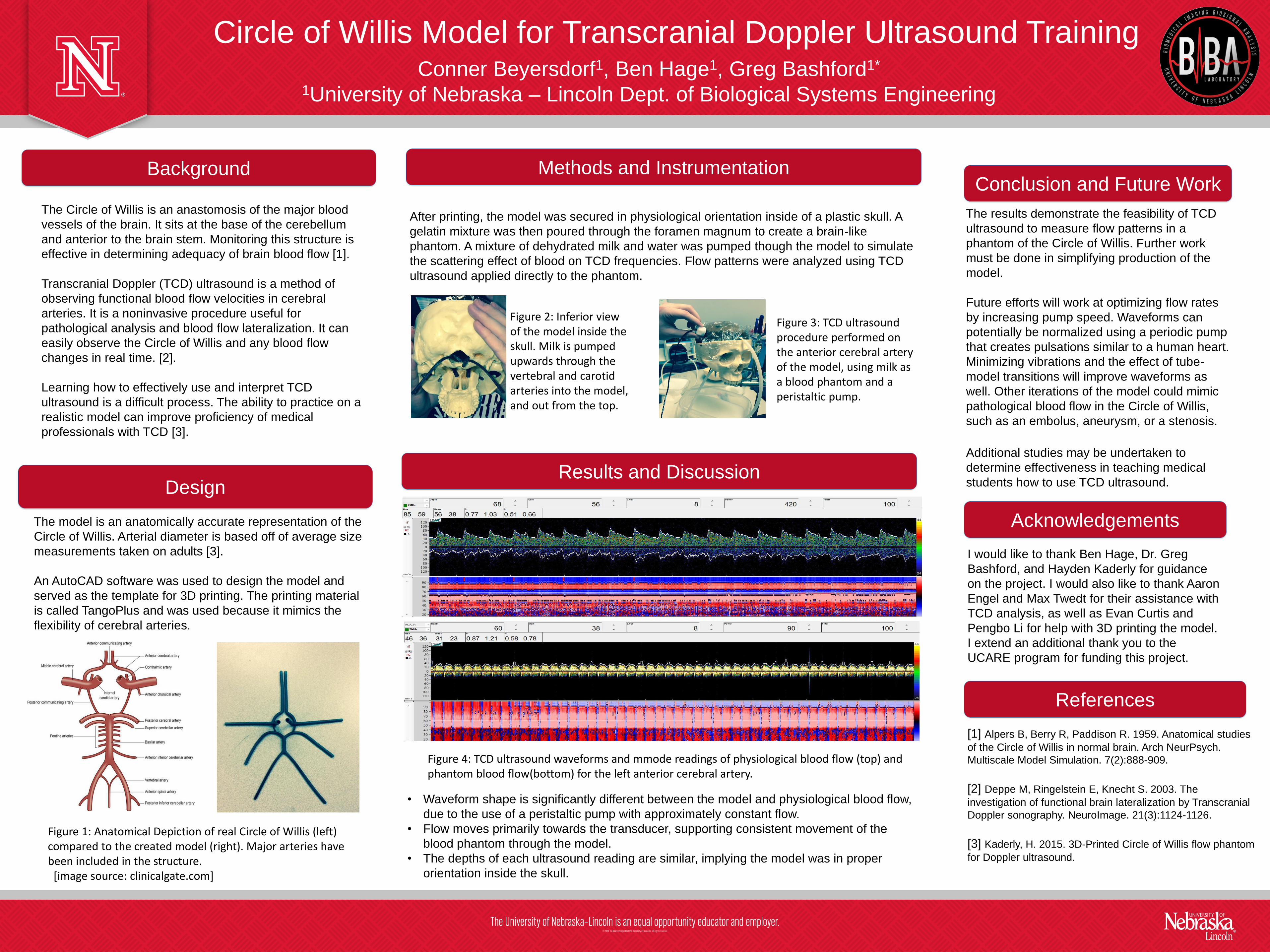
Figure 1: Anatomical Depiction of real Circle of Willis (left) compared to the created model (right). Major arteries have been included in the structure.
[image source: clinicalgate.com]
• Waveform shape is significantly different between the model and physiological blood flow,
due to the use of a peristaltic pump with approximately constant flow.
• Flow moves primarily towards the transducer, supporting consistent movement of the
blood phantom through the model.
• The depths of each ultrasound reading are similar, implying the model was in proper
orientation inside the skull.
Figure 4: TCD ultrasound waveforms and mmode readings of physiological blood flow (top) and phantom blood flow(bottom) for the left anterior cerebral artery.
Figure 2: Inferior view of the model inside the skull. Milk is pumped upwards through the vertebral and carotid arteries into the model, and out from the top.
Figure 3: TCD ultrasound procedure performed on the anterior cerebral artery of the model, using milk as a blood phantom and a peristaltic pump.


![Review Article Transcranial Doppler Ultrasound: A Review ...downloads.hindawi.com/journals/ijvm/2013/629378.pdf · Transcranial Doppler (TCD), rst described in [ ], is a noninvasive](https://static.documents.pub/doc/80x56/5f56cc40d1215262b86320d4/review-article-transcranial-doppler-ultrasound-a-review-transcranial-doppler.jpg)


