Int J Anat Res 2014, 2(2):344-53. ISSN 2321-4287 344 Original Article CIRCLE OF WILLIS: VARIANT FORMS AND THEIR EMBRYOLOGY USING GROSS DISSECTION AND MAGNETIC RESONANCE ANGIOGRAPHY Bishwajeet Saikia * 1 , Akash Handique 2 , Pranjal Phukan 3 , Donboklang Lynser 4 , Amitav Sarma 5 . ABSTRACT Address for Correspondence: Dr. Bishwajeet Saikia, Department of Anatomy, North Eastern Indira Gandhi Regional Institute of Health & Medical Sciences, Shillong–793018, Meghalaya, India. Phone: 91-9401059365. E-Mail: [email protected]Access this Article online Quick Response code Web site: *1,4 Senior Resident Doctor, 2,5 Associate Professor, 3 Assistant Professor. 1,5 Department of Anatomy, 2,3,4 Department of Radiology & Imaging. North Eastern Indira Gandhi Regional Institute of Health and Medical Sciences, Shillong, Meghalaya, India. Background: The circle of Willis is a large arterial anastomotic ring present at the base of the brain uniting the internal carotid and the vertebrobasilar systems. Branches from the internal carotid and vertebral arteries anastomose to form an arterial circle in the basal cisterns and then distribute to supply the brain. The anatomy of the circle is known to vary significantly; the vessels may be absent or sufficiently narrowed altering the hemodynamics of the circle of Willis and affecting its role as a collateral route. These variant forms can be correlated to their phylogeny and embryology. Prior knowledge of these variant forms is important in pathologies and treatment (e.g. parent artery occlusion for carotid aneurysms) resulting occlusion of carotid and vertebral arteries. Context and purpose: Our study was undertaken to observe and compare the morphology of circle of Willis using two entirely different methods; gross dissection (GD) and Magnetic resonance angiography (MRA) and to correlate the variant patterns encountered with the possible underlying developmental events. Gross dissection was carried out in 70 human cadavers and equal numbers of MRA’s of healthy individuals were studied retrospectively. Results: Only 31 cases (22.14%) presented with a complete circle of Willis, out of which 14 (20%) were cadaveric specimen and 17 (24.18%) were in MRA group. Unilateral hypoplastic posterior communicating artery was the most common variation observed in our study (19.28%). Conclusions: The wide variation in completeness of the circle of Willis in general population is similar to earlier observations. Review of phylogeny and embryology makes us familiar with variant forms which would be otherwise difficult to recognize and may be misinterpreted. MRA and gross dissection findings despite certain variations are comparable. KEYWORDS: Internal carotid artery, vertebral artery, Collateral flow. BACKGROUND International Journal of Anatomy and Research, Int J Anat Res 2014, Vol 2(2):344-53. ISSN 2321- 4287 Received: 10 April 2014 Peer Review: 10 April 2014 Published (O):31 May 2014 Accepted: 02 May 2014 Published (P):30 June 2014 International Journal of Anatomy and Research ISSN 2321-4287 www.ijmhr.org/ijar.htm Cerebrovascular accidents are one of the leading causes of death and disability through- out the world. The clinical manifestation of these cerebrovascular accidents reflects the area perfused by the cerebral vessels affected. However, the situation becomes more compli- cated with the presence or absence of adequate collateral blood flow provided by, an arterial ring of anastomosis present at the base of brain
Transcript
Int J Anat Res 2014, 2(2):344-53. ISSN 2321-4287 344
Original Article
CIRCLE OF WILLIS: VARIANT FORMS AND THEIR EMBRYOLOGYUSING GROSS DISSECTION AND MAGNETIC RESONANCEANGIOGRAPHYBishwajeet Saikia *1, Akash Handique 2, Pranjal Phukan 3, Donboklang Lynser 4,Amitav Sarma 5.
ABSTRACT
Address for Correspondence: Dr. Bishwajeet Saikia, Department of Anatomy, North Eastern IndiraGandhi Regional Institute of Health & Medical Sciences, Shillong–793018, Meghalaya, India.Phone: 91-9401059365. E-Mail: [email protected]
Access this Article online
Quick Response code Web site:
*1,4 Senior Resident Doctor, 2,5 Associate Professor, 3 Assistant Professor.1,5 Department of Anatomy, 2,3,4 Department of Radiology & Imaging.North Eastern Indira Gandhi Regional Institute of Health and Medical Sciences, Shillong, Meghalaya,India.
Background: The circle of Willis is a large arterial anastomotic ring present at the base of the brain uniting theinternal carotid and the vertebrobasilar systems. Branches from the internal carotid and vertebral arteriesanastomose to form an arterial circle in the basal cisterns and then distribute to supply the brain. The anatomyof the circle is known to vary significantly; the vessels may be absent or sufficiently narrowed altering thehemodynamics of the circle of Willis and affecting its role as a collateral route. These variant forms can becorrelated to their phylogeny and embryology. Prior knowledge of these variant forms is important in pathologiesand treatment (e.g. parent artery occlusion for carotid aneurysms) resulting occlusion of carotid and vertebralarteries.Context and purpose: Our study was undertaken to observe and compare the morphology of circle of Willisusing two entirely different methods; gross dissection (GD) and Magnetic resonance angiography (MRA) and tocorrelate the variant patterns encountered with the possible underlying developmental events. Gross dissectionwas carried out in 70 human cadavers and equal numbers of MRA’s of healthy individuals were studiedretrospectively.Results: Only 31 cases (22.14%) presented with a complete circle of Willis, out of which 14 (20%) were cadavericspecimen and 17 (24.18%) were in MRA group. Unilateral hypoplastic posterior communicating artery was themost common variation observed in our study (19.28%).Conclusions: The wide variation in completeness of the circle of Willis in general population is similar to earlierobservations. Review of phylogeny and embryology makes us familiar with variant forms which would beotherwise difficult to recognize and may be misinterpreted. MRA and gross dissection findings despite certainvariations are comparable.KEYWORDS: Internal carotid artery, vertebral artery, Collateral flow.
BACKGROUND
International Journal of Anatomy and Research,Int J Anat Res 2014, Vol 2(2):344-53. ISSN 2321- 4287
Received: 10 April 2014Peer Review: 10 April 2014 Published (O):31 May 2014Accepted: 02 May 2014 Published (P):30 June 2014
International Journal of Anatomy and ResearchISSN 2321-4287
www.ijmhr.org/ijar.htm
Cerebrovascular accidents are one of theleading causes of death and disability through-out the world. The clinical manifestation of thesecerebrovascular accidents reflects the area
perfused by the cerebral vessels affected.However, the situation becomes more compli-cated with the presence or absence of adequatecollateral blood flow provided by, an arterial ringof anastomosis present at the base of brain
Int J Anat Res 2014, 2(2):344-53. ISSN 2321-4287 345
Bishwajeet Saikia et al., CIRCLE OF WILLIS: VARIANT FORMS AND THEIR EMBRYOLOGY USING GROSS DISSECTION AND MAGNETIC RESONANCE ANGIOGRAPHY.
popularly known as the circle of Willis. The circleof Willis, unites the internal carotid andvertebrobasilar systems and is formed by anas-tomosis between the internal carotid,precommunicating part (A1) of anterior cerebral,anterior communicating, precommunicatingpart (P1) of posterior cerebral and posterior com-municating arteries (fig. 1). The collateral flowthus depends on the pattern and caliber of thesebranches forming the circle, which is known tovary. Some of these variations arise early duringvasculogenesis by alteration of vascularconstruction programme, due to some trigger-ing factors whereas; some of them represent themainstream of evolution [1].Variations can be described as patterns whichrepresent the mainstream of evolution of asystem whereas, an anomaly representsincreased rigidity of the system at its edgeswhich requires a minimal constrain to reveal itslimited flexibility [1]. Absence of a consistentrelationship between the size of the vessels andits territory, encountered in the variant forms ofthe circle can be interpreted as to be anabnormal one only during an occluding episodeof the feeders, whereas in normal circumstancesembryological events like territorial transfer andsharing of territories between the adjacentdeveloping vessels should results in normalcirculation [1]. The acquaintance of thesevariations further becomes important as withoutit diagnostic procedures may be misinterpretedor a neurosurgical procedure may becomecomplicated. The variations determine theangiographic filling pattern and their knowledgeenables distinction between hypoplasia andspasm [2]. During this study gross cadavericdissection was carried out to observe thevariations and later on for a further detail studyMagnetic Resonance Angiography was studiedretrospectively in living population.Magnetic Resonance angiography (MRA)enables evaluation of the intracranial vesselswithout the need for invasive procedures likecatheter angiography, avoiding small but definiterisk of embolism, complications likepsuedoaneurysms, contrast associated reactionsand vascular dissections [3]. Although MRA cannot reveal any arteries invisible in conventionalangiography, it provides a specificity of 100%,
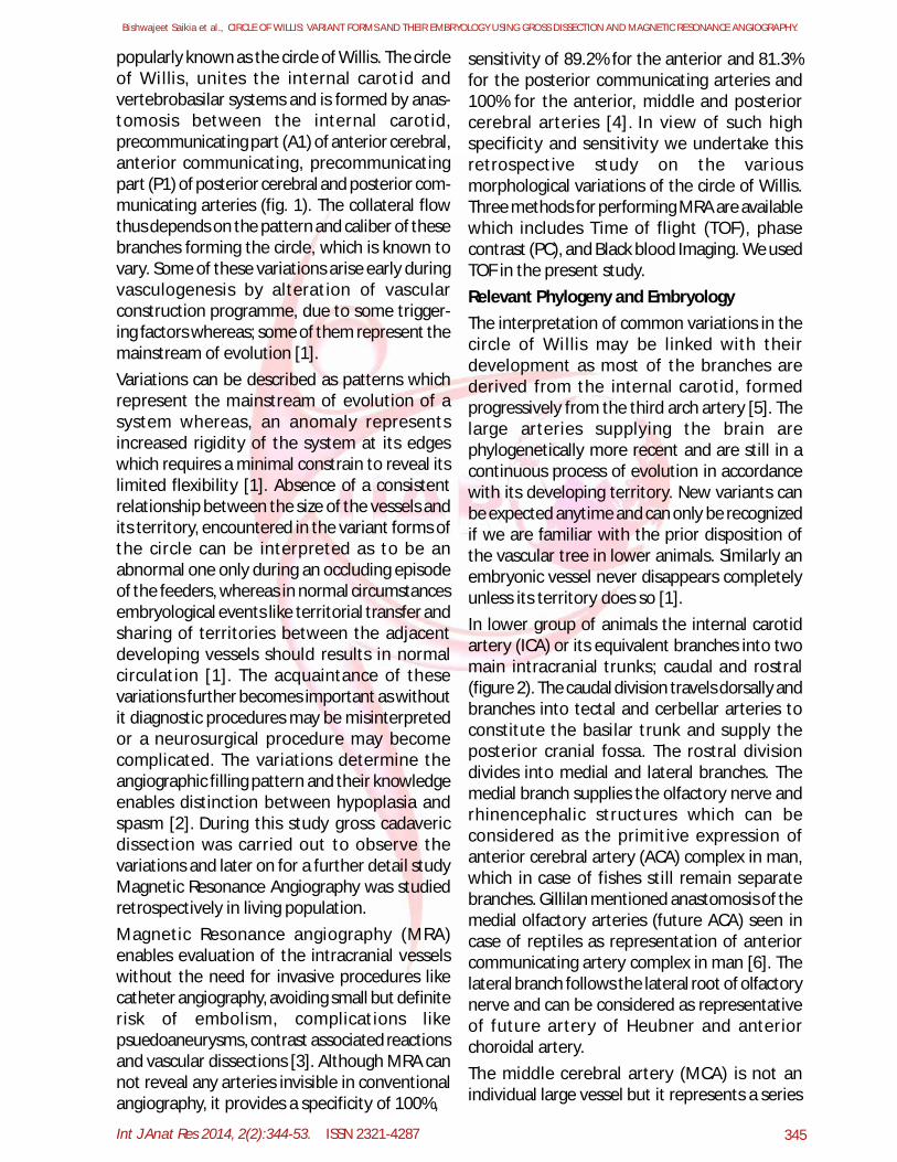
sensitivity of 89.2% for the anterior and 81.3%for the posterior communicating arteries and100% for the anterior, middle and posteriorcerebral arteries [4]. In view of such highspecificity and sensitivity we undertake thisretrospective study on the variousmorphological variations of the circle of Willis.Three methods for performing MRA are availablewhich includes Time of flight (TOF), phasecontrast (PC), and Black blood Imaging. We usedTOF in the present study.Relevant Phylogeny and EmbryologyThe interpretation of common variations in thecircle of Willis may be linked with theirdevelopment as most of the branches arederived from the internal carotid, formedprogressively from the third arch artery [5]. Thelarge arteries supplying the brain arephylogenetically more recent and are still in acontinuous process of evolution in accordancewith its developing territory. New variants canbe expected anytime and can only be recognizedif we are familiar with the prior disposition ofthe vascular tree in lower animals. Similarly anembryonic vessel never disappears completelyunless its territory does so [1].In lower group of animals the internal carotidartery (ICA) or its equivalent branches into twomain intracranial trunks; caudal and rostral(figure 2). The caudal division travels dorsally andbranches into tectal and cerbellar arteries toconstitute the basilar trunk and supply theposterior cranial fossa. The rostral divisiondivides into medial and lateral branches. Themedial branch supplies the olfactory nerve andrhinencephalic structures which can beconsidered as the primitive expression ofanterior cerebral artery (ACA) complex in man,which in case of fishes still remain separatebranches. Gillilan mentioned anastomosis of themedial olfactory arteries (future ACA) seen incase of reptiles as representation of anteriorcommunicating artery complex in man [6]. Thelateral branch follows the lateral root of olfactorynerve and can be considered as representativeof future artery of Heubner and anteriorchoroidal artery.The middle cerebral artery (MCA) is not anindividual large vessel but it represents a series
Int J Anat Res 2014, 2(2):344-53. ISSN 2321-4287 346
of small anastomotic vessels from the lateralstriate group. It develops after the “recurrentartery of Heubner” another branch from the ACAcomplex. They both share territories andvariations, and are in haemodynamic balancewith respect to their deep territories. Theanterior choroidal artery comes up as a branchfrom the ACA and most of its territories will betransformed to the tectal artery of the caudaldivision of the ICA, later to become the Posteriorcerebral artery (PCA) [1]. The PCA prolongs thecaudal division of the ICA. It supplies only thetectum; it becomes the true PCA when itannexes most of the cortical territories from theanterior choroidal artery. The basilar arteryresults from a cranio-caudal fusion of theposterior division of the ICA, so that the posteriorcommunicating artery (PCoA), the P1 segmentof PCA and upper basilar system, distal to theorigin of trigeminal artery, should be allconsidered as a single system; the caudal divisionof ICA [1].
MATERIALS AND METHODS
In the present study the circle of Willis (COW)was studied by gross dissection in 70 cadavericspecimens and by Magnetic resonanceangiography on equal number of livingindividuals. The study includes subjects of bothsexes which belong to different communities inthe Northeast India. In both studies the anteriorcerebral artery (ACA) were studied under; A1segment, defined as a part of ACA from its originat the internal carotid artery bifurcation till itsjunction with the anterior communicating arteryand A2 segment was defined as the course distalto it till the origin of pericallosal and calloso-marginal arteries. Similarly in the posteriorcerebral arteries (PCA), P1 segment originatesat basilar bifurcation up to the junction with theposterior communicating artery and the P2segment as the portion of PCA beyond it withinthe perimesencephalic cistern.A. Gross dissection:Gross dissection was carried out in thedepartment of Anatomy, Gauhati MedicalCollege, Assam, from July 2009 to September2010. A total number of 70 brain specimenswithout obvious pathological changes anddecomposition were collected from cadavers, for
a period from October 2009- September 2010 inthe department of Anatomy and unclaimed deadbodies in the department of Forensic Medicine,Gauhati Medical College, Assam, India.Dissection and processingThe brains from the cadavers with their arteriesintact were gently taken out by detaching thefalx cerebri and the ventral surfaces werecleared. The COW and its branches were clearedoff from the overlying meningeal coverings andany adhesions wherever present, to exposethem distinctly. The specimens were preservedin 10% formalin and dissected further at aconvenient time later. Wherever needed redfabric colour was used to enhance contrast andphotographs were taken. Relevant data wererecorded and compared.B. Magnetic Resonance Angiography:The Magnetic resonance angiographies (MRA)of 70 living individuals were studiedretrospectively in the Department of Radiology,NEIGRIHMS, Shillong, Meghalaya, India, for aperiod from 10th April 2012 to 22nd November2012. All patients underwent 3D time of flightMR angiography (3D TOF MRA) imaging using1.5 tesla MRI scanner (Avanto, Siemens,Germany). Following imaging parameters wereused repetition time/ echo time 23/7.0, flip angle25 degrees, slice thickness 0.7 mm, number ofslice 44/slab, number of slabs 4, slice overlap25%, flow direction feet to head with 40 mmsaturation at the head end, field of view 180 x158 and 256 matrix size. Reconstructions weredone in Syngo MR Workplace using 3D maximumintensity projection or MIP and Volumerendering technique or VRT. The data collectedfrom either method was compared. Theindividual variant forms were correlated with thepossible underlying embryological event and tocompare it with earlier established findings.
RESULTS
In the present study the circle of Willis werestudied by two different methods in two differ-ent groups; in 70 human cadaveric specimensby gross dissection and in 70 living individualsby Magnetic resonance angiography (MRA). Inorder to be consistent with the previous works,hypoplastic vessels were defined to be thosewith external diameters less than one millime-
Bishwajeet Saikia et al., CIRCLE OF WILLIS: VARIANT FORMS AND THEIR EMBRYOLOGY USING GROSS DISSECTION AND MAGNETIC RESONANCE ANGIOGRAPHY.
Int J Anat Res 2014, 2(2):344-53. ISSN 2321-4287 347
Group a b c d e f g h I j k l TotalGD 0 7.1 2.8 8.6 0 1.4 0 1.4 8.6 5.7 5.7 5.7 47.14%
Type of Variation (in percentage)Table1: Frequency of different varia-tions in anterior part of the “circle ofWillis” according to the type (classifi-cation) shown in Fig. 4.
Bishwajeet Saikia et al., CIRCLE OF WILLIS: VARIANT FORMS AND THEIR EMBRYOLOGY USING GROSS DISSECTION AND MAGNETIC RESONANCE ANGIOGRAPHY.
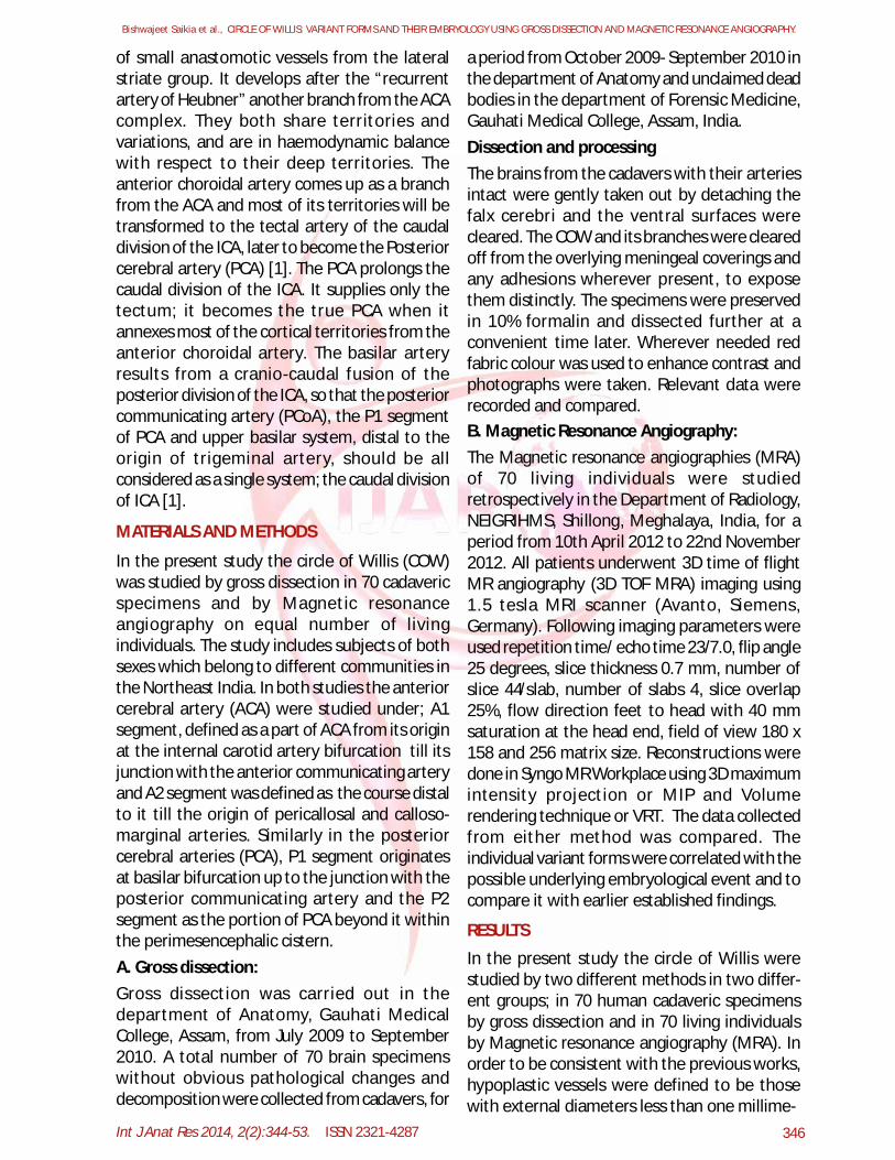
ter (1mm). Out of the total 140 cases, only 31cases (22.14%) presented with a complete(classic) circle of Willis (fig.3). A classic circle ofWillis was found in 14 (20%) of 70 cadavericspecimens and in 17 (24.18%) 70 MRA’s stud-ied. (fig.1). For a comprehensive understandingof the different patterns of the variationsobserved during our study, a schematicrepresentation for the variant forms of circle ofcircle of Willis in anterior and posterior part isshown in Fig. 3 and Fig. 4 respectively.
These labeled representations of the variantforms were used for classifying and numberingthe variations in table 1 and table 2.
Group a B c d e f g h TotalGD 11.4 1.4 5.7 2.8 18.6 14.3 1.4 0 55.7
Table 2: Frequency of different variations in posteriorpart of the “circle of Willis” according to the type (clas-sification) shown in Fig.5.
Fig. 1: Schematic representation (vessels forming thecircle of Willis) of the anterior part of the circle formedby the pre-communicating segments (A1) of the rightand left anterior cerebral arteries (ACA) and an anteriorcommunicating artery (ACoA) between them. The pos-terior part of the circle formed by the pre-communicat-ing segments (P1) of the right and left posterior cere-bral arteries (PCA), together with the right and left pos-terior communicating arteries (PCoA). The right and leftPCoAs originate from the right and left internal carotidarteries (ICAs) The A2 and P2 segments are the post -communicating portions of the anterior and posteriorcerebral arteries respectively. BA: basilar artery, MCA:middle cerebral artery.
Fig. 1
Table 3: Incidence of Normal Pattern of Circle of Willis.
Fig. 3: Schematic diagram to show the variations in thecircle of Willis in the anterior circulation, i.e. anteriorcerebral artery (ACA) & anterior communicating artery(ACoA): a) absent A1 segment with both ACA arising fromthe opposite ICA. b) Hypoplastic A1, c) Hypoplastic A2.d) Fenestration of A1. e) Median artery of Corpus callo-sum. f) Fused ACA with absent ACoA. g) Absent ACoAwith early origin of the Callosomarginal branch. h) Ab-sent ACoA. i) Hypoplastic ACoA. j) Double ACoA. k) Plexi-form ACoA. l) Fenestrated ACoA. ICA: Internal carotidartery.
Fig. 2: The position of anterior choroidal artery (AChA)in man shows its complete homology with therepresentation of posterior artery in bird. Both thearteries can be compared with each other and with theirposition in respect to the lateral geniculate body (LGB)confirming their identity. The pseudo-posterior cerebralartery and the tectal artery (in birds) together constitutethe components of posterior cerebral artery in man.
Fig. 2
Int J Anat Res 2014, 2(2):344-53. ISSN 2321-4287 348
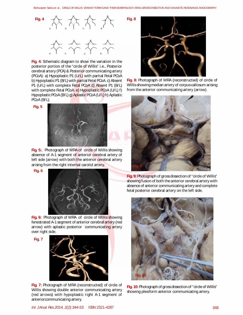
Fig. 4: Schematic diagram to show the variation in theposterior portion of the “circle of Willis” i.e., Posteriorcerebral artery (PCA) & Posterior communicating artery(PCoA): a) Hypoplastic P1 (U/L) with partial Fetal PCoAb) Hypoplastic P1 (B/L) with partial Fetal PCoA. c) AbsentP1 (U/L) with complete Fetal PCoA d) Absent P1 (B/L)with complete Fetal PCoA. e) Hypoplastic PCoA (U/L) f)Hypoplastic PCoA (B/L) g) Aplastic PCoA (U/L) h) AplasticPCoA (B/L).
Fig. 4
Bishwajeet Saikia et al., CIRCLE OF WILLIS: VARIANT FORMS AND THEIR EMBRYOLOGY USING GROSS DISSECTION AND MAGNETIC RESONANCE ANGIOGRAPHY.
Fig. 5:. Photograph of MRA of circle of Willis showingabsence of A-1 segment of anterior cerebral artery ofleft side (arrow) with both the anterior cerebral arteryarising from the right internal carotid artery.
Fig. 5
Fig. 6: Photograph of MRA of circle of Willis showingfenestrated A-1 segment of anterior cerebral artery (redarrow) with aplastic posterior communicating arteryover right side.
Fig. 6
Fig. 7
Fig. 7: Photograph of MRA (reconstructed) of circle ofWillis showing double anterior communicating artery(red arrows) with hypoplastic right A-1 segment ofanteriorcommunicating artery.
Fig. 8
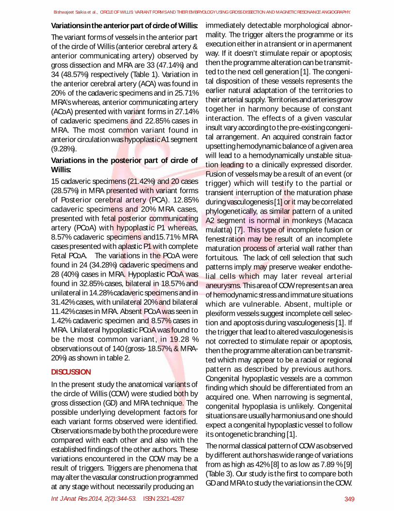
Fig. 8: Photograph of MRA (reconstructed) of circle ofWillis showing median artery of corpus callosum arisingfrom the anterior communicating artery (arrow).
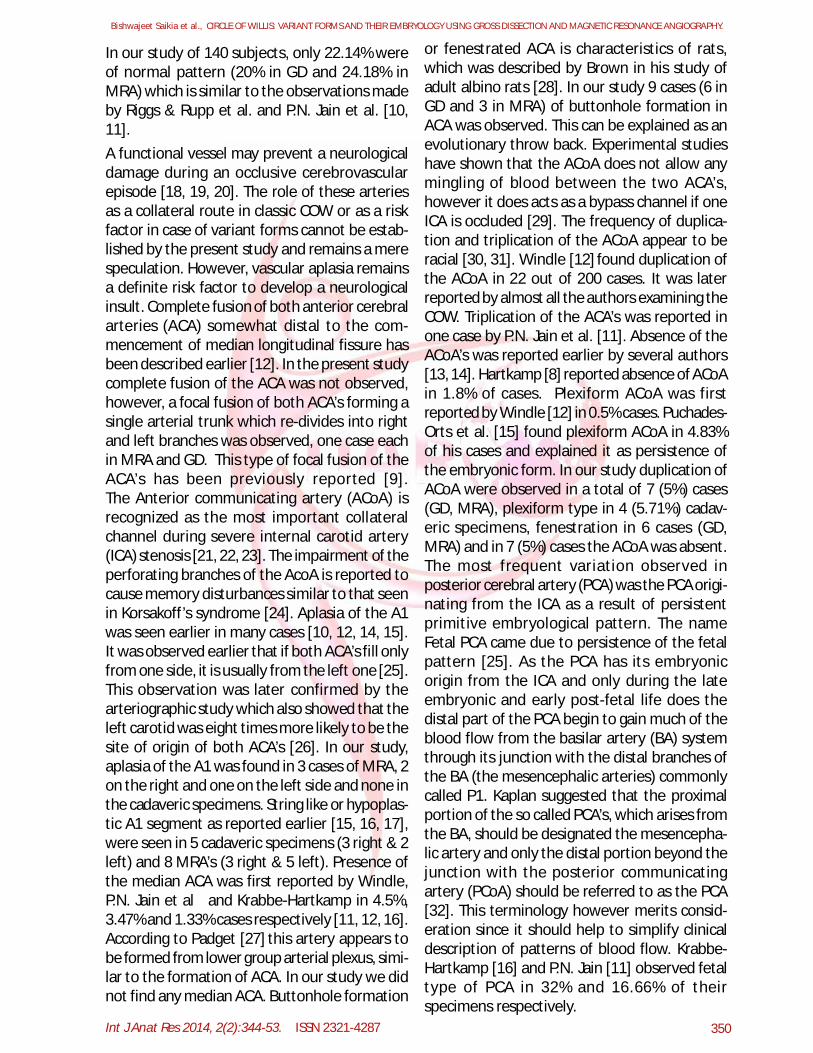
Fig. 9: Photograph of gross dissection of “circle of Willis”showing fusion of both the anterior cerebral artery withabsence of anterior communicating artery and completefetal posterior cerebral artery on the left side.
Fig. 9
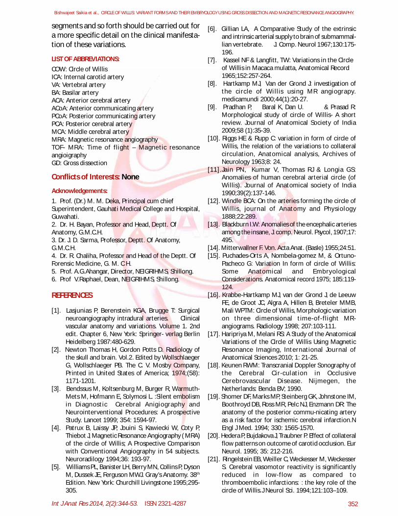
Fig. 10: Photograph of gross dissection of “circle of Willis”showing plexiform anterior communicating artery.
Fig. 10
Int J Anat Res 2014, 2(2):344-53. ISSN 2321-4287 349
DISCUSSION
Bishwajeet Saikia et al., CIRCLE OF WILLIS: VARIANT FORMS AND THEIR EMBRYOLOGY USING GROSS DISSECTION AND MAGNETIC RESONANCE ANGIOGRAPHY.
Variations in the anterior part of circle of Willis:The variant forms of vessels in the anterior partof the circle of Willis (anterior cerebral artery &anterior communicating artery) observed bygross dissection and MRA are 33 (47.14%) and34 (48.57%) respectively (Table 1). Variation inthe anterior cerebral artery (ACA) was found in20% of the cadaveric specimens and in 25.71%MRA’s whereas, anterior communicating artery(ACoA) presented with variant forms in 27.14%of cadaveric specimens and 22.85% cases inMRA. The most common variant found inanterior circulation was hypoplastic A1 segment(9.28%).Variations in the posterior part of circle ofWillis:15 cadaveric specimens (21.42%) and 20 cases(28.57%) in MRA presented with variant formsof Posterior cerebral artery (PCA). 12.85%cadaveric specimens and 20% MRA cases,presented with fetal posterior communicatingartery (PCoA) with hypoplastic P1 whereas,8.57% cadaveric specimens and15.71% MRAcases presented with aplastic P1 with completeFetal PCoA. The variations in the PCoA werefound in 24 (34.28%) cadaveric specimens and28 (40%) cases in MRA. Hypoplastic PCoA wasfound in 32.85% cases, bilateral in 18.57% andunilateral in 14.28% cadaveric specimens and in31.42% cases, with unilateral 20% and bilateral11.42% cases in MRA. Absent PCoA was seen in1.42% cadaveric specimen and 8.57% cases inMRA. Unilateral hypoplastic PCoA was found tobe the most common variant, in 19.28 %observations out of 140 (gross- 18.57%, & MRA-20%) as shown in table 2.
In the present study the anatomical variants ofthe circle of Willis (COW) were studied both bygross dissection (GD) and MRA technique. Thepossible underlying development factors foreach variant forms observed were identified.Observations made by both the procedure werecompared with each other and also with theestablished findings of the other authors. Thesevariations encountered in the COW may be aresult of triggers. Triggers are phenomena thatmay alter the vascular construction programmedat any stage without necessarily producing an
immediately detectable morphological abnor-mality. The trigger alters the programme or itsexecution either in a transient or in a permanentway. If it doesn’t stimulate repair or apoptosis;then the programme alteration can be transmit-ted to the next cell generation [1]. The congeni-tal disposition of these vessels represents theearlier natural adaptation of the territories totheir arterial supply. Territories and arteries growtogether in harmony because of constantinteraction. The effects of a given vascularinsult vary according to the pre-existing congeni-tal arrangement. An acquired constrain factorupsetting hemodynamic balance of a given areawill lead to a hemodynamically unstable situa-tion leading to a clinically expressed disorder.Fusion of vessels may be a result of an event (ortrigger) which will testify to the partial ortransient interruption of the maturation phaseduring vasculogenesis [1] or it may be correlatedphylogenetically, as similar pattern of a unitedA2 segment is normal in monkeys (Macacamulatta) [7]. This type of incomplete fusion orfenestration may be result of an incompletematuration process of arterial wall rather thanfortuitous. The lack of cell selection that suchpatterns imply may preserve weaker endothe-lial cells which may later reveal arterialaneurysms. This area of COW represents an areaof hemodynamic stress and immature situationswhich are vulnerable. Absent, multiple orplexiform vessels suggest incomplete cell selec-tion and apoptosis during vasculogenesis [1]. Ifthe trigger that lead to altered vasculogenesis isnot corrected to stimulate repair or apoptosis,then the programme alteration can be transmit-ted which may appear to be a racial or regionalpattern as described by previous authors.Congenital hypoplastic vessels are a commonfinding which should be differentiated from anacquired one. When narrowing is segmental,congenital hypoplasia is unlikely. Congenitalsituations are usually harmonius and one shouldexpect a congenital hypoplastic vessel to followits ontogenetic branching [1].The normal classical pattern of COW as observedby different authors has wide range of variationsfrom as high as 42% [8] to as low as 7.89 % [9](Table 3). Our study is the first to compare bothGD and MRA to study the variations in the COW.
Int J Anat Res 2014, 2(2):344-53. ISSN 2321-4287 350
In our study of 140 subjects, only 22.14% wereof normal pattern (20% in GD and 24.18% inMRA) which is similar to the observations madeby Riggs & Rupp et al. and P.N. Jain et al. [10,11].A functional vessel may prevent a neurologicaldamage during an occlusive cerebrovascularepisode [18, 19, 20]. The role of these arteriesas a collateral route in classic COW or as a riskfactor in case of variant forms cannot be estab-lished by the present study and remains a merespeculation. However, vascular aplasia remainsa definite risk factor to develop a neurologicalinsult. Complete fusion of both anterior cerebralarteries (ACA) somewhat distal to the com-mencement of median longitudinal fissure hasbeen described earlier [12]. In the present studycomplete fusion of the ACA was not observed,however, a focal fusion of both ACA’s forming asingle arterial trunk which re-divides into rightand left branches was observed, one case eachin MRA and GD. This type of focal fusion of theACA’s has been previously reported [9].The Anterior communicating artery (ACoA) isrecognized as the most important collateralchannel during severe internal carotid artery(ICA) stenosis [21, 22, 23]. The impairment of theperforating branches of the AcoA is reported tocause memory disturbances similar to that seenin Korsakoff’s syndrome [24]. Aplasia of the A1was seen earlier in many cases [10, 12, 14, 15].It was observed earlier that if both ACA’s fill onlyfrom one side, it is usually from the left one [25].This observation was later confirmed by thearteriographic study which also showed that theleft carotid was eight times more likely to be thesite of origin of both ACA’s [26]. In our study,aplasia of the A1 was found in 3 cases of MRA, 2on the right and one on the left side and none inthe cadaveric specimens. String like or hypoplas-tic A1 segment as reported earlier [15, 16, 17],were seen in 5 cadaveric specimens (3 right & 2left) and 8 MRA’s (3 right & 5 left). Presence ofthe median ACA was first reported by Windle,P.N. Jain et al and Krabbe-Hartkamp in 4.5%,3.47% and 1.33% cases respectively [11, 12, 16].According to Padget [27] this artery appears tobe formed from lower group arterial plexus, simi-lar to the formation of ACA. In our study we didnot find any median ACA. Buttonhole formation
or fenestrated ACA is characteristics of rats,which was described by Brown in his study ofadult albino rats [28]. In our study 9 cases (6 inGD and 3 in MRA) of buttonhole formation inACA was observed. This can be explained as anevolutionary throw back. Experimental studieshave shown that the ACoA does not allow anymingling of blood between the two ACA’s,however it does acts as a bypass channel if oneICA is occluded [29]. The frequency of duplica-tion and triplication of the ACoA appear to beracial [30, 31]. Windle [12] found duplication ofthe ACoA in 22 out of 200 cases. It was laterreported by almost all the authors examining theCOW. Triplication of the ACA’s was reported inone case by P.N. Jain et al. [11]. Absence of theACoA’s was reported earlier by several authors[13, 14]. Hartkamp [8] reported absence of ACoAin 1.8% of cases. Plexiform ACoA was firstreported by Windle [12] in 0.5% cases. Puchades-Orts et al. [15] found plexiform ACoA in 4.83%of his cases and explained it as persistence ofthe embryonic form. In our study duplication ofACoA were observed in a total of 7 (5%) cases(GD, MRA), plexiform type in 4 (5.71%) cadav-eric specimens, fenestration in 6 cases (GD,MRA) and in 7 (5%) cases the ACoA was absent.The most frequent variation observed inposterior cerebral artery (PCA) was the PCA origi-nating from the ICA as a result of persistentprimitive embryological pattern. The nameFetal PCA came due to persistence of the fetalpattern [25]. As the PCA has its embryonicorigin from the ICA and only during the lateembryonic and early post-fetal life does thedistal part of the PCA begin to gain much of theblood flow from the basilar artery (BA) systemthrough its junction with the distal branches ofthe BA (the mesencephalic arteries) commonlycalled P1. Kaplan suggested that the proximalportion of the so called PCA’s, which arises fromthe BA, should be designated the mesencepha-lic artery and only the distal portion beyond thejunction with the posterior communicatingartery (PCoA) should be referred to as the PCA[32]. This terminology however merits consid-eration since it should help to simplify clinicaldescription of patterns of blood flow. Krabbe-Hartkamp [16] and P.N. Jain [11] observed fetaltype of PCA in 32% and 16.66% of theirspecimens respectively.
Bishwajeet Saikia et al., CIRCLE OF WILLIS: VARIANT FORMS AND THEIR EMBRYOLOGY USING GROSS DISSECTION AND MAGNETIC RESONANCE ANGIOGRAPHY.
Int J Anat Res 2014, 2(2):344-53. ISSN 2321-4287 351
It was observed that patients with fetal-type PCAcould be more prone to develop vascular insuf-ficiency [33]. In our study complete fetal PCA wasseen in 16.42% of subjects whereas, incompletefetal PCA was seen 12.14%. PCA hypoplasia couldbe a contributory risk factor for ischemic stroke,even in absence of ICA occlusion [34]. In ourstudy 9 cadaveric specimens and 14 MRA’spresented with hypoplastic P1 with partial fetalPCA whereas, 6 cadaveric specimens and 11MRA’s presented with absent P1 with completePCA arising from ICA. Anomalies of the PCoAhave a great significance since it forms a linkbetween two major arterial systems – the inter-nal carotid and the vertebrobasilar circuit.A hypoplastic PCoA may be a risk factor fordeveloping neurological deficit in patients withICA occlusion [19]. Windle [12] found the PCoA’ssmall in 2.33% and absent in 1.5% of specimens.He observed anomalies in 49% and described theorigin of the PCA’s from one or both ICA’s as themost common variant. Mitchell [35] described16 patients with anomalies in the PCoA’s, with 9of them absent. P.N. Jain et al. [11] found 20absent and 26 hypoplastic PCoA and Partopratimet al. [9] found 44 hypoplastic forms of PCoA’s.In our study out of 140 cases, the most commonvariant observed was unilateral hypoplasticPCoA which was seen in a total of 27 (13 GD &14 MRA) cases. This observation was similar withprevious authors [8, 11, 15]. Bilateral hypoplas-tic PCoA was seen in a total of 18 (10 GD & 8MRA) cases. The PCoA was unilaterally absentin 2 cases and bilaterally in 5 cases. The incidenceof aneurysms of the circle of Willis reported inpublished literature is 0.25% to 4.9%. In ourstudy a single aneurysm in the A1 segment ofthe right ACA (1.42%) was observed.
CONCLUSION
The present study is based on the analysis madefrom the gross dissection in 70 post mortemspecimens of brain and in 70 MRA’s. In a fetal-type posterior cerebral artery (PCA) there is anembryonic derivation of the PCA from theinternal carotid artery (ICA). In completefetal-type PCA, the possibility for collateralcirculation to develop between the anterior andposterior part of the cerebral circulation by wayof leptomeningeal vessels is impossible as the
vascularization of the PCA flow territory iscompletely dependent on the ICA. Whether thisis a risk factor for stroke should be subject offurther investigation. Both MRA and cadavericspecimens show that morphological variationsare quite frequent in the circle of Willis (COW)in human beings. The foundation of arterial COWlies in vessels which are formed in the earlystages of fetal life some of these vessels succumbto involution while others are evolving tosupply blood to the developing structures.However, functional changes as well as theanatomical factors may influence the effective-ness of anastomosis. The COW is unquestionablyimportant as a potential source of anastomoticblood flow. Segments of the COW which are nar-row or string-like, or even absent are a result ofagenesis or involution during embryonicdevelopment. Because of this variability, theclinical manifestation of occlusion of thevertebral or carotid arteries may vary consider-ably from one individual to another, and theeffectiveness of collateral circulation may begreatly influenced. Our study comprising grossdissection and MRA is first of its kind to studygiving an indirect opportunity to compare thevariations of the COW in patient who havedecreased due to various causes and in livingpersons. Postmortem dissections have somedisadvantages. There may be a minor change inthe diameter of the vessels, the diameter of‘hypoplastic’ vessels may increase with time andexceed the 1 mm criteria. Degree of preserva-tion of each cadaver may vary leading to subtledifferences. Since the results cannot be directlycompared with the angiography, as in angiogra-phy we can measure only the internal diameterand that too in living subjects. Preparing acryliccast of the blood vessels can be more accuratebut is time consuming and costly. In TOF-MRAlimitations include dependence on amount anddirection of flow as well as the exact techniqueemployed. Therefore smaller arteries with verylittle flow might not be detected and leading tointerpretation as an aplastic artery instead of itbeing actually hypoplastic. Despite theselimitations we were able to document severalvariations during this study, by both methods.Further studies based on quantitative measure-ments of the luminal diameters and flow of the
Bishwajeet Saikia et al., CIRCLE OF WILLIS: VARIANT FORMS AND THEIR EMBRYOLOGY USING GROSS DISSECTION AND MAGNETIC RESONANCE ANGIOGRAPHY.
Int J Anat Res 2014, 2(2):344-53. ISSN 2321-4287 352
Conflicts of Interests: None
REFERENCES
segments and so forth should be carried out fora more specific detail on the clinical manifesta-tion of these variations.
LIST OF ABBREVIATIONS:COW: Circle of WillisICA: Internal carotid arteryVA: Vertebral arteryBA: Basilar arteryACA: Anterior cerebral arteryACoA: Anterior communicating arteryPCoA: Posterior communicating arteryPCA: Posterior cerebral arteryMCA: Middle cerebral arteryMRA: Magnetic resonance angiographyTOF- MRA: Time of flight – Magnetic resonanceangioigraphyGD: Gross dissection
Acknowledgements:1. Prof. (Dr.) M. M. Deka, Principal cum chiefSuperintendent, Gauhati Medical College and Hospital,Guwahati.2. Dr. H. Bayan, Professor and Head, Deptt. OfAnatomy, G.M.C.H.3. Dr. J. D. Sarma, Professor, Deptt. Of Anatomy,G.M.C.H.4. Dr. R. Chaliha, Professor and Head of the Deptt. OfForensic Medicine, G. M. C H.5. Prof. A.G.Ahangar, Director, NEIGRIHMS, Shillong.6. Prof V.Raphael, Dean, NEIGRIHMS, Shillong.
[6]. Gillian LA, A Comparative Study of the extrinsicand intrinsic arterial supply to brain of submammal-lian vertebrate. J. Comp. Neurol 1967;130:175-196.
[7]. Kassel NF & Langfitt, TW: Variations in the Circleof Willis in Macaca mulatta, Anatomical Record1965;152:257-264.
[8]. Hartkamp MJ, Van der Grond J: investigation ofthe circle of Wi ll is using MR angiograpy.medicamundi 2000;44(1):20-27.
[9]. Pradhan P, Baral K, Dan U. & Prasad R:Morphological study of circle of Willis- A shortreview. Journal of Anatomical Society of India2009;58 (1):35-39.
[10]. Riggs HE & Rupp C: variation in form of circle ofWillis, the relation of the variations to collateralcirculation, Anatomical analysis, Archives ofNeurology 1963;8: 24.
[11].Jain PN, Kumar V, Thomas RJ & Longia GS:Anomalies of human cerebral arterial circle (ofWillis). Journal of Anatomical society of India1990;39(2):137-146.
[12]. Windle BCA: On the arteries forming the circle ofWill is, journal of Anatomy and Physiology1888;22:289.
[13]. Blackburn I.W: Anomalies of the encephalic arteriesamong the insane, J. comp. Neurol. Psycol, 1907;17:495.
[14]. Mitterwallner F. Von. Acta Anat. (Basle) 1955;24:51.[15]. Puchades-Orts A, Nombela-gomez M, & Ortuno-
Pacheco G: Variation In form of circle of Willis:Some Anatomical and EmbryologicalConsiderations. Anatomical record 1975; 185:119-124.
[16]. Krabbe-Hartkamp MJ, van der Grond J, de LeeuwFE, de Groot JC, Algra A, Hillen B, Breteler MMB,Mali WPTM: Circle of Willis, Morphologic variationon three dimensional time-of-fl ight MR-angiograms. Radiology 1998; 207:103-111.
[17]. Haripriya M, Melani RS: A Study of the AnatomicalVariations of the Circle of Willis Using MagneticResonance Imaging, International Journal ofAnatomical Sciences 2010; 1: 21-25.
[19]. Shomer DF, Marks MP, Steinberg GK, Johnstone IM,Boothroyd DB, Ross MR, Pelc NJ, Enzmann DR: Theanatomy of the posterior commu-nicating arteryas a risk factor for ischemic cerebral infarction.NEngl J Med. 1994; 330: 1565-1570.
[20]. Hedera P, Bujdakova J, Traubner P. Effect of collateralflow patterns on outcome of carotid occlusion. EurNeurol. 1995; 35: 212-216.
[21]. Ringelstein EB, Weiller C, Weckesser M, WeckesserS. Cerebral vasomotor reactivity is significantlyreduced in low-flow as compared tothromboembolic infarctions: : the key role of thecircle of Willis.J Neurol Sci. 1994;121:103–109.
[2]. Newton Thomas H, Gordon Potts D. Radiology ofthe skull and brain. Vol.2. Edited by WollschlaegerG, Wollschlaeger PB. The C. V. Mosby Company,Printed in United States of America; 1974;(58):1171-1201.
[3]. Bendssus M, Koltsenburg M, Burger R, Warmuth-Mets M, Hofmann E, Solymosi L. :Silent embolismin Diagnostic Cerebral Anigiography andNeurointerventional Procedures: A prospectiveStudy. Lancet 1999; 354: 1594-97.
[4]. Patrux B, Laissy JP, Jouini S, Kawiecki W, Coty P,Thiebot J, Magnetic Resonance Angiography ( MRA)of the circle of Willis; A Prospective Comparisonwith Conventional Angiography in 54 subjects.Neuroradilogy 1994;36: 193-97.
[5]. Williams PL, Banister LH, Berry MN, Collins P, DysonM, Dussek JE, Ferguson MWJ. Gray’s Anatomy. 38th
Edition. New York: Churchill Livingstone 1995;295-305.
Bishwajeet Saikia et al., CIRCLE OF WILLIS: VARIANT FORMS AND THEIR EMBRYOLOGY USING GROSS DISSECTION AND MAGNETIC RESONANCE ANGIOGRAPHY.
Int J Anat Res 2014, 2(2):344-53. ISSN 2321-4287 353
Bishwajeet Saikia et al., CIRCLE OF WILLIS: VARIANT FORMS AND THEIR EMBRYOLOGY USING GROSS DISSECTION AND MAGNETIC RESONANCE ANGIOGRAPHY.
How to cite this article:Bishwajeet Saikia, Akash Handique, Pranjal Phukan, DonboklangLynser, Amitav Sarma. CIRCLE OF WILLIS: VARIANT FORMS ANDTHEIR EMBRYOLOGY USING GROSS DISSECTION AND MAGNETICRESONANCE ANGIOGRAPHY. Int J Anat Res 2014;2(2):344-53.
[22].Kja¨llman L, Blomstrand C, Holm J, Lundh T,Volkmann R. Patients with low stump pressure andpossible pressure fall in the middle cerebral arteryduring carotid surgery may be identifiedpreoperatively by transcranial Doppler.Eur Neurol.1995; 35: 259-263.
[23]. Hoksbergen AWJ, Legemate DA, Ubbink DT, de VosHJ, Jacobs MJHM. Influence of the collateralfunction of the circle of Willis on hemi-sphericalperfusion during carotid occlusion as assessed bytranscranial colour-coded duplexultrasonography.Eur J Vasc Endovasc Surg.1997;17:486-492.
[24].Parkin AJ, Leng NRC, Stanhope N: Memoryimpairment following ruptured aneurysm of theanterior communicating artery. Brain Cogn 7,1988;231-143.
[25]. Bullen F St John: Absence of one anterior cerebralwith both coming from opposite carotid, J.Ment.Sci1980;36:32.
[26]. Fields WS & Webel, J: Effects of vascular disorderson the vestibular system, in Neurological aspectsof Audotory & Vestibular disorders,1964, edited byfields, W.S & Alford, B.R. ,Thomas,C.CSpringfield,III.1996:22-29.
[27]. Padget DH: The development of the cranial arteriesin the human embryo, Contrib Embryol1948;32:205.
[28]. Brown James Oliver, The morphology of circulusarteriosus -cerebri in rats, Anatomical Record 1966;156(1): 99-106.
[29]. Kramer S.P: On the function of the circle of Willis,Journal Exp.Med 1912, 15: 348.
[30]. Vare AM & Bansal PC: Arterial pattern at the baseof the human brain, Journal of Anatomical Societyof India 1970; (19)3: 71-79.
[31]. Raja Reddy D & Prabhakar, V, & dayananda Rao, B,Anatomical study of the circle of Willis, NeurologyIndia 1972; 99/1: 8-12.
[32]. Kaplan H.A.: Arteries of the brain, Anatomic study,Acta Radiologica 1956; 46: 364.
[33]. Raamt A, Malib Williem PTM, Laarb Peter Jan van,Graafa Yolanda van der: The Fetal Variant of theCircle of Willis and Its Influence on the CerebralCollateral Circulation. Cerebrovasc Dis 2006;22:217-224.
[34]. Chuang YM, Liu CY, Pan PJ. Posterior communicatingartery hypoplasia as a risk factor for acute ischemicstroke in the absence of carotid artery occlusion. J.Clin.Neurosci 2008;15: 1376-1381.
[35]. Mitchell N & Angrist A: Intracranial aneurysms areport of 36 cases, Annals of Internal Medicine1943; 19: 909.