42
Circulation Chapter 3
| Date post: | 24-Dec-2015 |
| Category: |
Documents |
| Upload: | godfrey-mitchell |
| View: | 223 times |
| Download: | 4 times |
Circulation
Chapter 3
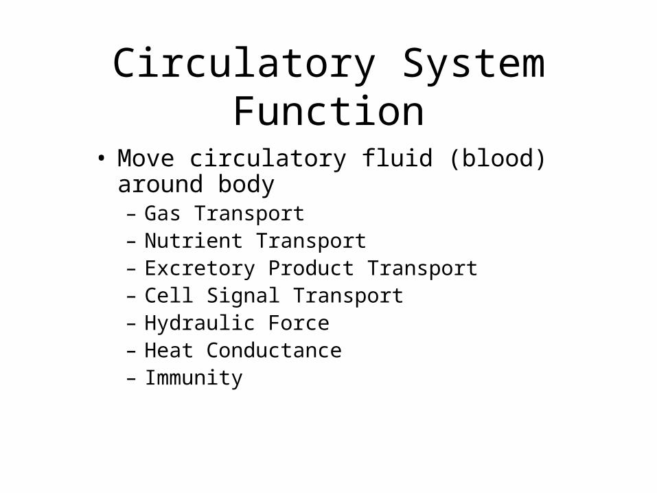
Circulatory System Function
• Move circulatory fluid (blood) around body– Gas Transport– Nutrient Transport– Excretory Product Transport– Cell Signal Transport– Hydraulic Force– Heat Conductance– Immunity
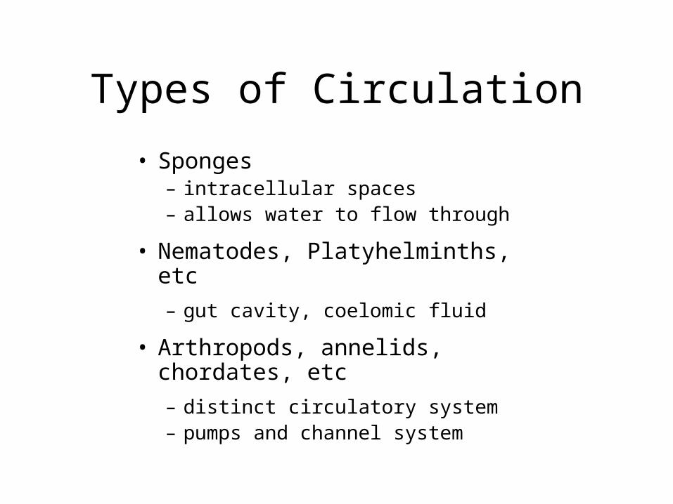
Types of Circulation
• Sponges– intracellular spaces– allows water to flow through
• Nematodes, Platyhelminths, etc
– gut cavity, coelomic fluid
• Arthropods, annelids, chordates, etc
– distinct circulatory system – pumps and channel system
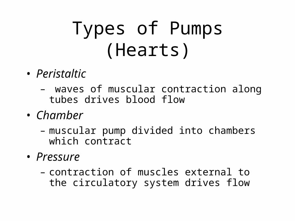
Types of Pumps (Hearts)
• Peristaltic– waves of muscular contraction along tubes drives
blood flow
• Chamber – muscular pump divided into chambers which contract
• Pressure – contraction of muscles external to the circulatory
system drives flow
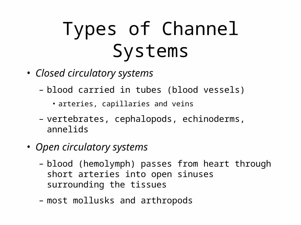
Types of Channel Systems
• Closed circulatory systems
– blood carried in tubes (blood vessels)
• arteries, capillaries and veins
– vertebrates, cephalopods, echinoderms, annelids
• Open circulatory systems
– blood (hemolymph) passes from heart through short arteries into open sinuses surrounding the tissues
– most mollusks and arthropods
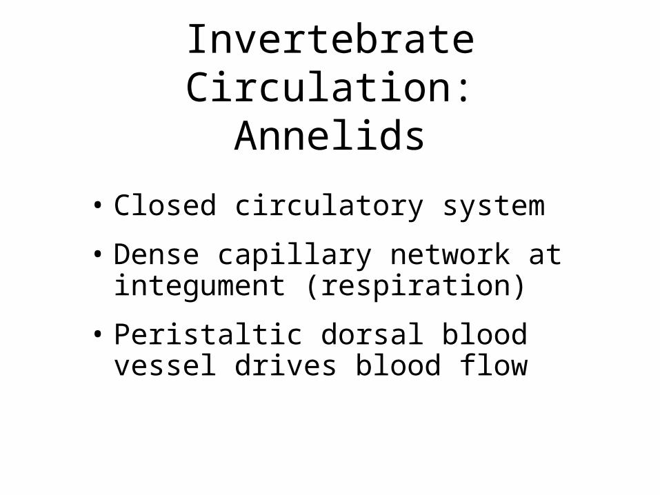
Invertebrate Circulation:Annelids
• Closed circulatory system
• Dense capillary network at integument (respiration)
• Peristaltic dorsal blood vessel drives blood flow
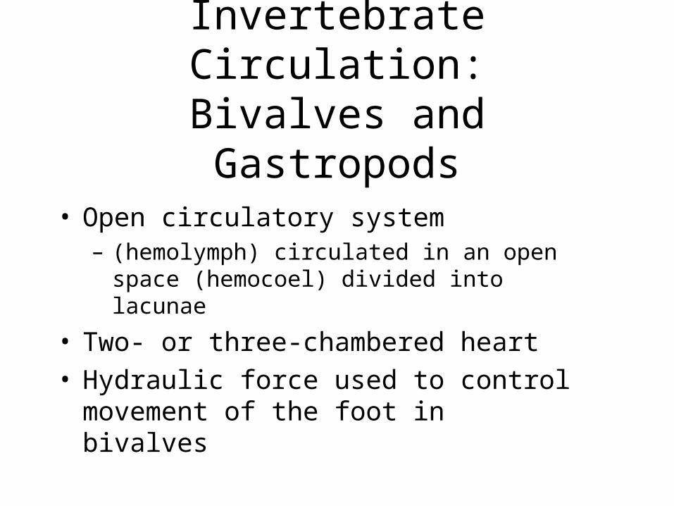
Invertebrate Circulation:Bivalves and Gastropods
• Open circulatory system– (hemolymph) circulated in an open space
(hemocoel) divided into lacunae
• Two- or three-chambered heart• Hydraulic force used to control movement of
the foot in bivalves
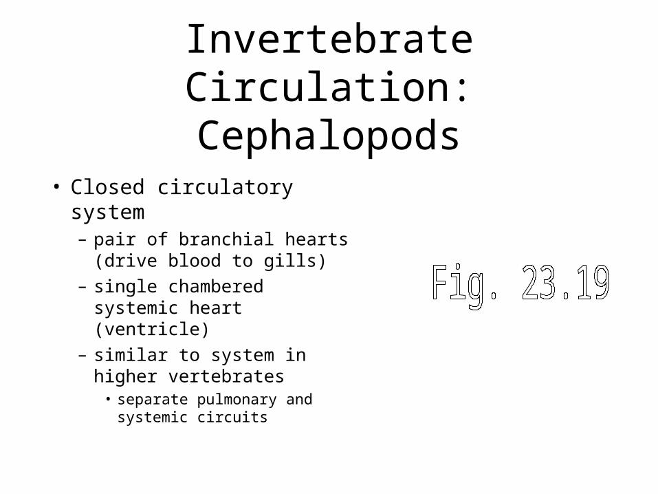
Invertebrate Circulation:Cephalopods
• Closed circulatory system– pair of branchial hearts
(drive blood to gills)
– single chambered systemic heart (ventricle)
– similar to system in higher vertebrates
• separate pulmonary and systemic circuits
Invertebrate Circulation:Insects
• Open circulatory system• Minimal gas transport• Large dorsal vessel w/peristaltic
heart in posterior segment– hemolymph runs anteriorly to head,
then ends in hemocoel– flow directed through hemocoel by
longitudinal membranes– flows back to posterior dorsal vessel
• Auxillary pumps supply wings limbs, and antennae
Invertebrate Circulation:Arachnids
• Similar to insect design• Hemolymph contains higher [hemocyanin]
– O2 transport
• More extensive arterial systems in arachnids with books lungs
• Specific arteries supply hydraulic pressure to legs for locomotion – legs of spiders lack extensor muscles
Invertebrate Circulation:Crustaceans
• Some small or sessile spp. lack heart or blood vessels
• Larger spp possess open system similar to insects
• Extensive circulation in gills– heart receives oxygenated
hemolymph from the gills then pumps it to the rest of the body
Vertebrate Circulation: General Patterns
• Single passage through heart during circuit (e.g., fish)– Single circuit
• Double passage through heart during circuit (e.g., mammals)– Separate pulmonary and
systemic circuits
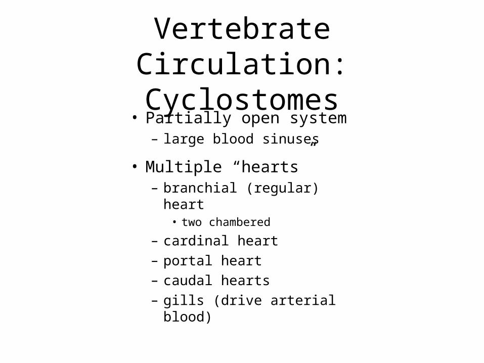
Vertebrate Circulation:Cyclostomes
• Partially open system– large blood sinuses
• Multiple “hearts”– branchial (regular) heart
• two chambered
– cardinal heart
– portal heart
– caudal hearts
– gills (drive arterial blood)
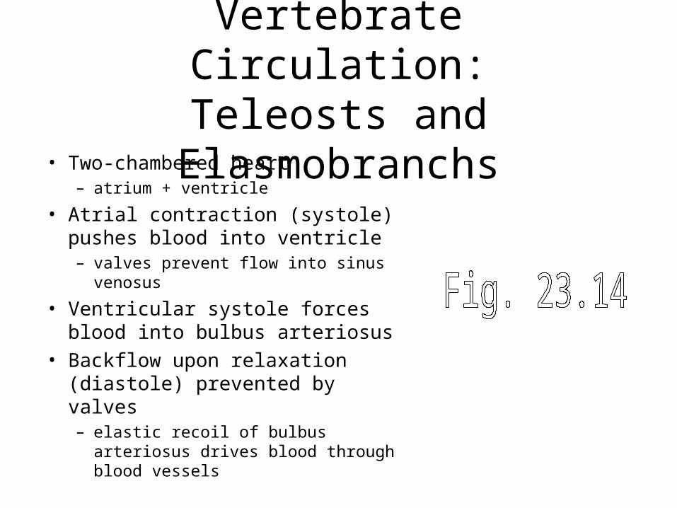
Vertebrate Circulation:Teleosts and Elasmobranchs
• Two-chambered heart – atrium + ventricle
• Atrial contraction (systole) pushes blood into ventricle– valves prevent flow into sinus venosus
• Ventricular systole forces blood into bulbus arteriosus
• Backflow upon relaxation (diastole) prevented by valves– elastic recoil of bulbus arteriosus
drives blood through blood vessels
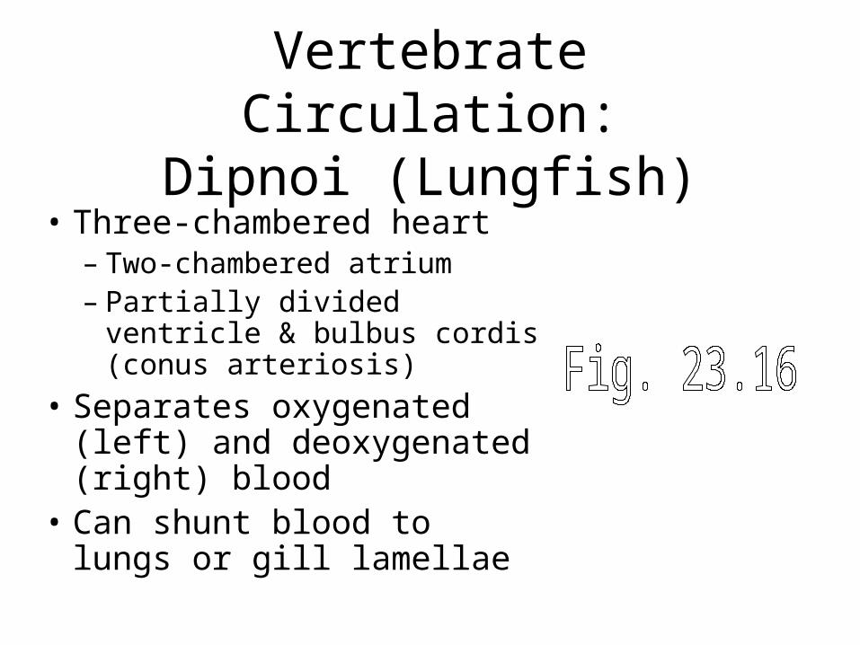
Vertebrate Circulation:Dipnoi (Lungfish)
• Three-chambered heart– Two-chambered atrium– Partially divided ventricle &
bulbus cordis (conus arteriosis)
• Separates oxygenated (left) and deoxygenated (right) blood
• Can shunt blood to lungs or gill lamellae
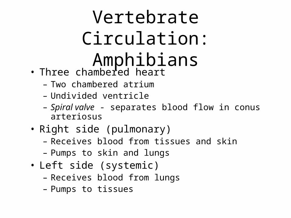
Vertebrate Circulation:Amphibians
• Three chambered heart– Two chambered atrium– Undivided ventricle– Spiral valve - separates blood flow in conus arteriosus
• Right side (pulmonary)– Receives blood from tissues and skin– Pumps to skin and lungs
• Left side (systemic)– Receives blood from lungs– Pumps to tissues
Vertebrate Circulation:Non-Archosaur Reptiles
• Three chambered heart– Two chambered atrium
– partly divided ventricle
• Ventricle contains three sub-chambers– divided upon contraction
– “five-chambered” heart
– allows heart to redirect blood flow btw pulmonary and systemic circuits
– “cardiac shunting”
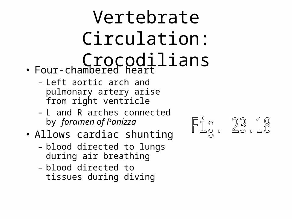
Vertebrate Circulation:Crocodilians
• Four-chambered heart– Left aortic arch and pulmonary
artery arise from right ventricle– L and R arches connected by
foramen of Panizza
• Allows cardiac shunting– blood directed to lungs during
air breathing– blood directed to tissues during
diving
Vertebrate Circulation:Mammals and Birds
• Four-chambered heart• Complete separation into
right and left halves • Blood pressure can differ
between pulmonary and systemic circuits– systemic BP = 95 mmHg
– pulmonary BP = 14 mmHg
Vertebrate Circulation:Mammals and Birds
• Atria– Thin walled, support ventricular filling
• Ventricles– Primary pumps for driving blood
through circulation
• One-way valves – Atrioventricular valves
– Arterial (semilunar) valves
– ensure unidirectional flow • veins → atria → ventricles → arteries
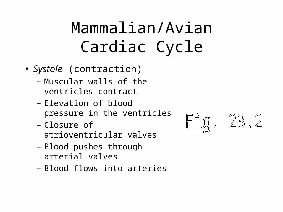
Mammalian/AvianCardiac Cycle
• Systole (contraction)– Muscular walls of the ventricles
contract
– Elevation of blood pressure in the ventricles
– Closure of atrioventricular valves
– Blood pushes through arterial valves
– Blood flows into arteries
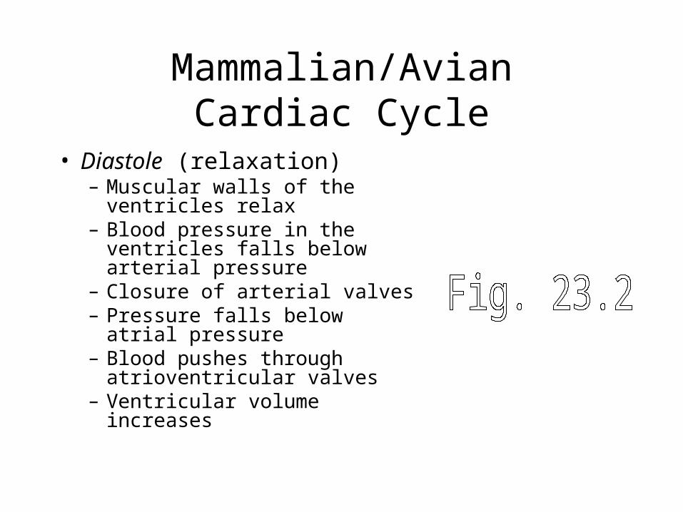
Mammalian/AvianCardiac Cycle
• Diastole (relaxation)– Muscular walls of the ventricles
relax– Blood pressure in the ventricles
falls below arterial pressure– Closure of arterial valves– Pressure falls below atrial
pressure– Blood pushes through
atrioventricular valves– Ventricular volume increases
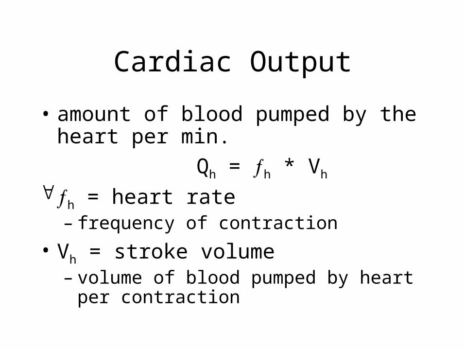
Cardiac Output
• amount of blood pumped by the heart per min.
Qh = h * Vh
h = heart rate – frequency of contraction
• Vh = stroke volume – volume of blood pumped by heart per
contraction
Cardiac Output
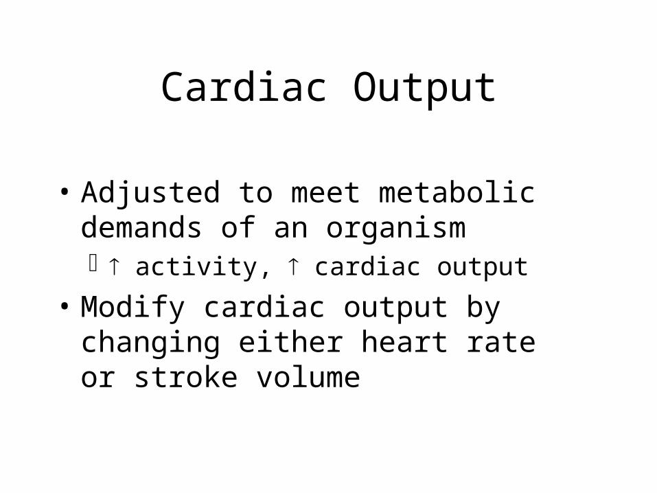
• Adjusted to meet metabolic demands of an organism activity, cardiac output
• Modify cardiac output by changing either heart rate or stroke volume
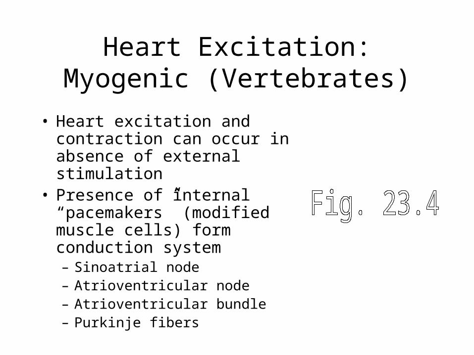
Heart Excitation:Myogenic (Vertebrates)
• Heart excitation and contraction can occur in absence of external stimulation
• Presence of internal “pacemakers” (modified muscle cells) form conduction system– Sinoatrial node– Atrioventricular node– Atrioventricular bundle– Purkinje fibers
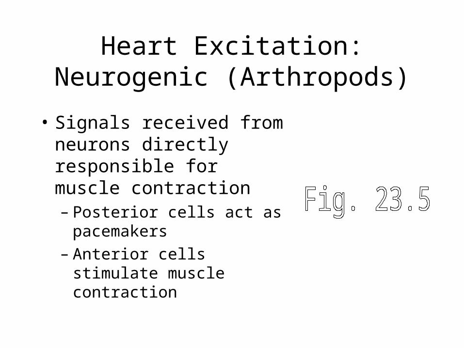
Heart Excitation:Neurogenic (Arthropods)
• Signals received from neurons directly responsible for muscle contraction– Posterior cells act as
pacemakers– Anterior cells stimulate
muscle contraction
Regulation of Cardiac Output (Mammals)
• Heart rate (modify pacemaker activity):– The autonomic nervous system:
• Parasympathetic nervous system (vagus nerve)– acetylcholine slows HR
• Sympathetic nervous system (accelerans nerve)– norepinephrine increases HR
– Hormones• Epinephrine (released from adrenal glands)
– increased HR
Regulation of Cardiac Output (Mammals)
• Stroke volume (modify force of contraction):– neural/hormonal
• epinephrine and norepinephrine– increases force of muscle contraction
– autoregulation • Frank-Starling Law
– increased venous return increases stretch on the heart
– increased stretch leads to stronger contractions
Oxygen Delivery During Exercise
activity, O2 requirements and CO2 production
• Three mechanisms of obtaining more O2
O2 extraction from the blood
• only 25% of O2 removed from blood at rest
• increase to 80-90% during exercise
Heart Rate Stroke Volume
Animal Size and Cardiac Output
• Smaller animals have relatively higher metabolic rates (b ~ 0.75)
• Smaller animals have relatively higher cardiac outputs (b ~ 0.75)
• Higher cardiac output due to higher heart rates, not larger stroke volumes
Blood Vessels
• Arteries - large, elastic tubes, multiple layers of muscles
• Arterioles - smaller diameter, less elastic, fewer muscle layers
• Capillaries - thin diameter, thin walls, low diffusion resistance
• Venules - larger diameter, thin walled, no muscle
• Veins - large diameter, elastic walls, little muscle, may possess valves
Blood Vessels• Structural Patterns
diameter, number, cross-sectional area
• Functional Patterns– Blood volume: largest in veins, smallest in capillaries
– Blood pressure: with distance passed
– Blood flow velocity: with diameter and cross-sectional area
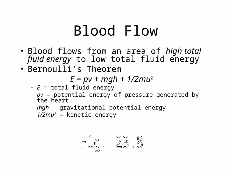
Blood Flow• Blood flows from an area of high total fluid
energy to low total fluid energy• Bernoulli’s Theorem
E = pv + mgh + 1/2mu2
– E = total fluid energy– pv = potential energy of pressure generated by the heart– mgh = gravitational potential energy – 1/2mu2 = kinetic energy
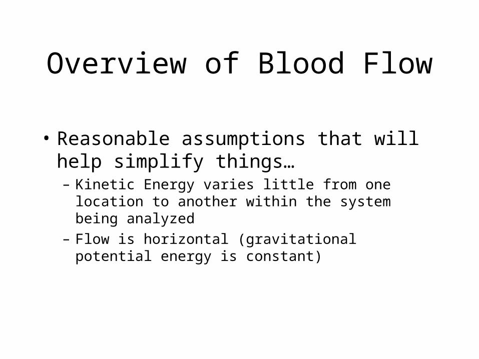
Overview of Blood Flow
• Reasonable assumptions that will help simplify things…– Kinetic Energy varies little from one location to
another within the system being analyzed
– Flow is horizontal (gravitational potential energy is constant)
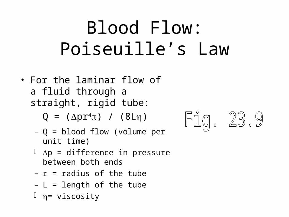
Blood Flow:Poiseuille’s Law
• For the laminar flow of a fluid through a straight, rigid tube:
Q = (pr4) / (8L)
– Q = blood flow (volume per unit time) p = difference in pressure between
both ends
– r = radius of the tube
– L = length of the tube = viscosity
Blood Flow:Poiseuille’s Law
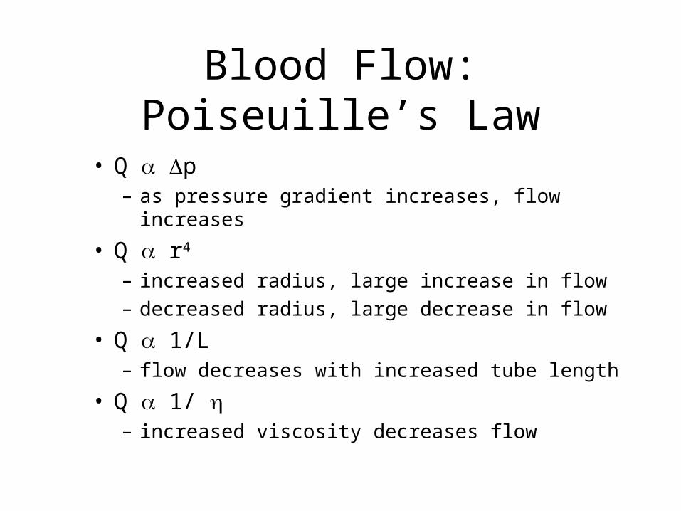
• Q p– as pressure gradient increases, flow increases
• Q r4
– increased radius, large increase in flow
– decreased radius, large decrease in flow
• Q 1/L– flow decreases with increased tube length
• Q 1/ – increased viscosity decreases flow
Gravity Effects on Blood Pressure
• As height ’s, gravitational potential energy ’s, pressure ’s
• Venous return– blood pressure in lower body greater
than upper body due to gravity • pressure in veins exceeds arterial
pressure
• blood pools in leg veins
• returned by venous pressure pumps
Gravity Effects on Blood Pressure
• Head perfusion– arterial blood pressure must be high
enough for blood to reach head– giraffes - long vertical neck
• high arterial BP
• venous values prevent backflow when head brought to ground level
Capillaries
• Enormous number of capillaries– overall large cross-sectional area
• Extremely thin diameter– slow blood flow– high SA/V ratio
• Thin walls (simple squamous endothelium)– low diffusion distance
Ultrafiltration• Small molecules can diffuse into and out of
capillaries• Additional amounts of fluid driven out by
hydraulic pressure inside the capillaries = ultrafiltration.– Small particles driven out with water
– large molecules (e.g. plasma proteins) remain in blood
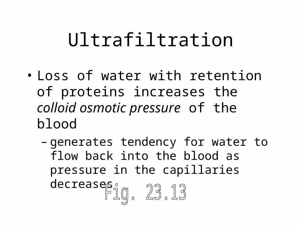
Ultrafiltration
• Loss of water with retention of proteins increases the colloid osmotic pressure of the blood– generates tendency for water to flow back into
the blood as pressure in the capillaries decreases
Lymphatic System
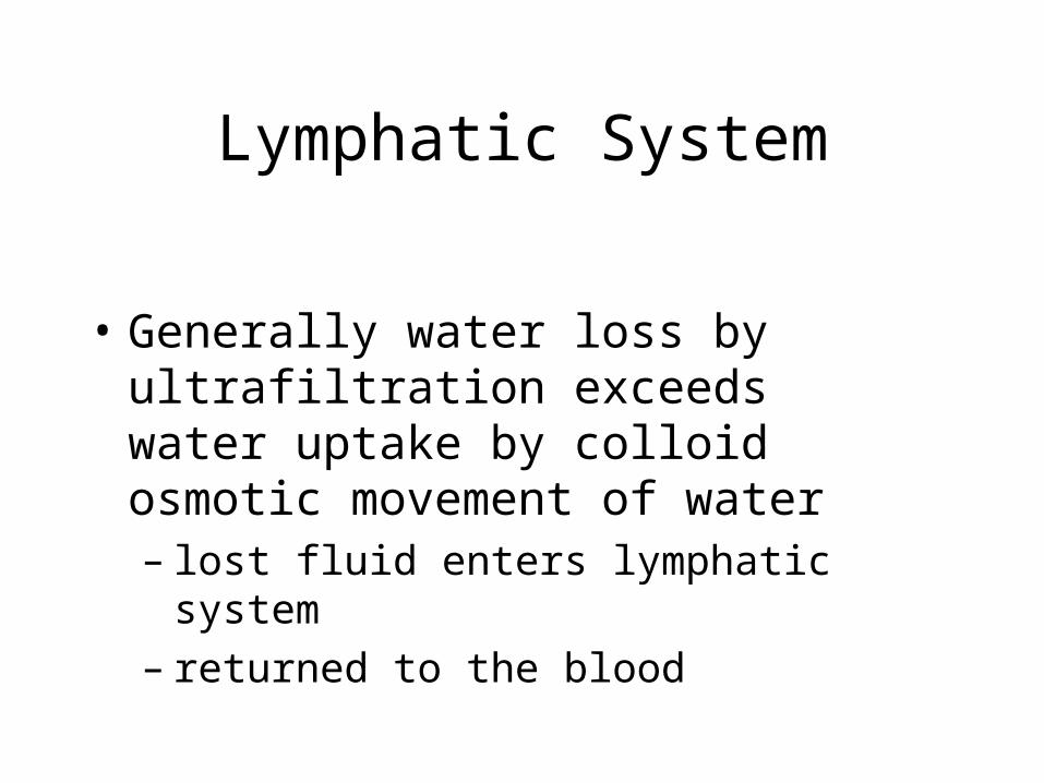
• Generally water loss by ultrafiltration exceeds water uptake by colloid osmotic movement of water– lost fluid enters lymphatic system– returned to the blood


