Page 1
CISPLATIN NEPHROTOXICITY
PhD thesis
Dr. Csaba Máthé MD
Clinical Medicine Doctoral School
Semmelweis University
Supervisor: Dr. György Losonczy MD, D.Sc
Official reviewers:
Dr. Attila Szabó MD, D.Sc
Dr. Krisztina Bogos MD, Ph.D.
Head of the Final Examination Committee:
Dr. Endre Cserháti PhD, D.Sc
Members of the Final Examination Committee:
Dr. Gyula Ostoros Gyula MD, Ph.D.
Dr. Zsuzsanna Orosz MD, Ph.D.
Budapest, 2014
Page 2
2
1. INTRODUCTION
In our Time lung cancer incidence increase. With new drugs the treatments
possibilities are higher and with this the side effects of chemotherapy also increase.
High-dose Cisplatin (Cp) – based combination chemotherapy regimens are used as
front-line treatments of small cell (SCLC) and non small cell lung cancer (NSCLC).
The therapeutic effects of Cp are significantly improved by dose escalation, but in this
time the nephrotoxicity is also escalated. The modality to prevent this now is only
hydration. Different hydration protocols were developed, but one certain effective it
isn’t. The medical studies showed, that age, female sex, smoking decreased the Cp-
induced renal function damage. They are no data about influence of comorbidities as,
for example, hypertension (HT), ischemic heart disease (CD) and diabetes mellitus
(DM). Our patients are older, with this they comorbidities incidence increased. This
can have influence for Cp-induced nephrtoxicity. This patients could have
nephrosclerosis sooner they serum creatinin level [creat] increased.
Vitamin E and C, selen, melatonin, erythropoietin, amifostine, etc. can
protect kidneys from Cp nephrotoxicity in animals. Amifostine, with approbation from
Food and Drug Administration (FDA) to prevent Cp nephrotoxicity, is not used for
expensiveness. CV 247 (CV) is composed of manganese and cupper gluconates, sodium
salicylate and ascorbic acid, which components are known in animal experiences Cp
nephrotoxicities preventive substance, could have nephroprotective effect.
Page 3
3
2. OBJECTIVES
1. Could pre- Cp [creat] predict Cp nephrotoxicity?
2. Can predict calculated isotopic GFR before Cp treatment the Cp-induced
nephrotoxicity?
3. Have influence the patient’s comorbidities for incidence of Cp nephrotoxicity?
4. Is CV protective against Cp nephrotoxicy?
3. METHODS
3.1. Prospective clinical study
The Pulmo-Oncology Unit of Dept. of Pulmonology at Semmelweis University
(Budapest, Hungary) treats 250-300 nonsmall and small cell lung cancer patients
annually. Since Cp-induced reversible or persistent uraemia was estimated to occur in
30% of our patients, in order to investigate whether GFR was already reduced before Cp
treatment when [creat] was still normal, in prospective study, we measured GFR by
clearance of 99m
Tc-DTPA- diethylene thiamine pentaacetic acid- (Izotóp Intézet Kft,
Budapest, Hungary) in 38 stage IIIB-IV lung cancer patients with normal [creat]
scheduled for Cp-based chemotherapy. 99m
Tc-DTPA clearance was measured after
administration of 40 MBq i.v. at the Dept. of Radiology- and Oncotherapy of
Semmelweis University by L. Duffek. Patients were then grouped according to their
highest post-Cp [creat] either below (n=15) or above (n=23) the upper limit of the
reference range (>106 µmol·L-1
) any time during their chemotherapy (2-4 cycles). Cp
was administrated at 75mg·m-2
i.v. (Teva Hungary, Budapest) in each cycle. Cp
infusions were ≥ 21 days part.
3.2. Retrospective clinical study
We retrospectively analysed records of patients (n=242) suffering from stage
IIIA-IV nonsmall or small cell lung cancer and receiving chemotherapy between
January and December 2006. Based on initial evaluation of the 242 patients, three major
subgroups were formed according the absence or presence of the comorbidities CD
and/or DMIH. The NC subgroup had no hypertension, ischemic heart disease or
Page 4
4
diabetes mellitus. The CD subgroup was formed based on presence of long-term,
medically controlled hypertension and ischaemic heart disease (together cardiovascular
disease; n=110), and the DMIH subgroup was based on the combinated presence of
diabetes mellitus and ischaemic heart disease without hypertension (n=52). The
diagnosis of chronic arterial hypertension was based on history and the use of
antihypertensive medications. Ischaemic heart disease was diagnosed based on history,
ECG abnormalities and previous treatment with coronary vasodilatators, platelet
aggregation inhibitors or percutaneous transluminal coronary angioplasty. None of the
CD patients suffered from uncontrolled hypertension, angina pectoris, acute myocardial
infarction or cardiac decompensation, or from any other acute or severe cardiovascular
comorbidity that could have contraindicated chemotherapy with high-dose Cp. Diabetes
mellitus was diagnosed based on history, treatment with insulin (n=5) or oral
antidiabetic treatment (n=47) and higher than normal fasting serum glucose
concentration. None of the DMIH patients suffered from uncontrolled hyperglycaemia
or had symptoms of major complications of diabetes. Urinary protein test showed
opalescence (≥1 g·day−1
) in two patients and slight opalescence (0.5–1.0 g·day−1
) in two
other patients; the majority had negative (<0.5 g·day−1
) results. Patients received several
subsequent combined chemotherapy courses, always containing high-dose Cp (75
mg·m−2
i.v.), and each pre- and the highest post-Cp [creat] concentration values were
recorded. Cp-induced persistent uraemia (which indicates Cp nephrotoxicity) was a
frequent cause of exclusion from further Cp treatment. The number of these patients
was compared between the three groups. Clinical data, such as age, sex, chronic
comorbidity, blood pressure and stage of lung cancer were collected. With regard to
laboratory data, serum glucose and [creat] concentrations were analysed. [creat] was
determined based on the modified Jaffe two-point kinetic reaction using commercially
available test from Dialab (Wiener Neudorf, Austria). Ccreat (eGFR) was calculated
according the Cockcroft–Gault equation. This calculation was selected because the
mean age of our patients was <65 yers.
Cp was provided by Teva Hungary (Budapest, Hungary) and EBEWE Pharma
(Unterach, Austria) and administered at a dose of 75 mg·m−2
. One of three additional
chemotherapeutic agents was given in combination with Cp: gemcitabine (gem, 1,250
mg·m−2
; Eli Lilly, Houten, the Netherlands), etoposide (etop, 3×120 mg·m−2
) and
Page 5
5
paclitaxel (pac, 175 mg·m−2
; both Bristol-Myers Squibb, Princeton, NJ, USA).
Neutropenia was treated with granulocyte colony-stimulating factor (filgrastim, 48 mU;
Amgen, Breda, the Netherlands), severe thrombocytopenia with platelet transfusion, and
anaemia with erythropoietin (Epoetin alfa, 40,000 IU·week−1
; Janssen-Cilag, Centocor,
Leiden, the Netherlands) and/or transfusion as indicated. Patients received
antinociceptive and antiemetic drugs, bisphosphonate, methylprednisolone and other
symptom relievers, as needed.
An i.v. infusion of 500 mL 0.9% NaCl was followed by either gemcitabine,
taxol or etoposide in a further 500 mL saline. After a third 500-mL saline infusion, Cp
was infused again in 500 mL. In our hands, infusion of 500 mL usually takes 20–30
min. Following Cp, the fifth 500 mL saline was infused (total volume of saline 2.500
mL within ∼2.5 h) and the infusion treatment was ended with 100 mL 20% mannitol
(Baxter, Deerfield, IL, USA) i.v.
Data are presented as means±SEM. Statistical analysis was performed using
GraphPad software (Graph Pad Prism 5.0; Graph Pad Software, Inc., San Diego, CA,
USA) using Fisher’s exact test, the Chi-squared test and t-tests (paired and unpaired) as
appropriate. One-or two-way ANOVA and the Kruskal–Wallis test was used to
compared more than two groups. Normally distributed data were analysed by ANOVA
and non-Gaussian distributed or nonparametric values were analysed by Kruskal–Wallis
test. After one-way ANOVA, if significant difference (p<0.05) was found, the
Newman–Keuls multiple comparison post hoc test was used for further analysis. After
two-way ANOVA, a Bonferroni post-test was used. After the Kruskal–Wallis test,
Dunn’s multiple comparison post hoc test was performed. The applied tests are
described in the table and figure legends.
3.3. Animal experience
The study was conducted on 40 male, 8-week old Wistar rats weighing 175-
190 g. The animals were randomly divided into 4 groups (n=10/group). They were kept
individually under standard conventional conditions according to European Council
Directive 123. The study conformed to the Declaration of Helsinki guidelines and was
approved by the local Animal Ethic Committee. Cp (10 mg in 20 ml) was obtained from
TEVA, Israel. The composition of CV247 (Pharmaserve Ltd, Manchester UK) was the
Page 6
6
following: 40 mg ascorbic acid, 2 mg manganese gluconate (unique selling proposition-
USP), 2 mg copper gluconate and 35 mg sodium salicylate per millilitre solution. The
dose given was 2 x 120 mg·kg-1
·day-1
vitamin C (Vit. C), 2 x 10⁵ mg·kg
-1·day
-1 sodium
salicylate, 2x6 mg·kg-1
·day-1
copper gluconate and 2.6 mg·kg-1
·day-1
manganese
gluconate. Methyl cellulose mucilage (Dow Chemicals, Midland, MI, USA) was
prepared in distilled water (1 %).
Control group (C) received 1% methyl cellulose at 10 ml·kg body weight-1
, per
oral (p.o.) by gastric gavage twice daily for 14 days. Another group of rats received
CV247 at 3 ml·kg body weight-1
, p.o. twice daily for 14 days (CV). Two groups were
intraperitoneally injected with a single dose of Cp at 6.5 mg·kg body weight-1
. Cp was
suspended in 10 ml·kg-1
1% methyl cellulose. One of the groups
injected with Cp was
subsequently treated with vehicle (C) or CV at 3 ml·kg body weight-1
, p.o. twice daily
for 14 days (CV+Cp). All rats were weighed and food and water consumptions were
also measured daily. On day 12 were 1.5 ml blood samples taken from all rats by retro-
orbital puncture under isoflurane anaesthesia after a 20-hour food deprivation. The
blood was anticoagulated with citrate and centrifuged twice at 2500 r·min-1
for 10 min
at +4 C to obtain plasma. (creat) and blood urea nitrogen (BUN) were determined from
the plasma by colorimetric tests using commercially available kits. Rats were terminally
anaesthetised with an overdose of pentobarbital on day 14. Blood was collected by
aortic puncture and one kidney from each animal was removed and weighed. Kidneys
were fixed in 8% buffered formalin (pH 7.4), paraffin sections were prepared and
stained with haematoxylin–eosin. Renal histological changes were blindly evaluated
using a 5-grade severity scale (0 =no change; 1= minimal changes; 2=mild changes;
3=moderate changes; 4=severe changes). The cyclooxygenase-2 (COX-2)
immunohistochemistry was done using a mouse monoclonal COX-2 primary antibody
(Novocastra, UK) at 1:100 dilution. The secondary antibody was a peroxidase-
conjugated mouse/rabbit polymer (Dako Real™ Envision™ /HRP, Rabbit/Mouse).
Diaminobenzidine was used for visualisation.
Means±SD are given throughout. The statistical comparisons were performed
by two-way repeated measures ANOVA with Bonferroni post hoc test or Mann-
Whitney U test using GraphPad Prism 5 for Windows, or by two-way ANOVA using
Page 7
7
the SPSS 17 for Windows, when appropriate. The level of significance was set at
p<0.05.
4. RESULTS
4.1. Clinical studies results
4.1.1. Results of prospective clinical study
Out of the 38 lung cancer patients, 23 patients responded with pathologically
increased [creat] after Cp (table I), although pre-treatment [creat] values were normal in
both groups. Pre-treatment GFR, as measured by clearance of 99m
Tc-DPTA, was
significantly reduced by ~25% in those lung cancer patients with azotaemia who
responded to 2-4 cycles of Cp treatment.
Table I. Pre-Cp glomerular filtration rate (GFR) of 38 initially non uraemic lung
cancer patients
Subgroup
pre-Cp
creat
post-Cp
creat pre-Cp GFR
(mL·min-1
·m-2
)
Age
(yrs)
Males/
females
( mol.L-1
)
Cp nephrot.
(n=23) 79 4
1 167 12
1 73.51 4
1 63.6 1.5
ns1 13/10
ns2
No Cp nephrot.
(n=15) 68 3 87 4 98.6 4.6 60.5 2.8 8/7
: p 0.05, not significant (ns): p 0.05 compared with „No Cp nephrotoxicity” group;
1: unpaired t-test; 2: Fischer’s exact test
4.1.2. Results of retrospective clinical study
Table II contain all patient’s clinical data and in table III we can see patients
[creat] values before and after Cp treatments.
Page 8
8
Table II. Clinical data of patients recevieving high-dose Cp for lung cancer
NC(n=80) CD (n=110) DMIH(n=52)
Age (yrs) 56 1 60 1 62 1
Males/females (n) 45/35 66/44ns
33/19ns
BMI (kg·m-2
) 24.3 0.4 25.4 0.4ns
25.4 0.6ns
Dose of Cp per treatment (mg) 123 2 126 2ns
124 4ns
Total dose of Cp (mg) 375 22 399 22ns
392 43ns
Mean number of cycles (n) 3.1 0.2 3.2 0.2ns
3.2 0.3ns
Cp+gem/etop/tax(patients
number)
38/41/1 52/55/3ns
25/24/3ns
Systolic/diastolic blood pressure
(mmHg)
132 2/81 1 134 2/81 1ns
137 3/84 2ns
Cardiac frequency beats (beats
·min-1
)
81 1 82 1ns
86 2ns
: p 0.05, ns: p 0.05 compared with NC
Page 9
9
Table III.[creat] in lung cancer patients receiving 1-4 cycles of high-dose Cp
Cp cycle NC Cd DMIH
1st Patients number
[creat] µmol·L-1
Pre-Cp
Post-Cp
80
77 1
82 2
110
78 1 ns
95 4 †
52
77 3 ns
94 4 †
2 2nd Patients number
[creat] µmol·L-1
Pre-Cp
Post-Cp
68
80 2
88 4
96
86 2 ns
105 5 †
49
82 3 ns
110 6 †
3rd Patients number
[creat] µmol·L-1
Pre-Cp
Post-Cp
42
85 3
91 5
64
91 3ns
113 5 †
31
83 4ns ns
117 9 †
4th Patients number
[creat] µmol·L-1
Pre-Cp
Post-Cp
35
84 4
93 5
47
95 3 ns
117 8 †
30
89 3 ns
121 5 †
: p 0.05 compared with pre-Cp (paired t-test); not significant (ns): p 0.05;
†: p 0.05 compared with NC (two-way ANOVA with Bonferroni after test).
Pre- treatments [creat] are not increased. After every treatment [creat]
increased significant, what shows Cp nephrotoxicity. In NC group [creat] was every
time physiologic, but in other two groups were increased. Before the new cycles of
chemotherapy [creat] level decreased in normal range, these need to continue Cp
therapy. DMIH patients’ kidney pre- treatments were not intact, that is responsible for
this high number of post-Cp azotaemia. Table III. shows, that this comorbidities
predispose patients to Cp nephrotoxocity. They incidence can’t predict by pre treatment
[creat] values.
Cp induced azotaemia was in NC group 7.5, in CD group 20.9 (p<0.05
compared with NC) and in DMIH group 30.8% (p<0.01 compared with NC) (Figure 1.)
Page 10
10
Figure 1. Frequency of nephrotoxicity (dark) and nephrotoxicity-related drop-out
from futher Cp treatment (bright) in lung cancer patients receiving 1-4 cycles of
high-dose Cp and suffering from hypertension and ischaemic heart disease (CD),
or diabetes mellitus and ischaemic heart disease (DMIH), or being free from these
severe comorbidities (NC).
*: p 0.05 compared with NC, **: p 0.01 compared with NC; #: p 0.01 compared with
CD (two-tailed difference tests).
Calculated GFR before Cp treatments where significant higher in that group,
which later don’t have azotaemia. That show, calculated GFR could be predictive for
Cp nephrotoxicity. This reproduce table I. results.
Figure 2. Summary of pre-Cp treatment calculated GFR values together in 3
patients groups (NC, CD, DMIH)
Difference was calculated with Mann Whitney test.*: p<0.01 Cp releated azotaemia
with no azotaemia.
Page 11
11
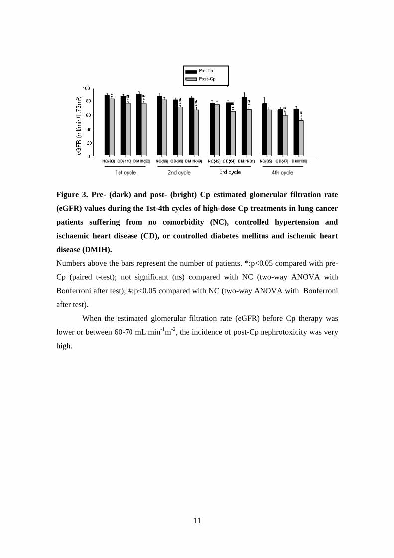
Figure 3. Pre- (dark) and post- (bright) Cp estimated glomerular filtration rate
(eGFR) values during the 1st-4th cycles of high-dose Cp treatments in lung cancer
patients suffering from no comorbidity (NC), controlled hypertension and
ischaemic heart disease (CD), or controlled diabetes mellitus and ischemic heart
disease (DMIH).
Numbers above the bars represent the number of patients. *:p<0.05 compared with pre-
Cp (paired t-test); not significant (ns) compared with NC (two-way ANOVA with
Bonferroni after test); #:p<0.05 compared with NC (two-way ANOVA with Bonferroni
after test).
When the estimated glomerular filtration rate (eGFR) before Cp therapy was
lower or between 60-70 mL·min-1
m-2
, the incidence of post-Cp nephrotoxicity was very
high.
Page 12
12
4.2. Animal experience
4.2.1.Body weight
Figure 4. Cp dicreased significant body weight of rats from 2nd day.
Body weight of rats steadily increased in control group from 171±8 g to
234±15 g over a study period of 14 days with a drop on day 11 after the overnight food
deprivation before blood sampling. In comparison with baseline values, Cp caused a
5.5% peak body weight loss (p<0.001) on day 3 after treatment. CV treatment did not
influenced body weight in comparison with C and Cp groups.
4.2.2. Water consumption
CV consistently increased water consumption in comparison with C group
(Figure 5) which was statistically significant on days 8, 9 and 11 (p<0.05 all). Cp
caused short, non-significant decrease in water consumption on day 2 after its
administration (daily -5- -10 mL). Therefore, from day 4, rats in Cp and CV+Cp groups
drank significantly more water than rats in group C. Co-administration of CV to Cp did
not alter water consumption in comparison with the group treated with Cp only. In
comparison with C group animals with Cp consume more water. CV alone increased
water consumption.
Page 13
13
Figure 5. From 5 day (*) water consumption was significantly increased in groups
treated with Cp and CV+Cp in comparison to the C group. The open circles show
that on days 8, 9 and 11 water consumption was significantly increased in the
group treated with CV in comparison to the group treated with vehicle.
The statistical analysis was performed by two-way ANOVA with Bonferroni after test.
4.2.3. Renal function
[creat] and [BUN] values were within physiological limit in groups C and CV.
([creat]: 17.0- 22.5 μmol·L-1
, [BUN]: 6.63- 10.48 mmol·L-1). Cp increased both
significant (p<0.01) in day 12 taked blood samples. CV did not alter (p>0.05 both) these
effects of Cp on renal function (Figure 6 and 7).
Figure 6. CV did not alter Cp caused azotaemia. (two-way ANOVA)
Page 14
14
Figure 7. CV have no effect of [BUN]. (two-way ANOVA)
4.2.4. Kidney histology and immunohistochemistry
Figure 8. CV have nephroprotective effect. (haematoxylin-eosin staining).
A.C:normal histology; B+E.Cp:severe degree of tubulointerstitial abnormality
(3.67±0.50); C.CV: normal histology; D+F.CV+Cp: significantly less severe alterations
we in group Cp (2.67±0.71; p<0.01) E and F higher magnification of sections.
Abnormalities were statistifically compared by two-way ANOVA.
Page 15
15
No histological changes were seen in kidneys in groups C and CV; that show
CV have no kidney nephrotoxicity. Blind assessment demonstrated a significant
(p<0.01) reduction in the mean of histological kidney injury from 3.67±0.50 in Cp to
2.67±0.71 in CV+Cp group.
Immunohistochemistry related a moderate degree of focal COX-2 activity in the
cytoplasm of tubular epithelium in the interstitial space and in the walls of major blood
vessels (C:1,20±0.42, CV:1,0±0,0). Blind assessment of COX-2 immunoreactivity
markedly increased in the groups treated with Cp and CV+Cp. Treatment with CV did
not alter COX-2 immunoreactivity in comparison C but slightly (Cp:3.00±0.71 versus
CV+Cp:2.44±0.53, p=0.097). Our study showed that CV could prevent Cp
nephrotoxicity.
Figure 9. Cp effect and CV protective effect on COX-2 immunohistochemistry in
renal cortex.
A.C: mild activity in the interstitium and tubular epithelium; B+E. Cp increased COX-2
activity int he damaged areas of the kidney (3.67±0.50); C.CV: mild activity in the
interstitium and tubular epithelium; D+F. CV+Cp: Cp effect is significant decreased by
Page 16
16
CV (2.67±0.71; p<0.01) E and F higher magnification of sections. Abnormalities were
statistifically compared by Mann Whitney test.
Page 17
17
5. CONCLUSIONS
1.The risk of nephrotoxicity should always be evaluated based on eGFR.
2.Lower eGFR (60-80 ml·perc-1
) with normal [creat] and [BUN] before Cp therapies
predict Cp nephrotoxicity.
3.Coexisting CD or DMIH, because narrow the tolerance of the kidneys to Cp,
increased the incidence of Cp nephrotoxicity.
4. CV 247 can attenuate Cp caused nephrotoxicity.
Page 18
18
6. BIBLIOGRAPHY OF THE CANDIDATE’S PUBLICATIONS
Publications related to the PhD thesis:
1.Máthé C, Bohács A, Duffek L, Lukácsovits J, Komlosi ZI, Szondy K, Horváth I,
Müller Vand Losonczy G. (2011) Cisplatin nephrotoxicity aggravated by
cardiovascular disease and diabetes in lung cancer patients. Eur Resp J, 37:888-894.
IF:5.895
2.Máthé C, Szénási G, Sebestény A, Blazovics A, Szentmihályi K, Hamar P and Albert
M. (2013) Protective effect of CV247 against cisplatin nephrotoxicity in rats. Hum Exp
Toxicol, May 7. [Epub ahead of print] IF:1.453
3.Máthé Cs, Bohács A, Duffek L, Lukácsovits J, Komlósi Zs, Szondy K, Horváth I,
Müller V, Losonczy Gy. (2011) Cisplatin nephrotoxicity in our patients with lung
cancer. Med Thor, 64: 33-41.
4.Losonczy Gy, Máthé Cs, Müller V, Szondy K, Moldvay J. (2010) Incidence, risk
factors and prevention of cisplatin- induced nephrotoxicity in patients with lung cancer.
Magy Onkol, 54:289-296.
5.Révai T, Máthé Cs, Winkler G, Bártfai Z. (2003) Cisplatin –induced nephropathie’s
diagnostic and therapy possibilities in apropos of one case. Hypertonia és nephrológia,
7:123-125.
Book chapters related to the PhD thesis:
1.Máthé Cs, Bártfai Z. Role of platinum compounds in lung cancer therapy. In: Révai
T, Máthé Cs, Bártfai Z (eds.), Cisplatin-nephropathy. Medition Kiadó Kft, Budapest,
2005:7-12.
2.Máthé Cs, Révai T. Case review. In: Révai T, Máthé Cs, Bártfai Z (eds.), Cisplatin-
nephropathy, Medition Kiadó Kft, Budapest, 2005:20-21.
3.Kocsis J, Bártfai Z, Máthé Cs. Effect and role in malignant tumors treatment of
cisplatin. In: Révai T, Máthé Cs, Bártfai Z (eds.) Cisplatin-nephropathy, Medition
Kiadó Kft, Budapest, 2005:13-16.
Page 19
19
Book editing related to the PhD thesis:
Révai T, Máthé Cs, Bártfai Z. Ciszplatin-nephropathy, Medion Kiadó Kft., Budapest,
2005.
Other publications:
1.Máthé Cs, Bártfai Z. (2004) Rhinitis allergica, Praxis, 13: 15-19.
2.Máthé Cs. (2007) Diagnosis and therapy modalities of lung cancer. Háziorvos
továbbképző szemle, 12: 652-658.
3.Szentmihályi K, May Z, Szénási G, Máthé C, Sebestény A, Albert M, Blázovics A.
(2014) Cisplatin administration influences on toxic and non-essential element
metabolism in rats. J Trace Elem Med Biol. Feb 25. [Epub ahead of print] IF:1,959
4.Vajda E, Emődi K, Lippai N, Egri G, Nagy A, Ruby E, Sápi Z, Máthé Cs, Magyar P.
(2007) Primery pulmonary paragangliom. Med Thor, 60:367-377.
5.Ostoros Gy, Tallósy I, Horváth Á, Máthé Cs. (1992) Examination of non small cell
lung cancer conservative therapy. Med Thor, 45:285-289.
Other book chapters:
1.Máthé Cs. Pulmonary embolism. In: Magyar P, Pálfy L, Bártfai Z (eds.)
Pulmonology manual for medical professional employee Medicina Könyvkiadó,
Budapest, 2006: 297-302.
2.Máthé Cs, Kismarton J: Law regarding with tuberculosis. In: Magyar P, Somoskövi
Á (szerk.), Pulmonal and extrapulmonal tuberculosis. Medicina Könyvkiadó, Budapest,
2007:245-250.
3.Máthé Cs: Pulmonary disease caused by irradiaton. In: Magyar P, Losonczy Gy
(eds.), Pulmonology handbook. Medicina Könyvkiadó, Budapest, 2012:526-529.
4.Máthé Cs, Tamási L: Mediastinal diseases. In: Magyar P, Losonczy Gy (eds.),
Pulmonology handbook. Medicina Könyvkiadó, Budapest, 2012:743-746.
5. Máthé Cs: Respiratory disease. In: Kalabay L (eds.) General pratitioner medicine
theory and practice, E-learning textbook, Semmelweis University, Budapest, 2012:792-
810.
6. Máthé Cs: Tuberculosis and rare infection lung diseases. In: Somfay A (eds.),
SpringMeg Kiadó, Budapest, 2013:138-169.