Analogy and Gesture for Mental Visualization of DNA Structure Anveshna Srivastava and Jayashree Ramadas Tata Institute of Fundamental Research India Abstract In this chapter, we report five beginning undergraduate students' understanding of the 3-D structure of DNA, and the use of gesture and analogy to enhance their 3-D visualization of DNA structure. Through clinical interview-cum-teaching sessions we first recapitulated their background knowledge of basic biology and chemistry prerequisites. We then proceeded to a microgenetic study of the students’ understanding of DNA structure, in which we found that initially all of them interpreted their familiar textbook diagrams as 2-D structures rather than 2-D representations of the 3-D structures. We subjected video data of these interview-cum-teaching sessions to a microgenetic time-sequence analysis, where we identified episodes during which the students used multiple representations of DNA backbone and nitrogenous bases and showed positive, that is, 2-D to 3-D transitions, and “Aha!” moments. We traced these students’ learning episodes to their use of gesture in combination with character viewpoint simulation in terms of ladder analogy of the DNA molecule. Gesture, analogy, and mental simulation —involving changing the viewpoint of an observer—were found useful for linking together DNA’s multiple external representations into its integrated internal representation, and thus perhaps for bringing about the observer’s mental visualization of its 3-D structure. Keywords: Analogy; DNA structure; Gesture, Model-based reasoning; Visualization Introduction The birth of molecular biology was significantly marked by the discovery of the double helical structure of the DNA molecule by Watson and Crick (1953a). The general correctness of this structure was gradually proven in the subsequent years by substantial research on the structural as well as functional aspects of the DNA molecule. The structure of DNA had immediate functional implications: “It follows that in a long molecule many different permutations are possible, and it therefore seems likely that the precise sequence of the bases is the code which carries the genetical information” (Watson & Crick, 1953b, p. 965). Conceptual understanding in molecular biology involves integration of the macro (genetic traits), micro (cell), and molecular (gene) levels. Building up of the DNA molecular structure and its location at the cellular level leads to an understanding of its biological significance, for example, in genetic expression. Marbach-Ad and Stavy (2000) remarked that the difficulty in understanding and linking these different organizational levels is “because sometimes one level (e.g., the macro level) belongs to one discipline (e.g., biology), and the other level (e.g., the molecular level) belongs to different discipline (e.g., chemistry)” (p. 201). In fact, the integration occurs in multiple ways—one that includes concepts from various disciplines, another that involves the macro, micro, and the molecular levels, and finally, the structure-function linkages within and across these levels. Structural-functional linkages have been identified as a problem area in elementary genetics (Marbach- Ad, 2001; Lewis, 2004). In a study of major problem areas in biological sciences as identified by 1 Citation: Srivastava, A. & Ramadas, J. (2013). Analogy and Gesture for Mental Visualization of DNA Structure. In Treagust, D.F. & Tsui, C.-Y. (Eds.), Multiple Representations in Biological Education. Dordrecht, The Netherlands: Springer, pp. 311-329.
Transcript
Citation: Srivastava, A. & Ramadas, J. (2013). Analogy and Gesture for Mental Visualization of DNA Structure. In Treagust, D.F. & Tsui, C.-Y. (Eds.), Multiple Representations in Biological Education (pp.
311-329). Dordrecht, The Netherlands: Springer.
Analogy and Gesture for Mental Visualization of DNA Structure
Anveshna Srivastava and Jayashree RamadasTata Institute of Fundamental Research
India
AbstractIn this chapter, we report five beginning undergraduate students' understanding of the 3-D structure of DNA, and the use of gesture and analogy to enhance their 3-D visualization of DNA structure. Through clinical interview-cum-teaching sessions we first recapitulated their background knowledge of basic biology and chemistry prerequisites. We then proceeded to a microgenetic study of the students’ understanding of DNA structure, in which we found that initially all of them interpreted their familiar textbook diagrams as 2-D structures rather than 2-D representations of the 3-D structures. We subjected video data of these interview-cum-teaching sessions to a microgenetic time-sequence analysis, where we identified episodes during which the students used multiple representations of DNA backbone and nitrogenous bases and showed positive, that is, 2-D to 3-D transitions, and “Aha!” moments. We traced these students’ learning episodes to their use of gesture in combination with character viewpoint simulation in terms of ladder analogy of the DNA molecule. Gesture, analogy, and mental simulation—involving changing the viewpoint of an observer—were found useful for linking together DNA’s multiple external representations into its integrated internal representation, and thus perhaps for bringing about the observer’s mental visualization of its 3-D structure.
Keywords: Analogy; DNA structure; Gesture, Model-based reasoning; Visualization
IntroductionThe birth of molecular biology was significantly marked by the discovery of the double helical structure of the DNA molecule by Watson and Crick (1953a). The general correctness of this structure was gradually proven in the subsequent years by substantial research on the structural as well as functional aspects of the DNA molecule. The structure of DNA had immediate functional implications: “It follows that in a long molecule many different permutations are possible, and it therefore seems likely that the precise sequence of the bases is the code which carries the genetical information” (Watson & Crick, 1953b, p. 965).
Conceptual understanding in molecular biology involves integration of the macro (genetic traits), micro (cell), and molecular (gene) levels. Building up of the DNA molecular structure and its location at the cellular level leads to an understanding of its biological significance, for example, in genetic expression. Marbach-Ad and Stavy (2000) remarked that the difficulty in understanding and linking these different organizational levels is “because sometimes one level (e.g., the macro level) belongs to one discipline (e.g., biology), and the other level (e.g., the molecular level) belongs to different discipline (e.g., chemistry)” (p. 201). In fact, the integration occurs in multiple ways—one that includes concepts from various disciplines, another that involves the macro, micro, and the molecular levels, and finally, the structure-function linkages within and across these levels.Structural-functional linkages have been identified as a problem area in elementary genetics (Marbach-Ad, 2001; Lewis, 2004). In a study of major problem areas in biological sciences as identified by
1
Citation: Srivastava, A. & Ramadas, J. (2013). Analogy and Gesture for Mental Visualization of DNA Structure. In Treagust, D.F. & Tsui, C.-Y. (Eds.), Multiple Representations in Biological Education. Dordrecht, The Netherlands: Springer, pp. 311-329.
students, Bahar, Johnstone, and Hansell (1999) reported that the structure and function of the DNA and RNA molecule was considered as a topic of relatively low difficulty. However, we make a case here that students do have a problem in understanding the basic 3-D structure of the DNA molecule.
Structure of the DNA MoleculeThe double-helical structure of the DNA molecule can be visualized as two right-handed helices coiled around a central axis. Each helix is composed of a sugar-phosphate backbone and each (deoxyribose) sugar molecule in this backbone is attached with a nitrogenous base through a glycosidic bond to form a nucleoside unit. The nitrogenous bases—purines (adenine or guanine) or pyrimidines (thymine or cytosine) are paired in a complementary fashion where adenine (A) forms two hydrogen bonds with thymine (T) and, guanine (G) forms three hydrogen bonds with cytosine (C). The nitrogen bases of the DNA molecule are planar ring structures of equal length which are perpendicular to the central DNA axis and also to their attached sugar molecules. Orientation of the nitrogenous base pairs and the specific hydrogen bonding between the complementary base pairs give rise to a basic ladder shape, which is coiled into a right handed helix of specific dimensions.
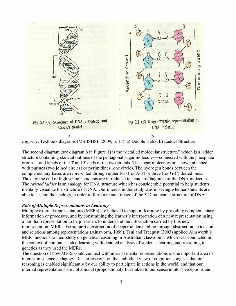
Textbook Representations of DNA StructureIn Indian schools, the chemical prerequisites for learning the DNA molecule in biology are built up from middle school till the higher secondary level (for students aged 17) as part of the chemistry curriculum. The higher secondary biology textbook used in our study, published by Maharashtra State Board of Secondary and Higher Secondary Education (MSBSHSE, 2009), introduces the DNA molecule by describing the components of nucleotides, the pentose sugar, phosphate group and the nitrogenous bases, with their chemical formulas. The analogy of a twisted ladder is usually followed by two kinds of diagrammatic representations. The first is a schematic representation of the DNA double helix, depicting two criss-crossing wavy ribbon-like strands, in which there are (sugar-phosphate) links labeled “S-P-S-P” in the backbone (see diagram a in Figure 1). Connecting the backbone are the skeletal structures of the nitrogenous base pairs with the respective number of hydrogen bonds with dimensional details indicated in Angstroms (Å). The accompanying text mentions the angle between successive base pairs and also that each “spiral turn” contains 10 pairs of nucleotides (p. 15).
2
a b Figure 1. Textbook diagrams (MSBSHSE, 2009, p. 15): a) Double Helix; b) Ladder Structure
The second diagram (see diagram b in Figure 1) is the “detailed molecular structure,” which is a ladder structure containing skeletal outlines of the pentagonal sugar molecules—connected with the phosphate groups—and labels of the 3' and 5' ends of the two strands. The sugar molecules are shown attached with purines (two joined circles) or pyrimidines (one circle). The hydrogen bonds between the complementary bases are represented through either two (for A-T) or three (for G-C) dotted lines. Thus, by the end of high school, students are introduced to standard diagrams of the DNA molecule. The twisted ladder is an analogy for DNA structure which has considerable potential to help students mentally visualize the structure of DNA. Our interest in this study was in seeing whether students are able to sustain the analogy in order to form a mental image of the 3-D molecular structure of DNA.
Role of Multiple Representations in LearningMultiple external representations (MERs) are believed to support learning by providing complementary information or processes, and by constraining the learner’s interpretation of a new representation using a familiar representation to help learners to understand the information carried by this new representation; MERs also support construction of deeper understanding through abstraction, extension, and relations among representations (Ainsworth, 1999). Tsui and Treagust (2003) applied Ainsworth’s MER functions to their study on genetics reasoning in Australian classrooms, which was conducted in the context of computer-aided learning with detailed analysis of students' learning and reasoning in genetics as they used the MERs.The question of how MERs could connect with internal mental representations is one important area of interest in science pedagogy. Recent research on the embodied view of cognition suggests that our reasoning is enabled significantly by our ability to participate in actions in the world, and that our internal representations are not amodal (propositional), but linked to our sensorimotor perceptions and
3
actions (Clark, 1997; Barsalaou, 1999). One direct implication of the embodied view is that MERs connect to internal representations through the learner's perceptions and actions.Drawing on the embodied view of cognition, we suggest that a possible pedagogical route from external to internal (mental) representations might be taken through the use of gesture. Goldin-Meadow and Beilock (2010) argued that gestures affect thinking by grounding it in action and may even have a more powerful influence on thoughts than action itself resulting in a rich internal representation that incorporates the sensorimotor properties required to act out in the world. This insight from cognitive science was used by Padalkar and Ramadas (2011) in proposing a pedagogical purpose for deliberately designing gestures in science. Padalkar and Ramadas argued that gestures might be used to internalize a natural phenomenon, a scientific model, or properties of space. It is important that the gestures in Padalkar and Ramadas’ study served not only to link external representations with internal mental ones, but they were also designed to link two types of external representations (concrete models and diagrams).Mathai and Ramadas (2009) proposed tasks calling for changing the viewpoint of an observer to encourage mental visualization of body systems. This parallels Goldin-Meadow and Beilock's (2010) hierarchies of gestures and actions—character viewpoint gestures reflect actual movements, observer viewpoint gestures capture the goal object or its trajectory, and metaphorical gestures represent abstractions—as well as the suggestion that character and observer viewpoint gestures, if used in sequence, could provide a bridge between concrete actions and more abstract representations. We therefore suggest that: (a) gestures could be used to link external and internal representations; (b) gestures could be used to link together different MERs into an integrated internal representation; (c) real or imagined manipulations or transformations of structure, and imagining a change in one's viewpoint could enable mental visualization of the structure; and (d) character viewpoint gestures or actions could help in making a molecular structure (e.g., DNA) more comprehensible to students. A complementary approach to building internal mental representations, particularly visual ones, is that of analogy. Gentner (1989) defined analogy as a mapping from a base (familiar) domain to a target (unfamiliar) one. Previous research has shown that analogy is useful in visualization, model-based reasoning, knowledge construction, and understanding (e.g., Duit, 1991; Justi & Gilbert, 2006; Harrison & Treagust, 2006). Like gesture, analogy has potential to help students construct mental visual models from multiple external representations. Therefore, in this study we used a combination of gesture and analogy of the twisted ladder using a character viewpoint simulation for encouraging visualization of the 3-D structure of the DNA molecule.
This StudyIn this study, we examined students' reasoning processes in understanding the 3-D nature of the DNA molecule through the integration of prerequisite concepts from physics and chemistry, supported by appropriate simple and low-cost multiple external representations (MERs) of DNA structure in terms of the following research questions: 1. Are students able to link the ladder analogy with common 2-D diagrams of DNA structure to form a
mental model of the 3-D structure of the molecule?2. Can we use gesture to link the 2-D representations and the ladder analogy with the 3-D concrete
models of DNA structure?3. Can we use mental simulation of changing observer viewpoint to link the 2-D representations and
the ladder analogy with the 3-D concrete models of DNA structure?
Through a screening test we selected five students of 17 to 19 years old, who were enrolled in the first year of a three-year bachelor degree course in biological sciences at a university in India (see Table 1).
4
Table 1Demographic Information of Participants in this StudyName of the student1
Age (in years)
Gender Mother Tongue
Degree pursuing (Bachelors)
Courses taken in the current semester2
Anuja 18 F Marathi Microbiology MPC
Sharada 18 F Oriya Biotechnology BMC
Nitin 19 M Marathi Microbiology MPC
Sandhya 17 F Telugu Biotechnology BMC
Aakriti 18 F Hindi Microbiology MPC
1Names are changed to preserve anonymity 2MPC: Microbiology, Physics, Chemistry; BMC: Biotechnology, Microbiology, Chemistry
We used a microgenetic research design (Siegler and Crowley, 1991; Siegler, 2006; Flynn and Siegler, 2007; Aalsvoort et al., 2009) which is appropriate for situations that involve rapid transitions in learning by tracing the processes of the students’ learning under dynamic, in vivo conditions. The three important attributes of a microgenetic research design developed in Siegler and Crowley (1991) and modified in Siegler (2006) are:
1. Observations span the period of rapidly changing competence. 2. Within this period, the density of observations is high, relative to the rate of change.3. Observations are analyzed intensively, with the goal of inferring the representations and
processes that gave rise to them. (p. 469). The students are observed very closely during the period of learning and then these observations are revisited again and again for a finer understanding of the patterns that depict “change in real-time as how it occurs” (Aalsvoort et al., 2009, p. 9).In our study, observations were carried out during six individual sessions held over nine days. Each session involved a clinical interview-cum-teaching sequence for 1 to 1.5 hours for each student per day. The language of the interview was English except for some occasions when Marathi and occasionally Hindi were used for two of the interviewees: Nitin and Aakriti. The prerequisites for the sessions lay within the syllabus for secondary and higher secondary schools recommended by the State Board. The sessions on Days 1 through 4 focused on initial assessment and recall of prerequisite concepts in biology and chemistry. Brief sequences of direct instruction were included in order to bridge some inevitable gaps in understanding. The issue of three-dimensionality of DNA structure was addressed on Days 4 through 6 and these data were analyzed microgenetically.
Multiple Representations of the DNA Backbone and the Nitrogenous Base PairsThe students were asked to draw the textbook diagrams (the ladder and helical structures in diagrams a and b in Figure 1) and recall the well-known ladder analogy for DNA structure. The DNA backbone was represented by five simple models (M1 to M5 in Table 2). M1 was comprised of a sheet of paper laid on a table and the students were asked to consider its long edges to represent the two DNA backbones. M2 consisted of two antiparallel pencils laid on the table and considered as the two DNA backbones. M3 was a variant of M2 where the two antiparallel pencils (the backbones) were made to stand erect on the table. M4 was a cutout model depicting the two backbones, each consisting of two
5
phosphate groups attached with one sugar molecule at its 3' and 5' positions, fixed on a cardboard base. M4 showed the molecular details of the two sugar-phosphate backbones.
Table 2 Multiple Representations of the DNA Backbone
Model No. Backbone representation
M1 Long edges of a sheet of paper (laid on the table)
M2 Two (anti) parallel pencils (laid on table)
M3 Two (anti) parallel pencils (held to stand erect on table)
M4 Cardboard cutout of a sugar molecule attached with two phosphate molecules (two sets) standing on a cardboard base
M5 Clothespin model (ladder representation of DNA which can be assembled on a table and then twisted to form a helix)
M5, or the clothespin model, was adapted from Venville (2008). The students were provided with two plastic tubes along which they could string interlocking clothespins of four different colors (green, yellow, blue, and pink) to represent the complementary DNA bases. The students were asked to construct the M5 model to depict first the ladder structure and then the helical representation of the DNA molecule.In combination with models representing the DNA backbone, two types of representations of the nitrogenous base pairs were introduced. The first representation consisted of cardboard cutouts of the different nitrogenous bases (see Figure 2) which was first suggested by James Watson's own account of his discovery of base-pairing as recounted in an online video program (Cold Spring Harbor Laboratory's DNA Learning Center, n.d.). The students were to use these cutouts against the M4 model to depict the orientation of the base pairs in the molecular model, while indicating the position of attachment of the bases with the sugar molecules in the DNA backbone.
6
a b
Figure 2. Cutouts of molecules of nitrogenous bases – (a) Purine base (b) Pyrimidine base
The second base pair representation comprised the palm gesture, in which the palm represents a nitrogenous base (purine or pyrimidine) and the straightened fingers represent the complementary nitrogenous base (pyrimidine or purine) (see Figure 3). The students used this gesture to imitate the orientation of the base pairs in the ladder against the models M1-M5, as appropriate.
Figure 3. Palm gesture with palm and straightened fingers representing a complementary base pair.
The last type of representation was the ladder analogy, in which the backbone and the base pair representations were combined. The students were asked to visualize, first a straight ladder, and then a twisted ladder. The ladder analogy was used as a reminder to the students about the DNA structure while they attempted to show the base pair orientation with the help of the palm gesture or the cutouts. If the analogy by itself did not work, then the students were instructed to mentally simulate the action of walking up the straight ladder, and in that situation consider how the steps of the ladder would be oriented. The gesture and mental simulation were also used for the helical ladder structure in model M5. The mental visualization (of the straight or the twisted ladder) and the simulation (of walking up the ladder) correspond, respectively, to the observer viewpoint and character viewpoint gestures/actions discussed by Goldin-Meadow and Beilock (2010). Here the actions are, of course, not actually carried out, but mentally simulated.
7
Preparing the Background (Days 1, 2, and 3)On Day 1, we examined students' understanding of the concept of DNA as the genetic material. We probed their familiarity with terms such as genetic material, gene, heredity, and so on. The students were asked about cells, the location of genetic material, and DNA as genetic material. All the students except Nitin had problems in understanding the relationship between a gene and DNA, for example, Anuja said, “I am confused that it (gene) is inside the DNA or the DNA is inside the gene.” Also, all the students did not understand Hershey and Chase’s classical experiment which proved that DNA is the genetic material. Each day, from Day 2 till Day 6, began with students' diagrammatic representations of the DNA ladder and the double helix as some approximation of the two familiar textbook diagrams (see Figure 1). On Day 2, we focused on recapitulating elementary background related to the chemistry of the DNA molecule and re-introduced to them the idea of nitrogenous bases (purines and pyrimidines) with the electronegative nitrogen atoms which can form hydrogen bonds with another nitrogenous base. On Day 3, the students explored different pairing possibilities between the bases using cardboard cutout models of the bases (see Figure 2). They eventually used the cutouts to form the A-T double hydrogen bond and G-C triple hydrogen bonds, to demonstrate that the base pairs were planar and of identical lengths.
Introduction to the Nucleoside (Day 4)At the start of Day 4, the students were introduced to the palm gesture (see Figure 3), and were then asked to imagine its correspondence with the planar base pairs, and to use the gesture against the M1 and/or M2 model. All the students began with an incorrect gesture, that is, they showed the base pairs in the plane of the straightened parallel backbones (Episode 1 to be discussed in the following sections). Day 4 then continued with questions and tasks which required revisiting of the concepts such as chemical bonds and the valencies of atoms depicted in the cutouts of the nitrogenous bases and the sugar molecule. The students were shown the M4 model of the sugar phosphate backbone and were then asked to depict base pair orientation against it through the palm gesture as well as through the cutouts of the bases. The day also involved instruction regarding heterocyclic molecules, functional groups and IUPAC numbering conventions for bases and sugars. This line of discussion was significant to help students understand the structure of the nucleotide unit and the antiparallel nature of the two DNA strands. Sharada and Aakriti were unclear about concepts—such as atomic structure, valency, electronegativity and bonding of atoms comprising the bases—and hence, the whole of Day 4 session was directed toward building of their chemistry background pertaining to atomic structure and bond formation which would be introduced to M4 only on Day 5. The purpose of Days 2, 3, and 4 was to familiarize the students with the planar structures formed through the bonding of the purines and pyrimidines and the chemistry involved in the formation of individual DNA units along with the introduction of gesture and analogy as tools for visualizing the orientation of the nitrogenous base pairs. Student interactions on Days 5 and 6 then dealt largely with the three-dimensionality of DNA structure, which was analyzed microgenetically.
Data AnalysisVideo recordings of all the six days sessions were the major data source along with journal notes and students' written data. The video data from Day 4 to Day 6 were subjected to a time-sequence analysis with microgenetic method. The video recordings of the five students, of a time interval from between 189 and 235 minutes, were scanned for episodes that consisted of continuous stretches of time during which the students engaged themselves with the three-dimensionality of the DNA molecule. An episode had either one or more events where the learner made a guided or a spontaneous attempt to
8
depict base pair orientation or twisting of the M5 backbone. The base pair orientation was indicated by their palm gesture, that is, placing of the palm against the DNA backbones (M1-M5), or through similar placing of the cutouts of the base pairs (against M4 only) (see Figure 4). The backbone models (M1-M5) in use during that episode were noted, along with the correctness (“+” event) or the incorrectness (“-” event) of placing of the base pairs. The time interval was counted from the start of Day 4 as “t = 0.”
a b Figure 4. Palm gesture used with M4 model – (a) Incorrect (-) gesture; (b) Correct (+) gesture We discuss here the detailed analysis of the sequence of correct (+) and incorrect (-) events for two of the five students, Anuja and Sandhya, as examples; and the specific backbone models (M1-M5) referred to in each event of the episode (see Table 3 and 4). A summary of the “+ve” transitions, that is from incorrect (-) to correct (+) events, for all the five students are given in Table 5. The unshaded events in Tables 3 and 4 indicate that the straight ladder structure is under discussion. Models M1-M4 are always straight ladder structures. If model M5 is being used, or if the gesture is being made in air (i.e., without support of one of the backbone models), then the ladder structure under discussion could be straight (an unshaded event) or helical (a shaded event).
Students' Understanding of the Ladder StructureAt the beginning of Day 4, it was clear to us that all the students were visualizing the “steps” of the DNA ladder to be flat. The first event on Day 4 for every student was a “-” event, referring to a straight ladder structure where the students depicted the base pair orientation in the plane of the DNA backbones. This turned out to be a strongly held misconception, probably reinforced by diagrams (see diagram b in Figure 1) which are common in textbooks.
The initial incorrect palm gesture in Episode I on Day 4 was followed up by between 30 to 55 minutes of questions-cum-instruction related to the formation of the nucleoside and bonding of the DNA base pairs, after which the students were asked to repeat the palm gesture (Episode II). Although all the students began with the incorrect in-the-plane-of-the-backbone gesture, Tables 3 and 4 show that they quickly changed to the correct gesture (in Episode II or Episode III). We refer to this as a “+ve” transition, indicating a realization of the three-dimensionality of the ladder structure. It was striking, however, that the correct response was not stable in any of the students. As the interviews proceeded, all the students showed a series of “-ve” and “+ve” transitions, that is, they kept switching between the correct and incorrect responses. This was notwithstanding the fact that the correct response was often accompanied by an “Aha!” moment (to be described later) and positive encouraging feedback (e.g., a broad shared smile, and “good!” or “very good!”) from the interviewer. The type of model being used during the episode was one factor which may have determined their responses.
9
Table 3
Microgenetic Analysis of Episodes Related to Three-Dimensionality of DNA Structure for Anuja
Day Day 4 Day 5 Day 6
1Start time 7.5 min 37.1 min 55.5 min
74.09 min 122.3 min
125.6 min
134.4 min 164.2 min
Episode No.
(Duration)
I (0.3 min)
II (5.6 min) III IV (0.4 min)
V VI (1.1 min)
VII (3.0 min)
VIII (2.2 min)
2Event + M3 M2 M4 M4 (c)
M4 (c)
Airz
Airz
M5x
M5 M5 M5z
M5z
M4 M4 (c)
M4 (c)
3Event - M1 M1 M4 M1 M2 M2 M4 (c)
M4 M4 (c)
M5 ladder construction (Start time - 75.0 min) M5 helix formation (Start time - 119.3 min)
1Start Time : The start time denotes the beginning of the episode with Day 4 starting at t=02Event + : Palm gesture or cutout orientation (c) perpendicular to DNA axis (correct)3Event - : Palm gesture or cutout orientation (c) parallel to DNA axis (incorrect) M4 (c) indicates that the cutouts of the N-bases were being used to show orientation. In all other cases, the palm gesture was being used. The shaded events depict palm gesture in reference to the helical model, in M5 or in Air. 0: none of the base pairs twisting; x: Only two base pairs twisting; y: Partial or non-uniform twisting; z: uniform twisting
10
Table 4
Microgenetic Analysis of Episodes Related to Three-Dimensionality of DNA Structure for Sandhya
Day Day 4 Day 5
1Start time 4.4 min 36.2 min 42.6 min 46.6 min 52.4 min
57.3 min 71.1 min
Episode No. (Duration)
I (0.8 min)
II (2.3 min) III (0.3 min) IV (2.2 min) V VI (2.0 min) VII
M5 ladder construction (Start time – 71.2 min)M5 helix formation (Start time – 106.5 min)
Table 4 for Sandhya (continued...)
Day Day 5 (Cont’d) Day 6
1Start time 121.3 min 151.4 min 156.4 min
Episode No. (Duration)
VIII (4.3 min) IX (3.0 min) X
2Event + M1 M5 M5 M1 M2 Air M5x
M5y
Air Airy
Airz
AirZ
3Event - M5 Air M3 M1 M5 Air0
Air0
M5y
1Start Time : The start time denotes the beginning of the episode with Day 4 starting at t=02Event + : Palm gesture or cutout orientation (c) perpendicular to DNA axis (correct)3Event - : Palm gesture or cutout orientation (c) parallel to DNA axis (incorrect)M4 (c) indicates that the cutouts of the N-bases were being used to show orientation. In all other cases, the palm gesture was being used. The shaded events depict palm gesture in reference to the helical model, in M5 or in Air. 0: none of the base pairs twisting; x: Only two base pairs twisting; y: Partial or non-uniform twisting; z: uniform twisting.
For Anuja, the first “+ve” transition happened with her use of M3, that is, when she picked up the parallel pencils (representing the
11
DNA backbone) lying on the table and held them to stand vertically (Episode II). She sustained the correct orientation through Day 4 and even Day 5, when she worked with M5, the clothespin model. But on Day 6, when she returned to the M4 (cutout) model, she reverted to a series of incorrect and correct orientations (Episode VIII) (see Table 3).For Sandhya, the first “+ve” transition happened on Day 4, using the palm gesture with M4. However, when in the next episode, four minutes later, she had to place the base pair cutouts against the M4 model, and she reverted to the incorrect orientation. Over a total interval of 16.7 minutes on the same Day (Episodes III – VI), using the M4 (c) base cutouts, Sandhya showed a series of three “-ve” and three “+ve” transitions. In Episodes VIII and IX, too, Sandhya showed four “-ve” and four “+ve” transitions while working with the straight and then helical M5 model (see Table 4).
Students' Understanding of the Helical StructureThe palm gesture was used with models M1-M4 to represent the fact that the base pairs of DNA were planar (of equal lengths), parallel to each other, and perpendicular to the two backbones, just like the steps of a ladder. The next task for the students was to depict the base pairs orientation in a helical ladder. In this task, they had to maintain the base pairs locally perpendicular to the two backbones and to the axis of the helix, but show that each base pair was twisted (by 36º) with respect to its adjacent base pair. This could be indicated by the students positioning their two palms in parallel planes, but angularly displaced with respect to each other, either in air, or against the M5 (clothespin) model.In Tables 3 and 4, the shaded events indicate that the students were showing the base pair orientation in the helical structure. A “+” or “-” event indicates that the base pair is shown perpendicular (correct) or parallel (incorrect) to the axis of the helix. The twisting of the base pairs is shown by a “0,” “x,” “y” or “z” in the shaded boxes, with 0 for no twisting of the bases, x for relative twisting of two base pairs only, y for non-uniform or partial twisting of some base pairs, and z for uniform or continuous twisting of all base pairs such that the first pair is aligned with the eleventh one (correct response).Before the M5 model was constructed, the students were asked whether the base pair orientation would change if the straight ladder was twisted to form a helical one. It was interesting that only Anuja and Sharada stated that the base pair orientation would change in the helix while the other three students stated that the bases would remain parallel, exactly as in the straight ladder structure. Anuja and Sharada indicated a continuous twisting in air with the base pairs perpendicular to the DNA axis (Anuja, Episode IV) (see Table 3). The construction of the M5 model is indicated by two arrows below Tables 3 and 4, a hollow arrow for the straight ladder and a shaded one for the twisted ladder. The straight ladder construction involved attaching the clothespins (bases) to the plastic tubing (backbone) and pairing the A-T and G-C bases. With some help, Anuja, Sandhya, and Sharada placed the bases equidistant along the backbone. However, when it came to twisting the ladder something unexpected happened. Anuja and Sandhya crossed the two backbones and, instead of making a helix, pressed the backbones and the bases flat on to the table, so that the ladder looked like a textbook diagram (see diagram a in Figure 1). Nitin did the same, even before he was asked to form the helix. All the five students except Nitin remembered that there were 10 base pairs in one helical turn, and there was a 36º angle involved
12
somewhere, but none guessed that 36º was the constant angle between the base pairs. Even as she handled the M5 helical model, Anuja still thought that only the two base pairs at the “center” were turning (Episode V). This was in contradiction to the correct gestures in air that she had shown in Episode IV (see Table 3). Notwithstanding their problems with the M5 model, all the students except Nitin had some idea of a helical shape as in a telephone cord, spiral-bound note-book or a spiral staircase. Nitin, however, was misled by the Marathi term sarpil for helix, meaning “snake-like,” which he illustrated with a wavy 2-D shape made from stiff wire. When shown a wire wound around a pencil, he said in Marathi, “It is like a snake wound around a tree.” Next there was a pedagogical intervention to remind the students about “10 base pairs in a helical turn,” “one turn is 360º,” and “10*36º = 360º.” In all the students, this led to an “Aha!” moment, that is, sudden realization or acceptance of the fact of uniform turning of the base pairs, indicated verbally or through a convincing facial expression. The intervention took place in or after the final gesture episode for all the students except Anuja, for whom the intervention happened in Episode VII (see Table 3). We cannot tell about the stability of this learning, since it happened at the very end of the sessions. The “Aha!” moments were more prominent in the contexts of the “+ve” transitions (parallel to perpendicular orientation of the base pairs) which are analyzed next.
Context of the “+ve” TransitionsThroughout Days 4-6 when students were questioned about the orientation of the base pairs, they frequently switched between a “-” (incorrect) response (base pairs locally in the plane of the backbone) and a “+” (correct) one (base pairs locally perpendicular to the plane of the backbone). The “-ve” (“+” to “-”) transitions were all unconscious ones, whereas the “+ve” (“-” to “+”) transitions were usually the result of an interjection or a hint by the interviewer. Of the 19 “-” ve transitions for all the students, 12 took place when the students used the cutouts with the M4 model. Here they had to simultaneously grapple with the chemical bonding between the bases and the sugar molecule, and the orientation of the base pairs with respect to the backbones. They had to recall that the bases were to be bonded with the carbon atom at the “first (prime)” position of the sugar molecule, and that it was the nitrogen atom at the first and the ninth position of a purine and a pyrimidine, which bonded, respectively, with the sugar molecule. For Sandhya, several negative transitions happened while using the M5 model where she had the twin task to consider the perpendicular orientation of the bases to the backbone or axis, as well as the angular turn of base pairs (see Table 4).
The “+ve” transitions were interesting because they represented a learning episode. Hence we asked: what were the types of intervention that led to “+ve” transitions? Table 5 summarizes the number of “+ve” transitions for each student and the context of each transition. The first “+ve” transition for each student occurred after they were given the ladder analogy: “Have you seen a ladder?” Initially, for Anuja, Nitin, and Sandhya, the ladder analogy by itself did not help. So the interviewer followed it up with instruction to the student to (mentally): “Try to climb the ladder. Where will you step? How will you place your foot?” This instruction to mentally simulate walking up the ladder immediately led to an “Aha!” moment and a quick correction of the gesture or the cutout orientation. Anuja, Sharada, Sandhya, and Aakriti spontaneously laughed out aloud. Sharada asked incredulously, “The real ladder?!” She then proceeded to correct her orientation without further instruction for mental simulation. Nitin was generally more reserved in his expression but he, too, gave a hint of a smile with vigorous shaking of his head, showing that he had realized something.
13
Table 5Summary of Number of “+ve” Transitions and their Contexts
Name of the student
No. of “+ve” transitions
Context of the transitions
Anuja 3 1. 1Ladder analogy with mental simulation; 2. reminder about gesture against M1; 3. reminder about orientation.
Sharada 2 1. Ladder analogy; 2. palm gesture.
Nitin 7 1. Ladder analogy with mental simulation; 2. palm gesture; 3. palm gesture; 4. reminder of earlier orientation; 5. reminder of earlier orientation; 6. ladder analogy with mental simulation; 7. ladder analogy with mental simulation.
Sandhya 8 1. Ladder analogy with mental simulation; 2. ladder analogy; 3. reminder about base positioning; 4. reminder about earlier gesture; 5. palm gesture; 6. ladder analogy with mental simulation; 7. ladder analogy; 8. reminder about the base placement.
Total 24 Ladder analogy (6), ladder analogy with mental simulation (7), palm gesture (4), reminders (7)
1All contexts which had direct bearing on the “Aha!” moment of the student are given in bold font.
Out of the total of 24 “+ve” transitions for the five students, 13 transitions came about when the interviewer gave the ladder analogy, by itself or accompanied by instruction to mentally simulate walking up the ladder. Sandhya and Aakriti had a second “Aha!” moment with just the ladder analogy, after the instruction to simulate had been given in a previous episode or event. Possibly mental simulation recurred in those events, spontaneously, without students being cued explicitly by the interviewer.
After the initial “Aha!” moment, seven of the subsequent “+ve” transitions occurred simply with a reminder to the students about their previous gesture or orientation. Four of these transitions occurred when the students spontaneously corrected their gesture. Of these self-corrections, two occurred while gesturing with the M1 model. The other two occurred with the M4 model, when the students were asked to use the palm gesture. Thus, after the “Aha!” moment, a simple reminder about the use of the palm gesture was sufficient to bring about a “+ve” transition.
Visualizing the 3-D Structure of DNAThe results of this study were striking and surprising to us. We anticipated that biology students might have some problem in visualizing the precise 3-D structure of the DNA molecule. We were not too surprised when all the students in our sample initially
14
thought that the DNA base pairs (the “steps” of the ladder) were in the plane of the backbone. This was a misconception from the common textbook diagrams (e.g., diagram b in Figure 1), and we found the same misconception in senior biologists.
What surprised us then was the difficulty that students had in correcting their apparently simple misconception. All of them had one or more “Aha!” moments when they realized that the base pairs were “really” like the steps of a ladder, that is, planar and perpendicular to the backbone. But, while dealing with the molecular model M4 (which required students' demonstration of palm gesture only against M4) and M4 (c) (which required students to place base cutouts against M4) or the helical models (M5), they rapidly and repeatedly forgot this simple fact. The difficulty here probably lay in a limitation of the working memory to simultaneously hold in their mind the molecular structure as well as orientation of the base pairs.
The second surprise came when Anuja, Nitin, and Sandhya on Day 5 constructed the DNA helix as two criss-crossing backbones with base pairs between them, forcibly flattening them to lie flat on the table! Undergraduate science students in urban India are exposed to the image of the DNA helix not only in their classrooms but also in the media and the Internet. All the students in our sample had attended tutorial classes, in which they had been exposed to clear and more detailed diagrams about DNA structure compared to those in their regular textbooks. Despite this considerable exposure they had not realized the essential three-dimensionality of DNA structure. The palm gesture with analogy and mental simulation helped convert the 2-D representations to 3-D ones. Pozzer-Ardenghi and Roth's (2005) work made salient the role of gestures and body orientations as semiotic resources which are usually unavailable in textbooks. We have proposed further that analogy and mental simulation can crucially enhance the effectiveness of gestures.
Palm Gesture as an Instructional and a Diagnostic ToolThe palm gesture could be a basic, simple tool to convey the orientation of the base pairs in the ladder structure. We used this gesture as a means to connect the multiple models (M1-M5) of the DNA backbone. The palm gesture is powerful and flexible enough that it is not tied to any specific orientation of the backbone. Models M1 and M2 were laid flat on the table, M3 and M4 were standing up, and M5 could be rotated in any direction. Gestures in air could be done in any direction, as did the students sometimes during this study. The palm gesture served to abstract out the idea of base pair orientation, independent of the particular model that was being used. It was a diagnostic tool for us to begin with, but as the interaction proceeded, it also became an instructional tool.
Use of Analogy for VisualizationThe ladder analogy was crucial in correcting the students' base pair orientation. The planarity of the base pairs arises due to the hydrogen bonds between them, while their perpendicularity to the DNA backbone comes from glycosidic bonds between the bases and the sugar molecules. The helical ladder structure of DNA is formed due to the tendency of the bases to avoid contact with water and stack one above the other, an arrangement that is further stabilized by Van der Waals forces and polar interactions between the adjacent bases (Woski & Schmidt, 2002).
Structure-function linkages in biology help students make sense of what they learn, and are thought to play a role in mental
15
visualization in understanding the human body systems (Mathai & Ramadas, 2009). The structural peculiarity of the DNA molecule is directly consequential to Chargaff's rule whereby the ratios of the adenine base to thymine base and that of guanine base to cytosine base are always very close to unity (Kauffman, 2003). Implications of the DNA physical structure are evident in the functions of DNA replication, transcription, and translation, whereby DNA copies itself to maintain genetic constancy, forms RNA and proteins contributing to phenotypic expression, and affords mutation and evolution. In the absence of this deep knowledge about functional features, the ladder analogy in this study helped students find a beautiful and pleasing consistency between a simple structure that they knew and DNA structure that they had to learn.
In the framework of Goldin-Meadow and Beilock (2010), the ladder analogy by itself is observer-centric, and the palm gesture is an observer viewpoint gesture. We found that these were not sufficient in most cases to bring about learning. We then had to ask students to imagine that they were actually stepping on the ladder, that is, getting inside the model. This could be seen as the equivalent of character viewpoint gestures or actions, which might have provided for the students a bridge between an imagined concrete action and the abstract representation of base pair orientation. Our results showed that, though students did not spontaneously link the ladder analogy with their textbook diagrams, gesture could be used to link 2-D representations with multiple 3-D models of DNA structure; and mental simulation—involving changing the observer viewpoint, to one from inside the molecule—could effectively link the ladder analogy with the molecular structure of DNA.
ReferencesAinsworth, S. E. (1999). The functions of multiple representations. Computers and Education, 33, 131-152.Bahar, M., Johnstone, A.H., & Hansell, M.H. (1999). Revisiting learning difficulties in biology. Journal of Biological Education, 33
(2), 84-86.Barsalaou, L.W. (1999). Perceptual symbol systems. Behavioral and Brain Sciences, 22, 577-560.Clark, A. (1997). Being there: Putting brain body and world together again. Cambridge, MA: MIT Press. Cold Spring Harbor Laboratory's DNA Learning Center (n.d.). Discovering the double helix structure of DNA, James Watson, video
with 3D animation and narration. Retrieved April 9, 2011, from http://www.dnalc.org/view/15492-Discovering-the-double-helix-structure-of DNA-James Watson-video-with-3D-animation-and- narration.html .
Duit, R. (1991). On the role of analogies and metaphors in learning science. Science Education, 75 (6), 649-672. Flynn, E., & Siegler, R. (2007). Measuring change: Current trends and future directions in microgenetic research. Infant and Child
Development, 16, 135-149.Gentner, D. (1989). The mechanisms of analogical learning. In Vosniadou, S and Ortony, A. (1989). Similarity and analogical
reasoning (pp. 199-24). Cambridge, MA: Cambridge University Press.Goldin-Meadow, S., Beilock, S. L. (2010). Action's influence on thought: The case of gesture. Perspectives on Psychological Science,
5 (6), 664-674. Harrison, A. G., & Treagust, D. F. (2006). Teaching and learning with analogies. In P. J. Aubusson, A. G. Harrison, and S. M. Ritchie
(Eds.), Metaphor and analogy in science education (pp. 11-24). Dordrecht, The Netherlands: Springer. Justi, R. and Gilbert, J. (2006). The role of analog models in the understanding of the nature of models in chemistry. In P. J. Aubusson,
A. G. Harrison, and S. M. Ritchie (Eds.), Metaphor and analogy in science education (pp. 119-130). Dordrecht, The Netherlands: Springer.
Kauffman, G.B. (2003). DNA Structure: Happy 50th Birthday! Chemistry Educator, 8, 219-230. Lewis, J. (2004). Traits, genes, particles and information: re-visiting students' understandings of genetics. International Journal of
Science Education, 26 (2), 195-206. Maharashtra State Board of Secondary and Higher Secondary Education. (2009). Biotechnology. In Standard XII 'Biology', Chapter 2
(pp. 13-33). Pune: MSBSHSE.Marbach-Ad, G. (2001). Attempting to break the code in student comprehension of genetic concepts. Journal of Biological Education,
35(4), 183-189.Marbach-Ad, G., & Stavy, R. (2000). Students' cellular and molecular explanations of genetic phenomena. Journal of Biological
Education, 34 (4), 200-205.Mathai, S., & Ramadas, J. (2009). Visuals and visualization of human body systems. International Journal of Science Education, 31
(3), 439-458.Padalkar, S., & Ramadas, J. (2011). Designed and spontaneous gestures in elementary astronomy education. International Journal of
Science Education, 33 (12), 1703-1739.Pozzer-Ardenghi, L. & Roth, W.-M. (2005). Photographs in lectures: Gestures as meaning-making resources. Linguistics and
Education, 15, 275-293.Siegler, R. S. (2006). Microgenetic analyses of learning. In D. Kuhn and R. S. Siegler (Eds.), Handbook of Child Psychology (Vol. 2,
pp. 464-510). NJ: John Wiley & Sons Inc. Siegler, R. S. & Crowley, K. (1991). The microgenetic method: A direct means for studying cognitive development. American
Psychologist, 46, 606-620.Tsui, C.-Y., & Treagust, D.F. (2003). Genetics reasoning with multiple external representations. Research in Science Education, 33,
111-135. van der Aalsvoort, G.M., van Geert, P. & Steenbeek, H.W. (2009). Microgenetic methodology: Possibilities with regard to research on
learning and instruction. In K. Kumpulainen, C. Hmelo-Silver, & M. Cesar (Eds.), Investigating classroom interaction: Methodologies in action. Rotterdam: Sense.
Venville, G. J. (2008). Effective biology analogies. In A. G. Harrison and R. K. Coll (Eds.), Using analogies in middle and secondary science classrooms: The FAR Guide – An interesting way to teach with analogies (pp. 113-116). Thousand Oaks, CA: Corwin Press.
Watson, J.D., & Crick, F.H.C. (1953a). Molecular structure of nucleic acids: A structure for deoxyribose nucleic acid. Nature, 171 (4356), 737-738.
17
Watson, J.D., & Crick, F.H.C. (1953b). Genetical implications of the structure of deoxyribonucleic acid. Nature, 171 (4361), 964-967.Woski, S. A., & Schmidt, F. J. (2002). DNA and RNA: Composition and structure. In T. M. Devlin (Ed.), Textbook of biochemistry
with clinical correlations (5th Ed.) (pp. 27-92). New York: Wiley-Liss.