94
Cell Organelles Class Presentation on Topic 2 4A– IB Biology HL
| Date post: | 17-Dec-2015 |
| Category: |
Documents |
| Upload: | lindsay-robertson |
| View: | 217 times |
| Download: | 1 times |

Cell Organelles
Class Presentation on Topic 2
4A– IB Biology HL

*RIBOSOMES*
Definition: Small spherical structures, consisting of two subunits.
Key: ABC – from the HLB textbookABC – from the AP Bio textbook*some sentences may contain the pg. #’s from where the info is derived from.Angel Alabe/Abbey Currence

RIBOSOMES Structure & Function
-Carry out protein synthesis. (More protein synthesis, more ribosomes found)
-One large subunit on top with one small subunit attached to bottom
-Occur in all prokaryotic cells and function as sites of protein synthesis
-They occur in very large numbers in cells with high protein production, and when numerous, impart a granular appearance to an electron micrograph of the prokaryotic cell.
Small subunit
Larger subunit

RIBOSOMES Structure & Function (contd.) -Ribosomes are complex structures within the
plasma membrane, but they have no exterior membrane.
-The part of the cell where proteins are made. Made of rRNA and have two subunits (513)
-Rough ER has ribosomes on the exterior of the channels (22)
-Decode strands of mRNA to produce polypeptides in the space between the two subunits. (204)

RIBOSOMES Structure & Function (contd.)
-Between subunits: binding sites for mRNA and tRNA (site A, P, and E) (204)
A: holds rRNA carrying the next amino acid to be added to the polypeptide chain
P: holds tRNA carrying the growing polypeptide chain
E: site from which tRNA that has lost its amino acid is discharged
RIBOSOMES
Contents -Composed of a
type of RNA and protein. (22)
-Subunits composed of ribosomal RNA (rRNA) molecules and many distinct proteins. (203)
-About 2/3 ribosomes mass is rRNA. (203)
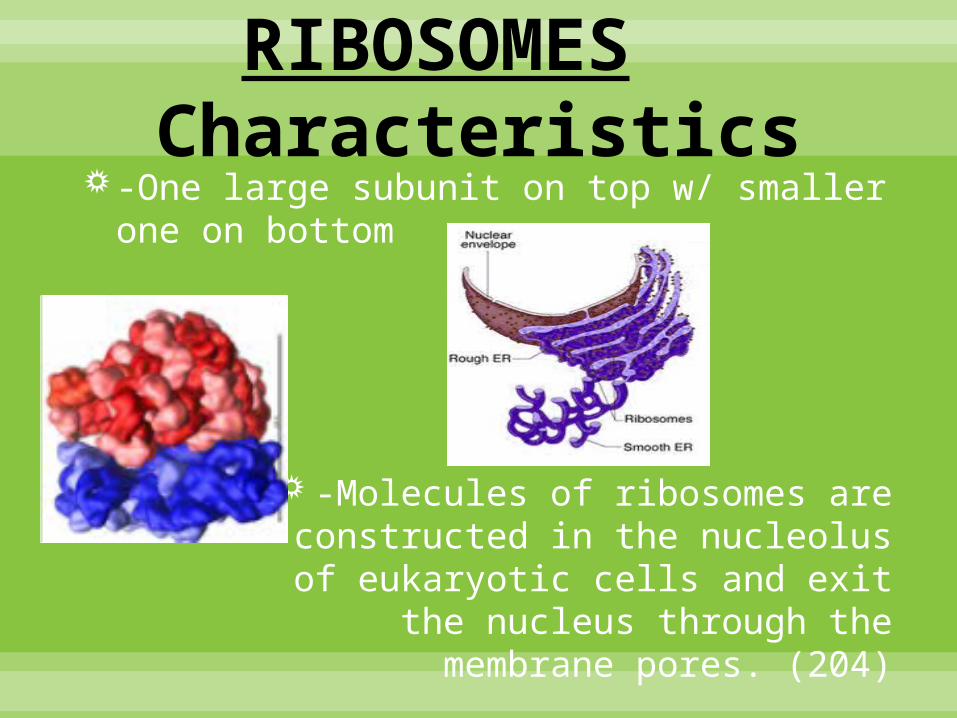
RIBOSOMES Characteristics
-One large subunit on top w/ smaller one on bottom
-Molecules of ribosomes are constructed in the nucleolus of
eukaryotic cells and exit the nucleus through the membrane
pores. (204)

RIBOSOMES Proteins Produced: Free vs. Attached
-Both structurally identical
-Free: suspended in cytosol (cytoplasm)
-Bound: attached to outside of endoplasmic reticulum or nuclear envelope
Generally make proteins that are destined for insertion into membranes, for packaging within certain organelles (such as lysosomes), or for export from the cell (secretion)
RIBOSOMES Prokaryotic vs. Eukaryotic Cell’s Ribosomes
Euk: Larger and denser than Prok. (22)
• Subunits equal 80S (22)
• Animal -Small structures, free in the cytoplasm or associated with the endoplasmic reticulum (ER)
• Plant –Small (20nm) stuctures which manufacture proteins. May be free in the cytoplasm or associated with the surface of the endoplasmic reticulum
Prok: Smaller and less dense than Euk (22)
• Subunits equal 70S
• Difference in molecular makeup (204)

RIBOSOMES 70S Ribosomes
-Prok. Subunits together equal 70S (22)
-Euk. Subunits together equal 80S
(S= Svedberg units, which indicate the relative rate of sedimentation during high-speed centrifugation) (22)


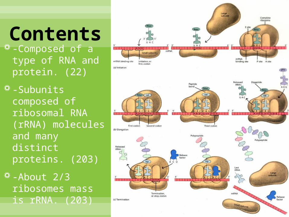
RIBOSOMES Translation & Channeling of Proteins
Translation
– The process of producing a protein once mRNA has been produced from the DNA template. (RNA protein)
The process is referred to as translation because it changes the language of DNA to the language of protein
The centre of this process is the ribosome.
Process involves several phases:
• Initiation
• Elongation
• Translocation
• Termination
co·don noun /ˈkōˌdän/ A sequence of three nucleotides which together form a unit of genetic code in a DNA or RNA molecule
*addressing codons*-codons carry the genetic code from DNA to the ribosomes via mRNA. -there are 64 possible codons.-three codons have no complementary tRNA anticodon ( referred to as stop codons)-there is a start codon (AUG) that signals the beginning of a polypeptide chain. -start codon also encodes the amino acid methionine.

RIBOSOMES Differences Between Free & rER Ribosomes
Free Ribosomes vs.
• Move freely in the cytoplasm
• Proteins produced are primarily used within the cell (206)
rER Ribosomes
• Attached to the exterior channels of rER
• Involved in protein synthesis
• Proteins produced are primarily secreted from the cell or used in lysosomes (206)
rER – rough endoplasmic reticulum

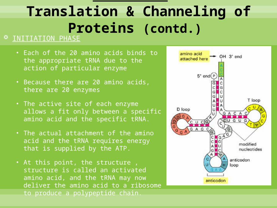
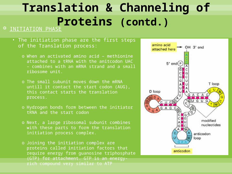
INITIATION PHASE
• The start codon (AUG) is on the 5’ end on all mRNAs.
• Each codon, other than the three stop codons, attaches to a particular tRNA. The tRNA has a 5’end and a 3’ end like all other nucleic acid strands.
• The 3’ end of tRNA is free and has the base sequence of CCA which is the site of amino acid attachment.
• Hydrogen bonds form in four areas because there are complementary bases in the
single stranded tRNA, which causes the tRNA to fold and tane-on a three dimensional structure.
• If the molecule is flattened, it has a two dimensional appearance of a clover leaf.
• One of the loops of the clover leaf contains an exposed anticodon which is unique to each type of tRNA. It’s also the anitcodon that pair with a specific codon of mRNA
RIBOSOMES Translation & Channeling of Proteins (contd.)
INITIATION PHASE
• Each of the 20 amino acids binds to the appropriate tRNA due to the action of particular enzyme
• Because there are 20 amino acids, there are 20 enzymes
• The active site of each enzyme allows a fit only between a specific amino acid and the specific tRNA.
• The actual attachment of the amino acid and the tRNA requires energy that is supplied by the ATP.
• At this point, the structure , structure is called an activated amino acid, and the tRNA may now deliver the amino acid to a ribosome to produce a polypeptide chain.
RIBOSOMES Translation & Channeling of Proteins (contd.)
INITIATION PHASE
• The initiation phase are the first steps of the Translation process:
o When an activated amino acid – methionine attached to a tRNA with the anitcodon UAC – combines with an mRNA strand and a small ribosome unit.
o The small subunit moves down the mRNA untill it contact the start codon (AUG), this contact starts the translation process.
o Hydrogen bonds form between the initiator tRNA and the start codon
o Next, a large ribosomal subunit combines with these parts to form the translation initiation process complex.
o Joining the initiation complex are proteins called initiation factors that require energy from guanosine triphosphate (GTP) for attachment. GTP is an energy-rich compound very similar to ATP
RIBOSOMES Translation & Channeling of Proteins (contd.)

ELONGATION PHASE
Once initiation is complete, elongation occurs
• Phase involves tRNAs bringing amino acids to the mRNA – ribosomal complex in the order of specified by the codons of the mRNA.
• Proteins called elongation factors assist in the binding the tRNAs to the exposed mRNA codons at the A site.
• The initiator tRNA then moves to the P site. The ribosomes catalyse the formation of peptide bonds between adjacent amino acids brought to the polypeptide assembling area.
RIBOSOMES Translation & Channeling of Proteins (contd.)

TRANSLOCATION PHASE
This phase happens during the elongation phase
• Involves the movement of the tRNAs from one site of the mRNA to another.
• First, a tRNA binds with the A site & its amino acid is then added to the growing polypeptide chain by a peptide bond.
• This causes the polypeptide chain to be attached to the tRNA at the A site.
• The tRNA then moves to the P site & transfers its polypeptide chain to the new tRNA that moves into the now exposed A site.
• Empty tRNA is transferred to the E where it’s released. This process occurs in the 5’ to 3’ direction.
• Therefore, the ribosomal complex is moving along the mRNA toward the 3’ end.
• Remember that the start codon was near the 5’ end of the mRNA.
RIBOSOMES Translation & Channeling of Proteins (contd.)

RIBOSOMES Translation & Channeling of Proteins (contd.)
TRANSLOCATION PHASE

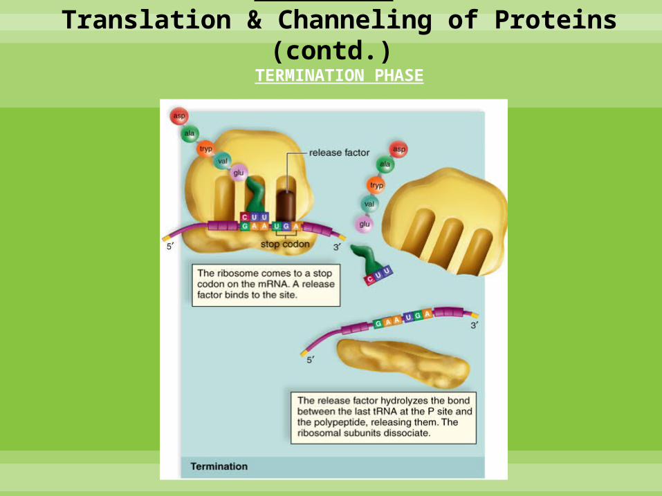
TERMINATION PHASE
Termination phase completes the translation process
• Begins when one of the three stop codons appears at the open of A site
• A protein called a release factor then fills the A site (protein factor does not carry an amino acid).
• It catalyses the hydrolysis of the bond linking the tRNA in the P site with the polypeptide chain.
• This frees the polypeptide, releasing it from the ribosome.
• Ribosome then separates frmom the mRNA & splits into its 2 subunits.
• Proteins synthesized in this manner have several different destinations.
• If they are produced by free ribosomes, proteins are primarily used within the cell.
• If produced by ribosomes bound to the ER, they’re primarily secreted from the cell or used in lysosomes.
RIBOSOMES Translation & Channeling of Proteins (contd.)
RIBOSOMES Translation & Channeling of Proteins (contd.)
TERMINATION PHASE

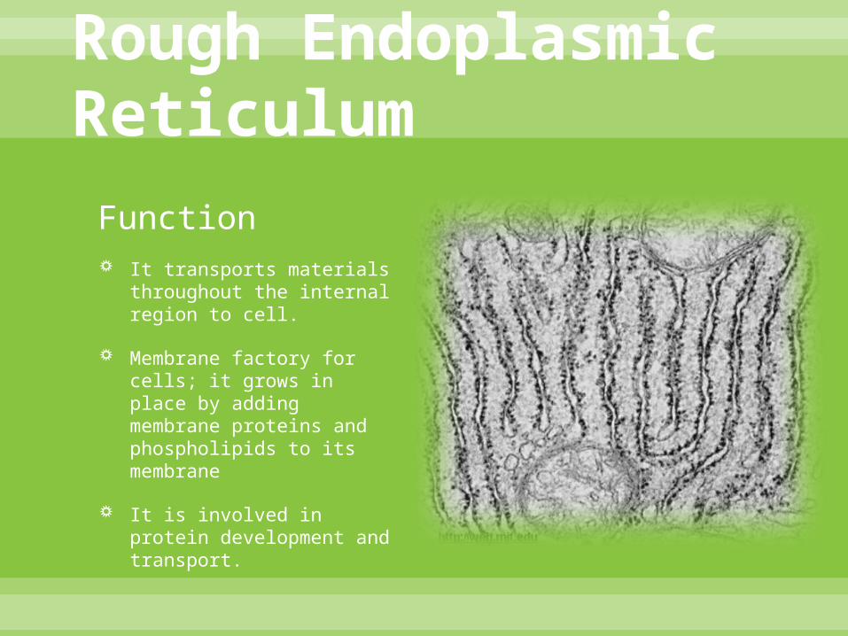
Rough Endoplasmic Reticulum
Structure A membranous system of
interconnected tubules and flattened sacs called cisternae.
Rough Endoplasmic Reticulum – studded on its outer surface with ribosomes.
Rough Endoplasmic Reticulum
Function It transports materials
throughout the internal region to cell.
Membrane factory for cells; it grows in place by adding membrane proteins and phospholipids to its membrane
It is involved in protein development and transport.

Rough Endoplasmic Reticulum
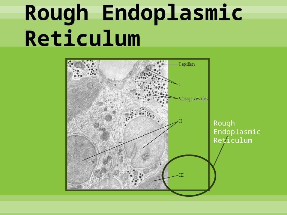
Content Most secretory proteins in
the organelle are glycoprotein (proteins that have carbohydrates covalently bonded to them). The carbohydrates are then attached to the proteins the rough endoplasmic reticulum by specialized molecules built into its membrane.
Rough Endoplasmic Reticulum
C ap illa ry
I
S to rag e v es ic le s
II
III
Rough Endoplasmic Reticulum
Cell Membrane Definition: The organelle that “functions as a selective
barrier or boundary for every cell, and regulates the sufficient passage of oxygen, nutrients, and wastes to service the entire cell.”
Also known as:
Membrane
Plasma membrane
Cell surface membrane
Organelles have membranes, too, that are similar in structure

Cell Membrane

Cell MembraneMicrograph image of part of a cell:
I = Membrane (of the nuclear envelope)
II = Mitochondrion

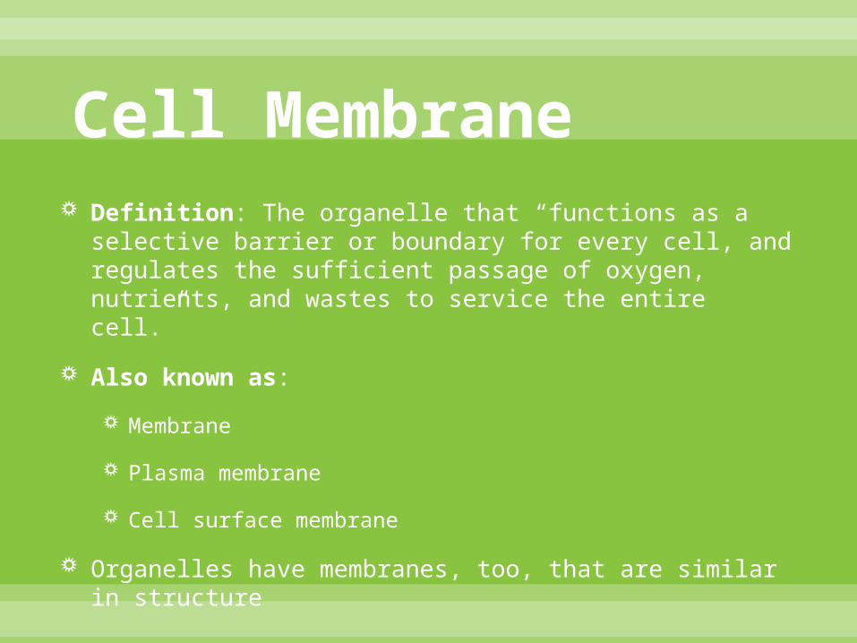
Cell MembranePhospholipid Structure
Structure Overview: There is a bilayer (double layer) in which lipids (fats) stay on the outer part, and proteins pass through the middle among the lipid tails
Phospholipids: Made up of glycerol (3 carbon compound)
2 of glycerol carbons are attached to fatty acids
3rd glycerol carbon attached to highly polar organic alcohol (related to phosphate)
Cell Membrane
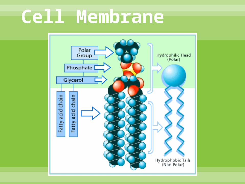
Cell MembranePhospholipid Structure
Has a hydrophilic (polar, water soluble) head
Has two hydrophobic (nonpolar, water insoluble) tails
The tails have a weak bond, causing the cell membrane to be very flexible
The heads are connected by hydrogen bonds, and they maintain the structure of the membrane
Cell Membrane

Cell Membrane

Cell MembranePhospholipid Structure
Cholesterol:
Found in animal cells ONLY
Located in the tails of phospholipids
Determines the membrane fluidity
Membranes MUST be fluid in order to function properly
Consistency is similar to olive oil
Temperature change affects fluidity
Instead of cholesterol, plant cells use unsaturated and saturated fatty acids

Cell MembraneProteins
Many different varieties – Also known as amino acids
Two most prominent types are:
Integral proteins
Peripheral proteins

Cell MembraneProteins
Integral:
Hydrophilic regions – polar amino acids (outer regions)
Hydrophobic regions – nonpolar amino acids (central regions)
Regulate the entrance and exit of molecules through the cell membrane
I
I = Integral Protein

Cell MembraneProteins
Peripheral:
Stays on the surface of cell membrane
Attached to integral proteins
Also attach to glycoproteins (another type )

Cell Membrane
I = Glycoprotein
II = Integral protein
III = Hydrophilic phosphate head

Cell Membrane

Cell MembraneProtein Functions
6 basic functions of proteins:
1) Enzymatic action: relates to metabolic reactions
2) Cell adhesion: when proteins connect (junctions)
3) Cell-to-cell communication: for identification
4) Hormone binding sites
5) Passive transport channels
6) Active transport pumps
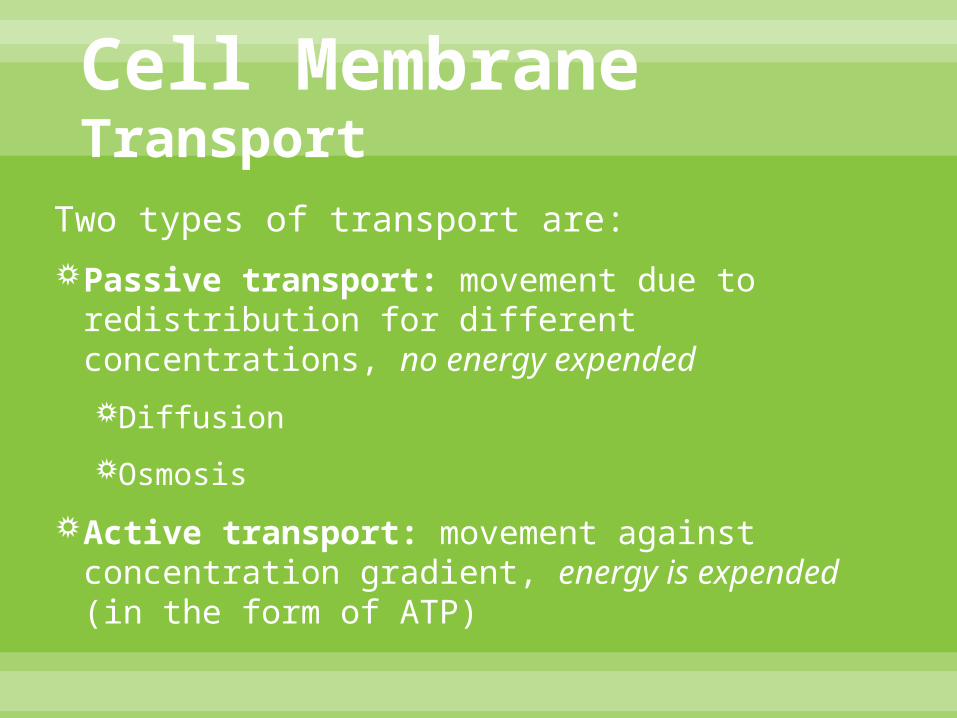
Cell MembraneTransport
Two types of transport are:
Passive transport: movement due to redistribution for different concentrations, no energy expended
Diffusion
Osmosis
Active transport: movement against concentration gradient, energy is expended (in the form of ATP)
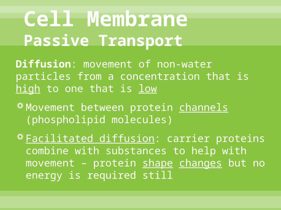
Cell MembranePassive Transport
Diffusion: movement of non-water particles from a concentration that is high to one that is low
Movement between protein channels (phospholipid molecules)
Facilitated diffusion: carrier proteins combine with substances to help with movement – protein shape changes but no energy is required still
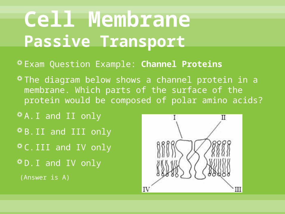
Cell MembranePassive Transport
Exam Question Example: Channel Proteins
The diagram below shows a channel protein in a membrane. Which parts of the surface of the protein would be composed of polar amino acids?
A. I and II only
B. II and III only
C. III and IV only
D. I and IV only
(Answer is A)

Cell MembranePassive Transport

Cell MembranePassive Transport
Osmosis:
Happens along the concentration gradient
Involves ONLY water molecules
Happens with partially permeable membranes
Movement caused by concentration difference inside and outside the cell membrane
Goal of passive transport is to reach equilibrium on both sides
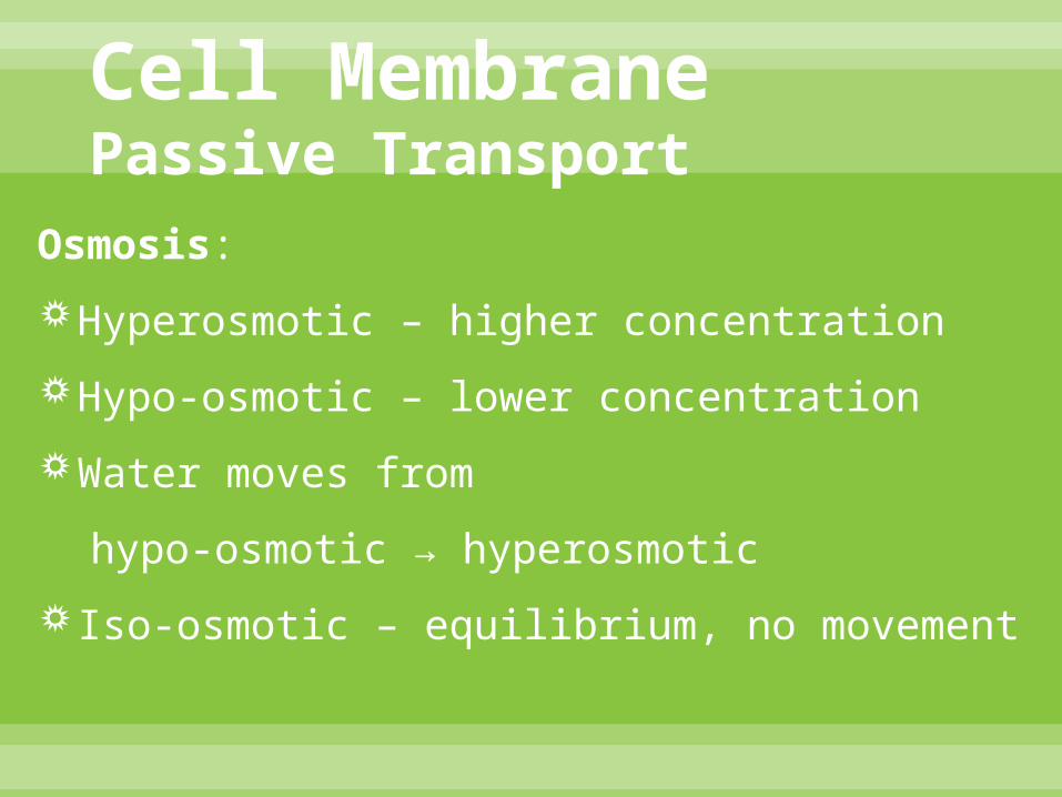
Cell MembranePassive Transport
Osmosis:
Hyperosmotic – higher concentration
Hypo-osmotic – lower concentration
Water moves from
hypo-osmotic → hyperosmotic
Iso-osmotic – equilibrium, no movement

Cell MembranePassive Transport
Osmosis: influencing factors of substances that are being transported are…
Size – small = easy movement
large = difficult movement
Charge – nonpolar = easy movement
polar = difficult movement

Cell MembranePassive Transport
(Partially permeable)
Trying to achieve equilibrium (iso-osmotic)
Uses aquaporins (proteins with specialized water channels)

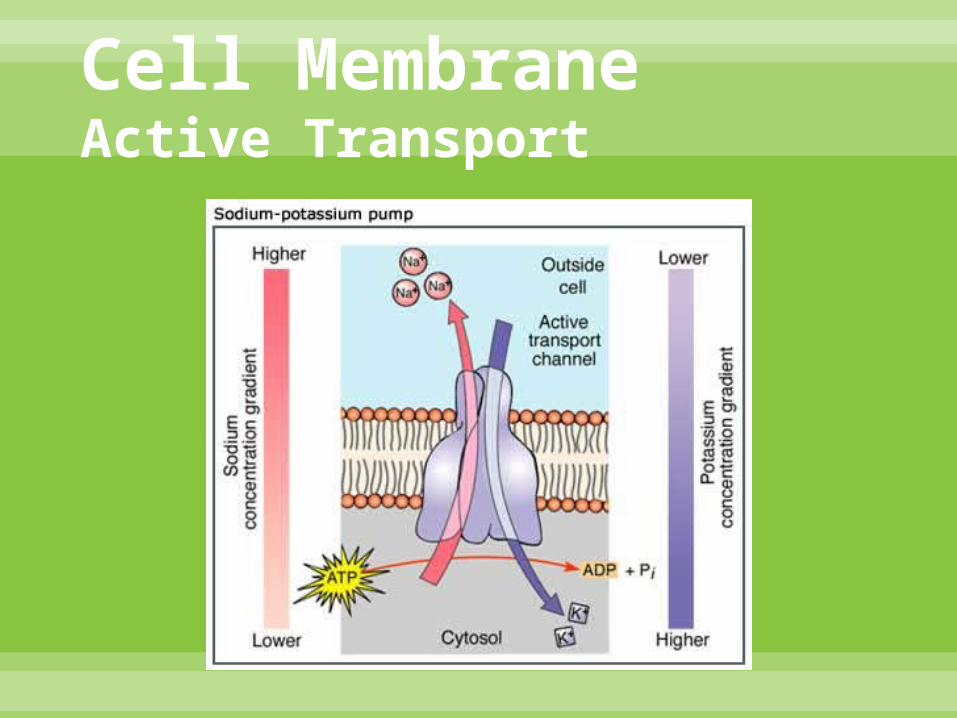
Cell MembraneActive Transport
Does require energy in the form of ATP
Moves against a concentration gradient that needs energy
Process helps cell maintain equal concentrations inside and outside the membrane

Cell MembraneActive Transport
Sodium-potassium pump: (way for moving these ions)
1. Specific protein in cell membrane binds to three sodium ions from inside the cell
2. Binding of sodium ions causes phosphorylation (the activation of the protein enzymes through the addition of PO4
-3 from the phosphate molecule from the AT
3. Phosphorylation causes protein to change shape and expel sodium ions that were inside the protein to outside the cell
4. 2 potassium ions enter the protein from outside the cell which causes the phosphate to be released
5. When the phosphate is released, protein restores to its original shape and the potassium ions are released into cell
Cell MembraneActive Transport

Cell MembraneEndocytosis & Exocytosis
Endocytosis: process that allows macromolecules to enter the cell
Exocytosis: process that allows macromolecules to leave the cell
In endocytosis, portion of membrane is pinched to surround a particle, and a vesicle forms leading to the cytoplasm
Vesicle allows protein to eventually reach nucleus

Cell Organelles - Cell Wall
Class Presentation on Topic 2
4A– IB Biology HL
By: Robert Jennings

Cell Wall
The cell wall is a semi-rigid structure composed mainly of cellulose, and its purpose is to give the cell structure and protection.

Structure/Composistion-Cell Wall Consist of three layers.
Middle lamella
1. Formed during cell division
2. Makes up outer wall of cell
3. Composed of proteins and pectic compounds.
Primary wall
1. Formed after middle lamella.
2. Consist of rigid skeleton made of cellulose microfibrils embedded in a gel-like matrix composed of pectic compounds, hemicellulose, and glycoproteins.
Secondary wall
1. After cell enlargement is completed
2. Very rigid and provides incredible strength.
3. Layered and made of cellulose, lignin, and hemicellulose.

Function-Cell Wall
Determines the shape of the particular cell.
Provides strength to the cell but is still somewhat flexible.
Controls the turgor pressure in a cell. Turgor is the pressure applied to the cell from the constituents inside it.
Since the cell wall is permeable it allows for proteins and other small molecules to come in and out of the cell.
Protects cells from pathogens and microorganisms.
Reserves carbohydrates that can be used in times of need.
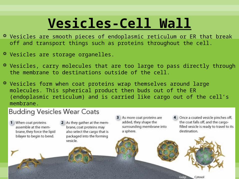
Vesicles-Cell Wall Vesicles are smooth pieces of endoplasmic reticulum or ER that break
off and transport things such as proteins throughout the cell.
Vesicles are storage organelles.
Vesicles, carry molecules that are too large to pass directly through the membrane to destinations outside of the cell.
Vesicles form when coat proteins wrap themselves around large molecules. This spherical product then buds out of the ER (endoplasmic reticulum) and is carried like cargo out of the cell’s membrane.

Miscellaneous-Cell Wall
Prokaryotic cells have a cell wall as well as plant cells.

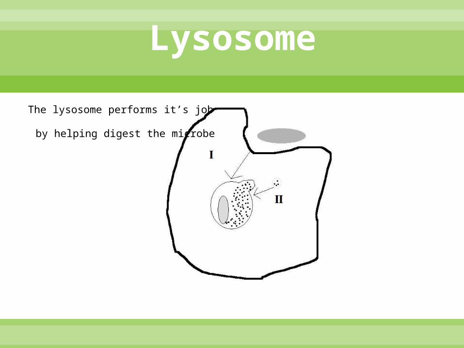
Lysosome:Structure
•Composed of:
• Membrane
• Proteins and Enzymes
If the lysosome stops or explodes, the cell will break down

Lysosome:Function
Scientifically, lysosomes are organelles that contain digestive enzymes. So, in other words, the lysosomes are essentially the stomach of the cell.
Intracellular digestive centers that derive from the Golgi apparatus
The enzymes that lysosomes have can breaks down the substances within the cell
Lysosome
The lysosome performs it’s job
by helping digest the microbe

Function Collects, packages, modifies, and distributes
materials synthesized in the cell
Is the post office of the cell
Tells products where to go in or out of the cell.

Contents Main body is cisternae. Which are flat and stacked
on top of each other. Products travel through cisternae.
Two sides. Cis side is close to the rough ER. Trans side.
Vesicles go in one side and come out the other.
Modifies Vesicles by adding enzymes or removing sugar and adding it own.

Class Presentation on Topic 2
4A– IB Biology HL
Nucleus
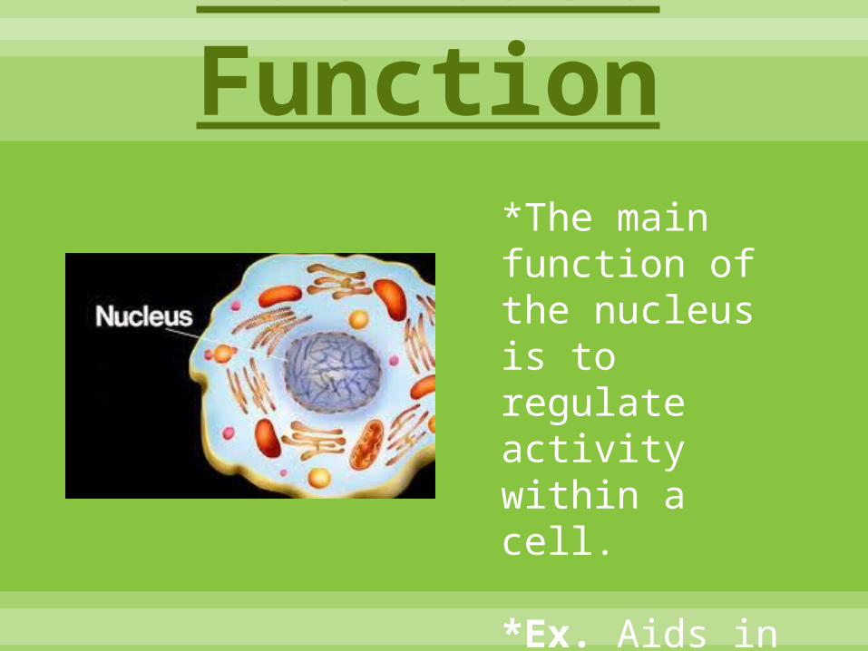
Nucleus: Function*The main function of the nucleus is to regulate activity within a cell.
*Ex. Aids in reproduction
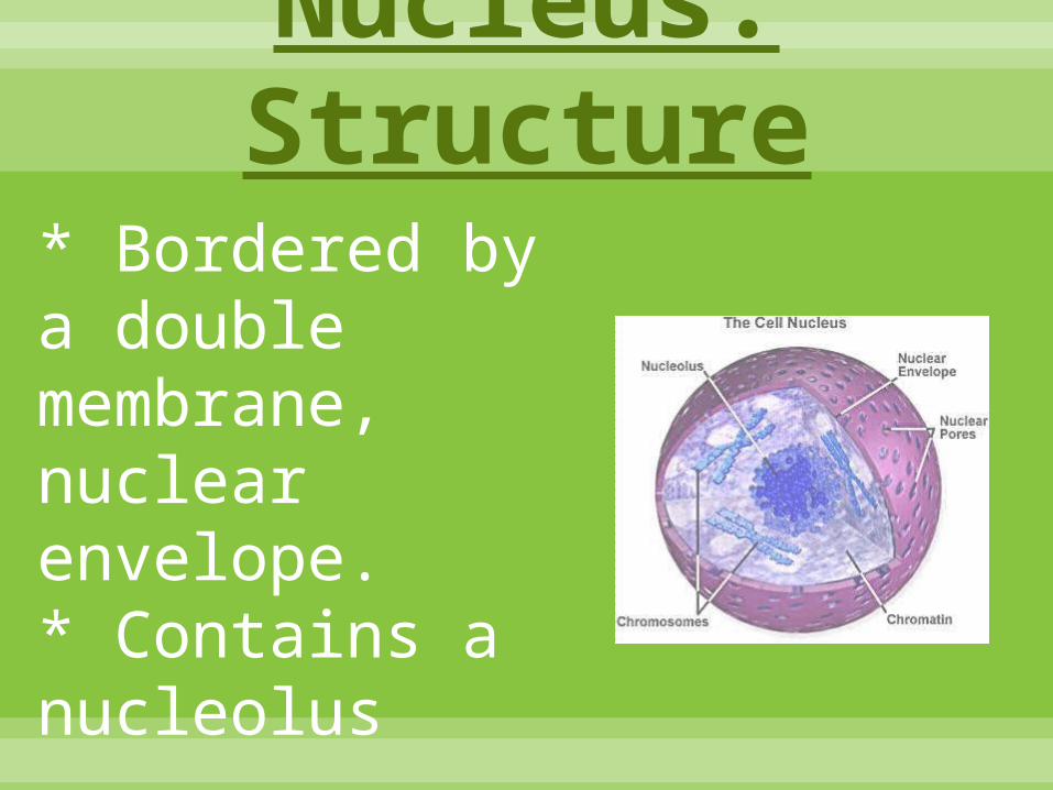
Nucleus: Structure* Bordered by a double membrane, nuclear envelope.* Contains a nucleolus

Nucleus: Contents
* Contains most of
the cell’s DNA *

Nucleus: Prokaryotic vs. Eukaryotic:
*Prokaryotic cells do NOT have a nucleus.* Eukaryotic cells DO have a nucleus.
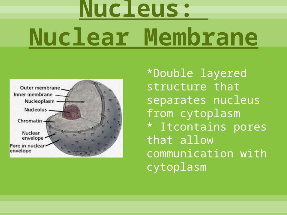
Nucleus: Nuclear Membrane
*Double layered structure that separates nucleus from cytoplasm* Itcontains pores that allow communication with cytoplasm

Nucleus: Locations
* A nucleus is located in the center of a cell.

Nucleus: Micrograph
* A Micrograph is a photo taken by a microscope!

Nucleus: DNA and RNA
* DNA carries the genetics of a cell and consists of thousands of genes* The RNA is processed so that non-coding parts are removed. After this, the RNA is removed from the nucleus.

Nucleus: Haploid nucleus + Diploid nuclei
In cell division:*Haploid nuclei contain only half of the number of chromosomes (23 chromosomes)* Diploid nuclei contain both pairs of chromosomes (which is 46 chromosomes)

Mitochondrion
By
Sarah Liles, Natashia Gavarrete, and Matthew Juve
Period 4A
IB Biology Year 1 HL

Mitochondrion Structure Within (Contents):
Rod-shaped organelles that appear throughout the cytoplasm.
Have their own DNA.
Circular chromosome similar to that in bacteria cells, allowing for autonomy within the cell.
Has a smooth outer membrane. The inner membrane is a semi-fluid substance called a matrix, and lies between the two membranes.
Cristae provides a huge internal surface area for the chemical reactions of the mitochondria to occur.
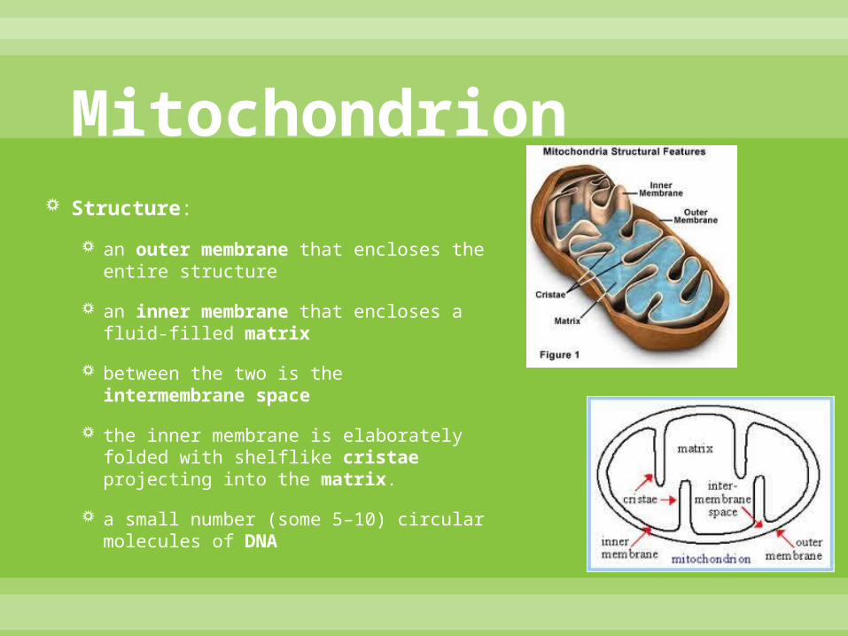
Mitochondrion Structure:
an outer membrane that encloses the entire structure
an inner membrane that encloses a fluid-filled matrix
between the two is the intermembrane space
the inner membrane is elaborately folded with shelflike cristae projecting into the matrix.
a small number (some 5–10) circular molecules of DNA

Mitochondrion Uses in prokaryotic cells:
They are not present in prokaryotic cells.
Uses in eukaryotic cells:
the energy producing structures of Eukaryotic cells and supply the cells with ATP.
Structures which produce the cell’s energy.
Powerhouses of the cell.
Prokaryotic Cell
Eukaryotic Cell

Mitochondrion Functions:
Main function: production of energy in the form of adenosine triphosphate (ATP)
Performs the process of programmed cell death. It occurs during development as the organism is pruning away unwanted, excess cells.
involved in building, breaking down, and recycling products needed for proper cell functioning.

Mitochondrion Mitochondria’s role in cellular respiration:
known as the powerhouses of the cell.
They are organelles that act as a digestive system that takes in nutrients and breaks them down.
This process creates energy for the cell.
By producing this nutrients and power it contributes to creating cell energy other wise known as cellular respiration.
The Mitochondrion's perfect shape allows for a good place for cellular respiration to occur, and maximize the effort given.

Mitochondrion Acetyl-CoA: produced in mitochondria through the oxidation of
fats.
Pyruvate is converted to Acetyl Coenzyme (CoA) via active transport when entering the mitochondria.
The entry compound for the citric acid cycle in cellular respiration, formed from a fragment of pyruvate attached to coenzyme while transporting through the organelle.

Mitochondrion Carries out key reactions in the cells of eukaryotes.
ATP Synthetase occurs in these organelles.
It is the location of Chemiosmosis.
The diffusion of ions across a selectively-permeable membrane. More specifically, it relates to the generation of ATP by the movement of hydrogen ions across a membrane during cellular respiration
Electron Micrograph
Mitochondrion appear as bacteria sized ellipses.
Vary in size and width, but this is hard to see on a two dimensional electron micrograph.

Mitochondrion Krebs cycle- Cycle that
accounts for the oxidation of carbohydrates in prokaryotes and eukaryotes.
Mitochondria produce the ATP in the Krebs cycle in eukaryotes.
There are no mitochondria in bacteria.

Mitochondrion Mitochondria vs. chloroplasts
Both are circular in shape.
Both are involved in ATP production.
Chloroplasts are slightly larger.
Mitochondrion generate ATP with glucose during cell respiration.
Chloroplasts generate ATP through sunlight.
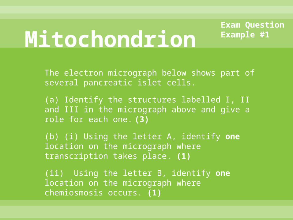
MitochondrionThe electron micrograph below shows part of several pancreatic islet cells.
(a) Identify the structures labelled I, II and III in the micrograph above and give a role for each one. (3)
(b) (i) Using the letter A, identify one location on the micrograph where transcription takes place. (1)
(ii) Using the letter B, identify one location on the micrograph where chemiosmosis occurs. (1)
Exam Question Example #1

MitochondrionCapillary
Mitochondria
Storage vesicles
Nucleus
(Rough) Endoplasmic Reticulum
Structure Role
mitochondriaproduce ATP / site of (aerobic) respiration;
nucleus
contains genetic information / produces RNA / site of replication;
(rough) endoplasmic reticulum
(site of) translation / protein production / proteintransport;
*Transcription takes place** Chemiosmosis
Exam Question Example #1Markscheme

MitochondrionThe electron micrographs below show mitochondria in longitudinal section. The mitochondrion in A is from a bat pancreas cell and that in B is from a mouse liver cell. (Next Slide)
(a) Annotate the micrographs to show two similarities in the structure of the mitochondria. (2)
(b) The mitochondria differ in size. State two other differences that are visible in the mitochondria. (2)
(c) Predict, with two reasons, which of the mitochondria would have been able to produce ATP at a greater rate.
Exam Question Example #2

Mitochondrion
.
Cristae
Matrix/Ribosomes
Differences:ShapeArrangement of cristae Density of cristaeAmount of matrix granules/any reference to dark dots *ribosomes not accepted
Similarities
Which mitochondria would have been able to produce ATP at a greater rate & why:Bat’sLarger size/volumeGreater surface area of cristae/more cristaeCloseness of mitochondria in mouse reduces rate
BATMOUSE
Exam Question Example #2Markscheme

Mitochondrion(a) Distinguish between the terms resolution
and magnification when applied to electron microscopy. (2)
The electron micrograph below shows part of a cell. (Next Slide)
(b) Identify the structures labelled I and II. (2)
(c)State one function of the structure labelled II. (1)
(d) Deduce, with a reason, whether this cell is eukaryotic or prokaryotic. (1)
(Total 6 marks)
Exam Question Example #7

Mitochondrion
Eukaryotic: internal membranes / membrane bound organelles / presence of mitochondria / double nuclear membrane;
I: membrane / (nuclear) envelopeII: mitochondrion / mitochondria
Function of mitochondrion (II):aerobic respiration;correct specific reaction / pathway occurring in mitochondria / ATP production; Do not accept “energy production” alone.
resolution: separate points / focus clearly / greater detail / clarity;magnification: size of image / view / picture
Exam Question Example #7Markscheme
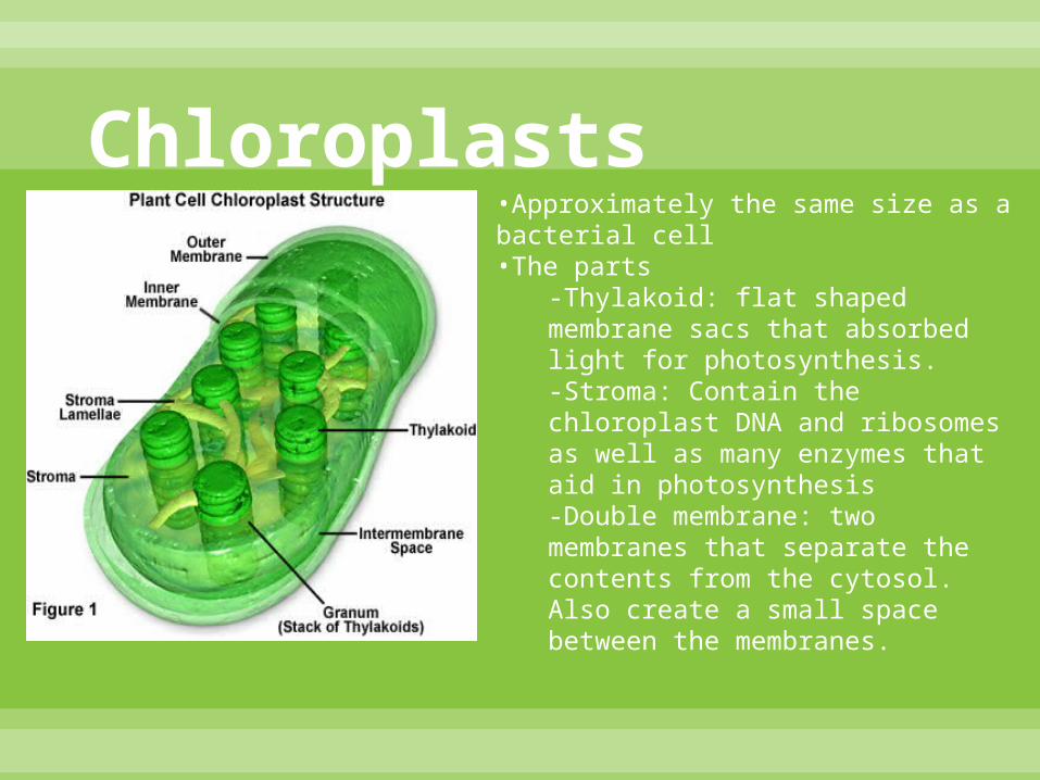
Chloroplasts•Approximately the same size as a bacterial cell•The parts
-Thylakoid: flat shaped membrane sacs that absorbed light for photosynthesis.-Stroma: Contain the chloroplast DNA and ribosomes as well as many enzymes that aid in photosynthesis-Double membrane: two membranes that separate the contents from the cytosol. Also create a small space between the membranes.

Chloroplasts•Contains enzymes in the Stroma which helps complete photosynthesis. - RuBP carboxylase enzyme- catalyzes the first step of the Calvin Cycle.•Main function is photosynthesis•The Calvin Cycle- In the Calvin Cycle, the enzymes of the chloroplast are in the stroma.•Tylakoids of chloroplasts get positive protons using energy obtained from light sources, thus the small volume size is advantageous to concentrate protons more rapidly.•Plastids•Have three distinct parts: the intermembrane space, the stroma, and the thylakoid space.

ChloroplastsChloroplasts Both Mitochondria
Contain stroma
Used for photosynthesisOnly in plant
cells
Contain matrixes
ATP energyCellular
respiration
-Same size-Double membranes- 70S ribosomes
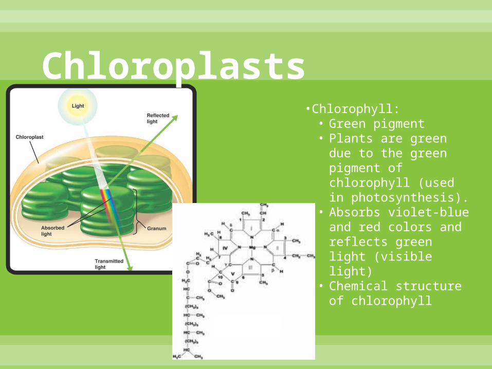
Chloroplasts•Chlorophyll:• Green pigment• Plants are green due to
the green pigment of chlorophyll (used in photosynthesis).
• Absorbs violet-blue and red colors and reflects green light (visible light)
• Chemical structure of chlorophyll

Works Cited
http://learn.genetics.utah.edu/content/begin/cells/vesicles/
http://biology.clc.uc.edu/courses/bio104/cells.htm
AP Edition Biology Textbook.
Biology HL Textbook.


