Cleavage follows fertilization. Functions of cleavage: Multicellular for differentiation The zygote is partitioned into blastomeres. Each blastomere contains different regions of the undivided cytoplasm and thus different cytoplasmic determinants. Restores Somatic Nuclear to Cytoplasmic Ratio 1:500 -> ->-> 1:6 sea urchin fertilized egg at end of cleavage - somatic cell Controls on # of cleavage divisions? 2n = 6 divisions 4n = ?
Transcript
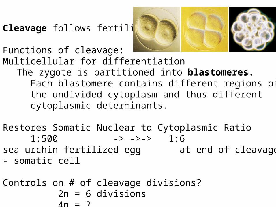
Cleavage follows fertilization.
Functions of cleavage:Multicellular for differentiation
The zygote is partitioned into blastomeres.Each blastomere contains different regions of the undivided cytoplasm and thus different cytoplasmic determinants.
Restores Somatic Nuclear to Cytoplasmic Ratio1:500 -> ->-> 1:6
sea urchin fertilized egg at end of cleavage - somatic cell
Controls on # of cleavage divisions?2n = 6 divisions4n = ?n = ?
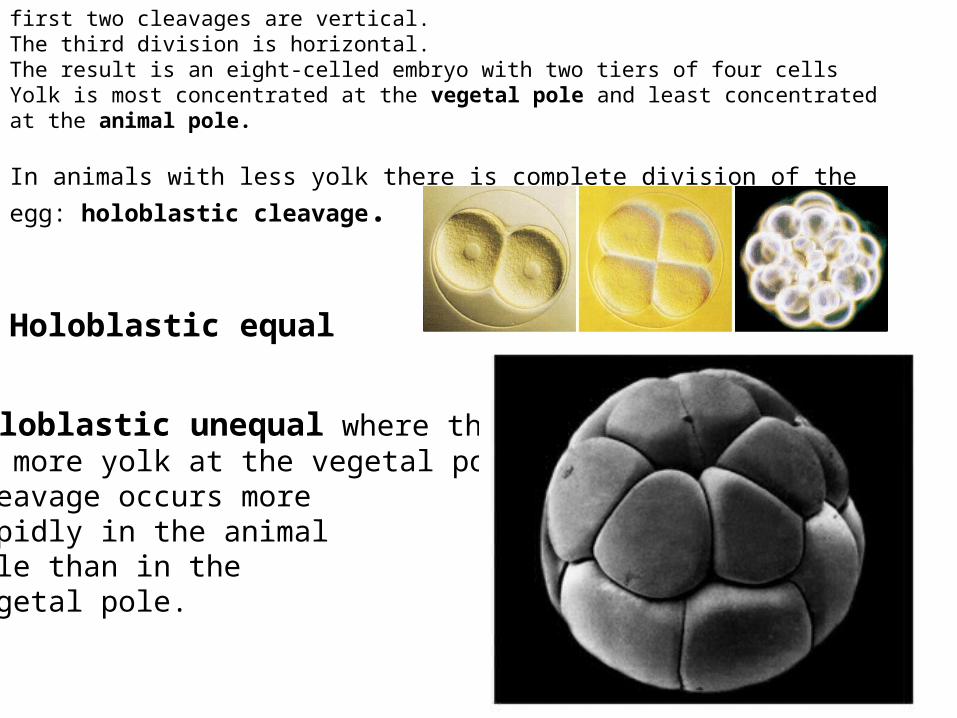
Holoblastic unequal where thereis more yolk at the vegetal pole.Cleavage occurs morerapidly in the animalpole than in thevegetal pole.
first two cleavages are vertical.The third division is horizontal.The result is an eight-celled embryo with two tiers of four cells Yolk is most concentrated at the vegetal pole and least concentrated at the animal pole.
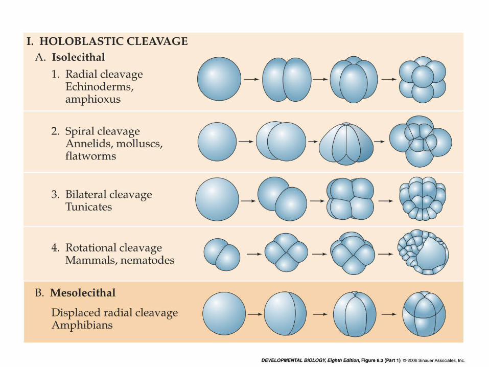
In animals with less yolk there is complete division of the egg: holoblastic cleavage.
Holoblastic equal
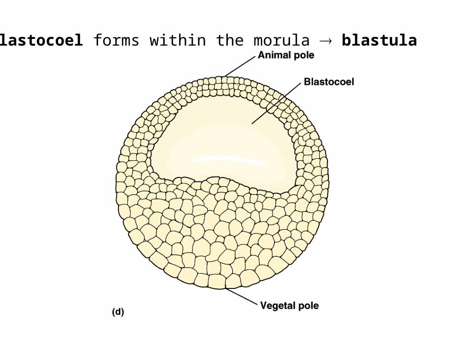
A blastocoel forms within the morula blastula
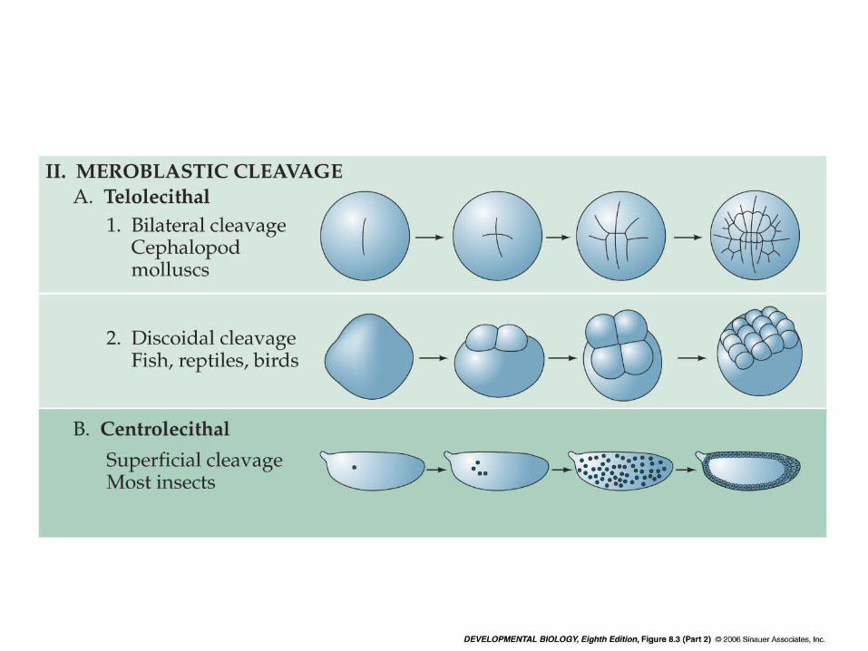
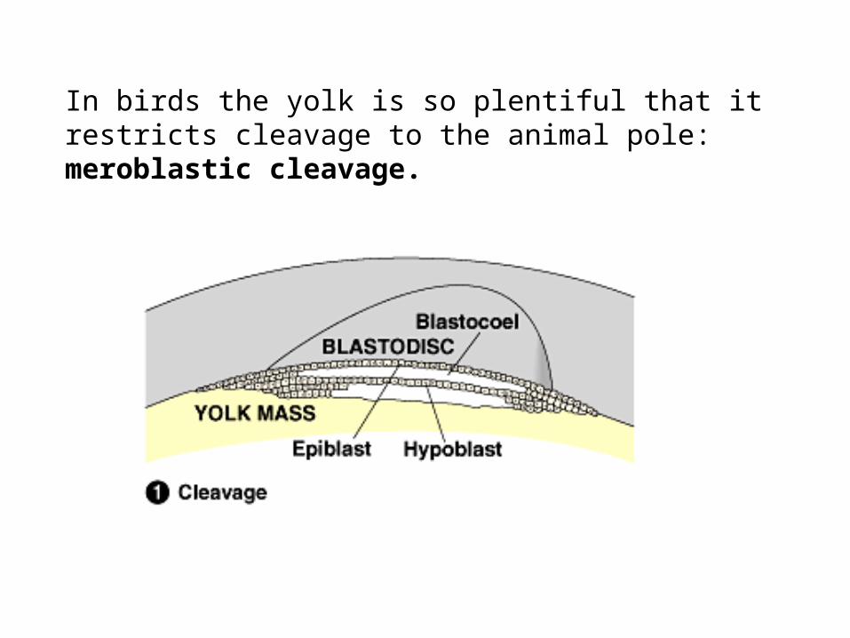
In birds the yolk is so plentiful that it restricts cleavage to the animal pole: meroblastic cleavage.
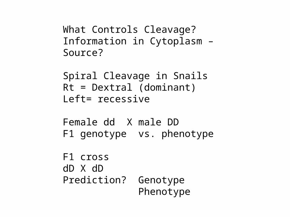
What Controls Cleavage?Information in Cytoplasm – Source?
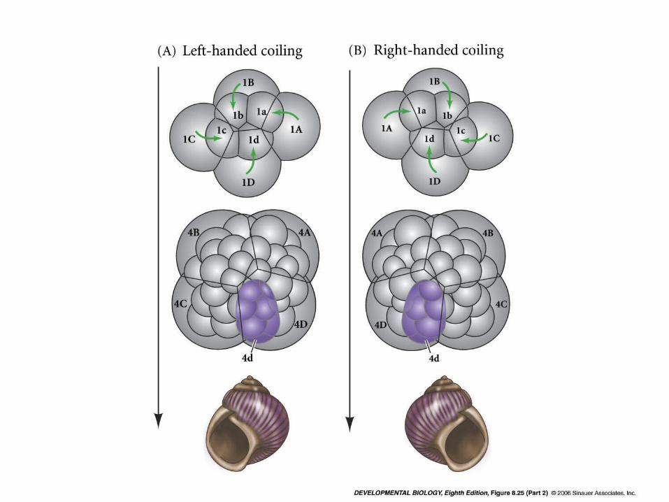
Spiral Cleavage in SnailsRt = Dextral (dominant)Left= recessive
Female dd X male DDF1 genotype vs. phenotype
F1 crossdD X dDPrediction? Genotype
Phenotype
Gastrulation rearranges the embryo into a triploblastic gastrula.– The embryonic germ layers are the ectoderm,
mesoderm, and endoderm.
Gastrulation rearranges the blastula to form a three-layered
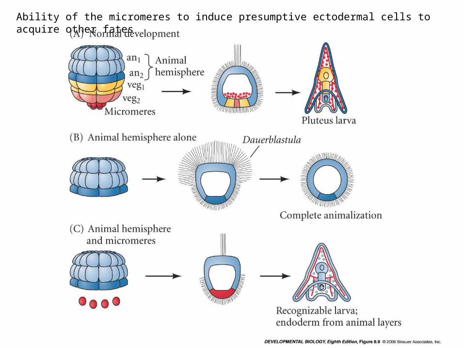
Ability of the micromeres to induce presumptive ectodermal cells to acquire other fates
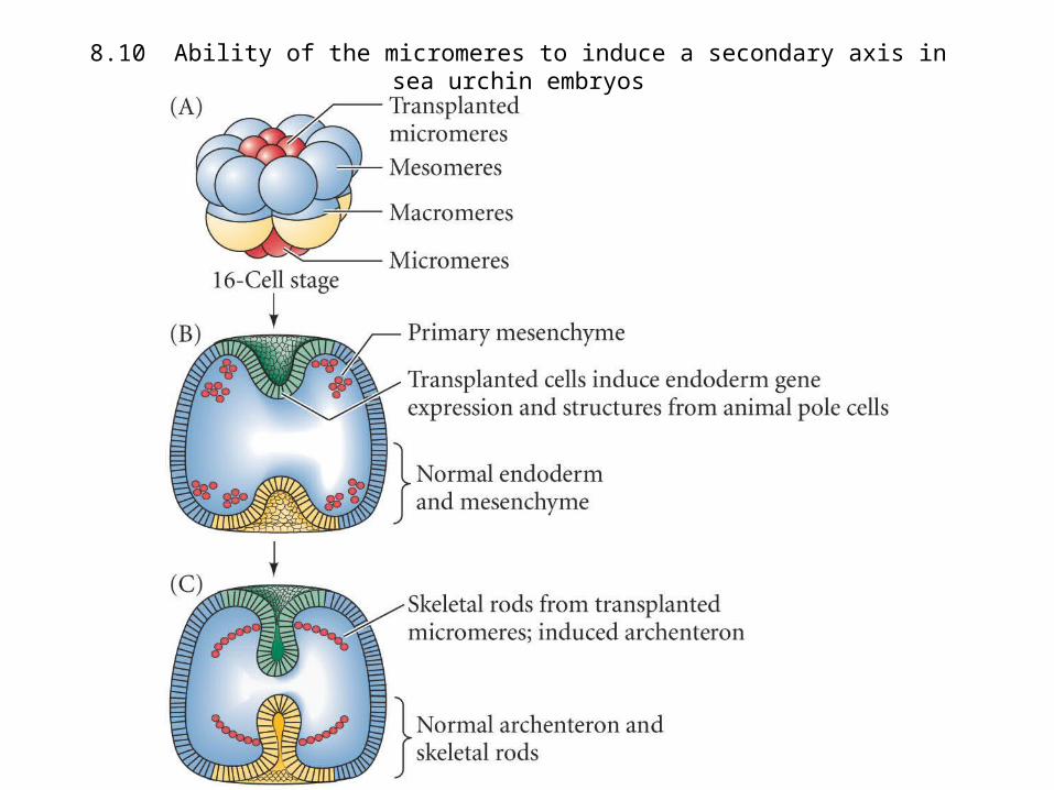
8.10 Ability of the micromeres to induce a secondary axis in sea urchin embryos
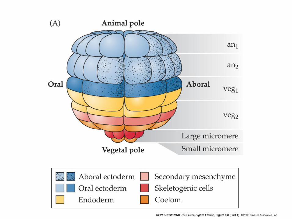
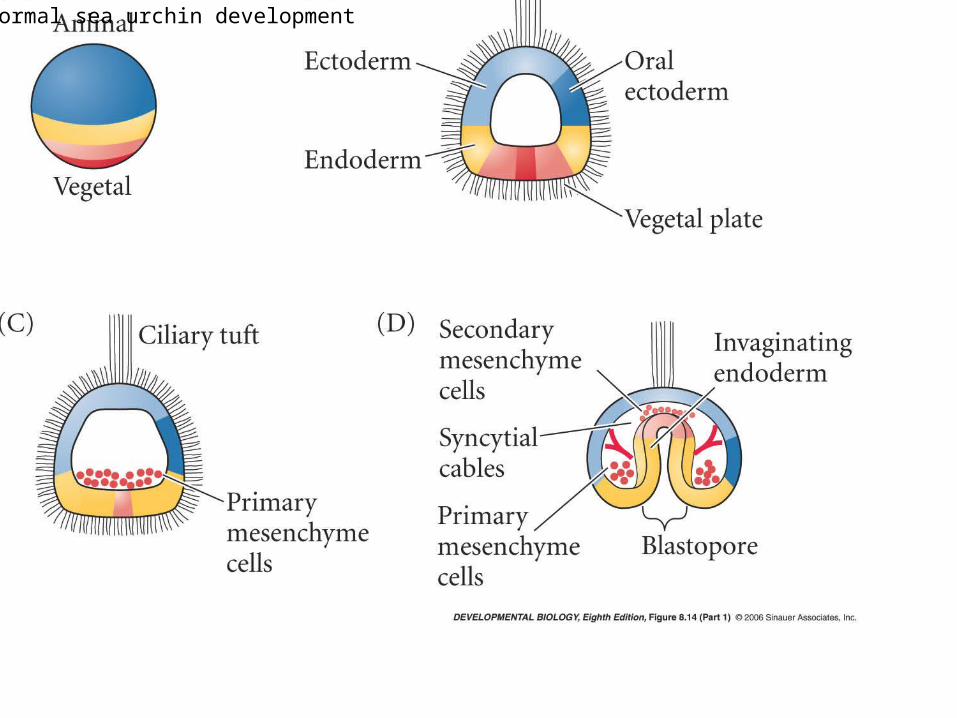
Normal sea urchin development
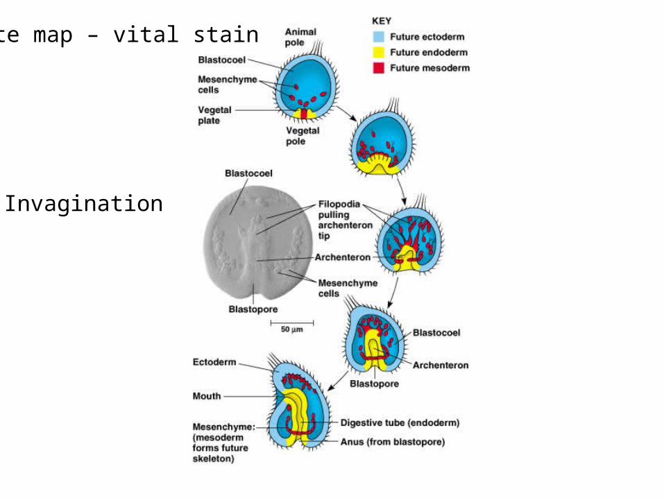
Fate map – vital stain
Invagination
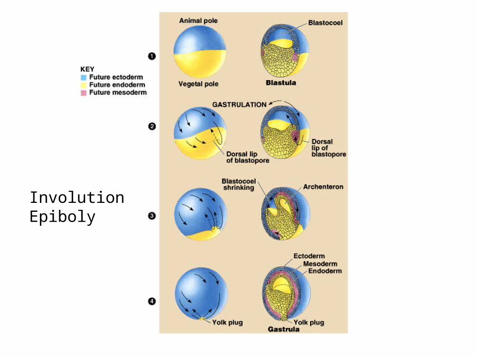
InvolutionEpiboly
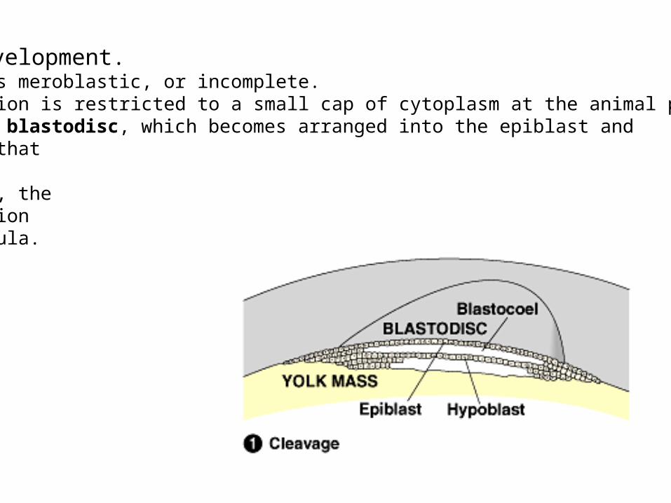
Avian Development.Cleavage is meroblastic, or incomplete.Cell division is restricted to a small cap of cytoplasm at the animal pole.Produces a blastodisc, which becomes arranged into the epiblast andhypoblast thatbound theblastocoel, theavian versionof a blastula.
During gastrulation some cells of the epiblast migrate (arrows) towards the interior of the embryo through the primitive streak. The primitive knot is where the future notochoral mesoderm forms.
Ingression(immigration)
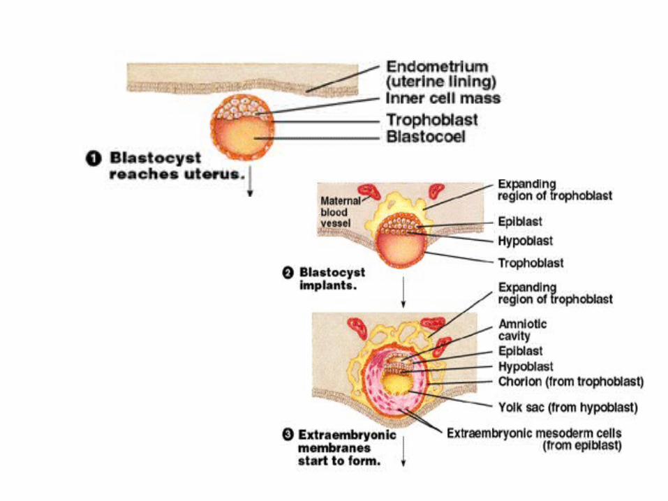
Once again, the embryonic membranes – homologous with those of shelled eggs.
Chorion: completely surrounds the embryo and other embryonic membranes.Amnion: encloses the embryo in a fluid-filled amniotic cavity.Yolk sac: found below the developing embryo.
Develops from the hypoblast.Site of early formation of blood cells which later migrate to the embryo.
Allantois: develops as an outpocketing of the embryo’s rudimentary gut.Incorporated into the umbilical cord, where it forms blood vessels.
Activation of embryonic genome
Mexican axolotl o-mutant strain The “o” gene is a recessive gene “O” gene is the normal, dominant gene
In embryos obtained from female axolotls homozygous for gene “o”, development is always arrested during gastrulation.
WHY? “O” protein is necessary to activate the embryonic genome.