EDUCATIONAL OBJECTIVE:Readers will take steps to minimize the risk of venous thromboembolism in their patients who are contemplating long-distance air travel Air travel and v enous thromboembolism: Minimizing the risk ■ ABSTRACT For those traveling on long flights, the risk of deep vein thrombosis or pulmonary embolism, generally referred to as venous thromboembolism (VTE), is real and dangerous if left unrecognized or untreated. T he goal of this publica- tion is to provide an overview of how best to prevent VTE during travel, and how to diagnose and t reat it. ■ KEY POINTS The risk of VTE is about three times higher in passengers on long-distance flights than in the general population, although the absolute risk is still low. All long-distance air passengers should perform stretch- ing exercises once an hour while in flight to prevent VTE. They should also stay hydrated. For patients at higher risk due to hypercoagulable condi- tions, physician s can consider prescribing compression stockings or an anticoagulant drug (a low-molecular- weight heparin or a factor Xa inhibitor) to be taken before the flight, or both. The evaluation of a patient with suspected VTE should include an estimation of the pretest probability of dis- ease. If symptoms dictate, dup lex ultrasonography of the upper or lower extremity to detect deep vein thrombosis or spiral computed tomography , ventilation-perfusion lung scan, or pulmonary angiography (where available) to diagnose an acute pulmonary embolism should be ordered. 111 Editor’s Note: The views expressed in this article are solely those of the authors and do not re flect the of ficial policy or position of the Department of State or the United States Government. This ver- sion of the article was peer-reviewed. V (VTE) as- sociated with travel has emerged as an important public health concern over the past decade. Numerous epidemiologic and case control studies have reported air travel as a risk factor for the development of VTE and have attempted to determine who is at risk and which precautions need to be taken to prevent this potentially fatal event. 1–7 Of- ten referred to as “traveler’s thrombosis” or “flight-related deep vein thrombosis,” VTE can also develop after long trips by automo- bile, bus, or train. 8,9 Although the absolute risk is very low , this threat appears to be about three times higher in travelers and increases with longer trips. 3 See related patient information material, page 121 This article focuses on defining VTE and recognizing its clinical features, as well as pro- viding recommendations and guidelines to prevent, diagnose, and treat this complication in people who travel. ■ WHAT IS VENOUS THROMBOEMBOLISM? Deep vein thrombosis and pulmonary embo- lism represent different manifestations of the same clinical entity, ie, VTE. VTE is a com- mon, lethal disease that affects hospitalized and nonhospitalized patients, frequently re- REVIEW doi:10.3949/ccjm.78a.10138 CREDIT CME JOHN R. BARTHOLOMEW, MD Professor of Medicine, Cleveland Clinic Lerner College of Medicine of Case Western Reserve University, Cleveland, OH; Head, Section of Vascular Medi- cine, Departments of Cardiovascular Medicine and Hematology/Oncology , Heart and Vascular Institute, Cleveland Clinic JONATHAN L. SCHAFFER, MD, MBA Managing Director, eClevelandClinic, Information Techno logy Division, Cleveland Clini c GEORGES F. McCORMICK, MD Office of Medical Services, US Department of State, Washington, DC on August 27, 2013. For personal use only. All other uses require permission. www.ccjm.org Downloaded from
curs, is often overlooked, may be asymptomat-ic, and may result in long-term complicationsthat include pulmonary hypertension and thepostthrombotic syndrome.
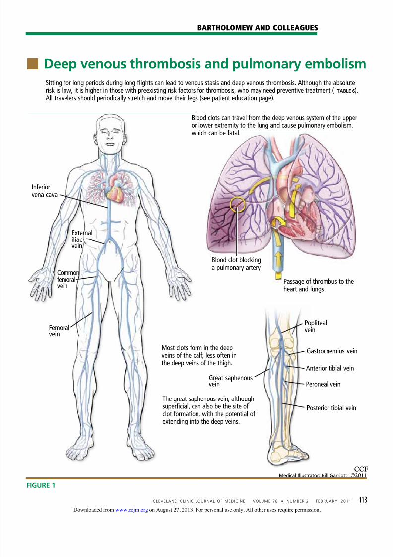
The leg veins are the most common siteof deep vein thrombosis, accounting for nearly90% of all cases; other locations include thearm and pelvic veins (FIGURE 1). Deep veinthrombosis in a proximal lower extremity (ie,involving the popliteal, femoral, common fem-oral, or external iliac vein) has an estimated50% risk of migrating and leading to an acutepulmonary embolism if not treated, while ap-proximately 25% of deep vein thromboses inthe calf veins will, if not treated, propagate toinvolve the aforementioned veins.
Deep vein thrombosis of the upper ex-tremities is generally related to an indwellingvenous catheter or a central line being usedfor long-term administration of antibiot-ics, chemotherapy, or nutrition. A conditionknown as Paget-Schroetter syndrome or “ef-fort thrombosis” may be seen in younger orathletic people who have a history of strenu-ous or unusual arm exercise.
■ RISK FACTORS FOR VTE
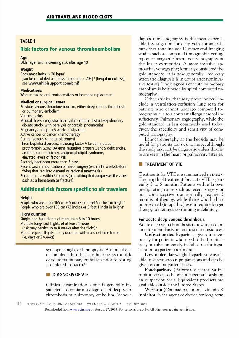
Most patients who develop VTE have one ormore risk factors for it (TABLE 1), the presence ofwhich is often referred to as a hypercoagulablestate or thrombophilia. These risk factors aregenerally classified as either genetic (inher-ited) or acquired (environmental). Most VTEevents are in fact associated with a combina-tion of genetic and acquired risk factors. Common inherited risk factors include: Factor V Leiden mutation Prothrombin gene mutation G20210A
Hyperhomocysteinemia Deficiency of the natural anticoagulantproteins C, S, or antithrombin
Elevated levels of factor VIII (may be in-herited or acquired).
Acquired risk factors include: Older age Immobilization or stasis (such as sitting for
long periods of time while traveling) Surgery (most notably orthopedic proce-
dures including hip and knee replacementand repair of a hip fracture)
The antiphospholipid syndrome (consist-ing of a lupus anticoagulant, anticardio-lipin antibodies, or both)
Pregnancy and the postpartum state Use of oral contraceptives or hormone re-
placement therapy Cancer (including the myeloproliferative
disorders) and certain chemotherapeuticagents
Obesity (a body mass index > 30 kg/m2, seewww.nhlbisupport.com/bmi/)
Inflammatory bowel disease Previous VTE A central venous catheter or pacemaker Nephrotic syndrome. In addition, emerging risk factors morerecently recognized include male sex, persis-tence of elevated factor VIII levels, and thecontinued presence of an elevated D-dimerlevel or deep vein thrombosis on duplex ultra-sonography once anticoagulation treatment iscompleted. There is also evidence of an asso-ciation between VTE and risk factors for ath-erosclerotic arterial disease such as smoking,hypertension, hyperlipidemia, and diabetes.
■ CLINICAL MANIFESTATIONS OF VTE
Patients with deep vein thrombosis may com-plain of pain, swelling, or both in the leg orarm. Physical examination may reveal increasedwarmth, tenderness, erythema, edema, or dilat-ed (collateral) veins, most notable on the upperthigh or calf (for deep vein thrombosis in thelower extremity) or the chest wall (for upper-
extremity deep vein thrombosis). The examinermay also observe a tender, palpable cord, whichrepresents a superficial vein thrombosis involv-ing the great and small saphenous veins (FIGURE
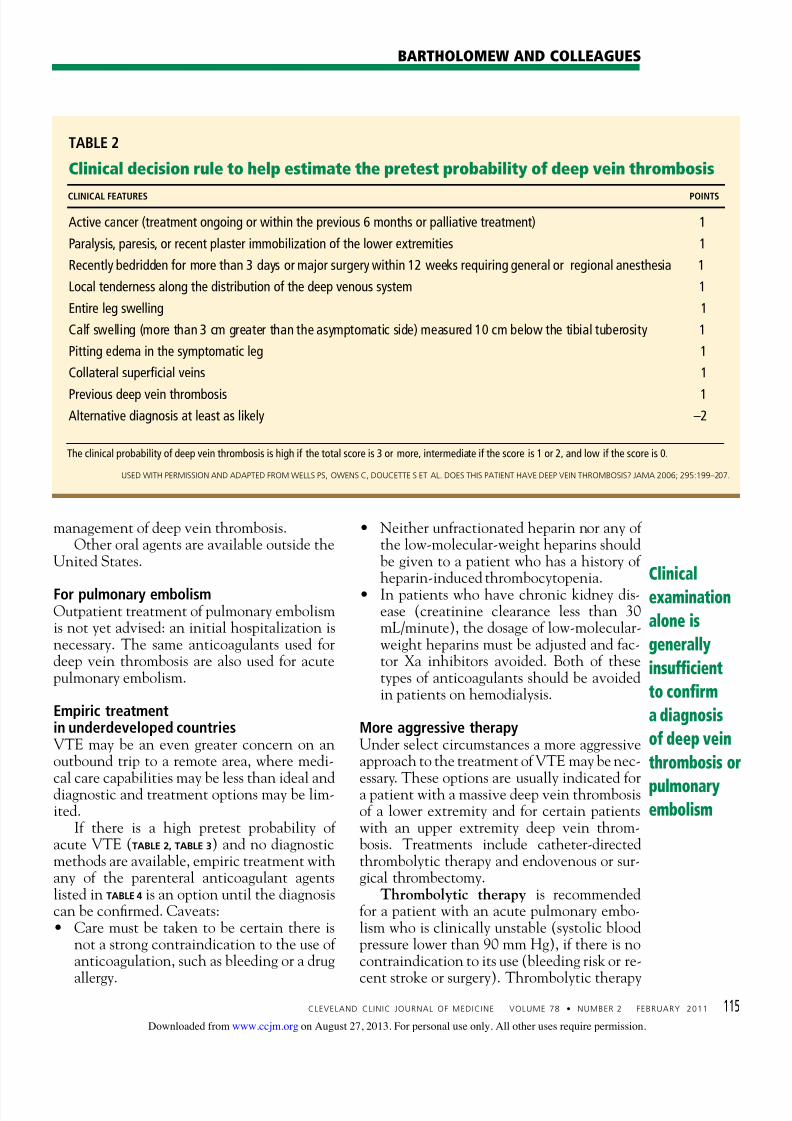
1). In extreme situations, the limb may be cya-notic or gangrenous. A recommended clinical decision algo-rithm that can help assess a patient’s risk foran acute deep vein thrombosis prior to test-ing is depicted in TABLE 2.10 Patients with acutepulmonary embolism are likely to complain ofthe sudden onset of shortness of breath, pleu-
ritic chest pain (especially with breathing),
When VTE
occurs,
it is a life-
altering and
life-threatening
event
on August 27, 2013. For personal use only. All other uses require permission.www.ccjm.orgDownloaded from
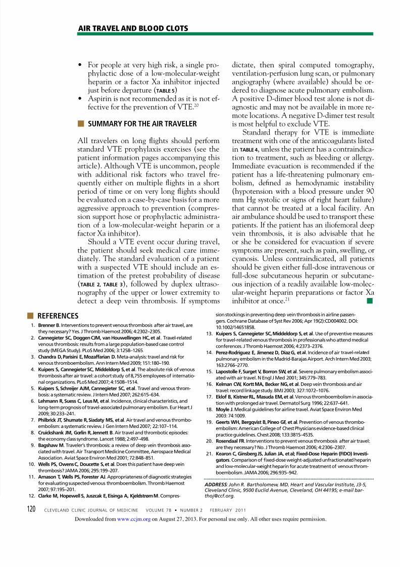
Sitting for long periods during long flights can lead to venous stasis and deep venous thrombosis. Although the absoluterisk is low, it is higher in those with preexisting risk factors for thrombosis, who may need preventive treatment ( TABLE 6).
All travelers should periodically stretch and move their legs (see patient education page).
Most clots form in the deepveins of the calf; less often inthe deep veins of the thigh.
Blood clots can travel from the deep venous system of the upperor lower extremity to the lung and cause pulmonary embolism,which can be fatal.
Passage of thrombus to the
heart and lungs
Blood clot blockinga pulmonary artery
Externaliliacvein
Commonfemoralvein
Femoralvein
Inferior
vena cava
Poplitealvein
Gastrocnemius vein
Anterior tibial vein
Peroneal vein
The great saphenous vein, althoughsuperficial, can also be the site ofclot formation, with the potential ofextending into the deep veins.
Great saphenousvein
Posterior tibial vein
on August 27, 2013. For personal use only. All other uses require permission.www.ccjm.orgDownloaded from
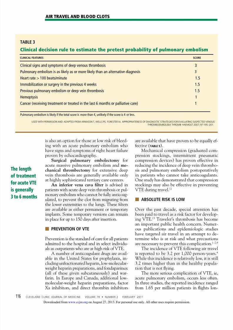
syncope, cough, or hemoptysis. A clinical de-cision algorithm that can help assess the riskof acute pulmonary embolism prior to testingis depicted in TABLE 3.11
■ DIAGNOSIS OF VTE
Clinical examination alone is generally in-suf ficient to confirm a diagnosis of deep vein
thrombosis or pulmonary embolism. Venous
duplex ultrasonography is the most depend-able investigation for deep vein thrombosis,but other tests include D-dimer and imagingstudies such as computed tomographic venog-
raphy or magnetic resonance venography ofthe lower extremities. A more invasive ap-proach is venography; formerly considered thegold standard, it is now generally used onlywhen the diagnosis is in doubt after noninva-sive testing. The diagnosis of acute pulmonaryembolism is best made by spiral computed to-mography. Other studies that may prove helpful in-clude a ventilation-perfusion lung scan forpatients who cannot undergo computed to-mography due to a contrast allergy or renal in-
suf ficiency. Pulmonary angiography, while thegold standard, is less commonly used today,given the specificity and sensitivity of com-puted tomography.
Echocardiography at the bedside may beuseful for patients too sick to move, althoughthe study may not be diagnostic unless throm-bi are seen in the heart or pulmonary arteries.
■ TREATMENT OF VTE
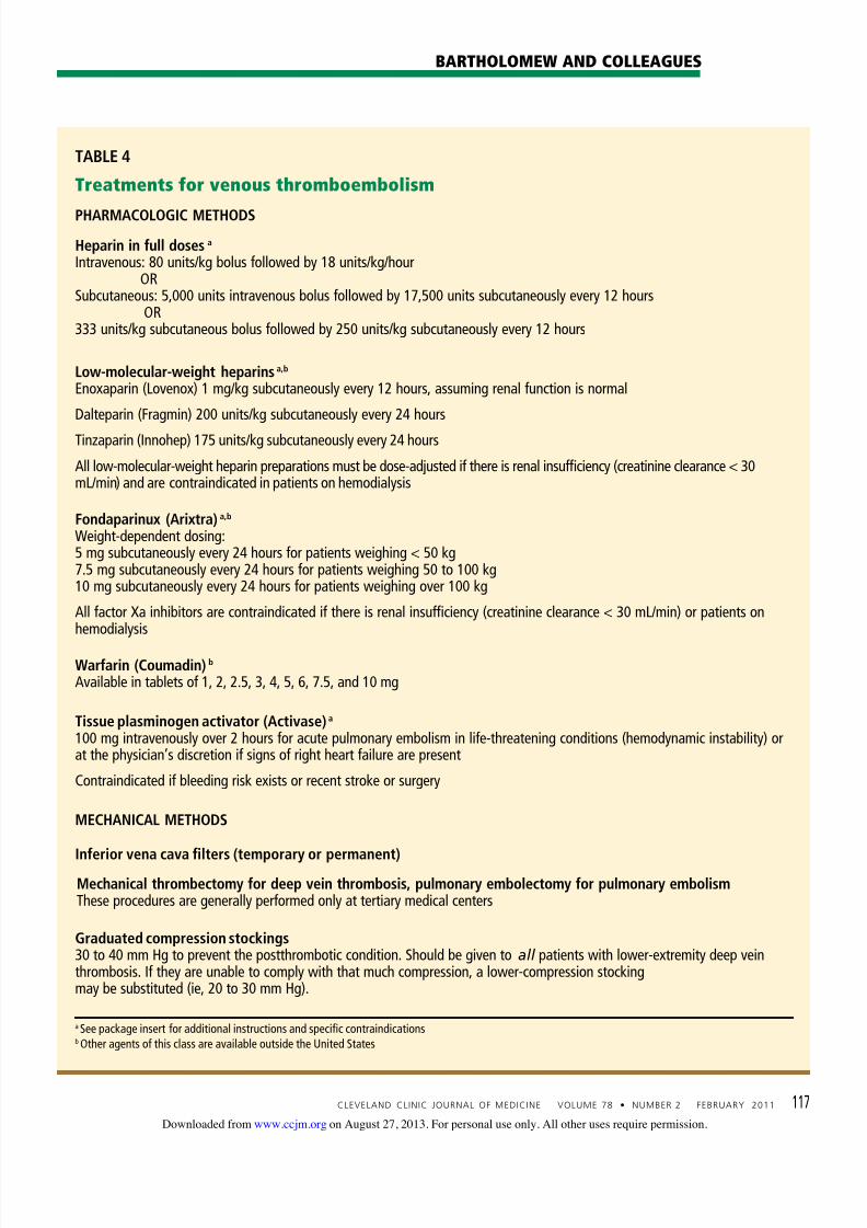
Treatments for VTE are summarized in TABLE 4.
The length of treatment for acute VTE is gen-erally 3 to 6 months. Patients with a knownprecipitating cause such as recent surgery ororal contraceptive use normally require 3months of therapy, while those who had anunprovoked (idiopathic) event require longertherapy, sometimes continuing indefinitely.
For acute deep venous thrombosisAcute deep vein thrombosis is now treated onan outpatient basis under most circumstances.
Unfractionated heparin is given intrave-nously for patients who need to be hospital-ized, or subcutaneously in full dose for inpa-tient or outpatient treatment. Low-molecular-weight heparins are avail-able in subcutaneous preparations and can begiven on an outpatient basis. Fondaparinux (Arixtra), a factor Xa in-hibitor, can also be given subcutaneously onan outpatient basis. Equivalent products areavailable outside the United States. Warfarin (Coumadin), an oral vitamin K
inhibitor, is the agent of choice for long-term
TABLE 1
Risk factors for venous thromboembolism
AgeOlder age, with increasing risk after age 40
WeightBody mass index > 30 kg/m2
(can be calculated as [mass in pounds × 703] / [height in inches2];see www.nhlbisupport.com/bmi/)
MedicationsWomen taking oral contraceptives or hormone replacement
Medical or surgical issuesPrevious venous thromboembolism, either deep venous thrombosisor pulmonary embolism
Varicose veinsMedical illness (congestive heart failure, chronic obstructive pulmonarydisease, stroke with paralysis or paresis, pneumonia)
Pregnancy and up to 6 weeks postpartumActive cancer or cancer chemotherapyCentral venous catheter placementThrombophilia disorders, including factor V Leiden mutation,prothrombin G20210A gene mutation, protein C and S deficiencies,antithrombin deficiency, antiphospholipid syndrome,elevated levels of factor VIII
Recently bedridden more than 3 daysRecent cast immobilization or major surgery (within 12 weeks beforeflying that required general or regional anesthesia)
Recent trauma within 3 months (or anything that compresses the veinssuch as a hematoma or fracture)
Additional risk factors specific to air travelers
HeightPeople who are under 165 cm (65 inches or 5 feet 5 inches) in height4 People who are over 185 cm (73 inches or 6 feet 1 inch) in height4
Flight durationSingle long-haul flights of more than 8 to 10 hoursMultiple long-haul flights of at least 4 hours(risk may persist up to 8 weeks after the flight)4
More frequent flights of any duration within a short time frame
(ie, days or 3 weeks)
on August 27, 2013. For personal use only. All other uses require permission.www.ccjm.orgDownloaded from
management of deep vein thrombosis.Other oral agents are available outside the
United States.
For pulmonary embolismOutpatient treatment of pulmonary embolismis not yet advised: an initial hospitalization isnecessary. The same anticoagulants used fordeep vein thrombosis are also used for acutepulmonary embolism.
Empiric treatmentin underdeveloped countriesVTE may be an even greater concern on anoutbound trip to a remote area, where medi-
cal care capabilities may be less than ideal anddiagnostic and treatment options may be lim-ited. If there is a high pretest probability ofacute VTE (TABLE 2, TABLE 3) and no diagnosticmethods are available, empiric treatment withany of the parenteral anticoagulant agentslisted in TABLE 4 is an option until the diagnosiscan be confirmed. Caveats: Care must be taken to be certain there is
not a strong contraindication to the use ofanticoagulation, such as bleeding or a drug
allergy.
Neither unfractionated heparin nor any ofthe low-molecular-weight heparins shouldbe given to a patient who has a history ofheparin-induced thrombocytopenia.
In patients who have chronic kidney dis-ease (creatinine clearance less than 30mL/minute), the dosage of low-molecular-weight heparins must be adjusted and fac-tor Xa inhibitors avoided. Both of thesetypes of anticoagulants should be avoidedin patients on hemodialysis.
More aggressive therapyUnder select circumstances a more aggressiveapproach to the treatment of VTE may be nec-
essary. These options are usually indicated fora patient with a massive deep vein thrombosisof a lower extremity and for certain patientswith an upper extremity deep vein throm-bosis. Treatments include catheter-directedthrombolytic therapy and endovenous or sur-gical thrombectomy. Thrombolytic therapy is recommendedfor a patient with an acute pulmonary embo-lism who is clinically unstable (systolic bloodpressure lower than 90 mm Hg), if there is nocontraindication to its use (bleeding risk or re-
cent stroke or surgery). Thrombolytic therapy
Clinical
examination
alone is
generally
insufficient
to confirm
a diagnosis
of deep vein
thrombosis or
pulmonaryembolism
TABLE 2
Clinical decision rule to help estimate the pretest probability of deep vein thrombosis
CLINICAL FEATURES POINTS
Active cancer (treatment ongoing or within the previous 6 months or palliative treatment) 1
Paralysis, paresis, or recent plaster immobilization of the lower extremities 1
Recently bedridden for more than 3 days or major surgery within 12 weeks requiring general or regional anesthesia 1
Local tenderness along the distribution of the deep venous system 1
Entire leg swelling 1
Calf swelling (more than 3 cm greater than the asymptomatic side) measured 10 cm below the tibial tuberosity 1
Pitting edema in the symptomatic leg 1
Collateral superficial veins 1
Previous deep vein thrombosis 1Alternative diagnosis at least as likely –2
The clinical probability of deep vein thrombosis is high if the total score is 3 or more, intermediate if the score is 1 or 2, and low if the score is 0.
on August 27, 2013. For personal use only. All other uses require permission.www.ccjm.orgDownloaded from
is also an option for those at low risk of bleed-ing with an acute pulmonary embolism whohave signs and symptoms of right heart failureproven by echocardiography.
Surgical pulmonary embolectomy foracute massive pulmonary embolism and me-chanical thrombectomy for extensive deepvein thrombosis are generally available onlyat highly sophisticated tertiary care centers. An inferior vena cava filter is advised inpatients with acute deep vein thrombosis or pul-monary embolism who cannot be fully anticoag-ulated, to prevent the clot from migrating fromthe lower extremities to the lungs. These filtersare available as either permanent or temporaryimplants. Some temporary versions can remainin place for up to 150 days after insertion.
■
PREVENTION OF VTEPrevention is the standard of care for all patientsadmitted to the hospital and in select individu-als as outpatients who are at high risk of VTE. A number of anticoagulant drugs are avail-able in the United States for prophylaxis, in-cluding unfractionated heparin, low-molecular-weight heparin preparations, and fondaparinux(all of these given subcutaneously) and war-farin. In Europe and Canada, additional low-molecular-weight heparin preparations, factor
Xa inhibitors, and direct thrombin inhibitors
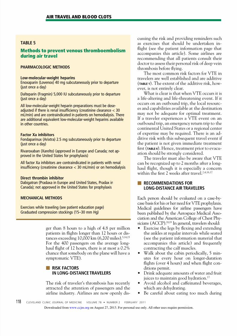
are available that have proven to be equally ef-fective (TABLE 5). Mechanical compression (graduated com-pression stockings, intermittent pneumaticcompression devices) has proven effective inreducing the incidence of deep vein thrombo-sis and pulmonary embolism postoperativelyin patients who cannot take anticoagulants.One study has demonstrated that compressionstockings may also be effective in preventingVTE during travel.12
■ ABSOLUTE RISK IS LOW
Over the past decade, special attention hasbeen paid to travel as a risk factor for develop-ing VTE.13 Traveler’s thrombosis has becomean important public health concern. Numer-ous publications and epidemiologic studieshave targeted air travel in an attempt to de-termine who is at risk and what precautionsare necessary to prevent this complication.1–7,9
The incidence of VTE following air travelis reported to be 3.2 per 1,000 person-years.4 While this incidence is relatively low, it is still3.2 times higher than in the healthy popula-tion that is not flying. The more serious complication of VTE, ie,acute pulmonary embolism, occurs less often.In three studies, the reported incidence ranged
from 1.65 per million patients in flights lon-
TABLE 3
Clinical decision rule to estimate the pretest probability of pulmonary embolism
CLINICAL FEATURES SCORE
Clinical signs and symptoms of deep venous thrombosis 3
Pulmonary embolism is as likely as or more likely than an alternative diagnosis 3
Heart rate > 100 beats/minute 1.5
Immobilization or surgery in the previous 4 weeks 1.5
Previous pulmonary embolism or deep vein thrombosis 1.5
Hemoptysis 1
Cancer (receiving treatment or treated in the last 6 months or palliative care) 1
Pulmonary embolism is likely if the total score is more than 4, unlikely if the score is 4 or less.
USED WITH PERMISSION AND ADAPTED FROM ARNASON T, WELLS PS, FORESTER AJ. APPROPRIATENESS OF DIAGNOSTIC STRATEGIES FOR EVALUATING SUSPECTED VENOUS
on August 27, 2013. For personal use only. All other uses require permission.www.ccjm.orgDownloaded from
Intravenous: 80 units/kg bolus followed by 18 units/kg/hourOR
Subcutaneous: 5,000 units intravenous bolus followed by 17,500 units subcutaneously every 12 hoursOR
333 units/kg subcutaneous bolus followed by 250 units/kg subcutaneously every 12 hours
Low-molecular-weight heparins a,b Enoxaparin (Lovenox) 1 mg/kg subcutaneously every 12 hours, assuming renal function is normal
Dalteparin (Fragmin) 200 units/kg subcutaneously every 24 hours
Tinzaparin (Innohep) 175 units/kg subcutaneously every 24 hours
All low-molecular-weight heparin preparations must be dose-adjusted if there is renal insufficiency (creatinine clearance < 30mL/min) and are contraindicated in patients on hemodialysis
Fondaparinux (Arixtra) a,b
Weight-dependent dosing:5 mg subcutaneously every 24 hours for patients weighing < 50 kg7.5 mg subcutaneously every 24 hours for patients weighing 50 to 100 kg10 mg subcutaneously every 24 hours for patients weighing over 100 kg
All factor Xa inhibitors are contraindicated if there is renal insufficiency (creatinine clearance < 30 mL/min) or patients onhemodialysis
Warfarin (Coumadin) b Available in tablets of 1, 2, 2.5, 3, 4, 5, 6, 7.5, and 10 mg
Tissue plasminogen activator (Activase) a
100 mg intravenously over 2 hours for acute pulmonary embolism in life-threatening conditions (hemodynamic instability) orat the physician’s discretion if signs of right heart failure are present
Contraindicated if bleeding risk exists or recent stroke or surgery
MECHANICAL METHODS
Inferior vena cava filters (temporary or permanent)Mechanical thrombectomy for deep vein thrombosis, pulmonary embolectomy for pulmonary embolismThese procedures are generally performed only at tertiary medical centers
Graduated compression stockings30 to 40 mm Hg to prevent the postthrombotic condition. Should be given to all patients with lower-extremity deep veinthrombosis. If they are unable to comply with that much compression, a lower-compression stockingmay be substituted (ie, 20 to 30 mm Hg).
a See package insert for additional instructions and specific contraindicationsb Other agents of this class are available outside the United States
on August 27, 2013. For personal use only. All other uses require permission.www.ccjm.orgDownloaded from
ger than 8 hours to a high of 4.8 per millionpatients in flights longer than 12 hours or dis-tances exceeding 10,000 km (6,200 miles).5,14,15 For the 400 passengers on the average long-haul flight of 12 hours, there is at most a 0.2%chance that somebody on the plane will have asymptomatic VTE).
■ RISK FACTORSIN LONG-DISTANCE TRAVELERS
The risk of traveler’s thrombosis has recentlyattracted the attention of passengers and the
airline industry. Airlines are now openly dis-
cussing the risk and providing reminders suchas exercises that should be undertaken in-flight (see the patient information page thataccompanies this article). Some airlines are
recommending that all patients consult theirdoctor to assess their personal risk of deep veinthrombosis before flying. The most common risk factors for VTE intravelers are well established and are additive(TABLE 1). The extent of the additive risk, how-ever, is not entirely clear. What is clear is that when VTE occurs it isa life-altering and life-threatening event. If itoccurs on an outbound trip, the local resourc-es and capabilities available at the destinationmay not be adequate for optimal treatment.
If a traveler experiences a VTE event on anoutbound trip, an emergency return trip to thecontinental United States or a regional centerof expertise may be required. There is an ad-ditive risk with this subsequent travel event ifthe patient is not given immediate treatmentfirst (TABLE 4). Hence, treatment prior to evacu-ation should be strongly considered. The traveler must also be aware that VTEcan be recognized up to 2 months after a long-haul flight, though it is especially a concernwithin the first 2 weeks after travel.2,4,16,17
■ RECOMMENDATIONS FORLONG-DISTANCE AIR TRAVELERS
Each person should be evaluated on a case-by-case basis for his or her need for VTE prophylaxis.Medical guidelines for airline passengers havebeen published by the Aerospace Medical Asso-ciation and the American College of Chest Phy-sicians (ACCP).18,19 In general, travelers should: Exercise the legs by flexing and extending
the ankles at regular intervals while seated(see the patient information material thataccompanies this article) and frequentlycontracting the calf muscles.
Walk about the cabin periodically, 5 min-utes for every hour on longer-durationflights (over 4 hours) and when flight con-ditions permit.
Drink adequate amounts of water and fruitjuices to maintain good hydration.17
Avoid alcohol and caffeinated beverages,which are dehydrating.
Be careful about eating too much during
TABLE 5
Methods to prevent venous thromboembolismduring air travel
PHARMACOLOGIC METHODS
Low-molecular-weight heparinsEnoxaparin (Lovenox) 40 mg subcutaneously prior to departure(just once a day)
Dalteparin (Fragmin) 5,000 IU subcutaneously prior to departure(just once a day)
All low-molecular-weight heparin preparations must be dose-adjusted if there is renal insufficiency (creatinine clearance < 30mL/min) and are contraindicated in patients on hemodialysis. Thereare additional equivalent low-molecular-weight heparins availablein other countries.
Factor Xa inhibitorsFondaparinux (Arixtra) 2.5 mg subcutaneously prior to departure(just once a day)
Rivaroxaban (Xarelto) (approved in Europe and Canada; not ap-proved in the United States for prophylaxis)
All factor Xa inhibitors are contraindicated in patients with renalinsufficiency (creatinine clearance < 30 mL/min) or on hemodialysis
Direct thrombin inhibitorDabigatran (Pradaxa in Europe and United States, Pradax inCanada); not approved in the United States for prophylaxis
MECHANICAL METHODS
Exercises while traveling (see patient education page)Graduated compression stockings (15–30 mm Hg)
on August 27, 2013. For personal use only. All other uses require permission.www.ccjm.orgDownloaded from
the flight. Request an aisle seat if you are at risk Do not place baggage underneath the seat
in front of you, because that reduces theability to move the legs.
Do not sleep in a cramped position, andavoid the use of any type of sleep aid.
Avoid wearing constrictive clothing aroundthe lower extremities or waist.
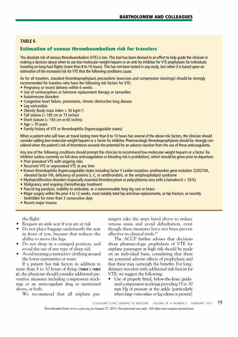
If a patient has risk factors in addition tomore than 8 to 10 hours of flying (TABLE 1, TABLE
6), the physician should consider additional pre-ventive measures including compression stock-ings or an anticoagulant drug as mentionedabove, or both.
We recommend that all airplane pas-
sengers take the steps listed above to reducevenous stasis and avoid dehydration, eventhough these measures have not been proveneffective in clinical trials.19
The ACCP further advises that decisionsabout pharmacologic prophylaxis of VTE forairplane passengers at high risk should be madeon an individual basis, considering that thereare potential adverse effects of prophylaxis andthat these may outweigh the benefits. For long-distance travelers with additional risk factors forVTE, we suggest the following: Use of properly fitted, below-the-knee gradu-
ated compression stockings providing 15 to 30mm Hg of pressure at the ankle (particularly
when large varicosities or leg edema is present)
TABLE 6
Estimation of venous thromboembolism risk for travelers
The absolute risk of venous thromboembolism (VTE) is low. This tool has been devised in an effort to help guide the clinician inmaking a decision about when to use low-molecular-weight heparin or an anti-Xa inhibitor for VTE prophylaxis for individualstraveling on long-haul flights (more than 8 to 10 hours). This has not been tested in any study, but rather it is based upon anestimation of the increased risk for VTE that the following conditions cause.
As for all travelers, standard thromboprophylaxis precautions (exercises and compression stockings) should be stronglyrecommended for travelers who have the following risk factors for VTE: Pregnancy or recent delivery within 6 weeks Use of contraceptives or hormone replacement therapy or tamoxifen Autoimmune disorders Congestive heart failure, pneumonia, chronic obstructive lung disease Leg varicosities Obesity (body mass index > 30 kg/m2) Tall stature (> 185 cm or 73 inches) Short stature (< 165 cm or 65 inches) Age > 70 years Family history of VTE or thrombophilia (hypercoagulable states)
When a patient who will have air travel lasting more than 8 to 10 hours has several of the above risk factors, the clinician shouldconsider adding low-molecular-weight heparin or a factor Xa inhibitor. Pharmacologic thromboprophylaxis should be strongly con-sidered when the patient’s risk of thrombosis exceeds the potential for an adverse reaction from the use of these anticoagulants.
Any one of the following conditions should prompt the clinician to recommend low-molecular-weight heparin or a factor Xainhibitor (unless currently on full-dose anticoagulation or bleeding risk is prohibitive), which should be given prior to departure: Prior provoked VTE with ongoing risks Recurrent VTE or unprovoked VTE at any time Known thrombophilia (hypercoagulable states including factor V Leiden mutation, prothrombin gene mutation G20210A,
elevated factor VIII, deficiency of proteins S, C, or antithrombin), or the antiphospholipid syndrome Myeloproliferative disorders (especially essential thrombocytosis or polycythemia vera with a hematocrit > 55%) Malignancy and ongoing chemotherapy treatment Flaccid leg paralysis, inability to ambulate, or a nonremovable long leg cast or brace Major surgery within the prior 4 to 12 weeks, most notably total hip and knee replacements, or hip fracture, or recently
bedridden for more than 3 consecutive days Recent major trauma
on August 27, 2013. For personal use only. All other uses require permission.www.ccjm.orgDownloaded from
For people at very high risk, a single pro-phylactic dose of a low-molecular-weightheparin or a factor Xa inhibitor injectedjust before departure (TABLE 5)
Aspirin is not recommended as it is not ef-fective for the prevention of VTE.20
■ SUMMARY FOR THE AIR TRAVELER
All travelers on long flights should performstandard VTE prophylaxis exercises (see thepatient information pages accompanying thisarticle). Although VTE is uncommon, peoplewith additional risk factors who travel fre-quently either on multiple flights in a shortperiod of time or on very long flights should
be evaluated on a case-by-case basis for a moreaggressive approach to prevention (compres-sion support hose or prophylactic administra-tion of a low-molecular-weight heparin or afactor Xa inhibitor). Should a VTE event occur during travel,the patient should seek medical care imme-diately. The standard evaluation of a patientwith a suspected VTE should include an es-timation of the pretest probability of disease(TABLE 2, TABLE 3), followed by duplex ultraso-nography of the upper or lower extremity todetect a deep vein thrombosis. If symptoms
dictate, then spiral computed tomography,ventilation-perfusion lung scan, or pulmonaryangiography (where available) should be or-dered to diagnose acute pulmonary embolism.
A positive D-dimer blood test alone is not di-agnostic and may not be available in more re-mote locations. A negative D-dimer test resultis most helpful to exclude VTE.
Standard therapy for VTE is immediatetreatment with one of the anticoagulants listedin TABLE 4, unless the patient has a contraindica-tion to treatment, such as bleeding or allergy.Immediate evacuation is recommended if thepatient has a life-threatening pulmonary em-bolism, defined as hemodynamic instability(hypotension with a blood pressure under 90
mm Hg systolic or signs of right heart failure)that cannot be treated at a local facility. Anair ambulance should be used to transport thesepatients. If the patient has an iliofemoral deepvein thrombosis, it is also advisable that heor she be considered for evacuation if severesymptoms are present, such as pain, swelling, orcyanosis. Unless contraindicated, all patientsshould be given either full-dose intravenous orfull-dose subcutaneous heparin or subcutane-ous injection of a readily available low-molec-ular-weight heparin preparations or factor Xainhibitor at once.21
■
■ REFERENCES 1. Brenner B. Interventions to prevent venous thrombosis after air travel, are
they necessary? Yes. J Thromb Haemost 2006; 4:2302–2305.
2. Cannegieter SC, Doggen CJM, van Houwellingen HC, et al. Travel-related
venous thrombosis: results from a large population-based case control
study (MEGA Study). PLoS Med 2006; 3:1258–1265.
3. Chandra D, Parisini E, Mozaffarian D. Meta-analysis: travel and risk for
venous thromboembolism. Ann Intern Med 2009; 151:180–190.
4. Kuipers S, Cannegieter SC, Middeldorp S, et al. The absolute risk of venous
thrombosis after air travel: a cohort study of 8,755 employees of internatio-
nal organizations. PLoS Med 2007; 4:1508–1514.
5. Kuipers S, Schreijer AJM, Cannegieter SC, et al. Travel and venous throm-
bosis: a systematic review. J Intern Med 2007; 262:615–634.
6. Lehmann R, Suess C, Leus M, et al. Incidence, clinical characteristics, and
long-term prognosis of travel-associated pulmonary embolism. Eur Heart J
2009; 30:233–241.
7. Philbrick JT, Shumate R, Siadaty MS, et al. Air travel and venous thrombo-
embolism: a systematic review. J Gen Intern Med 2007; 22:107–114.
8. Cruickshank JM, Gorlin R, Jennett B. Air travel and thrombotic episodes:
the economy class syndrome. Lancet 1988; 2:497–498.
9. Bagshaw M. Traveler’s thrombosis: a review of deep vein thrombosis asso-
ciated with travel. Air Transport Medicine Committee, Aerospace Medical
Association. Aviat Space Environ Med 2001; 72:848–851.
10. Wells PS, Owens C, Doucette S, et al. Does this patient have deep vein
thrombosis? JAMA 2006; 295:199–207.
11. Arnason T, Wells PS, Forester AJ. Appropriateness of diagnostic strategies
for evaluating suspected venous thromboembolism. Thromb Haemost
2007; 97:195–201.
12. Clarke M, Hopewell S, Juszcak E, Eisinga A, Kjeldstrøm M. Compres-
sion stockings in preventing deep vein thrombosis in airline passen-
gers. Cochrane Database of Syst Rev 2006; Apr 19(2):CD004002. DOI:
10.1002/14651858.
13. Kuipers S, Cannegieter SC, Middeldorp S, et al. Use of preventive measures
for travel-related venous thrombosis in professionals who attend medical
conferences. J Thromb Haemost 2006; 4:2373–2376.
14. Perez-Rodriguez E, Jimenez D, Diaz G, et al. Incidence of air travel-related
pulmonary embolism in the Madrid-Barajas Airport. Arch Intern Med 2003;
163:2766–2770.
15. Lapostolle F, Surget V, Borron SW, et al. Severe pulmonary embolism associ-
ated with air travel. N Engl J Med 2001; 345:779–783.
16. Kelman CW, Kortt MA, Becker NG, et al. Deep vein thrombosis and air
travel: record linkage study. BMJ 2003; 327:1072–1076.
17. Eklof B, Kistner RL, Masuda EM, et al. Venous thromboembolism in associa-tion with prolonged air travel. Dermatol Surg 1996; 22:637–641.
18. Moyle J. Medical guidelines for airline travel. Aviat Space Environ Med
2003: 74:1009.
19. Geerts WH, Bergqvist B, Pineo GF, et al. Prevention of venous thrombo-
embolism: American College of Chest Physicians evidence-based clinical
practice guidelines. Chest 2008; 133:381S–453S.
20. Rosendaal FR. Interventions to prevent venous thrombosis after air travel:
are they necessary? No. J Thromb Haemost 2006; 4:2306–2307.