June 2018 C hronic total occlusions (CTOs) can be found in multiple vessels and represent the most challeng- ing aspect of treating patients. CTOs represent dense collagenous lesions with varying degrees of calcification obstructing blood flow to distal vascular beds. The obstructive nature of this ma- terial limits the success rate of travers- ing these CTOs during catheter-based interventions. Unless a CTO can be traversed, no therapy can be delivered. A deeper understanding of CTOs in tibial arteries was recently described in the Journal of Endovascular Therapy. The study was designed to determine if cap morphologies analyzed via angiograms A recent study conducted by the CLI Global Society reviewed ad- ministrative claims on all fee- for-service Medicare beneficiaries and identified more than 72,000 with an initial diagnosis of critical limb ischemia (CLI) during 2011. Patients were fol- lowed over a 4-year period. The medi- an time from CLI diagnosis to first re- vascularization or amputation procedure was 9 days. Following diagnosis, 29% of patients either died or underwent ma- jor amputation in the first year. Through 4 years, 54% of patients died, 3% were alive with major amputation, and 42% were alive and free from major ampu- tation. The median duration of survival following CLI diagnosis was 3.5 years. 1 Patients with CLI have a poor long- term prognosis subsequent to the ini- tial diagnosis. Considerable efforts are needed to raise disease awareness, refine diagnostic algorithms, and establish evi- dence-based treatment pathways to ad- dress the high mortality rates associated with this diagnosis. 1 In many regions of the United States, most patients with severe peripheral artery disease (PAD) undergo amputation without even a di- agnostic arteriogram performed in the year before amputation. 2 It is reported that 51% of CLI patients have no diag- nostic vascular evaluation prior to pri- mary amputation. 3 There is a current grassroots movement of physicians known as the CLI Fighters. They embrace a multidisciplinary approach to the treatment of CLI and focus the majority of their practice on treating CLI patients. The CLI Fighters extend across multiple disciplines to include interventional radiology, vas- cular surgery, interventional cardiol- ogy, podiatry, wound care, and vascular medicine. “The CLI Fighters’ mission revolves around increasing awareness of this deadly disease and advocating for patients’ rights to preserve their limbs and avoid deadly amputations,” said Dr. Fadi Saab. Members of the CLI Global Society Board and 25 CLI Fighters assembled re- cently for the first CLI Fighters Summit in Minneapolis, Minnesota (Figures 1-3). Board members Dr. Barry Katzen, Dr. Jihad Mustapha, Dr. Robert Lookstein, and Dr. Vicki Driver reviewed available data and guidelines (Figure 4). Dr. Mary Yost provided an overview of the scope and economic impact of the PAD/CLI epidemic, and Dr. Christopher Boyes shared examples in CLI practice. “In the treatment of CLI, major am- putation is bad for the patient, bad for the hospital, and bad for the economy,” said Dr. Yost. Due to the aging popula- tion, CLI is projected to increase from about 3.4 million in 2015 to 4.7 mil- lion in 2030 (Figure 5). CLI is an ex- tremely expensive disease, with an esti- mated economic cost of $134 to $248 billion.The majority of these costs (about The CTOP Trial: Finally, Some Data to Suggest When to Utilize Pedal Access Guest Editor: Fadi Saab, MD Advanced Cardiac & Vascular Amputation Prevention Centers, Grand Rapids, MI CLI Global Society and CLI Fighters Join Forces at First CLI Fighter Summit CLI The Official Publication of the Critical Limb Ischemia Global Society GLOBAL Continued on page 22 Continued on page 18 Fadi Saab, MD #CLIfighters Figure 1. CLI Fighters engaged in discussion at the first CLI Fighter Summit. Figure 2. Mary Yost, MBA, with CLI Global Society Board Members Vicki Driver, DPM, and Jihad Mustapha, MD (left to right).
Transcript
June 2018
Chronic total occlusions (CTOs) can be found in multiple vessels and represent the most challeng-
ing aspect of treating patients. CTOs represent dense collagenous lesions with varying degrees of calcification
obstructing blood flow to distal vascular beds. The obstructive nature of this ma-terial limits the success rate of travers-ing these CTOs during catheter-based interventions. Unless a CTO can be traversed, no therapy can be delivered.
A deeper understanding of CTOs in tibial arteries was recently described in the Journal of Endovascular Therapy. The study was designed to determine if cap morphologies analyzed via angiograms
A recent study conducted by the CLI Global Society reviewed ad-ministrative claims on all fee-
for-service Medicare beneficiaries and identified more than 72,000 with an initial diagnosis of critical limb ischemia (CLI) during 2011. Patients were fol-lowed over a 4-year period. The medi-an time from CLI diagnosis to first re-vascularization or amputation procedure was 9 days. Following diagnosis, 29% of patients either died or underwent ma-jor amputation in the first year. Through 4 years, 54% of patients died, 3% were alive with major amputation, and 42% were alive and free from major ampu-tation. The median duration of survival following CLI diagnosis was 3.5 years.1
Patients with CLI have a poor long-term prognosis subsequent to the ini-tial diagnosis. Considerable efforts are needed to raise disease awareness, refine diagnostic algorithms, and establish evi-dence-based treatment pathways to ad-dress the high mortality rates associated with this diagnosis.1 In many regions of the United States, most patients with severe peripheral artery disease (PAD) undergo amputation without even a di-agnostic arteriogram performed in the year before amputation.2 It is reported that 51% of CLI patients have no diag-nostic vascular evaluation prior to pri-mary amputation.3
There is a current grassroots movement of physicians known as the CLI Fighters. They embrace a multidisciplinary
approach to the treatment of CLI and focus the majority of their practice on treating CLI patients. The CLI Fighters extend across multiple disciplines to include interventional radiology, vas-cular surgery, interventional cardiol-ogy, podiatry, wound care, and vascular medicine. “The CLI Fighters’ mission revolves around increasing awareness of this deadly disease and advocating for patients’ rights to preserve their limbs and avoid deadly amputations,” said Dr. Fadi Saab.
Members of the CLI Global Society Board and 25 CLI Fighters assembled re-cently for the first CLI Fighters Summit in Minneapolis, Minnesota (Figures 1-3). Board members Dr. Barry Katzen, Dr. Jihad Mustapha, Dr. Robert Lookstein, and Dr. Vicki Driver reviewed available data and guidelines (Figure 4). Dr. Mary Yost provided an overview of the scope and economic impact of the PAD/CLI epidemic, and Dr. Christopher Boyes shared examples in CLI practice.
“In the treatment of CLI, major am-putation is bad for the patient, bad for the hospital, and bad for the economy,” said Dr. Yost. Due to the aging popula-tion, CLI is projected to increase from about 3.4 million in 2015 to 4.7 mil-lion in 2030 (Figure 5). CLI is an ex-tremely expensive disease, with an esti-mated economic cost of $134 to $248 billion. The majority of these costs (about
The CTOP Trial: Finally, Some Data to Suggest When to Utilize Pedal AccessGuest Editor: Fadi Saab, MDAdvanced Cardiac & Vascular Amputation Prevention Centers, Grand Rapids, MI
CLI Global Society and CLI Fighters Join Forces at First CLI Fighter Summit
CLI The Official Publication of the Critical Limb Ischemia Global Society
GLOBAL
Continued on page 22
Continued on page 18
Fadi Saab, MD
#CLIfighters
Figure 1. CLI Fighters engaged in discussion at the first CLI Fighter Summit.
Figure 2. Mary Yost, MBA, with CLI Global Society Board Members Vicki Driver, DPM, and Jihad Mustapha, MD (left to right).
MASTERYOUR BTK CASES
Minimize risk of dissection and decrease the need for bailout stenting with the Chocolate™ PTA balloon. The balloon’s unique nitinol cage creates pillows and grooves to help ensure predictable, uniform and atraumatic dilatation.
She was but one traveler among thousands that night on the long and lonely highways connecting
her home state of Maryland to a doctor she had yet to meet in Michigan.
But the odyssey of Juanita “Nita” McDaniel-Williams does as much as any other story to underscore the gravity ren-dered by her fight with peripheral artery disease (PAD) and the psychosocial fac-tors that continue to affect her care and outcomes—and those of untold millions.
A single mother of three daughters, Nita, now in her early 50s, was diag-nosed with diabetes some 20 years ago. In discovering that virtually “everybody on my father’s side had it, I just figured it was now a part of my life, and that I was going to have to accept it.”
What she didn’t count on were com-plications in 2017 that put her face-to-face with the prospect of amputations of her foot, leg, or both. “My world was destroyed because of PAD,” she says.
Agreeing to a first opinion presented by a podiatrist in Maryland, Nita says she succumbed to the partial amputation of her left foot. But when faced with the possibility of additional amputations, she reached out to her daughters, who glad-ly went to work exploring alternatives.
One of them found Dr. Jihad A. Mustapha online, and after reading about the pioneering work he was doing
to prevent amputations, reached out to him with a tweet, briefly explaining her mother’s plight.
Nita was grateful but figured, “this man isn’t going to call me.”
She was wrong.Within 30 minutes, Dr. Mustapha
was on the phone with Nita’s daughter.
J.A. MUSTAPHA, MD, FACC, FSCAIClinical EditorAdvanced Cardiac & Vascular Amputation Prevention CentersGrand Rapids, MIClinical Associate Professor of MedicineMichigan State University COM, East Lansing, MI
Jeff Martin, PublisherCarmen Heaney, Executive EditorRebecca Kapur, Managing EditorAndrea Modica, Special Projects EditorVic Geanopulos, Creative DirectorElizabeth Vasil, Graphic Production Manager
EDITORIAL CORRESPONDENCE: Laurie Gustafson, Executive Editor HMP 70 East Swedesford Road, Suite 100 [email protected]
GEORGE ADAMS, MDGarner, NC
VICKIE R. DRIVER, DPM, MSBoston, MA
LAWRENCE GARCIA, MDBoston, MA
PHILIP P. GOODNEY, MDLebanon, NH
MICHAEL R. JAFF, DONewton, MA
BARRY T. KATZEN, MDMiami, FL
ROBERT LOOKSTEIN, MDNew York, NY
D. CHRIS METZGER, MDKingsport, TN
RICHARD F. NEVILLE, MDFairfax, VA
CONSTANTINO S. PENA, MDMiami, FL
FADI A. SAAB, MDGrand Rapids, MI
DIERK SCHEINERT, MDLeipzig, Germany
ANDREJ SCHMIDT, MDLeipzig, Germany
RAMON VARCOE, MBBS, MSSydney, Australia
FRANK J. VEITH, MDNew York, NY
RENU VIRMANI, MDGaithersburg, MD
CRAIG M. WALKER, MDHouma, LA
THOMAS ZELLER, MDBad Krozingen, Germany
Published in collaboration with
Editor’s note: Articles in this supplement to Cath Lab Digest did not undergo peer review.
EDITORIAL
SCIENTIFIC ADVISORY BOARD
Continued on page 14
TABLE OF CONTENTS
CLI Global Society and CLI Fighters Join Forces at First CLI Fighter Summit ......................................................................1
The CTOP Trial: Finally, Some Data to Suggest When to Utilize Pedal Access ............................................................1
Amputee Candidate Moves from East Coast to Michigan to Embrace Progressive Options .......................3
Long Tibial CTO and No Pedal Arch: CTOP Classifica-tion Helps Guide Treatment Approach ...............................4
Scientifically-Driven Approach to Crossing a CTOP Type IV Occlusion .....................................................................6
Highly Complex Bilateral Rutherford Stage 6 Patient with Multilevel Occlusive Disease Treated with Bilateral Posterior Tibial and Left Radial Access ............8
Upcoming Meetings and Events .......................................... 19
Content may not be reproduced in any form without written permission. Contact [email protected] for rights and permission.
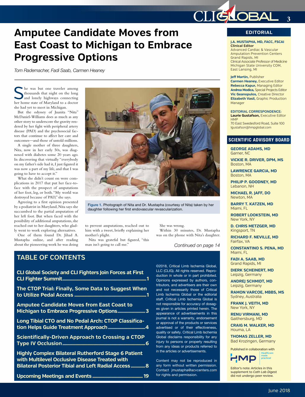
Amputee Candidate Moves from East Coast to Michigan to Embrace Progressive OptionsTom Rademacher, Fadi Saab, Carmen Heaney
Figure 1. Photograph of Nita and Dr. Mustapha (courtesy of Nita) taken by her daughter following her first endovascular revascularization.
4
June 2018
CLIGLOBAL
A 79-year-old man with critical limb ischemia (CLI) presented to my outpatient vascular clinic with
a several-month history of severe left foot rest pain, non-healing ulcers, and gan-grene. He was evaluated by a local vas-cular surgeon who recommended ampu-tation, so the patient came to see me for a second opinion. His past medical his-tory was significant for severe peripheral arterial disease (PAD) with CLI, hyper-tension, and hyperlipidemia. Although he had been seen by a vascular specialist in the past, he never had an angiogram or bypass surgery. Although he had a two-pack-per-day (for 20 years) smoking his-tory, he quit several years prior to pre-senting to my clinic.
His labs were normal (CBC, BMP, PT/PTT/INR), and his medications includ-ed losartan, amlodipine, furosemide, ator-vastatin, omeprazole, gabapentin, clopi-dogrel, and baby aspirin.
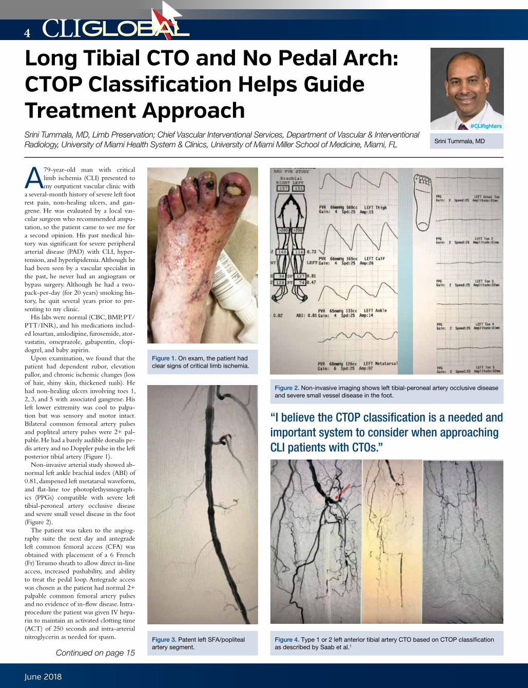
Upon examination, we found that the patient had dependent rubor, elevation pallor, and chronic ischemic changes (loss of hair, shiny skin, thickened nails). He had non-healing ulcers involving toes 1, 2, 3, and 5 with associated gangrene. His left lower extremity was cool to palpa-tion but was sensory and motor intact. Bilateral common femoral artery pulses and popliteal artery pulses were 2+ pal-pable. He had a barely audible dorsalis pe-dis artery and no Doppler pulse in the left posterior tibial artery (Figure 1).
Non-invasive arterial study showed ab-normal left ankle brachial index (ABI) of 0.81, dampened left metatarsal waveform, and flat-line toe photoplethysmograph-ics (PPGs) compatible with severe left tibial-peroneal artery occlusive disease and severe small vessel disease in the foot (Figure 2).
The patient was taken to the angiog-raphy suite the next day and antegrade left common femoral access (CFA) was obtained with placement of a 6 French (Fr) Terumo sheath to allow direct in-line access, increased pushability, and ability to treat the pedal loop. Antegrade access was chosen as the patient had normal 2+ palpable common femoral artery pulses and no evidence of in-flow disease. Intra-procedure the patient was given IV hepa-rin to maintain an activated clotting time (ACT) of 250 seconds and intra-arterial nitroglycerin as needed for spasm.
Long Tibial CTO and No Pedal Arch: CTOP Classification Helps Guide Treatment Approach Srini Tummala, MD, Limb Preservation; Chief Vascular Interventional Services, Department of Vascular & Interventional Radiology, University of Miami Health System & Clinics, University of Miami Miller School of Medicine, Miami, FL Srini Tummala, MD
#CLIfighters
Figure 1. On exam, the patient had clear signs of critical limb ischemia.
Figure 3. Patent left SFA/popliteal artery segment.
Figure 2. Non-invasive imaging shows left tibial-peroneal artery occlusive disease and severe small vessel disease in the foot.
Figure 4. Type 1 or 2 left anterior tibial artery CTO based on CTOP classification as described by Saab et al.1
“I believe the CTOP classification is a needed and important system to consider when approaching CLI patients with CTOs.”
Continued on page 15
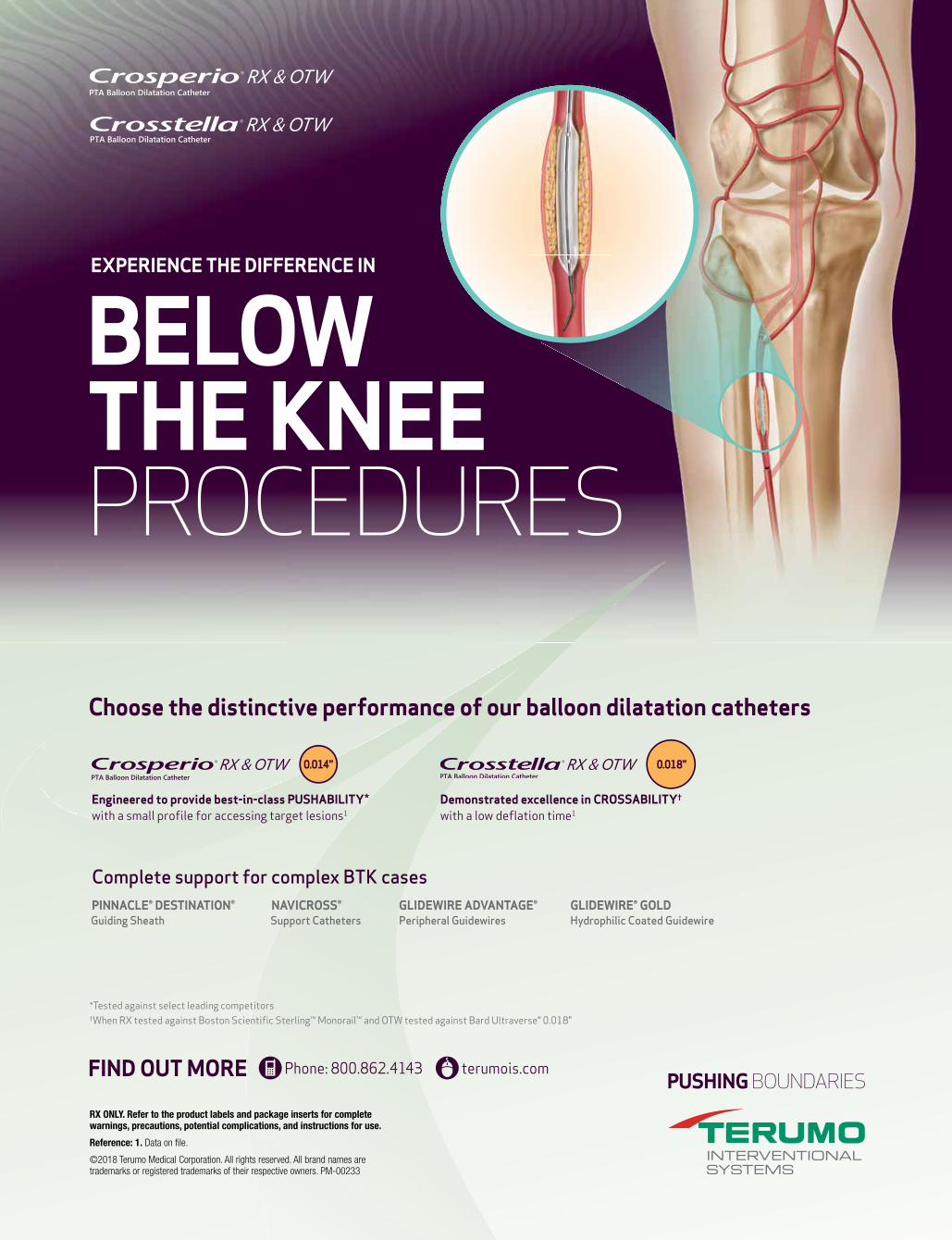
Choose the distinctive performance of our balloon dilatation catheters
*Tested against select leading competitors†When RX tested against Boston Scientifi c Sterling™ Monorail™ and OTW tested against Bard Ultraverse® 0.018"
RX ONLY. Refer to the product labels and package inserts for complete warnings, precautions, potential complications, and instructions for use.
Complete support for complex BTK casesPINNACLE® DESTINATION® Guiding Sheath
NAVICROSS® Support Catheters
GLIDEWIRE ADVANTAGE®Peripheral Guidewires
GLIDEWIRE® GOLDHydrophilic Coated Guidewire
Engineered to provide best-in-class PUSHABILITY* with a small profi le for accessing target lesions1
Demonstrated excellence in CROSSABILITY† with a low defl ation time1
46696ID02-BTK_CLi.indd 1 4/30/18 9:20 AMTerumo_BTK_CLIG0618.indd 1 5/7/18 9:34 AM
6
June 2018
CLIGLOBAL
A 65-year-old, 6'4", 192-pound (87 kg) man with Rutherford class IV peripheral arterial disease
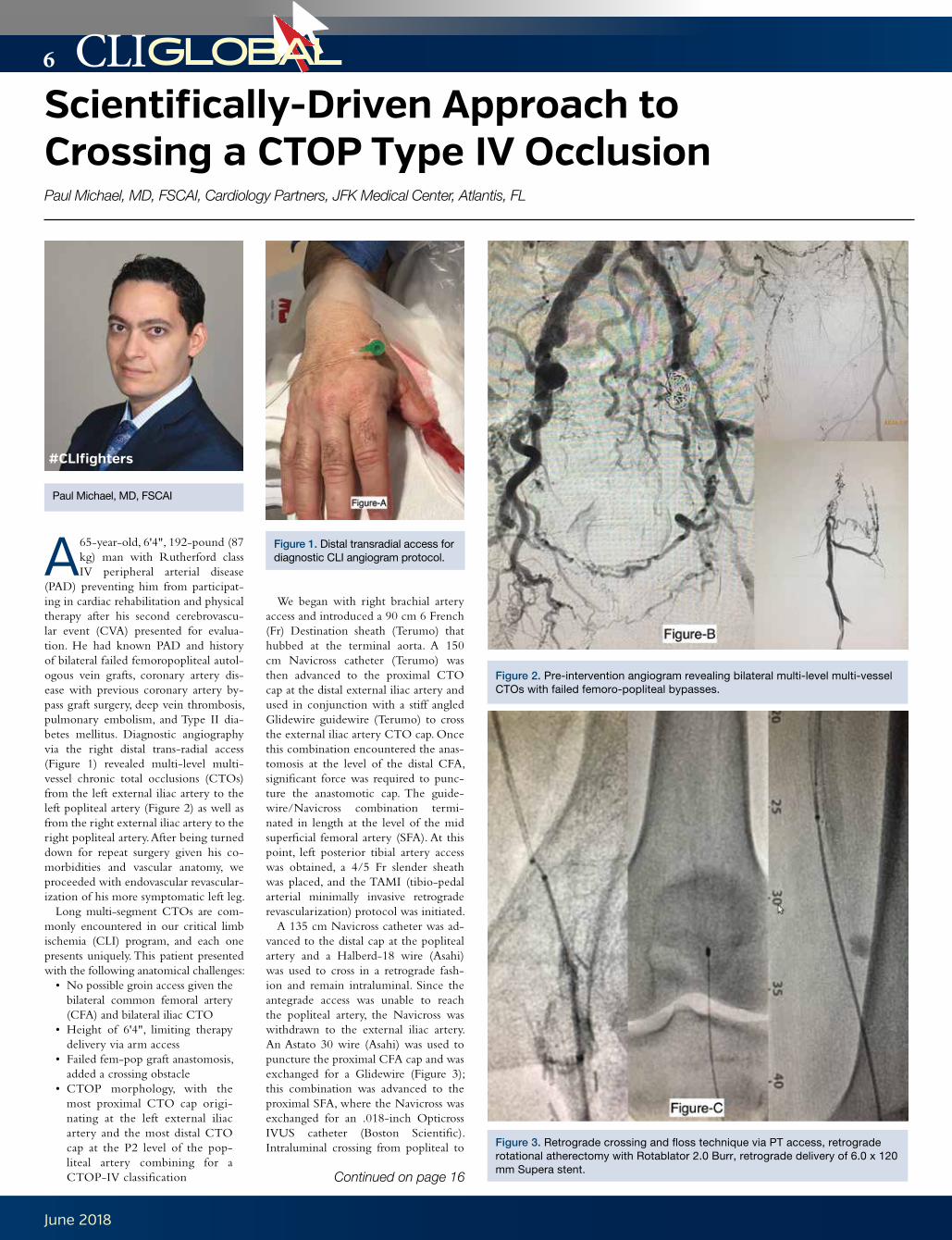
(PAD) preventing him from participat-ing in cardiac rehabilitation and physical therapy after his second cerebrovascu-lar event (CVA) presented for evalua-tion. He had known PAD and history of bilateral failed femoropopliteal autol-ogous vein grafts, coronary artery dis-ease with previous coronary artery by-pass graft surgery, deep vein thrombosis, pulmonary embolism, and Type II dia-betes mellitus. Diagnostic angiography via the right distal trans-radial access (Figure 1) revealed multi-level multi-vessel chronic total occlusions (CTOs) from the left external iliac artery to the left popliteal artery (Figure 2) as well as from the right external iliac artery to the right popliteal artery. After being turned down for repeat surgery given his co-morbidities and vascular anatomy, we proceeded with endovascular revascular-ization of his more symptomatic left leg.
Long multi-segment CTOs are com-monly encountered in our critical limb ischemia (CLI) program, and each one presents uniquely. This patient presented with the following anatomical challenges:
• No possible groin access given the bilateral common femoral artery (CFA) and bilateral iliac CTO
• Height of 6'4", limiting therapy delivery via arm access
• Failed fem-pop graft anastomosis, added a crossing obstacle
• CTOP morphology, with the most proximal CTO cap origi-nating at the left external iliac artery and the most distal CTO cap at the P2 level of the pop-liteal artery combining for a CTOP-IV classification
We began with right brachial artery access and introduced a 90 cm 6 French (Fr) Destination sheath (Terumo) that hubbed at the terminal aorta. A 150 cm Navicross catheter (Terumo) was then advanced to the proximal CTO cap at the distal external iliac artery and used in conjunction with a stiff angled Glidewire guidewire (Terumo) to cross the external iliac artery CTO cap. Once this combination encountered the anas-tomosis at the level of the distal CFA, significant force was required to punc-ture the anastomotic cap. The guide-wire/Navicross combination termi-nated in length at the level of the mid superficial femoral artery (SFA). At this point, left posterior tibial artery access was obtained, a 4/5 Fr slender sheath was placed, and the TAMI (tibio-pedal arterial minimally invasive retrograde revascularization) protocol was initiated.
A 135 cm Navicross catheter was ad-vanced to the distal cap at the popliteal artery and a Halberd-18 wire (Asahi) was used to cross in a retrograde fash-ion and remain intraluminal. Since the antegrade access was unable to reach the popliteal artery, the Navicross was withdrawn to the external iliac artery. An Astato 30 wire (Asahi) was used to puncture the proximal CFA cap and was exchanged for a Glidewire (Figure 3); this combination was advanced to the proximal SFA, where the Navicross was exchanged for an .018-inch Opticross IVUS catheter (Boston Scientific). Intraluminal crossing from popliteal to
Continued on page 16
Scientifically-Driven Approach to Crossing a CTOP Type IV Occlusion Paul Michael, MD, FSCAI, Cardiology Partners, JFK Medical Center, Atlantis, FL
Paul Michael, MD, FSCAI
#CLIfighters
Figure 1. Distal transradial access for diagnostic CLI angiogram protocol.
Figure 3. Retrograde crossing and floss technique via PT access, retrograde rotational atherectomy with Rotablator 2.0 Burr, retrograde delivery of 6.0 x 120 mm Supera stent.
New Guide WiresNEW Peripheral Guide Wires
Now available withASAHI’s unique guidewire technology
PRECISION ENGINEERED for Tough Peripheral Cases
®
ASAHI INTECC USA, INC.2500 Red Hill Avenue, Suite 210, Santa Ana, CA 92705Toll-Free [email protected]
Learn more atasahi-inteccusa-medical.com
The ASAHI INTECC peripheral guide wires are intended to facilitate the placement and exchange of diagnostic and therapeutic devices during intravascular procedures.These devices are intended for peripheral vascular use only.
The ASAHI® Corsair® Armet® is intended to provide support to facilitate the placement of guide wires in the peripheral vasculature, and can be used to exchange one guide wire for another.The ASAHI Corsair Armet is also intended to assist in the delivery of contrast media into the peripheral vasculature. This device should not be used in coronary vasculature or neurovasculature.
®
ASAHI Gladius® 0.014/0.018Workhorse
ASAHI® Halberd® 0.014/0.018Complex Lesion
ASAHI Gaia® PV 0.018Complex Lesion
New Microcatheter
Durable metal tip
Super SHINKA-ShaftLow profile, supportive catheter body,excellent torque
®
®
Ⓒ2016 ASAHI INTECC., LTD.“ASAHI,” “Halberd,” “ASAHI Gaia,” and “ASAHI Gladius” are trademarks or registered trademarks of ASAHI INTECC CO., LTD. in Japan and other countries.
C
M
Y
CM
MY
CY
CMY
K
2016_12_CLI-Global-TabloidAd_printer.pdf 1 11/21/2016 9:21:48 AM
Asahi_CLIC_1216.indd 1 11/21/16 9:26 AM
8
June 2018
CLIGLOBAL
A 67-year-old male was sent by his wound care physician to the emergency department for wors-
ening dry gangrene of the toes of both feet as well as right leg rest pain, especial-ly below the knee. He has a history of pe-ripheral arterial occlusive disease and had been treated 6 months earlier at an out-side institution with stenting of the right common iliac, right external iliac, right common femoral, and proximal right su-perficial femoral arteries. He has a histo-ry of coronary artery disease status post-coronary artery bypass graft surgery. His past medical history also included multi-ple ischemic strokes with residual right-sided weakness and chronic kidney dis-ease stage III. He had Rutherford stage VI arterial insufficiency in both feet.
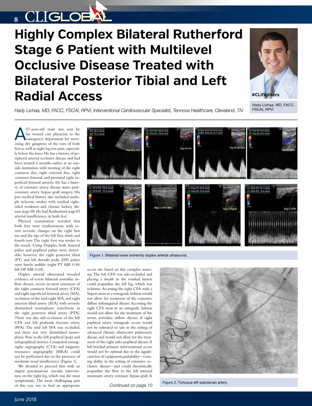
Physical examination revealed that both feet were erythematous with se-vere necrotic changes on the right first toe and the tips of the left first, third, and fourth toes. The right foot was tender to the touch. Using Doppler, both femoral pulses and popliteal pulses were detect-able; however, the right posterior tibial (PT) and left dorsalis pedis (DP) pulses were barely audible (right PT ABI: 0.48, left DP ABI: 0.68).
Duplex arterial ultrasound revealed evidence of severe bilateral aortoiliac in-flow disease, severe in-stent restenosis of the right common femoral artery (CFA) and right superficial femoral artery (SFA), occlusion of the mid right SFA, and right anterior tibial artery (ATA) with severely diminished monophasic waveforms in the right posterior tibial artery (PTA). There was also sub-occlusion of the left CFA and left profunda femoris artery (PFA). The mid left SFA was occluded, and there was very diminished mono-phasic flow in the left popliteal (pop) and infrapopliteal arteries. Computed tomog-raphy angiography (CTA) and magnetic resonance angiography (MRA) could not be performed due to the presence of moderate renal insufficiency (Figure 1).
We decided to proceed first with an urgent percutaneous vascular interven-tion on the right leg, which was the most symptomatic. The most challenging part of this case was to find an appropriate
access site based on this complex anato-my. The left CFA was sub-occluded and placing a sheath in the residual lumen could jeopardize the left leg, which was ischemic. Accessing the right CFA with a Supera stent in a retrograde fashion would not allow for treatment of the extensive diffuse infrainguinal disease. Accessing the right CFA stent in an antegrade fashion would not allow for the treatment of the severe aortoiliac inflow disease. A right popliteal artery retrograde access would not be tolerated or safe in the setting of advanced chronic obstructive pulmonary disease and would not allow for the treat-ment of the right infra-popliteal disease. A left brachial primary interventional access would not be optimal due to the signifi-cant loss of equipment pushability—cross-ing ability in the setting of extensive oc-clusive disease—and could theoretically jeopardize the flow to the left internal mammary artery coronary bypass graft. It
Highly Complex Bilateral Rutherford Stage 6 Patient with Multilevel Occlusive Disease Treated with Bilateral Posterior Tibial and Left Radial Access Hady Lichaa, MD, FACC, FSCAI, RPVI, Interventional Cardiovascular Specialist, Tennova Healthcare, Cleveland, TN
would also not allow for treatment of the infra-popliteal disease.
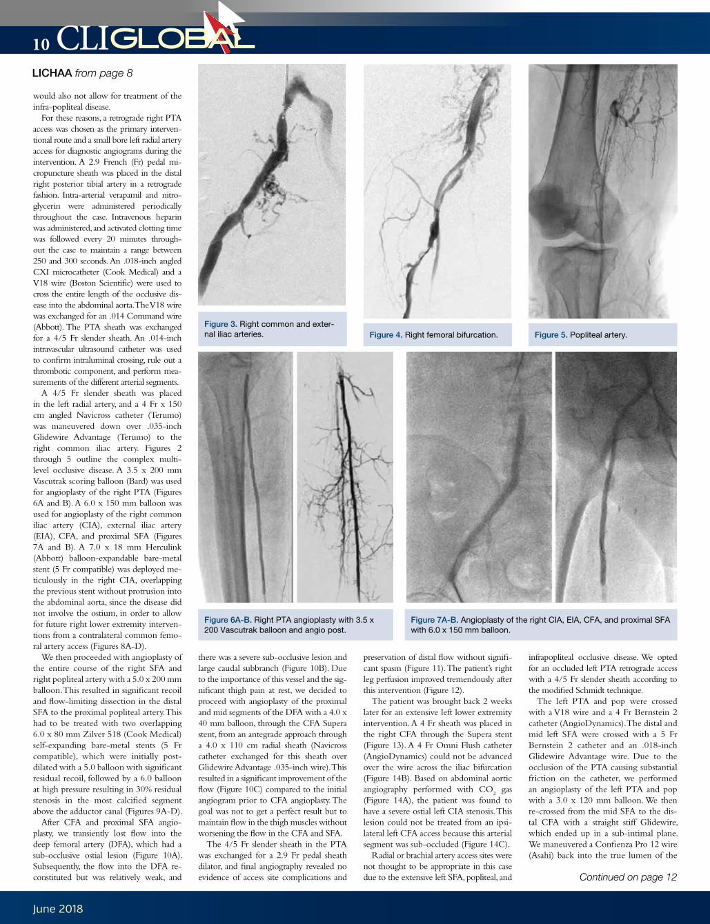
For these reasons, a retrograde right PTA access was chosen as the primary interven-tional route and a small bore left radial artery access for diagnostic angiograms during the intervention. A 2.9 French (Fr) pedal mi-cropuncture sheath was placed in the distal right posterior tibial artery in a retrograde fashion. Intra-arterial verapamil and nitro-glycerin were administered periodically throughout the case. Intravenous heparin was administered, and activated clotting time was followed every 20 minutes through-out the case to maintain a range between 250 and 300 seconds. An .018-inch angled CXI microcatheter (Cook Medical) and a V18 wire (Boston Scientific) were used to cross the entire length of the occlusive dis-ease into the abdominal aorta. The V18 wire was exchanged for an .014 Command wire (Abbott). The PTA sheath was exchanged for a 4/5 Fr slender sheath. An .014-inch intravascular ultrasound catheter was used to confirm intraluminal crossing, rule out a thrombotic component, and perform mea-surements of the different arterial segments.
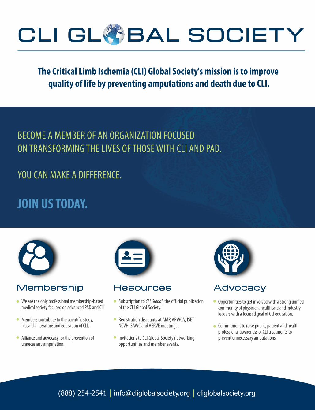
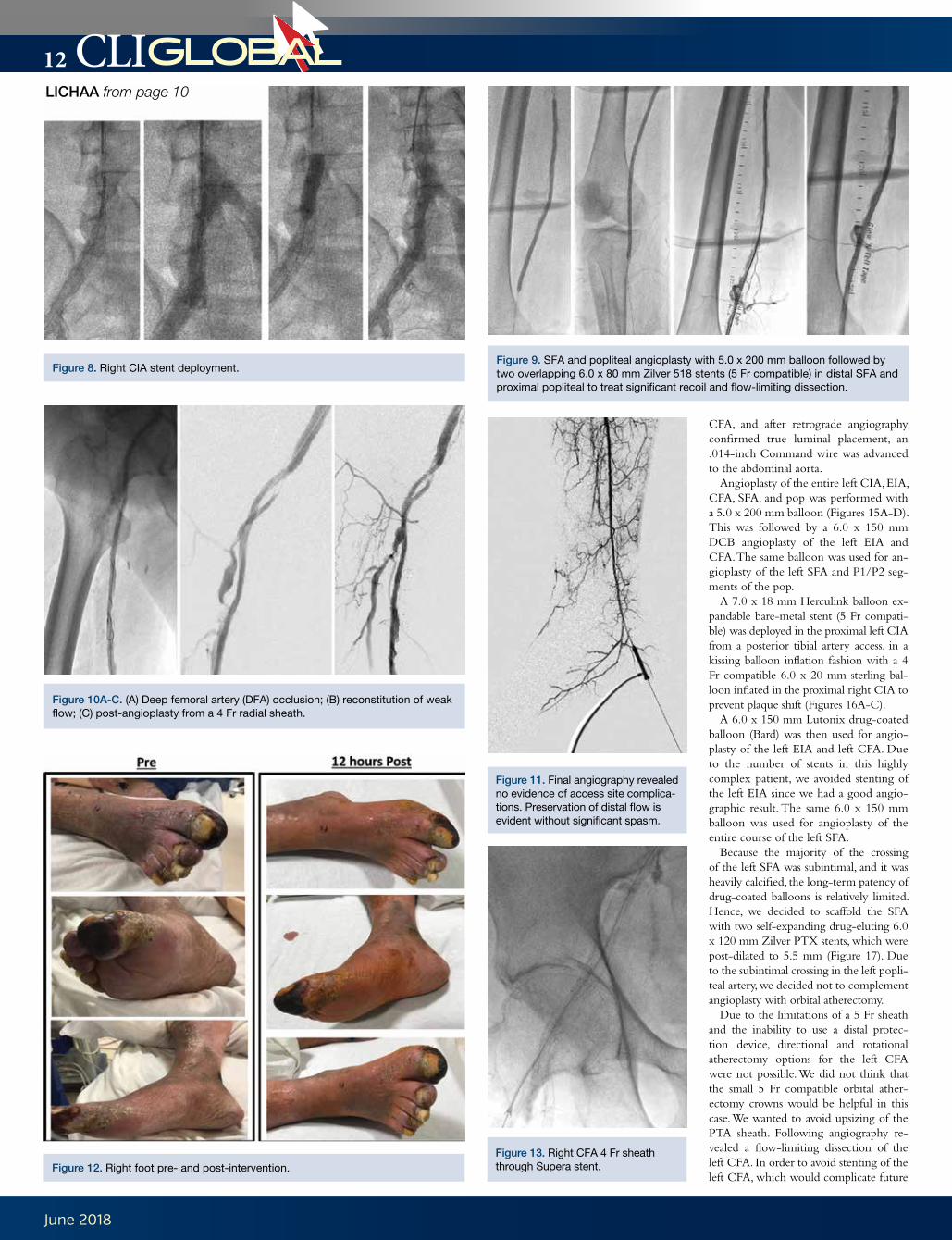
A 4/5 Fr slender sheath was placed in the left radial artery, and a 4 Fr x 150 cm angled Navicross catheter (Terumo) was maneuvered down over .035-inch Glidewire Advantage (Terumo) to the right common iliac artery. Figures 2 through 5 outline the complex multi-level occlusive disease. A 3.5 x 200 mm Vascutrak scoring balloon (Bard) was used for angioplasty of the right PTA (Figures 6A and B). A 6.0 x 150 mm balloon was used for angioplasty of the right common iliac artery (CIA), external iliac artery (EIA), CFA, and proximal SFA (Figures 7A and B). A 7.0 x 18 mm Herculink (Abbott) balloon-expandable bare-metal stent (5 Fr compatible) was deployed me-ticulously in the right CIA, overlapping the previous stent without protrusion into the abdominal aorta, since the disease did not involve the ostium, in order to allow for future right lower extremity interven-tions from a contralateral common femo-ral artery access (Figures 8A-D).
We then proceeded with angioplasty of the entire course of the right SFA and right popliteal artery with a 5.0 x 200 mm balloon. This resulted in significant recoil and flow-limiting dissection in the distal SFA to the proximal popliteal artery. This had to be treated with two overlapping 6.0 x 80 mm Zilver 518 (Cook Medical) self-expanding bare-metal stents (5 Fr compatible), which were initially post-dilated with a 5.0 balloon with significant residual recoil, followed by a 6.0 balloon at high pressure resulting in 30% residual stenosis in the most calcified segment above the adductor canal (Figures 9A-D).
After CFA and proximal SFA angio-plasty, we transiently lost flow into the deep femoral artery (DFA), which had a sub-occlusive ostial lesion (Figure 10A). Subsequently, the flow into the DFA re-constituted but was relatively weak, and
there was a severe sub-occlusive lesion and large caudal subbranch (Figure 10B). Due to the importance of this vessel and the sig-nificant thigh pain at rest, we decided to proceed with angioplasty of the proximal and mid segments of the DFA with a 4.0 x 40 mm balloon, through the CFA Supera stent, from an antegrade approach through a 4.0 x 110 cm radial sheath (Navicross catheter exchanged for this sheath over Glidewire Advantage .035-inch wire). This resulted in a significant improvement of the flow (Figure 10C) compared to the initial angiogram prior to CFA angioplasty. The goal was not to get a perfect result but to maintain flow in the thigh muscles without worsening the flow in the CFA and SFA.
The 4/5 Fr slender sheath in the PTA was exchanged for a 2.9 Fr pedal sheath dilator, and final angiography revealed no evidence of access site complications and
preservation of distal flow without signifi-cant spasm (Figure 11). The patient’s right leg perfusion improved tremendously after this intervention (Figure 12).
The patient was brought back 2 weeks later for an extensive left lower extremity intervention. A 4 Fr sheath was placed in the right CFA through the Supera stent (Figure 13). A 4 Fr Omni Flush catheter (AngioDynamics) could not be advanced over the wire across the iliac bifurcation (Figure 14B). Based on abdominal aortic angiography performed with CO
2 gas
(Figure 14A), the patient was found to have a severe ostial left CIA stenosis. This lesion could not be treated from an ipsi-lateral left CFA access because this arterial segment was sub-occluded (Figure 14C).
Radial or brachial artery access sites were not thought to be appropriate in this case due to the extensive left SFA, popliteal, and
infrapopliteal occlusive disease. We opted for an occluded left PTA retrograde access with a 4/5 Fr slender sheath according to the modified Schmidt technique.
The left PTA and pop were crossed with a V18 wire and a 4 Fr Bernstein 2 catheter (AngioDynamics). The distal and mid left SFA were crossed with a 5 Fr Bernstein 2 catheter and an .018-inch Glidewire Advantage wire. Due to the occlusion of the PTA causing substantial friction on the catheter, we performed an angioplasty of the left PTA and pop with a 3.0 x 120 mm balloon. We then re-crossed from the mid SFA to the dis-tal CFA with a straight stiff Glidewire, which ended up in a sub-intimal plane. We maneuvered a Confienza Pro 12 wire (Asahi) back into the true lumen of the
LICHAA from page 8
Continued on page 12
Figure 3. Right common and exter-nal iliac arteries.
Figure 6A-B. Right PTA angioplasty with 3.5 x 200 Vascutrak balloon and angio post.
Figure 7A-B. Angioplasty of the right CIA, EIA, CFA, and proximal SFA with 6.0 x 150 mm balloon.
Figure 4. Right femoral bifurcation. Figure 5. Popliteal artery.
CLISociety_CLIG0318.indd 1 1/31/18 10:34 AM
12
June 2018
CLIGLOBAL
CFA, and after retrograde angiography confirmed true luminal placement, an .014-inch Command wire was advanced to the abdominal aorta.
Angioplasty of the entire left CIA, EIA, CFA, SFA, and pop was performed with a 5.0 x 200 mm balloon (Figures 15A-D). This was followed by a 6.0 x 150 mm DCB angioplasty of the left EIA and CFA. The same balloon was used for an-gioplasty of the left SFA and P1/P2 seg-ments of the pop.
A 7.0 x 18 mm Herculink balloon ex-pandable bare-metal stent (5 Fr compati-ble) was deployed in the proximal left CIA from a posterior tibial artery access, in a kissing balloon inflation fashion with a 4 Fr compatible 6.0 x 20 mm sterling bal-loon inflated in the proximal right CIA to prevent plaque shift (Figures 16A-C).
A 6.0 x 150 mm Lutonix drug-coated balloon (Bard) was then used for angio-plasty of the left EIA and left CFA. Due to the number of stents in this highly complex patient, we avoided stenting of the left EIA since we had a good angio-graphic result. The same 6.0 x 150 mm balloon was used for angioplasty of the entire course of the left SFA.
Because the majority of the crossing of the left SFA was subintimal, and it was heavily calcified, the long-term patency of drug-coated balloons is relatively limited. Hence, we decided to scaffold the SFA with two self-expanding drug-eluting 6.0 x 120 mm Zilver PTX stents, which were post-dilated to 5.5 mm (Figure 17). Due to the subintimal crossing in the left popli-teal artery, we decided not to complement angioplasty with orbital atherectomy.
Due to the limitations of a 5 Fr sheath and the inability to use a distal protec-tion device, directional and rotational atherectomy options for the left CFA were not possible. We did not think that the small 5 Fr compatible orbital ather-ectomy crowns would be helpful in this case. We wanted to avoid upsizing of the PTA sheath. Following angiography re-vealed a flow-limiting dissection of the left CFA. In order to avoid stenting of the left CFA, which would complicate future
Figure 11. Final angiography revealed no evidence of access site complica-tions. Preservation of distal flow is evident without significant spasm.
Figure 13. Right CFA 4 Fr sheath through Supera stent.
Figure 8. Right CIA stent deployment.Figure 9. SFA and popliteal angioplasty with 5.0 x 200 mm balloon followed by two overlapping 6.0 x 80 mm Zilver 518 stents (5 Fr compatible) in distal SFA and proximal popliteal to treat significant recoil and flow-limiting dissection.
Figure 10A-C. (A) Deep femoral artery (DFA) occlusion; (B) reconstitution of weak flow; (C) post-angioplasty from a 4 Fr radial sheath.
LICHAA from page 10
Figure 12. Right foot pre- and post-intervention.
13
June 2018
CLIGLOBAL
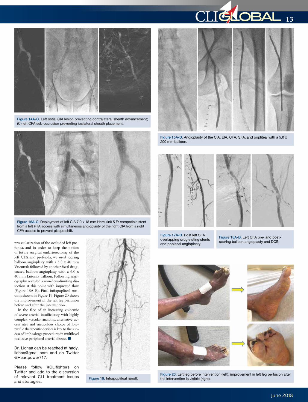
revascularization of the occluded left pro-funda, and in order to keep the option of future surgical endarterectomy of the left CFA and profunda, we used scoring balloon angioplasty with a 5.0 x 40 mm Vascutrak followed by another focal drug-coated balloon angioplasty with a 6.0 x 40 mm Lutonix balloon. Following angi-ography revealed a non-flow-limiting dis-section at this point with improved flow (Figure 18A-B). Final infrapopliteal run-off is shown in Figure 19. Figure 20 shows the improvement in the left leg perfusion before and after the intervention.
In the face of an increasing epidemic of severe arterial insufficiency with highly complex vascular anatomy, alternative ac-cess sites and meticulous choice of low-profile therapeutic devices is key to the suc-cess of limb salvage procedures in multilevel occlusive peripheral arterial disease. n
Dr. Lichaa can be reached at [email protected] and on Twitter @Heartpower717.
Please follow #CLIfighters on Twitter and add to the discussion of relevant CLI treatment issues and strategies.
Figure 14A-C. Left ostial CIA lesion preventing contralateral sheath advancement; (C) left CFA sub-occlusion preventing ipsilateral sheath placement.
Figure 16A-C. Deployment of left CIA 7.0 x 18 mm Herculink 5 Fr compatible stent from a left PTA access with simultaneous angioplasty of the right CIA from a right CFA access to prevent plaque shift.
Figure 17A-B. Post left SFA overlapping drug eluting stents and popliteal angioplasty.
Figure 18A-B. Left CFA pre- and post- scoring balloon angioplasty and DCB.
Figure 19. Infrapopliteal runoff.
Figure 15A-D. Angioplasty of the CIA, EIA, CFA, SFA, and popliteal with a 5.0 x 200 mm balloon.
Figure 20. Left leg before intervention (left); improvement in left leg perfusion after the intervention is visible (right).
14
June 2018
CLIGLOBAL
“Tell me what’s going on with your mom,” he said.
“Your mom is too young to go through this,” Dr. Mustapha replied, after listening to a litany of concerns.
“Bring her to me.”Nita’s eyes well with tears when re-
calling those four words, words that would change the very compass of her life and put her within a whole new realm of healthcare. She joined forces with Dr. Mustapha, who now directs the Advanced Cardiac and Vascular Amputation Prevention Center that he co-founded with Dr. Fadi Saab in Grand Rapids, Michigan.
Buoyed by hope, Nita climbed into the backseat of her car that same evening. With her sleuthing daughter at the wheel and accompanied by her three grandchil-dren, they drove non-stop for 12 hours from Maryland to West Michigan.
“Was it worth it?” she asks, and the question sparks a laugh. “Look at the smile on my face,” she says. “It was worth every mile.”
Within hours of arriving in West Michigan, Nita placed herself under the care of Dr. Mustapha, whose ground-breaking work in the area of critical limb ischemia (CLI) revascularization therapies has been able to stave off any further debilitating surgeries for her (Figure 1).
“I give Nita a lot of credit for the courage it took for her to drive that very night in search of something differ-ent,” says Dr. Mustapha. “By exerting her right to command a second opinion, she recoiled against a dangerous trend that is affecting a disproportionate number of African-Americans and Hispanics suf-fering from the threat of amputation.
“In leaving her former medical care in the rearview mirror that evening, she chose to not become another statistic.”
Dr. Mustapha isn’t speaking from con-jecture. In 2017, he served as lead au-thor on a seminal paper published in the Journal of Racial and Ethnic Health Disparities that found that “African-Americans and Hispanics have less ac-cess to care because they are being ad-mitted when sicker and more likely on an emergent basis.”1
“For the better part of two decades, the U.S. Department of Health and Human Services has led efforts to elimi-nate healthcare disparities as one of its overarching goals,” says Dr. Mustapha. “But what we discovered through a care-ful analysis of the data and anecdotal in-formation is there is virtually no evidence that attempts to explain the factors be-hind these disparities.”
Dr. Mustapha points to some sober-ing statistics that back this up. Among them: in the United States alone, African-Americans and Hispanics are up to two times more likely than their Caucasian counterparts to lose a leg to amputation.
“We have a lot of data to mine in order to get at the underlying reasons for this,” says Dr. Mustapha. “But what’s already obvious is that there are indeed racial and ethnic disparities when it comes to the incidence and frequency of amputations.”
In Nita’s case, there is no evidence that she suffered on account of her race. She’s just happy to now be living in Michigan full-time, making the decision to move closer to Dr. Mustapha and his practice, which is a few miles from a home she leases on the city’s South Side.
“I’ve met so many wonderful people who have blessed my life,” she says, “and it all started with Dr. Mustapha. He told me to call if I ever needed anything, and when I do, he always answers.”
Nita underwent several complex endo-vascular peripheral vascular revasculariza-tions including arteriovenous (AV) rever-sal, which is gaining some traction in the treatment of patients with end-stage CLI who have an arterial system that cannot be opened. The venous system is preferentially pressurized, and oxygenated blood flow is routed through this system by taking ad-vantage of the non-diseased path (i.e., the
veins) to reverse flow in major veins and also the smaller veins of the foot. Nita’s wound healing took place over a matter of months and was successful due to the highly coordinated multidisciplinary approach to her care, which included endovascular re-vascularization specialists, podiatry, wound care, and primary care.
“He’s for real,” says Nita of Dr. Mustapha, “a unique doctor who listens to you and pays attention to everything you have to say, no matter how long it takes. He’ll sit by your bedside and hold your hand and explain what he wants and hopes to do.
“I feel safe.”Looking back at the care she received
prior to moving to Michigan, especially the amputation of several of her toes, Nita re-members that she was close to giving up when her daughters interceded.
“I had always told them to believe in mir-acles,” says Nita, “and now it was their turn. They said, ‘Mama, you always told us to step out on faith and believe in miracles. Now you’re our miracle, and we believe in you.’”
Nita says she found solace in Dr. Mustapha and developed an abiding trust in him the first time he conveyed an invitation
to consult him, emphasizing that “it made me anxious to see him, to see this man that some were calling ‘The Leg Saver’.”
Nita spent most of her adult career in the restaurant business, and flirts with the idea of opening a bistro in the Grand Rapids area that would showcase her se-cret-recipe gumbo.
Until then, she’ll continue to rely on Dr. Mustapha’s “blessed hands” and the team with which he surrounds himself.
Though her daughters wonder wheth-er their mom should move back to Maryland, Nita says she’s committed to remaining in Michigan, primarily to avail herself of Dr. Mustapha’s continuing care.
racial disparities in amputation rates for the treatment of peripheral artery disease (PAD) using decomposition methods. J Racial Ethn Health Disparities. 2017. doi: 10.1007/s40615-016-0261-9. [Epub ahead of print]
Please follow #CLIfighters on Twitter and add to the discussion of relevant CLI treatment issues and strategies.
“Nita’s wound healing took place over a matter of months and was successful due to the highly coordinated multidisciplinary approach to her care, which included endovascular revascularization specialists, podiatry, wound care, and primary care.”
RADEMACHER from page 3
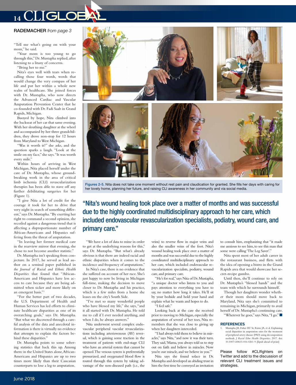
Figures 2-5. Nita does not take one moment without rest pain and claudication for granted. She fills her days with caring for her lovely home, planning her future, and raising CLI awareness in her community and via social media.
15
June 2018
CLIGLOBAL
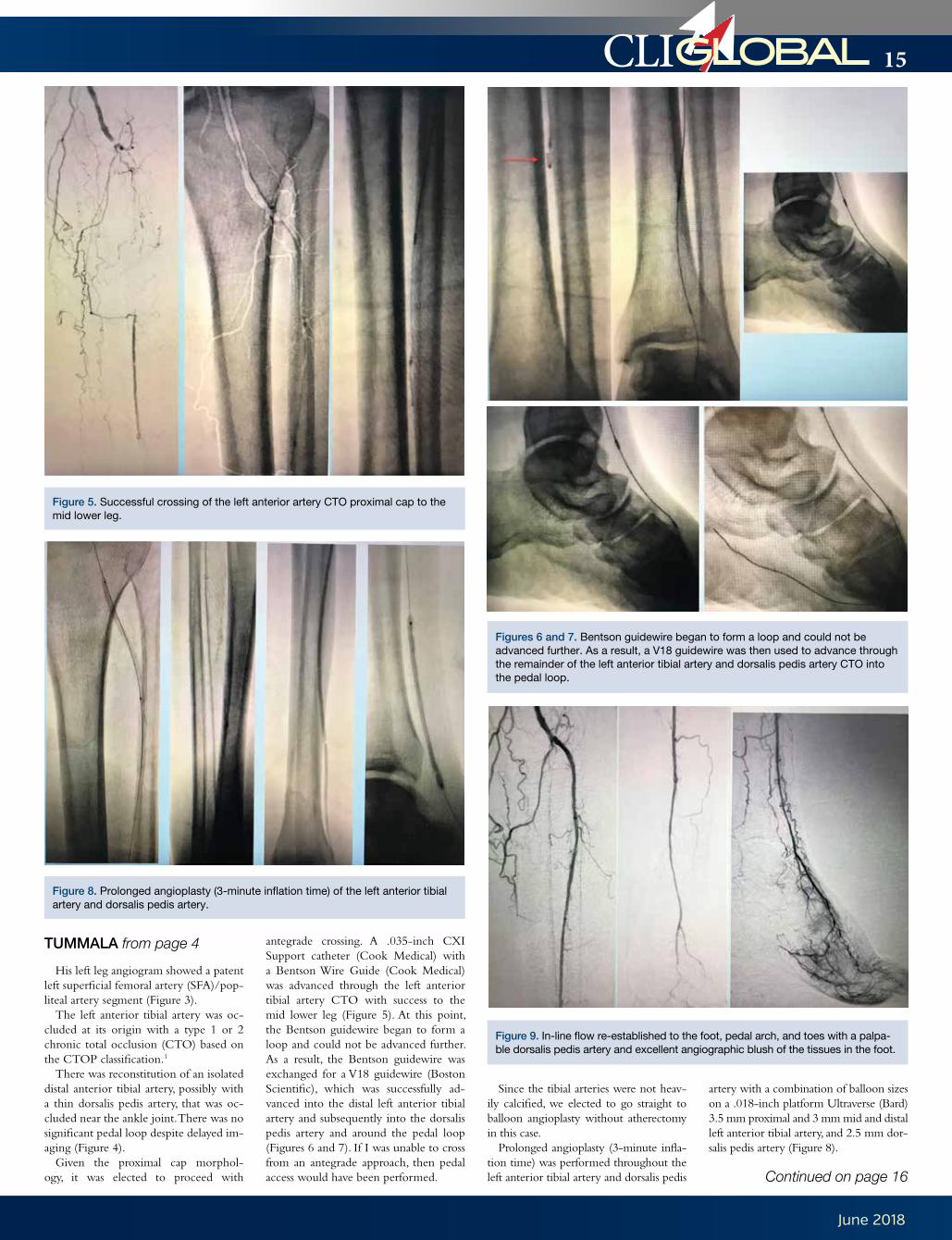
His left leg angiogram showed a patent left superficial femoral artery (SFA)/pop-liteal artery segment (Figure 3).
The left anterior tibial artery was oc-cluded at its origin with a type 1 or 2 chronic total occlusion (CTO) based on the CTOP classification.1
There was reconstitution of an isolated distal anterior tibial artery, possibly with a thin dorsalis pedis artery, that was oc-cluded near the ankle joint. There was no significant pedal loop despite delayed im-aging (Figure 4).
Given the proximal cap morphol-ogy, it was elected to proceed with
antegrade crossing. A .035-inch CXI Support catheter (Cook Medical) with a Bentson Wire Guide (Cook Medical) was advanced through the left anterior tibial artery CTO with success to the mid lower leg (Figure 5). At this point, the Bentson guidewire began to form a loop and could not be advanced further. As a result, the Bentson guidewire was exchanged for a V18 guidewire (Boston Scientific), which was successfully ad-vanced into the distal left anterior tibial artery and subsequently into the dorsalis pedis artery and around the pedal loop (Figures 6 and 7). If I was unable to cross from an antegrade approach, then pedal access would have been performed.
Since the tibial arteries were not heav-ily calcified, we elected to go straight to balloon angioplasty without atherectomy in this case.
Prolonged angioplasty (3-minute infla-tion time) was performed throughout the left anterior tibial artery and dorsalis pedis
artery with a combination of balloon sizes on a .018-inch platform Ultraverse (Bard) 3.5 mm proximal and 3 mm mid and distal left anterior tibial artery, and 2.5 mm dor-salis pedis artery (Figure 8).
Figure 5. Successful crossing of the left anterior artery CTO proximal cap to the mid lower leg.
Figure 8. Prolonged angioplasty (3-minute inflation time) of the left anterior tibial artery and dorsalis pedis artery.
Figure 9. In-line flow re-established to the foot, pedal arch, and toes with a palpa-ble dorsalis pedis artery and excellent angiographic blush of the tissues in the foot.
Figures 6 and 7. Bentson guidewire began to form a loop and could not be advanced further. As a result, a V18 guidewire was then used to advance through the remainder of the left anterior tibial artery and dorsalis pedis artery CTO into the pedal loop.
TUMMALA from page 4
Continued on page 16
16
June 2018
CLIGLOBAL
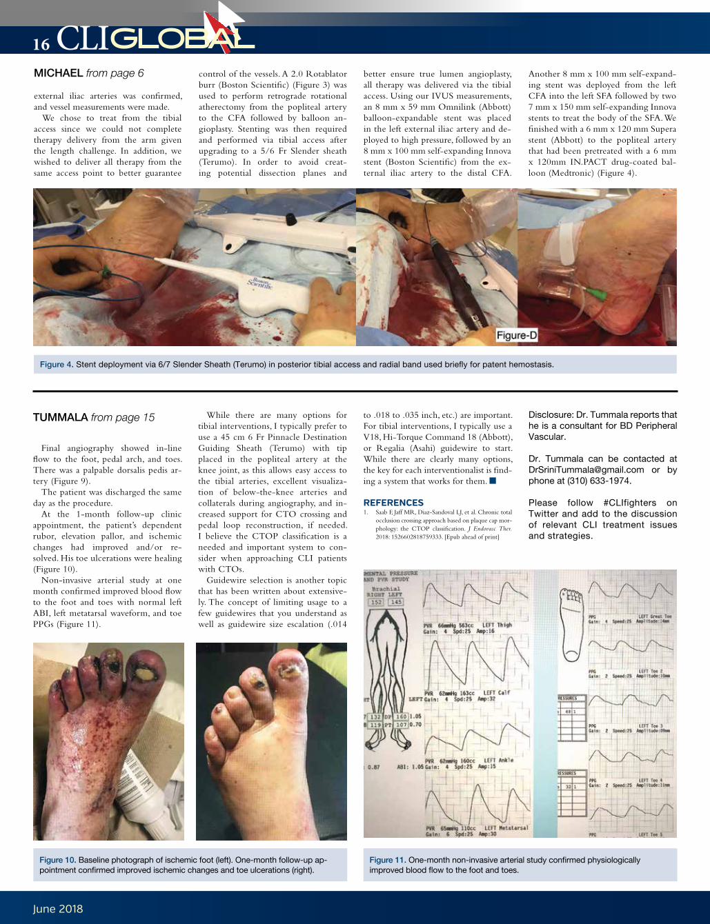
external iliac arteries was confirmed, and vessel measurements were made.
We chose to treat from the tibial access since we could not complete therapy delivery from the arm given the length challenge. In addition, we wished to deliver all therapy from the same access point to better guarantee
control of the vessels. A 2.0 Rotablator burr (Boston Scientific) (Figure 3) was used to perform retrograde rotational atherectomy from the popliteal artery to the CFA followed by balloon an-gioplasty. Stenting was then required and performed via tibial access after upgrading to a 5/6 Fr Slender sheath (Terumo). In order to avoid creat-ing potential dissection planes and
better ensure true lumen angioplasty, all therapy was delivered via the tibial access. Using our IVUS measurements, an 8 mm x 59 mm Omnilink (Abbott) balloon-expandable stent was placed in the left external iliac artery and de-ployed to high pressure, followed by an 8 mm x 100 mm self-expanding Innova stent (Boston Scientific) from the ex-ternal iliac artery to the distal CFA.
Another 8 mm x 100 mm self-expand-ing stent was deployed from the left CFA into the left SFA followed by two 7 mm x 150 mm self-expanding Innova stents to treat the body of the SFA. We finished with a 6 mm x 120 mm Supera stent (Abbott) to the popliteal artery that had been pretreated with a 6 mm x 120mm IN.PACT drug-coated bal-loon (Medtronic) (Figure 4).
MICHAEL from page 6
Final angiography showed in-line flow to the foot, pedal arch, and toes. There was a palpable dorsalis pedis ar-tery (Figure 9).
The patient was discharged the same day as the procedure.
At the 1-month follow-up clinic appointment, the patient’s dependent rubor, elevation pallor, and ischemic changes had improved and/or re-solved. His toe ulcerations were healing (Figure 10).
Non-invasive arterial study at one month confirmed improved blood flow to the foot and toes with normal left ABI, left metatarsal waveform, and toe PPGs (Figure 11).
While there are many options for tibial interventions, I typically prefer to use a 45 cm 6 Fr Pinnacle Destination Guiding Sheath (Terumo) with tip placed in the popliteal artery at the knee joint, as this allows easy access to the tibial arteries, excellent visualiza-tion of below-the-knee arteries and collaterals during angiography, and in-creased support for CTO crossing and pedal loop reconstruction, if needed. I believe the CTOP classification is a needed and important system to con-sider when approaching CLI patients with CTOs.
Guidewire selection is another topic that has been written about extensive-ly. The concept of limiting usage to a few guidewires that you understand as well as guidewire size escalation (.014
to .018 to .035 inch, etc.) are important. For tibial interventions, I typically use a V18, Hi-Torque Command 18 (Abbott), or Regalia (Asahi) guidewire to start. While there are clearly many options, the key for each interventionalist is find-ing a system that works for them. n
REFERENCES1. Saab F, Jaff MR, Diaz-Sandoval LJ, et al. Chronic total
occlusion crossing approach based on plaque cap mor-phology: the CTOP classification. J Endovasc Ther. 2018: 1526602818759333. [Epub ahead of print]
Disclosure: Dr. Tummala reports that he is a consultant for BD Peripheral Vascular.
Dr. Tummala can be contacted at [email protected] or by phone at (310) 633-1974.
Please follow #CLIfighters on Twitter and add to the discussion of relevant CLI treatment issues and strategies.
TUMMALA from page 15
Figure 10. Baseline photograph of ischemic foot (left). One-month follow-up ap-pointment confirmed improved ischemic changes and toe ulcerations (right).
Figure 11. One-month non-invasive arterial study confirmed physiologically improved blood flow to the foot and toes.
Figure 4. Stent deployment via 6/7 Slender Sheath (Terumo) in posterior tibial access and radial band used briefly for patent hemostasis.
17
June 2018
CLIGLOBAL
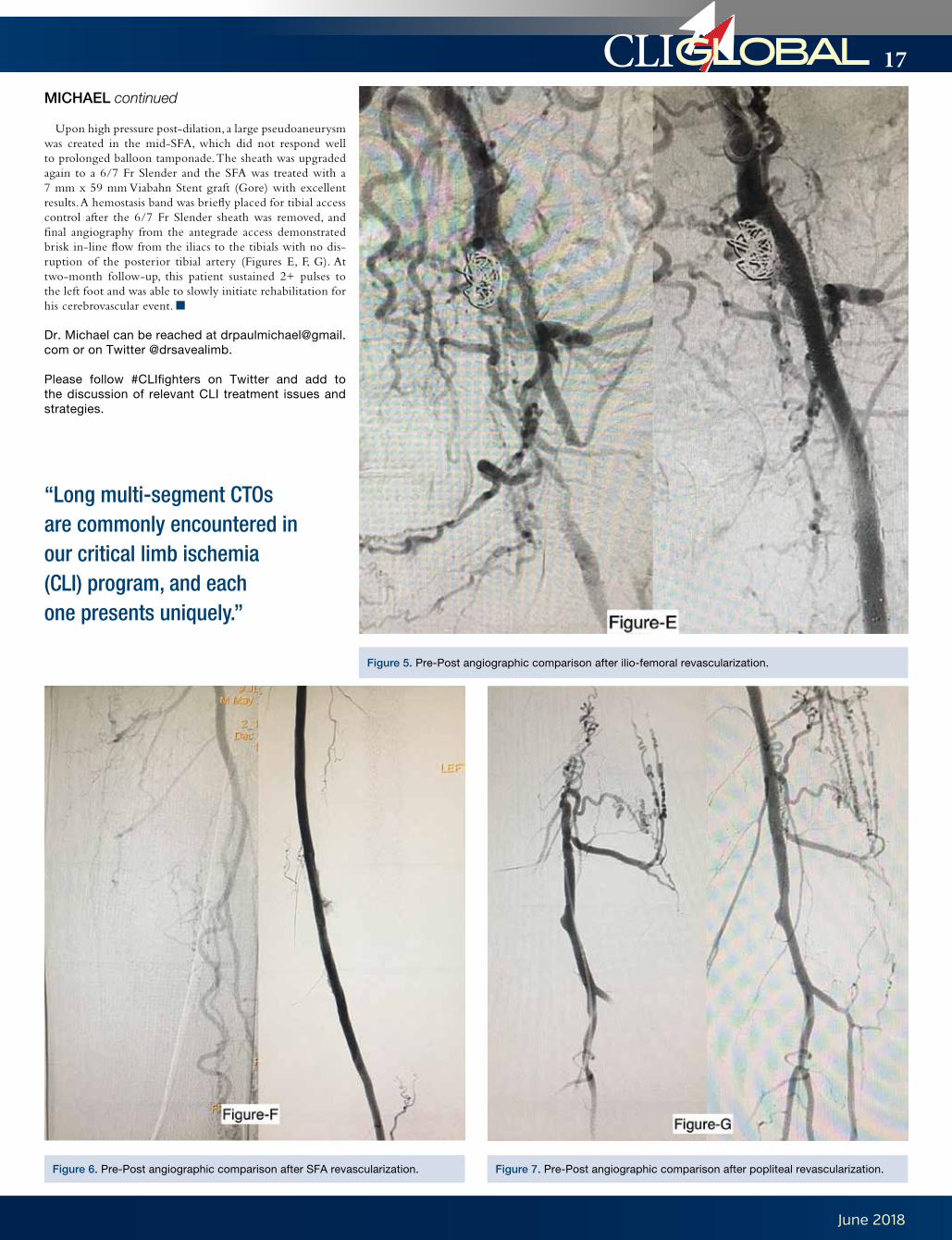
Upon high pressure post-dilation, a large pseudoaneurysm was created in the mid-SFA, which did not respond well to prolonged balloon tamponade. The sheath was upgraded again to a 6/7 Fr Slender and the SFA was treated with a 7 mm x 59 mm Viabahn Stent graft (Gore) with excellent results. A hemostasis band was briefly placed for tibial access control after the 6/7 Fr Slender sheath was removed, and final angiography from the antegrade access demonstrated brisk in-line flow from the iliacs to the tibials with no dis-ruption of the posterior tibial artery (Figures E, F, G). At two-month follow-up, this patient sustained 2+ pulses to the left foot and was able to slowly initiate rehabilitation for his cerebrovascular event. n
Dr. Michael can be reached at [email protected] or on Twitter @drsavealimb.
Please follow #CLIfighters on Twitter and add to the discussion of relevant CLI treatment issues and strategies.
MICHAEL continued
Figure 6. Pre-Post angiographic comparison after SFA revascularization.
Figure 5. Pre-Post angiographic comparison after ilio-femoral revascularization.
Figure 7. Pre-Post angiographic comparison after popliteal revascularization.
“Long multi-segment CTOs are commonly encountered in our critical limb ischemia (CLI) program, and each one presents uniquely.”
18
June 2018
CLIGLOBAL
80%) are paid for by Medicare—in other words, our tax dollars. Numerous factors increase CLI costs, including the use of major amputation rather than revascu-larization, which is a poor allocation of economic resources. It is also a waste of our tax dollars. The tragedy is that major amputation, as well as many of the other cost-drivers such as significant delays in diagnosis and treatment, suboptimal med-ical management, procedural complica-tions, and a high rate of unplanned re-admissions, are preventable or modifiable.
Dr. Sonya Noor shared her experience treating CLI: “Amputation isn’t just an-other operation. We talk about statistics
of death and cost. We have dehumanized it. Let me explain. Usually after treating and failing, it is all that’s left to do. You have to look your elderly, weak patient in the eye and tell them they are going to lose a part of their body, and with that goes their independence, self-respect, and dignity. Going to the bathroom in the middle of the night is a challenge. Getting out of bed and not falling, learning how to walk all over again, if they even can, presents more challenges. Few can drive, play golf, or bowl, which are common post-retirement activities. Then the feelings of being disfigured, inadequate, and unacceptable creep in. It’s not just another operation. It is a death sentence.”
Per Dr. Katzen, a major issue in imple-menting change is defining CLI in coding terms. The Society is committed to the pub-lic health goal of reducing the incidence of mortality and amputation due to CLI. To do so, we need the fundamental data. The Society’s work is focused on creating aware-ness clinically and within governing bod-ies to lead to these changes that will define endpoints and support the development of new techniques and technologies—all to support the treatment of patients with CLI.
Dr. Katzen emphasized, “We currently do not have a solid foundation on how to approach the cascade of events in CLI therapy. In my opinion, the current rate of amputation without a diagnostic angio-gram is completely unacceptable!”
CLI FIGHTERS from Cover
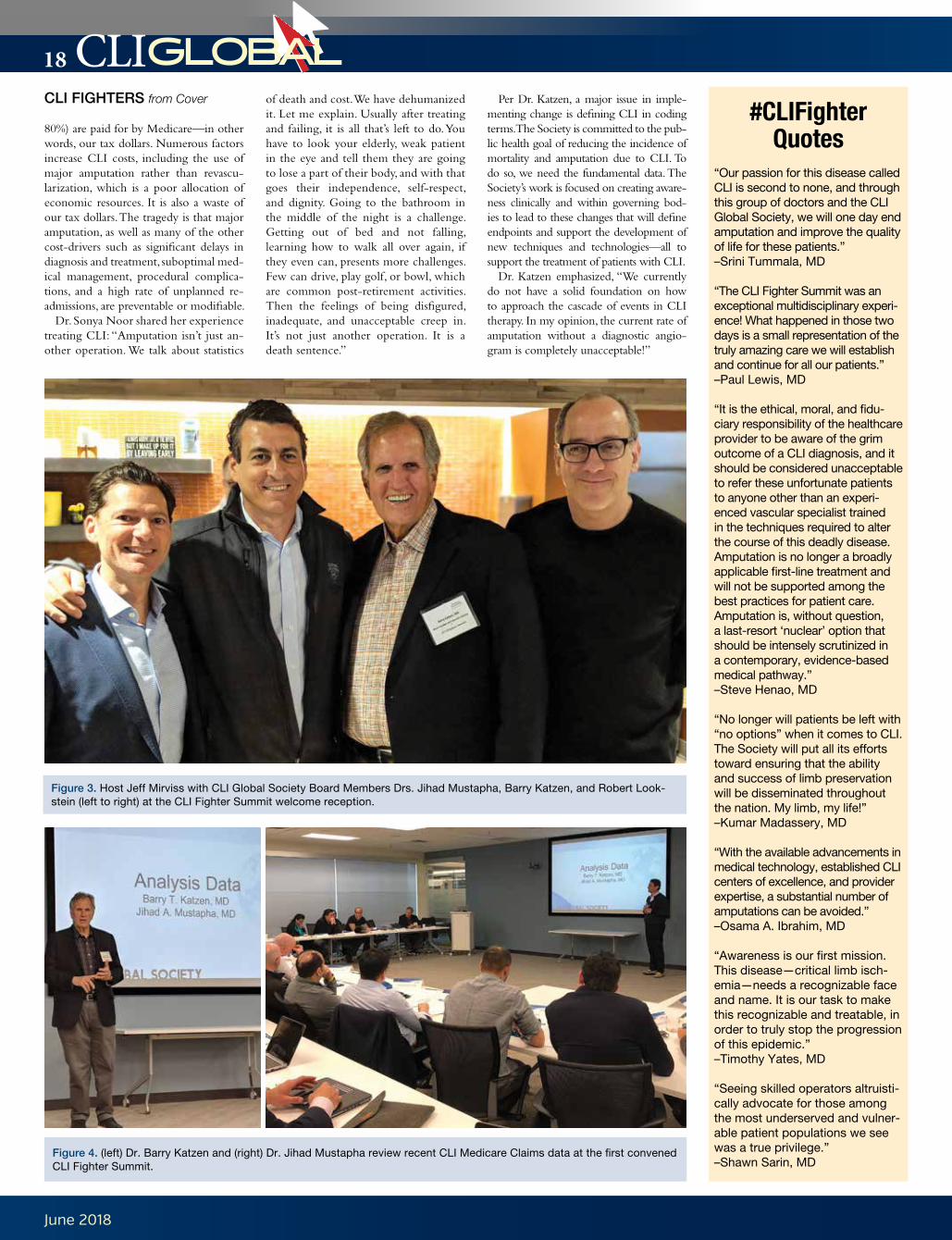
Figure 3. Host Jeff Mirviss with CLI Global Society Board Members Drs. Jihad Mustapha, Barry Katzen, and Robert Look-stein (left to right) at the CLI Fighter Summit welcome reception.
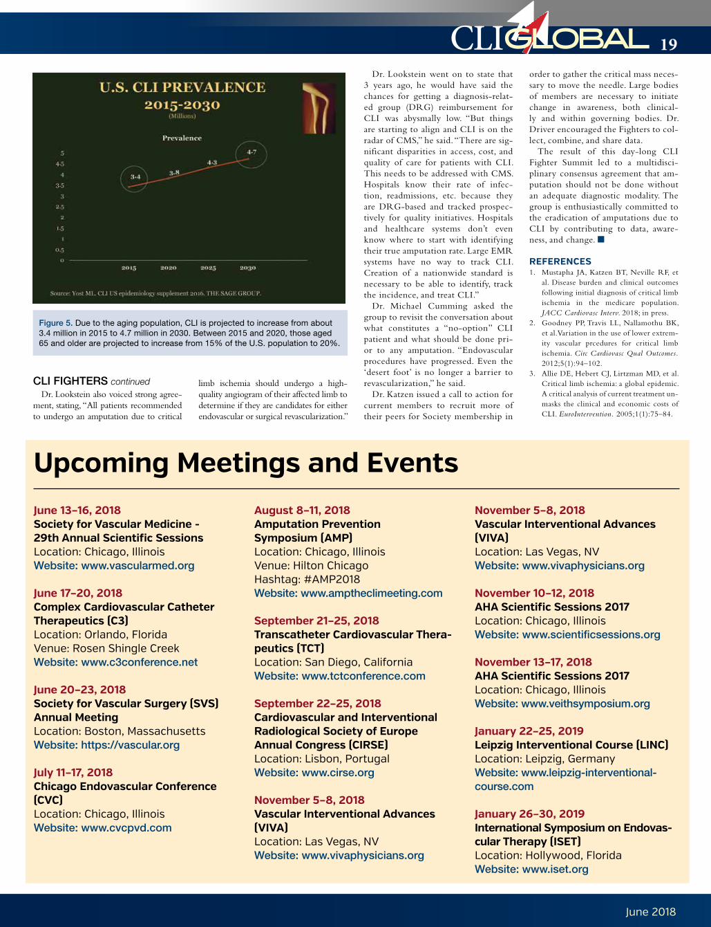
Figure 4. (left) Dr. Barry Katzen and (right) Dr. Jihad Mustapha review recent CLI Medicare Claims data at the first convened CLI Fighter Summit.
#CLIFighter Quotes
“Our passion for this disease called CLI is second to none, and through this group of doctors and the CLI Global Society, we will one day end amputation and improve the quality of life for these patients.” –Srini Tummala, MD
“The CLI Fighter Summit was an exceptional multidisciplinary experi-ence! What happened in those two days is a small representation of the truly amazing care we will establish and continue for all our patients.”–Paul Lewis, MD
“It is the ethical, moral, and fidu-ciary responsibility of the healthcare provider to be aware of the grim outcome of a CLI diagnosis, and it should be considered unacceptable to refer these unfortunate patients to anyone other than an experi-enced vascular specialist trained in the techniques required to alter the course of this deadly disease. Amputation is no longer a broadly applicable first-line treatment and will not be supported among the best practices for patient care. Amputation is, without question, a last-resort ‘nuclear’ option that should be intensely scrutinized in a contemporary, evidence-based medical pathway.”–Steve Henao, MD
“No longer will patients be left with “no options” when it comes to CLI. The Society will put all its efforts toward ensuring that the ability and success of limb preservation will be disseminated throughout the nation. My limb, my life!”–Kumar Madassery, MD
“With the available advancements in medical technology, established CLI centers of excellence, and provider expertise, a substantial number of amputations can be avoided.”–Osama A. Ibrahim, MD
“Awareness is our first mission. This disease—critical limb isch-emia—needs a recognizable face and name. It is our task to make this recognizable and treatable, in order to truly stop the progression of this epidemic.”–Timothy Yates, MD
“Seeing skilled operators altruisti-cally advocate for those among the most underserved and vulner-able patient populations we see was a true privilege.”–Shawn Sarin, MD
19
June 2018
CLIGLOBAL
Dr. Lookstein also voiced strong agree-ment, stating, “All patients recommended to undergo an amputation due to critical
limb ischemia should undergo a high-quality angiogram of their affected limb to determine if they are candidates for either endovascular or surgical revascularization.”
Dr. Lookstein went on to state that 3 years ago, he would have said the chances for getting a diagnosis-relat-ed group (DRG) reimbursement for CLI was abysmally low. “But things are starting to align and CLI is on the radar of CMS,” he said. “There are sig-nificant disparities in access, cost, and quality of care for patients with CLI. This needs to be addressed with CMS. Hospitals know their rate of infec-tion, readmissions, etc. because they are DRG-based and tracked prospec-tively for quality initiatives. Hospitals and healthcare systems don’t even know where to start with identifying their true amputation rate. Large EMR systems have no way to track CLI. Creation of a nationwide standard is necessary to be able to identify, track the incidence, and treat CLI.”
Dr. Michael Cumming asked the group to revisit the conversation about what constitutes a “no-option” CLI patient and what should be done pri-or to any amputation. “Endovascular procedures have progressed. Even the ‘desert foot’ is no longer a barrier to revascularization,” he said.
Dr. Katzen issued a call to action for current members to recruit more of their peers for Society membership in
order to gather the critical mass neces-sary to move the needle. Large bodies of members are necessary to initiate change in awareness, both clinical-ly and within governing bodies. Dr. Driver encouraged the Fighters to col-lect, combine, and share data.
The result of this day-long CLI Fighter Summit led to a multidisci-plinary consensus agreement that am-putation should not be done without an adequate diagnostic modality. The group is enthusiastically committed to the eradication of amputations due to CLI by contributing to data, aware-ness, and change. n
REFERENCES1. Mustapha JA, Katzen BT, Neville RF, et
al. Disease burden and clinical outcomes following initial diagnosis of critical limb ischemia in the medicare population. JACC Cardiovasc Interv. 2018; in press.
2. Goodney PP, Travis LL, Nallamothu BK, et al. Variation in the use of lower extrem-ity vascular prcedures for critical limb ischemia. Circ Cardiovasc Qual Outcomes. 2012;5(1):94–102.
3. Allie DE, Hebert CJ, Lirtzman MD, et al. Critical limb ischemia: a global epidemic. A critical analysis of current treatment un-masks the clinical and economic costs of CLI. EuroIntervention. 2005;1(1):75–84.
Figure 5. Due to the aging population, CLI is projected to increase from about 3.4 million in 2015 to 4.7 million in 2030. Between 2015 and 2020, those aged 65 and older are projected to increase from 15% of the U.S. population to 20%.
CLI FIGHTERS continued
June 13–16, 2018Society for Vascular Medicine - 29th Annual Scientific SessionsLocation: Chicago, IllinoisWebsite: www.vascularmed.org
September 21–25, 2018Transcatheter Cardiovascular Thera-peutics (TCT) Location: San Diego, CaliforniaWebsite: www.tctconference.com
September 22–25, 2018Cardiovascular and Interventional Radiological Society of Europe Annual Congress (CIRSE) Location: Lisbon, PortugalWebsite: www.cirse.org
November 5–8, 2018Vascular Interventional Advances (VIVA) Location: Las Vegas, NVWebsite: www.vivaphysicians.org
November 5–8, 2018Vascular Interventional Advances (VIVA) Location: Las Vegas, NVWebsite: www.vivaphysicians.org
November 10–12, 2018AHA Scientific Sessions 2017 Location: Chicago, IllinoisWebsite: www.scientificsessions.org
November 13–17, 2018AHA Scientific Sessions 2017Location: Chicago, IllinoisWebsite: www.veithsymposium.org
January 22–25, 2019Leipzig Interventional Course (LINC)Location: Leipzig, GermanyWebsite: www.leipzig-interventional-course.com
January 26–30, 2019International Symposium on Endovas-cular Therapy (ISET)Location: Hollywood, Florida Website: www.iset.org
Upcoming Meetings and Events
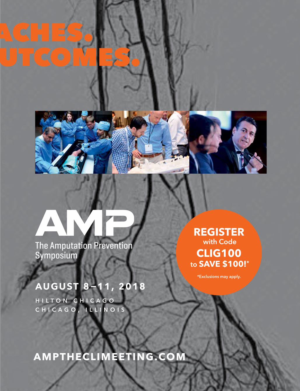
august 8–11, 2018H i l t o n C H i C a g oC H i C a g o , i l l i n o i s
AMPTHECLIMEETING.COM
REGISTER with Code
CLIG100 to SAVE $100!*
*Exclusions may apply.
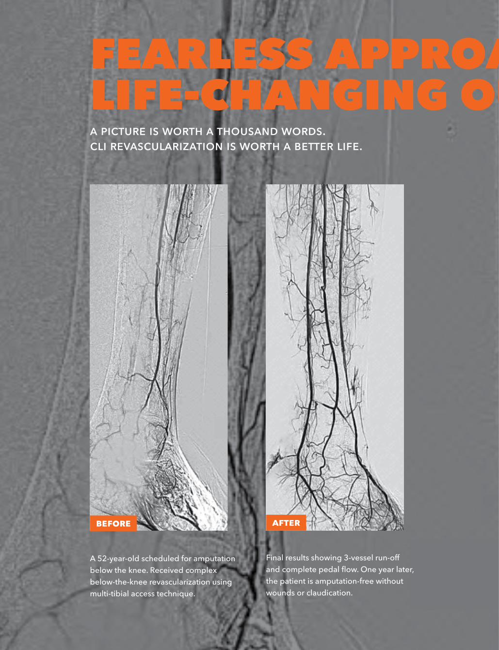
A 52-year-old scheduled for amputation below the knee. Received complex below-the-knee revascularization using multi-tibial access technique.
Final results showing 3-vessel run-off and complete pedal flow. One year later, the patient is amputation-free without wounds or claudication.
Afterbefore
fearless approaches. life-changing outcomes.a piCture is wortH a tHousand words. Cli revasCularization is wortH a better life.
2018-AMP_Journal_2pg_Ads_June.indd 16-17 5/11/18 9:26 AM
august 8–11, 2018H i l t o n C H i C a g oC H i C a g o , i l l i n o i s
AMPTHECLIMEETING.COM
REGISTER with Code
CLIG100 to SAVE $100!*
*Exclusions may apply.
A 52-year-old scheduled for amputation below the knee. Received complex below-the-knee revascularization using multi-tibial access technique.
Final results showing 3-vessel run-off and complete pedal flow. One year later, the patient is amputation-free without wounds or claudication.
Afterbefore
fearless approaches. life-changing outcomes.a piCture is wortH a tHousand words. Cli revasCularization is wortH a better life.
2018-AMP_Journal_2pg_Ads_June.indd 16-17 5/11/18 9:26 AM
22
June 2018
CLIGLOBAL
and ultrasound (US) performed in pa-tients with peripheral artery disease (PAD) can be used to predict the suc-cess of different interventional ap-proaches (antegrade vs retrograde vs combined antegrade-retrograde) in traversing CTOs.1 The CTOP tri-al included patients enrolled in the Peripheral Registry of Endovascular Outcomes (PRIME Registry). The general practice of clinicians to traverse a CTO depends on the physician’s ex-perience, tools available, and the cen-ter’s experience treating patients with CTOs. Based on the morphology of the CTO, the operator can choose how to cross it. The traditional way of crossing a CTO centers on obtaining common femoral artery (CFA) access and tra-versing the occlusion from a cranial ap-proach toward the foot. The success of this approach is inconsistent, with fail-ure rates up to 40%. Tibio-pedal access has been utilized to increase the success rate of crossing a CTO.2,3
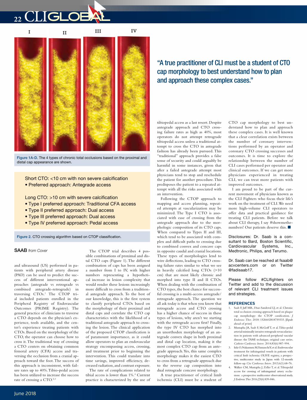
The CTOP trial describes 4 pos-sible combinations of proximal and dis-tal CTO caps (Figure 1). The different combination of caps has been assigned a number from I to IV, with higher numbers representing a hypotheti-cal increase in lesion complexity that would render these lesions increasingly more difficult to cross from a tradition-al antegrade approach. To the best of our knowledge, this is the first system to classify peripheral CTOs based on the morphology of their proximal and distal caps and correlate the CTO cap characteristics with the likelihood of a traditional antegrade approach to cross-ing the lesion. The clinical application of the proposed CTOP classification is of paramount importance, as it could allow operators to plan an endovascular strategy encompassing access, crossing, and treatment prior to beginning the intervention. This could translate into time savings, improved efficiency, de-creased radiation, and contrast exposure.
The rate of complications related to tibial access is lower than 1%.4 Current practice is characterized by the use of
tibiopedal access as a last resort. Despite antegrade approach and CTO cross-ing failure rates as high as 40%, most operators do not attempt retrograde tibiopedal access unless a traditional at-tempt to cross the CTO in antegrade fashion has already been pursued. This “traditional” approach provides a false sense of security and could arguably be harmful in some instances, given that after a failed antegrade attempt most physicians tend to stop and reschedule the patient for another procedure. This predisposes the patient to a repeated at-tempt with all the risks associated with an intervention.
Following the CTOP approach to mapping and access planning, repeat-ed attempts at vascularization may be minimized. The Type I CTO is asso-ciated with ease of crossing from the antegrade approach due to the mor-phologic composition of its CTO caps. When compared to Types II and III, which tend to be associated with com-plex and difficult paths to crossing due to combined convex and concave caps in opposite caudal and cranial locations. These types of morphologies lend to wire deflections, leading to CTO cross-ing failure rates similar to what we see in heavily calcified long CTOs (>10 cm) that are most likely chronic and morphed into type II and II CTOs. When dealing with the combination of CTO types, the best chance for success-ful crossing is a multi-access antegrade/retrograde approach. The question we all ask today is that when you know that retrograde access and CTO crossing has a higher chance of success in these types of lesions, why aren’t we starting with the retrograde access first? Finally, the type IV CTO has morphed into an unorthodox morphology of an an-tegrade convex shape in both proximal and distal cap location, making it the most complex CTO cap from an ante-grade approach. Yes, this same complex morphology makes it the easiest CTO to cross from a retrograde approach due to the reverse cap composition into dual retrograde concave morphology.
A true practitioner of critical limb ischemia (CLI) must be a student of
CTO cap morphology to best un-derstand how to plan and approach these complex cases. It is well known that a clear correlation exists between the number of coronary interven-tions performed by an operator and coronary CTO crossing successes and outcomes. It is time to explore the relationship between the number of CLI cases performed per operator and clinical outcomes. If we can get more physicians experienced in treating CLI, we can treat more patients with improved outcomes.
I am proud to be part of the cur-rent movement of physicians known as the CLI Fighters who focus their life’s work on the treatment of CLI. We need more high-volume CLI operators to offer data and practical guidance for treating CLI patients. Before we talk about CLI therapy, I say #showmethe-numbers! Our patients deserve this. n
Disclosures: Dr. Saab is a con-sultant to Bard, Boston Scientific, Cardiovascular Systems, Inc., Medtronic, Philips, and Terumo.
Dr. Saab can be reached at [email protected] or on Twitter @fadisaab17.
Please follow #CLIfighters on Twitter and add to the discussion of relevant CLI treatment issues and strategies.
REFERENCES1. Saab F, Jaff MR, Diaz-Sandoval LJ, et al. Chronic
total occlusion crossing approach based on plaque cap morphology: the CTOP cassification. J Endovasc Ther. 2018: 1526602818759333. [Epub ahead of print]
2. Mustapha JA, Saab F, McGoff T, et al. Tibio-pedal arterial minimally invasive retrograde revasculariza-tion in patients with advanced peripheral vascular disease: the TAMI technique, original case series. Catheter Cardiovasc Interv. 2014;83(6):987–994.
3. Iida O, Nakamura M, Yamauchi Y, et al. Endovascular treatment for infrainguinal vessels in patients with critical limb ischemia: OLIVE registry, a prospec-tive, multicenter study in Japan with 12-month follow-up. Circ Cardiovasc Interv. 2013;6(1):68–76.
4. Walker CM, Mustapha J, Zeller T, et al. Tibiopedal access for crossing of infrainguinal artery occlu-sions: a prospective multicenter observational study. J Endovasc Ther. 2016;23(6):839-846.
SAAB from Cover
Figure 1A-D. The 4 types of chronic total occlusions based on the proximal and distal cap appearance are shown.
Short CTO: <10 cm with non severe calcification• Preferred approach: Antegrade access
Long CTO: >10 cm with severe calcification• Type I preferred approach: Traditional CFA access• Type II preferred approach: Dual access• Type III preferred approach: Dual access• Type IV preferred approach: Pedal access
Figure 2. CTO crossing algorithm based on CTOP classification.
“A true practitioner of CLI must be a student of CTO cap morphology to best understand how to plan and approach these complex cases.”
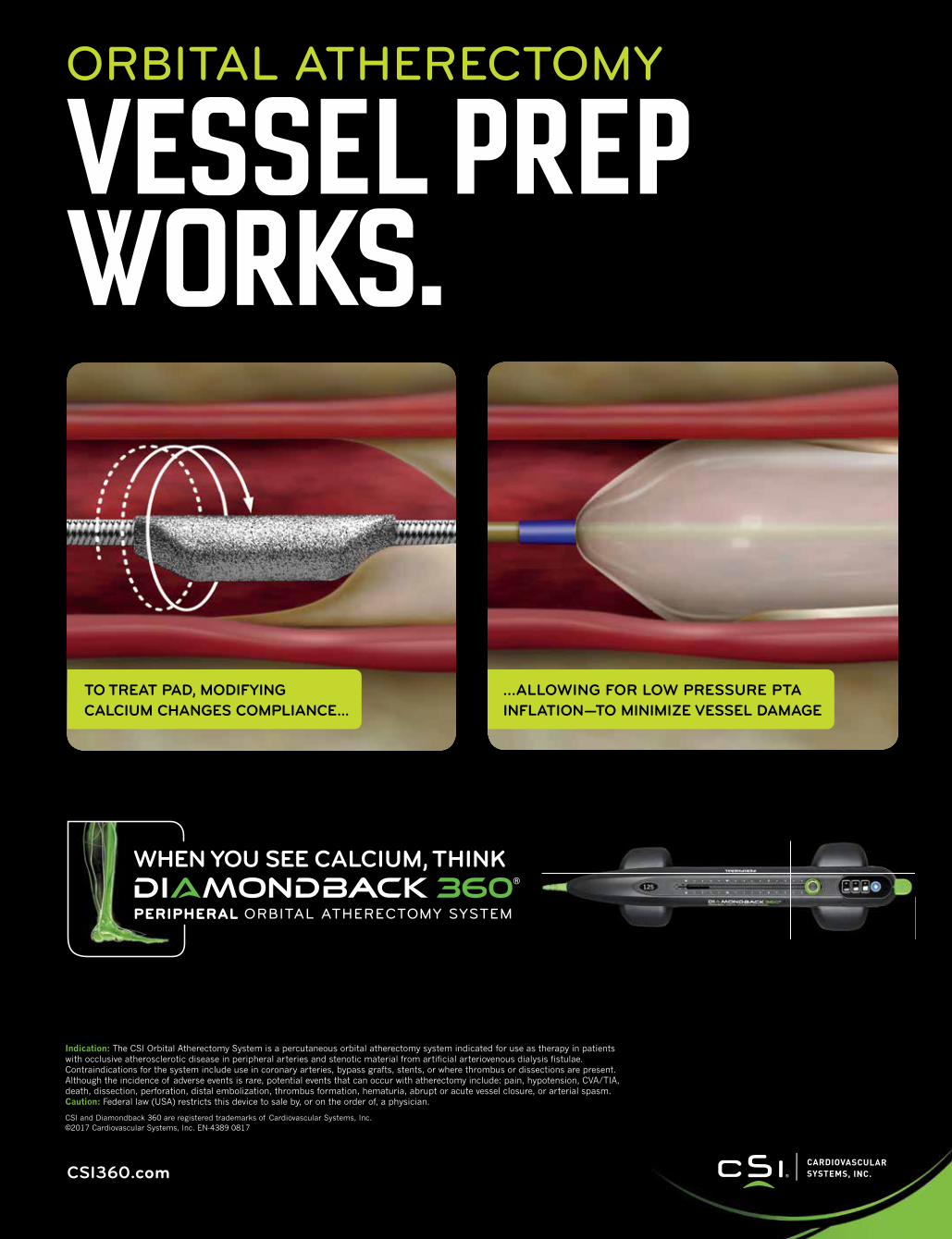
ORBITAL ATHERECTOMY
VESSEL PREPWORKS.
TO TREAT PAD, MODIFYING CALCIUM CHANGES COMPLIANCE...
...ALLOWING FOR LOW PRESSURE PTA INFLATION—TO MINIMIZE VESSEL DAMAGE
Indication: The CSI Orbital Atherectomy System is a percutaneous orbital atherectomy system indicated for use as therapy in patients with occlusive atherosclerotic disease in peripheral arteries and stenotic material from artificial arteriovenous dialysis fistulae. Contraindications for the system include use in coronary arteries, bypass grafts, stents, or where thrombus or dissections are present. Although the incidence of adverse events is rare, potential events that can occur with atherectomy include: pain, hypotension, CVA/TIA, death, dissection, perforation, distal embolization, thrombus formation, hematuria, abrupt or acute vessel closure, or arterial spasm. Caution: Federal law (USA) restricts this device to sale by, or on the order of, a physician.
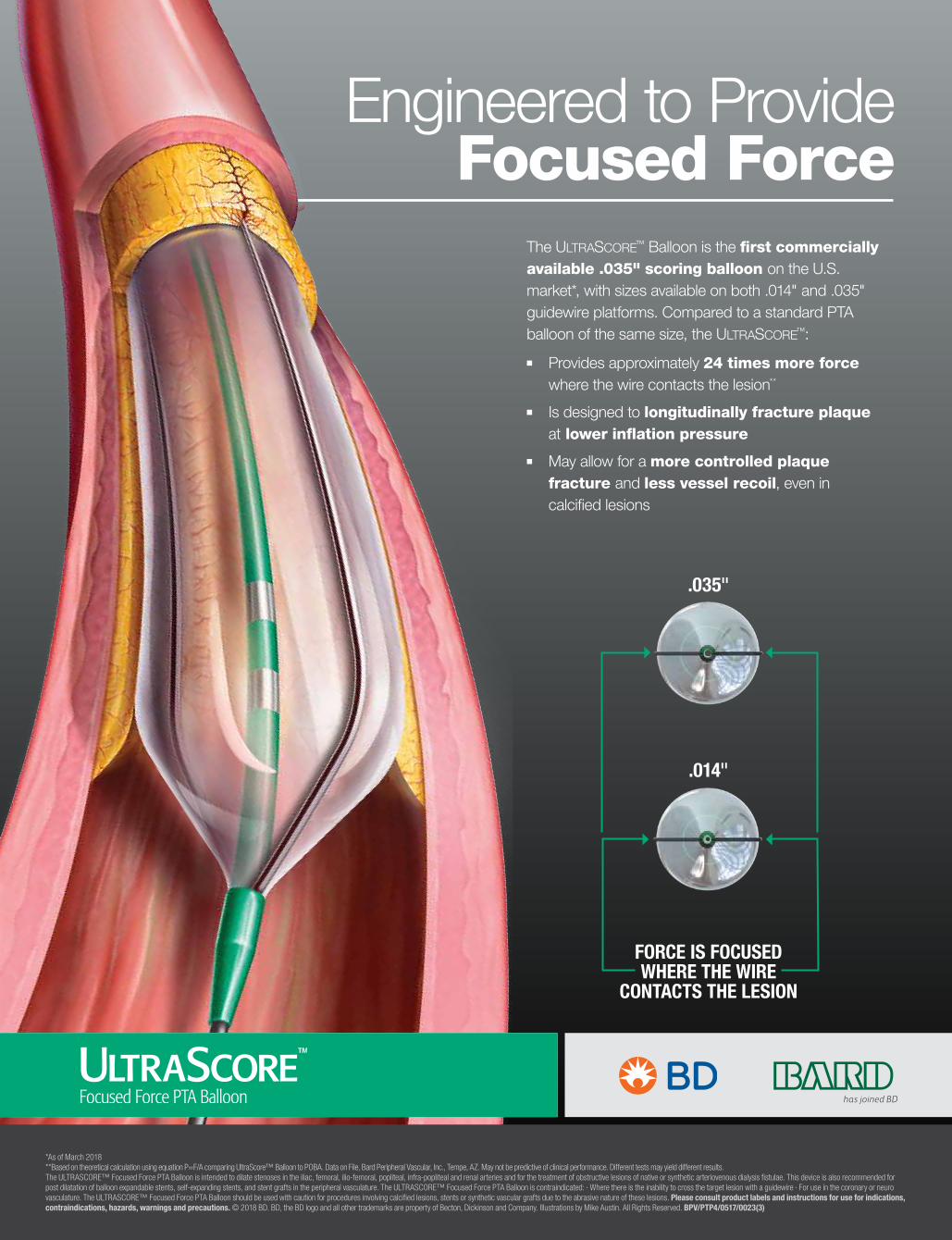
The UltraScore™ Balloon is the first commercially available .035" scoring balloon on the U.S. market*, with sizes available on both .014" and .035" guidewire platforms. Compared to a standard PTA balloon of the same size, the UltraScore™:
■■ Provides approximately 24 times more force where the wire contacts the lesion**
■■ Is designed to longitudinally fracture plaque at lower inflation pressure
■■ May allow for a more controlled plaque fracture and less vessel recoil, even in calcified lesions