Clinical and Medical Case Reports & Studies Case Report Nurjanti L. Cli Med Case Rep: CMCR-103. Very Severe (4 th Grade) Closed Comedonal Acne Vulgaris That Was Complicated by Staphylococcus Epidermidis And Pityrosporum Ovale Folliculities In 16 th Years Old Atopic Hystory Girl That Was Treated With 4x Tca 10% Chemical Peeling At 2 Weeks Intervals Lely Nurjanti * Dermatoveneorologist Samarinda East Kalimantan Indonesia *Corresponding author: Lely Nurjanti, Dermatoveneorologist Samarinda East Kalimantan Indonesia, Email: [email protected]Citation: Nurjanti L (2018) Very Severe (4th Grade) Closed Comedonal Acne Vulgaris That Was Complicated By Staphylococcus Epidermidis And Pityrosporum Ovale Folliculities In 16th Years Old Atopic Hystory Girl That Was Treated With 4x Tca 10% Chemical Peeling At 2 Weeks Intervals. Cli Med Case Rep: CMCR-103. Received Date: 06 June, 2018; Accepted Date: 13 June, 2018; Introduction Acne vulgaris was very common self limitting disease, affected approximately 85% of adolesence, that was defined as a chronic inflammation of pilosebaceous units. It was characterized (diagnosed) by the formation of comedones (as primarily acne lession), erythemathous papules and pustules, less frequently nodules and pseudocyst, and was accompanied by scarring in some cases that caused psychosocial problems. Cunliffe classified the severity of acne vulgaris into 4 types based on the kind and number of acne lessions : mild, moderate, severe, very severe. Four major factors were involved in the etiopathogenesis : follicular hyperkeratinization, increased sebum production, abnormality of microbial flora and inflammation process. The goal of therapy were : removed plugging of the pilar drainage, reduced sebum production, treated bacterial colonization, prevented from scaring. The complications were acne scar, persistent hyperpigmentation, pyogenic granuloma formation, persistent swelling, gram negative bacteria folliculitis, bacterial and fungal folliculitis as secondary infection, resistances to antibiotics. Published Date: 22 July, 2018 The purpose of this case report was to share experience in treating very severe comedonal acne vulgaris because of the chronicity course of acne vulgaris was difficult to be eradicated and there were many resistances problems to antibiotics in some literatures and journals. Case Presentation It was a very severe (4 th grade) closed comedonal acne vulgaris that was complicated by Staphylococcus epidermidis and Pityrosporum ovale folliculitis in 16 th years old atopic hystory girl based on anamnesis, clinical finding and laboratory examination. And this case was treated by 4x TCA 10% chemical peeling at 2 weeks intervals, 2 weeks antibiotic and 10 days ketokonazole after the result of culture and laboratory examination were positive.And the result was good. There was significant improvement in clinical stage (4 th grade to 1 st grade acne vulgaris), decreased the count of comedones, diminished papules-pustules and inflammation, no scar and post inflamatory hyperpigmentation were occurred. 1 Volume 2018 , Issue 01
Transcript
Clinical and Medical Case Reports amp Studies
Case Report
Nurjanti L Cli Med Case Rep CMCR-103
Very Severe (4th
Grade) Closed Comedonal Acne Vulgaris That Was Complicated by Staphylococcus Epidermidis And Pityrosporum Ovale Folliculities
In 16th
Years Old Atopic Hystory Girl That Was Treated With 4x Tca 10 Chemical Peeling At 2 Weeks Intervals
Lely Nurjanti
Dermatoveneorologist Samarinda East Kalimantan Indonesia
Corresponding author Lely Nurjanti Dermatoveneorologist Samarinda East Kalimantan Indonesia Email
lelynurjantiyahoocom
Citation Nurjanti L (2018) Very Severe (4th Grade) Closed Comedonal Acne Vulgaris That Was Complicated By Staphylococcus Epidermidis And Pityrosporum Ovale Folliculities In 16th Years Old Atopic Hystory Girl That Was Treated With 4x Tca 10 Chemical Peeling At 2 Weeks Intervals Cli Med Case Rep CMCR-103
Received Date 06 June 2018 Accepted Date 13 June 2018
Introduction
Acne vulgaris was very common self limitting disease
affected approximately 85 of adolesence that was defined
as a chronic inflammation of pilosebaceous units It was
characterized (diagnosed) by the formation of comedones (as
primarily acne lession) erythemathous papules and pustules
less frequently nodules and pseudocyst and was accompanied
by scarring in some cases that caused psychosocial problems
Cunliffe classified the severity of acne vulgaris into 4 types
based on the kind and number of acne lessions mild
moderate severe very severe Four major factors were involved in the etiopathogenesis follicular
hyperkeratinization increased sebum production abnormality
of microbial flora and inflammation process The goal of
therapy were removed plugging of the pilar drainage
reduced sebum production treated bacterial colonization
prevented from scaring The complications were acne scar
bacterial and fungal folliculitis as secondary infection
resistances to antibiotics
Published Date 22 July 2018
The purpose of this case report was to share experience in
treating very severe comedonal acne vulgaris because of the chronicity course of acne vulgaris was difficult to be
eradicated and there were many resistances problems to
antibiotics in some literatures and journals
Case Presentation
It was a very severe (4
th grade) closed comedonal acne
vulgaris that was complicated by Staphylococcus epidermidis and Pityrosporum ovale folliculitis in 16 th years old atopic hystory girl based on anamnesis clinical finding and laboratory examination And this case was treated by 4x TCA 10 chemical peeling at 2 weeks intervals 2 weeks antibiotic and 10 days ketokonazole after the result of culture and laboratory examination were positiveAnd the result was good
There was significant improvement in clinical stage (4th
grade
to 1st
grade acne vulgaris) decreased the count of comedones diminished papules-pustules and inflammation no scar and post inflamatory hyperpigmentation were occurred
1 Volume 2018 Issue 01
Citation Nurjanti L (2018) Very Severe (4th Grade) Closed Comedonal Acne Vulgaris That Was Complicated by Staphylococcus Epidermidis And Pityrosporum Ovale Folliculities In 16th Years Old Atopic Hystory Girl That Was Treated With
4x Tca 10 Chemical Peeling At 2 Weeks Intervals Cli Med Case Rep CMCR-103
Result of laboratory examination Hb 133 gdL (N) Hct 409 (N) Tr 321000 (N) Leu 8900 (N) ESR 37 mmHg (uarr) Ba 02(N) eo 43(uarr) neut 667(N) Lym 232(uarr) Mo 56(N) IgE atopy 694KIUL (upper N limit)
Chol 170 mgdL (middle N level) TG 75 mgdL (low N level) DHEAS 204 μgdL (middle N level)
KOHPovale GramSepidermidis Culture S Epidermidis
BEFORE PEELING AFTER 4X 10amp TCA PEELING
Resistance tests to antibiotic Resistance to cephalosporine erytromycinclindamycin azytromycin clarytromycin gentamycin ciprofloxacine ofloxacine clotrimoxazole
Sensitive to doxycycline tetracycline minocycline rifampicine nitrofurantoin linezolidtigecyclin vancomycin movifloxacine
Discussion
TCA 10 was superficial chemical peeling considered as adjunctive therapy to the first line acne therapy retinoids and
antibiotic and TCA was the first line therapy for acne scar and skin rejuvenationTCA was cheap and save because no
Good descriptive clinical improvement before and after 4x 10 of TCA chemical peelings
systemic absorbtion had keratolytic effect (comedolytic
action) and anti-inflammatory effect (bactericidal action) It
could be combined to antibiotics and antifungal therapy and
solved resistance problems to antibiotic and antifungal in acne
therapy
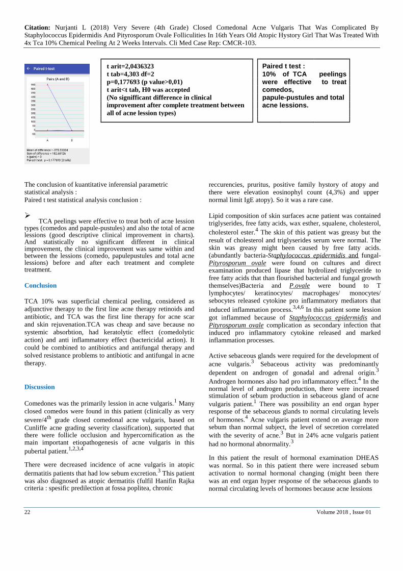
Paired t test 10 of TCA peelings were effective to treat comedos papule-pustules and total acne lessions
Minimal Post Inflammatory Hyperpigmentation
and no scar formation were reported
2 Volume 2018 Issue 01
Citation Nurjanti L (2018) Very Severe (4th Grade) Closed Comedonal Acne Vulgaris That Was Complicated By Staphylococcus Epidermidis And Pityrosporum Ovale Folliculities In 16th Years Old Atopic Hystory Girl That Was Treated With 4x Tca 10 Chemical Peeling At 2 Weeks Intervals Cli Med Case Rep CMCR-103
Keywords acne vulgaris etiopathogenesis TCA chemical
peeling
Introduction
Definition
Acne vulgaris was a very common self limitting disease that was seen primarily in adolescent It was defined as a chronic
inflammation of the pilosebaceous units1234
Acne was not
infectious3
Incidence
Acne vulgaris affected approximately 85 of young
people134
The age of onset was at puberty typically 12-15
years4 but could firstly appeared at 25 years old
14 Ihe peak
insidence of acne vulgaris was in 17-21 years (17-18 years in
female and 19-21 years in males)4 Acne vulgaris was more
severe in males than females1 The lower incidence of acne
vulgaris was in Asians and Africans1 Acne in Black
Americans were less evident than white Americans3
Diagnosis amp Clinical Manifestation
Acne vulgaris was characterized (diagnosed) by the formation of comedones (openblack head and closedwhite head) erythemathous papules and pustules less frequently nodules and pseudocysts (could been ruptured reencapsulated inflammed and formed abscesses) draining sinus tracts (round isolated single nodules and cysts coalesce to linear mounds)
that was accompanied by scarring in some cases3 Acne
vulgaris lessions were polymorphic due to inflammation
process of acne lessions4 Comedones were the primarily
lessions of acne but they were not unique and could been
found in other skin disease like senilis comedo1 The
predilection of acne vulgaris were on the face trunk upper
arms and buttock1 Seborrhea of the face and scalp frequently
presented and could been severe1
Duration of lession The duration of lession was weeks to
months1
Season Acne vulgaris was worsen at winter and fall
1
SymptomIt was Itchy and pain especially in nodulocystic
type1 Itchy was rare It could been found in early phase of
acne or in succesfully treatment cases Itchy was caused by releasing histamine like substances that were produced by P
acnes that were killed by treatment4 Pain was also rare could
been found in patients with nodule and sinus espescially on
the trunk4
Classification
Fitzpatrick classified acne vulgaris as non inflammatory
Plewig and Kligman in 1975 classified acne vulgaris in 3
types comedonal papulopustular and conglobata5 Cunliffe
classified the severity of acne vulgaris according to the number and typeskinds of the lession into 4 types of acne vulgaris mild moderate severe and very severe acne
vulgaris4 The types of lession were comedones
papulespustules nodulescystssinus tracts inflammation and
scarring4 This case report classified and judged the severity
and after treatment clinical improvement of acne vulgaris based on Cunliffe classification criteria
Laboratory Examination
No laboratory examination were required1 In the majority of
acne patients had normal hormonal levels1 If endocrine
disorders were suspected (especially in patients who had clinical manifestation of hyperandrogenism like irregular menses hirsutism hoarse voice alopecia androgenism) determined free testosterone follicle stimulating hormone luteinizing hormone and DHEAS to exclude
hyperandrogenism and polycystic ovary syndrome134
Recalcitrant acne could also been related to congenital adrenal
hyperplasia (11β atau 21β hydroxylase deficiency)1
Etiopatogenesis
Acne vulgaris was multifactorial disease of pilosebaceous
follicles The important patophysiology of acne vulgaris were
follicular hyperkeratinization increased sebum production P
acne colonization Inflammation1
Four major factors were involved in the etiopathogenesis 1234
Follicular hyperkeratinization and cornification of the
pilosebaceous duct
It was not been known whether the initial trigger for acne was
seborrhoea or ductal hyperkertanization or both4
Several factors that might been important as ductal hypercornificationfollicular hyperkeratinization mechanism
were 1234
Abnormal response to androgen Abnormal lipid composition of the ductal corneocytes
(local deficiency of linoleic acid)
Local cytokine activity (IL1α)
Microbial factors
Androgen that quantitatively and qualitatively normal in serum stimulated sebaceous glands to produce more sebum
there was high sebum secretion rate1 Essential fatty acid
linoleic deficiency were characterized by inducing follicular hyperkeratosis impacting of corneocyte and decreasing epithelial barrier function low level linoleic acid led
comedogenesis134
The changing in sebum secretion or
3 Volume 2018 Issue 01
Citation Nurjanti L (2018) Very Severe (4th Grade) Closed Comedonal Acne Vulgaris That Was Complicated By Staphylococcus Epidermidis And Pityrosporum Ovale Folliculities In 16th Years Old Atopic Hystory Girl That Was Treated With
4x Tca 10 Chemical Peeling At 2 Weeks Intervals Cli Med Case Rep CMCR-103 composition (could flourished microorganism growth that activated immune system) could led to release of IL 1α by follicular keratinocytes which in turn could stimulated
comedogenesis1 Pro inflamatory cytokine stimulated
abnormal keratinocyte proliferation and differentiation
revealed obstruction12
Follicular plugging was formed and
would prevented the drainage of sebum and androgens2 There
was follicle impaction and distention formed comedos that were disrupted and ruptured there was leakaged of follicular
materials that induced inflammed lessions24
The early hypercornification of acne was not been initiated by
bacteria but later there were microbacterials grew and
bacterial lipases converted triglycerides to free fatty acids
there was changed of sebum composition and diluted linoleic
acid concentration that led hypercornification (follicular
hyperkeratosis1) and comedogenesis
4
Comedones represented as the retention and
hyperproliferation of ductal corneocytes in the duct34
There
were accumulation of multiple corneocytes in the duct could been caused by either an increased in production of basal keratinocytes or failured of the keratinocytes to be expelled
from the duct4
Ductal hypercornification hystopathologically was presented as microcomedones and clinically as blackheads and
whiteheads3 There was a signifficant correlation between the
severity of acne and the number and size of follicular casts in
comedogenesis3 There was an increasing in proliferation of
ductal keratinocytes of non affected and affected follicles Hystologically microcomedos were found in normal nearby
sites of acne and area that was affected with acne3 The
primary abnormality that led to hypercornification was not been related to change in keratin expression hypercornification and comedogenesis might been related to
failure the ductal corneocytes to separate3
The primary changed in the sebaceous follicle in acne was an
alteration in the pattern of keratinization within the follicle1
Normally keratinous material in the follicle was loosely organized In ultrastructural level there were many lamellar granules and relatively few keratohyaline granules Comedo formation was firstly formed in the lower portion of follicular
infundibulum (infrainfundibulum)1 The keratinous materials
were denser the lamellar granules were less numerous keratohyalin granules were increasing and some of cells were containing amorphous materials (which were probably lipid)
were generated during the process of keratinization1 Kinetic
studies demonstrated that there was an increasing in cellular
turn over in comedones1
Corneocytes frequently contained about 20 water but they
were varies markedly with age4 The swelling of the epidermis
was caused by hydration that followed prolong soaking of the skin particularly in warm water was familiar in most people
Corniffied epithelium of the sebaceous follicle became hydrated that might increased sebum outflow resistance by
reducing the size of the pilosebaceous ostium4 This
obstruction was associated with a decreased in outflow of
sebum3 Acute obstruction of a particular pilosebaceous duct
might then occured and thus precipitated acne4 It explained
tropical acne and pre menstrual acne flared3
Comedogenesis was also related to the potential importance of
what was called the sebolemmal sheath3 It had been
suggested that the excretion of products from the sebaceous gland was occured through an organized acellular tubular conduit-the sebolemmal sheath was produced by sebaceous duct cells The rupture of this sheath might contributed to
comedogenesis3
Abnormal response to androgen influenced
hypercornification
The evidence was accumulating to propose that androgens (a male steroid hormone such as testosterone) might play an
important role in comedogenesis3 The cells of pilosebaceous
duct had androgen receptors and 5α reductase type I (enzym that converted testosterone to DHT) was also present in this
cells3
Androgen were known to regulate the development of
sebaceous gland and sebum production Androgen might
played indirect at the follicular hyperkeratinization was
supported by some obsevations 1
Androgen receptors had been localized to the outer root
sheath of the infrainfundibular region in the follicles The formation of follicular cast reduced in patients that
was treated with anti androgen Each of the key enzyme involved in androgen metabolism
had been identified in the follicles
Abnormal lipid composition in keratinocyter duct
influenced hypercornification
Follicular hyperkeratinization might related to a local
deficiency of linoleic acid production of IL 1α within the
follicle or possibilly the effect of androgens (high sebum
(which might parallel with the increased scale that was
found in comedo3)
Impacting of corneocytes that formed comedones4
Decreasing epithelial barier function13
(which might made the comedonal wall more permeable to
inflammatory substances3)
134 Membrane coated granules were probably more related to
barrier permeability than cell separation and that were
decreased in commedones3
4 Volume 2018 Issue 01
Citation Nurjanti L (2018) Very Severe (4th Grade) Closed Comedonal Acne Vulgaris That Was Complicated By Staphylococcus Epidermidis And Pityrosporum Ovale Folliculities In 16th Years Old Atopic Hystory Girl That Was Treated With
4x Tca 10 Chemical Peeling At 2 Weeks Intervals Cli Med Case Rep CMCR-103
In examination of polar lipids recovered from comedone showed that the acyl ceramides were contain only 6 linoleic acid among esterified fatty acids compared with 45 in
normal human epidermis3
Linoleic acid concentration was
decreased in acne patients sebum3
Other lipids had been incriminated there were free fatty acids squalene squalene peroxides oleic acid isopropyl niristate liquid parafin wax ester ceramid linoleic acid Low ceramides and low level linoleic acid essential fatty acid in
ceramide had been blamed for inducing comedones34
There
were correlation between lipid peroxidase levels (an oxidative degradation of lipids resulting in cell damage) and the size of comedos There were low level of linoleic acid and high
sebum level in acne patients4 UVA radiation in lipid
nyristate liquid paraffin) induced comedogenesis4 There was
high sebum flowed that produced a local deficiency of vitamin
A in the duct that induced ductal cornification4 Than the
changing in sebum secretion or composition could led to release of IL 1α by follicular keratinocytes which in turn
could stimulated comedogenesis1
The primary site of the developing comedone in the sebaceous follicle in acne vulgaris was at the level of the
infrainfundibulum4 It was proposed that at the time of cell
division when the sebaceous cells still had an contact with the basement membrane they still had and access to circulating
lipid including linoleat4 Once sebum synthesis began no
further lipids were accepted from circulation so that more sebum was synthezised per cell and linoleate content would
been diluted4 This linoleate content would been released at
the time of final cell ruptured and incorporated into various lipids in proportion to be relative rates (linoleic acid was diluted by sebum and the concentration would been low) at which these lipids were being synthesized at the time of cell
ruptured4 Linoleic acid was essential fatty acid that could not
been synthesized by human cell tissue34
The pilosebaceous unit comprised an matured epiuthelium and developed sebocytes through which the hair and sebum passed Anatomically the pilosebaceous unit was divided into smaller units infundibulum (acroinfundibulum and infrainfundibulum)
and sebaceous duct4 The sebocytes rest on the basement
membrane that were contiguous with the dermis and extending from this basal layer into the central part of the gland The
sebaceous gland was a holocrine gland the secretion was the result of self destruction of the sebocytes The nucleus was moved to the periphery of the cell The cell then entered the pilosebaceous duct The sebum was secreted than it was
moved up with desquamated corneocytes and presented
microbes to the surface4
Local cytokine influenced comedo formation
Inteleukine 1ɑ was found in comedo it was important in
comedogenesis and it was produced by keratinocytes of the
duct3 It was proved by in vitro study This effect could been
blocked by Interleukine 1 antagonists4 and the formation was
totally disrupted by EGF (Epidermal Growth Factor)34
Two studies had failed to incriminate bacteria in the initiation
of comedones and it was proved by the fact that there were no
bacterias that had been shown in some early comedones
Ultrastructurally and cultures of some early non inflamed
biopsy material that were taken from lessions were sterile34
P acnes was not involved in the initiation of comedones but
might been involved in the later stages of comedogenesis34
The early hypercornification of acne was not been initiated by bacteria but later there were bacterias colonization that produced lipase that converted triglyserida to free fatty acid and increasing sebaceous free fatty acids would been changed sebum composition and diluted linoleic acid concentration
that led hypercornification (follicular hyperkeratosis)13
corneocytes impaction that formed comedo4 and decreasing
epithelial barier function that increased permeability of
comedonal wall124
Biopsy and culture of early non inflammed lessions had shown that 30 of these were without bacteria suggesting that
ductal bacteria were not needed for initiation of cornification3
Electron microscopy of early non inflammed lessions that were taken from prepubertal and early pubertal individual had
demonstrated few or no bacteria3 Quantification of bacteria
from comedones suggested that follicular colonization might
been unrelated to comedogenesis3
Increased sebum production
Normal or abnormal androgens stimulated sebaceous glands
to produce more sebum or there was end organ androgen
hypersensitivity response in normal hormonal level of acne
vulgaris that made bacterial and fungal were flourished 134
There was much debated concerning the prime trigger to acne it was the increased of sebum production or formation of comedones or both abnormality developed paralel in the same
Sebum excretion increased in acne patient than normal people
and the increasing of sebum exctretion was equally with acne
severity3
Increased production of sebum in acne patients was explained
as 4 possibilities 4
An elevated level of circulating hormone that was caused
by an abnormal pituitary drive
5 Volume 2018 Issue 01
Citation Nurjanti L (2018) Very Severe (4th Grade) Closed Comedonal Acne Vulgaris That Was Complicated By Staphylococcus Epidermidis And Pityrosporum Ovale Folliculities In 16th Years Old Atopic Hystory Girl That Was Treated With
4x Tca 10 Chemical Peeling At 2 Weeks Intervals Cli Med Case Rep CMCR-103
An abnormal increase in the production of androgen in the adrenal and gonad
An end organ hyper response of the sebaceous glands to normal circulating level of hormone
Combination
In most of acne patients had no hormonal misfit Most patients in clinic did not require investigations of sex hormones simply because the patients seem otherwise normal they responded well to an appropriate treatment reasonablly and thus did not
need detailed endocrinological examination4 There was rare
cases that acne female patients had clinical sign of abnormal hormonal level like excessively hairy hoarse voice irregular menses and they got on well with the men and could been pregnant In this patient could been found an elevation levels
of circulating androgens or an abnormal pituitary drive4
There was an end organ hyper response of the pilosebaceous
unit to normal levels of circulating androgens And it was
supported by the finding that the sebaceous glands in acne
prone areas function differently to those in non prone areas so
acne could been found only on the trunk and none on the face
or acne just on the face and none on the back and chest4
A connection between acne and high rates of sebum secretion
was supported by at least 3 types of evidence1
Children did not get acne during the age range from
approximately 2-6 years when sebum secretion was extremely low
Average rates of sebum secretion were higher in individuals with acne than those without acne
Treatment that reduced sebum secretion (such as estrogen 13 cis retinoic acid) improved acne
Increased sebum production was presented as patient`s
seborrhoea (greasy skin)3Active sebaceous glands were a
prerequisite for the development of acne Acne patients male and female excreted on average more sebum than normal subjects and the level of sebum secretion correlated
reasonably well with the severity of the acne3 Sebaceous
activity was predominantly dependent on androgens sex
hormones of gonadal or adrenal origin3 Abnormally high
levels of sebum secretion could been thus resulted from high overall androgen production or increased availability of free androgen because of a deficiency in sex hormone binding
globulin (SHBG)3 Equally they could involved an amplified
target response mediated either through 5α reductase of testosterone or an increased capacity of the intracelluler
receptor to bind the hormone3
Lawrence et al found that only 41 of the acne patients had
free testosterone level above normal Lucky et al measured a number of androgens and their precursors as well as and found that 52 of non hirsute women with acne had at least
one abnormal hormone level Darley et al found high sebum testosterone in 26 low SHBG in 45 and high prolactin in 45 of 38 woman with acne However 24 of the total had no
might been important for example increased androsterone metabolism had also been reported in normo androgenic
females3
In some published papers it would seem that androgenic hormonal balance was disturbed in 50-75 of female acne
patients3 However this had not been established that it was
the critical factor and at least a quarter of all cases remain
unexplained3 The development of acne were simply related to
systemic hormone levels But in general acne patient had not
frequently had endocrine misfit3
The acne did not occur simultaneously on all susceptible sites was consonant with the finding that sebum secretion were varies from follicle to follicle In acne patients there were marked heterogenicity in individual follicular sebum
excretion3 This suggested that certain follicles might been
proned to acne3 An enhanced peripheral response should
been considered as a factor in many subjects3
The possible role of increased 5ɑ reductase of testosterone to its more active metabolite was indicated both by the deminstration that sebaceous glands in acne prone regions showed abnormallity high 5ɑ reductase activity in vitro and by the finding of abnormally high ammount of 5 alfa
androstenediols in the urine of female acne patients3 There
were 2 forms of 5ɑ reductase type I and type II and the type I 5ɑ reductase was more relevant The activity of type I 5ɑ reductase in isolated sebaceous glands also supported the end
organ hyperresponsiveness theory for acne3 Androgen action
on the sebaceous gland might been independent from serum
hormone levels3
There was possibility that other hormones affected the sebaceous glands either directly or by enhancing their
response to androgens3 Low sebum excretion rate was low in
individuals with isolated growth hormone deficiency but this
endocrinopathies was rare3
Sebum consisted of mixture of squalene wax and sterol esters cholesterol polar lipid and triglyserides As the sebum moved up the duct bacteria especially P acnes hydrolized the triglycerides to free fatty acids which eventually appeared at the skin surface Free fatty acid fraction of the sebum was
considered to be important in the causation of inflammation13
Triglycerides fraction in sebum was probably responsible for
acne1
The role of individual lipid components in causing acne was
uncertain Lipid might been involved in ductal
hypercornification or might been essential to the growth of
bacteria3 Sampling of skin surface lipids had shown that
6 Volume 2018 Issue 01
Citation Nurjanti L (2018) Very Severe (4th Grade) Closed Comedonal Acne Vulgaris That Was Complicated By Staphylococcus Epidermidis And Pityrosporum Ovale Folliculities In 16th Years Old Atopic Hystory Girl That Was Treated With
4x Tca 10 Chemical Peeling At 2 Weeks Intervals Cli Med Case Rep CMCR-103 patients with acne tended to have higher levels of squalene and wax esters and lower levels of essential fatty acids linoleic acid and a more frequent occurences of particular free
fatty acids3 Linoleic acid was significantly reduced in ductal
hypercornifications3 Linoleic acid levels were signifantly
decreased in acne patients and there was inverse relationship between sebum secretion and linoleic acid esential fatty acid
concentration of sebum1 Linoleic acid could not been
synthesized in mammalian tissue and its concentration was diluted by subsequent endogenous lipid synthesis in sebaceous
cell1
In was unclear why elevated rates of sebum secretion led to
acne The triglyceride fraction of sebum which was unique to
humans was probably responsible for acne The bacterial
population of the follicle hydrolized triglycerides to fatty
acids which eventually appeared on the skin surface In the
past the free fatty acid fraction of sebum was considered to be
important in the causation of inflammation but in recent years
it had become evident that there were probably other more
important causes of inflammation1
The sebaceous glands produced a considerable amount of
sebum in the first 3 months of life which then gradually
reduced to zero at 6 months of age This neonatal stimulus
was likely to be an effect of the fetal and neonatal adrenal
androgens After 6 months of age the sebaceous gkands
remained quiscent until early adrenarche At adrenarche
around 7-8 years there was an increased in adrenal androgens
in particular dehydroepiandrostenedione with the resultant
increased in sebum excretion In the early pubertal years there
was a further increased in adrenal androgens and gonadal
androgen stimulus to the sebaceous gland There was an
obvious increased in greasiness of the skin (seborrhoea) even
in subjects who did not have acne The sebaceous gland was
under endocrine control The main stimulus to the sebaceous
glands was androgens The pituitary had an important role in
controlling the androgen production via the adrenals and gonads The adrenals in particular produced
dihydroepiandrostenedione and the gonads in both sexes
produced testosterone The circulating androgens in particular
testosterone were bound to the sex hormone binding globulin
and it was the 1-2 of free testosterone was dictated
sebaceous gland activity4
In both sexes independent of the presence or absence of acne
there was a gradual increased in sebum excretion from
puberty and beyond reaching a peak at about the age of 16-20
years Thereafter the level remained constant until there was a
gradual decreased from about 40 years onwards in women and
from about 50 years in males In general the sebum exretion
rate (SER) in men was signifficantly higher than in women4
Patients with acne also had seborrhoea indeed many patients complained that as acne developed so there was an increased greasiness of the skin and on the scalp There was a
reasonable correlation between the amount of sebum
production and the severity of acne There was an evidence
that those subjects with seborrhoea and acne had a higher
number of sebaceous lobules per gland Indeed one of the
disappointing features of acne therapy with most therapies
were the fact that despite an improvement in the acne the
sebaceous was persisted But in Dianette (cyproterone
acetatate+ethinyl estradiols) and isotretinoin therapy there
was a significant reduction in sebum excretion and acne
improvement4
Measurements of sebum excretion also showed that
individuals with acne produced more sebum than individuals
who had never had acne There was a gradual decreased in
sebum excretion beyond the age of about 40 years Thus
reduction in sebum alone was not accounted for resolution of
acne4
There were differences of the lipid composition between the
skin surfaces and in the sebaceous glands Skin surface lipid
composition had less triglycerides and more free fatty acids
levels and equally same levels of wax esther squalene
cholesterol esters and cholesterol It was caused by lipolityc
enzymes that were produced predominantly by P acnes and
Staphylococcus epidermidis that hydrolized triglycerides into
free fatty acids when the sebum was moved up from the
pilosebaceous duct4
The abnormality of the microbial flora
Bacterial and Pityrosporum ovale `s lipase hydrolyzed triglycerides to free fatty acids that flourished bacterials and
Pityrosporum ovales themselves4 There was lipid
composition changing and free fatty acid would marked
inflammation process34
Free fatty acid was comedogenic4
Bacterial and fungi were bound to the receptor of monocytes keratinocytes perifollicular and peribulbar macrophages sebocytes langerhans cells and other inflammatory cells (through TLR2 Receptor or others) and T lymphocytes (through CD4) than produced proinflammatory mediators
(IL1α TNFα etc) that led to an inflammatory response1346
P acnes induced TLR 2 Receptor expresion and play role in
acne inflammation6
Adolescence and its attendance seborrhoea were associated
with a significant increased in P acnes3 But there was a little
or no relationship between the number of bacterias on the skin
surface or in the duct with the severity of acne3 But in other
books Cunliffe said that there was a correlation between the reduction in P acnes counts and the clinical manifestation of
acne4 The development of resistance to P acnes might
equated with clinical failure to treat the acne4 There was no P
acnes colonization in non acne vulgaris patients3 P acnes
colonization were at anterior nares4 And P acnes were
important in acne pathogenesis1 Sebum excretion rate and
ductal cornification correlated well with clinical severity3
7 Volume 2018 Issue 01
Citation Nurjanti L (2018) Very Severe (4th Grade) Closed Comedonal Acne Vulgaris That Was Complicated By Staphylococcus Epidermidis And Pityrosporum Ovale Folliculities In 16th Years Old Atopic Hystory Girl That Was Treated With
4x Tca 10 Chemical Peeling At 2 Weeks Intervals Cli Med Case Rep CMCR-103
Acne was not infectious3 The three major organisms were
isolated from the surface of the skin and the duct of patients with acne were Propinibacterium acnes Staphylococcus epidermidis and Malassezia furfurPityrosporum ovale
3
There were three major subgroups of the propionibacterias P
acnes P granulosum and P avidum3 Almost certainly P
acnes and to lesser extended P granulosum were the most
important3 Nevertheless as they lived in association with the
Staphylococcus epidermidis and Malassezia furfur three organisms had probably some control over the growth of P
acnes3 And Staphylococci were the first organism that
colonized the normal skin people4
Staphylococcus epidermidis were found as comensal (normal
colony at nares head and axilla) and patogen (as chronic
nosocomial infection that infected through contaminated stuff
in cardiac cathetherization or other procedures) It was
difficult to be eradicated it had high resistances it was easy to
be infected again after it was treated (by hands or
contaminated stuff) and it was clinically found as chronic
infection But this colonization inhibited Staphylococcus
aureus virulencies7
Pityrosporum ovale was lipophilic saprophytic budding unipolar dimorphic gram positive double walled oval to round yeast They were normal part of the follicular skin flora and alteration in flora caused uncontroled growth of yeasts
and would been pathogenic4 They needed free fatty acid for
survival (they had lipase that hydrolized triglyceride to free fatty acid) They were found in stratum corneum and in pilar follicles in areas with increasing sebaceous gland activity such
as chest and back8
P acnes were gram positive non motile rods that tended to be irregular when the first isolated - some were short branching
and required free fatty acid to colonize34
P acnes should been clumped free fatty acid aided clumping and so bacterial lipases might been necessary for clumping and for ductal
colonization3 Isolates required 7 days of incubation under an
aerob conditiom in 35-37oC (but this organism was not
strickly an aerob)4 The physiological microenvironment of
the follicle and the microenvironmental adaptation of P acnes might been important factor in the penetration of this non
motile bacterium into the follicle duct4 They grew optimum
at 30-37oC (temperature in the follicle)
4
The environment of the bacteria was probably more important
than their absolute number for development of acne lession3
In vitro it had been shown that low oxygen tension acidic pH (36-67) and nutrient supply [nitrogen carbon hydrogen carbohydrate amino acid minerals vitamin (biotin nicotinic acid and thiamin)] markedly affected the growth of P acnes and the bacterial of active substances production such as lipase proteaseshyaluronate lyase phosphatase and smooth
muscle contracting substance34
In the presence of light at high oxygen concentrations P acnes
grew well but later the growth was inhibited because of
photodamaging reactions involving excess oxygen and the
endogenous microbial porphyrins3
The development of acne vulgaris was likely linked with the P acnes and very occasionally with the transient flora that were involved in acne (The transient flora was gram negative bacteria that was shed from anterior nares onto the adjacent skin after the resident flora was supressed by long term
systemic or topical antibiotics)34
The limitted species of
organism (resident organism) colonized the skin surface such as propionibacteria staphylococci aerobic coryneform
bacteria and the yeast Malassezia furfur4 Some
microorganisms were appeared and dissapeared from the skin
environment and constitued transient flora4
Inflamation processes
Cunlife reported that hystologically CD4+ T lymphocytes were found in eatly 6 hours papular acne inflammation CD4+ T lymphocytes and neutrophyl were found in 24-48 hours CD4+ T lymphocytes macrophage and giant cells were found
in 72 hours4
The dermal inflammation was not been caused by bacteria in the dermis It was probably resulted from biologically active mediators (IL 1ɑ IL β TNFαetc) that diffused from the follicle where they were produced by the binding of bacterials to TLR2 receptor (or others) of monocytes sebocytes keratinocytes perifollicular and peribulbar macrophages langerhans cells and other immune cells or CD4+ of T
lymphocytes461011
There was an ability of innate immune
system to use TLR2 receptor to recognonized microbial
pattern and initiated immune response in cutaneous disease9
TLR 2 receptor induced inflammatory response and the
development of antigen spesific adaptive immunity6
Pro inflammatory cytokine stimulated abnormal keratinocyte proliferationdifferentiation and hypercornification that revealed obstructions than there were follicles impaction and
distention that formed comedos239
As the retained cells blocked the follicular opening the lower portion of the follicle was dilated by entrapped sebum Disruption of the follicular
epithelium permitted the discharged of the follicular dermis2
The combination of keratin sebum and microorganism led to pro inflammatory mediators releasing and lymphocytes
neutrophyls and foreign body giant cells accumulating2 In the
early inflammation inflammation was due to pro inflammatory mediators that moved through the duct wall into
the dermis and had not been caused by the duct ruptured3
Interleukine1α was a dominant proinflamatory cytokine that played role in comedonal acne vulgaris inflammation
process4 Other pro inflamatory cytokines that were produced
were IL6 IL8 IL12 IL4 IL10 TNFα4
Some kind of pro inflammatory cytokines that were produced by innate immunity in acne vulgaris in some journals were
8 Volume 2018 Issue 01
Citation Nurjanti L (2018) Very Severe (4th Grade) Closed Comedonal Acne Vulgaris That Was Complicated By Staphylococcus Epidermidis And Pityrosporum Ovale Folliculities In 16th Years Old Atopic Hystory Girl That Was Treated With
4x Tca 10 Chemical Peeling At 2 Weeks Intervals Cli Med Case Rep CMCR-103
TLR 2 receptor of the monocytes bound to P acnes to
produce IL12 IL8 pro inflammatory cytokine10
NLRP3 inflammasome of the human sebocytes and
monocytes as mediated pathway bound to live P acnes in the sebaceous glands through caspase 1
expression amp activation to produce IL1β1112
TLR 2 receptor of the keratinocytes bound to P acnes
to produce IL8 human defensin 2 pro inflammatory
cytokine10
TLR2 receptor of human keratinocytes bound to PacnesthroughPAMPs-PathogenAssociated Mollecular Paterns to produce IL1α in 7 days of
exposures that induced comedogenesis13
PAMPs were such as peptidiglycan (PG) and
lipopolysacharida (LP) of P acnes6
PAR 2 of the keratinocytes bound to P acnes to
produce IL 1α IL8 TNFɑ10
TLR2 receptor of human monocytes and skin surface macrophages in human pilosebceous bound to microbial agent (P acnes gram positive coccus ) through NFƙBNuclear Factor kappa light chain
enhancer of activated B cells activation614
and
MAPK (Mitogen Activated protein Kinase) cascade6
to sinthesize and release of IL12 IL8 TNFɑ IL1β14
TLR 2 receptor of monocytes bound to gram positive
coccus to produce IL1214
Monocytes bound to P ovale (lived or heat killed opsonized P ovale through alternative complement activation pathway more stimulated than non
opsonized) to produce IL8 IL1ɑ15
Monocytes bound to gram positive bacteria to produce
TNFɑ IL615
Monocytes bound to Gram negative bacteria bound
to produce TNFɑ IL1 IL615
IL 8 induced chemotactic factors might played an important role in attracted neutrophyl to the pilosebaceous unit that led to release lysosomal enzime that led to rupture follicular
epithelium and further inflammation14
Furthermore P acnes
released lipases proteases hyaluronidase which contributed
to tissue injury14
IL 8 induced chemotaxis and activation of neutrophyl and T
cells15
IL12 promoted development of Th1 mediated immune
response And overproduction of Th1 cytokine such as IL 12 was implicated in the development of tissue injury in a certain
autoimmune and inflammatory disease14
IL1ɑ was low activated by lymphocytes chemotaxis
activation of neutrophyl than induced inflammation15
Therefore interaction of P ovale and phagocytic cells might served to amplify the inflammatory response and encourage
regulated of pro inflammatory cytokine with removed lipid15
Langerhansmacrophages were able to take up antigen (acted as antigen presenting cellsAPC) and then were presented to T
cells15
P acnes activated monocytes cytokine relased through the pattern recognition receptors (PPRs) for example TLR2
receptor of the innate immune system14
So TLR 2 receptor
was PPRs14
TLR2 receptor activation contributed to the pathogenesis of acne suggesting that these cells promoted inflammatory responses at the site of the disease activity and
induced pro inflammatory cytokine production14
Release of pro inflammatory cytokines that were mediated through TLR2 receptor had harmful effect in acne by promoting
inflammation and tisuue destruction14
So TLR 2 receptor was a logical target for therapeutic intervention to block inflammatory cytokine response in acne and other inflammation condition which tissue injury was detrimental to
the host14
Isotretinoin down regulated TLR2 that induced
cytokine response6
Infestation of the organism itself was not the main caused of
the disease but was rather caused by the various inflammatory
responses that were initiated by microbial agents that led to
destruction of the host tissue Such responses were the
formation of immune complex the recruitment and activation
of neutrophyl and monocytes the released of cytokines
released of dependent enzymes14
P ovale had 2 phenotypes immunostimulated and
immunosupressed phenotypes15
P ovale (through an alternative pathway of complement activation) activated
cellular immune response and humoral immune response15
Complement was the part of immune system that enhanced the ability of antibodies and phagocytic cells to clear microbes and damage cells from an organism promoted inflammation and attacked the pathogen plasma membrane It was part of innate immune system (which was not adaptable and did not change over the course of individual`s lifetime) and it could been recruitted by the adaptive immune system to finish the
action There were 2 complement activation classical
pathway (that was mediated by immune complex) and altenative pathway (that was mediated by yeast or bacterial
cells)15
There was complement activation that involed in the early to later stages of inflammation and P acnes were capable for triggering both the alternative and classical complement
pathways13415
Complement activation caused lysis bacteria
and virus opsonization inflammation15
In the early non inflamed and inflammed lession had shown that there were activation of the classical and alternative
pathways3 And there were the type 4 immunological reaction
to a non spesific antigens in the prior of obvious duct
ruptured3
9 Volume 2018 Issue 01
Citation Nurjanti L (2018) Very Severe (4th Grade) Closed Comedonal Acne Vulgaris That Was Complicated By Staphylococcus Epidermidis And Pityrosporum Ovale Folliculities In 16th Years Old Atopic Hystory Girl That Was Treated With
4x Tca 10 Chemical Peeling At 2 Weeks Intervals Cli Med Case Rep CMCR-103 The majority comedones clearly represented a dermal pool of
pro inflammatory IL 1α4 Spongiosis of the pilosebaceous
follicle wall was the feature of early inflammation changed this could led to leakage of comedonal IL 1α into the
epidermis4 The consequence was the activation of dermal
microvascular endhothelial cells selective accumulation of antigen non spesific to mononuclear cells and inititation of antigen independent cutaneous inflammation that consistent
with the hystological features of early inflammation in acne4
In the later in the moderate and severe inflammation there was disruption of the duct and macrophage giant cell foreign
body reaction3 An amplification phase via antigen dependent
T cell responses to other comedonal components for example
P acnes might then developed4 The intensity and duration of
the subsequent cell mediated response would been depended on many factors including the degree of individual
sensitization to their cutaneous microflora4 Following the
disruption of cell wall neutrophyl would been attracted into the duct by microbial chemotactic factor that was proved by a study that demonstrated the capability of P acnes to attract
neutrophyl in vitro4 So P acnes might caused inflammation
because this organism had been shown to secrete chemotactic factors and the chemotactic activity had been shown in
comedones1 Low mollecular weight chemotactic factor did
not require serum complement for activation and because of its small size it could probably escaped from follicle and attracted polymorphonuclear leukocytes
1 If
polymorphonuclear leukocytes enter the follicle they could ingested P acnes organisms resulted the release of hydrolytic anzymes which in turn might been importance in producing
ingested P acnes was anti P acnes dependent antibody
(ADCC)1
Bacterial cell walls fractions of P acnes were a potent
chemoattractan for polymorphonuclear and mononuclear cells
could also produced prostaglandine like substance that acted
as non streroidal anti inflammatory drugs that had small anti
acne effect3
It was likely but was not been proven that P acnes played an
important role in acne inflamation4 Whether P acnes played a
role in the initiation of inflammation in acne was questionable since it had not been colonized at all of the early lessions Nevertheless there was an increasing in the number of lessions colonized by P acnes following early inflammatory change P acnes were a potent adjuvant that induced a chronic inflammatory tissue response because it was resistant to
phagocyte killing and degradation4 So P acnes caused
chronic inflammation process because of its resistance to
phagocyte cell and could not been degraded4
In the late phase of inflammation P acnes dependent T cell lymphocyte response could been found there were variations in Cell Mediated Immune Response depended on individual
microflora sensitization14
Circulating antibody to P acnes
were elevated in patient with severe acne1 Patients with
severe acne were significantly more sensitized to P acnes than normal individuals and the overall immunological status of patients were elevated compared with acne free individuals of
the same age4 But this observation did not provide direct
evidence for a pathogenic role of P acnes in initiating inflammatory acne and might merely reflected an increased exposure of patient to the organism as result of their condition and might played a role in the exacerbation of chronic
inflammatory response4
Lipid that got into dermis when the duct ruptured acted as an irritant and some lipids like linoleic acid could down regulated neutrophyl oxygen metabolism and phagocytosis and
contributed inflammation3 Inflammation was resulted from
the production of free fatty acid and it showed that P acnes was the main source of follicular lipase that hydrolized
trigliserida to free fatty acid1 The sebolemmal sheath
accumulated inspisated sebum and formed a firm calculus
which eroded the duct wall and contributed to inflammation3
DHT was the main driver of androgen induced sebum
production of the skin DHEA was another hormone for
activity of activator protein (AP1) and NFƙB led to
recruitment of inflammatory cells AP 1 inflammatory cascade
led activation of matrix metalloproteinase which contributed
to local tissue destruction and scar formation13
P acnes Hydrolized triglycerides to pro inflammatory free fatty acid through lypase enzyme of P acnes FFA spur
production of antimicrobial peptidesAMPs (Such as Human β
defensin 1HBD1 cathelicidine human β defensin 2HBD2) thus leading to further inflammation That inflammation in acne lession was broken in the deep
layer and formed nodules16
10 Volume 2018 Issue 01
Citation Nurjanti L (2018) Very Severe (4th Grade) Closed Comedonal Acne Vulgaris That Was Complicated By Staphylococcus Epidermidis And Pityrosporum Ovale Folliculities In 16th Years Old Atopic Hystory Girl That Was Treated With
4x Tca 10 Chemical Peeling At 2 Weeks Intervals Cli Med Case Rep CMCR-103 In study was reported the elevated IgE levels that related with clinical severity in a group but another group was found no
changed in total IgE levels3 Female had better defence
mechanism than male against P acnes3 Most acne patient had
no misfit immunological reaction3 There were no circulating
immune complexes in acne sera patients3 Skin testing with
heat killed suspensies of P acnes demonstrated that subject with severe acne produced a greater inflammatory reaction at 48 h than other subject suggested that host response might
been important3 Changing in neutrophyl chemotaxis might
been the secondary event3 P acnes polypeptide were detected
in serum of the acne patients but were not in normal individu3
GeneticThere were multifactorial genetical background and
familial predisposition that had been proved in twin study13
Acne was polymorphous dermatosis with a polygenetic
background that did not follow Mendelian rules4 Most
individual with cystic acne had parents with a hystory of
severe acne1 Several study had shown that genetic factor
influenced susceptibility to acne3 There were 45 acne
parents`s in schoolboys acne patient in Germany and were
also supported by genetic study in twins34
Besides genetic factor the exogenous factor also influenced the severity of disease inflammation process for example bacterial
colonization4 Severe acne might been associated with XYY
syndrome (rare)1
Racial Acne in Black Americans were less evident than white Americans Americans had more severe acne than
Japanese3 Acne vulgaris was lower incidence in Asians and
Africans1 AtopicThere was decreased incidence of acne
vulgaris in atopic dermatitis patients that had low sebum
production3 Seborrhoeic Seborrhoeic dermatitis was
concomitantly found with acne vulgaris in some cases but the
relationship had not been known1
Menstruation About 70 patient reported 2-7 days premenstrual flared up of acne vulgaris related to sebaceous pores size changing that influenced the hydration of
pilocebaceous epithelium34
There was an alteration of
progesterone and estrogen levels34
Estrogen therapy increased SHBG and reduced free testosterone so there were
decreasing of sebum production and libido17
The orifice of pilosebaceous duct was smallest between days 16-20 of the menstrual cycle It reduced the flowed of sebum produced relative obstruction and so increased the possibility
of pro inflammatory cytokine mediators to concentrate in the lumen of sebaceous glands duct thus stimulated the flare of
acne premenstrually4 There was premenstrual changing in
hydration of pilosebaceous epithelium and variation in sebum
excretion during pre menstrual cycle that flared acne 3
Testosterone was produced by ovarium and adrenal gonad
testosterone than converted to estrogen and progesterone17
Testosterone levels peaked at the middle of menses phase
(was around of ovulation) and there was increased libido17
Testosterone was converted to DHT by 5ɑ reductase enzyme Testosterone and DHT were androgen that stimulated and were binded to androgen receptor in the sebaceous gland thus
stimulated sebum production17
Most of acne female had normal menstrual cycle and normal
hormonal level4
Hormonal misfit In 24 acne vulgaris patient had no
hormonal abnormality3 Most acne patients had normal
hormone levels or levels at the upper end of normal range3
Most acne patients had no hormonal misfit and had no need
to investigate the hormonal problems in female patient4
Active sebaceous glands were required for the development of
acne vulgaris3 Sebaceous activity was predominantly
depended on androgen of gonadal and adrenal origin3 In the
normal level of androgen production there were increasing stimulation of sebum production in sebaceous gland of acne
vulgaris patient 1 There was also a possibility of an end organ
hyper response of the pilosebaceous glands to normal
circulating levels of androgen hormones4 And it was
supported by the finding that the sebaceous glands in acne prone areas function differently to those in non prone areas so acne could been found only on the trunk and none on the face
or acne just on the face and none on the back and chest4
Acne vulgaris patient extended on average more sebum than normal subject the level of secretion was correlated with the
severity of acne3 Androgen hormone had pro inflammatory
effect so androgen levels and antiandrogen therapy
influenced acne inflammation severity4
There were rare cases with excessive androgen production of
ovarium adrenal and pituitary that were found in some
exceptional case like acne in children (5-7 years) individual
who poorly responded to 3 course oral isotretinoin acne
therapy patient with clinical skin androgenic manifestations
like excessive hair hoarse voice irreguler menstruation could
not got on well with the men could not been pregnant and
female pattern alopecia (in polycystic ovarian syndrome and
congenital adrenal hyperplasia)4
Diets Cunliffe said that overall dietary factor did not cause
acne4 In study proved no correlation between acne severity
and whatever food ingestion4 In personal study there were no
link between acne severity calory intake carbohydrate lipids
protein minerals amino acid and vitamin4
11 Volume 2018 Issue 01
Citation Nurjanti L (2018) Very Severe (4th Grade) Closed Comedonal Acne Vulgaris That Was Complicated By Staphylococcus Epidermidis And Pityrosporum Ovale Folliculities In 16th Years Old Atopic Hystory Girl That Was Treated With
4x Tca 10 Chemical Peeling At 2 Weeks Intervals Cli Med Case Rep CMCR-103
But the possible effect of nutrition on the age of puberty might
been relevant as acne was more likely occured after the
started of sexual development and this occured when the body
weight attained about 48 kg3
The insidens of acne was low in people who had eaten rich fish diets and that was markly increased acne insidens in
people who had eaten western diet with saturated fat3 It could
been due to genetic factor4 Environmental factor also
influenced the kind of people diet3
Chocholates caramels and fatty acids were accused of
aggravating acne4 High glycemic diets aggravated acne
2
Chocholate had insuline like substance2 In high insulin
levels there were low SHBG levels and high free testosterone
levels that increased sebum production Insulin might affected
SHBG thereby influencing androgen clearance3 There was
an inverse relationship between the serum levels of insulin
and SHBG in woman17
In obesity there was raised insulin levels lower SHBG levels amp total testosterone in both
sexes14
Lower SHBG resulted in increasing of free testosterone the effect of high free testosterone levels resulted masculinization and high sebum production Estrogen therapy increased SHBG and reduced free testosterone so there were
decreasing of sebum production and libido17
However post meal transient hyperinsulinemia did not play a
role in hyperandrogenaemic acne patients3 And in study high
chocholate diet did not modulate the natural course of acne4
Chocholate appeared to have no significant influence in acne
course study3
Reduced skimmed milk diet with calsium and vitamin D
suplementation were benefit in acne patient and obesity2
Continuous low calori intake such as in anorexia nervosa patient and in patient with crash diet might improved the disease and there were reducing of sebum excretion rate changing sebum composition decreasing sex hormones such
as DHEA that explained clinical improvement of acne4
Dietary restriction resulted a mark weight loss and reduced seborhoea but it could not been considered as routine
treatment for acne3 Crash diets that were combined with
strong physical stress could increased androgen release4
Environment Acne insidence increased in people who
migrated from east to western countries because of the dietary
changing (rich fish diet to saturated fat diet) due to
environment factor that influenced the people diet4
UV radiation UV radiation was known to have wide ranging effect on cellular immunological system but controled study
on the therapetic effects were lacking4 UVA could converted
squalene into squalene peroxidase which could enhanced
comedogenesis4 But the other UV radiation like UVB visible
light (blue and red) and natural light 400-450 wavelength
were beneficial to improve acne lession4 Artificial UV
radiation appeared to be less satisfactory than natural
radiation3 UVB produced tanning of the skin that produced
camouflage that led to a subjective improvement of acne4
Erythemathous and suberythemathous dose of UVB could led to scalling of the interfollicular epidermis and might helped
corneocyte desquamated from around of acroinfundibulum4
Narrow band UVB particularly helped in eczema and
psoriasis4
The wavelength 400-450 nm could activate porphyrins (in the bacteria) that were produced by P acnes and could helped to destroy P acnes in the acne follicles
themselves4 Visible light in both red and blue light range had
been shown to improve acne as effective as benzoil peroxide
It was suggested that red light was antimicrobial4
Photodynamic therapy was under invenstigation4
Sweating Up to 15 patient noticed that sweating caused a
deterioration in their acne especially if they lived or worked
in a hot humid environment for example as a cook Ductal
hydration might been a responsible factor3
Hot and Humidity Acne could been worsen dramatically if
patients were exposed to tropical and subtropical climates4
The holiday to humid environment frequently precipitated
acne4 It might related to increase poral occlusive effect of
skin hydration4 Excess humidity aggravated acne by an effect
on sebum outflow4
Friction Friction might contributed additional acne by
irritating the upper parts of pilosebaceous duct4
Occupation Hydration of the ductal stratum corneum
induced acne in such occupation like catering4 Acneiform oil
folliculitis and chloracne were the occupational acne4
Stress It was unlikely that stress alone induced the formation
of acne lession4 However acne itself induced stress and
picking of the spot would aggravated the appearance4 That
was particularly obvious in young females who presented
acne excoriee4 Questionaire study had shown that many
patients experience shame(70) embarrassment and anxiety (63) lack of confidence (67) impaired social contact (57) and significant problem with unemployment
4 Severe
acne might been related to increase anger and anxiety4 There
were psychological and social effect of acne in inducing
anxiety depression and impaired the quality of life4
Cosmetic It had shown that some cosmetic contained lanolin
petrolatum certain vegetables oil butylstearate lauryl alkohol
oleic acid isopropyl myristate propylene glycol D and C red
dyes were comedogenic3
Pomade Pomades were comedogenic greasy preparation3
CourseampPrognosis
Acne vulgaris frequently cleared spontaneously by the early
12 Volume 2018 Issue 01
Citation Nurjanti L (2018) Very Severe (4th Grade) Closed Comedonal Acne Vulgaris That Was Complicated By Staphylococcus Epidermidis And Pityrosporum Ovale Folliculities In 16th Years Old Atopic Hystory Girl That Was Treated With
4x Tca 10 Chemical Peeling At 2 Weeks Intervals Cli Med Case Rep CMCR-103
twentieth but could persisted to the fourth decade or older1
Treatment for acne might only required for 3-4 years but the many patients with obvious clinical acne therapy would required for 8-12 years until the acne went into spontaneous
remision4 Spontaneous remission frequently was around the
age of 25 years 93 of acne cases were resolved within 25 years and in 7 acne could persisted well into the mid forties or early fifties (up to the age of 45 years) and they were called
as mature acne4
Inflammed lessions developed dynamically with the majority exhibiting polymorphic clinical and hystological appearance
before resolving4 Papule might become pustular before
resolving ussually through the macular phase4 Over 50 of
superficial lessions were resolved within 7-10 days whereas the deep seated nodules and pustules might persisted for 10-30
days or even longer4 The lessions would been healed and
exacerbated by many factors and made acne vulgaris as one of
the chronic pilosebacebaceous disease13
Flares occured in winter and with the onset of menses1
Several factors that affected predisposed triggered influenced exacerbated or aggravated acne vulgaris were genetic racial atopic seborhoeic mestruation hormonal misfit diet environment ultraviolet light heat and humidity
Citation Nurjanti L (2018) Very Severe (4th Grade) Closed Comedonal Acne Vulgaris That Was Complicated By Staphylococcus Epidermidis And Pityrosporum Ovale Folliculities In 16th Years Old Atopic Hystory Girl That Was Treated With
4x Tca 10 Chemical Peeling At 2 Weeks Intervals Cli Med Case Rep CMCR-103
Goal of therapy The goal of therapy were removed plugging of the pilar drainage reduced sebum production
treated bacterial colonization and prevented from scarring14
The treatment might been required for 3-4 years patient with obvious clinical acne required 8-12 years therapy until the spontaneous remission was occured Spontaneous remission
wouldl been occured in 25 years old 34
and 7 could
persisted until mid forties-early fifties and were called as
mature acne4
Early recognition and treatment of acne was
important to prevent acne scarring that caused physiological
The rare long term treatment of acne with antibiotics
complication was gram negative folliculitis3 Ampicillin and
trimetoprim were the treatment of choice Oral isotretinoin
was choosen for the antibiotic resistance cases23
Chemical Peeling
Chemical peeling was one of the treatment of choice for acne and acne scar
1718 Chemical peelingchemical
resurfacingchemoexfoliationchemosurgery involved an application of one or more exfoliating agents to the skin resulting in the destruction of portion of the epidermis andor dermis with subsequent regeneration This produced
controling wound and reepithelialization19
Acne vulgaris might been improved by superficial peeling
although medium peeling could aggravated or actually
produced acne19
In rosasea the existing erythema of the
disease made medium peeling more risky because of persistent
tenderness or erythema19
Chemical peeling generally treated
superficial acne scar4 Medium depth peeling with solid CO2
to efface the rims or the edge of depressed scar was combined
with immediate repetitive application of 35-50 TCA to the
rims had resulted in substantial improvement19
Chemical peeling could been used to improve the appearance
of ageing wrinkled or sun damaged skin17
It was less effective in dealing with acne scars but it was a valid dermatological manouvre for these and the other superficial
lessions on the face17
Chemical face peeling was given to
conjunction with or as an alternative to dermabrasion There
were many protocols involved in different combination of chemical peeling some were given in combination with laser
dermablation4
A variety of preparation on differing concentration could been given alone or in combination depending on the desired
outcome17
Peels were categorized by the level of injury they
caused17
Chemical peeling wounding classification divided into 3
types 1819
Superficial peeling wounding-to stratum
granulosumpapillary dermis
14 Volume 2018 Issue 01
Citation Nurjanti L (2018) Very Severe (4th Grade) Closed Comedonal Acne Vulgaris That Was Complicated By Staphylococcus Epidermidis And Pityrosporum Ovale Folliculities In 16th Years Old Atopic Hystory Girl That Was Treated With
4x Tca 10 Chemical Peeling At 2 Weeks Intervals Cli Med Case Rep CMCR-103
Very light-stratum corneum exfoliation or stratum ghranulosum depth (ɑ hydroxy acid salisilic acid TCA10-25 resorsinol jessner`s solution solid carbondioxide tretinoin)
Light-basal layer or upper papillary dermal depth (35 TCA unoccluded single or multiple application)
Medium depth peeling wounding-through the papillary dermis to upper reticular dermis
Combination peels single or multiple applications(CO2+TCA35 Jessner`s solutions+TCA 35 Glycolic acid +TCA35 50 TCA unoccluded single
applications) Full strengthth (phenol 99 unoccluded) Deep depth peeling wounding to the mid reticular dermis
(Baker Gordon baker phenol croton oil unoccluded and occluded)
Patients with dry skin and fair complexion were the best
subjects17
Fitzpatrick`s classification measured pigmentary responsiveness of the skin to ultraviolet light most often based on ethnic background Skin type I-III were ideal for peeling types IV-VI could also been peeled with all peeling agent but
the risk of unwanted pigmentation was greater19
The neck should only been included with caution as the skin in this area
was more prone to scars and hyperpigmentations17
Weaker preparation were generally applied on eyelids and the care should not been taken to cause hypertrophic scars which
might occured around the mouth or mandible17
Prolonged erythema and increased sensitivity to sunlight and pigmentary changed (both hyperpigmentation and hypopigmentation)
might followed the procedure17
In acne vulgaris chemical peeling had keratolytic effect by
dissolved intercellular cement and reduced corneocyte
adhesion anti inflammatory effect by commedolytic action
(Salisilic Acid was better) and bactericidal action (Glycolic
acid was better)20
Peeling in acne Chemical peeling was given as an adjunct to medical therapy in acne because it produced complimentary rapid therapeutic effect and improvement in skin appearance
and texture21
Primary effect was in comedone with a
concomitant reduction in inflammatory lessions Peels allowed topical agent to penetrate more efficiently into the
skin and might improved PIH21
Peeling agent for acne SA
GA LHA Jessner`s TCA1721
Peeling for post acne hyperpigmentation and rejuvenation Chemical peels were evidence based in treatment of post acne pigmented macules and atrophic scars as they improved coexisting comedonal and papular acne reduced post acne erythema and have a lightening effect on pigmentation at the
base of healed lessions and scars22
Chemical peeling
improved the depth contour and caused softening associated scars by their action on collagen remodelling and stimulation
of new collagen activity22
All peels also added improvement
in texture glow22
Chemical peeling also acted as priming for
treatment of pigmented acne marks in skin of colors before
resurfacing therapies with lasers and lights were sought for22
Chemical peeling was thinning the stratum corneum and
regenerating a compact epidermis which reflected light evenly acrossed the skin surface and imparted a textural improvement
and lightening effect by the elimination of epidermal melanin and
prevention of transfer of melanin to keratinocytes22
Chemical
peeling was resultant improvement in dyschromias texture and
fine lines And rejuvenation effect was enhanced if the patients
was well primed espescially in dark skin types22
Priming Daily application of 01 tretinoin for 2 weeks prior to 35 TCA peels significantly enhanced the healing time of the facial forearm and hand skin in a double blind placebo
controlled study19
However tretinoin application before and
after TCA did not significantly enhance the clinical efficacy
of the peel19
Topical tretinoin all trans retinoic acid was a
suplement to most peel regimens along daily application of
sunscreen19
Contraindication of chemical peeling Those patient who were not closely cooperating with physician should not been
treated4 There was evidence that patients should been off oral
isotretioin for 1 year Frequently relapsing herpes simplex was a relative contraindication
4 Occasionally herpes simplex
infection was an absolut contraindication and acyclovir
prophylactic could been prescribed4 Peeling should not
normally been performed during the sunny time of the year because of the greater possibility to produce
hyperpigmentation post therapy417
Complication of chemical peeling Pigmentary changed in
the form of hyperpigmentatiom might occured in darker skin
types and was caused by sunlight estrogens photosensitizing
drugs or pregnancy Baker gordon depth peels could made
hypopimentation and scarring TCA 50 was capable of
unpredictable hypertrophic scarring and hypopigmentation
Bacterial fungal viral infection might occur after peeling
Prolong erythema might occur after peeling and might been
treated with topical hydrocortisone Redness occured in
patient who took alcohol beverages suffered from contact
dermatitis and took isotretinoin prior peel Textural skin
changed and the form of large pore occured temporarily after
exacerbation of koebnerizing pemphigus like disease might
occured in phenolcroton oil peels19
Peeling Agent
TCA
TCA was probably the most commonly applied agent17
Weak preparation 10-15 might been applied for light freshening peels and higher concentrations for medium depth or deep
peels17
The depth of injury was depended on acid concentration and the number of application Need no
15 Volume 2018 Issue 01
Citation Nurjanti L (2018) Very Severe (4th Grade) Closed Comedonal Acne Vulgaris That Was Complicated By Staphylococcus Epidermidis And Pityrosporum Ovale Folliculities In 16th Years Old Atopic Hystory Girl That Was Treated With
4x Tca 10 Chemical Peeling At 2 Weeks Intervals Cli Med Case Rep CMCR-103
neutralization17
TCA 10 was superficial chemical peeling considered as adjunctive therapy in acne often added to the first line therapy such as retinoids and antibiotic and as the
first line therapy for acne scar and skin rejuvenation19
In high concentration was good for treating acne scar (CROSSChemical Reconstruction of Skin Scars
technique)419
TCA was cheap and save because no systemic
absorbtion but it felt pain (more than SAless severe than
phenol)19
TCA was an effective haemostatic caustic which had many
uses17
The 30-50 concentration could been given as styptic
and was frequently employed as conjunction with superficial
curretage in the treatment of solar keratosis seborrhoeic
warts etc17
The supersaturated solution could been applied
on its own to treat many benign and dysplastic skin lession17
TCA was useful treatment for xanthelasmata and solar
lentigos17
It should been applied with great care however
especially around the eyes17
Its action was rapid and white
frosting occured within a few seconds of application17
The caustic action could been partially neutralized by applying alcohol water or sodium bicarbonat soaked gauze but this was unlikely to have any effect once the acid had penetrated the
skin17
Excess sebum should first been removed using detergent
ether or acetone17
TCA should then been applied with an almost dry applicator The concentration that was applied could vary according to the site the condition to be treated and whether the TCA was being applied as a styptic or a
superficial skin caustic3 Weaker solution of TCA were
sometimes given for treating wider areas of skin17
Because of deliquisence TCA should been kept in close coloured and
corrosion resistant bottle17
TCA 50 was similar to phenol in its destructive effect on the
epidermis17
TCA chemical peeling caused epidermal
coagulation and collagen necrosis up to the upper reticular dermis reepithelialization begun from survival islets of keratinocytes and skin appendages and the clinical effects were due to resultant increased in dermal volume of collagen
glycosaminoglycans and elastin1719
Keratolitic and
comedolitic effect of TCA diminished free fatty acid that was important for bacteria and fungal growth so it acted as antibiotic and antifungal adjuvant therapy and could solved resistance problems to antibiotic and antifungal in acne therapy
ɑ hydroxy acid for example glycolic acid could acted as superficial peels or freshening peels and at high concentration as medium depth chemical
peels17
The depth of injury depend on pH concentration of
the acid amount appliedlayer and durationlength of
treatment time17
Should been neutralized17
AHA chemical peeling therapy started with a 2 weeks course of 10-15 glycolic acid daily application then increased
application up to 20-35 weekly or every second week4 To
induce better and deeper effects one had to use in
concentration up to 704 The application time until the
neutralization of the pH could also be increased4 The glycolic
acid concentration time of application and interval between therapies could be adapted to the patient`s individual needed deeper effect could been obtained by 20 TCA in water TCA
concentration up to 45 were also in use4
GA could normalized keratinization and increase epidermal and dermal hyaluronic acid and collagen gene expression GA 70 reduced comedo In lower concentration GA improved
both inflammation and non inflammation lessions21
GA
improved pigmentation and diminished acne flared after the
first treatment21
Salycilic Acid (β hydroxy acid)
It had comedolytic and keratolytic effect Need no
neutralization17
Treated comedo and inflammation in acne
whereas SA was better than GA21
Jessners
Jessner`s solution was mild17
Jessner`s solution was contained of phenol salicylic acid lactic acid in ethanol It was self neutralizing the depth penetrance was depended on
times of appliedlayer17
Jessner`s had significant greater degree of exfoliation and reduced sebum secretion compared
with GA21
Baker Gordon formula (88 phenol crotton oil)
Phenol acted as deep peels17
Phenol had systemic absorbtion
the side effect were cardiac arytmia nephrotoxic Phenol caused complete coagulation of epidermal keratin protein that blocking further penetration Croton oil had keratolytic effect
and potentiated the depth penetration of phenol17
Superficial Peeling
Superficial peels caused wounding to the epidermis and might
reach the papillary dermis (dermal epiderrmal interface)1721
Superficial peeling agent induced increasing upper dermal collagen production in response to repeated epidermal
slough19
Superficial peels exerted their action by decreasing
corneocytes adhesion and increasing dermal collagen21
These peels were good methods for rejuvenating the epidermis and
upper dermal layers of the skin21
After superficial peels epidermal regeneration could been expected within 3 to 5 days
16 Volume 2018 Issue 01
Citation Nurjanti L (2018) Very Severe (4th Grade) Closed Comedonal Acne Vulgaris That Was Complicated By Staphylococcus Epidermidis And Pityrosporum Ovale Folliculities In 16th Years Old Atopic Hystory Girl That Was Treated With
4x Tca 10 Chemical Peeling At 2 Weeks Intervals Cli Med Case Rep CMCR-103
and desquamated was frequently well accepted21
These peels were well tolerated by patients who required limitted down
time after treatment17
Superficial peeling which were
generally epidermal and pose little risk of scarring 19
They could been used in all fitzpatrick`s skin types skin colors and
body areas19
Superficial peels were given in the treatment of photoaging acne actinic keratosis solar lentigines and pigmentary
dyschromias17
Given the limitted nature of the injury was induced by this peels patients frequently needed multiple treatments on the weekly or monthly for effectiveness basis to
reach a desired result1719
They did not vesiculate and patient
generally continue to normal activity19
Minimal post operative care was needed for superficial peels and patient might returned to their normal daily activity immediately
could appllied cosmetics to conceal erythema19
However patients needed to be properly counceled regarding the limited benefit of superficial peels which could not improved wrinkles and deep furrows that might been possible
improved by deeper injury peels17
Repeated superficial peels
could not produced the same result as a single deeper peels17
ɑ hydroxy acids (AHA`s) Naturally occuring agents that were typically derived from foods included glycolic acid (sugaracne) lactic acid (sour milk) malic acid (apples) and citric acid (citrus fruits) Glycolic acid had smallest moleculear size and thus greater bioavailibility that made it
was the one of the most frequently applied AHAs17
The depth of injury was determined by the pH concentration of
the acid amount applied and length of treatment time17
Glycolic acid in concentration up to 70 was frequently
needed to applies for melasma acne and photoaging17
Following rapid application to the entire face it should be
neutralized with sodium bicarbonat or plain water17
Glycolic acid had been given in combination with 5FU for the
treatment of actinic keratosis17
Salicylic acid Salicylic acid was β hydroxy acid could been given in concentration of 20-30 for the treatment of acne
and mild photoaging17
It was especially useful as an adjunctive treatment for acne because of both the keratolytic
and the comedolytic properties of the salicylic acid17
It was also given in combination with other agents as part of
Jessner`s solution17
Salicylic acid tended to be less
inflammatory than other superficial chemical peels17
After application patients experience some mild stinging and
discomfort17
A whitening of the skin was termed as frosting from the precipitation of salicylic acid crystals was noted
within several minutes of application17
Salicylic acid did not require neutralization although cool compresses after
application could sooth the skin17
Trichlor Acetic Acid TCA in concentration at 10-25 was
applied extensively as a superficial peel The depth of injury
was related to the concentration and the the number of application with repeated coats of a low concentration TCA
leading to greater penetration17
The agent was applied and erythema and a white frost were noted within 1 minute
Patients experienced a burning sensation17
Handheld faned and post procedural cool compreses could reduced
discomfort17
TCA did not require neutralization after
application17
TCA 10-35 needed to accomplish superficial peeling on facial and non facial areas such peel might be
repeated every 7-28 days19
Jessner Jessner`s solution was a combined of resorcinol
salicylic acid and lactic acid in ethanol17
This superficial peel had keratolytic activity and it was typically given for acne or
hyperkeratotic lessions17
It was self neutralizing and multiple
application could been performed to obtain a deeper injury17
Solid CO2 (Dry ice) It had been given alone or in and
combination with TCA to obtain a deeper peel17
It had been
proposed as an effective treatment for acne scars and as a way
to potentiate the effect of TCA to achieve a deeper peel17
Medium Depth Peeling
Medium depth chemical peeling was defined as a controlled wound through the epidermis and down to the deep papillary
dermis17
In contrast to multiple treatments that were frequently performed with superficial peels medium depth peels were generally done as single procedure because of the more significant injury produced and more robust clinical
response17
This peels caused epidermal necrosis and dermal injury which resulted in increased collagen production during
the wound healing process over the next several months17
Medium depth peels healing process was longer with full
epithelialization occuring in about 1 week21
Medium depth peeling might been repeated every 3 to 12 months based on the amount of active damage that remaining or reccuring after
the peel on for continue scar effacement19
Medium depth peels were indicated for treatment of mild to moderate photodamaged rhytids pigmentary dyschromia actinic
keratosis solar lentigines and other epidermal growth17
The medium depth peel was 50 TCA17
However it was not generally applied currently as a single agent peels because of the unpredictable results and increased incidence of scarring and dyspigmentation (hypopigmentation)
1719 Rather
combining 35 TCA with an initial application of another agent such as solid CO2 jessner`s solution or glycolic acid could produced a medium depth injury (equal with 50 TCA) without the complications associated with higher
concentrations of TCA alone1819
As the result of the damage
to the epidermis produced with the initial peel the lower strength TCA was able to penetrate deeper and produced a
more significant and even result17
Side effect of medium
depth peeling was hyperpigmentation espescially in dark skinned patients and sun protection was recommended for
several weeks after treatment21
17 Volume 2018 Issue 01
Citation Nurjanti L (2018) Very Severe (4th Grade) Closed Comedonal Acne Vulgaris That Was Complicated By Staphylococcus Epidermidis And Pityrosporum Ovale Folliculities In 16th Years Old Atopic Hystory Girl That Was Treated With
4x Tca 10 Chemical Peeling At 2 Weeks Intervals Cli Med Case Rep CMCR-103
Deep Peeling
Deep chemical peels were defined as those that caused an
injury down to the mid reticular dermis17
Deep peeling caused rapid denaturization of surface keratin and other proteins in
dermis and outer dermis21
Penetrating into the reticular dermis the deep peels maximized the generation of new
collagen21
Epithelialization occured in 5 to 10 days but deep peels required significant healing time ussually 2 months or
more and sun protection should been given21
This peels were indicated for patients with moderate to severe
photodamage and advanced rhytids17
Deep peels produced
more significant injury and patients had to extended period of
postoperative healing17
Baker Gordon formula phenol peel was the traditional deep
peel17
Undilluted 88 phenol did not produce a deep or consistent injury because it caused complete coagulation of epidermal keratin proteins thus blocking further
penetration17
The Baker Gordon formula reduced the concentration of phenol to 55 the croton oil acted as a keratolytic and potentiated the depth of penetration of the
phenol17
Cardiac monitoring was required because phenol
could produced arrythmias17
Intravenous fluids were given before and during the peel to limit the serum concentration of
phenol and any potential renal complications17
In addition the face was divided into smaller cosmetic units which were
treated individually17
A 15 minutes waiting was required between treating each subunit spreading the entire procedure over 1-2 hours thus further limitting the systemic
concentration of phenol17
Following application occlusive
tape could been applied if deeper wound was desired17
The patients were managed conservatively in the postoperative period with petrolatum and wound care until the skin was
healed17
In addition to the cardiac and systemic concerns assosiated with deep peels other risk included
hypopigmentation textural abnormality and scarring17
If any of the phenol solution accidentally contacted with the eyes mineral oil should been given to flush because water could
potentiated the effect of the phenol17
Antiviral prophylactic
should been administered17
Case Presentation
A case of very severe (4th
grade) closed comedonal acne
vulgaris in 16th
years old stopic girl that was complicated by Staphylococcus epidermis foliculitis and Pityrosporum ovale folicullitis was reported based on anamnesis clinical finding and laboratory examination She got some
`pimples`erythemathous papules-pustules (less than 3 lession)
and so many white head comedones (more than 100 closed
comedos) on her face since 3 months before By the time
three of the lessions were got inflammed (because of the
course of the disease itself and some because of picking of the
lession) and seem more erythemathous and suppurative
She had hystory of atopic skin disease (Fufil Hanifin rajka
criteria spesific predilection at fossa poplitea chronic reccurencies pruritus positive family hystory of atopy and
had abnormal eosinophyl count 43 and upper normal limit
Ig E titer)
The skin was slightly greasy but no abnormallity of serum
lipid composition (normal cholesterol and triglyserides serum) No sign of clinical hyperandrogenism no menstrual
irregularity no alopecia no hoarse voice no hirsutisme and the result of DHEA serum was at the upper normal limit
There was premenstrual dependence in acne flared up (that
supported relative pores obstruction) There was genetic
hystory of acne vulgaris (that supported polygenetical
background in acne vulgaris) There was food dependence in
acne flared up (if she took lot of saturated fatty diets) The
acne lession were more severe on jilbab`s covered areas (that
obstructed pores) No hystory of taking oral or topical
medication (that induced acneiform eruption) before If she
felt stress she picked 1-2 of the acne lessions on her face and
it made them more inflamed No cosmetic application and no
exposure to other comedogenic substances She did not work
or live at hot and humid environment (like cooking or holiday
to hot climates areas that made suddenly pores obstruction)
she was a student
The result of laboratory examinations were some abnormality
inflammation process that activated by proinflamatory
cytokine that produced by CD4+ of T lymphocytes and TLR2
of monocytesebocyte macrophage and keratinocyte binding
bacteriafungi activation In theory hystopathology
examination of the papules showed that T lymphocyte could
found in early 6 hours papules than the other cells like
PMNs(neutrophyls) in 24-48 hours of papules and
macrophages in 72 hours of papules)
The result of blood examinations were IgE atopic 694
KIUL(upper normal limit) Cholesterol total 170 mgdL ( middle normal level) Trigliceryde 75 mgdL (low normal
level) DHEAS 204 μgdL (middle normal level) Haemoglobine 133 gdL Hematocrite 409 Trombocyte 321000 Leucocyte 8900 (Within Normal Limit)
Result of laboratory examination Hb 133 gdL (N) Hct 409 (N) Tr 321000 (N)
Leu 8900 (N) ESR 37 mmHg (uarr) Ba 02(N) eo
43(uarr) neut 667(N)
18 Volume 2018 Issue 01
Citation Nurjanti L (2018) Very Severe (4th Grade) Closed Comedonal Acne Vulgaris That Was Complicated By Staphylococcus Epidermidis And Pityrosporum Ovale Folliculities In 16th Years Old Atopic Hystory Girl That Was Treated With
4x Tca 10 Chemical Peeling At 2 Weeks Intervals Cli Med Case Rep CMCR-103
She was treated by four times 10 of TCA chemical peeling
at 2 weeks intervals Two weeks antibiotic and 10 days
ketokonazole 1x200 mg were given (after the result of
labolatory examinations were positive) And the result was
good There were significant improvement in clinical stage
There was decreased Cunliffe acne vulgaris grading and
severity from very severe4th
grade closed comedonal acne
vulgaris (more than 100 closed comedo on the face) to
mild1st
grade papulopustular acne vulgaris (less than 3
papulopustular lessions on the face) diminished all of closed comedones reduced papules-pustules and inflammation no scar and post inflammatory hyperpigmentation were occured and no redness of the skin face
BEFORE POSTPEELI POSTPEELII POSTPEEL III POST PEEL IV
19 Volume 2018 Issue 01
Citation Nurjanti L (2018) Very Severe (4th Grade) Closed Comedonal Acne Vulgaris That Was Complicated By Staphylococcus Epidermidis And Pityrosporum Ovale Folliculities In 16th Years Old Atopic Hystory Girl That Was Treated With
4x Tca 10 Chemical Peeling At 2 Weeks Intervals Cli Med Case Rep CMCR-103
BEFORE POSTPEEL I POSTPEEL II POSTPEEL III POSTPEEL IV
BEFORE POSTPEEL I POSTPEEL II POSTPEEL III POSTPEEL IV
QUANTITATIVE DESCRIPTIVE STATISTICAL ANALYSIS AS CHARTS
Good descriptive clinical improvement before and after 4x 10 of TCA chemical peelings therapy in acne grading severity (decreased amount of lessions from total 568 to
8 (4th to 1st grade)
Minimal Post Inflammatory Hyperpigmentation
and no scar formation were reported
The conclusion of kuantitative descriptive statistical analysis
There were significant improvement before and after 4x 10 of TCA chemical peelings therapy in acne grading severity that were described in charts as decreased amount of comedos papuleputules and
total acne lessions according Cunliffe acne grading
severity classification (from total 568 to 8 lession 4th
grade to 1st
grade) There were some minimal post Inflammatory
hyperpigmentations after 4x 10 of TCA chemical peelings therapy that was described in chart
20 Volume 2018 Issue 01
Citation Nurjanti L (2018) Very Severe (4th Grade) Closed Comedonal Acne Vulgaris That Was Complicated By Staphylococcus Epidermidis And Pityrosporum Ovale Folliculities In 16th Years Old Atopic Hystory Girl That Was Treated With
4x Tca 10 Chemical Peeling At 2 Weeks Intervals Cli Med Case Rep CMCR-103
ONE SAMPLE tTEST
t arit=6409603 t arit=100697565
t tab=5840909 df=3 t tab=5840909 df=3
p=0007694 (p p=0002085 (p
valuelt001) valuelt001)
t aritgtt tab H0 was t aritgtt tab H0 was
rejected rejected
(+ signifficant difference (+ signifficant
t arit=6450641 t tab=5840909 df=3
p=0007556(p valuelt001) t aritgtt tab H0 was rejected
(+ signifficant difference in
amount of total lessions
before-after treatment)
The conclusion of kuantitative inferensial parametric statistical analysis
One sample t test conclusion
PAIRED t TEST BEFORE AFTER TREATMENT
TCA peelings were effective to treat comedos papule-pustules and total acne lessions (significant different statistically in clinical improvement within comedos papule-pustules and total acne lession type)
t arit=2082757 t
tab=3182 df=3
p=0128663 (p valuegt001) t aritltt tab H0 was accepted
(No signifficant difference in
clinical improvement within
comedos)
t arit=20 t tab=3182 df=3
p=0139326 (p valuegt001) t aritltt tab H0 was accepted
(No signifficant difference in
clinical improvement within
papule-pustules)
t arit=20954342 t
tab=3182 df=3
p=0127117(p valuegt001)
t aritltt tab H0 was accepted (No signifficant difference in clinical
improvement within total acne lessions)
21 Volume 2018 Issue 01
Citation Nurjanti L (2018) Very Severe (4th Grade) Closed Comedonal Acne Vulgaris That Was Complicated By Staphylococcus Epidermidis And Pityrosporum Ovale Folliculities In 16th Years Old Atopic Hystory Girl That Was Treated With
4x Tca 10 Chemical Peeling At 2 Weeks Intervals Cli Med Case Rep CMCR-103
t arit=20436323 Paired t test t tab=4303 df=2 10 of TCA peelings p=0177693 (p valuegt001) were effective to treat t aritltt tab H0 was accepted comedos
(No signifficant difference in clinical papule-pustules and total improvement after complete treatment between acne lessions
all of acne lession types)
The conclusion of kuantitative inferensial parametric statistical analysis Paired t test statistical analysis conclusion
TCA peelings were effective to treat both of acne lession types (comedos and papule-pustules) and also the total of acne lessions (good descriptive clinical improvement in charts) And statistically no significant different in clinical improvement the clinical improvement was same within and between the lessions (comedo papulepustules and total acne lessions) before and after each treatment and complete treatment
Conclusion
TCA 10 was superficial chemical peeling considered as
adjunctive therapy to the first line acne therapy retinoids and
antibiotic and TCA was the first line therapy for acne scar
and skin rejuvenationTCA was cheap and save because no
systemic absorbtion had keratolytic effect (comedolytic
action) and anti inflammatory effect (bactericidal action) It
could be combined to antibiotics and antifungal therapy and
solved resistance problems to antibiotic and antifungal in acne
therapy
Discussion
Comedones was the primarily lession in acne vulgaris1 Many
closed comedos were found in this patient (clinically as very
severe4th
grade closed comedonal acne vulgaris based on
Cunliffe acne grading severity classification) supported that there were follicle occlusion and hypercornification as the main important etiopathogenesis of acne vulgaris in this
pubertal patient1234
There were decreased incidence of acne vulgaris in atopic
dermatitis patients that had low sebum excretion3 This patient
was also diagnosed as atopic dermatitis (fulfil Hanifin Rajka criteria spesific predilection at fossa poplitea chronic
reccurencies pruritus positive family hystory of atopy and there were elevation eosinophyl count (43) and upper normal limit IgE atopy) So it was a rare case
Lipid composition of skin surfaces acne patient was contained triglyserides free fatty acids wax esther squalene cholesterol
cholesterol ester4 The skin of this patient was greasy but the
result of cholesterol and triglyserides serum were normal The skin was greasy might been caused by free fatty acids (abundantly bacteria-Staphylococcus epidermidis and fungal-Pityrosporum ovale were found on cultures and direct examination produced lipase that hydrolized triglyceride to free fatty acids that than flourished bacterial and fungal growth themselves)Bacteria and Povale were bound to T lymphocytes keratinocytes macrophages monocytes sebocytes released cytokine pro inflammatory mediators that
induced inflammation process346
In this patient some lession
got inflammed because of Staphylococcus epidermidis and Pityrosporum ovale complication as secondary infection that induced pro inflammatory cytokine released and marked inflammation processes Active sebaceous glands were required for the development of
acne vulgaris3 Sebaceous activity was predominantly
dependent on androgen of gonadal and adrenal origin3
Androgen hormones also had pro inflammatory effect4 In the
normal level of androgen production there were increased stimulation of sebum production in sebaceous gland of acne
vulgaris patient1 There was possibility an end organ hyper
response of the sebaceous glands to normal circulating levels
of hormones4 Acne vulgaris patient extend on average more
sebum than normal subject the level of secretion correlated
with the severity of acne3 But in 24 acne vulgaris patient
had no hormonal abnormality3
In this patient the result of hormonal examination DHEAS
was normal So in this patient there were increased sebum
activation to normal hormonal changing (might been there was an end organ hyper response of the sebaceous glands to
normal circulating levels of hormones because acne lessions
22 Volume 2018 Issue 01
Citation Nurjanti L (2018) Very Severe (4th Grade) Closed Comedonal Acne Vulgaris That Was Complicated By Staphylococcus Epidermidis And Pityrosporum Ovale Folliculities In 16th Years Old Atopic Hystory Girl That Was Treated With
4x Tca 10 Chemical Peeling At 2 Weeks Intervals Cli Med Case Rep CMCR-103
were appeared only on her face and there was no acne on the other part of her body) and still supported the role of sebum in acne vulgaris etiopathogenesis
About 70 of women were complaint of a flare 2-7 days pre menstrually related to premenstrual change in hydration of
pilocebaceous epithelium progesterone and estrogen3
Pilocebaseous duct orifice was smallest in 16-20th
day of the menstrual cycle reduced the flowed of sebum produced relative obstruction and increased pro inflamatory cytokine mediators to concentrate in the sebaceous glandduct lumen
and stimulated premenstrual acne flared up4 Estrogen might
reduced SHBG levels so there was increased of free
There was premenstrually flare up of acne vulgaris in this patient
Acne was polymorphous dermatosis with a polygenetic
background did not follow Mendelian rules4 Several studies
had shown that genetic factors influenced susceptibility in acne (there were 45 acne parents`s in schoolboys acne patient in Germany and genetic role in acne was also
supported by genetic study in twins)34
But the exogenous
also factor influenced the severity of disease inflammation
process (bacterial colonization hot humid environmentetc)4
There was family hystory of acne in this patient (brother`s and sister`s both of her parent) A personal study of 100 acne patients found no link between acne severity calory intake carbohydrate lipid proteins minerals amino acid or vitamins Food had no significant
influence in acne vulgaris34
Rooks said that overall there
were no correlation between acne and food4
But Andrews reported that high glycemic food aggravated
acne2 Chocholates caramels and fatty acids were accused of
aggravating acne4 Chocholate had insuline like substance
2 In
high insulin levels there were low SHBG levels and high free
testosterone levels that increased sebum production In some
study showed that rich fish diets people had less acne than
more saturated fat diets people4 The possible effect of
nutrition on the age of puberty might been relevant as acne was more likely after the started of sexual development and
occured when the body weight attained about 48 kg3 Diets
with marked weight loss improved acne4
But Crash diets combined with strong physical stress increased androgen
release4
This patient had food influence in acne flared up The acnes were more and severe when she took lot of saturated fatty
diets and firstly acne occured in her puberty age (16th
) with
marked sexual development and her body weight attained to 48 kg
The acne lession appeared more on areas that covered by jilbab supporting that ductal hydration and relative obstruction (sweating humid environment) were the
responsible factor3 UVA radiation enhanced the
comedogenicity of sebum (convert squalene into squalene
peroxidase which enhanced comedogenesis) 34
Some kind of
UV beneficial in acne treatment (in photodynamic therapy)4
The wavelength 400-450nm light activated P acnes porpyrins
that destroyed P acnes in the follicle themselves4 Visible light
(blue and red) had antimicrobial activity4 Erythemathous and
suberythemathous dose UVB made tanning and scaling of the interfollicular epidermis and helped desquamate corneocyte
from around acroinfundibulum4 Acne lession in this patient
also found on open areas that exposed to sunlight
In this patient acne itself induced stress and picking acne
aggravated the appearance in some lessions3 Some cosmetic
were occlusive and comedogenic3 but there was no cosmetic
application in this patient Some topical and oral drugs were
acne induced 34
but no topical and oral medication that she got before
In theory hystopathology examination of the papules showed
that T lymphocyte could be found in early 6 hours papules
than the other cells like PMNs(neutrophyls) and CD4+ T
ymphocytes in 24-48 hours of papules and CD4+ T
lymphocytes macrophages and giant cells in 72 hours of
papules4
In this patient the result of blood laboratory examination were
some abnormality ESR 37 mmh Leucocyte 8900 (slightly
elevated but no fever supported mild acne papules-pustules
inflammation process) In differential count Eosinophyl
43 (Slightly abnormal supported atopic diagnosed)
Neutrophyl 667 Lymphocyte 232 Monocyte 56
(supported acne vulgaris inflammation process that activated
There was a little or no relationship between the number of bacteria on the skin surface or in the duct and the severity of
acne3 Sebum excretion rate and ductal cornification correlated
well with clinical severity3
The three major organisms were isolated from the surface of the skin and the duct of patients with acne were Propinibacterium acnes Staphylococcus epidermidis and
Malassezia furfur3 Staphylococci were the first organism that
colonized the normal skin people4 Acne was not infectious
3
In this patient the result of direct KOH 10 examination showed budding yeast+ that supported Pityrosporum ovale foliculitis complication The result of direct gram examination and blood agar cultured revealed coccus possitive gram bacteria that supported Staphylococcus epidermidis folliculitis complication
The acne lessions of this case were complicated by Staphylococcus epidermidis and Pitirosporum ovale and there
23 Volume 2018 Issue 01
Citation Nurjanti L (2018) Very Severe (4th Grade) Closed Comedonal Acne Vulgaris That Was Complicated By Staphylococcus Epidermidis And Pityrosporum Ovale Folliculities In 16th Years Old Atopic Hystory Girl That Was Treated With
4x Tca 10 Chemical Peeling At 2 Weeks Intervals Cli Med Case Rep CMCR-103
were microbial flora abnormality34
that supported one of the
acne vulgaris etiopathogenesis in this patient Actually there were no Propionibaxterium acnes were found (in gram examination revealed no positive rods gram bacteria and in direct examination revealed no non motile positive gram rods bacteria)
The resistance tests to antibiotics were performed and revealed that Staphylococcus epidermidis was resistance to cephalosporine erytromycin clindamycin azytromycin
Staphylococcus epidermidis were found as comensal (normal colony at nares head and axilla) and patogen (as chronic nosocomial infection that infected through contaminated stuff in cardiac cathetherization or other procedures) It was difficult to be eradicated it had high resistances it was easy to be infected again after it was treated (by hands or contaminated stuff) and it was clinically found as chronic infection But this colonization inhibited Staphylococcus
aureus virulencies7 Staphylococcus epidermidis was the first
normal flora on the skin surface4
Pityrosporum ovale was lipophilic saprophytic budding unipolar dimorphic gram positive double walled oval to round yeast They were normal part of the follicular skin flora and alteration in flora caused uncontroled growth of yeasts
and would been pathogenic4 They needed free fatty acid for
survival (they had lipase that hydrolized triglyceride to free fatty acid) They were found in stratum corneum and in pilar follicles in areas with increased sebaceous gland activity such
as chest and back8
So the anamnesis clinical finding and labotatory
examinations supported the diagnosed as very severe (4th
grade) closed comedonal acne vulgaris that was complicated by Staphylococcus epidermidis and Pityrosporum ovale foliculitis in atopic hystory 16
th years
old girl patient
Chemical peelingchemical
resurfacingchemoexfoliationchemosurgery involved an
application of one or more exfoliating agents to the skin
resulting in the destruction of portion of the epidermis andor
dermis with subsequent regeneration This produced
controling wound and reepithelialization19
TCA 10 was superficial chemical peeling considered as
adjunctive therapy in acne frequently added to first line therapy
such as retinoids and antibiotic and as first line therapy for acne
scar and skin rejuvenation19
In high concentration was good for
treating acne scar (CROSS Chemical reconstruction of Skin
Scars technique)181920
TCA
was cheap and save because no systemic absorbtion but it felt pain (more than SAless severe than phenol) Chemical peeling had keratolytic effect by dissolved intercellular cement and reduced corneocyte adhesion anti inflammatory effect by commedolytic action (Salisilic Acid peels better) and
bactericidal action (Glycolic acid better) in acne vulgaris20
TCA chemical peeling caused epidermal coagulation and collagen necrosis up to the upper reticular dermis reepithelialization begun from survival islets of keratinocytes and skin appendages and the clinical effect were due to resultant increased in dermal volume of collagen
glycosaminoglycans and elastin171920
Keratolitic and comedolitic effect of TCA diminished hypercornification-
ductal obstruction and bacterial-fungal colonization so
decreased inflammation and free fatty acid that was important for bacteria and fungal growth so it acted as antibiotic and
antifungal adjunctive therapy and could be combined with
antibiotic and antifungal and solved resistance problems to antibiotic and antifungal in acne therapy
In this case 4x 10TCA chemical peeling in 2 weeks
intervals showed good result for treating very severe (4th
grade) commedonal acne vulgaris without scars formation and revealed no post inflamatory hyperpigmentation There were diminished of all closed comedos on her face
There was decreased severity from very severe (4th
grade) comedonal acne (more than 100 closed comedos on the face)
to mild (1st
grade) papulopustular acne vulgaris (only 3 papulopustular acne lession on her face) TCA was good in comedolytic activity And topical therapy with retinoid 0025 to supress comedo formation and maintain therapy was chosen up to now
Referrences
1 Thiboutot DM Strauss JS Diseases of The Sebaceous
Glands In Freedberg IM etal Fitzpatrick`s
Dermatology in General Medicine12th
ed New York McGraw-Hill Inc2013p672-86
2 James WD Berger TG Elston DM Neuhaus IM Acne
In Andrew`s Diseases of the skin 12th
ed Philadelphia WB Saunders Company 2016p225-38
3 Cunliffe WJ Simpson NB Disorders of Sebaceous Glands In Champion RH Burton JL Burns DA Breathnach SM RooksWilkinsonEbling Textbook of
Dermatology10th
ed USA Blackwell Science
Ltd2013p1927-82 4 Cunlife WJ Gollnick HPM Acne Diagnosis and
Management 1st
ed United KingdomMartin Dunitz Ltd2001p1-146
5 Wasitaatmadja Sjarif M Akne erupsi akneiformis
rosasea rinofima In Djuanda Adhi Hamzah Mochtar Aisah Siti Ilmu Penyakit Kulit dan Kelamin 5edJakartaBalai Penerbit FKUI Indonesia2007p253-9
6 Amal Fathy Roshby Wasfi Mohamed nader Ali Ismael
24 Volume 2018 Issue 01
Citation Nurjanti L (2018) Very Severe (4th Grade) Closed Comedonal Acne Vulgaris That Was Complicated By
Staphylococcus Epidermidis And Pityrosporum Ovale Folliculities In 16th Years Old Atopic Hystory Girl That Was Treated
With 4x Tca 10 Chemical Peeling At 2 Weeks Intervals Cli Med Case Rep CMCR-103
Mung Atef El Akhras Expression of Toll Like Receptor 2 on Peripheral Blood Monocytes of Patient wirth Inflammatory and Non Infklammatory Acne Vulgaris The Egyptian Journal of
Immunologyvol18(1)2009127-134 Available from httpwwwAppegyptlearncom 13 Amalfatehy1 [7screen]
7 Otto M Stapylococcus epidermidis an Accidental PathogenNature Review Microbiology2009Aug7(8)555-67 Available from httpwwwncbinlmnihgov PMC free
Article][PubMed][145 screen]
8 Thayikkannu AB Kindo AJ Veeraraghavan Malassezia
Can It Be Ignore Indian Journal of
Dermatology2015Jul-Aug60(4)332-9Available from httpwwwncbinlmnihgov [PMC free Article][PubMed][101 screen]
9 MclnturffJE Kim J The Role of Toll Like Receptors in The Patophysiology of Acne SeminCutanMedSurg 2005Jun24(2)73-8Availablefrom httpwwwncbinlmnihgov[PMC free article][Pubmed][5 screen]
10 Emil A Tanghetti MD The role of inflamation in The Pathology of Acne The Journal of Clinical and Aesthetic Dermatology2013Sep6(9)27-35 Available from httpwwwncbinlmnihgov[PMC free article][Pubmed][8 screen]
11 Min Qin et al Propionibacterium Acnes Induces IL 1 beta secretion via The NLRP3 inflammasome in Human
Monocytes JInvest Dermatol 2014 Feb134(2)381-388 Available from httpwwwncbinlmnihgov [PMC free
article][Pubmed[7 screen) 12 Li Z J et al Propionibacterium Acnes Activates the
NLRP3 Inflammasome in Human Sebocytes JInvestDermatol 2014 Nov134(11)2747-56Available from httpwwwncbinlmnihgov[PMC free article][Pubmed[9 screen)
13 Selway J Kurczab T Kealey T Langhands K Toll Like Receptor 2 Activation and Comedogenesis Implications for The Pathogenesis of Acne BMC Dermatol 2013 Sep 613(10)1471-5945Available from
httpwwwncbinlmnihgov[PMC free article][Pubmed] 14 Strain G Zumoff B Rosne W P Sunyer The Relationship
Between Serum Levels of Insulin and SHBG in men The
15 H Ruth Ashbee E Gyn V Evans Immunology of Diseases Associated with Malassezia Species
ClinMicrobiolRev 2003 Jan15(1)21-57Available from httpwwwncbinlmnihgov [PMC free
article][Pubmed[36 screen) 16 Blemishes Acne Pathophysiology and Diagnosis In
Acne Vulgaris Available from
httpwwwenwikipediaorgwiki Acne Vulgaris
17 RPR Dawber NPJ Walker CM Lawrence Physical and Laser Therapies In Champion RH Burton JL Burns DA Breathnach SM RooksWilkinsonEbling Textbook
of Dermatology10th
ed USA Blackwell Science
Ltd2013p3582-3 18 James WD Berger TG Elston DM Neuhaus IM
Cosmetic Dermatology In Andrew`s Diseases of the
skin 12th
ed Philadelphia WB Saunders Company 2016p922-24
19 Harold J Brody Skin Resurfacing Chemical Peels In Freedberg IM etal Fitzpatrick`s Dermatology in General Medicine12
thed New York McGraw-Hill
Inc2013p2530-5 20 Handog EB Datuin MS Singzon IA Chemical Peels for
Acne and Acne Scars in Asians Evidence Based review JournalofCutaneousandAesthetic Surgery2012Oct5(4)239-46Available from httpwwwncbinlmnihgov [PMC free Article][PubMed][71 screen]
21 Marta I Rendon Diane S BersonJoel L Cohen Wendy E Robert Isaac Starker Beatrice Wang Evidence and Considerations in The Application of Chemical Peels in Skin Disorders and Aesthetic Resurfacing JClinAesthetDermatol 2010 Jul3(7)32-43Available from httpwwwncbinlmnihgov [PMC free Article][PubMed][13 screen]
22 Shehcaz Z Arsiwala Chemical Peels for Post Acne
Hyperpigmentation in skin color Journal of Pigmentary
Disorder 20152(2)1000162 Available from
httpdxdoiorg1041722376-04271000162[online][5
screen]
Copyright copy2018 Nurjanti L This is an open-access article
distributed under the terms of the Creative Commons
Attribution License which permits unrestricted use
distribution and reproduction in any medium provided the
original author and source are credited
25 Volume 2018 Issue 01
Citation Nurjanti L (2018) Very Severe (4th Grade) Closed Comedonal Acne Vulgaris That Was Complicated by Staphylococcus Epidermidis And Pityrosporum Ovale Folliculities In 16th Years Old Atopic Hystory Girl That Was Treated With
4x Tca 10 Chemical Peeling At 2 Weeks Intervals Cli Med Case Rep CMCR-103
Result of laboratory examination Hb 133 gdL (N) Hct 409 (N) Tr 321000 (N) Leu 8900 (N) ESR 37 mmHg (uarr) Ba 02(N) eo 43(uarr) neut 667(N) Lym 232(uarr) Mo 56(N) IgE atopy 694KIUL (upper N limit)
Chol 170 mgdL (middle N level) TG 75 mgdL (low N level) DHEAS 204 μgdL (middle N level)
KOHPovale GramSepidermidis Culture S Epidermidis
BEFORE PEELING AFTER 4X 10amp TCA PEELING
Resistance tests to antibiotic Resistance to cephalosporine erytromycinclindamycin azytromycin clarytromycin gentamycin ciprofloxacine ofloxacine clotrimoxazole
Sensitive to doxycycline tetracycline minocycline rifampicine nitrofurantoin linezolidtigecyclin vancomycin movifloxacine
Discussion
TCA 10 was superficial chemical peeling considered as adjunctive therapy to the first line acne therapy retinoids and
antibiotic and TCA was the first line therapy for acne scar and skin rejuvenationTCA was cheap and save because no
Good descriptive clinical improvement before and after 4x 10 of TCA chemical peelings
systemic absorbtion had keratolytic effect (comedolytic
action) and anti-inflammatory effect (bactericidal action) It
could be combined to antibiotics and antifungal therapy and
solved resistance problems to antibiotic and antifungal in acne
therapy
Paired t test 10 of TCA peelings were effective to treat comedos papule-pustules and total acne lessions
Minimal Post Inflammatory Hyperpigmentation
and no scar formation were reported
2 Volume 2018 Issue 01
Citation Nurjanti L (2018) Very Severe (4th Grade) Closed Comedonal Acne Vulgaris That Was Complicated By Staphylococcus Epidermidis And Pityrosporum Ovale Folliculities In 16th Years Old Atopic Hystory Girl That Was Treated With 4x Tca 10 Chemical Peeling At 2 Weeks Intervals Cli Med Case Rep CMCR-103
Keywords acne vulgaris etiopathogenesis TCA chemical
peeling
Introduction
Definition
Acne vulgaris was a very common self limitting disease that was seen primarily in adolescent It was defined as a chronic
inflammation of the pilosebaceous units1234
Acne was not
infectious3
Incidence
Acne vulgaris affected approximately 85 of young
people134
The age of onset was at puberty typically 12-15
years4 but could firstly appeared at 25 years old
14 Ihe peak
insidence of acne vulgaris was in 17-21 years (17-18 years in
female and 19-21 years in males)4 Acne vulgaris was more
severe in males than females1 The lower incidence of acne
vulgaris was in Asians and Africans1 Acne in Black
Americans were less evident than white Americans3
Diagnosis amp Clinical Manifestation
Acne vulgaris was characterized (diagnosed) by the formation of comedones (openblack head and closedwhite head) erythemathous papules and pustules less frequently nodules and pseudocysts (could been ruptured reencapsulated inflammed and formed abscesses) draining sinus tracts (round isolated single nodules and cysts coalesce to linear mounds)
that was accompanied by scarring in some cases3 Acne
vulgaris lessions were polymorphic due to inflammation
process of acne lessions4 Comedones were the primarily
lessions of acne but they were not unique and could been
found in other skin disease like senilis comedo1 The
predilection of acne vulgaris were on the face trunk upper
arms and buttock1 Seborrhea of the face and scalp frequently
presented and could been severe1
Duration of lession The duration of lession was weeks to
months1
Season Acne vulgaris was worsen at winter and fall
1
SymptomIt was Itchy and pain especially in nodulocystic
type1 Itchy was rare It could been found in early phase of
acne or in succesfully treatment cases Itchy was caused by releasing histamine like substances that were produced by P
acnes that were killed by treatment4 Pain was also rare could
been found in patients with nodule and sinus espescially on
the trunk4
Classification
Fitzpatrick classified acne vulgaris as non inflammatory
Plewig and Kligman in 1975 classified acne vulgaris in 3
types comedonal papulopustular and conglobata5 Cunliffe
classified the severity of acne vulgaris according to the number and typeskinds of the lession into 4 types of acne vulgaris mild moderate severe and very severe acne
vulgaris4 The types of lession were comedones
papulespustules nodulescystssinus tracts inflammation and
scarring4 This case report classified and judged the severity
and after treatment clinical improvement of acne vulgaris based on Cunliffe classification criteria
Laboratory Examination
No laboratory examination were required1 In the majority of
acne patients had normal hormonal levels1 If endocrine
disorders were suspected (especially in patients who had clinical manifestation of hyperandrogenism like irregular menses hirsutism hoarse voice alopecia androgenism) determined free testosterone follicle stimulating hormone luteinizing hormone and DHEAS to exclude
hyperandrogenism and polycystic ovary syndrome134
Recalcitrant acne could also been related to congenital adrenal
hyperplasia (11β atau 21β hydroxylase deficiency)1
Etiopatogenesis
Acne vulgaris was multifactorial disease of pilosebaceous
follicles The important patophysiology of acne vulgaris were
follicular hyperkeratinization increased sebum production P
acne colonization Inflammation1
Four major factors were involved in the etiopathogenesis 1234
Follicular hyperkeratinization and cornification of the
pilosebaceous duct
It was not been known whether the initial trigger for acne was
seborrhoea or ductal hyperkertanization or both4
Several factors that might been important as ductal hypercornificationfollicular hyperkeratinization mechanism
were 1234
Abnormal response to androgen Abnormal lipid composition of the ductal corneocytes
(local deficiency of linoleic acid)
Local cytokine activity (IL1α)
Microbial factors
Androgen that quantitatively and qualitatively normal in serum stimulated sebaceous glands to produce more sebum
there was high sebum secretion rate1 Essential fatty acid
linoleic deficiency were characterized by inducing follicular hyperkeratosis impacting of corneocyte and decreasing epithelial barrier function low level linoleic acid led
comedogenesis134
The changing in sebum secretion or
3 Volume 2018 Issue 01
Citation Nurjanti L (2018) Very Severe (4th Grade) Closed Comedonal Acne Vulgaris That Was Complicated By Staphylococcus Epidermidis And Pityrosporum Ovale Folliculities In 16th Years Old Atopic Hystory Girl That Was Treated With
4x Tca 10 Chemical Peeling At 2 Weeks Intervals Cli Med Case Rep CMCR-103 composition (could flourished microorganism growth that activated immune system) could led to release of IL 1α by follicular keratinocytes which in turn could stimulated
comedogenesis1 Pro inflamatory cytokine stimulated
abnormal keratinocyte proliferation and differentiation
revealed obstruction12
Follicular plugging was formed and
would prevented the drainage of sebum and androgens2 There
was follicle impaction and distention formed comedos that were disrupted and ruptured there was leakaged of follicular
materials that induced inflammed lessions24
The early hypercornification of acne was not been initiated by
bacteria but later there were microbacterials grew and
bacterial lipases converted triglycerides to free fatty acids
there was changed of sebum composition and diluted linoleic
acid concentration that led hypercornification (follicular
hyperkeratosis1) and comedogenesis
4
Comedones represented as the retention and
hyperproliferation of ductal corneocytes in the duct34
There
were accumulation of multiple corneocytes in the duct could been caused by either an increased in production of basal keratinocytes or failured of the keratinocytes to be expelled
from the duct4
Ductal hypercornification hystopathologically was presented as microcomedones and clinically as blackheads and
whiteheads3 There was a signifficant correlation between the
severity of acne and the number and size of follicular casts in
comedogenesis3 There was an increasing in proliferation of
ductal keratinocytes of non affected and affected follicles Hystologically microcomedos were found in normal nearby
sites of acne and area that was affected with acne3 The
primary abnormality that led to hypercornification was not been related to change in keratin expression hypercornification and comedogenesis might been related to
failure the ductal corneocytes to separate3
The primary changed in the sebaceous follicle in acne was an
alteration in the pattern of keratinization within the follicle1
Normally keratinous material in the follicle was loosely organized In ultrastructural level there were many lamellar granules and relatively few keratohyaline granules Comedo formation was firstly formed in the lower portion of follicular
infundibulum (infrainfundibulum)1 The keratinous materials
were denser the lamellar granules were less numerous keratohyalin granules were increasing and some of cells were containing amorphous materials (which were probably lipid)
were generated during the process of keratinization1 Kinetic
studies demonstrated that there was an increasing in cellular
turn over in comedones1
Corneocytes frequently contained about 20 water but they
were varies markedly with age4 The swelling of the epidermis
was caused by hydration that followed prolong soaking of the skin particularly in warm water was familiar in most people
Corniffied epithelium of the sebaceous follicle became hydrated that might increased sebum outflow resistance by
reducing the size of the pilosebaceous ostium4 This
obstruction was associated with a decreased in outflow of
sebum3 Acute obstruction of a particular pilosebaceous duct
might then occured and thus precipitated acne4 It explained
tropical acne and pre menstrual acne flared3
Comedogenesis was also related to the potential importance of
what was called the sebolemmal sheath3 It had been
suggested that the excretion of products from the sebaceous gland was occured through an organized acellular tubular conduit-the sebolemmal sheath was produced by sebaceous duct cells The rupture of this sheath might contributed to
comedogenesis3
Abnormal response to androgen influenced
hypercornification
The evidence was accumulating to propose that androgens (a male steroid hormone such as testosterone) might play an
important role in comedogenesis3 The cells of pilosebaceous
duct had androgen receptors and 5α reductase type I (enzym that converted testosterone to DHT) was also present in this
cells3
Androgen were known to regulate the development of
sebaceous gland and sebum production Androgen might
played indirect at the follicular hyperkeratinization was
supported by some obsevations 1
Androgen receptors had been localized to the outer root
sheath of the infrainfundibular region in the follicles The formation of follicular cast reduced in patients that
was treated with anti androgen Each of the key enzyme involved in androgen metabolism
had been identified in the follicles
Abnormal lipid composition in keratinocyter duct
influenced hypercornification
Follicular hyperkeratinization might related to a local
deficiency of linoleic acid production of IL 1α within the
follicle or possibilly the effect of androgens (high sebum
(which might parallel with the increased scale that was
found in comedo3)
Impacting of corneocytes that formed comedones4
Decreasing epithelial barier function13
(which might made the comedonal wall more permeable to
inflammatory substances3)
134 Membrane coated granules were probably more related to
barrier permeability than cell separation and that were
decreased in commedones3
4 Volume 2018 Issue 01
Citation Nurjanti L (2018) Very Severe (4th Grade) Closed Comedonal Acne Vulgaris That Was Complicated By Staphylococcus Epidermidis And Pityrosporum Ovale Folliculities In 16th Years Old Atopic Hystory Girl That Was Treated With
4x Tca 10 Chemical Peeling At 2 Weeks Intervals Cli Med Case Rep CMCR-103
In examination of polar lipids recovered from comedone showed that the acyl ceramides were contain only 6 linoleic acid among esterified fatty acids compared with 45 in
normal human epidermis3
Linoleic acid concentration was
decreased in acne patients sebum3
Other lipids had been incriminated there were free fatty acids squalene squalene peroxides oleic acid isopropyl niristate liquid parafin wax ester ceramid linoleic acid Low ceramides and low level linoleic acid essential fatty acid in
ceramide had been blamed for inducing comedones34
There
were correlation between lipid peroxidase levels (an oxidative degradation of lipids resulting in cell damage) and the size of comedos There were low level of linoleic acid and high
sebum level in acne patients4 UVA radiation in lipid
nyristate liquid paraffin) induced comedogenesis4 There was
high sebum flowed that produced a local deficiency of vitamin
A in the duct that induced ductal cornification4 Than the
changing in sebum secretion or composition could led to release of IL 1α by follicular keratinocytes which in turn
could stimulated comedogenesis1
The primary site of the developing comedone in the sebaceous follicle in acne vulgaris was at the level of the
infrainfundibulum4 It was proposed that at the time of cell
division when the sebaceous cells still had an contact with the basement membrane they still had and access to circulating
lipid including linoleat4 Once sebum synthesis began no
further lipids were accepted from circulation so that more sebum was synthezised per cell and linoleate content would
been diluted4 This linoleate content would been released at
the time of final cell ruptured and incorporated into various lipids in proportion to be relative rates (linoleic acid was diluted by sebum and the concentration would been low) at which these lipids were being synthesized at the time of cell
ruptured4 Linoleic acid was essential fatty acid that could not
been synthesized by human cell tissue34
The pilosebaceous unit comprised an matured epiuthelium and developed sebocytes through which the hair and sebum passed Anatomically the pilosebaceous unit was divided into smaller units infundibulum (acroinfundibulum and infrainfundibulum)
and sebaceous duct4 The sebocytes rest on the basement
membrane that were contiguous with the dermis and extending from this basal layer into the central part of the gland The
sebaceous gland was a holocrine gland the secretion was the result of self destruction of the sebocytes The nucleus was moved to the periphery of the cell The cell then entered the pilosebaceous duct The sebum was secreted than it was
moved up with desquamated corneocytes and presented
microbes to the surface4
Local cytokine influenced comedo formation
Inteleukine 1ɑ was found in comedo it was important in
comedogenesis and it was produced by keratinocytes of the
duct3 It was proved by in vitro study This effect could been
blocked by Interleukine 1 antagonists4 and the formation was
totally disrupted by EGF (Epidermal Growth Factor)34
Two studies had failed to incriminate bacteria in the initiation
of comedones and it was proved by the fact that there were no
bacterias that had been shown in some early comedones
Ultrastructurally and cultures of some early non inflamed
biopsy material that were taken from lessions were sterile34
P acnes was not involved in the initiation of comedones but
might been involved in the later stages of comedogenesis34
The early hypercornification of acne was not been initiated by bacteria but later there were bacterias colonization that produced lipase that converted triglyserida to free fatty acid and increasing sebaceous free fatty acids would been changed sebum composition and diluted linoleic acid concentration
that led hypercornification (follicular hyperkeratosis)13
corneocytes impaction that formed comedo4 and decreasing
epithelial barier function that increased permeability of
comedonal wall124
Biopsy and culture of early non inflammed lessions had shown that 30 of these were without bacteria suggesting that
ductal bacteria were not needed for initiation of cornification3
Electron microscopy of early non inflammed lessions that were taken from prepubertal and early pubertal individual had
demonstrated few or no bacteria3 Quantification of bacteria
from comedones suggested that follicular colonization might
been unrelated to comedogenesis3
Increased sebum production
Normal or abnormal androgens stimulated sebaceous glands
to produce more sebum or there was end organ androgen
hypersensitivity response in normal hormonal level of acne
vulgaris that made bacterial and fungal were flourished 134
There was much debated concerning the prime trigger to acne it was the increased of sebum production or formation of comedones or both abnormality developed paralel in the same
Sebum excretion increased in acne patient than normal people
and the increasing of sebum exctretion was equally with acne
severity3
Increased production of sebum in acne patients was explained
as 4 possibilities 4
An elevated level of circulating hormone that was caused
by an abnormal pituitary drive
5 Volume 2018 Issue 01
Citation Nurjanti L (2018) Very Severe (4th Grade) Closed Comedonal Acne Vulgaris That Was Complicated By Staphylococcus Epidermidis And Pityrosporum Ovale Folliculities In 16th Years Old Atopic Hystory Girl That Was Treated With
4x Tca 10 Chemical Peeling At 2 Weeks Intervals Cli Med Case Rep CMCR-103
An abnormal increase in the production of androgen in the adrenal and gonad
An end organ hyper response of the sebaceous glands to normal circulating level of hormone
Combination
In most of acne patients had no hormonal misfit Most patients in clinic did not require investigations of sex hormones simply because the patients seem otherwise normal they responded well to an appropriate treatment reasonablly and thus did not
need detailed endocrinological examination4 There was rare
cases that acne female patients had clinical sign of abnormal hormonal level like excessively hairy hoarse voice irregular menses and they got on well with the men and could been pregnant In this patient could been found an elevation levels
of circulating androgens or an abnormal pituitary drive4
There was an end organ hyper response of the pilosebaceous
unit to normal levels of circulating androgens And it was
supported by the finding that the sebaceous glands in acne
prone areas function differently to those in non prone areas so
acne could been found only on the trunk and none on the face
or acne just on the face and none on the back and chest4
A connection between acne and high rates of sebum secretion
was supported by at least 3 types of evidence1
Children did not get acne during the age range from
approximately 2-6 years when sebum secretion was extremely low
Average rates of sebum secretion were higher in individuals with acne than those without acne
Treatment that reduced sebum secretion (such as estrogen 13 cis retinoic acid) improved acne
Increased sebum production was presented as patient`s
seborrhoea (greasy skin)3Active sebaceous glands were a
prerequisite for the development of acne Acne patients male and female excreted on average more sebum than normal subjects and the level of sebum secretion correlated
reasonably well with the severity of the acne3 Sebaceous
activity was predominantly dependent on androgens sex
hormones of gonadal or adrenal origin3 Abnormally high
levels of sebum secretion could been thus resulted from high overall androgen production or increased availability of free androgen because of a deficiency in sex hormone binding
globulin (SHBG)3 Equally they could involved an amplified
target response mediated either through 5α reductase of testosterone or an increased capacity of the intracelluler
receptor to bind the hormone3
Lawrence et al found that only 41 of the acne patients had
free testosterone level above normal Lucky et al measured a number of androgens and their precursors as well as and found that 52 of non hirsute women with acne had at least
one abnormal hormone level Darley et al found high sebum testosterone in 26 low SHBG in 45 and high prolactin in 45 of 38 woman with acne However 24 of the total had no
might been important for example increased androsterone metabolism had also been reported in normo androgenic
females3
In some published papers it would seem that androgenic hormonal balance was disturbed in 50-75 of female acne
patients3 However this had not been established that it was
the critical factor and at least a quarter of all cases remain
unexplained3 The development of acne were simply related to
systemic hormone levels But in general acne patient had not
frequently had endocrine misfit3
The acne did not occur simultaneously on all susceptible sites was consonant with the finding that sebum secretion were varies from follicle to follicle In acne patients there were marked heterogenicity in individual follicular sebum
excretion3 This suggested that certain follicles might been
proned to acne3 An enhanced peripheral response should
been considered as a factor in many subjects3
The possible role of increased 5ɑ reductase of testosterone to its more active metabolite was indicated both by the deminstration that sebaceous glands in acne prone regions showed abnormallity high 5ɑ reductase activity in vitro and by the finding of abnormally high ammount of 5 alfa
androstenediols in the urine of female acne patients3 There
were 2 forms of 5ɑ reductase type I and type II and the type I 5ɑ reductase was more relevant The activity of type I 5ɑ reductase in isolated sebaceous glands also supported the end
organ hyperresponsiveness theory for acne3 Androgen action
on the sebaceous gland might been independent from serum
hormone levels3
There was possibility that other hormones affected the sebaceous glands either directly or by enhancing their
response to androgens3 Low sebum excretion rate was low in
individuals with isolated growth hormone deficiency but this
endocrinopathies was rare3
Sebum consisted of mixture of squalene wax and sterol esters cholesterol polar lipid and triglyserides As the sebum moved up the duct bacteria especially P acnes hydrolized the triglycerides to free fatty acids which eventually appeared at the skin surface Free fatty acid fraction of the sebum was
considered to be important in the causation of inflammation13
Triglycerides fraction in sebum was probably responsible for
acne1
The role of individual lipid components in causing acne was
uncertain Lipid might been involved in ductal
hypercornification or might been essential to the growth of
bacteria3 Sampling of skin surface lipids had shown that
6 Volume 2018 Issue 01
Citation Nurjanti L (2018) Very Severe (4th Grade) Closed Comedonal Acne Vulgaris That Was Complicated By Staphylococcus Epidermidis And Pityrosporum Ovale Folliculities In 16th Years Old Atopic Hystory Girl That Was Treated With
4x Tca 10 Chemical Peeling At 2 Weeks Intervals Cli Med Case Rep CMCR-103 patients with acne tended to have higher levels of squalene and wax esters and lower levels of essential fatty acids linoleic acid and a more frequent occurences of particular free
fatty acids3 Linoleic acid was significantly reduced in ductal
hypercornifications3 Linoleic acid levels were signifantly
decreased in acne patients and there was inverse relationship between sebum secretion and linoleic acid esential fatty acid
concentration of sebum1 Linoleic acid could not been
synthesized in mammalian tissue and its concentration was diluted by subsequent endogenous lipid synthesis in sebaceous
cell1
In was unclear why elevated rates of sebum secretion led to
acne The triglyceride fraction of sebum which was unique to
humans was probably responsible for acne The bacterial
population of the follicle hydrolized triglycerides to fatty
acids which eventually appeared on the skin surface In the
past the free fatty acid fraction of sebum was considered to be
important in the causation of inflammation but in recent years
it had become evident that there were probably other more
important causes of inflammation1
The sebaceous glands produced a considerable amount of
sebum in the first 3 months of life which then gradually
reduced to zero at 6 months of age This neonatal stimulus
was likely to be an effect of the fetal and neonatal adrenal
androgens After 6 months of age the sebaceous gkands
remained quiscent until early adrenarche At adrenarche
around 7-8 years there was an increased in adrenal androgens
in particular dehydroepiandrostenedione with the resultant
increased in sebum excretion In the early pubertal years there
was a further increased in adrenal androgens and gonadal
androgen stimulus to the sebaceous gland There was an
obvious increased in greasiness of the skin (seborrhoea) even
in subjects who did not have acne The sebaceous gland was
under endocrine control The main stimulus to the sebaceous
glands was androgens The pituitary had an important role in
controlling the androgen production via the adrenals and gonads The adrenals in particular produced
dihydroepiandrostenedione and the gonads in both sexes
produced testosterone The circulating androgens in particular
testosterone were bound to the sex hormone binding globulin
and it was the 1-2 of free testosterone was dictated
sebaceous gland activity4
In both sexes independent of the presence or absence of acne
there was a gradual increased in sebum excretion from
puberty and beyond reaching a peak at about the age of 16-20
years Thereafter the level remained constant until there was a
gradual decreased from about 40 years onwards in women and
from about 50 years in males In general the sebum exretion
rate (SER) in men was signifficantly higher than in women4
Patients with acne also had seborrhoea indeed many patients complained that as acne developed so there was an increased greasiness of the skin and on the scalp There was a
reasonable correlation between the amount of sebum
production and the severity of acne There was an evidence
that those subjects with seborrhoea and acne had a higher
number of sebaceous lobules per gland Indeed one of the
disappointing features of acne therapy with most therapies
were the fact that despite an improvement in the acne the
sebaceous was persisted But in Dianette (cyproterone
acetatate+ethinyl estradiols) and isotretinoin therapy there
was a significant reduction in sebum excretion and acne
improvement4
Measurements of sebum excretion also showed that
individuals with acne produced more sebum than individuals
who had never had acne There was a gradual decreased in
sebum excretion beyond the age of about 40 years Thus
reduction in sebum alone was not accounted for resolution of
acne4
There were differences of the lipid composition between the
skin surfaces and in the sebaceous glands Skin surface lipid
composition had less triglycerides and more free fatty acids
levels and equally same levels of wax esther squalene
cholesterol esters and cholesterol It was caused by lipolityc
enzymes that were produced predominantly by P acnes and
Staphylococcus epidermidis that hydrolized triglycerides into
free fatty acids when the sebum was moved up from the
pilosebaceous duct4
The abnormality of the microbial flora
Bacterial and Pityrosporum ovale `s lipase hydrolyzed triglycerides to free fatty acids that flourished bacterials and
Pityrosporum ovales themselves4 There was lipid
composition changing and free fatty acid would marked
inflammation process34
Free fatty acid was comedogenic4
Bacterial and fungi were bound to the receptor of monocytes keratinocytes perifollicular and peribulbar macrophages sebocytes langerhans cells and other inflammatory cells (through TLR2 Receptor or others) and T lymphocytes (through CD4) than produced proinflammatory mediators
(IL1α TNFα etc) that led to an inflammatory response1346
P acnes induced TLR 2 Receptor expresion and play role in
acne inflammation6
Adolescence and its attendance seborrhoea were associated
with a significant increased in P acnes3 But there was a little
or no relationship between the number of bacterias on the skin
surface or in the duct with the severity of acne3 But in other
books Cunliffe said that there was a correlation between the reduction in P acnes counts and the clinical manifestation of
acne4 The development of resistance to P acnes might
equated with clinical failure to treat the acne4 There was no P
acnes colonization in non acne vulgaris patients3 P acnes
colonization were at anterior nares4 And P acnes were
important in acne pathogenesis1 Sebum excretion rate and
ductal cornification correlated well with clinical severity3
7 Volume 2018 Issue 01
Citation Nurjanti L (2018) Very Severe (4th Grade) Closed Comedonal Acne Vulgaris That Was Complicated By Staphylococcus Epidermidis And Pityrosporum Ovale Folliculities In 16th Years Old Atopic Hystory Girl That Was Treated With
4x Tca 10 Chemical Peeling At 2 Weeks Intervals Cli Med Case Rep CMCR-103
Acne was not infectious3 The three major organisms were
isolated from the surface of the skin and the duct of patients with acne were Propinibacterium acnes Staphylococcus epidermidis and Malassezia furfurPityrosporum ovale
3
There were three major subgroups of the propionibacterias P
acnes P granulosum and P avidum3 Almost certainly P
acnes and to lesser extended P granulosum were the most
important3 Nevertheless as they lived in association with the
Staphylococcus epidermidis and Malassezia furfur three organisms had probably some control over the growth of P
acnes3 And Staphylococci were the first organism that
colonized the normal skin people4
Staphylococcus epidermidis were found as comensal (normal
colony at nares head and axilla) and patogen (as chronic
nosocomial infection that infected through contaminated stuff
in cardiac cathetherization or other procedures) It was
difficult to be eradicated it had high resistances it was easy to
be infected again after it was treated (by hands or
contaminated stuff) and it was clinically found as chronic
infection But this colonization inhibited Staphylococcus
aureus virulencies7
Pityrosporum ovale was lipophilic saprophytic budding unipolar dimorphic gram positive double walled oval to round yeast They were normal part of the follicular skin flora and alteration in flora caused uncontroled growth of yeasts
and would been pathogenic4 They needed free fatty acid for
survival (they had lipase that hydrolized triglyceride to free fatty acid) They were found in stratum corneum and in pilar follicles in areas with increasing sebaceous gland activity such
as chest and back8
P acnes were gram positive non motile rods that tended to be irregular when the first isolated - some were short branching
and required free fatty acid to colonize34
P acnes should been clumped free fatty acid aided clumping and so bacterial lipases might been necessary for clumping and for ductal
colonization3 Isolates required 7 days of incubation under an
aerob conditiom in 35-37oC (but this organism was not
strickly an aerob)4 The physiological microenvironment of
the follicle and the microenvironmental adaptation of P acnes might been important factor in the penetration of this non
motile bacterium into the follicle duct4 They grew optimum
at 30-37oC (temperature in the follicle)
4
The environment of the bacteria was probably more important
than their absolute number for development of acne lession3
In vitro it had been shown that low oxygen tension acidic pH (36-67) and nutrient supply [nitrogen carbon hydrogen carbohydrate amino acid minerals vitamin (biotin nicotinic acid and thiamin)] markedly affected the growth of P acnes and the bacterial of active substances production such as lipase proteaseshyaluronate lyase phosphatase and smooth
muscle contracting substance34
In the presence of light at high oxygen concentrations P acnes
grew well but later the growth was inhibited because of
photodamaging reactions involving excess oxygen and the
endogenous microbial porphyrins3
The development of acne vulgaris was likely linked with the P acnes and very occasionally with the transient flora that were involved in acne (The transient flora was gram negative bacteria that was shed from anterior nares onto the adjacent skin after the resident flora was supressed by long term
systemic or topical antibiotics)34
The limitted species of
organism (resident organism) colonized the skin surface such as propionibacteria staphylococci aerobic coryneform
bacteria and the yeast Malassezia furfur4 Some
microorganisms were appeared and dissapeared from the skin
environment and constitued transient flora4
Inflamation processes
Cunlife reported that hystologically CD4+ T lymphocytes were found in eatly 6 hours papular acne inflammation CD4+ T lymphocytes and neutrophyl were found in 24-48 hours CD4+ T lymphocytes macrophage and giant cells were found
in 72 hours4
The dermal inflammation was not been caused by bacteria in the dermis It was probably resulted from biologically active mediators (IL 1ɑ IL β TNFαetc) that diffused from the follicle where they were produced by the binding of bacterials to TLR2 receptor (or others) of monocytes sebocytes keratinocytes perifollicular and peribulbar macrophages langerhans cells and other immune cells or CD4+ of T
lymphocytes461011
There was an ability of innate immune
system to use TLR2 receptor to recognonized microbial
pattern and initiated immune response in cutaneous disease9
TLR 2 receptor induced inflammatory response and the
development of antigen spesific adaptive immunity6
Pro inflammatory cytokine stimulated abnormal keratinocyte proliferationdifferentiation and hypercornification that revealed obstructions than there were follicles impaction and
distention that formed comedos239
As the retained cells blocked the follicular opening the lower portion of the follicle was dilated by entrapped sebum Disruption of the follicular
epithelium permitted the discharged of the follicular dermis2
The combination of keratin sebum and microorganism led to pro inflammatory mediators releasing and lymphocytes
neutrophyls and foreign body giant cells accumulating2 In the
early inflammation inflammation was due to pro inflammatory mediators that moved through the duct wall into
the dermis and had not been caused by the duct ruptured3
Interleukine1α was a dominant proinflamatory cytokine that played role in comedonal acne vulgaris inflammation
process4 Other pro inflamatory cytokines that were produced
were IL6 IL8 IL12 IL4 IL10 TNFα4
Some kind of pro inflammatory cytokines that were produced by innate immunity in acne vulgaris in some journals were
8 Volume 2018 Issue 01
Citation Nurjanti L (2018) Very Severe (4th Grade) Closed Comedonal Acne Vulgaris That Was Complicated By Staphylococcus Epidermidis And Pityrosporum Ovale Folliculities In 16th Years Old Atopic Hystory Girl That Was Treated With
4x Tca 10 Chemical Peeling At 2 Weeks Intervals Cli Med Case Rep CMCR-103
TLR 2 receptor of the monocytes bound to P acnes to
produce IL12 IL8 pro inflammatory cytokine10
NLRP3 inflammasome of the human sebocytes and
monocytes as mediated pathway bound to live P acnes in the sebaceous glands through caspase 1
expression amp activation to produce IL1β1112
TLR 2 receptor of the keratinocytes bound to P acnes
to produce IL8 human defensin 2 pro inflammatory
cytokine10
TLR2 receptor of human keratinocytes bound to PacnesthroughPAMPs-PathogenAssociated Mollecular Paterns to produce IL1α in 7 days of
exposures that induced comedogenesis13
PAMPs were such as peptidiglycan (PG) and
lipopolysacharida (LP) of P acnes6
PAR 2 of the keratinocytes bound to P acnes to
produce IL 1α IL8 TNFɑ10
TLR2 receptor of human monocytes and skin surface macrophages in human pilosebceous bound to microbial agent (P acnes gram positive coccus ) through NFƙBNuclear Factor kappa light chain
enhancer of activated B cells activation614
and
MAPK (Mitogen Activated protein Kinase) cascade6
to sinthesize and release of IL12 IL8 TNFɑ IL1β14
TLR 2 receptor of monocytes bound to gram positive
coccus to produce IL1214
Monocytes bound to P ovale (lived or heat killed opsonized P ovale through alternative complement activation pathway more stimulated than non
opsonized) to produce IL8 IL1ɑ15
Monocytes bound to gram positive bacteria to produce
TNFɑ IL615
Monocytes bound to Gram negative bacteria bound
to produce TNFɑ IL1 IL615
IL 8 induced chemotactic factors might played an important role in attracted neutrophyl to the pilosebaceous unit that led to release lysosomal enzime that led to rupture follicular
epithelium and further inflammation14
Furthermore P acnes
released lipases proteases hyaluronidase which contributed
to tissue injury14
IL 8 induced chemotaxis and activation of neutrophyl and T
cells15
IL12 promoted development of Th1 mediated immune
response And overproduction of Th1 cytokine such as IL 12 was implicated in the development of tissue injury in a certain
autoimmune and inflammatory disease14
IL1ɑ was low activated by lymphocytes chemotaxis
activation of neutrophyl than induced inflammation15
Therefore interaction of P ovale and phagocytic cells might served to amplify the inflammatory response and encourage
regulated of pro inflammatory cytokine with removed lipid15
Langerhansmacrophages were able to take up antigen (acted as antigen presenting cellsAPC) and then were presented to T
cells15
P acnes activated monocytes cytokine relased through the pattern recognition receptors (PPRs) for example TLR2
receptor of the innate immune system14
So TLR 2 receptor
was PPRs14
TLR2 receptor activation contributed to the pathogenesis of acne suggesting that these cells promoted inflammatory responses at the site of the disease activity and
induced pro inflammatory cytokine production14
Release of pro inflammatory cytokines that were mediated through TLR2 receptor had harmful effect in acne by promoting
inflammation and tisuue destruction14
So TLR 2 receptor was a logical target for therapeutic intervention to block inflammatory cytokine response in acne and other inflammation condition which tissue injury was detrimental to
the host14
Isotretinoin down regulated TLR2 that induced
cytokine response6
Infestation of the organism itself was not the main caused of
the disease but was rather caused by the various inflammatory
responses that were initiated by microbial agents that led to
destruction of the host tissue Such responses were the
formation of immune complex the recruitment and activation
of neutrophyl and monocytes the released of cytokines
released of dependent enzymes14
P ovale had 2 phenotypes immunostimulated and
immunosupressed phenotypes15
P ovale (through an alternative pathway of complement activation) activated
cellular immune response and humoral immune response15
Complement was the part of immune system that enhanced the ability of antibodies and phagocytic cells to clear microbes and damage cells from an organism promoted inflammation and attacked the pathogen plasma membrane It was part of innate immune system (which was not adaptable and did not change over the course of individual`s lifetime) and it could been recruitted by the adaptive immune system to finish the
action There were 2 complement activation classical
pathway (that was mediated by immune complex) and altenative pathway (that was mediated by yeast or bacterial
cells)15
There was complement activation that involed in the early to later stages of inflammation and P acnes were capable for triggering both the alternative and classical complement
pathways13415
Complement activation caused lysis bacteria
and virus opsonization inflammation15
In the early non inflamed and inflammed lession had shown that there were activation of the classical and alternative
pathways3 And there were the type 4 immunological reaction
to a non spesific antigens in the prior of obvious duct
ruptured3
9 Volume 2018 Issue 01
Citation Nurjanti L (2018) Very Severe (4th Grade) Closed Comedonal Acne Vulgaris That Was Complicated By Staphylococcus Epidermidis And Pityrosporum Ovale Folliculities In 16th Years Old Atopic Hystory Girl That Was Treated With
4x Tca 10 Chemical Peeling At 2 Weeks Intervals Cli Med Case Rep CMCR-103 The majority comedones clearly represented a dermal pool of
pro inflammatory IL 1α4 Spongiosis of the pilosebaceous
follicle wall was the feature of early inflammation changed this could led to leakage of comedonal IL 1α into the
epidermis4 The consequence was the activation of dermal
microvascular endhothelial cells selective accumulation of antigen non spesific to mononuclear cells and inititation of antigen independent cutaneous inflammation that consistent
with the hystological features of early inflammation in acne4
In the later in the moderate and severe inflammation there was disruption of the duct and macrophage giant cell foreign
body reaction3 An amplification phase via antigen dependent
T cell responses to other comedonal components for example
P acnes might then developed4 The intensity and duration of
the subsequent cell mediated response would been depended on many factors including the degree of individual
sensitization to their cutaneous microflora4 Following the
disruption of cell wall neutrophyl would been attracted into the duct by microbial chemotactic factor that was proved by a study that demonstrated the capability of P acnes to attract
neutrophyl in vitro4 So P acnes might caused inflammation
because this organism had been shown to secrete chemotactic factors and the chemotactic activity had been shown in
comedones1 Low mollecular weight chemotactic factor did
not require serum complement for activation and because of its small size it could probably escaped from follicle and attracted polymorphonuclear leukocytes
1 If
polymorphonuclear leukocytes enter the follicle they could ingested P acnes organisms resulted the release of hydrolytic anzymes which in turn might been importance in producing
ingested P acnes was anti P acnes dependent antibody
(ADCC)1
Bacterial cell walls fractions of P acnes were a potent
chemoattractan for polymorphonuclear and mononuclear cells
could also produced prostaglandine like substance that acted
as non streroidal anti inflammatory drugs that had small anti
acne effect3
It was likely but was not been proven that P acnes played an
important role in acne inflamation4 Whether P acnes played a
role in the initiation of inflammation in acne was questionable since it had not been colonized at all of the early lessions Nevertheless there was an increasing in the number of lessions colonized by P acnes following early inflammatory change P acnes were a potent adjuvant that induced a chronic inflammatory tissue response because it was resistant to
phagocyte killing and degradation4 So P acnes caused
chronic inflammation process because of its resistance to
phagocyte cell and could not been degraded4
In the late phase of inflammation P acnes dependent T cell lymphocyte response could been found there were variations in Cell Mediated Immune Response depended on individual
microflora sensitization14
Circulating antibody to P acnes
were elevated in patient with severe acne1 Patients with
severe acne were significantly more sensitized to P acnes than normal individuals and the overall immunological status of patients were elevated compared with acne free individuals of
the same age4 But this observation did not provide direct
evidence for a pathogenic role of P acnes in initiating inflammatory acne and might merely reflected an increased exposure of patient to the organism as result of their condition and might played a role in the exacerbation of chronic
inflammatory response4
Lipid that got into dermis when the duct ruptured acted as an irritant and some lipids like linoleic acid could down regulated neutrophyl oxygen metabolism and phagocytosis and
contributed inflammation3 Inflammation was resulted from
the production of free fatty acid and it showed that P acnes was the main source of follicular lipase that hydrolized
trigliserida to free fatty acid1 The sebolemmal sheath
accumulated inspisated sebum and formed a firm calculus
which eroded the duct wall and contributed to inflammation3
DHT was the main driver of androgen induced sebum
production of the skin DHEA was another hormone for
activity of activator protein (AP1) and NFƙB led to
recruitment of inflammatory cells AP 1 inflammatory cascade
led activation of matrix metalloproteinase which contributed
to local tissue destruction and scar formation13
P acnes Hydrolized triglycerides to pro inflammatory free fatty acid through lypase enzyme of P acnes FFA spur
production of antimicrobial peptidesAMPs (Such as Human β
defensin 1HBD1 cathelicidine human β defensin 2HBD2) thus leading to further inflammation That inflammation in acne lession was broken in the deep
layer and formed nodules16
10 Volume 2018 Issue 01
Citation Nurjanti L (2018) Very Severe (4th Grade) Closed Comedonal Acne Vulgaris That Was Complicated By Staphylococcus Epidermidis And Pityrosporum Ovale Folliculities In 16th Years Old Atopic Hystory Girl That Was Treated With
4x Tca 10 Chemical Peeling At 2 Weeks Intervals Cli Med Case Rep CMCR-103 In study was reported the elevated IgE levels that related with clinical severity in a group but another group was found no
changed in total IgE levels3 Female had better defence
mechanism than male against P acnes3 Most acne patient had
no misfit immunological reaction3 There were no circulating
immune complexes in acne sera patients3 Skin testing with
heat killed suspensies of P acnes demonstrated that subject with severe acne produced a greater inflammatory reaction at 48 h than other subject suggested that host response might
been important3 Changing in neutrophyl chemotaxis might
been the secondary event3 P acnes polypeptide were detected
in serum of the acne patients but were not in normal individu3
GeneticThere were multifactorial genetical background and
familial predisposition that had been proved in twin study13
Acne was polymorphous dermatosis with a polygenetic
background that did not follow Mendelian rules4 Most
individual with cystic acne had parents with a hystory of
severe acne1 Several study had shown that genetic factor
influenced susceptibility to acne3 There were 45 acne
parents`s in schoolboys acne patient in Germany and were
also supported by genetic study in twins34
Besides genetic factor the exogenous factor also influenced the severity of disease inflammation process for example bacterial
colonization4 Severe acne might been associated with XYY
syndrome (rare)1
Racial Acne in Black Americans were less evident than white Americans Americans had more severe acne than
Japanese3 Acne vulgaris was lower incidence in Asians and
Africans1 AtopicThere was decreased incidence of acne
vulgaris in atopic dermatitis patients that had low sebum
production3 Seborrhoeic Seborrhoeic dermatitis was
concomitantly found with acne vulgaris in some cases but the
relationship had not been known1
Menstruation About 70 patient reported 2-7 days premenstrual flared up of acne vulgaris related to sebaceous pores size changing that influenced the hydration of
pilocebaceous epithelium34
There was an alteration of
progesterone and estrogen levels34
Estrogen therapy increased SHBG and reduced free testosterone so there were
decreasing of sebum production and libido17
The orifice of pilosebaceous duct was smallest between days 16-20 of the menstrual cycle It reduced the flowed of sebum produced relative obstruction and so increased the possibility
of pro inflammatory cytokine mediators to concentrate in the lumen of sebaceous glands duct thus stimulated the flare of
acne premenstrually4 There was premenstrual changing in
hydration of pilosebaceous epithelium and variation in sebum
excretion during pre menstrual cycle that flared acne 3
Testosterone was produced by ovarium and adrenal gonad
testosterone than converted to estrogen and progesterone17
Testosterone levels peaked at the middle of menses phase
(was around of ovulation) and there was increased libido17
Testosterone was converted to DHT by 5ɑ reductase enzyme Testosterone and DHT were androgen that stimulated and were binded to androgen receptor in the sebaceous gland thus
stimulated sebum production17
Most of acne female had normal menstrual cycle and normal
hormonal level4
Hormonal misfit In 24 acne vulgaris patient had no
hormonal abnormality3 Most acne patients had normal
hormone levels or levels at the upper end of normal range3
Most acne patients had no hormonal misfit and had no need
to investigate the hormonal problems in female patient4
Active sebaceous glands were required for the development of
acne vulgaris3 Sebaceous activity was predominantly
depended on androgen of gonadal and adrenal origin3 In the
normal level of androgen production there were increasing stimulation of sebum production in sebaceous gland of acne
vulgaris patient 1 There was also a possibility of an end organ
hyper response of the pilosebaceous glands to normal
circulating levels of androgen hormones4 And it was
supported by the finding that the sebaceous glands in acne prone areas function differently to those in non prone areas so acne could been found only on the trunk and none on the face
or acne just on the face and none on the back and chest4
Acne vulgaris patient extended on average more sebum than normal subject the level of secretion was correlated with the
severity of acne3 Androgen hormone had pro inflammatory
effect so androgen levels and antiandrogen therapy
influenced acne inflammation severity4
There were rare cases with excessive androgen production of
ovarium adrenal and pituitary that were found in some
exceptional case like acne in children (5-7 years) individual
who poorly responded to 3 course oral isotretinoin acne
therapy patient with clinical skin androgenic manifestations
like excessive hair hoarse voice irreguler menstruation could
not got on well with the men could not been pregnant and
female pattern alopecia (in polycystic ovarian syndrome and
congenital adrenal hyperplasia)4
Diets Cunliffe said that overall dietary factor did not cause
acne4 In study proved no correlation between acne severity
and whatever food ingestion4 In personal study there were no
link between acne severity calory intake carbohydrate lipids
protein minerals amino acid and vitamin4
11 Volume 2018 Issue 01
Citation Nurjanti L (2018) Very Severe (4th Grade) Closed Comedonal Acne Vulgaris That Was Complicated By Staphylococcus Epidermidis And Pityrosporum Ovale Folliculities In 16th Years Old Atopic Hystory Girl That Was Treated With
4x Tca 10 Chemical Peeling At 2 Weeks Intervals Cli Med Case Rep CMCR-103
But the possible effect of nutrition on the age of puberty might
been relevant as acne was more likely occured after the
started of sexual development and this occured when the body
weight attained about 48 kg3
The insidens of acne was low in people who had eaten rich fish diets and that was markly increased acne insidens in
people who had eaten western diet with saturated fat3 It could
been due to genetic factor4 Environmental factor also
influenced the kind of people diet3
Chocholates caramels and fatty acids were accused of
aggravating acne4 High glycemic diets aggravated acne
2
Chocholate had insuline like substance2 In high insulin
levels there were low SHBG levels and high free testosterone
levels that increased sebum production Insulin might affected
SHBG thereby influencing androgen clearance3 There was
an inverse relationship between the serum levels of insulin
and SHBG in woman17
In obesity there was raised insulin levels lower SHBG levels amp total testosterone in both
sexes14
Lower SHBG resulted in increasing of free testosterone the effect of high free testosterone levels resulted masculinization and high sebum production Estrogen therapy increased SHBG and reduced free testosterone so there were
decreasing of sebum production and libido17
However post meal transient hyperinsulinemia did not play a
role in hyperandrogenaemic acne patients3 And in study high
chocholate diet did not modulate the natural course of acne4
Chocholate appeared to have no significant influence in acne
course study3
Reduced skimmed milk diet with calsium and vitamin D
suplementation were benefit in acne patient and obesity2
Continuous low calori intake such as in anorexia nervosa patient and in patient with crash diet might improved the disease and there were reducing of sebum excretion rate changing sebum composition decreasing sex hormones such
as DHEA that explained clinical improvement of acne4
Dietary restriction resulted a mark weight loss and reduced seborhoea but it could not been considered as routine
treatment for acne3 Crash diets that were combined with
strong physical stress could increased androgen release4
Environment Acne insidence increased in people who
migrated from east to western countries because of the dietary
changing (rich fish diet to saturated fat diet) due to
environment factor that influenced the people diet4
UV radiation UV radiation was known to have wide ranging effect on cellular immunological system but controled study
on the therapetic effects were lacking4 UVA could converted
squalene into squalene peroxidase which could enhanced
comedogenesis4 But the other UV radiation like UVB visible
light (blue and red) and natural light 400-450 wavelength
were beneficial to improve acne lession4 Artificial UV
radiation appeared to be less satisfactory than natural
radiation3 UVB produced tanning of the skin that produced
camouflage that led to a subjective improvement of acne4
Erythemathous and suberythemathous dose of UVB could led to scalling of the interfollicular epidermis and might helped
corneocyte desquamated from around of acroinfundibulum4
Narrow band UVB particularly helped in eczema and
psoriasis4
The wavelength 400-450 nm could activate porphyrins (in the bacteria) that were produced by P acnes and could helped to destroy P acnes in the acne follicles
themselves4 Visible light in both red and blue light range had
been shown to improve acne as effective as benzoil peroxide
It was suggested that red light was antimicrobial4
Photodynamic therapy was under invenstigation4
Sweating Up to 15 patient noticed that sweating caused a
deterioration in their acne especially if they lived or worked
in a hot humid environment for example as a cook Ductal
hydration might been a responsible factor3
Hot and Humidity Acne could been worsen dramatically if
patients were exposed to tropical and subtropical climates4
The holiday to humid environment frequently precipitated
acne4 It might related to increase poral occlusive effect of
skin hydration4 Excess humidity aggravated acne by an effect
on sebum outflow4
Friction Friction might contributed additional acne by
irritating the upper parts of pilosebaceous duct4
Occupation Hydration of the ductal stratum corneum
induced acne in such occupation like catering4 Acneiform oil
folliculitis and chloracne were the occupational acne4
Stress It was unlikely that stress alone induced the formation
of acne lession4 However acne itself induced stress and
picking of the spot would aggravated the appearance4 That
was particularly obvious in young females who presented
acne excoriee4 Questionaire study had shown that many
patients experience shame(70) embarrassment and anxiety (63) lack of confidence (67) impaired social contact (57) and significant problem with unemployment
4 Severe
acne might been related to increase anger and anxiety4 There
were psychological and social effect of acne in inducing
anxiety depression and impaired the quality of life4
Cosmetic It had shown that some cosmetic contained lanolin
petrolatum certain vegetables oil butylstearate lauryl alkohol
oleic acid isopropyl myristate propylene glycol D and C red
dyes were comedogenic3
Pomade Pomades were comedogenic greasy preparation3
CourseampPrognosis
Acne vulgaris frequently cleared spontaneously by the early
12 Volume 2018 Issue 01
Citation Nurjanti L (2018) Very Severe (4th Grade) Closed Comedonal Acne Vulgaris That Was Complicated By Staphylococcus Epidermidis And Pityrosporum Ovale Folliculities In 16th Years Old Atopic Hystory Girl That Was Treated With
4x Tca 10 Chemical Peeling At 2 Weeks Intervals Cli Med Case Rep CMCR-103
twentieth but could persisted to the fourth decade or older1
Treatment for acne might only required for 3-4 years but the many patients with obvious clinical acne therapy would required for 8-12 years until the acne went into spontaneous
remision4 Spontaneous remission frequently was around the
age of 25 years 93 of acne cases were resolved within 25 years and in 7 acne could persisted well into the mid forties or early fifties (up to the age of 45 years) and they were called
as mature acne4
Inflammed lessions developed dynamically with the majority exhibiting polymorphic clinical and hystological appearance
before resolving4 Papule might become pustular before
resolving ussually through the macular phase4 Over 50 of
superficial lessions were resolved within 7-10 days whereas the deep seated nodules and pustules might persisted for 10-30
days or even longer4 The lessions would been healed and
exacerbated by many factors and made acne vulgaris as one of
the chronic pilosebacebaceous disease13
Flares occured in winter and with the onset of menses1
Several factors that affected predisposed triggered influenced exacerbated or aggravated acne vulgaris were genetic racial atopic seborhoeic mestruation hormonal misfit diet environment ultraviolet light heat and humidity
Citation Nurjanti L (2018) Very Severe (4th Grade) Closed Comedonal Acne Vulgaris That Was Complicated By Staphylococcus Epidermidis And Pityrosporum Ovale Folliculities In 16th Years Old Atopic Hystory Girl That Was Treated With
4x Tca 10 Chemical Peeling At 2 Weeks Intervals Cli Med Case Rep CMCR-103
Goal of therapy The goal of therapy were removed plugging of the pilar drainage reduced sebum production
treated bacterial colonization and prevented from scarring14
The treatment might been required for 3-4 years patient with obvious clinical acne required 8-12 years therapy until the spontaneous remission was occured Spontaneous remission
wouldl been occured in 25 years old 34
and 7 could
persisted until mid forties-early fifties and were called as
mature acne4
Early recognition and treatment of acne was
important to prevent acne scarring that caused physiological
The rare long term treatment of acne with antibiotics
complication was gram negative folliculitis3 Ampicillin and
trimetoprim were the treatment of choice Oral isotretinoin
was choosen for the antibiotic resistance cases23
Chemical Peeling
Chemical peeling was one of the treatment of choice for acne and acne scar
1718 Chemical peelingchemical
resurfacingchemoexfoliationchemosurgery involved an application of one or more exfoliating agents to the skin resulting in the destruction of portion of the epidermis andor dermis with subsequent regeneration This produced
controling wound and reepithelialization19
Acne vulgaris might been improved by superficial peeling
although medium peeling could aggravated or actually
produced acne19
In rosasea the existing erythema of the
disease made medium peeling more risky because of persistent
tenderness or erythema19
Chemical peeling generally treated
superficial acne scar4 Medium depth peeling with solid CO2
to efface the rims or the edge of depressed scar was combined
with immediate repetitive application of 35-50 TCA to the
rims had resulted in substantial improvement19
Chemical peeling could been used to improve the appearance
of ageing wrinkled or sun damaged skin17
It was less effective in dealing with acne scars but it was a valid dermatological manouvre for these and the other superficial
lessions on the face17
Chemical face peeling was given to
conjunction with or as an alternative to dermabrasion There
were many protocols involved in different combination of chemical peeling some were given in combination with laser
dermablation4
A variety of preparation on differing concentration could been given alone or in combination depending on the desired
outcome17
Peels were categorized by the level of injury they
caused17
Chemical peeling wounding classification divided into 3
types 1819
Superficial peeling wounding-to stratum
granulosumpapillary dermis
14 Volume 2018 Issue 01
Citation Nurjanti L (2018) Very Severe (4th Grade) Closed Comedonal Acne Vulgaris That Was Complicated By Staphylococcus Epidermidis And Pityrosporum Ovale Folliculities In 16th Years Old Atopic Hystory Girl That Was Treated With
4x Tca 10 Chemical Peeling At 2 Weeks Intervals Cli Med Case Rep CMCR-103
Very light-stratum corneum exfoliation or stratum ghranulosum depth (ɑ hydroxy acid salisilic acid TCA10-25 resorsinol jessner`s solution solid carbondioxide tretinoin)
Light-basal layer or upper papillary dermal depth (35 TCA unoccluded single or multiple application)
Medium depth peeling wounding-through the papillary dermis to upper reticular dermis
Combination peels single or multiple applications(CO2+TCA35 Jessner`s solutions+TCA 35 Glycolic acid +TCA35 50 TCA unoccluded single
applications) Full strengthth (phenol 99 unoccluded) Deep depth peeling wounding to the mid reticular dermis
(Baker Gordon baker phenol croton oil unoccluded and occluded)
Patients with dry skin and fair complexion were the best
subjects17
Fitzpatrick`s classification measured pigmentary responsiveness of the skin to ultraviolet light most often based on ethnic background Skin type I-III were ideal for peeling types IV-VI could also been peeled with all peeling agent but
the risk of unwanted pigmentation was greater19
The neck should only been included with caution as the skin in this area
was more prone to scars and hyperpigmentations17
Weaker preparation were generally applied on eyelids and the care should not been taken to cause hypertrophic scars which
might occured around the mouth or mandible17
Prolonged erythema and increased sensitivity to sunlight and pigmentary changed (both hyperpigmentation and hypopigmentation)
might followed the procedure17
In acne vulgaris chemical peeling had keratolytic effect by
dissolved intercellular cement and reduced corneocyte
adhesion anti inflammatory effect by commedolytic action
(Salisilic Acid was better) and bactericidal action (Glycolic
acid was better)20
Peeling in acne Chemical peeling was given as an adjunct to medical therapy in acne because it produced complimentary rapid therapeutic effect and improvement in skin appearance
and texture21
Primary effect was in comedone with a
concomitant reduction in inflammatory lessions Peels allowed topical agent to penetrate more efficiently into the
skin and might improved PIH21
Peeling agent for acne SA
GA LHA Jessner`s TCA1721
Peeling for post acne hyperpigmentation and rejuvenation Chemical peels were evidence based in treatment of post acne pigmented macules and atrophic scars as they improved coexisting comedonal and papular acne reduced post acne erythema and have a lightening effect on pigmentation at the
base of healed lessions and scars22
Chemical peeling
improved the depth contour and caused softening associated scars by their action on collagen remodelling and stimulation
of new collagen activity22
All peels also added improvement
in texture glow22
Chemical peeling also acted as priming for
treatment of pigmented acne marks in skin of colors before
resurfacing therapies with lasers and lights were sought for22
Chemical peeling was thinning the stratum corneum and
regenerating a compact epidermis which reflected light evenly acrossed the skin surface and imparted a textural improvement
and lightening effect by the elimination of epidermal melanin and
prevention of transfer of melanin to keratinocytes22
Chemical
peeling was resultant improvement in dyschromias texture and
fine lines And rejuvenation effect was enhanced if the patients
was well primed espescially in dark skin types22
Priming Daily application of 01 tretinoin for 2 weeks prior to 35 TCA peels significantly enhanced the healing time of the facial forearm and hand skin in a double blind placebo
controlled study19
However tretinoin application before and
after TCA did not significantly enhance the clinical efficacy
of the peel19
Topical tretinoin all trans retinoic acid was a
suplement to most peel regimens along daily application of
sunscreen19
Contraindication of chemical peeling Those patient who were not closely cooperating with physician should not been
treated4 There was evidence that patients should been off oral
isotretioin for 1 year Frequently relapsing herpes simplex was a relative contraindication
4 Occasionally herpes simplex
infection was an absolut contraindication and acyclovir
prophylactic could been prescribed4 Peeling should not
normally been performed during the sunny time of the year because of the greater possibility to produce
hyperpigmentation post therapy417
Complication of chemical peeling Pigmentary changed in
the form of hyperpigmentatiom might occured in darker skin
types and was caused by sunlight estrogens photosensitizing
drugs or pregnancy Baker gordon depth peels could made
hypopimentation and scarring TCA 50 was capable of
unpredictable hypertrophic scarring and hypopigmentation
Bacterial fungal viral infection might occur after peeling
Prolong erythema might occur after peeling and might been
treated with topical hydrocortisone Redness occured in
patient who took alcohol beverages suffered from contact
dermatitis and took isotretinoin prior peel Textural skin
changed and the form of large pore occured temporarily after
exacerbation of koebnerizing pemphigus like disease might
occured in phenolcroton oil peels19
Peeling Agent
TCA
TCA was probably the most commonly applied agent17
Weak preparation 10-15 might been applied for light freshening peels and higher concentrations for medium depth or deep
peels17
The depth of injury was depended on acid concentration and the number of application Need no
15 Volume 2018 Issue 01
Citation Nurjanti L (2018) Very Severe (4th Grade) Closed Comedonal Acne Vulgaris That Was Complicated By Staphylococcus Epidermidis And Pityrosporum Ovale Folliculities In 16th Years Old Atopic Hystory Girl That Was Treated With
4x Tca 10 Chemical Peeling At 2 Weeks Intervals Cli Med Case Rep CMCR-103
neutralization17
TCA 10 was superficial chemical peeling considered as adjunctive therapy in acne often added to the first line therapy such as retinoids and antibiotic and as the
first line therapy for acne scar and skin rejuvenation19
In high concentration was good for treating acne scar (CROSSChemical Reconstruction of Skin Scars
technique)419
TCA was cheap and save because no systemic
absorbtion but it felt pain (more than SAless severe than
phenol)19
TCA was an effective haemostatic caustic which had many
uses17
The 30-50 concentration could been given as styptic
and was frequently employed as conjunction with superficial
curretage in the treatment of solar keratosis seborrhoeic
warts etc17
The supersaturated solution could been applied
on its own to treat many benign and dysplastic skin lession17
TCA was useful treatment for xanthelasmata and solar
lentigos17
It should been applied with great care however
especially around the eyes17
Its action was rapid and white
frosting occured within a few seconds of application17
The caustic action could been partially neutralized by applying alcohol water or sodium bicarbonat soaked gauze but this was unlikely to have any effect once the acid had penetrated the
skin17
Excess sebum should first been removed using detergent
ether or acetone17
TCA should then been applied with an almost dry applicator The concentration that was applied could vary according to the site the condition to be treated and whether the TCA was being applied as a styptic or a
superficial skin caustic3 Weaker solution of TCA were
sometimes given for treating wider areas of skin17
Because of deliquisence TCA should been kept in close coloured and
corrosion resistant bottle17
TCA 50 was similar to phenol in its destructive effect on the
epidermis17
TCA chemical peeling caused epidermal
coagulation and collagen necrosis up to the upper reticular dermis reepithelialization begun from survival islets of keratinocytes and skin appendages and the clinical effects were due to resultant increased in dermal volume of collagen
glycosaminoglycans and elastin1719
Keratolitic and
comedolitic effect of TCA diminished free fatty acid that was important for bacteria and fungal growth so it acted as antibiotic and antifungal adjuvant therapy and could solved resistance problems to antibiotic and antifungal in acne therapy
ɑ hydroxy acid for example glycolic acid could acted as superficial peels or freshening peels and at high concentration as medium depth chemical
peels17
The depth of injury depend on pH concentration of
the acid amount appliedlayer and durationlength of
treatment time17
Should been neutralized17
AHA chemical peeling therapy started with a 2 weeks course of 10-15 glycolic acid daily application then increased
application up to 20-35 weekly or every second week4 To
induce better and deeper effects one had to use in
concentration up to 704 The application time until the
neutralization of the pH could also be increased4 The glycolic
acid concentration time of application and interval between therapies could be adapted to the patient`s individual needed deeper effect could been obtained by 20 TCA in water TCA
concentration up to 45 were also in use4
GA could normalized keratinization and increase epidermal and dermal hyaluronic acid and collagen gene expression GA 70 reduced comedo In lower concentration GA improved
both inflammation and non inflammation lessions21
GA
improved pigmentation and diminished acne flared after the
first treatment21
Salycilic Acid (β hydroxy acid)
It had comedolytic and keratolytic effect Need no
neutralization17
Treated comedo and inflammation in acne
whereas SA was better than GA21
Jessners
Jessner`s solution was mild17
Jessner`s solution was contained of phenol salicylic acid lactic acid in ethanol It was self neutralizing the depth penetrance was depended on
times of appliedlayer17
Jessner`s had significant greater degree of exfoliation and reduced sebum secretion compared
with GA21
Baker Gordon formula (88 phenol crotton oil)
Phenol acted as deep peels17
Phenol had systemic absorbtion
the side effect were cardiac arytmia nephrotoxic Phenol caused complete coagulation of epidermal keratin protein that blocking further penetration Croton oil had keratolytic effect
and potentiated the depth penetration of phenol17
Superficial Peeling
Superficial peels caused wounding to the epidermis and might
reach the papillary dermis (dermal epiderrmal interface)1721
Superficial peeling agent induced increasing upper dermal collagen production in response to repeated epidermal
slough19
Superficial peels exerted their action by decreasing
corneocytes adhesion and increasing dermal collagen21
These peels were good methods for rejuvenating the epidermis and
upper dermal layers of the skin21
After superficial peels epidermal regeneration could been expected within 3 to 5 days
16 Volume 2018 Issue 01
Citation Nurjanti L (2018) Very Severe (4th Grade) Closed Comedonal Acne Vulgaris That Was Complicated By Staphylococcus Epidermidis And Pityrosporum Ovale Folliculities In 16th Years Old Atopic Hystory Girl That Was Treated With
4x Tca 10 Chemical Peeling At 2 Weeks Intervals Cli Med Case Rep CMCR-103
and desquamated was frequently well accepted21
These peels were well tolerated by patients who required limitted down
time after treatment17
Superficial peeling which were
generally epidermal and pose little risk of scarring 19
They could been used in all fitzpatrick`s skin types skin colors and
body areas19
Superficial peels were given in the treatment of photoaging acne actinic keratosis solar lentigines and pigmentary
dyschromias17
Given the limitted nature of the injury was induced by this peels patients frequently needed multiple treatments on the weekly or monthly for effectiveness basis to
reach a desired result1719
They did not vesiculate and patient
generally continue to normal activity19
Minimal post operative care was needed for superficial peels and patient might returned to their normal daily activity immediately
could appllied cosmetics to conceal erythema19
However patients needed to be properly counceled regarding the limited benefit of superficial peels which could not improved wrinkles and deep furrows that might been possible
improved by deeper injury peels17
Repeated superficial peels
could not produced the same result as a single deeper peels17
ɑ hydroxy acids (AHA`s) Naturally occuring agents that were typically derived from foods included glycolic acid (sugaracne) lactic acid (sour milk) malic acid (apples) and citric acid (citrus fruits) Glycolic acid had smallest moleculear size and thus greater bioavailibility that made it
was the one of the most frequently applied AHAs17
The depth of injury was determined by the pH concentration of
the acid amount applied and length of treatment time17
Glycolic acid in concentration up to 70 was frequently
needed to applies for melasma acne and photoaging17
Following rapid application to the entire face it should be
neutralized with sodium bicarbonat or plain water17
Glycolic acid had been given in combination with 5FU for the
treatment of actinic keratosis17
Salicylic acid Salicylic acid was β hydroxy acid could been given in concentration of 20-30 for the treatment of acne
and mild photoaging17
It was especially useful as an adjunctive treatment for acne because of both the keratolytic
and the comedolytic properties of the salicylic acid17
It was also given in combination with other agents as part of
Jessner`s solution17
Salicylic acid tended to be less
inflammatory than other superficial chemical peels17
After application patients experience some mild stinging and
discomfort17
A whitening of the skin was termed as frosting from the precipitation of salicylic acid crystals was noted
within several minutes of application17
Salicylic acid did not require neutralization although cool compresses after
application could sooth the skin17
Trichlor Acetic Acid TCA in concentration at 10-25 was
applied extensively as a superficial peel The depth of injury
was related to the concentration and the the number of application with repeated coats of a low concentration TCA
leading to greater penetration17
The agent was applied and erythema and a white frost were noted within 1 minute
Patients experienced a burning sensation17
Handheld faned and post procedural cool compreses could reduced
discomfort17
TCA did not require neutralization after
application17
TCA 10-35 needed to accomplish superficial peeling on facial and non facial areas such peel might be
repeated every 7-28 days19
Jessner Jessner`s solution was a combined of resorcinol
salicylic acid and lactic acid in ethanol17
This superficial peel had keratolytic activity and it was typically given for acne or
hyperkeratotic lessions17
It was self neutralizing and multiple
application could been performed to obtain a deeper injury17
Solid CO2 (Dry ice) It had been given alone or in and
combination with TCA to obtain a deeper peel17
It had been
proposed as an effective treatment for acne scars and as a way
to potentiate the effect of TCA to achieve a deeper peel17
Medium Depth Peeling
Medium depth chemical peeling was defined as a controlled wound through the epidermis and down to the deep papillary
dermis17
In contrast to multiple treatments that were frequently performed with superficial peels medium depth peels were generally done as single procedure because of the more significant injury produced and more robust clinical
response17
This peels caused epidermal necrosis and dermal injury which resulted in increased collagen production during
the wound healing process over the next several months17
Medium depth peels healing process was longer with full
epithelialization occuring in about 1 week21
Medium depth peeling might been repeated every 3 to 12 months based on the amount of active damage that remaining or reccuring after
the peel on for continue scar effacement19
Medium depth peels were indicated for treatment of mild to moderate photodamaged rhytids pigmentary dyschromia actinic
keratosis solar lentigines and other epidermal growth17
The medium depth peel was 50 TCA17
However it was not generally applied currently as a single agent peels because of the unpredictable results and increased incidence of scarring and dyspigmentation (hypopigmentation)
1719 Rather
combining 35 TCA with an initial application of another agent such as solid CO2 jessner`s solution or glycolic acid could produced a medium depth injury (equal with 50 TCA) without the complications associated with higher
concentrations of TCA alone1819
As the result of the damage
to the epidermis produced with the initial peel the lower strength TCA was able to penetrate deeper and produced a
more significant and even result17
Side effect of medium
depth peeling was hyperpigmentation espescially in dark skinned patients and sun protection was recommended for
several weeks after treatment21
17 Volume 2018 Issue 01
Citation Nurjanti L (2018) Very Severe (4th Grade) Closed Comedonal Acne Vulgaris That Was Complicated By Staphylococcus Epidermidis And Pityrosporum Ovale Folliculities In 16th Years Old Atopic Hystory Girl That Was Treated With
4x Tca 10 Chemical Peeling At 2 Weeks Intervals Cli Med Case Rep CMCR-103
Deep Peeling
Deep chemical peels were defined as those that caused an
injury down to the mid reticular dermis17
Deep peeling caused rapid denaturization of surface keratin and other proteins in
dermis and outer dermis21
Penetrating into the reticular dermis the deep peels maximized the generation of new
collagen21
Epithelialization occured in 5 to 10 days but deep peels required significant healing time ussually 2 months or
more and sun protection should been given21
This peels were indicated for patients with moderate to severe
photodamage and advanced rhytids17
Deep peels produced
more significant injury and patients had to extended period of
postoperative healing17
Baker Gordon formula phenol peel was the traditional deep
peel17
Undilluted 88 phenol did not produce a deep or consistent injury because it caused complete coagulation of epidermal keratin proteins thus blocking further
penetration17
The Baker Gordon formula reduced the concentration of phenol to 55 the croton oil acted as a keratolytic and potentiated the depth of penetration of the
phenol17
Cardiac monitoring was required because phenol
could produced arrythmias17
Intravenous fluids were given before and during the peel to limit the serum concentration of
phenol and any potential renal complications17
In addition the face was divided into smaller cosmetic units which were
treated individually17
A 15 minutes waiting was required between treating each subunit spreading the entire procedure over 1-2 hours thus further limitting the systemic
concentration of phenol17
Following application occlusive
tape could been applied if deeper wound was desired17
The patients were managed conservatively in the postoperative period with petrolatum and wound care until the skin was
healed17
In addition to the cardiac and systemic concerns assosiated with deep peels other risk included
hypopigmentation textural abnormality and scarring17
If any of the phenol solution accidentally contacted with the eyes mineral oil should been given to flush because water could
potentiated the effect of the phenol17
Antiviral prophylactic
should been administered17
Case Presentation
A case of very severe (4th
grade) closed comedonal acne
vulgaris in 16th
years old stopic girl that was complicated by Staphylococcus epidermis foliculitis and Pityrosporum ovale folicullitis was reported based on anamnesis clinical finding and laboratory examination She got some
`pimples`erythemathous papules-pustules (less than 3 lession)
and so many white head comedones (more than 100 closed
comedos) on her face since 3 months before By the time
three of the lessions were got inflammed (because of the
course of the disease itself and some because of picking of the
lession) and seem more erythemathous and suppurative
She had hystory of atopic skin disease (Fufil Hanifin rajka
criteria spesific predilection at fossa poplitea chronic reccurencies pruritus positive family hystory of atopy and
had abnormal eosinophyl count 43 and upper normal limit
Ig E titer)
The skin was slightly greasy but no abnormallity of serum
lipid composition (normal cholesterol and triglyserides serum) No sign of clinical hyperandrogenism no menstrual
irregularity no alopecia no hoarse voice no hirsutisme and the result of DHEA serum was at the upper normal limit
There was premenstrual dependence in acne flared up (that
supported relative pores obstruction) There was genetic
hystory of acne vulgaris (that supported polygenetical
background in acne vulgaris) There was food dependence in
acne flared up (if she took lot of saturated fatty diets) The
acne lession were more severe on jilbab`s covered areas (that
obstructed pores) No hystory of taking oral or topical
medication (that induced acneiform eruption) before If she
felt stress she picked 1-2 of the acne lessions on her face and
it made them more inflamed No cosmetic application and no
exposure to other comedogenic substances She did not work
or live at hot and humid environment (like cooking or holiday
to hot climates areas that made suddenly pores obstruction)
she was a student
The result of laboratory examinations were some abnormality
inflammation process that activated by proinflamatory
cytokine that produced by CD4+ of T lymphocytes and TLR2
of monocytesebocyte macrophage and keratinocyte binding
bacteriafungi activation In theory hystopathology
examination of the papules showed that T lymphocyte could
found in early 6 hours papules than the other cells like
PMNs(neutrophyls) in 24-48 hours of papules and
macrophages in 72 hours of papules)
The result of blood examinations were IgE atopic 694
KIUL(upper normal limit) Cholesterol total 170 mgdL ( middle normal level) Trigliceryde 75 mgdL (low normal
level) DHEAS 204 μgdL (middle normal level) Haemoglobine 133 gdL Hematocrite 409 Trombocyte 321000 Leucocyte 8900 (Within Normal Limit)
Result of laboratory examination Hb 133 gdL (N) Hct 409 (N) Tr 321000 (N)
Leu 8900 (N) ESR 37 mmHg (uarr) Ba 02(N) eo
43(uarr) neut 667(N)
18 Volume 2018 Issue 01
Citation Nurjanti L (2018) Very Severe (4th Grade) Closed Comedonal Acne Vulgaris That Was Complicated By Staphylococcus Epidermidis And Pityrosporum Ovale Folliculities In 16th Years Old Atopic Hystory Girl That Was Treated With
4x Tca 10 Chemical Peeling At 2 Weeks Intervals Cli Med Case Rep CMCR-103
She was treated by four times 10 of TCA chemical peeling
at 2 weeks intervals Two weeks antibiotic and 10 days
ketokonazole 1x200 mg were given (after the result of
labolatory examinations were positive) And the result was
good There were significant improvement in clinical stage
There was decreased Cunliffe acne vulgaris grading and
severity from very severe4th
grade closed comedonal acne
vulgaris (more than 100 closed comedo on the face) to
mild1st
grade papulopustular acne vulgaris (less than 3
papulopustular lessions on the face) diminished all of closed comedones reduced papules-pustules and inflammation no scar and post inflammatory hyperpigmentation were occured and no redness of the skin face
BEFORE POSTPEELI POSTPEELII POSTPEEL III POST PEEL IV
19 Volume 2018 Issue 01
Citation Nurjanti L (2018) Very Severe (4th Grade) Closed Comedonal Acne Vulgaris That Was Complicated By Staphylococcus Epidermidis And Pityrosporum Ovale Folliculities In 16th Years Old Atopic Hystory Girl That Was Treated With
4x Tca 10 Chemical Peeling At 2 Weeks Intervals Cli Med Case Rep CMCR-103
BEFORE POSTPEEL I POSTPEEL II POSTPEEL III POSTPEEL IV
BEFORE POSTPEEL I POSTPEEL II POSTPEEL III POSTPEEL IV
QUANTITATIVE DESCRIPTIVE STATISTICAL ANALYSIS AS CHARTS
Good descriptive clinical improvement before and after 4x 10 of TCA chemical peelings therapy in acne grading severity (decreased amount of lessions from total 568 to
8 (4th to 1st grade)
Minimal Post Inflammatory Hyperpigmentation
and no scar formation were reported
The conclusion of kuantitative descriptive statistical analysis
There were significant improvement before and after 4x 10 of TCA chemical peelings therapy in acne grading severity that were described in charts as decreased amount of comedos papuleputules and
total acne lessions according Cunliffe acne grading
severity classification (from total 568 to 8 lession 4th
grade to 1st
grade) There were some minimal post Inflammatory
hyperpigmentations after 4x 10 of TCA chemical peelings therapy that was described in chart
20 Volume 2018 Issue 01
Citation Nurjanti L (2018) Very Severe (4th Grade) Closed Comedonal Acne Vulgaris That Was Complicated By Staphylococcus Epidermidis And Pityrosporum Ovale Folliculities In 16th Years Old Atopic Hystory Girl That Was Treated With
4x Tca 10 Chemical Peeling At 2 Weeks Intervals Cli Med Case Rep CMCR-103
ONE SAMPLE tTEST
t arit=6409603 t arit=100697565
t tab=5840909 df=3 t tab=5840909 df=3
p=0007694 (p p=0002085 (p
valuelt001) valuelt001)
t aritgtt tab H0 was t aritgtt tab H0 was
rejected rejected
(+ signifficant difference (+ signifficant
t arit=6450641 t tab=5840909 df=3
p=0007556(p valuelt001) t aritgtt tab H0 was rejected
(+ signifficant difference in
amount of total lessions
before-after treatment)
The conclusion of kuantitative inferensial parametric statistical analysis
One sample t test conclusion
PAIRED t TEST BEFORE AFTER TREATMENT
TCA peelings were effective to treat comedos papule-pustules and total acne lessions (significant different statistically in clinical improvement within comedos papule-pustules and total acne lession type)
t arit=2082757 t
tab=3182 df=3
p=0128663 (p valuegt001) t aritltt tab H0 was accepted
(No signifficant difference in
clinical improvement within
comedos)
t arit=20 t tab=3182 df=3
p=0139326 (p valuegt001) t aritltt tab H0 was accepted
(No signifficant difference in
clinical improvement within
papule-pustules)
t arit=20954342 t
tab=3182 df=3
p=0127117(p valuegt001)
t aritltt tab H0 was accepted (No signifficant difference in clinical
improvement within total acne lessions)
21 Volume 2018 Issue 01
Citation Nurjanti L (2018) Very Severe (4th Grade) Closed Comedonal Acne Vulgaris That Was Complicated By Staphylococcus Epidermidis And Pityrosporum Ovale Folliculities In 16th Years Old Atopic Hystory Girl That Was Treated With
4x Tca 10 Chemical Peeling At 2 Weeks Intervals Cli Med Case Rep CMCR-103
t arit=20436323 Paired t test t tab=4303 df=2 10 of TCA peelings p=0177693 (p valuegt001) were effective to treat t aritltt tab H0 was accepted comedos
(No signifficant difference in clinical papule-pustules and total improvement after complete treatment between acne lessions
all of acne lession types)
The conclusion of kuantitative inferensial parametric statistical analysis Paired t test statistical analysis conclusion
TCA peelings were effective to treat both of acne lession types (comedos and papule-pustules) and also the total of acne lessions (good descriptive clinical improvement in charts) And statistically no significant different in clinical improvement the clinical improvement was same within and between the lessions (comedo papulepustules and total acne lessions) before and after each treatment and complete treatment
Conclusion
TCA 10 was superficial chemical peeling considered as
adjunctive therapy to the first line acne therapy retinoids and
antibiotic and TCA was the first line therapy for acne scar
and skin rejuvenationTCA was cheap and save because no
systemic absorbtion had keratolytic effect (comedolytic
action) and anti inflammatory effect (bactericidal action) It
could be combined to antibiotics and antifungal therapy and
solved resistance problems to antibiotic and antifungal in acne
therapy
Discussion
Comedones was the primarily lession in acne vulgaris1 Many
closed comedos were found in this patient (clinically as very
severe4th
grade closed comedonal acne vulgaris based on
Cunliffe acne grading severity classification) supported that there were follicle occlusion and hypercornification as the main important etiopathogenesis of acne vulgaris in this
pubertal patient1234
There were decreased incidence of acne vulgaris in atopic
dermatitis patients that had low sebum excretion3 This patient
was also diagnosed as atopic dermatitis (fulfil Hanifin Rajka criteria spesific predilection at fossa poplitea chronic
reccurencies pruritus positive family hystory of atopy and there were elevation eosinophyl count (43) and upper normal limit IgE atopy) So it was a rare case
Lipid composition of skin surfaces acne patient was contained triglyserides free fatty acids wax esther squalene cholesterol
cholesterol ester4 The skin of this patient was greasy but the
result of cholesterol and triglyserides serum were normal The skin was greasy might been caused by free fatty acids (abundantly bacteria-Staphylococcus epidermidis and fungal-Pityrosporum ovale were found on cultures and direct examination produced lipase that hydrolized triglyceride to free fatty acids that than flourished bacterial and fungal growth themselves)Bacteria and Povale were bound to T lymphocytes keratinocytes macrophages monocytes sebocytes released cytokine pro inflammatory mediators that
induced inflammation process346
In this patient some lession
got inflammed because of Staphylococcus epidermidis and Pityrosporum ovale complication as secondary infection that induced pro inflammatory cytokine released and marked inflammation processes Active sebaceous glands were required for the development of
acne vulgaris3 Sebaceous activity was predominantly
dependent on androgen of gonadal and adrenal origin3
Androgen hormones also had pro inflammatory effect4 In the
normal level of androgen production there were increased stimulation of sebum production in sebaceous gland of acne
vulgaris patient1 There was possibility an end organ hyper
response of the sebaceous glands to normal circulating levels
of hormones4 Acne vulgaris patient extend on average more
sebum than normal subject the level of secretion correlated
with the severity of acne3 But in 24 acne vulgaris patient
had no hormonal abnormality3
In this patient the result of hormonal examination DHEAS
was normal So in this patient there were increased sebum
activation to normal hormonal changing (might been there was an end organ hyper response of the sebaceous glands to
normal circulating levels of hormones because acne lessions
22 Volume 2018 Issue 01
Citation Nurjanti L (2018) Very Severe (4th Grade) Closed Comedonal Acne Vulgaris That Was Complicated By Staphylococcus Epidermidis And Pityrosporum Ovale Folliculities In 16th Years Old Atopic Hystory Girl That Was Treated With
4x Tca 10 Chemical Peeling At 2 Weeks Intervals Cli Med Case Rep CMCR-103
were appeared only on her face and there was no acne on the other part of her body) and still supported the role of sebum in acne vulgaris etiopathogenesis
About 70 of women were complaint of a flare 2-7 days pre menstrually related to premenstrual change in hydration of
pilocebaceous epithelium progesterone and estrogen3
Pilocebaseous duct orifice was smallest in 16-20th
day of the menstrual cycle reduced the flowed of sebum produced relative obstruction and increased pro inflamatory cytokine mediators to concentrate in the sebaceous glandduct lumen
and stimulated premenstrual acne flared up4 Estrogen might
reduced SHBG levels so there was increased of free
There was premenstrually flare up of acne vulgaris in this patient
Acne was polymorphous dermatosis with a polygenetic
background did not follow Mendelian rules4 Several studies
had shown that genetic factors influenced susceptibility in acne (there were 45 acne parents`s in schoolboys acne patient in Germany and genetic role in acne was also
supported by genetic study in twins)34
But the exogenous
also factor influenced the severity of disease inflammation
process (bacterial colonization hot humid environmentetc)4
There was family hystory of acne in this patient (brother`s and sister`s both of her parent) A personal study of 100 acne patients found no link between acne severity calory intake carbohydrate lipid proteins minerals amino acid or vitamins Food had no significant
influence in acne vulgaris34
Rooks said that overall there
were no correlation between acne and food4
But Andrews reported that high glycemic food aggravated
acne2 Chocholates caramels and fatty acids were accused of
aggravating acne4 Chocholate had insuline like substance
2 In
high insulin levels there were low SHBG levels and high free
testosterone levels that increased sebum production In some
study showed that rich fish diets people had less acne than
more saturated fat diets people4 The possible effect of
nutrition on the age of puberty might been relevant as acne was more likely after the started of sexual development and
occured when the body weight attained about 48 kg3 Diets
with marked weight loss improved acne4
But Crash diets combined with strong physical stress increased androgen
release4
This patient had food influence in acne flared up The acnes were more and severe when she took lot of saturated fatty
diets and firstly acne occured in her puberty age (16th
) with
marked sexual development and her body weight attained to 48 kg
The acne lession appeared more on areas that covered by jilbab supporting that ductal hydration and relative obstruction (sweating humid environment) were the
responsible factor3 UVA radiation enhanced the
comedogenicity of sebum (convert squalene into squalene
peroxidase which enhanced comedogenesis) 34
Some kind of
UV beneficial in acne treatment (in photodynamic therapy)4
The wavelength 400-450nm light activated P acnes porpyrins
that destroyed P acnes in the follicle themselves4 Visible light
(blue and red) had antimicrobial activity4 Erythemathous and
suberythemathous dose UVB made tanning and scaling of the interfollicular epidermis and helped desquamate corneocyte
from around acroinfundibulum4 Acne lession in this patient
also found on open areas that exposed to sunlight
In this patient acne itself induced stress and picking acne
aggravated the appearance in some lessions3 Some cosmetic
were occlusive and comedogenic3 but there was no cosmetic
application in this patient Some topical and oral drugs were
acne induced 34
but no topical and oral medication that she got before
In theory hystopathology examination of the papules showed
that T lymphocyte could be found in early 6 hours papules
than the other cells like PMNs(neutrophyls) and CD4+ T
ymphocytes in 24-48 hours of papules and CD4+ T
lymphocytes macrophages and giant cells in 72 hours of
papules4
In this patient the result of blood laboratory examination were
some abnormality ESR 37 mmh Leucocyte 8900 (slightly
elevated but no fever supported mild acne papules-pustules
inflammation process) In differential count Eosinophyl
43 (Slightly abnormal supported atopic diagnosed)
Neutrophyl 667 Lymphocyte 232 Monocyte 56
(supported acne vulgaris inflammation process that activated
There was a little or no relationship between the number of bacteria on the skin surface or in the duct and the severity of
acne3 Sebum excretion rate and ductal cornification correlated
well with clinical severity3
The three major organisms were isolated from the surface of the skin and the duct of patients with acne were Propinibacterium acnes Staphylococcus epidermidis and
Malassezia furfur3 Staphylococci were the first organism that
colonized the normal skin people4 Acne was not infectious
3
In this patient the result of direct KOH 10 examination showed budding yeast+ that supported Pityrosporum ovale foliculitis complication The result of direct gram examination and blood agar cultured revealed coccus possitive gram bacteria that supported Staphylococcus epidermidis folliculitis complication
The acne lessions of this case were complicated by Staphylococcus epidermidis and Pitirosporum ovale and there
23 Volume 2018 Issue 01
Citation Nurjanti L (2018) Very Severe (4th Grade) Closed Comedonal Acne Vulgaris That Was Complicated By Staphylococcus Epidermidis And Pityrosporum Ovale Folliculities In 16th Years Old Atopic Hystory Girl That Was Treated With
4x Tca 10 Chemical Peeling At 2 Weeks Intervals Cli Med Case Rep CMCR-103
were microbial flora abnormality34
that supported one of the
acne vulgaris etiopathogenesis in this patient Actually there were no Propionibaxterium acnes were found (in gram examination revealed no positive rods gram bacteria and in direct examination revealed no non motile positive gram rods bacteria)
The resistance tests to antibiotics were performed and revealed that Staphylococcus epidermidis was resistance to cephalosporine erytromycin clindamycin azytromycin
Staphylococcus epidermidis were found as comensal (normal colony at nares head and axilla) and patogen (as chronic nosocomial infection that infected through contaminated stuff in cardiac cathetherization or other procedures) It was difficult to be eradicated it had high resistances it was easy to be infected again after it was treated (by hands or contaminated stuff) and it was clinically found as chronic infection But this colonization inhibited Staphylococcus
aureus virulencies7 Staphylococcus epidermidis was the first
normal flora on the skin surface4
Pityrosporum ovale was lipophilic saprophytic budding unipolar dimorphic gram positive double walled oval to round yeast They were normal part of the follicular skin flora and alteration in flora caused uncontroled growth of yeasts
and would been pathogenic4 They needed free fatty acid for
survival (they had lipase that hydrolized triglyceride to free fatty acid) They were found in stratum corneum and in pilar follicles in areas with increased sebaceous gland activity such
as chest and back8
So the anamnesis clinical finding and labotatory
examinations supported the diagnosed as very severe (4th
grade) closed comedonal acne vulgaris that was complicated by Staphylococcus epidermidis and Pityrosporum ovale foliculitis in atopic hystory 16
th years
old girl patient
Chemical peelingchemical
resurfacingchemoexfoliationchemosurgery involved an
application of one or more exfoliating agents to the skin
resulting in the destruction of portion of the epidermis andor
dermis with subsequent regeneration This produced
controling wound and reepithelialization19
TCA 10 was superficial chemical peeling considered as
adjunctive therapy in acne frequently added to first line therapy
such as retinoids and antibiotic and as first line therapy for acne
scar and skin rejuvenation19
In high concentration was good for
treating acne scar (CROSS Chemical reconstruction of Skin
Scars technique)181920
TCA
was cheap and save because no systemic absorbtion but it felt pain (more than SAless severe than phenol) Chemical peeling had keratolytic effect by dissolved intercellular cement and reduced corneocyte adhesion anti inflammatory effect by commedolytic action (Salisilic Acid peels better) and
bactericidal action (Glycolic acid better) in acne vulgaris20
TCA chemical peeling caused epidermal coagulation and collagen necrosis up to the upper reticular dermis reepithelialization begun from survival islets of keratinocytes and skin appendages and the clinical effect were due to resultant increased in dermal volume of collagen
glycosaminoglycans and elastin171920
Keratolitic and comedolitic effect of TCA diminished hypercornification-
ductal obstruction and bacterial-fungal colonization so
decreased inflammation and free fatty acid that was important for bacteria and fungal growth so it acted as antibiotic and
antifungal adjunctive therapy and could be combined with
antibiotic and antifungal and solved resistance problems to antibiotic and antifungal in acne therapy
In this case 4x 10TCA chemical peeling in 2 weeks
intervals showed good result for treating very severe (4th
grade) commedonal acne vulgaris without scars formation and revealed no post inflamatory hyperpigmentation There were diminished of all closed comedos on her face
There was decreased severity from very severe (4th
grade) comedonal acne (more than 100 closed comedos on the face)
to mild (1st
grade) papulopustular acne vulgaris (only 3 papulopustular acne lession on her face) TCA was good in comedolytic activity And topical therapy with retinoid 0025 to supress comedo formation and maintain therapy was chosen up to now
Referrences
1 Thiboutot DM Strauss JS Diseases of The Sebaceous
Glands In Freedberg IM etal Fitzpatrick`s
Dermatology in General Medicine12th
ed New York McGraw-Hill Inc2013p672-86
2 James WD Berger TG Elston DM Neuhaus IM Acne
In Andrew`s Diseases of the skin 12th
ed Philadelphia WB Saunders Company 2016p225-38
3 Cunliffe WJ Simpson NB Disorders of Sebaceous Glands In Champion RH Burton JL Burns DA Breathnach SM RooksWilkinsonEbling Textbook of
Dermatology10th
ed USA Blackwell Science
Ltd2013p1927-82 4 Cunlife WJ Gollnick HPM Acne Diagnosis and
Management 1st
ed United KingdomMartin Dunitz Ltd2001p1-146
5 Wasitaatmadja Sjarif M Akne erupsi akneiformis
rosasea rinofima In Djuanda Adhi Hamzah Mochtar Aisah Siti Ilmu Penyakit Kulit dan Kelamin 5edJakartaBalai Penerbit FKUI Indonesia2007p253-9
6 Amal Fathy Roshby Wasfi Mohamed nader Ali Ismael
24 Volume 2018 Issue 01
Citation Nurjanti L (2018) Very Severe (4th Grade) Closed Comedonal Acne Vulgaris That Was Complicated By
Staphylococcus Epidermidis And Pityrosporum Ovale Folliculities In 16th Years Old Atopic Hystory Girl That Was Treated
With 4x Tca 10 Chemical Peeling At 2 Weeks Intervals Cli Med Case Rep CMCR-103
Mung Atef El Akhras Expression of Toll Like Receptor 2 on Peripheral Blood Monocytes of Patient wirth Inflammatory and Non Infklammatory Acne Vulgaris The Egyptian Journal of
Immunologyvol18(1)2009127-134 Available from httpwwwAppegyptlearncom 13 Amalfatehy1 [7screen]
7 Otto M Stapylococcus epidermidis an Accidental PathogenNature Review Microbiology2009Aug7(8)555-67 Available from httpwwwncbinlmnihgov PMC free
Article][PubMed][145 screen]
8 Thayikkannu AB Kindo AJ Veeraraghavan Malassezia
Can It Be Ignore Indian Journal of
Dermatology2015Jul-Aug60(4)332-9Available from httpwwwncbinlmnihgov [PMC free Article][PubMed][101 screen]
9 MclnturffJE Kim J The Role of Toll Like Receptors in The Patophysiology of Acne SeminCutanMedSurg 2005Jun24(2)73-8Availablefrom httpwwwncbinlmnihgov[PMC free article][Pubmed][5 screen]
10 Emil A Tanghetti MD The role of inflamation in The Pathology of Acne The Journal of Clinical and Aesthetic Dermatology2013Sep6(9)27-35 Available from httpwwwncbinlmnihgov[PMC free article][Pubmed][8 screen]
11 Min Qin et al Propionibacterium Acnes Induces IL 1 beta secretion via The NLRP3 inflammasome in Human
Monocytes JInvest Dermatol 2014 Feb134(2)381-388 Available from httpwwwncbinlmnihgov [PMC free
article][Pubmed[7 screen) 12 Li Z J et al Propionibacterium Acnes Activates the
NLRP3 Inflammasome in Human Sebocytes JInvestDermatol 2014 Nov134(11)2747-56Available from httpwwwncbinlmnihgov[PMC free article][Pubmed[9 screen)
13 Selway J Kurczab T Kealey T Langhands K Toll Like Receptor 2 Activation and Comedogenesis Implications for The Pathogenesis of Acne BMC Dermatol 2013 Sep 613(10)1471-5945Available from
httpwwwncbinlmnihgov[PMC free article][Pubmed] 14 Strain G Zumoff B Rosne W P Sunyer The Relationship
Between Serum Levels of Insulin and SHBG in men The
15 H Ruth Ashbee E Gyn V Evans Immunology of Diseases Associated with Malassezia Species
ClinMicrobiolRev 2003 Jan15(1)21-57Available from httpwwwncbinlmnihgov [PMC free
article][Pubmed[36 screen) 16 Blemishes Acne Pathophysiology and Diagnosis In
Acne Vulgaris Available from
httpwwwenwikipediaorgwiki Acne Vulgaris
17 RPR Dawber NPJ Walker CM Lawrence Physical and Laser Therapies In Champion RH Burton JL Burns DA Breathnach SM RooksWilkinsonEbling Textbook
of Dermatology10th
ed USA Blackwell Science
Ltd2013p3582-3 18 James WD Berger TG Elston DM Neuhaus IM
Cosmetic Dermatology In Andrew`s Diseases of the
skin 12th
ed Philadelphia WB Saunders Company 2016p922-24
19 Harold J Brody Skin Resurfacing Chemical Peels In Freedberg IM etal Fitzpatrick`s Dermatology in General Medicine12
thed New York McGraw-Hill
Inc2013p2530-5 20 Handog EB Datuin MS Singzon IA Chemical Peels for
Acne and Acne Scars in Asians Evidence Based review JournalofCutaneousandAesthetic Surgery2012Oct5(4)239-46Available from httpwwwncbinlmnihgov [PMC free Article][PubMed][71 screen]
21 Marta I Rendon Diane S BersonJoel L Cohen Wendy E Robert Isaac Starker Beatrice Wang Evidence and Considerations in The Application of Chemical Peels in Skin Disorders and Aesthetic Resurfacing JClinAesthetDermatol 2010 Jul3(7)32-43Available from httpwwwncbinlmnihgov [PMC free Article][PubMed][13 screen]
22 Shehcaz Z Arsiwala Chemical Peels for Post Acne
Hyperpigmentation in skin color Journal of Pigmentary
Disorder 20152(2)1000162 Available from
httpdxdoiorg1041722376-04271000162[online][5
screen]
Copyright copy2018 Nurjanti L This is an open-access article
distributed under the terms of the Creative Commons
Attribution License which permits unrestricted use
distribution and reproduction in any medium provided the
original author and source are credited
25 Volume 2018 Issue 01
Citation Nurjanti L (2018) Very Severe (4th Grade) Closed Comedonal Acne Vulgaris That Was Complicated By Staphylococcus Epidermidis And Pityrosporum Ovale Folliculities In 16th Years Old Atopic Hystory Girl That Was Treated With
4x Tca 10 Chemical Peeling At 2 Weeks Intervals Cli Med Case Rep CMCR-103
An abnormal increase in the production of androgen in the adrenal and gonad
An end organ hyper response of the sebaceous glands to normal circulating level of hormone
Combination
In most of acne patients had no hormonal misfit Most patients in clinic did not require investigations of sex hormones simply because the patients seem otherwise normal they responded well to an appropriate treatment reasonablly and thus did not
need detailed endocrinological examination4 There was rare
cases that acne female patients had clinical sign of abnormal hormonal level like excessively hairy hoarse voice irregular menses and they got on well with the men and could been pregnant In this patient could been found an elevation levels
of circulating androgens or an abnormal pituitary drive4
There was an end organ hyper response of the pilosebaceous
unit to normal levels of circulating androgens And it was
supported by the finding that the sebaceous glands in acne
prone areas function differently to those in non prone areas so
acne could been found only on the trunk and none on the face
or acne just on the face and none on the back and chest4
A connection between acne and high rates of sebum secretion
was supported by at least 3 types of evidence1
Children did not get acne during the age range from
approximately 2-6 years when sebum secretion was extremely low
Average rates of sebum secretion were higher in individuals with acne than those without acne
Treatment that reduced sebum secretion (such as estrogen 13 cis retinoic acid) improved acne
Increased sebum production was presented as patient`s
seborrhoea (greasy skin)3Active sebaceous glands were a
prerequisite for the development of acne Acne patients male and female excreted on average more sebum than normal subjects and the level of sebum secretion correlated
reasonably well with the severity of the acne3 Sebaceous
activity was predominantly dependent on androgens sex
hormones of gonadal or adrenal origin3 Abnormally high
levels of sebum secretion could been thus resulted from high overall androgen production or increased availability of free androgen because of a deficiency in sex hormone binding
globulin (SHBG)3 Equally they could involved an amplified
target response mediated either through 5α reductase of testosterone or an increased capacity of the intracelluler
receptor to bind the hormone3
Lawrence et al found that only 41 of the acne patients had
free testosterone level above normal Lucky et al measured a number of androgens and their precursors as well as and found that 52 of non hirsute women with acne had at least
one abnormal hormone level Darley et al found high sebum testosterone in 26 low SHBG in 45 and high prolactin in 45 of 38 woman with acne However 24 of the total had no
might been important for example increased androsterone metabolism had also been reported in normo androgenic
females3
In some published papers it would seem that androgenic hormonal balance was disturbed in 50-75 of female acne
patients3 However this had not been established that it was
the critical factor and at least a quarter of all cases remain
unexplained3 The development of acne were simply related to
systemic hormone levels But in general acne patient had not
frequently had endocrine misfit3
The acne did not occur simultaneously on all susceptible sites was consonant with the finding that sebum secretion were varies from follicle to follicle In acne patients there were marked heterogenicity in individual follicular sebum
excretion3 This suggested that certain follicles might been
proned to acne3 An enhanced peripheral response should
been considered as a factor in many subjects3
The possible role of increased 5ɑ reductase of testosterone to its more active metabolite was indicated both by the deminstration that sebaceous glands in acne prone regions showed abnormallity high 5ɑ reductase activity in vitro and by the finding of abnormally high ammount of 5 alfa
androstenediols in the urine of female acne patients3 There
were 2 forms of 5ɑ reductase type I and type II and the type I 5ɑ reductase was more relevant The activity of type I 5ɑ reductase in isolated sebaceous glands also supported the end
organ hyperresponsiveness theory for acne3 Androgen action
on the sebaceous gland might been independent from serum
hormone levels3
There was possibility that other hormones affected the sebaceous glands either directly or by enhancing their
response to androgens3 Low sebum excretion rate was low in
individuals with isolated growth hormone deficiency but this
endocrinopathies was rare3
Sebum consisted of mixture of squalene wax and sterol esters cholesterol polar lipid and triglyserides As the sebum moved up the duct bacteria especially P acnes hydrolized the triglycerides to free fatty acids which eventually appeared at the skin surface Free fatty acid fraction of the sebum was
considered to be important in the causation of inflammation13
Triglycerides fraction in sebum was probably responsible for
acne1
The role of individual lipid components in causing acne was
uncertain Lipid might been involved in ductal
hypercornification or might been essential to the growth of
bacteria3 Sampling of skin surface lipids had shown that
6 Volume 2018 Issue 01
Citation Nurjanti L (2018) Very Severe (4th Grade) Closed Comedonal Acne Vulgaris That Was Complicated By Staphylococcus Epidermidis And Pityrosporum Ovale Folliculities In 16th Years Old Atopic Hystory Girl That Was Treated With
4x Tca 10 Chemical Peeling At 2 Weeks Intervals Cli Med Case Rep CMCR-103 patients with acne tended to have higher levels of squalene and wax esters and lower levels of essential fatty acids linoleic acid and a more frequent occurences of particular free
fatty acids3 Linoleic acid was significantly reduced in ductal
hypercornifications3 Linoleic acid levels were signifantly
decreased in acne patients and there was inverse relationship between sebum secretion and linoleic acid esential fatty acid
concentration of sebum1 Linoleic acid could not been
synthesized in mammalian tissue and its concentration was diluted by subsequent endogenous lipid synthesis in sebaceous
cell1
In was unclear why elevated rates of sebum secretion led to
acne The triglyceride fraction of sebum which was unique to
humans was probably responsible for acne The bacterial
population of the follicle hydrolized triglycerides to fatty
acids which eventually appeared on the skin surface In the
past the free fatty acid fraction of sebum was considered to be
important in the causation of inflammation but in recent years
it had become evident that there were probably other more
important causes of inflammation1
The sebaceous glands produced a considerable amount of
sebum in the first 3 months of life which then gradually
reduced to zero at 6 months of age This neonatal stimulus
was likely to be an effect of the fetal and neonatal adrenal
androgens After 6 months of age the sebaceous gkands
remained quiscent until early adrenarche At adrenarche
around 7-8 years there was an increased in adrenal androgens
in particular dehydroepiandrostenedione with the resultant
increased in sebum excretion In the early pubertal years there
was a further increased in adrenal androgens and gonadal
androgen stimulus to the sebaceous gland There was an
obvious increased in greasiness of the skin (seborrhoea) even
in subjects who did not have acne The sebaceous gland was
under endocrine control The main stimulus to the sebaceous
glands was androgens The pituitary had an important role in
controlling the androgen production via the adrenals and gonads The adrenals in particular produced
dihydroepiandrostenedione and the gonads in both sexes
produced testosterone The circulating androgens in particular
testosterone were bound to the sex hormone binding globulin
and it was the 1-2 of free testosterone was dictated
sebaceous gland activity4
In both sexes independent of the presence or absence of acne
there was a gradual increased in sebum excretion from
puberty and beyond reaching a peak at about the age of 16-20
years Thereafter the level remained constant until there was a
gradual decreased from about 40 years onwards in women and
from about 50 years in males In general the sebum exretion
rate (SER) in men was signifficantly higher than in women4
Patients with acne also had seborrhoea indeed many patients complained that as acne developed so there was an increased greasiness of the skin and on the scalp There was a
reasonable correlation between the amount of sebum
production and the severity of acne There was an evidence
that those subjects with seborrhoea and acne had a higher
number of sebaceous lobules per gland Indeed one of the
disappointing features of acne therapy with most therapies
were the fact that despite an improvement in the acne the
sebaceous was persisted But in Dianette (cyproterone
acetatate+ethinyl estradiols) and isotretinoin therapy there
was a significant reduction in sebum excretion and acne
improvement4
Measurements of sebum excretion also showed that
individuals with acne produced more sebum than individuals
who had never had acne There was a gradual decreased in
sebum excretion beyond the age of about 40 years Thus
reduction in sebum alone was not accounted for resolution of
acne4
There were differences of the lipid composition between the
skin surfaces and in the sebaceous glands Skin surface lipid
composition had less triglycerides and more free fatty acids
levels and equally same levels of wax esther squalene
cholesterol esters and cholesterol It was caused by lipolityc
enzymes that were produced predominantly by P acnes and
Staphylococcus epidermidis that hydrolized triglycerides into
free fatty acids when the sebum was moved up from the
pilosebaceous duct4
The abnormality of the microbial flora
Bacterial and Pityrosporum ovale `s lipase hydrolyzed triglycerides to free fatty acids that flourished bacterials and
Pityrosporum ovales themselves4 There was lipid
composition changing and free fatty acid would marked
inflammation process34
Free fatty acid was comedogenic4
Bacterial and fungi were bound to the receptor of monocytes keratinocytes perifollicular and peribulbar macrophages sebocytes langerhans cells and other inflammatory cells (through TLR2 Receptor or others) and T lymphocytes (through CD4) than produced proinflammatory mediators
(IL1α TNFα etc) that led to an inflammatory response1346
P acnes induced TLR 2 Receptor expresion and play role in
acne inflammation6
Adolescence and its attendance seborrhoea were associated
with a significant increased in P acnes3 But there was a little
or no relationship between the number of bacterias on the skin
surface or in the duct with the severity of acne3 But in other
books Cunliffe said that there was a correlation between the reduction in P acnes counts and the clinical manifestation of
acne4 The development of resistance to P acnes might
equated with clinical failure to treat the acne4 There was no P
acnes colonization in non acne vulgaris patients3 P acnes
colonization were at anterior nares4 And P acnes were
important in acne pathogenesis1 Sebum excretion rate and
ductal cornification correlated well with clinical severity3
7 Volume 2018 Issue 01
Citation Nurjanti L (2018) Very Severe (4th Grade) Closed Comedonal Acne Vulgaris That Was Complicated By Staphylococcus Epidermidis And Pityrosporum Ovale Folliculities In 16th Years Old Atopic Hystory Girl That Was Treated With
4x Tca 10 Chemical Peeling At 2 Weeks Intervals Cli Med Case Rep CMCR-103
Acne was not infectious3 The three major organisms were
isolated from the surface of the skin and the duct of patients with acne were Propinibacterium acnes Staphylococcus epidermidis and Malassezia furfurPityrosporum ovale
3
There were three major subgroups of the propionibacterias P
acnes P granulosum and P avidum3 Almost certainly P
acnes and to lesser extended P granulosum were the most
important3 Nevertheless as they lived in association with the
Staphylococcus epidermidis and Malassezia furfur three organisms had probably some control over the growth of P
acnes3 And Staphylococci were the first organism that
colonized the normal skin people4
Staphylococcus epidermidis were found as comensal (normal
colony at nares head and axilla) and patogen (as chronic
nosocomial infection that infected through contaminated stuff
in cardiac cathetherization or other procedures) It was
difficult to be eradicated it had high resistances it was easy to
be infected again after it was treated (by hands or
contaminated stuff) and it was clinically found as chronic
infection But this colonization inhibited Staphylococcus
aureus virulencies7
Pityrosporum ovale was lipophilic saprophytic budding unipolar dimorphic gram positive double walled oval to round yeast They were normal part of the follicular skin flora and alteration in flora caused uncontroled growth of yeasts
and would been pathogenic4 They needed free fatty acid for
survival (they had lipase that hydrolized triglyceride to free fatty acid) They were found in stratum corneum and in pilar follicles in areas with increasing sebaceous gland activity such
as chest and back8
P acnes were gram positive non motile rods that tended to be irregular when the first isolated - some were short branching
and required free fatty acid to colonize34
P acnes should been clumped free fatty acid aided clumping and so bacterial lipases might been necessary for clumping and for ductal
colonization3 Isolates required 7 days of incubation under an
aerob conditiom in 35-37oC (but this organism was not
strickly an aerob)4 The physiological microenvironment of
the follicle and the microenvironmental adaptation of P acnes might been important factor in the penetration of this non
motile bacterium into the follicle duct4 They grew optimum
at 30-37oC (temperature in the follicle)
4
The environment of the bacteria was probably more important
than their absolute number for development of acne lession3
In vitro it had been shown that low oxygen tension acidic pH (36-67) and nutrient supply [nitrogen carbon hydrogen carbohydrate amino acid minerals vitamin (biotin nicotinic acid and thiamin)] markedly affected the growth of P acnes and the bacterial of active substances production such as lipase proteaseshyaluronate lyase phosphatase and smooth
muscle contracting substance34
In the presence of light at high oxygen concentrations P acnes
grew well but later the growth was inhibited because of
photodamaging reactions involving excess oxygen and the
endogenous microbial porphyrins3
The development of acne vulgaris was likely linked with the P acnes and very occasionally with the transient flora that were involved in acne (The transient flora was gram negative bacteria that was shed from anterior nares onto the adjacent skin after the resident flora was supressed by long term
systemic or topical antibiotics)34
The limitted species of
organism (resident organism) colonized the skin surface such as propionibacteria staphylococci aerobic coryneform
bacteria and the yeast Malassezia furfur4 Some
microorganisms were appeared and dissapeared from the skin
environment and constitued transient flora4
Inflamation processes
Cunlife reported that hystologically CD4+ T lymphocytes were found in eatly 6 hours papular acne inflammation CD4+ T lymphocytes and neutrophyl were found in 24-48 hours CD4+ T lymphocytes macrophage and giant cells were found
in 72 hours4
The dermal inflammation was not been caused by bacteria in the dermis It was probably resulted from biologically active mediators (IL 1ɑ IL β TNFαetc) that diffused from the follicle where they were produced by the binding of bacterials to TLR2 receptor (or others) of monocytes sebocytes keratinocytes perifollicular and peribulbar macrophages langerhans cells and other immune cells or CD4+ of T
lymphocytes461011
There was an ability of innate immune
system to use TLR2 receptor to recognonized microbial
pattern and initiated immune response in cutaneous disease9
TLR 2 receptor induced inflammatory response and the
development of antigen spesific adaptive immunity6
Pro inflammatory cytokine stimulated abnormal keratinocyte proliferationdifferentiation and hypercornification that revealed obstructions than there were follicles impaction and
distention that formed comedos239
As the retained cells blocked the follicular opening the lower portion of the follicle was dilated by entrapped sebum Disruption of the follicular
epithelium permitted the discharged of the follicular dermis2
The combination of keratin sebum and microorganism led to pro inflammatory mediators releasing and lymphocytes
neutrophyls and foreign body giant cells accumulating2 In the
early inflammation inflammation was due to pro inflammatory mediators that moved through the duct wall into
the dermis and had not been caused by the duct ruptured3
Interleukine1α was a dominant proinflamatory cytokine that played role in comedonal acne vulgaris inflammation
process4 Other pro inflamatory cytokines that were produced
were IL6 IL8 IL12 IL4 IL10 TNFα4
Some kind of pro inflammatory cytokines that were produced by innate immunity in acne vulgaris in some journals were
8 Volume 2018 Issue 01
Citation Nurjanti L (2018) Very Severe (4th Grade) Closed Comedonal Acne Vulgaris That Was Complicated By Staphylococcus Epidermidis And Pityrosporum Ovale Folliculities In 16th Years Old Atopic Hystory Girl That Was Treated With
4x Tca 10 Chemical Peeling At 2 Weeks Intervals Cli Med Case Rep CMCR-103
TLR 2 receptor of the monocytes bound to P acnes to
produce IL12 IL8 pro inflammatory cytokine10
NLRP3 inflammasome of the human sebocytes and
monocytes as mediated pathway bound to live P acnes in the sebaceous glands through caspase 1
expression amp activation to produce IL1β1112
TLR 2 receptor of the keratinocytes bound to P acnes
to produce IL8 human defensin 2 pro inflammatory
cytokine10
TLR2 receptor of human keratinocytes bound to PacnesthroughPAMPs-PathogenAssociated Mollecular Paterns to produce IL1α in 7 days of
exposures that induced comedogenesis13
PAMPs were such as peptidiglycan (PG) and
lipopolysacharida (LP) of P acnes6
PAR 2 of the keratinocytes bound to P acnes to
produce IL 1α IL8 TNFɑ10
TLR2 receptor of human monocytes and skin surface macrophages in human pilosebceous bound to microbial agent (P acnes gram positive coccus ) through NFƙBNuclear Factor kappa light chain
enhancer of activated B cells activation614
and
MAPK (Mitogen Activated protein Kinase) cascade6
to sinthesize and release of IL12 IL8 TNFɑ IL1β14
TLR 2 receptor of monocytes bound to gram positive
coccus to produce IL1214
Monocytes bound to P ovale (lived or heat killed opsonized P ovale through alternative complement activation pathway more stimulated than non
opsonized) to produce IL8 IL1ɑ15
Monocytes bound to gram positive bacteria to produce
TNFɑ IL615
Monocytes bound to Gram negative bacteria bound
to produce TNFɑ IL1 IL615
IL 8 induced chemotactic factors might played an important role in attracted neutrophyl to the pilosebaceous unit that led to release lysosomal enzime that led to rupture follicular
epithelium and further inflammation14
Furthermore P acnes
released lipases proteases hyaluronidase which contributed
to tissue injury14
IL 8 induced chemotaxis and activation of neutrophyl and T
cells15
IL12 promoted development of Th1 mediated immune
response And overproduction of Th1 cytokine such as IL 12 was implicated in the development of tissue injury in a certain
autoimmune and inflammatory disease14
IL1ɑ was low activated by lymphocytes chemotaxis
activation of neutrophyl than induced inflammation15
Therefore interaction of P ovale and phagocytic cells might served to amplify the inflammatory response and encourage
regulated of pro inflammatory cytokine with removed lipid15
Langerhansmacrophages were able to take up antigen (acted as antigen presenting cellsAPC) and then were presented to T
cells15
P acnes activated monocytes cytokine relased through the pattern recognition receptors (PPRs) for example TLR2
receptor of the innate immune system14
So TLR 2 receptor
was PPRs14
TLR2 receptor activation contributed to the pathogenesis of acne suggesting that these cells promoted inflammatory responses at the site of the disease activity and
induced pro inflammatory cytokine production14
Release of pro inflammatory cytokines that were mediated through TLR2 receptor had harmful effect in acne by promoting
inflammation and tisuue destruction14
So TLR 2 receptor was a logical target for therapeutic intervention to block inflammatory cytokine response in acne and other inflammation condition which tissue injury was detrimental to
the host14
Isotretinoin down regulated TLR2 that induced
cytokine response6
Infestation of the organism itself was not the main caused of
the disease but was rather caused by the various inflammatory
responses that were initiated by microbial agents that led to
destruction of the host tissue Such responses were the
formation of immune complex the recruitment and activation
of neutrophyl and monocytes the released of cytokines
released of dependent enzymes14
P ovale had 2 phenotypes immunostimulated and
immunosupressed phenotypes15
P ovale (through an alternative pathway of complement activation) activated
cellular immune response and humoral immune response15
Complement was the part of immune system that enhanced the ability of antibodies and phagocytic cells to clear microbes and damage cells from an organism promoted inflammation and attacked the pathogen plasma membrane It was part of innate immune system (which was not adaptable and did not change over the course of individual`s lifetime) and it could been recruitted by the adaptive immune system to finish the
action There were 2 complement activation classical
pathway (that was mediated by immune complex) and altenative pathway (that was mediated by yeast or bacterial
cells)15
There was complement activation that involed in the early to later stages of inflammation and P acnes were capable for triggering both the alternative and classical complement
pathways13415
Complement activation caused lysis bacteria
and virus opsonization inflammation15
In the early non inflamed and inflammed lession had shown that there were activation of the classical and alternative
pathways3 And there were the type 4 immunological reaction
to a non spesific antigens in the prior of obvious duct
ruptured3
9 Volume 2018 Issue 01
Citation Nurjanti L (2018) Very Severe (4th Grade) Closed Comedonal Acne Vulgaris That Was Complicated By Staphylococcus Epidermidis And Pityrosporum Ovale Folliculities In 16th Years Old Atopic Hystory Girl That Was Treated With
4x Tca 10 Chemical Peeling At 2 Weeks Intervals Cli Med Case Rep CMCR-103 The majority comedones clearly represented a dermal pool of
pro inflammatory IL 1α4 Spongiosis of the pilosebaceous
follicle wall was the feature of early inflammation changed this could led to leakage of comedonal IL 1α into the
epidermis4 The consequence was the activation of dermal
microvascular endhothelial cells selective accumulation of antigen non spesific to mononuclear cells and inititation of antigen independent cutaneous inflammation that consistent
with the hystological features of early inflammation in acne4
In the later in the moderate and severe inflammation there was disruption of the duct and macrophage giant cell foreign
body reaction3 An amplification phase via antigen dependent
T cell responses to other comedonal components for example
P acnes might then developed4 The intensity and duration of
the subsequent cell mediated response would been depended on many factors including the degree of individual
sensitization to their cutaneous microflora4 Following the
disruption of cell wall neutrophyl would been attracted into the duct by microbial chemotactic factor that was proved by a study that demonstrated the capability of P acnes to attract
neutrophyl in vitro4 So P acnes might caused inflammation
because this organism had been shown to secrete chemotactic factors and the chemotactic activity had been shown in
comedones1 Low mollecular weight chemotactic factor did
not require serum complement for activation and because of its small size it could probably escaped from follicle and attracted polymorphonuclear leukocytes
1 If
polymorphonuclear leukocytes enter the follicle they could ingested P acnes organisms resulted the release of hydrolytic anzymes which in turn might been importance in producing
ingested P acnes was anti P acnes dependent antibody
(ADCC)1
Bacterial cell walls fractions of P acnes were a potent
chemoattractan for polymorphonuclear and mononuclear cells
could also produced prostaglandine like substance that acted
as non streroidal anti inflammatory drugs that had small anti
acne effect3
It was likely but was not been proven that P acnes played an
important role in acne inflamation4 Whether P acnes played a
role in the initiation of inflammation in acne was questionable since it had not been colonized at all of the early lessions Nevertheless there was an increasing in the number of lessions colonized by P acnes following early inflammatory change P acnes were a potent adjuvant that induced a chronic inflammatory tissue response because it was resistant to
phagocyte killing and degradation4 So P acnes caused
chronic inflammation process because of its resistance to
phagocyte cell and could not been degraded4
In the late phase of inflammation P acnes dependent T cell lymphocyte response could been found there were variations in Cell Mediated Immune Response depended on individual
microflora sensitization14
Circulating antibody to P acnes
were elevated in patient with severe acne1 Patients with
severe acne were significantly more sensitized to P acnes than normal individuals and the overall immunological status of patients were elevated compared with acne free individuals of
the same age4 But this observation did not provide direct
evidence for a pathogenic role of P acnes in initiating inflammatory acne and might merely reflected an increased exposure of patient to the organism as result of their condition and might played a role in the exacerbation of chronic
inflammatory response4
Lipid that got into dermis when the duct ruptured acted as an irritant and some lipids like linoleic acid could down regulated neutrophyl oxygen metabolism and phagocytosis and
contributed inflammation3 Inflammation was resulted from
the production of free fatty acid and it showed that P acnes was the main source of follicular lipase that hydrolized
trigliserida to free fatty acid1 The sebolemmal sheath
accumulated inspisated sebum and formed a firm calculus
which eroded the duct wall and contributed to inflammation3
DHT was the main driver of androgen induced sebum
production of the skin DHEA was another hormone for
activity of activator protein (AP1) and NFƙB led to
recruitment of inflammatory cells AP 1 inflammatory cascade
led activation of matrix metalloproteinase which contributed
to local tissue destruction and scar formation13
P acnes Hydrolized triglycerides to pro inflammatory free fatty acid through lypase enzyme of P acnes FFA spur
production of antimicrobial peptidesAMPs (Such as Human β
defensin 1HBD1 cathelicidine human β defensin 2HBD2) thus leading to further inflammation That inflammation in acne lession was broken in the deep
layer and formed nodules16
10 Volume 2018 Issue 01
Citation Nurjanti L (2018) Very Severe (4th Grade) Closed Comedonal Acne Vulgaris That Was Complicated By Staphylococcus Epidermidis And Pityrosporum Ovale Folliculities In 16th Years Old Atopic Hystory Girl That Was Treated With
4x Tca 10 Chemical Peeling At 2 Weeks Intervals Cli Med Case Rep CMCR-103 In study was reported the elevated IgE levels that related with clinical severity in a group but another group was found no
changed in total IgE levels3 Female had better defence
mechanism than male against P acnes3 Most acne patient had
no misfit immunological reaction3 There were no circulating
immune complexes in acne sera patients3 Skin testing with
heat killed suspensies of P acnes demonstrated that subject with severe acne produced a greater inflammatory reaction at 48 h than other subject suggested that host response might
been important3 Changing in neutrophyl chemotaxis might
been the secondary event3 P acnes polypeptide were detected
in serum of the acne patients but were not in normal individu3
GeneticThere were multifactorial genetical background and
familial predisposition that had been proved in twin study13
Acne was polymorphous dermatosis with a polygenetic
background that did not follow Mendelian rules4 Most
individual with cystic acne had parents with a hystory of
severe acne1 Several study had shown that genetic factor
influenced susceptibility to acne3 There were 45 acne
parents`s in schoolboys acne patient in Germany and were
also supported by genetic study in twins34
Besides genetic factor the exogenous factor also influenced the severity of disease inflammation process for example bacterial
colonization4 Severe acne might been associated with XYY
syndrome (rare)1
Racial Acne in Black Americans were less evident than white Americans Americans had more severe acne than
Japanese3 Acne vulgaris was lower incidence in Asians and
Africans1 AtopicThere was decreased incidence of acne
vulgaris in atopic dermatitis patients that had low sebum
production3 Seborrhoeic Seborrhoeic dermatitis was
concomitantly found with acne vulgaris in some cases but the
relationship had not been known1
Menstruation About 70 patient reported 2-7 days premenstrual flared up of acne vulgaris related to sebaceous pores size changing that influenced the hydration of
pilocebaceous epithelium34
There was an alteration of
progesterone and estrogen levels34
Estrogen therapy increased SHBG and reduced free testosterone so there were
decreasing of sebum production and libido17
The orifice of pilosebaceous duct was smallest between days 16-20 of the menstrual cycle It reduced the flowed of sebum produced relative obstruction and so increased the possibility
of pro inflammatory cytokine mediators to concentrate in the lumen of sebaceous glands duct thus stimulated the flare of
acne premenstrually4 There was premenstrual changing in
hydration of pilosebaceous epithelium and variation in sebum
excretion during pre menstrual cycle that flared acne 3
Testosterone was produced by ovarium and adrenal gonad
testosterone than converted to estrogen and progesterone17
Testosterone levels peaked at the middle of menses phase
(was around of ovulation) and there was increased libido17
Testosterone was converted to DHT by 5ɑ reductase enzyme Testosterone and DHT were androgen that stimulated and were binded to androgen receptor in the sebaceous gland thus
stimulated sebum production17
Most of acne female had normal menstrual cycle and normal
hormonal level4
Hormonal misfit In 24 acne vulgaris patient had no
hormonal abnormality3 Most acne patients had normal
hormone levels or levels at the upper end of normal range3
Most acne patients had no hormonal misfit and had no need
to investigate the hormonal problems in female patient4
Active sebaceous glands were required for the development of
acne vulgaris3 Sebaceous activity was predominantly
depended on androgen of gonadal and adrenal origin3 In the
normal level of androgen production there were increasing stimulation of sebum production in sebaceous gland of acne
vulgaris patient 1 There was also a possibility of an end organ
hyper response of the pilosebaceous glands to normal
circulating levels of androgen hormones4 And it was
supported by the finding that the sebaceous glands in acne prone areas function differently to those in non prone areas so acne could been found only on the trunk and none on the face
or acne just on the face and none on the back and chest4
Acne vulgaris patient extended on average more sebum than normal subject the level of secretion was correlated with the
severity of acne3 Androgen hormone had pro inflammatory
effect so androgen levels and antiandrogen therapy
influenced acne inflammation severity4
There were rare cases with excessive androgen production of
ovarium adrenal and pituitary that were found in some
exceptional case like acne in children (5-7 years) individual
who poorly responded to 3 course oral isotretinoin acne
therapy patient with clinical skin androgenic manifestations
like excessive hair hoarse voice irreguler menstruation could
not got on well with the men could not been pregnant and
female pattern alopecia (in polycystic ovarian syndrome and
congenital adrenal hyperplasia)4
Diets Cunliffe said that overall dietary factor did not cause
acne4 In study proved no correlation between acne severity
and whatever food ingestion4 In personal study there were no
link between acne severity calory intake carbohydrate lipids
protein minerals amino acid and vitamin4
11 Volume 2018 Issue 01
Citation Nurjanti L (2018) Very Severe (4th Grade) Closed Comedonal Acne Vulgaris That Was Complicated By Staphylococcus Epidermidis And Pityrosporum Ovale Folliculities In 16th Years Old Atopic Hystory Girl That Was Treated With
4x Tca 10 Chemical Peeling At 2 Weeks Intervals Cli Med Case Rep CMCR-103
But the possible effect of nutrition on the age of puberty might
been relevant as acne was more likely occured after the
started of sexual development and this occured when the body
weight attained about 48 kg3
The insidens of acne was low in people who had eaten rich fish diets and that was markly increased acne insidens in
people who had eaten western diet with saturated fat3 It could
been due to genetic factor4 Environmental factor also
influenced the kind of people diet3
Chocholates caramels and fatty acids were accused of
aggravating acne4 High glycemic diets aggravated acne
2
Chocholate had insuline like substance2 In high insulin
levels there were low SHBG levels and high free testosterone
levels that increased sebum production Insulin might affected
SHBG thereby influencing androgen clearance3 There was
an inverse relationship between the serum levels of insulin
and SHBG in woman17
In obesity there was raised insulin levels lower SHBG levels amp total testosterone in both
sexes14
Lower SHBG resulted in increasing of free testosterone the effect of high free testosterone levels resulted masculinization and high sebum production Estrogen therapy increased SHBG and reduced free testosterone so there were
decreasing of sebum production and libido17
However post meal transient hyperinsulinemia did not play a
role in hyperandrogenaemic acne patients3 And in study high
chocholate diet did not modulate the natural course of acne4
Chocholate appeared to have no significant influence in acne
course study3
Reduced skimmed milk diet with calsium and vitamin D
suplementation were benefit in acne patient and obesity2
Continuous low calori intake such as in anorexia nervosa patient and in patient with crash diet might improved the disease and there were reducing of sebum excretion rate changing sebum composition decreasing sex hormones such
as DHEA that explained clinical improvement of acne4
Dietary restriction resulted a mark weight loss and reduced seborhoea but it could not been considered as routine
treatment for acne3 Crash diets that were combined with
strong physical stress could increased androgen release4
Environment Acne insidence increased in people who
migrated from east to western countries because of the dietary
changing (rich fish diet to saturated fat diet) due to
environment factor that influenced the people diet4
UV radiation UV radiation was known to have wide ranging effect on cellular immunological system but controled study
on the therapetic effects were lacking4 UVA could converted
squalene into squalene peroxidase which could enhanced
comedogenesis4 But the other UV radiation like UVB visible
light (blue and red) and natural light 400-450 wavelength
were beneficial to improve acne lession4 Artificial UV
radiation appeared to be less satisfactory than natural
radiation3 UVB produced tanning of the skin that produced
camouflage that led to a subjective improvement of acne4
Erythemathous and suberythemathous dose of UVB could led to scalling of the interfollicular epidermis and might helped
corneocyte desquamated from around of acroinfundibulum4
Narrow band UVB particularly helped in eczema and
psoriasis4
The wavelength 400-450 nm could activate porphyrins (in the bacteria) that were produced by P acnes and could helped to destroy P acnes in the acne follicles
themselves4 Visible light in both red and blue light range had
been shown to improve acne as effective as benzoil peroxide
It was suggested that red light was antimicrobial4
Photodynamic therapy was under invenstigation4
Sweating Up to 15 patient noticed that sweating caused a
deterioration in their acne especially if they lived or worked
in a hot humid environment for example as a cook Ductal
hydration might been a responsible factor3
Hot and Humidity Acne could been worsen dramatically if
patients were exposed to tropical and subtropical climates4
The holiday to humid environment frequently precipitated
acne4 It might related to increase poral occlusive effect of
skin hydration4 Excess humidity aggravated acne by an effect
on sebum outflow4
Friction Friction might contributed additional acne by
irritating the upper parts of pilosebaceous duct4
Occupation Hydration of the ductal stratum corneum
induced acne in such occupation like catering4 Acneiform oil
folliculitis and chloracne were the occupational acne4
Stress It was unlikely that stress alone induced the formation
of acne lession4 However acne itself induced stress and
picking of the spot would aggravated the appearance4 That
was particularly obvious in young females who presented
acne excoriee4 Questionaire study had shown that many
patients experience shame(70) embarrassment and anxiety (63) lack of confidence (67) impaired social contact (57) and significant problem with unemployment
4 Severe
acne might been related to increase anger and anxiety4 There
were psychological and social effect of acne in inducing
anxiety depression and impaired the quality of life4
Cosmetic It had shown that some cosmetic contained lanolin
petrolatum certain vegetables oil butylstearate lauryl alkohol
oleic acid isopropyl myristate propylene glycol D and C red
dyes were comedogenic3
Pomade Pomades were comedogenic greasy preparation3
CourseampPrognosis
Acne vulgaris frequently cleared spontaneously by the early
12 Volume 2018 Issue 01
Citation Nurjanti L (2018) Very Severe (4th Grade) Closed Comedonal Acne Vulgaris That Was Complicated By Staphylococcus Epidermidis And Pityrosporum Ovale Folliculities In 16th Years Old Atopic Hystory Girl That Was Treated With
4x Tca 10 Chemical Peeling At 2 Weeks Intervals Cli Med Case Rep CMCR-103
twentieth but could persisted to the fourth decade or older1
Treatment for acne might only required for 3-4 years but the many patients with obvious clinical acne therapy would required for 8-12 years until the acne went into spontaneous
remision4 Spontaneous remission frequently was around the
age of 25 years 93 of acne cases were resolved within 25 years and in 7 acne could persisted well into the mid forties or early fifties (up to the age of 45 years) and they were called
as mature acne4
Inflammed lessions developed dynamically with the majority exhibiting polymorphic clinical and hystological appearance
before resolving4 Papule might become pustular before
resolving ussually through the macular phase4 Over 50 of
superficial lessions were resolved within 7-10 days whereas the deep seated nodules and pustules might persisted for 10-30
days or even longer4 The lessions would been healed and
exacerbated by many factors and made acne vulgaris as one of
the chronic pilosebacebaceous disease13
Flares occured in winter and with the onset of menses1
Several factors that affected predisposed triggered influenced exacerbated or aggravated acne vulgaris were genetic racial atopic seborhoeic mestruation hormonal misfit diet environment ultraviolet light heat and humidity
Citation Nurjanti L (2018) Very Severe (4th Grade) Closed Comedonal Acne Vulgaris That Was Complicated By Staphylococcus Epidermidis And Pityrosporum Ovale Folliculities In 16th Years Old Atopic Hystory Girl That Was Treated With
4x Tca 10 Chemical Peeling At 2 Weeks Intervals Cli Med Case Rep CMCR-103
Goal of therapy The goal of therapy were removed plugging of the pilar drainage reduced sebum production
treated bacterial colonization and prevented from scarring14
The treatment might been required for 3-4 years patient with obvious clinical acne required 8-12 years therapy until the spontaneous remission was occured Spontaneous remission
wouldl been occured in 25 years old 34
and 7 could
persisted until mid forties-early fifties and were called as
mature acne4
Early recognition and treatment of acne was
important to prevent acne scarring that caused physiological
The rare long term treatment of acne with antibiotics
complication was gram negative folliculitis3 Ampicillin and
trimetoprim were the treatment of choice Oral isotretinoin
was choosen for the antibiotic resistance cases23
Chemical Peeling
Chemical peeling was one of the treatment of choice for acne and acne scar
1718 Chemical peelingchemical
resurfacingchemoexfoliationchemosurgery involved an application of one or more exfoliating agents to the skin resulting in the destruction of portion of the epidermis andor dermis with subsequent regeneration This produced
controling wound and reepithelialization19
Acne vulgaris might been improved by superficial peeling
although medium peeling could aggravated or actually
produced acne19
In rosasea the existing erythema of the
disease made medium peeling more risky because of persistent
tenderness or erythema19
Chemical peeling generally treated
superficial acne scar4 Medium depth peeling with solid CO2
to efface the rims or the edge of depressed scar was combined
with immediate repetitive application of 35-50 TCA to the
rims had resulted in substantial improvement19
Chemical peeling could been used to improve the appearance
of ageing wrinkled or sun damaged skin17
It was less effective in dealing with acne scars but it was a valid dermatological manouvre for these and the other superficial
lessions on the face17
Chemical face peeling was given to
conjunction with or as an alternative to dermabrasion There
were many protocols involved in different combination of chemical peeling some were given in combination with laser
dermablation4
A variety of preparation on differing concentration could been given alone or in combination depending on the desired
outcome17
Peels were categorized by the level of injury they
caused17
Chemical peeling wounding classification divided into 3
types 1819
Superficial peeling wounding-to stratum
granulosumpapillary dermis
14 Volume 2018 Issue 01
Citation Nurjanti L (2018) Very Severe (4th Grade) Closed Comedonal Acne Vulgaris That Was Complicated By Staphylococcus Epidermidis And Pityrosporum Ovale Folliculities In 16th Years Old Atopic Hystory Girl That Was Treated With
4x Tca 10 Chemical Peeling At 2 Weeks Intervals Cli Med Case Rep CMCR-103
Very light-stratum corneum exfoliation or stratum ghranulosum depth (ɑ hydroxy acid salisilic acid TCA10-25 resorsinol jessner`s solution solid carbondioxide tretinoin)
Light-basal layer or upper papillary dermal depth (35 TCA unoccluded single or multiple application)
Medium depth peeling wounding-through the papillary dermis to upper reticular dermis
Combination peels single or multiple applications(CO2+TCA35 Jessner`s solutions+TCA 35 Glycolic acid +TCA35 50 TCA unoccluded single
applications) Full strengthth (phenol 99 unoccluded) Deep depth peeling wounding to the mid reticular dermis
(Baker Gordon baker phenol croton oil unoccluded and occluded)
Patients with dry skin and fair complexion were the best
subjects17
Fitzpatrick`s classification measured pigmentary responsiveness of the skin to ultraviolet light most often based on ethnic background Skin type I-III were ideal for peeling types IV-VI could also been peeled with all peeling agent but
the risk of unwanted pigmentation was greater19
The neck should only been included with caution as the skin in this area
was more prone to scars and hyperpigmentations17
Weaker preparation were generally applied on eyelids and the care should not been taken to cause hypertrophic scars which
might occured around the mouth or mandible17
Prolonged erythema and increased sensitivity to sunlight and pigmentary changed (both hyperpigmentation and hypopigmentation)
might followed the procedure17
In acne vulgaris chemical peeling had keratolytic effect by
dissolved intercellular cement and reduced corneocyte
adhesion anti inflammatory effect by commedolytic action
(Salisilic Acid was better) and bactericidal action (Glycolic
acid was better)20
Peeling in acne Chemical peeling was given as an adjunct to medical therapy in acne because it produced complimentary rapid therapeutic effect and improvement in skin appearance
and texture21
Primary effect was in comedone with a
concomitant reduction in inflammatory lessions Peels allowed topical agent to penetrate more efficiently into the
skin and might improved PIH21
Peeling agent for acne SA
GA LHA Jessner`s TCA1721
Peeling for post acne hyperpigmentation and rejuvenation Chemical peels were evidence based in treatment of post acne pigmented macules and atrophic scars as they improved coexisting comedonal and papular acne reduced post acne erythema and have a lightening effect on pigmentation at the
base of healed lessions and scars22
Chemical peeling
improved the depth contour and caused softening associated scars by their action on collagen remodelling and stimulation
of new collagen activity22
All peels also added improvement
in texture glow22
Chemical peeling also acted as priming for
treatment of pigmented acne marks in skin of colors before
resurfacing therapies with lasers and lights were sought for22
Chemical peeling was thinning the stratum corneum and
regenerating a compact epidermis which reflected light evenly acrossed the skin surface and imparted a textural improvement
and lightening effect by the elimination of epidermal melanin and
prevention of transfer of melanin to keratinocytes22
Chemical
peeling was resultant improvement in dyschromias texture and
fine lines And rejuvenation effect was enhanced if the patients
was well primed espescially in dark skin types22
Priming Daily application of 01 tretinoin for 2 weeks prior to 35 TCA peels significantly enhanced the healing time of the facial forearm and hand skin in a double blind placebo
controlled study19
However tretinoin application before and
after TCA did not significantly enhance the clinical efficacy
of the peel19
Topical tretinoin all trans retinoic acid was a
suplement to most peel regimens along daily application of
sunscreen19
Contraindication of chemical peeling Those patient who were not closely cooperating with physician should not been
treated4 There was evidence that patients should been off oral
isotretioin for 1 year Frequently relapsing herpes simplex was a relative contraindication
4 Occasionally herpes simplex
infection was an absolut contraindication and acyclovir
prophylactic could been prescribed4 Peeling should not
normally been performed during the sunny time of the year because of the greater possibility to produce
hyperpigmentation post therapy417
Complication of chemical peeling Pigmentary changed in
the form of hyperpigmentatiom might occured in darker skin
types and was caused by sunlight estrogens photosensitizing
drugs or pregnancy Baker gordon depth peels could made
hypopimentation and scarring TCA 50 was capable of
unpredictable hypertrophic scarring and hypopigmentation
Bacterial fungal viral infection might occur after peeling
Prolong erythema might occur after peeling and might been
treated with topical hydrocortisone Redness occured in
patient who took alcohol beverages suffered from contact
dermatitis and took isotretinoin prior peel Textural skin
changed and the form of large pore occured temporarily after
exacerbation of koebnerizing pemphigus like disease might
occured in phenolcroton oil peels19
Peeling Agent
TCA
TCA was probably the most commonly applied agent17
Weak preparation 10-15 might been applied for light freshening peels and higher concentrations for medium depth or deep
peels17
The depth of injury was depended on acid concentration and the number of application Need no
15 Volume 2018 Issue 01
Citation Nurjanti L (2018) Very Severe (4th Grade) Closed Comedonal Acne Vulgaris That Was Complicated By Staphylococcus Epidermidis And Pityrosporum Ovale Folliculities In 16th Years Old Atopic Hystory Girl That Was Treated With
4x Tca 10 Chemical Peeling At 2 Weeks Intervals Cli Med Case Rep CMCR-103
neutralization17
TCA 10 was superficial chemical peeling considered as adjunctive therapy in acne often added to the first line therapy such as retinoids and antibiotic and as the
first line therapy for acne scar and skin rejuvenation19
In high concentration was good for treating acne scar (CROSSChemical Reconstruction of Skin Scars
technique)419
TCA was cheap and save because no systemic
absorbtion but it felt pain (more than SAless severe than
phenol)19
TCA was an effective haemostatic caustic which had many
uses17
The 30-50 concentration could been given as styptic
and was frequently employed as conjunction with superficial
curretage in the treatment of solar keratosis seborrhoeic
warts etc17
The supersaturated solution could been applied
on its own to treat many benign and dysplastic skin lession17
TCA was useful treatment for xanthelasmata and solar
lentigos17
It should been applied with great care however
especially around the eyes17
Its action was rapid and white
frosting occured within a few seconds of application17
The caustic action could been partially neutralized by applying alcohol water or sodium bicarbonat soaked gauze but this was unlikely to have any effect once the acid had penetrated the
skin17
Excess sebum should first been removed using detergent
ether or acetone17
TCA should then been applied with an almost dry applicator The concentration that was applied could vary according to the site the condition to be treated and whether the TCA was being applied as a styptic or a
superficial skin caustic3 Weaker solution of TCA were
sometimes given for treating wider areas of skin17
Because of deliquisence TCA should been kept in close coloured and
corrosion resistant bottle17
TCA 50 was similar to phenol in its destructive effect on the
epidermis17
TCA chemical peeling caused epidermal
coagulation and collagen necrosis up to the upper reticular dermis reepithelialization begun from survival islets of keratinocytes and skin appendages and the clinical effects were due to resultant increased in dermal volume of collagen
glycosaminoglycans and elastin1719
Keratolitic and
comedolitic effect of TCA diminished free fatty acid that was important for bacteria and fungal growth so it acted as antibiotic and antifungal adjuvant therapy and could solved resistance problems to antibiotic and antifungal in acne therapy
ɑ hydroxy acid for example glycolic acid could acted as superficial peels or freshening peels and at high concentration as medium depth chemical
peels17
The depth of injury depend on pH concentration of
the acid amount appliedlayer and durationlength of
treatment time17
Should been neutralized17
AHA chemical peeling therapy started with a 2 weeks course of 10-15 glycolic acid daily application then increased
application up to 20-35 weekly or every second week4 To
induce better and deeper effects one had to use in
concentration up to 704 The application time until the
neutralization of the pH could also be increased4 The glycolic
acid concentration time of application and interval between therapies could be adapted to the patient`s individual needed deeper effect could been obtained by 20 TCA in water TCA
concentration up to 45 were also in use4
GA could normalized keratinization and increase epidermal and dermal hyaluronic acid and collagen gene expression GA 70 reduced comedo In lower concentration GA improved
both inflammation and non inflammation lessions21
GA
improved pigmentation and diminished acne flared after the
first treatment21
Salycilic Acid (β hydroxy acid)
It had comedolytic and keratolytic effect Need no
neutralization17
Treated comedo and inflammation in acne
whereas SA was better than GA21
Jessners
Jessner`s solution was mild17
Jessner`s solution was contained of phenol salicylic acid lactic acid in ethanol It was self neutralizing the depth penetrance was depended on
times of appliedlayer17
Jessner`s had significant greater degree of exfoliation and reduced sebum secretion compared
with GA21
Baker Gordon formula (88 phenol crotton oil)
Phenol acted as deep peels17
Phenol had systemic absorbtion
the side effect were cardiac arytmia nephrotoxic Phenol caused complete coagulation of epidermal keratin protein that blocking further penetration Croton oil had keratolytic effect
and potentiated the depth penetration of phenol17
Superficial Peeling
Superficial peels caused wounding to the epidermis and might
reach the papillary dermis (dermal epiderrmal interface)1721
Superficial peeling agent induced increasing upper dermal collagen production in response to repeated epidermal
slough19
Superficial peels exerted their action by decreasing
corneocytes adhesion and increasing dermal collagen21
These peels were good methods for rejuvenating the epidermis and
upper dermal layers of the skin21
After superficial peels epidermal regeneration could been expected within 3 to 5 days
16 Volume 2018 Issue 01
Citation Nurjanti L (2018) Very Severe (4th Grade) Closed Comedonal Acne Vulgaris That Was Complicated By Staphylococcus Epidermidis And Pityrosporum Ovale Folliculities In 16th Years Old Atopic Hystory Girl That Was Treated With
4x Tca 10 Chemical Peeling At 2 Weeks Intervals Cli Med Case Rep CMCR-103
and desquamated was frequently well accepted21
These peels were well tolerated by patients who required limitted down
time after treatment17
Superficial peeling which were
generally epidermal and pose little risk of scarring 19
They could been used in all fitzpatrick`s skin types skin colors and
body areas19
Superficial peels were given in the treatment of photoaging acne actinic keratosis solar lentigines and pigmentary
dyschromias17
Given the limitted nature of the injury was induced by this peels patients frequently needed multiple treatments on the weekly or monthly for effectiveness basis to
reach a desired result1719
They did not vesiculate and patient
generally continue to normal activity19
Minimal post operative care was needed for superficial peels and patient might returned to their normal daily activity immediately
could appllied cosmetics to conceal erythema19
However patients needed to be properly counceled regarding the limited benefit of superficial peels which could not improved wrinkles and deep furrows that might been possible
improved by deeper injury peels17
Repeated superficial peels
could not produced the same result as a single deeper peels17
ɑ hydroxy acids (AHA`s) Naturally occuring agents that were typically derived from foods included glycolic acid (sugaracne) lactic acid (sour milk) malic acid (apples) and citric acid (citrus fruits) Glycolic acid had smallest moleculear size and thus greater bioavailibility that made it
was the one of the most frequently applied AHAs17
The depth of injury was determined by the pH concentration of
the acid amount applied and length of treatment time17
Glycolic acid in concentration up to 70 was frequently
needed to applies for melasma acne and photoaging17
Following rapid application to the entire face it should be
neutralized with sodium bicarbonat or plain water17
Glycolic acid had been given in combination with 5FU for the
treatment of actinic keratosis17
Salicylic acid Salicylic acid was β hydroxy acid could been given in concentration of 20-30 for the treatment of acne
and mild photoaging17
It was especially useful as an adjunctive treatment for acne because of both the keratolytic
and the comedolytic properties of the salicylic acid17
It was also given in combination with other agents as part of
Jessner`s solution17
Salicylic acid tended to be less
inflammatory than other superficial chemical peels17
After application patients experience some mild stinging and
discomfort17
A whitening of the skin was termed as frosting from the precipitation of salicylic acid crystals was noted
within several minutes of application17
Salicylic acid did not require neutralization although cool compresses after
application could sooth the skin17
Trichlor Acetic Acid TCA in concentration at 10-25 was
applied extensively as a superficial peel The depth of injury
was related to the concentration and the the number of application with repeated coats of a low concentration TCA
leading to greater penetration17
The agent was applied and erythema and a white frost were noted within 1 minute
Patients experienced a burning sensation17
Handheld faned and post procedural cool compreses could reduced
discomfort17
TCA did not require neutralization after
application17
TCA 10-35 needed to accomplish superficial peeling on facial and non facial areas such peel might be
repeated every 7-28 days19
Jessner Jessner`s solution was a combined of resorcinol
salicylic acid and lactic acid in ethanol17
This superficial peel had keratolytic activity and it was typically given for acne or
hyperkeratotic lessions17
It was self neutralizing and multiple
application could been performed to obtain a deeper injury17
Solid CO2 (Dry ice) It had been given alone or in and
combination with TCA to obtain a deeper peel17
It had been
proposed as an effective treatment for acne scars and as a way
to potentiate the effect of TCA to achieve a deeper peel17
Medium Depth Peeling
Medium depth chemical peeling was defined as a controlled wound through the epidermis and down to the deep papillary
dermis17
In contrast to multiple treatments that were frequently performed with superficial peels medium depth peels were generally done as single procedure because of the more significant injury produced and more robust clinical
response17
This peels caused epidermal necrosis and dermal injury which resulted in increased collagen production during
the wound healing process over the next several months17
Medium depth peels healing process was longer with full
epithelialization occuring in about 1 week21
Medium depth peeling might been repeated every 3 to 12 months based on the amount of active damage that remaining or reccuring after
the peel on for continue scar effacement19
Medium depth peels were indicated for treatment of mild to moderate photodamaged rhytids pigmentary dyschromia actinic
keratosis solar lentigines and other epidermal growth17
The medium depth peel was 50 TCA17
However it was not generally applied currently as a single agent peels because of the unpredictable results and increased incidence of scarring and dyspigmentation (hypopigmentation)
1719 Rather
combining 35 TCA with an initial application of another agent such as solid CO2 jessner`s solution or glycolic acid could produced a medium depth injury (equal with 50 TCA) without the complications associated with higher
concentrations of TCA alone1819
As the result of the damage
to the epidermis produced with the initial peel the lower strength TCA was able to penetrate deeper and produced a
more significant and even result17
Side effect of medium
depth peeling was hyperpigmentation espescially in dark skinned patients and sun protection was recommended for
several weeks after treatment21
17 Volume 2018 Issue 01
Citation Nurjanti L (2018) Very Severe (4th Grade) Closed Comedonal Acne Vulgaris That Was Complicated By Staphylococcus Epidermidis And Pityrosporum Ovale Folliculities In 16th Years Old Atopic Hystory Girl That Was Treated With
4x Tca 10 Chemical Peeling At 2 Weeks Intervals Cli Med Case Rep CMCR-103
Deep Peeling
Deep chemical peels were defined as those that caused an
injury down to the mid reticular dermis17
Deep peeling caused rapid denaturization of surface keratin and other proteins in
dermis and outer dermis21
Penetrating into the reticular dermis the deep peels maximized the generation of new
collagen21
Epithelialization occured in 5 to 10 days but deep peels required significant healing time ussually 2 months or
more and sun protection should been given21
This peels were indicated for patients with moderate to severe
photodamage and advanced rhytids17
Deep peels produced
more significant injury and patients had to extended period of
postoperative healing17
Baker Gordon formula phenol peel was the traditional deep
peel17
Undilluted 88 phenol did not produce a deep or consistent injury because it caused complete coagulation of epidermal keratin proteins thus blocking further
penetration17
The Baker Gordon formula reduced the concentration of phenol to 55 the croton oil acted as a keratolytic and potentiated the depth of penetration of the
phenol17
Cardiac monitoring was required because phenol
could produced arrythmias17
Intravenous fluids were given before and during the peel to limit the serum concentration of
phenol and any potential renal complications17
In addition the face was divided into smaller cosmetic units which were
treated individually17
A 15 minutes waiting was required between treating each subunit spreading the entire procedure over 1-2 hours thus further limitting the systemic
concentration of phenol17
Following application occlusive
tape could been applied if deeper wound was desired17
The patients were managed conservatively in the postoperative period with petrolatum and wound care until the skin was
healed17
In addition to the cardiac and systemic concerns assosiated with deep peels other risk included
hypopigmentation textural abnormality and scarring17
If any of the phenol solution accidentally contacted with the eyes mineral oil should been given to flush because water could
potentiated the effect of the phenol17
Antiviral prophylactic
should been administered17
Case Presentation
A case of very severe (4th
grade) closed comedonal acne
vulgaris in 16th
years old stopic girl that was complicated by Staphylococcus epidermis foliculitis and Pityrosporum ovale folicullitis was reported based on anamnesis clinical finding and laboratory examination She got some
`pimples`erythemathous papules-pustules (less than 3 lession)
and so many white head comedones (more than 100 closed
comedos) on her face since 3 months before By the time
three of the lessions were got inflammed (because of the
course of the disease itself and some because of picking of the
lession) and seem more erythemathous and suppurative
She had hystory of atopic skin disease (Fufil Hanifin rajka
criteria spesific predilection at fossa poplitea chronic reccurencies pruritus positive family hystory of atopy and
had abnormal eosinophyl count 43 and upper normal limit
Ig E titer)
The skin was slightly greasy but no abnormallity of serum
lipid composition (normal cholesterol and triglyserides serum) No sign of clinical hyperandrogenism no menstrual
irregularity no alopecia no hoarse voice no hirsutisme and the result of DHEA serum was at the upper normal limit
There was premenstrual dependence in acne flared up (that
supported relative pores obstruction) There was genetic
hystory of acne vulgaris (that supported polygenetical
background in acne vulgaris) There was food dependence in
acne flared up (if she took lot of saturated fatty diets) The
acne lession were more severe on jilbab`s covered areas (that
obstructed pores) No hystory of taking oral or topical
medication (that induced acneiform eruption) before If she
felt stress she picked 1-2 of the acne lessions on her face and
it made them more inflamed No cosmetic application and no
exposure to other comedogenic substances She did not work
or live at hot and humid environment (like cooking or holiday
to hot climates areas that made suddenly pores obstruction)
she was a student
The result of laboratory examinations were some abnormality
inflammation process that activated by proinflamatory
cytokine that produced by CD4+ of T lymphocytes and TLR2
of monocytesebocyte macrophage and keratinocyte binding
bacteriafungi activation In theory hystopathology
examination of the papules showed that T lymphocyte could
found in early 6 hours papules than the other cells like
PMNs(neutrophyls) in 24-48 hours of papules and
macrophages in 72 hours of papules)
The result of blood examinations were IgE atopic 694
KIUL(upper normal limit) Cholesterol total 170 mgdL ( middle normal level) Trigliceryde 75 mgdL (low normal
level) DHEAS 204 μgdL (middle normal level) Haemoglobine 133 gdL Hematocrite 409 Trombocyte 321000 Leucocyte 8900 (Within Normal Limit)
Result of laboratory examination Hb 133 gdL (N) Hct 409 (N) Tr 321000 (N)
Leu 8900 (N) ESR 37 mmHg (uarr) Ba 02(N) eo
43(uarr) neut 667(N)
18 Volume 2018 Issue 01
Citation Nurjanti L (2018) Very Severe (4th Grade) Closed Comedonal Acne Vulgaris That Was Complicated By Staphylococcus Epidermidis And Pityrosporum Ovale Folliculities In 16th Years Old Atopic Hystory Girl That Was Treated With
4x Tca 10 Chemical Peeling At 2 Weeks Intervals Cli Med Case Rep CMCR-103
She was treated by four times 10 of TCA chemical peeling
at 2 weeks intervals Two weeks antibiotic and 10 days
ketokonazole 1x200 mg were given (after the result of
labolatory examinations were positive) And the result was
good There were significant improvement in clinical stage
There was decreased Cunliffe acne vulgaris grading and
severity from very severe4th
grade closed comedonal acne
vulgaris (more than 100 closed comedo on the face) to
mild1st
grade papulopustular acne vulgaris (less than 3
papulopustular lessions on the face) diminished all of closed comedones reduced papules-pustules and inflammation no scar and post inflammatory hyperpigmentation were occured and no redness of the skin face
BEFORE POSTPEELI POSTPEELII POSTPEEL III POST PEEL IV
19 Volume 2018 Issue 01
Citation Nurjanti L (2018) Very Severe (4th Grade) Closed Comedonal Acne Vulgaris That Was Complicated By Staphylococcus Epidermidis And Pityrosporum Ovale Folliculities In 16th Years Old Atopic Hystory Girl That Was Treated With
4x Tca 10 Chemical Peeling At 2 Weeks Intervals Cli Med Case Rep CMCR-103
BEFORE POSTPEEL I POSTPEEL II POSTPEEL III POSTPEEL IV
BEFORE POSTPEEL I POSTPEEL II POSTPEEL III POSTPEEL IV
QUANTITATIVE DESCRIPTIVE STATISTICAL ANALYSIS AS CHARTS
Good descriptive clinical improvement before and after 4x 10 of TCA chemical peelings therapy in acne grading severity (decreased amount of lessions from total 568 to
8 (4th to 1st grade)
Minimal Post Inflammatory Hyperpigmentation
and no scar formation were reported
The conclusion of kuantitative descriptive statistical analysis
There were significant improvement before and after 4x 10 of TCA chemical peelings therapy in acne grading severity that were described in charts as decreased amount of comedos papuleputules and
total acne lessions according Cunliffe acne grading
severity classification (from total 568 to 8 lession 4th
grade to 1st
grade) There were some minimal post Inflammatory
hyperpigmentations after 4x 10 of TCA chemical peelings therapy that was described in chart
20 Volume 2018 Issue 01
Citation Nurjanti L (2018) Very Severe (4th Grade) Closed Comedonal Acne Vulgaris That Was Complicated By Staphylococcus Epidermidis And Pityrosporum Ovale Folliculities In 16th Years Old Atopic Hystory Girl That Was Treated With
4x Tca 10 Chemical Peeling At 2 Weeks Intervals Cli Med Case Rep CMCR-103
ONE SAMPLE tTEST
t arit=6409603 t arit=100697565
t tab=5840909 df=3 t tab=5840909 df=3
p=0007694 (p p=0002085 (p
valuelt001) valuelt001)
t aritgtt tab H0 was t aritgtt tab H0 was
rejected rejected
(+ signifficant difference (+ signifficant
t arit=6450641 t tab=5840909 df=3
p=0007556(p valuelt001) t aritgtt tab H0 was rejected
(+ signifficant difference in
amount of total lessions
before-after treatment)
The conclusion of kuantitative inferensial parametric statistical analysis
One sample t test conclusion
PAIRED t TEST BEFORE AFTER TREATMENT
TCA peelings were effective to treat comedos papule-pustules and total acne lessions (significant different statistically in clinical improvement within comedos papule-pustules and total acne lession type)
t arit=2082757 t
tab=3182 df=3
p=0128663 (p valuegt001) t aritltt tab H0 was accepted
(No signifficant difference in
clinical improvement within
comedos)
t arit=20 t tab=3182 df=3
p=0139326 (p valuegt001) t aritltt tab H0 was accepted
(No signifficant difference in
clinical improvement within
papule-pustules)
t arit=20954342 t
tab=3182 df=3
p=0127117(p valuegt001)
t aritltt tab H0 was accepted (No signifficant difference in clinical
improvement within total acne lessions)
21 Volume 2018 Issue 01
Citation Nurjanti L (2018) Very Severe (4th Grade) Closed Comedonal Acne Vulgaris That Was Complicated By Staphylococcus Epidermidis And Pityrosporum Ovale Folliculities In 16th Years Old Atopic Hystory Girl That Was Treated With
4x Tca 10 Chemical Peeling At 2 Weeks Intervals Cli Med Case Rep CMCR-103
t arit=20436323 Paired t test t tab=4303 df=2 10 of TCA peelings p=0177693 (p valuegt001) were effective to treat t aritltt tab H0 was accepted comedos
(No signifficant difference in clinical papule-pustules and total improvement after complete treatment between acne lessions
all of acne lession types)
The conclusion of kuantitative inferensial parametric statistical analysis Paired t test statistical analysis conclusion
TCA peelings were effective to treat both of acne lession types (comedos and papule-pustules) and also the total of acne lessions (good descriptive clinical improvement in charts) And statistically no significant different in clinical improvement the clinical improvement was same within and between the lessions (comedo papulepustules and total acne lessions) before and after each treatment and complete treatment
Conclusion
TCA 10 was superficial chemical peeling considered as
adjunctive therapy to the first line acne therapy retinoids and
antibiotic and TCA was the first line therapy for acne scar
and skin rejuvenationTCA was cheap and save because no
systemic absorbtion had keratolytic effect (comedolytic
action) and anti inflammatory effect (bactericidal action) It
could be combined to antibiotics and antifungal therapy and
solved resistance problems to antibiotic and antifungal in acne
therapy
Discussion
Comedones was the primarily lession in acne vulgaris1 Many
closed comedos were found in this patient (clinically as very
severe4th
grade closed comedonal acne vulgaris based on
Cunliffe acne grading severity classification) supported that there were follicle occlusion and hypercornification as the main important etiopathogenesis of acne vulgaris in this
pubertal patient1234
There were decreased incidence of acne vulgaris in atopic
dermatitis patients that had low sebum excretion3 This patient
was also diagnosed as atopic dermatitis (fulfil Hanifin Rajka criteria spesific predilection at fossa poplitea chronic
reccurencies pruritus positive family hystory of atopy and there were elevation eosinophyl count (43) and upper normal limit IgE atopy) So it was a rare case
Lipid composition of skin surfaces acne patient was contained triglyserides free fatty acids wax esther squalene cholesterol
cholesterol ester4 The skin of this patient was greasy but the
result of cholesterol and triglyserides serum were normal The skin was greasy might been caused by free fatty acids (abundantly bacteria-Staphylococcus epidermidis and fungal-Pityrosporum ovale were found on cultures and direct examination produced lipase that hydrolized triglyceride to free fatty acids that than flourished bacterial and fungal growth themselves)Bacteria and Povale were bound to T lymphocytes keratinocytes macrophages monocytes sebocytes released cytokine pro inflammatory mediators that
induced inflammation process346
In this patient some lession
got inflammed because of Staphylococcus epidermidis and Pityrosporum ovale complication as secondary infection that induced pro inflammatory cytokine released and marked inflammation processes Active sebaceous glands were required for the development of
acne vulgaris3 Sebaceous activity was predominantly
dependent on androgen of gonadal and adrenal origin3
Androgen hormones also had pro inflammatory effect4 In the
normal level of androgen production there were increased stimulation of sebum production in sebaceous gland of acne
vulgaris patient1 There was possibility an end organ hyper
response of the sebaceous glands to normal circulating levels
of hormones4 Acne vulgaris patient extend on average more
sebum than normal subject the level of secretion correlated
with the severity of acne3 But in 24 acne vulgaris patient
had no hormonal abnormality3
In this patient the result of hormonal examination DHEAS
was normal So in this patient there were increased sebum
activation to normal hormonal changing (might been there was an end organ hyper response of the sebaceous glands to
normal circulating levels of hormones because acne lessions
22 Volume 2018 Issue 01
Citation Nurjanti L (2018) Very Severe (4th Grade) Closed Comedonal Acne Vulgaris That Was Complicated By Staphylococcus Epidermidis And Pityrosporum Ovale Folliculities In 16th Years Old Atopic Hystory Girl That Was Treated With
4x Tca 10 Chemical Peeling At 2 Weeks Intervals Cli Med Case Rep CMCR-103
were appeared only on her face and there was no acne on the other part of her body) and still supported the role of sebum in acne vulgaris etiopathogenesis
About 70 of women were complaint of a flare 2-7 days pre menstrually related to premenstrual change in hydration of
pilocebaceous epithelium progesterone and estrogen3
Pilocebaseous duct orifice was smallest in 16-20th
day of the menstrual cycle reduced the flowed of sebum produced relative obstruction and increased pro inflamatory cytokine mediators to concentrate in the sebaceous glandduct lumen
and stimulated premenstrual acne flared up4 Estrogen might
reduced SHBG levels so there was increased of free
There was premenstrually flare up of acne vulgaris in this patient
Acne was polymorphous dermatosis with a polygenetic
background did not follow Mendelian rules4 Several studies
had shown that genetic factors influenced susceptibility in acne (there were 45 acne parents`s in schoolboys acne patient in Germany and genetic role in acne was also
supported by genetic study in twins)34
But the exogenous
also factor influenced the severity of disease inflammation
process (bacterial colonization hot humid environmentetc)4
There was family hystory of acne in this patient (brother`s and sister`s both of her parent) A personal study of 100 acne patients found no link between acne severity calory intake carbohydrate lipid proteins minerals amino acid or vitamins Food had no significant
influence in acne vulgaris34
Rooks said that overall there
were no correlation between acne and food4
But Andrews reported that high glycemic food aggravated
acne2 Chocholates caramels and fatty acids were accused of
aggravating acne4 Chocholate had insuline like substance
2 In
high insulin levels there were low SHBG levels and high free
testosterone levels that increased sebum production In some
study showed that rich fish diets people had less acne than
more saturated fat diets people4 The possible effect of
nutrition on the age of puberty might been relevant as acne was more likely after the started of sexual development and
occured when the body weight attained about 48 kg3 Diets
with marked weight loss improved acne4
But Crash diets combined with strong physical stress increased androgen
release4
This patient had food influence in acne flared up The acnes were more and severe when she took lot of saturated fatty
diets and firstly acne occured in her puberty age (16th
) with
marked sexual development and her body weight attained to 48 kg
The acne lession appeared more on areas that covered by jilbab supporting that ductal hydration and relative obstruction (sweating humid environment) were the
responsible factor3 UVA radiation enhanced the
comedogenicity of sebum (convert squalene into squalene
peroxidase which enhanced comedogenesis) 34
Some kind of
UV beneficial in acne treatment (in photodynamic therapy)4
The wavelength 400-450nm light activated P acnes porpyrins
that destroyed P acnes in the follicle themselves4 Visible light
(blue and red) had antimicrobial activity4 Erythemathous and
suberythemathous dose UVB made tanning and scaling of the interfollicular epidermis and helped desquamate corneocyte
from around acroinfundibulum4 Acne lession in this patient
also found on open areas that exposed to sunlight
In this patient acne itself induced stress and picking acne
aggravated the appearance in some lessions3 Some cosmetic
were occlusive and comedogenic3 but there was no cosmetic
application in this patient Some topical and oral drugs were
acne induced 34
but no topical and oral medication that she got before
In theory hystopathology examination of the papules showed
that T lymphocyte could be found in early 6 hours papules
than the other cells like PMNs(neutrophyls) and CD4+ T
ymphocytes in 24-48 hours of papules and CD4+ T
lymphocytes macrophages and giant cells in 72 hours of
papules4
In this patient the result of blood laboratory examination were
some abnormality ESR 37 mmh Leucocyte 8900 (slightly
elevated but no fever supported mild acne papules-pustules
inflammation process) In differential count Eosinophyl
43 (Slightly abnormal supported atopic diagnosed)
Neutrophyl 667 Lymphocyte 232 Monocyte 56
(supported acne vulgaris inflammation process that activated
There was a little or no relationship between the number of bacteria on the skin surface or in the duct and the severity of
acne3 Sebum excretion rate and ductal cornification correlated
well with clinical severity3
The three major organisms were isolated from the surface of the skin and the duct of patients with acne were Propinibacterium acnes Staphylococcus epidermidis and
Malassezia furfur3 Staphylococci were the first organism that
colonized the normal skin people4 Acne was not infectious
3
In this patient the result of direct KOH 10 examination showed budding yeast+ that supported Pityrosporum ovale foliculitis complication The result of direct gram examination and blood agar cultured revealed coccus possitive gram bacteria that supported Staphylococcus epidermidis folliculitis complication
The acne lessions of this case were complicated by Staphylococcus epidermidis and Pitirosporum ovale and there
23 Volume 2018 Issue 01
Citation Nurjanti L (2018) Very Severe (4th Grade) Closed Comedonal Acne Vulgaris That Was Complicated By Staphylococcus Epidermidis And Pityrosporum Ovale Folliculities In 16th Years Old Atopic Hystory Girl That Was Treated With
4x Tca 10 Chemical Peeling At 2 Weeks Intervals Cli Med Case Rep CMCR-103
were microbial flora abnormality34
that supported one of the
acne vulgaris etiopathogenesis in this patient Actually there were no Propionibaxterium acnes were found (in gram examination revealed no positive rods gram bacteria and in direct examination revealed no non motile positive gram rods bacteria)
The resistance tests to antibiotics were performed and revealed that Staphylococcus epidermidis was resistance to cephalosporine erytromycin clindamycin azytromycin
Staphylococcus epidermidis were found as comensal (normal colony at nares head and axilla) and patogen (as chronic nosocomial infection that infected through contaminated stuff in cardiac cathetherization or other procedures) It was difficult to be eradicated it had high resistances it was easy to be infected again after it was treated (by hands or contaminated stuff) and it was clinically found as chronic infection But this colonization inhibited Staphylococcus
aureus virulencies7 Staphylococcus epidermidis was the first
normal flora on the skin surface4
Pityrosporum ovale was lipophilic saprophytic budding unipolar dimorphic gram positive double walled oval to round yeast They were normal part of the follicular skin flora and alteration in flora caused uncontroled growth of yeasts
and would been pathogenic4 They needed free fatty acid for
survival (they had lipase that hydrolized triglyceride to free fatty acid) They were found in stratum corneum and in pilar follicles in areas with increased sebaceous gland activity such
as chest and back8
So the anamnesis clinical finding and labotatory
examinations supported the diagnosed as very severe (4th
grade) closed comedonal acne vulgaris that was complicated by Staphylococcus epidermidis and Pityrosporum ovale foliculitis in atopic hystory 16
th years
old girl patient
Chemical peelingchemical
resurfacingchemoexfoliationchemosurgery involved an
application of one or more exfoliating agents to the skin
resulting in the destruction of portion of the epidermis andor
dermis with subsequent regeneration This produced
controling wound and reepithelialization19
TCA 10 was superficial chemical peeling considered as
adjunctive therapy in acne frequently added to first line therapy
such as retinoids and antibiotic and as first line therapy for acne
scar and skin rejuvenation19
In high concentration was good for
treating acne scar (CROSS Chemical reconstruction of Skin
Scars technique)181920
TCA
was cheap and save because no systemic absorbtion but it felt pain (more than SAless severe than phenol) Chemical peeling had keratolytic effect by dissolved intercellular cement and reduced corneocyte adhesion anti inflammatory effect by commedolytic action (Salisilic Acid peels better) and
bactericidal action (Glycolic acid better) in acne vulgaris20
TCA chemical peeling caused epidermal coagulation and collagen necrosis up to the upper reticular dermis reepithelialization begun from survival islets of keratinocytes and skin appendages and the clinical effect were due to resultant increased in dermal volume of collagen
glycosaminoglycans and elastin171920
Keratolitic and comedolitic effect of TCA diminished hypercornification-
ductal obstruction and bacterial-fungal colonization so
decreased inflammation and free fatty acid that was important for bacteria and fungal growth so it acted as antibiotic and
antifungal adjunctive therapy and could be combined with
antibiotic and antifungal and solved resistance problems to antibiotic and antifungal in acne therapy
In this case 4x 10TCA chemical peeling in 2 weeks
intervals showed good result for treating very severe (4th
grade) commedonal acne vulgaris without scars formation and revealed no post inflamatory hyperpigmentation There were diminished of all closed comedos on her face
There was decreased severity from very severe (4th
grade) comedonal acne (more than 100 closed comedos on the face)
to mild (1st
grade) papulopustular acne vulgaris (only 3 papulopustular acne lession on her face) TCA was good in comedolytic activity And topical therapy with retinoid 0025 to supress comedo formation and maintain therapy was chosen up to now
Referrences
1 Thiboutot DM Strauss JS Diseases of The Sebaceous
Glands In Freedberg IM etal Fitzpatrick`s
Dermatology in General Medicine12th
ed New York McGraw-Hill Inc2013p672-86
2 James WD Berger TG Elston DM Neuhaus IM Acne
In Andrew`s Diseases of the skin 12th
ed Philadelphia WB Saunders Company 2016p225-38
3 Cunliffe WJ Simpson NB Disorders of Sebaceous Glands In Champion RH Burton JL Burns DA Breathnach SM RooksWilkinsonEbling Textbook of
Dermatology10th
ed USA Blackwell Science
Ltd2013p1927-82 4 Cunlife WJ Gollnick HPM Acne Diagnosis and
Management 1st
ed United KingdomMartin Dunitz Ltd2001p1-146
5 Wasitaatmadja Sjarif M Akne erupsi akneiformis
rosasea rinofima In Djuanda Adhi Hamzah Mochtar Aisah Siti Ilmu Penyakit Kulit dan Kelamin 5edJakartaBalai Penerbit FKUI Indonesia2007p253-9
6 Amal Fathy Roshby Wasfi Mohamed nader Ali Ismael
24 Volume 2018 Issue 01
Citation Nurjanti L (2018) Very Severe (4th Grade) Closed Comedonal Acne Vulgaris That Was Complicated By
Staphylococcus Epidermidis And Pityrosporum Ovale Folliculities In 16th Years Old Atopic Hystory Girl That Was Treated
With 4x Tca 10 Chemical Peeling At 2 Weeks Intervals Cli Med Case Rep CMCR-103
Mung Atef El Akhras Expression of Toll Like Receptor 2 on Peripheral Blood Monocytes of Patient wirth Inflammatory and Non Infklammatory Acne Vulgaris The Egyptian Journal of
Immunologyvol18(1)2009127-134 Available from httpwwwAppegyptlearncom 13 Amalfatehy1 [7screen]
7 Otto M Stapylococcus epidermidis an Accidental PathogenNature Review Microbiology2009Aug7(8)555-67 Available from httpwwwncbinlmnihgov PMC free
Article][PubMed][145 screen]
8 Thayikkannu AB Kindo AJ Veeraraghavan Malassezia
Can It Be Ignore Indian Journal of
Dermatology2015Jul-Aug60(4)332-9Available from httpwwwncbinlmnihgov [PMC free Article][PubMed][101 screen]
9 MclnturffJE Kim J The Role of Toll Like Receptors in The Patophysiology of Acne SeminCutanMedSurg 2005Jun24(2)73-8Availablefrom httpwwwncbinlmnihgov[PMC free article][Pubmed][5 screen]
10 Emil A Tanghetti MD The role of inflamation in The Pathology of Acne The Journal of Clinical and Aesthetic Dermatology2013Sep6(9)27-35 Available from httpwwwncbinlmnihgov[PMC free article][Pubmed][8 screen]
11 Min Qin et al Propionibacterium Acnes Induces IL 1 beta secretion via The NLRP3 inflammasome in Human
Monocytes JInvest Dermatol 2014 Feb134(2)381-388 Available from httpwwwncbinlmnihgov [PMC free
article][Pubmed[7 screen) 12 Li Z J et al Propionibacterium Acnes Activates the
NLRP3 Inflammasome in Human Sebocytes JInvestDermatol 2014 Nov134(11)2747-56Available from httpwwwncbinlmnihgov[PMC free article][Pubmed[9 screen)
13 Selway J Kurczab T Kealey T Langhands K Toll Like Receptor 2 Activation and Comedogenesis Implications for The Pathogenesis of Acne BMC Dermatol 2013 Sep 613(10)1471-5945Available from
httpwwwncbinlmnihgov[PMC free article][Pubmed] 14 Strain G Zumoff B Rosne W P Sunyer The Relationship
Between Serum Levels of Insulin and SHBG in men The
15 H Ruth Ashbee E Gyn V Evans Immunology of Diseases Associated with Malassezia Species
ClinMicrobiolRev 2003 Jan15(1)21-57Available from httpwwwncbinlmnihgov [PMC free
article][Pubmed[36 screen) 16 Blemishes Acne Pathophysiology and Diagnosis In
Acne Vulgaris Available from
httpwwwenwikipediaorgwiki Acne Vulgaris
17 RPR Dawber NPJ Walker CM Lawrence Physical and Laser Therapies In Champion RH Burton JL Burns DA Breathnach SM RooksWilkinsonEbling Textbook
of Dermatology10th
ed USA Blackwell Science
Ltd2013p3582-3 18 James WD Berger TG Elston DM Neuhaus IM
Cosmetic Dermatology In Andrew`s Diseases of the
skin 12th
ed Philadelphia WB Saunders Company 2016p922-24
19 Harold J Brody Skin Resurfacing Chemical Peels In Freedberg IM etal Fitzpatrick`s Dermatology in General Medicine12
thed New York McGraw-Hill
Inc2013p2530-5 20 Handog EB Datuin MS Singzon IA Chemical Peels for
Acne and Acne Scars in Asians Evidence Based review JournalofCutaneousandAesthetic Surgery2012Oct5(4)239-46Available from httpwwwncbinlmnihgov [PMC free Article][PubMed][71 screen]
21 Marta I Rendon Diane S BersonJoel L Cohen Wendy E Robert Isaac Starker Beatrice Wang Evidence and Considerations in The Application of Chemical Peels in Skin Disorders and Aesthetic Resurfacing JClinAesthetDermatol 2010 Jul3(7)32-43Available from httpwwwncbinlmnihgov [PMC free Article][PubMed][13 screen]
22 Shehcaz Z Arsiwala Chemical Peels for Post Acne
Hyperpigmentation in skin color Journal of Pigmentary
Disorder 20152(2)1000162 Available from
httpdxdoiorg1041722376-04271000162[online][5
screen]
Copyright copy2018 Nurjanti L This is an open-access article
distributed under the terms of the Creative Commons
Attribution License which permits unrestricted use
distribution and reproduction in any medium provided the
original author and source are credited
25 Volume 2018 Issue 01
Citation Nurjanti L (2018) Very Severe (4th Grade) Closed Comedonal Acne Vulgaris That Was Complicated By Staphylococcus Epidermidis And Pityrosporum Ovale Folliculities In 16th Years Old Atopic Hystory Girl That Was Treated With
4x Tca 10 Chemical Peeling At 2 Weeks Intervals Cli Med Case Rep CMCR-103
TLR 2 receptor of the monocytes bound to P acnes to
produce IL12 IL8 pro inflammatory cytokine10
NLRP3 inflammasome of the human sebocytes and
monocytes as mediated pathway bound to live P acnes in the sebaceous glands through caspase 1
expression amp activation to produce IL1β1112
TLR 2 receptor of the keratinocytes bound to P acnes
to produce IL8 human defensin 2 pro inflammatory
cytokine10
TLR2 receptor of human keratinocytes bound to PacnesthroughPAMPs-PathogenAssociated Mollecular Paterns to produce IL1α in 7 days of
exposures that induced comedogenesis13
PAMPs were such as peptidiglycan (PG) and
lipopolysacharida (LP) of P acnes6
PAR 2 of the keratinocytes bound to P acnes to
produce IL 1α IL8 TNFɑ10
TLR2 receptor of human monocytes and skin surface macrophages in human pilosebceous bound to microbial agent (P acnes gram positive coccus ) through NFƙBNuclear Factor kappa light chain
enhancer of activated B cells activation614
and
MAPK (Mitogen Activated protein Kinase) cascade6
to sinthesize and release of IL12 IL8 TNFɑ IL1β14
TLR 2 receptor of monocytes bound to gram positive
coccus to produce IL1214
Monocytes bound to P ovale (lived or heat killed opsonized P ovale through alternative complement activation pathway more stimulated than non
opsonized) to produce IL8 IL1ɑ15
Monocytes bound to gram positive bacteria to produce
TNFɑ IL615
Monocytes bound to Gram negative bacteria bound
to produce TNFɑ IL1 IL615
IL 8 induced chemotactic factors might played an important role in attracted neutrophyl to the pilosebaceous unit that led to release lysosomal enzime that led to rupture follicular
epithelium and further inflammation14
Furthermore P acnes
released lipases proteases hyaluronidase which contributed
to tissue injury14
IL 8 induced chemotaxis and activation of neutrophyl and T
cells15
IL12 promoted development of Th1 mediated immune
response And overproduction of Th1 cytokine such as IL 12 was implicated in the development of tissue injury in a certain
autoimmune and inflammatory disease14
IL1ɑ was low activated by lymphocytes chemotaxis
activation of neutrophyl than induced inflammation15
Therefore interaction of P ovale and phagocytic cells might served to amplify the inflammatory response and encourage
regulated of pro inflammatory cytokine with removed lipid15
Langerhansmacrophages were able to take up antigen (acted as antigen presenting cellsAPC) and then were presented to T
cells15
P acnes activated monocytes cytokine relased through the pattern recognition receptors (PPRs) for example TLR2
receptor of the innate immune system14
So TLR 2 receptor
was PPRs14
TLR2 receptor activation contributed to the pathogenesis of acne suggesting that these cells promoted inflammatory responses at the site of the disease activity and
induced pro inflammatory cytokine production14
Release of pro inflammatory cytokines that were mediated through TLR2 receptor had harmful effect in acne by promoting
inflammation and tisuue destruction14
So TLR 2 receptor was a logical target for therapeutic intervention to block inflammatory cytokine response in acne and other inflammation condition which tissue injury was detrimental to
the host14
Isotretinoin down regulated TLR2 that induced
cytokine response6
Infestation of the organism itself was not the main caused of
the disease but was rather caused by the various inflammatory
responses that were initiated by microbial agents that led to
destruction of the host tissue Such responses were the
formation of immune complex the recruitment and activation
of neutrophyl and monocytes the released of cytokines
released of dependent enzymes14
P ovale had 2 phenotypes immunostimulated and
immunosupressed phenotypes15
P ovale (through an alternative pathway of complement activation) activated
cellular immune response and humoral immune response15
Complement was the part of immune system that enhanced the ability of antibodies and phagocytic cells to clear microbes and damage cells from an organism promoted inflammation and attacked the pathogen plasma membrane It was part of innate immune system (which was not adaptable and did not change over the course of individual`s lifetime) and it could been recruitted by the adaptive immune system to finish the
action There were 2 complement activation classical
pathway (that was mediated by immune complex) and altenative pathway (that was mediated by yeast or bacterial
cells)15
There was complement activation that involed in the early to later stages of inflammation and P acnes were capable for triggering both the alternative and classical complement
pathways13415
Complement activation caused lysis bacteria
and virus opsonization inflammation15
In the early non inflamed and inflammed lession had shown that there were activation of the classical and alternative
pathways3 And there were the type 4 immunological reaction
to a non spesific antigens in the prior of obvious duct
ruptured3
9 Volume 2018 Issue 01
Citation Nurjanti L (2018) Very Severe (4th Grade) Closed Comedonal Acne Vulgaris That Was Complicated By Staphylococcus Epidermidis And Pityrosporum Ovale Folliculities In 16th Years Old Atopic Hystory Girl That Was Treated With
4x Tca 10 Chemical Peeling At 2 Weeks Intervals Cli Med Case Rep CMCR-103 The majority comedones clearly represented a dermal pool of
pro inflammatory IL 1α4 Spongiosis of the pilosebaceous
follicle wall was the feature of early inflammation changed this could led to leakage of comedonal IL 1α into the
epidermis4 The consequence was the activation of dermal
microvascular endhothelial cells selective accumulation of antigen non spesific to mononuclear cells and inititation of antigen independent cutaneous inflammation that consistent
with the hystological features of early inflammation in acne4
In the later in the moderate and severe inflammation there was disruption of the duct and macrophage giant cell foreign
body reaction3 An amplification phase via antigen dependent
T cell responses to other comedonal components for example
P acnes might then developed4 The intensity and duration of
the subsequent cell mediated response would been depended on many factors including the degree of individual
sensitization to their cutaneous microflora4 Following the
disruption of cell wall neutrophyl would been attracted into the duct by microbial chemotactic factor that was proved by a study that demonstrated the capability of P acnes to attract
neutrophyl in vitro4 So P acnes might caused inflammation
because this organism had been shown to secrete chemotactic factors and the chemotactic activity had been shown in
comedones1 Low mollecular weight chemotactic factor did
not require serum complement for activation and because of its small size it could probably escaped from follicle and attracted polymorphonuclear leukocytes
1 If
polymorphonuclear leukocytes enter the follicle they could ingested P acnes organisms resulted the release of hydrolytic anzymes which in turn might been importance in producing
ingested P acnes was anti P acnes dependent antibody
(ADCC)1
Bacterial cell walls fractions of P acnes were a potent
chemoattractan for polymorphonuclear and mononuclear cells
could also produced prostaglandine like substance that acted
as non streroidal anti inflammatory drugs that had small anti
acne effect3
It was likely but was not been proven that P acnes played an
important role in acne inflamation4 Whether P acnes played a
role in the initiation of inflammation in acne was questionable since it had not been colonized at all of the early lessions Nevertheless there was an increasing in the number of lessions colonized by P acnes following early inflammatory change P acnes were a potent adjuvant that induced a chronic inflammatory tissue response because it was resistant to
phagocyte killing and degradation4 So P acnes caused
chronic inflammation process because of its resistance to
phagocyte cell and could not been degraded4
In the late phase of inflammation P acnes dependent T cell lymphocyte response could been found there were variations in Cell Mediated Immune Response depended on individual
microflora sensitization14
Circulating antibody to P acnes
were elevated in patient with severe acne1 Patients with
severe acne were significantly more sensitized to P acnes than normal individuals and the overall immunological status of patients were elevated compared with acne free individuals of
the same age4 But this observation did not provide direct
evidence for a pathogenic role of P acnes in initiating inflammatory acne and might merely reflected an increased exposure of patient to the organism as result of their condition and might played a role in the exacerbation of chronic
inflammatory response4
Lipid that got into dermis when the duct ruptured acted as an irritant and some lipids like linoleic acid could down regulated neutrophyl oxygen metabolism and phagocytosis and
contributed inflammation3 Inflammation was resulted from
the production of free fatty acid and it showed that P acnes was the main source of follicular lipase that hydrolized
trigliserida to free fatty acid1 The sebolemmal sheath
accumulated inspisated sebum and formed a firm calculus
which eroded the duct wall and contributed to inflammation3
DHT was the main driver of androgen induced sebum
production of the skin DHEA was another hormone for
activity of activator protein (AP1) and NFƙB led to
recruitment of inflammatory cells AP 1 inflammatory cascade
led activation of matrix metalloproteinase which contributed
to local tissue destruction and scar formation13
P acnes Hydrolized triglycerides to pro inflammatory free fatty acid through lypase enzyme of P acnes FFA spur
production of antimicrobial peptidesAMPs (Such as Human β
defensin 1HBD1 cathelicidine human β defensin 2HBD2) thus leading to further inflammation That inflammation in acne lession was broken in the deep
layer and formed nodules16
10 Volume 2018 Issue 01
Citation Nurjanti L (2018) Very Severe (4th Grade) Closed Comedonal Acne Vulgaris That Was Complicated By Staphylococcus Epidermidis And Pityrosporum Ovale Folliculities In 16th Years Old Atopic Hystory Girl That Was Treated With
4x Tca 10 Chemical Peeling At 2 Weeks Intervals Cli Med Case Rep CMCR-103 In study was reported the elevated IgE levels that related with clinical severity in a group but another group was found no
changed in total IgE levels3 Female had better defence
mechanism than male against P acnes3 Most acne patient had
no misfit immunological reaction3 There were no circulating
immune complexes in acne sera patients3 Skin testing with
heat killed suspensies of P acnes demonstrated that subject with severe acne produced a greater inflammatory reaction at 48 h than other subject suggested that host response might
been important3 Changing in neutrophyl chemotaxis might
been the secondary event3 P acnes polypeptide were detected
in serum of the acne patients but were not in normal individu3
GeneticThere were multifactorial genetical background and
familial predisposition that had been proved in twin study13
Acne was polymorphous dermatosis with a polygenetic
background that did not follow Mendelian rules4 Most
individual with cystic acne had parents with a hystory of
severe acne1 Several study had shown that genetic factor
influenced susceptibility to acne3 There were 45 acne
parents`s in schoolboys acne patient in Germany and were
also supported by genetic study in twins34
Besides genetic factor the exogenous factor also influenced the severity of disease inflammation process for example bacterial
colonization4 Severe acne might been associated with XYY
syndrome (rare)1
Racial Acne in Black Americans were less evident than white Americans Americans had more severe acne than
Japanese3 Acne vulgaris was lower incidence in Asians and
Africans1 AtopicThere was decreased incidence of acne
vulgaris in atopic dermatitis patients that had low sebum
production3 Seborrhoeic Seborrhoeic dermatitis was
concomitantly found with acne vulgaris in some cases but the
relationship had not been known1
Menstruation About 70 patient reported 2-7 days premenstrual flared up of acne vulgaris related to sebaceous pores size changing that influenced the hydration of
pilocebaceous epithelium34
There was an alteration of
progesterone and estrogen levels34
Estrogen therapy increased SHBG and reduced free testosterone so there were
decreasing of sebum production and libido17
The orifice of pilosebaceous duct was smallest between days 16-20 of the menstrual cycle It reduced the flowed of sebum produced relative obstruction and so increased the possibility
of pro inflammatory cytokine mediators to concentrate in the lumen of sebaceous glands duct thus stimulated the flare of
acne premenstrually4 There was premenstrual changing in
hydration of pilosebaceous epithelium and variation in sebum
excretion during pre menstrual cycle that flared acne 3
Testosterone was produced by ovarium and adrenal gonad
testosterone than converted to estrogen and progesterone17
Testosterone levels peaked at the middle of menses phase
(was around of ovulation) and there was increased libido17
Testosterone was converted to DHT by 5ɑ reductase enzyme Testosterone and DHT were androgen that stimulated and were binded to androgen receptor in the sebaceous gland thus
stimulated sebum production17
Most of acne female had normal menstrual cycle and normal
hormonal level4
Hormonal misfit In 24 acne vulgaris patient had no
hormonal abnormality3 Most acne patients had normal
hormone levels or levels at the upper end of normal range3
Most acne patients had no hormonal misfit and had no need
to investigate the hormonal problems in female patient4
Active sebaceous glands were required for the development of
acne vulgaris3 Sebaceous activity was predominantly
depended on androgen of gonadal and adrenal origin3 In the
normal level of androgen production there were increasing stimulation of sebum production in sebaceous gland of acne
vulgaris patient 1 There was also a possibility of an end organ
hyper response of the pilosebaceous glands to normal
circulating levels of androgen hormones4 And it was
supported by the finding that the sebaceous glands in acne prone areas function differently to those in non prone areas so acne could been found only on the trunk and none on the face
or acne just on the face and none on the back and chest4
Acne vulgaris patient extended on average more sebum than normal subject the level of secretion was correlated with the
severity of acne3 Androgen hormone had pro inflammatory
effect so androgen levels and antiandrogen therapy
influenced acne inflammation severity4
There were rare cases with excessive androgen production of
ovarium adrenal and pituitary that were found in some
exceptional case like acne in children (5-7 years) individual
who poorly responded to 3 course oral isotretinoin acne
therapy patient with clinical skin androgenic manifestations
like excessive hair hoarse voice irreguler menstruation could
not got on well with the men could not been pregnant and
female pattern alopecia (in polycystic ovarian syndrome and
congenital adrenal hyperplasia)4
Diets Cunliffe said that overall dietary factor did not cause
acne4 In study proved no correlation between acne severity
and whatever food ingestion4 In personal study there were no
link between acne severity calory intake carbohydrate lipids
protein minerals amino acid and vitamin4
11 Volume 2018 Issue 01
Citation Nurjanti L (2018) Very Severe (4th Grade) Closed Comedonal Acne Vulgaris That Was Complicated By Staphylococcus Epidermidis And Pityrosporum Ovale Folliculities In 16th Years Old Atopic Hystory Girl That Was Treated With
4x Tca 10 Chemical Peeling At 2 Weeks Intervals Cli Med Case Rep CMCR-103
But the possible effect of nutrition on the age of puberty might
been relevant as acne was more likely occured after the
started of sexual development and this occured when the body
weight attained about 48 kg3
The insidens of acne was low in people who had eaten rich fish diets and that was markly increased acne insidens in
people who had eaten western diet with saturated fat3 It could
been due to genetic factor4 Environmental factor also
influenced the kind of people diet3
Chocholates caramels and fatty acids were accused of
aggravating acne4 High glycemic diets aggravated acne
2
Chocholate had insuline like substance2 In high insulin
levels there were low SHBG levels and high free testosterone
levels that increased sebum production Insulin might affected
SHBG thereby influencing androgen clearance3 There was
an inverse relationship between the serum levels of insulin
and SHBG in woman17
In obesity there was raised insulin levels lower SHBG levels amp total testosterone in both
sexes14
Lower SHBG resulted in increasing of free testosterone the effect of high free testosterone levels resulted masculinization and high sebum production Estrogen therapy increased SHBG and reduced free testosterone so there were
decreasing of sebum production and libido17
However post meal transient hyperinsulinemia did not play a
role in hyperandrogenaemic acne patients3 And in study high
chocholate diet did not modulate the natural course of acne4
Chocholate appeared to have no significant influence in acne
course study3
Reduced skimmed milk diet with calsium and vitamin D
suplementation were benefit in acne patient and obesity2
Continuous low calori intake such as in anorexia nervosa patient and in patient with crash diet might improved the disease and there were reducing of sebum excretion rate changing sebum composition decreasing sex hormones such
as DHEA that explained clinical improvement of acne4
Dietary restriction resulted a mark weight loss and reduced seborhoea but it could not been considered as routine
treatment for acne3 Crash diets that were combined with
strong physical stress could increased androgen release4
Environment Acne insidence increased in people who
migrated from east to western countries because of the dietary
changing (rich fish diet to saturated fat diet) due to
environment factor that influenced the people diet4
UV radiation UV radiation was known to have wide ranging effect on cellular immunological system but controled study
on the therapetic effects were lacking4 UVA could converted
squalene into squalene peroxidase which could enhanced
comedogenesis4 But the other UV radiation like UVB visible
light (blue and red) and natural light 400-450 wavelength
were beneficial to improve acne lession4 Artificial UV
radiation appeared to be less satisfactory than natural
radiation3 UVB produced tanning of the skin that produced
camouflage that led to a subjective improvement of acne4
Erythemathous and suberythemathous dose of UVB could led to scalling of the interfollicular epidermis and might helped
corneocyte desquamated from around of acroinfundibulum4
Narrow band UVB particularly helped in eczema and
psoriasis4
The wavelength 400-450 nm could activate porphyrins (in the bacteria) that were produced by P acnes and could helped to destroy P acnes in the acne follicles
themselves4 Visible light in both red and blue light range had
been shown to improve acne as effective as benzoil peroxide
It was suggested that red light was antimicrobial4
Photodynamic therapy was under invenstigation4
Sweating Up to 15 patient noticed that sweating caused a
deterioration in their acne especially if they lived or worked
in a hot humid environment for example as a cook Ductal
hydration might been a responsible factor3
Hot and Humidity Acne could been worsen dramatically if
patients were exposed to tropical and subtropical climates4
The holiday to humid environment frequently precipitated
acne4 It might related to increase poral occlusive effect of
skin hydration4 Excess humidity aggravated acne by an effect
on sebum outflow4
Friction Friction might contributed additional acne by
irritating the upper parts of pilosebaceous duct4
Occupation Hydration of the ductal stratum corneum
induced acne in such occupation like catering4 Acneiform oil
folliculitis and chloracne were the occupational acne4
Stress It was unlikely that stress alone induced the formation
of acne lession4 However acne itself induced stress and
picking of the spot would aggravated the appearance4 That
was particularly obvious in young females who presented
acne excoriee4 Questionaire study had shown that many
patients experience shame(70) embarrassment and anxiety (63) lack of confidence (67) impaired social contact (57) and significant problem with unemployment
4 Severe
acne might been related to increase anger and anxiety4 There
were psychological and social effect of acne in inducing
anxiety depression and impaired the quality of life4
Cosmetic It had shown that some cosmetic contained lanolin
petrolatum certain vegetables oil butylstearate lauryl alkohol
oleic acid isopropyl myristate propylene glycol D and C red
dyes were comedogenic3
Pomade Pomades were comedogenic greasy preparation3
CourseampPrognosis
Acne vulgaris frequently cleared spontaneously by the early
12 Volume 2018 Issue 01
Citation Nurjanti L (2018) Very Severe (4th Grade) Closed Comedonal Acne Vulgaris That Was Complicated By Staphylococcus Epidermidis And Pityrosporum Ovale Folliculities In 16th Years Old Atopic Hystory Girl That Was Treated With
4x Tca 10 Chemical Peeling At 2 Weeks Intervals Cli Med Case Rep CMCR-103
twentieth but could persisted to the fourth decade or older1
Treatment for acne might only required for 3-4 years but the many patients with obvious clinical acne therapy would required for 8-12 years until the acne went into spontaneous
remision4 Spontaneous remission frequently was around the
age of 25 years 93 of acne cases were resolved within 25 years and in 7 acne could persisted well into the mid forties or early fifties (up to the age of 45 years) and they were called
as mature acne4
Inflammed lessions developed dynamically with the majority exhibiting polymorphic clinical and hystological appearance
before resolving4 Papule might become pustular before
resolving ussually through the macular phase4 Over 50 of
superficial lessions were resolved within 7-10 days whereas the deep seated nodules and pustules might persisted for 10-30
days or even longer4 The lessions would been healed and
exacerbated by many factors and made acne vulgaris as one of
the chronic pilosebacebaceous disease13
Flares occured in winter and with the onset of menses1
Several factors that affected predisposed triggered influenced exacerbated or aggravated acne vulgaris were genetic racial atopic seborhoeic mestruation hormonal misfit diet environment ultraviolet light heat and humidity
Citation Nurjanti L (2018) Very Severe (4th Grade) Closed Comedonal Acne Vulgaris That Was Complicated By Staphylococcus Epidermidis And Pityrosporum Ovale Folliculities In 16th Years Old Atopic Hystory Girl That Was Treated With
4x Tca 10 Chemical Peeling At 2 Weeks Intervals Cli Med Case Rep CMCR-103
Goal of therapy The goal of therapy were removed plugging of the pilar drainage reduced sebum production
treated bacterial colonization and prevented from scarring14
The treatment might been required for 3-4 years patient with obvious clinical acne required 8-12 years therapy until the spontaneous remission was occured Spontaneous remission
wouldl been occured in 25 years old 34
and 7 could
persisted until mid forties-early fifties and were called as
mature acne4
Early recognition and treatment of acne was
important to prevent acne scarring that caused physiological
The rare long term treatment of acne with antibiotics
complication was gram negative folliculitis3 Ampicillin and
trimetoprim were the treatment of choice Oral isotretinoin
was choosen for the antibiotic resistance cases23
Chemical Peeling
Chemical peeling was one of the treatment of choice for acne and acne scar
1718 Chemical peelingchemical
resurfacingchemoexfoliationchemosurgery involved an application of one or more exfoliating agents to the skin resulting in the destruction of portion of the epidermis andor dermis with subsequent regeneration This produced
controling wound and reepithelialization19
Acne vulgaris might been improved by superficial peeling
although medium peeling could aggravated or actually
produced acne19
In rosasea the existing erythema of the
disease made medium peeling more risky because of persistent
tenderness or erythema19
Chemical peeling generally treated
superficial acne scar4 Medium depth peeling with solid CO2
to efface the rims or the edge of depressed scar was combined
with immediate repetitive application of 35-50 TCA to the
rims had resulted in substantial improvement19
Chemical peeling could been used to improve the appearance
of ageing wrinkled or sun damaged skin17
It was less effective in dealing with acne scars but it was a valid dermatological manouvre for these and the other superficial
lessions on the face17
Chemical face peeling was given to
conjunction with or as an alternative to dermabrasion There
were many protocols involved in different combination of chemical peeling some were given in combination with laser
dermablation4
A variety of preparation on differing concentration could been given alone or in combination depending on the desired
outcome17
Peels were categorized by the level of injury they
caused17
Chemical peeling wounding classification divided into 3
types 1819
Superficial peeling wounding-to stratum
granulosumpapillary dermis
14 Volume 2018 Issue 01
Citation Nurjanti L (2018) Very Severe (4th Grade) Closed Comedonal Acne Vulgaris That Was Complicated By Staphylococcus Epidermidis And Pityrosporum Ovale Folliculities In 16th Years Old Atopic Hystory Girl That Was Treated With
4x Tca 10 Chemical Peeling At 2 Weeks Intervals Cli Med Case Rep CMCR-103
Very light-stratum corneum exfoliation or stratum ghranulosum depth (ɑ hydroxy acid salisilic acid TCA10-25 resorsinol jessner`s solution solid carbondioxide tretinoin)
Light-basal layer or upper papillary dermal depth (35 TCA unoccluded single or multiple application)
Medium depth peeling wounding-through the papillary dermis to upper reticular dermis
Combination peels single or multiple applications(CO2+TCA35 Jessner`s solutions+TCA 35 Glycolic acid +TCA35 50 TCA unoccluded single
applications) Full strengthth (phenol 99 unoccluded) Deep depth peeling wounding to the mid reticular dermis
(Baker Gordon baker phenol croton oil unoccluded and occluded)
Patients with dry skin and fair complexion were the best
subjects17
Fitzpatrick`s classification measured pigmentary responsiveness of the skin to ultraviolet light most often based on ethnic background Skin type I-III were ideal for peeling types IV-VI could also been peeled with all peeling agent but
the risk of unwanted pigmentation was greater19
The neck should only been included with caution as the skin in this area
was more prone to scars and hyperpigmentations17
Weaker preparation were generally applied on eyelids and the care should not been taken to cause hypertrophic scars which
might occured around the mouth or mandible17
Prolonged erythema and increased sensitivity to sunlight and pigmentary changed (both hyperpigmentation and hypopigmentation)
might followed the procedure17
In acne vulgaris chemical peeling had keratolytic effect by
dissolved intercellular cement and reduced corneocyte
adhesion anti inflammatory effect by commedolytic action
(Salisilic Acid was better) and bactericidal action (Glycolic
acid was better)20
Peeling in acne Chemical peeling was given as an adjunct to medical therapy in acne because it produced complimentary rapid therapeutic effect and improvement in skin appearance
and texture21
Primary effect was in comedone with a
concomitant reduction in inflammatory lessions Peels allowed topical agent to penetrate more efficiently into the
skin and might improved PIH21
Peeling agent for acne SA
GA LHA Jessner`s TCA1721
Peeling for post acne hyperpigmentation and rejuvenation Chemical peels were evidence based in treatment of post acne pigmented macules and atrophic scars as they improved coexisting comedonal and papular acne reduced post acne erythema and have a lightening effect on pigmentation at the
base of healed lessions and scars22
Chemical peeling
improved the depth contour and caused softening associated scars by their action on collagen remodelling and stimulation
of new collagen activity22
All peels also added improvement
in texture glow22
Chemical peeling also acted as priming for
treatment of pigmented acne marks in skin of colors before
resurfacing therapies with lasers and lights were sought for22
Chemical peeling was thinning the stratum corneum and
regenerating a compact epidermis which reflected light evenly acrossed the skin surface and imparted a textural improvement
and lightening effect by the elimination of epidermal melanin and
prevention of transfer of melanin to keratinocytes22
Chemical
peeling was resultant improvement in dyschromias texture and
fine lines And rejuvenation effect was enhanced if the patients
was well primed espescially in dark skin types22
Priming Daily application of 01 tretinoin for 2 weeks prior to 35 TCA peels significantly enhanced the healing time of the facial forearm and hand skin in a double blind placebo
controlled study19
However tretinoin application before and
after TCA did not significantly enhance the clinical efficacy
of the peel19
Topical tretinoin all trans retinoic acid was a
suplement to most peel regimens along daily application of
sunscreen19
Contraindication of chemical peeling Those patient who were not closely cooperating with physician should not been
treated4 There was evidence that patients should been off oral
isotretioin for 1 year Frequently relapsing herpes simplex was a relative contraindication
4 Occasionally herpes simplex
infection was an absolut contraindication and acyclovir
prophylactic could been prescribed4 Peeling should not
normally been performed during the sunny time of the year because of the greater possibility to produce
hyperpigmentation post therapy417
Complication of chemical peeling Pigmentary changed in
the form of hyperpigmentatiom might occured in darker skin
types and was caused by sunlight estrogens photosensitizing
drugs or pregnancy Baker gordon depth peels could made
hypopimentation and scarring TCA 50 was capable of
unpredictable hypertrophic scarring and hypopigmentation
Bacterial fungal viral infection might occur after peeling
Prolong erythema might occur after peeling and might been
treated with topical hydrocortisone Redness occured in
patient who took alcohol beverages suffered from contact
dermatitis and took isotretinoin prior peel Textural skin
changed and the form of large pore occured temporarily after
exacerbation of koebnerizing pemphigus like disease might
occured in phenolcroton oil peels19
Peeling Agent
TCA
TCA was probably the most commonly applied agent17
Weak preparation 10-15 might been applied for light freshening peels and higher concentrations for medium depth or deep
peels17
The depth of injury was depended on acid concentration and the number of application Need no
15 Volume 2018 Issue 01
Citation Nurjanti L (2018) Very Severe (4th Grade) Closed Comedonal Acne Vulgaris That Was Complicated By Staphylococcus Epidermidis And Pityrosporum Ovale Folliculities In 16th Years Old Atopic Hystory Girl That Was Treated With
4x Tca 10 Chemical Peeling At 2 Weeks Intervals Cli Med Case Rep CMCR-103
neutralization17
TCA 10 was superficial chemical peeling considered as adjunctive therapy in acne often added to the first line therapy such as retinoids and antibiotic and as the
first line therapy for acne scar and skin rejuvenation19
In high concentration was good for treating acne scar (CROSSChemical Reconstruction of Skin Scars
technique)419
TCA was cheap and save because no systemic
absorbtion but it felt pain (more than SAless severe than
phenol)19
TCA was an effective haemostatic caustic which had many
uses17
The 30-50 concentration could been given as styptic
and was frequently employed as conjunction with superficial
curretage in the treatment of solar keratosis seborrhoeic
warts etc17
The supersaturated solution could been applied
on its own to treat many benign and dysplastic skin lession17
TCA was useful treatment for xanthelasmata and solar
lentigos17
It should been applied with great care however
especially around the eyes17
Its action was rapid and white
frosting occured within a few seconds of application17
The caustic action could been partially neutralized by applying alcohol water or sodium bicarbonat soaked gauze but this was unlikely to have any effect once the acid had penetrated the
skin17
Excess sebum should first been removed using detergent
ether or acetone17
TCA should then been applied with an almost dry applicator The concentration that was applied could vary according to the site the condition to be treated and whether the TCA was being applied as a styptic or a
superficial skin caustic3 Weaker solution of TCA were
sometimes given for treating wider areas of skin17
Because of deliquisence TCA should been kept in close coloured and
corrosion resistant bottle17
TCA 50 was similar to phenol in its destructive effect on the
epidermis17
TCA chemical peeling caused epidermal
coagulation and collagen necrosis up to the upper reticular dermis reepithelialization begun from survival islets of keratinocytes and skin appendages and the clinical effects were due to resultant increased in dermal volume of collagen
glycosaminoglycans and elastin1719
Keratolitic and
comedolitic effect of TCA diminished free fatty acid that was important for bacteria and fungal growth so it acted as antibiotic and antifungal adjuvant therapy and could solved resistance problems to antibiotic and antifungal in acne therapy
ɑ hydroxy acid for example glycolic acid could acted as superficial peels or freshening peels and at high concentration as medium depth chemical
peels17
The depth of injury depend on pH concentration of
the acid amount appliedlayer and durationlength of
treatment time17
Should been neutralized17
AHA chemical peeling therapy started with a 2 weeks course of 10-15 glycolic acid daily application then increased
application up to 20-35 weekly or every second week4 To
induce better and deeper effects one had to use in
concentration up to 704 The application time until the
neutralization of the pH could also be increased4 The glycolic
acid concentration time of application and interval between therapies could be adapted to the patient`s individual needed deeper effect could been obtained by 20 TCA in water TCA
concentration up to 45 were also in use4
GA could normalized keratinization and increase epidermal and dermal hyaluronic acid and collagen gene expression GA 70 reduced comedo In lower concentration GA improved
both inflammation and non inflammation lessions21
GA
improved pigmentation and diminished acne flared after the
first treatment21
Salycilic Acid (β hydroxy acid)
It had comedolytic and keratolytic effect Need no
neutralization17
Treated comedo and inflammation in acne
whereas SA was better than GA21
Jessners
Jessner`s solution was mild17
Jessner`s solution was contained of phenol salicylic acid lactic acid in ethanol It was self neutralizing the depth penetrance was depended on
times of appliedlayer17
Jessner`s had significant greater degree of exfoliation and reduced sebum secretion compared
with GA21
Baker Gordon formula (88 phenol crotton oil)
Phenol acted as deep peels17
Phenol had systemic absorbtion
the side effect were cardiac arytmia nephrotoxic Phenol caused complete coagulation of epidermal keratin protein that blocking further penetration Croton oil had keratolytic effect
and potentiated the depth penetration of phenol17
Superficial Peeling
Superficial peels caused wounding to the epidermis and might
reach the papillary dermis (dermal epiderrmal interface)1721
Superficial peeling agent induced increasing upper dermal collagen production in response to repeated epidermal
slough19
Superficial peels exerted their action by decreasing
corneocytes adhesion and increasing dermal collagen21
These peels were good methods for rejuvenating the epidermis and
upper dermal layers of the skin21
After superficial peels epidermal regeneration could been expected within 3 to 5 days
16 Volume 2018 Issue 01
Citation Nurjanti L (2018) Very Severe (4th Grade) Closed Comedonal Acne Vulgaris That Was Complicated By Staphylococcus Epidermidis And Pityrosporum Ovale Folliculities In 16th Years Old Atopic Hystory Girl That Was Treated With
4x Tca 10 Chemical Peeling At 2 Weeks Intervals Cli Med Case Rep CMCR-103
and desquamated was frequently well accepted21
These peels were well tolerated by patients who required limitted down
time after treatment17
Superficial peeling which were
generally epidermal and pose little risk of scarring 19
They could been used in all fitzpatrick`s skin types skin colors and
body areas19
Superficial peels were given in the treatment of photoaging acne actinic keratosis solar lentigines and pigmentary
dyschromias17
Given the limitted nature of the injury was induced by this peels patients frequently needed multiple treatments on the weekly or monthly for effectiveness basis to
reach a desired result1719
They did not vesiculate and patient
generally continue to normal activity19
Minimal post operative care was needed for superficial peels and patient might returned to their normal daily activity immediately
could appllied cosmetics to conceal erythema19
However patients needed to be properly counceled regarding the limited benefit of superficial peels which could not improved wrinkles and deep furrows that might been possible
improved by deeper injury peels17
Repeated superficial peels
could not produced the same result as a single deeper peels17
ɑ hydroxy acids (AHA`s) Naturally occuring agents that were typically derived from foods included glycolic acid (sugaracne) lactic acid (sour milk) malic acid (apples) and citric acid (citrus fruits) Glycolic acid had smallest moleculear size and thus greater bioavailibility that made it
was the one of the most frequently applied AHAs17
The depth of injury was determined by the pH concentration of
the acid amount applied and length of treatment time17
Glycolic acid in concentration up to 70 was frequently
needed to applies for melasma acne and photoaging17
Following rapid application to the entire face it should be
neutralized with sodium bicarbonat or plain water17
Glycolic acid had been given in combination with 5FU for the
treatment of actinic keratosis17
Salicylic acid Salicylic acid was β hydroxy acid could been given in concentration of 20-30 for the treatment of acne
and mild photoaging17
It was especially useful as an adjunctive treatment for acne because of both the keratolytic
and the comedolytic properties of the salicylic acid17
It was also given in combination with other agents as part of
Jessner`s solution17
Salicylic acid tended to be less
inflammatory than other superficial chemical peels17
After application patients experience some mild stinging and
discomfort17
A whitening of the skin was termed as frosting from the precipitation of salicylic acid crystals was noted
within several minutes of application17
Salicylic acid did not require neutralization although cool compresses after
application could sooth the skin17
Trichlor Acetic Acid TCA in concentration at 10-25 was
applied extensively as a superficial peel The depth of injury
was related to the concentration and the the number of application with repeated coats of a low concentration TCA
leading to greater penetration17
The agent was applied and erythema and a white frost were noted within 1 minute
Patients experienced a burning sensation17
Handheld faned and post procedural cool compreses could reduced
discomfort17
TCA did not require neutralization after
application17
TCA 10-35 needed to accomplish superficial peeling on facial and non facial areas such peel might be
repeated every 7-28 days19
Jessner Jessner`s solution was a combined of resorcinol
salicylic acid and lactic acid in ethanol17
This superficial peel had keratolytic activity and it was typically given for acne or
hyperkeratotic lessions17
It was self neutralizing and multiple
application could been performed to obtain a deeper injury17
Solid CO2 (Dry ice) It had been given alone or in and
combination with TCA to obtain a deeper peel17
It had been
proposed as an effective treatment for acne scars and as a way
to potentiate the effect of TCA to achieve a deeper peel17
Medium Depth Peeling
Medium depth chemical peeling was defined as a controlled wound through the epidermis and down to the deep papillary
dermis17
In contrast to multiple treatments that were frequently performed with superficial peels medium depth peels were generally done as single procedure because of the more significant injury produced and more robust clinical
response17
This peels caused epidermal necrosis and dermal injury which resulted in increased collagen production during
the wound healing process over the next several months17
Medium depth peels healing process was longer with full
epithelialization occuring in about 1 week21
Medium depth peeling might been repeated every 3 to 12 months based on the amount of active damage that remaining or reccuring after
the peel on for continue scar effacement19
Medium depth peels were indicated for treatment of mild to moderate photodamaged rhytids pigmentary dyschromia actinic
keratosis solar lentigines and other epidermal growth17
The medium depth peel was 50 TCA17
However it was not generally applied currently as a single agent peels because of the unpredictable results and increased incidence of scarring and dyspigmentation (hypopigmentation)
1719 Rather
combining 35 TCA with an initial application of another agent such as solid CO2 jessner`s solution or glycolic acid could produced a medium depth injury (equal with 50 TCA) without the complications associated with higher
concentrations of TCA alone1819
As the result of the damage
to the epidermis produced with the initial peel the lower strength TCA was able to penetrate deeper and produced a
more significant and even result17
Side effect of medium
depth peeling was hyperpigmentation espescially in dark skinned patients and sun protection was recommended for
several weeks after treatment21
17 Volume 2018 Issue 01
Citation Nurjanti L (2018) Very Severe (4th Grade) Closed Comedonal Acne Vulgaris That Was Complicated By Staphylococcus Epidermidis And Pityrosporum Ovale Folliculities In 16th Years Old Atopic Hystory Girl That Was Treated With
4x Tca 10 Chemical Peeling At 2 Weeks Intervals Cli Med Case Rep CMCR-103
Deep Peeling
Deep chemical peels were defined as those that caused an
injury down to the mid reticular dermis17
Deep peeling caused rapid denaturization of surface keratin and other proteins in
dermis and outer dermis21
Penetrating into the reticular dermis the deep peels maximized the generation of new
collagen21
Epithelialization occured in 5 to 10 days but deep peels required significant healing time ussually 2 months or
more and sun protection should been given21
This peels were indicated for patients with moderate to severe
photodamage and advanced rhytids17
Deep peels produced
more significant injury and patients had to extended period of
postoperative healing17
Baker Gordon formula phenol peel was the traditional deep
peel17
Undilluted 88 phenol did not produce a deep or consistent injury because it caused complete coagulation of epidermal keratin proteins thus blocking further
penetration17
The Baker Gordon formula reduced the concentration of phenol to 55 the croton oil acted as a keratolytic and potentiated the depth of penetration of the
phenol17
Cardiac monitoring was required because phenol
could produced arrythmias17
Intravenous fluids were given before and during the peel to limit the serum concentration of
phenol and any potential renal complications17
In addition the face was divided into smaller cosmetic units which were
treated individually17
A 15 minutes waiting was required between treating each subunit spreading the entire procedure over 1-2 hours thus further limitting the systemic
concentration of phenol17
Following application occlusive
tape could been applied if deeper wound was desired17
The patients were managed conservatively in the postoperative period with petrolatum and wound care until the skin was
healed17
In addition to the cardiac and systemic concerns assosiated with deep peels other risk included
hypopigmentation textural abnormality and scarring17
If any of the phenol solution accidentally contacted with the eyes mineral oil should been given to flush because water could
potentiated the effect of the phenol17
Antiviral prophylactic
should been administered17
Case Presentation
A case of very severe (4th
grade) closed comedonal acne
vulgaris in 16th
years old stopic girl that was complicated by Staphylococcus epidermis foliculitis and Pityrosporum ovale folicullitis was reported based on anamnesis clinical finding and laboratory examination She got some
`pimples`erythemathous papules-pustules (less than 3 lession)
and so many white head comedones (more than 100 closed
comedos) on her face since 3 months before By the time
three of the lessions were got inflammed (because of the
course of the disease itself and some because of picking of the
lession) and seem more erythemathous and suppurative
She had hystory of atopic skin disease (Fufil Hanifin rajka
criteria spesific predilection at fossa poplitea chronic reccurencies pruritus positive family hystory of atopy and
had abnormal eosinophyl count 43 and upper normal limit
Ig E titer)
The skin was slightly greasy but no abnormallity of serum
lipid composition (normal cholesterol and triglyserides serum) No sign of clinical hyperandrogenism no menstrual
irregularity no alopecia no hoarse voice no hirsutisme and the result of DHEA serum was at the upper normal limit
There was premenstrual dependence in acne flared up (that
supported relative pores obstruction) There was genetic
hystory of acne vulgaris (that supported polygenetical
background in acne vulgaris) There was food dependence in
acne flared up (if she took lot of saturated fatty diets) The
acne lession were more severe on jilbab`s covered areas (that
obstructed pores) No hystory of taking oral or topical
medication (that induced acneiform eruption) before If she
felt stress she picked 1-2 of the acne lessions on her face and
it made them more inflamed No cosmetic application and no
exposure to other comedogenic substances She did not work
or live at hot and humid environment (like cooking or holiday
to hot climates areas that made suddenly pores obstruction)
she was a student
The result of laboratory examinations were some abnormality
inflammation process that activated by proinflamatory
cytokine that produced by CD4+ of T lymphocytes and TLR2
of monocytesebocyte macrophage and keratinocyte binding
bacteriafungi activation In theory hystopathology
examination of the papules showed that T lymphocyte could
found in early 6 hours papules than the other cells like
PMNs(neutrophyls) in 24-48 hours of papules and
macrophages in 72 hours of papules)
The result of blood examinations were IgE atopic 694
KIUL(upper normal limit) Cholesterol total 170 mgdL ( middle normal level) Trigliceryde 75 mgdL (low normal
level) DHEAS 204 μgdL (middle normal level) Haemoglobine 133 gdL Hematocrite 409 Trombocyte 321000 Leucocyte 8900 (Within Normal Limit)
Result of laboratory examination Hb 133 gdL (N) Hct 409 (N) Tr 321000 (N)
Leu 8900 (N) ESR 37 mmHg (uarr) Ba 02(N) eo
43(uarr) neut 667(N)
18 Volume 2018 Issue 01
Citation Nurjanti L (2018) Very Severe (4th Grade) Closed Comedonal Acne Vulgaris That Was Complicated By Staphylococcus Epidermidis And Pityrosporum Ovale Folliculities In 16th Years Old Atopic Hystory Girl That Was Treated With
4x Tca 10 Chemical Peeling At 2 Weeks Intervals Cli Med Case Rep CMCR-103
She was treated by four times 10 of TCA chemical peeling
at 2 weeks intervals Two weeks antibiotic and 10 days
ketokonazole 1x200 mg were given (after the result of
labolatory examinations were positive) And the result was
good There were significant improvement in clinical stage
There was decreased Cunliffe acne vulgaris grading and
severity from very severe4th
grade closed comedonal acne
vulgaris (more than 100 closed comedo on the face) to
mild1st
grade papulopustular acne vulgaris (less than 3
papulopustular lessions on the face) diminished all of closed comedones reduced papules-pustules and inflammation no scar and post inflammatory hyperpigmentation were occured and no redness of the skin face
BEFORE POSTPEELI POSTPEELII POSTPEEL III POST PEEL IV
19 Volume 2018 Issue 01
Citation Nurjanti L (2018) Very Severe (4th Grade) Closed Comedonal Acne Vulgaris That Was Complicated By Staphylococcus Epidermidis And Pityrosporum Ovale Folliculities In 16th Years Old Atopic Hystory Girl That Was Treated With
4x Tca 10 Chemical Peeling At 2 Weeks Intervals Cli Med Case Rep CMCR-103
BEFORE POSTPEEL I POSTPEEL II POSTPEEL III POSTPEEL IV
BEFORE POSTPEEL I POSTPEEL II POSTPEEL III POSTPEEL IV
QUANTITATIVE DESCRIPTIVE STATISTICAL ANALYSIS AS CHARTS
Good descriptive clinical improvement before and after 4x 10 of TCA chemical peelings therapy in acne grading severity (decreased amount of lessions from total 568 to
8 (4th to 1st grade)
Minimal Post Inflammatory Hyperpigmentation
and no scar formation were reported
The conclusion of kuantitative descriptive statistical analysis
There were significant improvement before and after 4x 10 of TCA chemical peelings therapy in acne grading severity that were described in charts as decreased amount of comedos papuleputules and
total acne lessions according Cunliffe acne grading
severity classification (from total 568 to 8 lession 4th
grade to 1st
grade) There were some minimal post Inflammatory
hyperpigmentations after 4x 10 of TCA chemical peelings therapy that was described in chart
20 Volume 2018 Issue 01
Citation Nurjanti L (2018) Very Severe (4th Grade) Closed Comedonal Acne Vulgaris That Was Complicated By Staphylococcus Epidermidis And Pityrosporum Ovale Folliculities In 16th Years Old Atopic Hystory Girl That Was Treated With
4x Tca 10 Chemical Peeling At 2 Weeks Intervals Cli Med Case Rep CMCR-103
ONE SAMPLE tTEST
t arit=6409603 t arit=100697565
t tab=5840909 df=3 t tab=5840909 df=3
p=0007694 (p p=0002085 (p
valuelt001) valuelt001)
t aritgtt tab H0 was t aritgtt tab H0 was
rejected rejected
(+ signifficant difference (+ signifficant
t arit=6450641 t tab=5840909 df=3
p=0007556(p valuelt001) t aritgtt tab H0 was rejected
(+ signifficant difference in
amount of total lessions
before-after treatment)
The conclusion of kuantitative inferensial parametric statistical analysis
One sample t test conclusion
PAIRED t TEST BEFORE AFTER TREATMENT
TCA peelings were effective to treat comedos papule-pustules and total acne lessions (significant different statistically in clinical improvement within comedos papule-pustules and total acne lession type)
t arit=2082757 t
tab=3182 df=3
p=0128663 (p valuegt001) t aritltt tab H0 was accepted
(No signifficant difference in
clinical improvement within
comedos)
t arit=20 t tab=3182 df=3
p=0139326 (p valuegt001) t aritltt tab H0 was accepted
(No signifficant difference in
clinical improvement within
papule-pustules)
t arit=20954342 t
tab=3182 df=3
p=0127117(p valuegt001)
t aritltt tab H0 was accepted (No signifficant difference in clinical
improvement within total acne lessions)
21 Volume 2018 Issue 01
Citation Nurjanti L (2018) Very Severe (4th Grade) Closed Comedonal Acne Vulgaris That Was Complicated By Staphylococcus Epidermidis And Pityrosporum Ovale Folliculities In 16th Years Old Atopic Hystory Girl That Was Treated With
4x Tca 10 Chemical Peeling At 2 Weeks Intervals Cli Med Case Rep CMCR-103
t arit=20436323 Paired t test t tab=4303 df=2 10 of TCA peelings p=0177693 (p valuegt001) were effective to treat t aritltt tab H0 was accepted comedos
(No signifficant difference in clinical papule-pustules and total improvement after complete treatment between acne lessions
all of acne lession types)
The conclusion of kuantitative inferensial parametric statistical analysis Paired t test statistical analysis conclusion
TCA peelings were effective to treat both of acne lession types (comedos and papule-pustules) and also the total of acne lessions (good descriptive clinical improvement in charts) And statistically no significant different in clinical improvement the clinical improvement was same within and between the lessions (comedo papulepustules and total acne lessions) before and after each treatment and complete treatment
Conclusion
TCA 10 was superficial chemical peeling considered as
adjunctive therapy to the first line acne therapy retinoids and
antibiotic and TCA was the first line therapy for acne scar
and skin rejuvenationTCA was cheap and save because no
systemic absorbtion had keratolytic effect (comedolytic
action) and anti inflammatory effect (bactericidal action) It
could be combined to antibiotics and antifungal therapy and
solved resistance problems to antibiotic and antifungal in acne
therapy
Discussion
Comedones was the primarily lession in acne vulgaris1 Many
closed comedos were found in this patient (clinically as very
severe4th
grade closed comedonal acne vulgaris based on
Cunliffe acne grading severity classification) supported that there were follicle occlusion and hypercornification as the main important etiopathogenesis of acne vulgaris in this
pubertal patient1234
There were decreased incidence of acne vulgaris in atopic
dermatitis patients that had low sebum excretion3 This patient
was also diagnosed as atopic dermatitis (fulfil Hanifin Rajka criteria spesific predilection at fossa poplitea chronic
reccurencies pruritus positive family hystory of atopy and there were elevation eosinophyl count (43) and upper normal limit IgE atopy) So it was a rare case
Lipid composition of skin surfaces acne patient was contained triglyserides free fatty acids wax esther squalene cholesterol
cholesterol ester4 The skin of this patient was greasy but the
result of cholesterol and triglyserides serum were normal The skin was greasy might been caused by free fatty acids (abundantly bacteria-Staphylococcus epidermidis and fungal-Pityrosporum ovale were found on cultures and direct examination produced lipase that hydrolized triglyceride to free fatty acids that than flourished bacterial and fungal growth themselves)Bacteria and Povale were bound to T lymphocytes keratinocytes macrophages monocytes sebocytes released cytokine pro inflammatory mediators that
induced inflammation process346
In this patient some lession
got inflammed because of Staphylococcus epidermidis and Pityrosporum ovale complication as secondary infection that induced pro inflammatory cytokine released and marked inflammation processes Active sebaceous glands were required for the development of
acne vulgaris3 Sebaceous activity was predominantly
dependent on androgen of gonadal and adrenal origin3
Androgen hormones also had pro inflammatory effect4 In the
normal level of androgen production there were increased stimulation of sebum production in sebaceous gland of acne
vulgaris patient1 There was possibility an end organ hyper
response of the sebaceous glands to normal circulating levels
of hormones4 Acne vulgaris patient extend on average more
sebum than normal subject the level of secretion correlated
with the severity of acne3 But in 24 acne vulgaris patient
had no hormonal abnormality3
In this patient the result of hormonal examination DHEAS
was normal So in this patient there were increased sebum
activation to normal hormonal changing (might been there was an end organ hyper response of the sebaceous glands to
normal circulating levels of hormones because acne lessions
22 Volume 2018 Issue 01
Citation Nurjanti L (2018) Very Severe (4th Grade) Closed Comedonal Acne Vulgaris That Was Complicated By Staphylococcus Epidermidis And Pityrosporum Ovale Folliculities In 16th Years Old Atopic Hystory Girl That Was Treated With
4x Tca 10 Chemical Peeling At 2 Weeks Intervals Cli Med Case Rep CMCR-103
were appeared only on her face and there was no acne on the other part of her body) and still supported the role of sebum in acne vulgaris etiopathogenesis
About 70 of women were complaint of a flare 2-7 days pre menstrually related to premenstrual change in hydration of
pilocebaceous epithelium progesterone and estrogen3
Pilocebaseous duct orifice was smallest in 16-20th
day of the menstrual cycle reduced the flowed of sebum produced relative obstruction and increased pro inflamatory cytokine mediators to concentrate in the sebaceous glandduct lumen
and stimulated premenstrual acne flared up4 Estrogen might
reduced SHBG levels so there was increased of free
There was premenstrually flare up of acne vulgaris in this patient
Acne was polymorphous dermatosis with a polygenetic
background did not follow Mendelian rules4 Several studies
had shown that genetic factors influenced susceptibility in acne (there were 45 acne parents`s in schoolboys acne patient in Germany and genetic role in acne was also
supported by genetic study in twins)34
But the exogenous
also factor influenced the severity of disease inflammation
process (bacterial colonization hot humid environmentetc)4
There was family hystory of acne in this patient (brother`s and sister`s both of her parent) A personal study of 100 acne patients found no link between acne severity calory intake carbohydrate lipid proteins minerals amino acid or vitamins Food had no significant
influence in acne vulgaris34
Rooks said that overall there
were no correlation between acne and food4
But Andrews reported that high glycemic food aggravated
acne2 Chocholates caramels and fatty acids were accused of
aggravating acne4 Chocholate had insuline like substance
2 In
high insulin levels there were low SHBG levels and high free
testosterone levels that increased sebum production In some
study showed that rich fish diets people had less acne than
more saturated fat diets people4 The possible effect of
nutrition on the age of puberty might been relevant as acne was more likely after the started of sexual development and
occured when the body weight attained about 48 kg3 Diets
with marked weight loss improved acne4
But Crash diets combined with strong physical stress increased androgen
release4
This patient had food influence in acne flared up The acnes were more and severe when she took lot of saturated fatty
diets and firstly acne occured in her puberty age (16th
) with
marked sexual development and her body weight attained to 48 kg
The acne lession appeared more on areas that covered by jilbab supporting that ductal hydration and relative obstruction (sweating humid environment) were the
responsible factor3 UVA radiation enhanced the
comedogenicity of sebum (convert squalene into squalene
peroxidase which enhanced comedogenesis) 34
Some kind of
UV beneficial in acne treatment (in photodynamic therapy)4
The wavelength 400-450nm light activated P acnes porpyrins
that destroyed P acnes in the follicle themselves4 Visible light
(blue and red) had antimicrobial activity4 Erythemathous and
suberythemathous dose UVB made tanning and scaling of the interfollicular epidermis and helped desquamate corneocyte
from around acroinfundibulum4 Acne lession in this patient
also found on open areas that exposed to sunlight
In this patient acne itself induced stress and picking acne
aggravated the appearance in some lessions3 Some cosmetic
were occlusive and comedogenic3 but there was no cosmetic
application in this patient Some topical and oral drugs were
acne induced 34
but no topical and oral medication that she got before
In theory hystopathology examination of the papules showed
that T lymphocyte could be found in early 6 hours papules
than the other cells like PMNs(neutrophyls) and CD4+ T
ymphocytes in 24-48 hours of papules and CD4+ T
lymphocytes macrophages and giant cells in 72 hours of
papules4
In this patient the result of blood laboratory examination were
some abnormality ESR 37 mmh Leucocyte 8900 (slightly
elevated but no fever supported mild acne papules-pustules
inflammation process) In differential count Eosinophyl
43 (Slightly abnormal supported atopic diagnosed)
Neutrophyl 667 Lymphocyte 232 Monocyte 56
(supported acne vulgaris inflammation process that activated
There was a little or no relationship between the number of bacteria on the skin surface or in the duct and the severity of
acne3 Sebum excretion rate and ductal cornification correlated
well with clinical severity3
The three major organisms were isolated from the surface of the skin and the duct of patients with acne were Propinibacterium acnes Staphylococcus epidermidis and
Malassezia furfur3 Staphylococci were the first organism that
colonized the normal skin people4 Acne was not infectious
3
In this patient the result of direct KOH 10 examination showed budding yeast+ that supported Pityrosporum ovale foliculitis complication The result of direct gram examination and blood agar cultured revealed coccus possitive gram bacteria that supported Staphylococcus epidermidis folliculitis complication
The acne lessions of this case were complicated by Staphylococcus epidermidis and Pitirosporum ovale and there
23 Volume 2018 Issue 01
Citation Nurjanti L (2018) Very Severe (4th Grade) Closed Comedonal Acne Vulgaris That Was Complicated By Staphylococcus Epidermidis And Pityrosporum Ovale Folliculities In 16th Years Old Atopic Hystory Girl That Was Treated With
4x Tca 10 Chemical Peeling At 2 Weeks Intervals Cli Med Case Rep CMCR-103
were microbial flora abnormality34
that supported one of the
acne vulgaris etiopathogenesis in this patient Actually there were no Propionibaxterium acnes were found (in gram examination revealed no positive rods gram bacteria and in direct examination revealed no non motile positive gram rods bacteria)
The resistance tests to antibiotics were performed and revealed that Staphylococcus epidermidis was resistance to cephalosporine erytromycin clindamycin azytromycin
Staphylococcus epidermidis were found as comensal (normal colony at nares head and axilla) and patogen (as chronic nosocomial infection that infected through contaminated stuff in cardiac cathetherization or other procedures) It was difficult to be eradicated it had high resistances it was easy to be infected again after it was treated (by hands or contaminated stuff) and it was clinically found as chronic infection But this colonization inhibited Staphylococcus
aureus virulencies7 Staphylococcus epidermidis was the first
normal flora on the skin surface4
Pityrosporum ovale was lipophilic saprophytic budding unipolar dimorphic gram positive double walled oval to round yeast They were normal part of the follicular skin flora and alteration in flora caused uncontroled growth of yeasts
and would been pathogenic4 They needed free fatty acid for
survival (they had lipase that hydrolized triglyceride to free fatty acid) They were found in stratum corneum and in pilar follicles in areas with increased sebaceous gland activity such
as chest and back8
So the anamnesis clinical finding and labotatory
examinations supported the diagnosed as very severe (4th
grade) closed comedonal acne vulgaris that was complicated by Staphylococcus epidermidis and Pityrosporum ovale foliculitis in atopic hystory 16
th years
old girl patient
Chemical peelingchemical
resurfacingchemoexfoliationchemosurgery involved an
application of one or more exfoliating agents to the skin
resulting in the destruction of portion of the epidermis andor
dermis with subsequent regeneration This produced
controling wound and reepithelialization19
TCA 10 was superficial chemical peeling considered as
adjunctive therapy in acne frequently added to first line therapy
such as retinoids and antibiotic and as first line therapy for acne
scar and skin rejuvenation19
In high concentration was good for
treating acne scar (CROSS Chemical reconstruction of Skin
Scars technique)181920
TCA
was cheap and save because no systemic absorbtion but it felt pain (more than SAless severe than phenol) Chemical peeling had keratolytic effect by dissolved intercellular cement and reduced corneocyte adhesion anti inflammatory effect by commedolytic action (Salisilic Acid peels better) and
bactericidal action (Glycolic acid better) in acne vulgaris20
TCA chemical peeling caused epidermal coagulation and collagen necrosis up to the upper reticular dermis reepithelialization begun from survival islets of keratinocytes and skin appendages and the clinical effect were due to resultant increased in dermal volume of collagen
glycosaminoglycans and elastin171920
Keratolitic and comedolitic effect of TCA diminished hypercornification-
ductal obstruction and bacterial-fungal colonization so
decreased inflammation and free fatty acid that was important for bacteria and fungal growth so it acted as antibiotic and
antifungal adjunctive therapy and could be combined with
antibiotic and antifungal and solved resistance problems to antibiotic and antifungal in acne therapy
In this case 4x 10TCA chemical peeling in 2 weeks
intervals showed good result for treating very severe (4th
grade) commedonal acne vulgaris without scars formation and revealed no post inflamatory hyperpigmentation There were diminished of all closed comedos on her face
There was decreased severity from very severe (4th
grade) comedonal acne (more than 100 closed comedos on the face)
to mild (1st
grade) papulopustular acne vulgaris (only 3 papulopustular acne lession on her face) TCA was good in comedolytic activity And topical therapy with retinoid 0025 to supress comedo formation and maintain therapy was chosen up to now
Referrences
1 Thiboutot DM Strauss JS Diseases of The Sebaceous
Glands In Freedberg IM etal Fitzpatrick`s
Dermatology in General Medicine12th
ed New York McGraw-Hill Inc2013p672-86
2 James WD Berger TG Elston DM Neuhaus IM Acne
In Andrew`s Diseases of the skin 12th
ed Philadelphia WB Saunders Company 2016p225-38
3 Cunliffe WJ Simpson NB Disorders of Sebaceous Glands In Champion RH Burton JL Burns DA Breathnach SM RooksWilkinsonEbling Textbook of
Dermatology10th
ed USA Blackwell Science
Ltd2013p1927-82 4 Cunlife WJ Gollnick HPM Acne Diagnosis and
Management 1st
ed United KingdomMartin Dunitz Ltd2001p1-146
5 Wasitaatmadja Sjarif M Akne erupsi akneiformis
rosasea rinofima In Djuanda Adhi Hamzah Mochtar Aisah Siti Ilmu Penyakit Kulit dan Kelamin 5edJakartaBalai Penerbit FKUI Indonesia2007p253-9
6 Amal Fathy Roshby Wasfi Mohamed nader Ali Ismael
24 Volume 2018 Issue 01
Citation Nurjanti L (2018) Very Severe (4th Grade) Closed Comedonal Acne Vulgaris That Was Complicated By
Staphylococcus Epidermidis And Pityrosporum Ovale Folliculities In 16th Years Old Atopic Hystory Girl That Was Treated
With 4x Tca 10 Chemical Peeling At 2 Weeks Intervals Cli Med Case Rep CMCR-103
Mung Atef El Akhras Expression of Toll Like Receptor 2 on Peripheral Blood Monocytes of Patient wirth Inflammatory and Non Infklammatory Acne Vulgaris The Egyptian Journal of
Immunologyvol18(1)2009127-134 Available from httpwwwAppegyptlearncom 13 Amalfatehy1 [7screen]
7 Otto M Stapylococcus epidermidis an Accidental PathogenNature Review Microbiology2009Aug7(8)555-67 Available from httpwwwncbinlmnihgov PMC free
Article][PubMed][145 screen]
8 Thayikkannu AB Kindo AJ Veeraraghavan Malassezia
Can It Be Ignore Indian Journal of
Dermatology2015Jul-Aug60(4)332-9Available from httpwwwncbinlmnihgov [PMC free Article][PubMed][101 screen]
9 MclnturffJE Kim J The Role of Toll Like Receptors in The Patophysiology of Acne SeminCutanMedSurg 2005Jun24(2)73-8Availablefrom httpwwwncbinlmnihgov[PMC free article][Pubmed][5 screen]
10 Emil A Tanghetti MD The role of inflamation in The Pathology of Acne The Journal of Clinical and Aesthetic Dermatology2013Sep6(9)27-35 Available from httpwwwncbinlmnihgov[PMC free article][Pubmed][8 screen]
11 Min Qin et al Propionibacterium Acnes Induces IL 1 beta secretion via The NLRP3 inflammasome in Human
Monocytes JInvest Dermatol 2014 Feb134(2)381-388 Available from httpwwwncbinlmnihgov [PMC free
article][Pubmed[7 screen) 12 Li Z J et al Propionibacterium Acnes Activates the
NLRP3 Inflammasome in Human Sebocytes JInvestDermatol 2014 Nov134(11)2747-56Available from httpwwwncbinlmnihgov[PMC free article][Pubmed[9 screen)
13 Selway J Kurczab T Kealey T Langhands K Toll Like Receptor 2 Activation and Comedogenesis Implications for The Pathogenesis of Acne BMC Dermatol 2013 Sep 613(10)1471-5945Available from
httpwwwncbinlmnihgov[PMC free article][Pubmed] 14 Strain G Zumoff B Rosne W P Sunyer The Relationship
Between Serum Levels of Insulin and SHBG in men The
15 H Ruth Ashbee E Gyn V Evans Immunology of Diseases Associated with Malassezia Species
ClinMicrobiolRev 2003 Jan15(1)21-57Available from httpwwwncbinlmnihgov [PMC free
article][Pubmed[36 screen) 16 Blemishes Acne Pathophysiology and Diagnosis In
Acne Vulgaris Available from
httpwwwenwikipediaorgwiki Acne Vulgaris
17 RPR Dawber NPJ Walker CM Lawrence Physical and Laser Therapies In Champion RH Burton JL Burns DA Breathnach SM RooksWilkinsonEbling Textbook
of Dermatology10th
ed USA Blackwell Science
Ltd2013p3582-3 18 James WD Berger TG Elston DM Neuhaus IM
Cosmetic Dermatology In Andrew`s Diseases of the
skin 12th
ed Philadelphia WB Saunders Company 2016p922-24
19 Harold J Brody Skin Resurfacing Chemical Peels In Freedberg IM etal Fitzpatrick`s Dermatology in General Medicine12
thed New York McGraw-Hill
Inc2013p2530-5 20 Handog EB Datuin MS Singzon IA Chemical Peels for
Acne and Acne Scars in Asians Evidence Based review JournalofCutaneousandAesthetic Surgery2012Oct5(4)239-46Available from httpwwwncbinlmnihgov [PMC free Article][PubMed][71 screen]
21 Marta I Rendon Diane S BersonJoel L Cohen Wendy E Robert Isaac Starker Beatrice Wang Evidence and Considerations in The Application of Chemical Peels in Skin Disorders and Aesthetic Resurfacing JClinAesthetDermatol 2010 Jul3(7)32-43Available from httpwwwncbinlmnihgov [PMC free Article][PubMed][13 screen]
22 Shehcaz Z Arsiwala Chemical Peels for Post Acne
Hyperpigmentation in skin color Journal of Pigmentary
Disorder 20152(2)1000162 Available from
httpdxdoiorg1041722376-04271000162[online][5
screen]
Copyright copy2018 Nurjanti L This is an open-access article
distributed under the terms of the Creative Commons
Attribution License which permits unrestricted use
distribution and reproduction in any medium provided the
original author and source are credited
25 Volume 2018 Issue 01
Citation Nurjanti L (2018) Very Severe (4th Grade) Closed Comedonal Acne Vulgaris That Was Complicated By Staphylococcus Epidermidis And Pityrosporum Ovale Folliculities In 16th Years Old Atopic Hystory Girl That Was Treated With
4x Tca 10 Chemical Peeling At 2 Weeks Intervals Cli Med Case Rep CMCR-103
neutralization17
TCA 10 was superficial chemical peeling considered as adjunctive therapy in acne often added to the first line therapy such as retinoids and antibiotic and as the
first line therapy for acne scar and skin rejuvenation19
In high concentration was good for treating acne scar (CROSSChemical Reconstruction of Skin Scars
technique)419
TCA was cheap and save because no systemic
absorbtion but it felt pain (more than SAless severe than
phenol)19
TCA was an effective haemostatic caustic which had many
uses17
The 30-50 concentration could been given as styptic
and was frequently employed as conjunction with superficial
curretage in the treatment of solar keratosis seborrhoeic
warts etc17
The supersaturated solution could been applied
on its own to treat many benign and dysplastic skin lession17
TCA was useful treatment for xanthelasmata and solar
lentigos17
It should been applied with great care however
especially around the eyes17
Its action was rapid and white
frosting occured within a few seconds of application17
The caustic action could been partially neutralized by applying alcohol water or sodium bicarbonat soaked gauze but this was unlikely to have any effect once the acid had penetrated the
skin17
Excess sebum should first been removed using detergent
ether or acetone17
TCA should then been applied with an almost dry applicator The concentration that was applied could vary according to the site the condition to be treated and whether the TCA was being applied as a styptic or a
superficial skin caustic3 Weaker solution of TCA were
sometimes given for treating wider areas of skin17
Because of deliquisence TCA should been kept in close coloured and
corrosion resistant bottle17
TCA 50 was similar to phenol in its destructive effect on the
epidermis17
TCA chemical peeling caused epidermal
coagulation and collagen necrosis up to the upper reticular dermis reepithelialization begun from survival islets of keratinocytes and skin appendages and the clinical effects were due to resultant increased in dermal volume of collagen
glycosaminoglycans and elastin1719
Keratolitic and
comedolitic effect of TCA diminished free fatty acid that was important for bacteria and fungal growth so it acted as antibiotic and antifungal adjuvant therapy and could solved resistance problems to antibiotic and antifungal in acne therapy
ɑ hydroxy acid for example glycolic acid could acted as superficial peels or freshening peels and at high concentration as medium depth chemical
peels17
The depth of injury depend on pH concentration of
the acid amount appliedlayer and durationlength of
treatment time17
Should been neutralized17
AHA chemical peeling therapy started with a 2 weeks course of 10-15 glycolic acid daily application then increased
application up to 20-35 weekly or every second week4 To
induce better and deeper effects one had to use in
concentration up to 704 The application time until the
neutralization of the pH could also be increased4 The glycolic
acid concentration time of application and interval between therapies could be adapted to the patient`s individual needed deeper effect could been obtained by 20 TCA in water TCA
concentration up to 45 were also in use4
GA could normalized keratinization and increase epidermal and dermal hyaluronic acid and collagen gene expression GA 70 reduced comedo In lower concentration GA improved
both inflammation and non inflammation lessions21
GA
improved pigmentation and diminished acne flared after the
first treatment21
Salycilic Acid (β hydroxy acid)
It had comedolytic and keratolytic effect Need no
neutralization17
Treated comedo and inflammation in acne
whereas SA was better than GA21
Jessners
Jessner`s solution was mild17
Jessner`s solution was contained of phenol salicylic acid lactic acid in ethanol It was self neutralizing the depth penetrance was depended on
times of appliedlayer17
Jessner`s had significant greater degree of exfoliation and reduced sebum secretion compared
with GA21
Baker Gordon formula (88 phenol crotton oil)
Phenol acted as deep peels17
Phenol had systemic absorbtion
the side effect were cardiac arytmia nephrotoxic Phenol caused complete coagulation of epidermal keratin protein that blocking further penetration Croton oil had keratolytic effect
and potentiated the depth penetration of phenol17
Superficial Peeling
Superficial peels caused wounding to the epidermis and might
reach the papillary dermis (dermal epiderrmal interface)1721
Superficial peeling agent induced increasing upper dermal collagen production in response to repeated epidermal
slough19
Superficial peels exerted their action by decreasing
corneocytes adhesion and increasing dermal collagen21
These peels were good methods for rejuvenating the epidermis and
upper dermal layers of the skin21
After superficial peels epidermal regeneration could been expected within 3 to 5 days
16 Volume 2018 Issue 01
Citation Nurjanti L (2018) Very Severe (4th Grade) Closed Comedonal Acne Vulgaris That Was Complicated By Staphylococcus Epidermidis And Pityrosporum Ovale Folliculities In 16th Years Old Atopic Hystory Girl That Was Treated With
4x Tca 10 Chemical Peeling At 2 Weeks Intervals Cli Med Case Rep CMCR-103
and desquamated was frequently well accepted21
These peels were well tolerated by patients who required limitted down
time after treatment17
Superficial peeling which were
generally epidermal and pose little risk of scarring 19
They could been used in all fitzpatrick`s skin types skin colors and
body areas19
Superficial peels were given in the treatment of photoaging acne actinic keratosis solar lentigines and pigmentary
dyschromias17
Given the limitted nature of the injury was induced by this peels patients frequently needed multiple treatments on the weekly or monthly for effectiveness basis to
reach a desired result1719
They did not vesiculate and patient
generally continue to normal activity19
Minimal post operative care was needed for superficial peels and patient might returned to their normal daily activity immediately
could appllied cosmetics to conceal erythema19
However patients needed to be properly counceled regarding the limited benefit of superficial peels which could not improved wrinkles and deep furrows that might been possible
improved by deeper injury peels17
Repeated superficial peels
could not produced the same result as a single deeper peels17
ɑ hydroxy acids (AHA`s) Naturally occuring agents that were typically derived from foods included glycolic acid (sugaracne) lactic acid (sour milk) malic acid (apples) and citric acid (citrus fruits) Glycolic acid had smallest moleculear size and thus greater bioavailibility that made it
was the one of the most frequently applied AHAs17
The depth of injury was determined by the pH concentration of
the acid amount applied and length of treatment time17
Glycolic acid in concentration up to 70 was frequently
needed to applies for melasma acne and photoaging17
Following rapid application to the entire face it should be
neutralized with sodium bicarbonat or plain water17
Glycolic acid had been given in combination with 5FU for the
treatment of actinic keratosis17
Salicylic acid Salicylic acid was β hydroxy acid could been given in concentration of 20-30 for the treatment of acne
and mild photoaging17
It was especially useful as an adjunctive treatment for acne because of both the keratolytic
and the comedolytic properties of the salicylic acid17
It was also given in combination with other agents as part of
Jessner`s solution17
Salicylic acid tended to be less
inflammatory than other superficial chemical peels17
After application patients experience some mild stinging and
discomfort17
A whitening of the skin was termed as frosting from the precipitation of salicylic acid crystals was noted
within several minutes of application17
Salicylic acid did not require neutralization although cool compresses after
application could sooth the skin17
Trichlor Acetic Acid TCA in concentration at 10-25 was
applied extensively as a superficial peel The depth of injury
was related to the concentration and the the number of application with repeated coats of a low concentration TCA
leading to greater penetration17
The agent was applied and erythema and a white frost were noted within 1 minute
Patients experienced a burning sensation17
Handheld faned and post procedural cool compreses could reduced
discomfort17
TCA did not require neutralization after
application17
TCA 10-35 needed to accomplish superficial peeling on facial and non facial areas such peel might be
repeated every 7-28 days19
Jessner Jessner`s solution was a combined of resorcinol
salicylic acid and lactic acid in ethanol17
This superficial peel had keratolytic activity and it was typically given for acne or
hyperkeratotic lessions17
It was self neutralizing and multiple
application could been performed to obtain a deeper injury17
Solid CO2 (Dry ice) It had been given alone or in and
combination with TCA to obtain a deeper peel17
It had been
proposed as an effective treatment for acne scars and as a way
to potentiate the effect of TCA to achieve a deeper peel17
Medium Depth Peeling
Medium depth chemical peeling was defined as a controlled wound through the epidermis and down to the deep papillary
dermis17
In contrast to multiple treatments that were frequently performed with superficial peels medium depth peels were generally done as single procedure because of the more significant injury produced and more robust clinical
response17
This peels caused epidermal necrosis and dermal injury which resulted in increased collagen production during
the wound healing process over the next several months17
Medium depth peels healing process was longer with full
epithelialization occuring in about 1 week21
Medium depth peeling might been repeated every 3 to 12 months based on the amount of active damage that remaining or reccuring after
the peel on for continue scar effacement19
Medium depth peels were indicated for treatment of mild to moderate photodamaged rhytids pigmentary dyschromia actinic
keratosis solar lentigines and other epidermal growth17
The medium depth peel was 50 TCA17
However it was not generally applied currently as a single agent peels because of the unpredictable results and increased incidence of scarring and dyspigmentation (hypopigmentation)
1719 Rather
combining 35 TCA with an initial application of another agent such as solid CO2 jessner`s solution or glycolic acid could produced a medium depth injury (equal with 50 TCA) without the complications associated with higher
concentrations of TCA alone1819
As the result of the damage
to the epidermis produced with the initial peel the lower strength TCA was able to penetrate deeper and produced a
more significant and even result17
Side effect of medium
depth peeling was hyperpigmentation espescially in dark skinned patients and sun protection was recommended for
several weeks after treatment21
17 Volume 2018 Issue 01
Citation Nurjanti L (2018) Very Severe (4th Grade) Closed Comedonal Acne Vulgaris That Was Complicated By Staphylococcus Epidermidis And Pityrosporum Ovale Folliculities In 16th Years Old Atopic Hystory Girl That Was Treated With
4x Tca 10 Chemical Peeling At 2 Weeks Intervals Cli Med Case Rep CMCR-103
Deep Peeling
Deep chemical peels were defined as those that caused an
injury down to the mid reticular dermis17
Deep peeling caused rapid denaturization of surface keratin and other proteins in
dermis and outer dermis21
Penetrating into the reticular dermis the deep peels maximized the generation of new
collagen21
Epithelialization occured in 5 to 10 days but deep peels required significant healing time ussually 2 months or
more and sun protection should been given21
This peels were indicated for patients with moderate to severe
photodamage and advanced rhytids17
Deep peels produced
more significant injury and patients had to extended period of
postoperative healing17
Baker Gordon formula phenol peel was the traditional deep
peel17
Undilluted 88 phenol did not produce a deep or consistent injury because it caused complete coagulation of epidermal keratin proteins thus blocking further
penetration17
The Baker Gordon formula reduced the concentration of phenol to 55 the croton oil acted as a keratolytic and potentiated the depth of penetration of the
phenol17
Cardiac monitoring was required because phenol
could produced arrythmias17
Intravenous fluids were given before and during the peel to limit the serum concentration of
phenol and any potential renal complications17
In addition the face was divided into smaller cosmetic units which were
treated individually17
A 15 minutes waiting was required between treating each subunit spreading the entire procedure over 1-2 hours thus further limitting the systemic
concentration of phenol17
Following application occlusive
tape could been applied if deeper wound was desired17
The patients were managed conservatively in the postoperative period with petrolatum and wound care until the skin was
healed17
In addition to the cardiac and systemic concerns assosiated with deep peels other risk included
hypopigmentation textural abnormality and scarring17
If any of the phenol solution accidentally contacted with the eyes mineral oil should been given to flush because water could
potentiated the effect of the phenol17
Antiviral prophylactic
should been administered17
Case Presentation
A case of very severe (4th
grade) closed comedonal acne
vulgaris in 16th
years old stopic girl that was complicated by Staphylococcus epidermis foliculitis and Pityrosporum ovale folicullitis was reported based on anamnesis clinical finding and laboratory examination She got some
`pimples`erythemathous papules-pustules (less than 3 lession)
and so many white head comedones (more than 100 closed
comedos) on her face since 3 months before By the time
three of the lessions were got inflammed (because of the
course of the disease itself and some because of picking of the
lession) and seem more erythemathous and suppurative
She had hystory of atopic skin disease (Fufil Hanifin rajka
criteria spesific predilection at fossa poplitea chronic reccurencies pruritus positive family hystory of atopy and
had abnormal eosinophyl count 43 and upper normal limit
Ig E titer)
The skin was slightly greasy but no abnormallity of serum
lipid composition (normal cholesterol and triglyserides serum) No sign of clinical hyperandrogenism no menstrual
irregularity no alopecia no hoarse voice no hirsutisme and the result of DHEA serum was at the upper normal limit
There was premenstrual dependence in acne flared up (that
supported relative pores obstruction) There was genetic
hystory of acne vulgaris (that supported polygenetical
background in acne vulgaris) There was food dependence in
acne flared up (if she took lot of saturated fatty diets) The
acne lession were more severe on jilbab`s covered areas (that
obstructed pores) No hystory of taking oral or topical
medication (that induced acneiform eruption) before If she
felt stress she picked 1-2 of the acne lessions on her face and
it made them more inflamed No cosmetic application and no
exposure to other comedogenic substances She did not work
or live at hot and humid environment (like cooking or holiday
to hot climates areas that made suddenly pores obstruction)
she was a student
The result of laboratory examinations were some abnormality
inflammation process that activated by proinflamatory
cytokine that produced by CD4+ of T lymphocytes and TLR2
of monocytesebocyte macrophage and keratinocyte binding
bacteriafungi activation In theory hystopathology
examination of the papules showed that T lymphocyte could
found in early 6 hours papules than the other cells like
PMNs(neutrophyls) in 24-48 hours of papules and
macrophages in 72 hours of papules)
The result of blood examinations were IgE atopic 694
KIUL(upper normal limit) Cholesterol total 170 mgdL ( middle normal level) Trigliceryde 75 mgdL (low normal
level) DHEAS 204 μgdL (middle normal level) Haemoglobine 133 gdL Hematocrite 409 Trombocyte 321000 Leucocyte 8900 (Within Normal Limit)
Result of laboratory examination Hb 133 gdL (N) Hct 409 (N) Tr 321000 (N)
Leu 8900 (N) ESR 37 mmHg (uarr) Ba 02(N) eo
43(uarr) neut 667(N)
18 Volume 2018 Issue 01
Citation Nurjanti L (2018) Very Severe (4th Grade) Closed Comedonal Acne Vulgaris That Was Complicated By Staphylococcus Epidermidis And Pityrosporum Ovale Folliculities In 16th Years Old Atopic Hystory Girl That Was Treated With
4x Tca 10 Chemical Peeling At 2 Weeks Intervals Cli Med Case Rep CMCR-103
She was treated by four times 10 of TCA chemical peeling
at 2 weeks intervals Two weeks antibiotic and 10 days
ketokonazole 1x200 mg were given (after the result of
labolatory examinations were positive) And the result was
good There were significant improvement in clinical stage
There was decreased Cunliffe acne vulgaris grading and
severity from very severe4th
grade closed comedonal acne
vulgaris (more than 100 closed comedo on the face) to
mild1st
grade papulopustular acne vulgaris (less than 3
papulopustular lessions on the face) diminished all of closed comedones reduced papules-pustules and inflammation no scar and post inflammatory hyperpigmentation were occured and no redness of the skin face
BEFORE POSTPEELI POSTPEELII POSTPEEL III POST PEEL IV
19 Volume 2018 Issue 01
Citation Nurjanti L (2018) Very Severe (4th Grade) Closed Comedonal Acne Vulgaris That Was Complicated By Staphylococcus Epidermidis And Pityrosporum Ovale Folliculities In 16th Years Old Atopic Hystory Girl That Was Treated With
4x Tca 10 Chemical Peeling At 2 Weeks Intervals Cli Med Case Rep CMCR-103
BEFORE POSTPEEL I POSTPEEL II POSTPEEL III POSTPEEL IV
BEFORE POSTPEEL I POSTPEEL II POSTPEEL III POSTPEEL IV
QUANTITATIVE DESCRIPTIVE STATISTICAL ANALYSIS AS CHARTS
Good descriptive clinical improvement before and after 4x 10 of TCA chemical peelings therapy in acne grading severity (decreased amount of lessions from total 568 to
8 (4th to 1st grade)
Minimal Post Inflammatory Hyperpigmentation
and no scar formation were reported
The conclusion of kuantitative descriptive statistical analysis
There were significant improvement before and after 4x 10 of TCA chemical peelings therapy in acne grading severity that were described in charts as decreased amount of comedos papuleputules and
total acne lessions according Cunliffe acne grading
severity classification (from total 568 to 8 lession 4th
grade to 1st
grade) There were some minimal post Inflammatory
hyperpigmentations after 4x 10 of TCA chemical peelings therapy that was described in chart
20 Volume 2018 Issue 01
Citation Nurjanti L (2018) Very Severe (4th Grade) Closed Comedonal Acne Vulgaris That Was Complicated By Staphylococcus Epidermidis And Pityrosporum Ovale Folliculities In 16th Years Old Atopic Hystory Girl That Was Treated With
4x Tca 10 Chemical Peeling At 2 Weeks Intervals Cli Med Case Rep CMCR-103
ONE SAMPLE tTEST
t arit=6409603 t arit=100697565
t tab=5840909 df=3 t tab=5840909 df=3
p=0007694 (p p=0002085 (p
valuelt001) valuelt001)
t aritgtt tab H0 was t aritgtt tab H0 was
rejected rejected
(+ signifficant difference (+ signifficant
t arit=6450641 t tab=5840909 df=3
p=0007556(p valuelt001) t aritgtt tab H0 was rejected
(+ signifficant difference in
amount of total lessions
before-after treatment)
The conclusion of kuantitative inferensial parametric statistical analysis
One sample t test conclusion
PAIRED t TEST BEFORE AFTER TREATMENT
TCA peelings were effective to treat comedos papule-pustules and total acne lessions (significant different statistically in clinical improvement within comedos papule-pustules and total acne lession type)
t arit=2082757 t
tab=3182 df=3
p=0128663 (p valuegt001) t aritltt tab H0 was accepted
(No signifficant difference in
clinical improvement within
comedos)
t arit=20 t tab=3182 df=3
p=0139326 (p valuegt001) t aritltt tab H0 was accepted
(No signifficant difference in
clinical improvement within
papule-pustules)
t arit=20954342 t
tab=3182 df=3
p=0127117(p valuegt001)
t aritltt tab H0 was accepted (No signifficant difference in clinical
improvement within total acne lessions)
21 Volume 2018 Issue 01
Citation Nurjanti L (2018) Very Severe (4th Grade) Closed Comedonal Acne Vulgaris That Was Complicated By Staphylococcus Epidermidis And Pityrosporum Ovale Folliculities In 16th Years Old Atopic Hystory Girl That Was Treated With
4x Tca 10 Chemical Peeling At 2 Weeks Intervals Cli Med Case Rep CMCR-103
t arit=20436323 Paired t test t tab=4303 df=2 10 of TCA peelings p=0177693 (p valuegt001) were effective to treat t aritltt tab H0 was accepted comedos
(No signifficant difference in clinical papule-pustules and total improvement after complete treatment between acne lessions
all of acne lession types)
The conclusion of kuantitative inferensial parametric statistical analysis Paired t test statistical analysis conclusion
TCA peelings were effective to treat both of acne lession types (comedos and papule-pustules) and also the total of acne lessions (good descriptive clinical improvement in charts) And statistically no significant different in clinical improvement the clinical improvement was same within and between the lessions (comedo papulepustules and total acne lessions) before and after each treatment and complete treatment
Conclusion
TCA 10 was superficial chemical peeling considered as
adjunctive therapy to the first line acne therapy retinoids and
antibiotic and TCA was the first line therapy for acne scar
and skin rejuvenationTCA was cheap and save because no
systemic absorbtion had keratolytic effect (comedolytic
action) and anti inflammatory effect (bactericidal action) It
could be combined to antibiotics and antifungal therapy and
solved resistance problems to antibiotic and antifungal in acne
therapy
Discussion
Comedones was the primarily lession in acne vulgaris1 Many
closed comedos were found in this patient (clinically as very
severe4th
grade closed comedonal acne vulgaris based on
Cunliffe acne grading severity classification) supported that there were follicle occlusion and hypercornification as the main important etiopathogenesis of acne vulgaris in this
pubertal patient1234
There were decreased incidence of acne vulgaris in atopic
dermatitis patients that had low sebum excretion3 This patient
was also diagnosed as atopic dermatitis (fulfil Hanifin Rajka criteria spesific predilection at fossa poplitea chronic
reccurencies pruritus positive family hystory of atopy and there were elevation eosinophyl count (43) and upper normal limit IgE atopy) So it was a rare case
Lipid composition of skin surfaces acne patient was contained triglyserides free fatty acids wax esther squalene cholesterol
cholesterol ester4 The skin of this patient was greasy but the
result of cholesterol and triglyserides serum were normal The skin was greasy might been caused by free fatty acids (abundantly bacteria-Staphylococcus epidermidis and fungal-Pityrosporum ovale were found on cultures and direct examination produced lipase that hydrolized triglyceride to free fatty acids that than flourished bacterial and fungal growth themselves)Bacteria and Povale were bound to T lymphocytes keratinocytes macrophages monocytes sebocytes released cytokine pro inflammatory mediators that
induced inflammation process346
In this patient some lession
got inflammed because of Staphylococcus epidermidis and Pityrosporum ovale complication as secondary infection that induced pro inflammatory cytokine released and marked inflammation processes Active sebaceous glands were required for the development of
acne vulgaris3 Sebaceous activity was predominantly
dependent on androgen of gonadal and adrenal origin3
Androgen hormones also had pro inflammatory effect4 In the
normal level of androgen production there were increased stimulation of sebum production in sebaceous gland of acne
vulgaris patient1 There was possibility an end organ hyper
response of the sebaceous glands to normal circulating levels
of hormones4 Acne vulgaris patient extend on average more
sebum than normal subject the level of secretion correlated
with the severity of acne3 But in 24 acne vulgaris patient
had no hormonal abnormality3
In this patient the result of hormonal examination DHEAS
was normal So in this patient there were increased sebum
activation to normal hormonal changing (might been there was an end organ hyper response of the sebaceous glands to
normal circulating levels of hormones because acne lessions
22 Volume 2018 Issue 01
Citation Nurjanti L (2018) Very Severe (4th Grade) Closed Comedonal Acne Vulgaris That Was Complicated By Staphylococcus Epidermidis And Pityrosporum Ovale Folliculities In 16th Years Old Atopic Hystory Girl That Was Treated With
4x Tca 10 Chemical Peeling At 2 Weeks Intervals Cli Med Case Rep CMCR-103
were appeared only on her face and there was no acne on the other part of her body) and still supported the role of sebum in acne vulgaris etiopathogenesis
About 70 of women were complaint of a flare 2-7 days pre menstrually related to premenstrual change in hydration of
pilocebaceous epithelium progesterone and estrogen3
Pilocebaseous duct orifice was smallest in 16-20th
day of the menstrual cycle reduced the flowed of sebum produced relative obstruction and increased pro inflamatory cytokine mediators to concentrate in the sebaceous glandduct lumen
and stimulated premenstrual acne flared up4 Estrogen might
reduced SHBG levels so there was increased of free
There was premenstrually flare up of acne vulgaris in this patient
Acne was polymorphous dermatosis with a polygenetic
background did not follow Mendelian rules4 Several studies
had shown that genetic factors influenced susceptibility in acne (there were 45 acne parents`s in schoolboys acne patient in Germany and genetic role in acne was also
supported by genetic study in twins)34
But the exogenous
also factor influenced the severity of disease inflammation
process (bacterial colonization hot humid environmentetc)4
There was family hystory of acne in this patient (brother`s and sister`s both of her parent) A personal study of 100 acne patients found no link between acne severity calory intake carbohydrate lipid proteins minerals amino acid or vitamins Food had no significant
influence in acne vulgaris34
Rooks said that overall there
were no correlation between acne and food4
But Andrews reported that high glycemic food aggravated
acne2 Chocholates caramels and fatty acids were accused of
aggravating acne4 Chocholate had insuline like substance
2 In
high insulin levels there were low SHBG levels and high free
testosterone levels that increased sebum production In some
study showed that rich fish diets people had less acne than
more saturated fat diets people4 The possible effect of
nutrition on the age of puberty might been relevant as acne was more likely after the started of sexual development and
occured when the body weight attained about 48 kg3 Diets
with marked weight loss improved acne4
But Crash diets combined with strong physical stress increased androgen
release4
This patient had food influence in acne flared up The acnes were more and severe when she took lot of saturated fatty
diets and firstly acne occured in her puberty age (16th
) with
marked sexual development and her body weight attained to 48 kg
The acne lession appeared more on areas that covered by jilbab supporting that ductal hydration and relative obstruction (sweating humid environment) were the
responsible factor3 UVA radiation enhanced the
comedogenicity of sebum (convert squalene into squalene
peroxidase which enhanced comedogenesis) 34
Some kind of
UV beneficial in acne treatment (in photodynamic therapy)4
The wavelength 400-450nm light activated P acnes porpyrins
that destroyed P acnes in the follicle themselves4 Visible light
(blue and red) had antimicrobial activity4 Erythemathous and
suberythemathous dose UVB made tanning and scaling of the interfollicular epidermis and helped desquamate corneocyte
from around acroinfundibulum4 Acne lession in this patient
also found on open areas that exposed to sunlight
In this patient acne itself induced stress and picking acne
aggravated the appearance in some lessions3 Some cosmetic
were occlusive and comedogenic3 but there was no cosmetic
application in this patient Some topical and oral drugs were
acne induced 34
but no topical and oral medication that she got before
In theory hystopathology examination of the papules showed
that T lymphocyte could be found in early 6 hours papules
than the other cells like PMNs(neutrophyls) and CD4+ T
ymphocytes in 24-48 hours of papules and CD4+ T
lymphocytes macrophages and giant cells in 72 hours of
papules4
In this patient the result of blood laboratory examination were
some abnormality ESR 37 mmh Leucocyte 8900 (slightly
elevated but no fever supported mild acne papules-pustules
inflammation process) In differential count Eosinophyl
43 (Slightly abnormal supported atopic diagnosed)
Neutrophyl 667 Lymphocyte 232 Monocyte 56
(supported acne vulgaris inflammation process that activated
There was a little or no relationship between the number of bacteria on the skin surface or in the duct and the severity of
acne3 Sebum excretion rate and ductal cornification correlated
well with clinical severity3
The three major organisms were isolated from the surface of the skin and the duct of patients with acne were Propinibacterium acnes Staphylococcus epidermidis and
Malassezia furfur3 Staphylococci were the first organism that
colonized the normal skin people4 Acne was not infectious
3
In this patient the result of direct KOH 10 examination showed budding yeast+ that supported Pityrosporum ovale foliculitis complication The result of direct gram examination and blood agar cultured revealed coccus possitive gram bacteria that supported Staphylococcus epidermidis folliculitis complication
The acne lessions of this case were complicated by Staphylococcus epidermidis and Pitirosporum ovale and there
23 Volume 2018 Issue 01
Citation Nurjanti L (2018) Very Severe (4th Grade) Closed Comedonal Acne Vulgaris That Was Complicated By Staphylococcus Epidermidis And Pityrosporum Ovale Folliculities In 16th Years Old Atopic Hystory Girl That Was Treated With
4x Tca 10 Chemical Peeling At 2 Weeks Intervals Cli Med Case Rep CMCR-103
were microbial flora abnormality34
that supported one of the
acne vulgaris etiopathogenesis in this patient Actually there were no Propionibaxterium acnes were found (in gram examination revealed no positive rods gram bacteria and in direct examination revealed no non motile positive gram rods bacteria)
The resistance tests to antibiotics were performed and revealed that Staphylococcus epidermidis was resistance to cephalosporine erytromycin clindamycin azytromycin
Staphylococcus epidermidis were found as comensal (normal colony at nares head and axilla) and patogen (as chronic nosocomial infection that infected through contaminated stuff in cardiac cathetherization or other procedures) It was difficult to be eradicated it had high resistances it was easy to be infected again after it was treated (by hands or contaminated stuff) and it was clinically found as chronic infection But this colonization inhibited Staphylococcus
aureus virulencies7 Staphylococcus epidermidis was the first
normal flora on the skin surface4
Pityrosporum ovale was lipophilic saprophytic budding unipolar dimorphic gram positive double walled oval to round yeast They were normal part of the follicular skin flora and alteration in flora caused uncontroled growth of yeasts
and would been pathogenic4 They needed free fatty acid for
survival (they had lipase that hydrolized triglyceride to free fatty acid) They were found in stratum corneum and in pilar follicles in areas with increased sebaceous gland activity such
as chest and back8
So the anamnesis clinical finding and labotatory
examinations supported the diagnosed as very severe (4th
grade) closed comedonal acne vulgaris that was complicated by Staphylococcus epidermidis and Pityrosporum ovale foliculitis in atopic hystory 16
th years
old girl patient
Chemical peelingchemical
resurfacingchemoexfoliationchemosurgery involved an
application of one or more exfoliating agents to the skin
resulting in the destruction of portion of the epidermis andor
dermis with subsequent regeneration This produced
controling wound and reepithelialization19
TCA 10 was superficial chemical peeling considered as
adjunctive therapy in acne frequently added to first line therapy
such as retinoids and antibiotic and as first line therapy for acne
scar and skin rejuvenation19
In high concentration was good for
treating acne scar (CROSS Chemical reconstruction of Skin
Scars technique)181920
TCA
was cheap and save because no systemic absorbtion but it felt pain (more than SAless severe than phenol) Chemical peeling had keratolytic effect by dissolved intercellular cement and reduced corneocyte adhesion anti inflammatory effect by commedolytic action (Salisilic Acid peels better) and
bactericidal action (Glycolic acid better) in acne vulgaris20
TCA chemical peeling caused epidermal coagulation and collagen necrosis up to the upper reticular dermis reepithelialization begun from survival islets of keratinocytes and skin appendages and the clinical effect were due to resultant increased in dermal volume of collagen
glycosaminoglycans and elastin171920
Keratolitic and comedolitic effect of TCA diminished hypercornification-
ductal obstruction and bacterial-fungal colonization so
decreased inflammation and free fatty acid that was important for bacteria and fungal growth so it acted as antibiotic and
antifungal adjunctive therapy and could be combined with
antibiotic and antifungal and solved resistance problems to antibiotic and antifungal in acne therapy
In this case 4x 10TCA chemical peeling in 2 weeks
intervals showed good result for treating very severe (4th
grade) commedonal acne vulgaris without scars formation and revealed no post inflamatory hyperpigmentation There were diminished of all closed comedos on her face
There was decreased severity from very severe (4th
grade) comedonal acne (more than 100 closed comedos on the face)
to mild (1st
grade) papulopustular acne vulgaris (only 3 papulopustular acne lession on her face) TCA was good in comedolytic activity And topical therapy with retinoid 0025 to supress comedo formation and maintain therapy was chosen up to now
Referrences
1 Thiboutot DM Strauss JS Diseases of The Sebaceous
Glands In Freedberg IM etal Fitzpatrick`s
Dermatology in General Medicine12th
ed New York McGraw-Hill Inc2013p672-86
2 James WD Berger TG Elston DM Neuhaus IM Acne
In Andrew`s Diseases of the skin 12th
ed Philadelphia WB Saunders Company 2016p225-38
3 Cunliffe WJ Simpson NB Disorders of Sebaceous Glands In Champion RH Burton JL Burns DA Breathnach SM RooksWilkinsonEbling Textbook of
Dermatology10th
ed USA Blackwell Science
Ltd2013p1927-82 4 Cunlife WJ Gollnick HPM Acne Diagnosis and
Management 1st
ed United KingdomMartin Dunitz Ltd2001p1-146
5 Wasitaatmadja Sjarif M Akne erupsi akneiformis
rosasea rinofima In Djuanda Adhi Hamzah Mochtar Aisah Siti Ilmu Penyakit Kulit dan Kelamin 5edJakartaBalai Penerbit FKUI Indonesia2007p253-9
6 Amal Fathy Roshby Wasfi Mohamed nader Ali Ismael
24 Volume 2018 Issue 01
Citation Nurjanti L (2018) Very Severe (4th Grade) Closed Comedonal Acne Vulgaris That Was Complicated By
Staphylococcus Epidermidis And Pityrosporum Ovale Folliculities In 16th Years Old Atopic Hystory Girl That Was Treated
With 4x Tca 10 Chemical Peeling At 2 Weeks Intervals Cli Med Case Rep CMCR-103
Mung Atef El Akhras Expression of Toll Like Receptor 2 on Peripheral Blood Monocytes of Patient wirth Inflammatory and Non Infklammatory Acne Vulgaris The Egyptian Journal of
Immunologyvol18(1)2009127-134 Available from httpwwwAppegyptlearncom 13 Amalfatehy1 [7screen]
7 Otto M Stapylococcus epidermidis an Accidental PathogenNature Review Microbiology2009Aug7(8)555-67 Available from httpwwwncbinlmnihgov PMC free
Article][PubMed][145 screen]
8 Thayikkannu AB Kindo AJ Veeraraghavan Malassezia
Can It Be Ignore Indian Journal of
Dermatology2015Jul-Aug60(4)332-9Available from httpwwwncbinlmnihgov [PMC free Article][PubMed][101 screen]
9 MclnturffJE Kim J The Role of Toll Like Receptors in The Patophysiology of Acne SeminCutanMedSurg 2005Jun24(2)73-8Availablefrom httpwwwncbinlmnihgov[PMC free article][Pubmed][5 screen]
10 Emil A Tanghetti MD The role of inflamation in The Pathology of Acne The Journal of Clinical and Aesthetic Dermatology2013Sep6(9)27-35 Available from httpwwwncbinlmnihgov[PMC free article][Pubmed][8 screen]
11 Min Qin et al Propionibacterium Acnes Induces IL 1 beta secretion via The NLRP3 inflammasome in Human
Monocytes JInvest Dermatol 2014 Feb134(2)381-388 Available from httpwwwncbinlmnihgov [PMC free
article][Pubmed[7 screen) 12 Li Z J et al Propionibacterium Acnes Activates the
NLRP3 Inflammasome in Human Sebocytes JInvestDermatol 2014 Nov134(11)2747-56Available from httpwwwncbinlmnihgov[PMC free article][Pubmed[9 screen)
13 Selway J Kurczab T Kealey T Langhands K Toll Like Receptor 2 Activation and Comedogenesis Implications for The Pathogenesis of Acne BMC Dermatol 2013 Sep 613(10)1471-5945Available from
httpwwwncbinlmnihgov[PMC free article][Pubmed] 14 Strain G Zumoff B Rosne W P Sunyer The Relationship
Between Serum Levels of Insulin and SHBG in men The
15 H Ruth Ashbee E Gyn V Evans Immunology of Diseases Associated with Malassezia Species
ClinMicrobiolRev 2003 Jan15(1)21-57Available from httpwwwncbinlmnihgov [PMC free
article][Pubmed[36 screen) 16 Blemishes Acne Pathophysiology and Diagnosis In
Acne Vulgaris Available from
httpwwwenwikipediaorgwiki Acne Vulgaris
17 RPR Dawber NPJ Walker CM Lawrence Physical and Laser Therapies In Champion RH Burton JL Burns DA Breathnach SM RooksWilkinsonEbling Textbook
of Dermatology10th
ed USA Blackwell Science
Ltd2013p3582-3 18 James WD Berger TG Elston DM Neuhaus IM
Cosmetic Dermatology In Andrew`s Diseases of the
skin 12th
ed Philadelphia WB Saunders Company 2016p922-24
19 Harold J Brody Skin Resurfacing Chemical Peels In Freedberg IM etal Fitzpatrick`s Dermatology in General Medicine12
thed New York McGraw-Hill
Inc2013p2530-5 20 Handog EB Datuin MS Singzon IA Chemical Peels for
Acne and Acne Scars in Asians Evidence Based review JournalofCutaneousandAesthetic Surgery2012Oct5(4)239-46Available from httpwwwncbinlmnihgov [PMC free Article][PubMed][71 screen]
21 Marta I Rendon Diane S BersonJoel L Cohen Wendy E Robert Isaac Starker Beatrice Wang Evidence and Considerations in The Application of Chemical Peels in Skin Disorders and Aesthetic Resurfacing JClinAesthetDermatol 2010 Jul3(7)32-43Available from httpwwwncbinlmnihgov [PMC free Article][PubMed][13 screen]
22 Shehcaz Z Arsiwala Chemical Peels for Post Acne
Hyperpigmentation in skin color Journal of Pigmentary
Disorder 20152(2)1000162 Available from
httpdxdoiorg1041722376-04271000162[online][5
screen]
Copyright copy2018 Nurjanti L This is an open-access article
distributed under the terms of the Creative Commons
Attribution License which permits unrestricted use
distribution and reproduction in any medium provided the
original author and source are credited
25 Volume 2018 Issue 01
Citation Nurjanti L (2018) Very Severe (4th Grade) Closed Comedonal Acne Vulgaris That Was Complicated By Staphylococcus Epidermidis And Pityrosporum Ovale Folliculities In 16th Years Old Atopic Hystory Girl That Was Treated With
4x Tca 10 Chemical Peeling At 2 Weeks Intervals Cli Med Case Rep CMCR-103
BEFORE POSTPEEL I POSTPEEL II POSTPEEL III POSTPEEL IV
BEFORE POSTPEEL I POSTPEEL II POSTPEEL III POSTPEEL IV
QUANTITATIVE DESCRIPTIVE STATISTICAL ANALYSIS AS CHARTS
Good descriptive clinical improvement before and after 4x 10 of TCA chemical peelings therapy in acne grading severity (decreased amount of lessions from total 568 to
8 (4th to 1st grade)
Minimal Post Inflammatory Hyperpigmentation
and no scar formation were reported
The conclusion of kuantitative descriptive statistical analysis
There were significant improvement before and after 4x 10 of TCA chemical peelings therapy in acne grading severity that were described in charts as decreased amount of comedos papuleputules and
total acne lessions according Cunliffe acne grading
severity classification (from total 568 to 8 lession 4th
grade to 1st
grade) There were some minimal post Inflammatory
hyperpigmentations after 4x 10 of TCA chemical peelings therapy that was described in chart
20 Volume 2018 Issue 01
Citation Nurjanti L (2018) Very Severe (4th Grade) Closed Comedonal Acne Vulgaris That Was Complicated By Staphylococcus Epidermidis And Pityrosporum Ovale Folliculities In 16th Years Old Atopic Hystory Girl That Was Treated With
4x Tca 10 Chemical Peeling At 2 Weeks Intervals Cli Med Case Rep CMCR-103
ONE SAMPLE tTEST
t arit=6409603 t arit=100697565
t tab=5840909 df=3 t tab=5840909 df=3
p=0007694 (p p=0002085 (p
valuelt001) valuelt001)
t aritgtt tab H0 was t aritgtt tab H0 was
rejected rejected
(+ signifficant difference (+ signifficant
t arit=6450641 t tab=5840909 df=3
p=0007556(p valuelt001) t aritgtt tab H0 was rejected
(+ signifficant difference in
amount of total lessions
before-after treatment)
The conclusion of kuantitative inferensial parametric statistical analysis
One sample t test conclusion
PAIRED t TEST BEFORE AFTER TREATMENT
TCA peelings were effective to treat comedos papule-pustules and total acne lessions (significant different statistically in clinical improvement within comedos papule-pustules and total acne lession type)
t arit=2082757 t
tab=3182 df=3
p=0128663 (p valuegt001) t aritltt tab H0 was accepted
(No signifficant difference in
clinical improvement within
comedos)
t arit=20 t tab=3182 df=3
p=0139326 (p valuegt001) t aritltt tab H0 was accepted
(No signifficant difference in
clinical improvement within
papule-pustules)
t arit=20954342 t
tab=3182 df=3
p=0127117(p valuegt001)
t aritltt tab H0 was accepted (No signifficant difference in clinical
improvement within total acne lessions)
21 Volume 2018 Issue 01
Citation Nurjanti L (2018) Very Severe (4th Grade) Closed Comedonal Acne Vulgaris That Was Complicated By Staphylococcus Epidermidis And Pityrosporum Ovale Folliculities In 16th Years Old Atopic Hystory Girl That Was Treated With
4x Tca 10 Chemical Peeling At 2 Weeks Intervals Cli Med Case Rep CMCR-103
t arit=20436323 Paired t test t tab=4303 df=2 10 of TCA peelings p=0177693 (p valuegt001) were effective to treat t aritltt tab H0 was accepted comedos
(No signifficant difference in clinical papule-pustules and total improvement after complete treatment between acne lessions
all of acne lession types)
The conclusion of kuantitative inferensial parametric statistical analysis Paired t test statistical analysis conclusion
TCA peelings were effective to treat both of acne lession types (comedos and papule-pustules) and also the total of acne lessions (good descriptive clinical improvement in charts) And statistically no significant different in clinical improvement the clinical improvement was same within and between the lessions (comedo papulepustules and total acne lessions) before and after each treatment and complete treatment
Conclusion
TCA 10 was superficial chemical peeling considered as
adjunctive therapy to the first line acne therapy retinoids and
antibiotic and TCA was the first line therapy for acne scar
and skin rejuvenationTCA was cheap and save because no
systemic absorbtion had keratolytic effect (comedolytic
action) and anti inflammatory effect (bactericidal action) It
could be combined to antibiotics and antifungal therapy and
solved resistance problems to antibiotic and antifungal in acne
therapy
Discussion
Comedones was the primarily lession in acne vulgaris1 Many
closed comedos were found in this patient (clinically as very
severe4th
grade closed comedonal acne vulgaris based on
Cunliffe acne grading severity classification) supported that there were follicle occlusion and hypercornification as the main important etiopathogenesis of acne vulgaris in this
pubertal patient1234
There were decreased incidence of acne vulgaris in atopic
dermatitis patients that had low sebum excretion3 This patient
was also diagnosed as atopic dermatitis (fulfil Hanifin Rajka criteria spesific predilection at fossa poplitea chronic
reccurencies pruritus positive family hystory of atopy and there were elevation eosinophyl count (43) and upper normal limit IgE atopy) So it was a rare case
Lipid composition of skin surfaces acne patient was contained triglyserides free fatty acids wax esther squalene cholesterol
cholesterol ester4 The skin of this patient was greasy but the
result of cholesterol and triglyserides serum were normal The skin was greasy might been caused by free fatty acids (abundantly bacteria-Staphylococcus epidermidis and fungal-Pityrosporum ovale were found on cultures and direct examination produced lipase that hydrolized triglyceride to free fatty acids that than flourished bacterial and fungal growth themselves)Bacteria and Povale were bound to T lymphocytes keratinocytes macrophages monocytes sebocytes released cytokine pro inflammatory mediators that
induced inflammation process346
In this patient some lession
got inflammed because of Staphylococcus epidermidis and Pityrosporum ovale complication as secondary infection that induced pro inflammatory cytokine released and marked inflammation processes Active sebaceous glands were required for the development of
acne vulgaris3 Sebaceous activity was predominantly
dependent on androgen of gonadal and adrenal origin3
Androgen hormones also had pro inflammatory effect4 In the
normal level of androgen production there were increased stimulation of sebum production in sebaceous gland of acne
vulgaris patient1 There was possibility an end organ hyper
response of the sebaceous glands to normal circulating levels
of hormones4 Acne vulgaris patient extend on average more
sebum than normal subject the level of secretion correlated
with the severity of acne3 But in 24 acne vulgaris patient
had no hormonal abnormality3
In this patient the result of hormonal examination DHEAS
was normal So in this patient there were increased sebum
activation to normal hormonal changing (might been there was an end organ hyper response of the sebaceous glands to
normal circulating levels of hormones because acne lessions
22 Volume 2018 Issue 01
Citation Nurjanti L (2018) Very Severe (4th Grade) Closed Comedonal Acne Vulgaris That Was Complicated By Staphylococcus Epidermidis And Pityrosporum Ovale Folliculities In 16th Years Old Atopic Hystory Girl That Was Treated With
4x Tca 10 Chemical Peeling At 2 Weeks Intervals Cli Med Case Rep CMCR-103
were appeared only on her face and there was no acne on the other part of her body) and still supported the role of sebum in acne vulgaris etiopathogenesis
About 70 of women were complaint of a flare 2-7 days pre menstrually related to premenstrual change in hydration of
pilocebaceous epithelium progesterone and estrogen3
Pilocebaseous duct orifice was smallest in 16-20th
day of the menstrual cycle reduced the flowed of sebum produced relative obstruction and increased pro inflamatory cytokine mediators to concentrate in the sebaceous glandduct lumen
and stimulated premenstrual acne flared up4 Estrogen might
reduced SHBG levels so there was increased of free
There was premenstrually flare up of acne vulgaris in this patient
Acne was polymorphous dermatosis with a polygenetic
background did not follow Mendelian rules4 Several studies
had shown that genetic factors influenced susceptibility in acne (there were 45 acne parents`s in schoolboys acne patient in Germany and genetic role in acne was also
supported by genetic study in twins)34
But the exogenous
also factor influenced the severity of disease inflammation
process (bacterial colonization hot humid environmentetc)4
There was family hystory of acne in this patient (brother`s and sister`s both of her parent) A personal study of 100 acne patients found no link between acne severity calory intake carbohydrate lipid proteins minerals amino acid or vitamins Food had no significant
influence in acne vulgaris34
Rooks said that overall there
were no correlation between acne and food4
But Andrews reported that high glycemic food aggravated
acne2 Chocholates caramels and fatty acids were accused of
aggravating acne4 Chocholate had insuline like substance
2 In
high insulin levels there were low SHBG levels and high free
testosterone levels that increased sebum production In some
study showed that rich fish diets people had less acne than
more saturated fat diets people4 The possible effect of
nutrition on the age of puberty might been relevant as acne was more likely after the started of sexual development and
occured when the body weight attained about 48 kg3 Diets
with marked weight loss improved acne4
But Crash diets combined with strong physical stress increased androgen
release4
This patient had food influence in acne flared up The acnes were more and severe when she took lot of saturated fatty
diets and firstly acne occured in her puberty age (16th
) with
marked sexual development and her body weight attained to 48 kg
The acne lession appeared more on areas that covered by jilbab supporting that ductal hydration and relative obstruction (sweating humid environment) were the
responsible factor3 UVA radiation enhanced the
comedogenicity of sebum (convert squalene into squalene
peroxidase which enhanced comedogenesis) 34
Some kind of
UV beneficial in acne treatment (in photodynamic therapy)4
The wavelength 400-450nm light activated P acnes porpyrins
that destroyed P acnes in the follicle themselves4 Visible light
(blue and red) had antimicrobial activity4 Erythemathous and
suberythemathous dose UVB made tanning and scaling of the interfollicular epidermis and helped desquamate corneocyte
from around acroinfundibulum4 Acne lession in this patient
also found on open areas that exposed to sunlight
In this patient acne itself induced stress and picking acne
aggravated the appearance in some lessions3 Some cosmetic
were occlusive and comedogenic3 but there was no cosmetic
application in this patient Some topical and oral drugs were
acne induced 34
but no topical and oral medication that she got before
In theory hystopathology examination of the papules showed
that T lymphocyte could be found in early 6 hours papules
than the other cells like PMNs(neutrophyls) and CD4+ T
ymphocytes in 24-48 hours of papules and CD4+ T
lymphocytes macrophages and giant cells in 72 hours of
papules4
In this patient the result of blood laboratory examination were
some abnormality ESR 37 mmh Leucocyte 8900 (slightly
elevated but no fever supported mild acne papules-pustules
inflammation process) In differential count Eosinophyl
43 (Slightly abnormal supported atopic diagnosed)
Neutrophyl 667 Lymphocyte 232 Monocyte 56
(supported acne vulgaris inflammation process that activated
There was a little or no relationship between the number of bacteria on the skin surface or in the duct and the severity of
acne3 Sebum excretion rate and ductal cornification correlated
well with clinical severity3
The three major organisms were isolated from the surface of the skin and the duct of patients with acne were Propinibacterium acnes Staphylococcus epidermidis and
Malassezia furfur3 Staphylococci were the first organism that
colonized the normal skin people4 Acne was not infectious
3
In this patient the result of direct KOH 10 examination showed budding yeast+ that supported Pityrosporum ovale foliculitis complication The result of direct gram examination and blood agar cultured revealed coccus possitive gram bacteria that supported Staphylococcus epidermidis folliculitis complication
The acne lessions of this case were complicated by Staphylococcus epidermidis and Pitirosporum ovale and there
23 Volume 2018 Issue 01
Citation Nurjanti L (2018) Very Severe (4th Grade) Closed Comedonal Acne Vulgaris That Was Complicated By Staphylococcus Epidermidis And Pityrosporum Ovale Folliculities In 16th Years Old Atopic Hystory Girl That Was Treated With
4x Tca 10 Chemical Peeling At 2 Weeks Intervals Cli Med Case Rep CMCR-103
were microbial flora abnormality34
that supported one of the
acne vulgaris etiopathogenesis in this patient Actually there were no Propionibaxterium acnes were found (in gram examination revealed no positive rods gram bacteria and in direct examination revealed no non motile positive gram rods bacteria)
The resistance tests to antibiotics were performed and revealed that Staphylococcus epidermidis was resistance to cephalosporine erytromycin clindamycin azytromycin
Staphylococcus epidermidis were found as comensal (normal colony at nares head and axilla) and patogen (as chronic nosocomial infection that infected through contaminated stuff in cardiac cathetherization or other procedures) It was difficult to be eradicated it had high resistances it was easy to be infected again after it was treated (by hands or contaminated stuff) and it was clinically found as chronic infection But this colonization inhibited Staphylococcus
aureus virulencies7 Staphylococcus epidermidis was the first
normal flora on the skin surface4
Pityrosporum ovale was lipophilic saprophytic budding unipolar dimorphic gram positive double walled oval to round yeast They were normal part of the follicular skin flora and alteration in flora caused uncontroled growth of yeasts
and would been pathogenic4 They needed free fatty acid for
survival (they had lipase that hydrolized triglyceride to free fatty acid) They were found in stratum corneum and in pilar follicles in areas with increased sebaceous gland activity such
as chest and back8
So the anamnesis clinical finding and labotatory
examinations supported the diagnosed as very severe (4th
grade) closed comedonal acne vulgaris that was complicated by Staphylococcus epidermidis and Pityrosporum ovale foliculitis in atopic hystory 16
th years
old girl patient
Chemical peelingchemical
resurfacingchemoexfoliationchemosurgery involved an
application of one or more exfoliating agents to the skin
resulting in the destruction of portion of the epidermis andor
dermis with subsequent regeneration This produced
controling wound and reepithelialization19
TCA 10 was superficial chemical peeling considered as
adjunctive therapy in acne frequently added to first line therapy
such as retinoids and antibiotic and as first line therapy for acne
scar and skin rejuvenation19
In high concentration was good for
treating acne scar (CROSS Chemical reconstruction of Skin
Scars technique)181920
TCA
was cheap and save because no systemic absorbtion but it felt pain (more than SAless severe than phenol) Chemical peeling had keratolytic effect by dissolved intercellular cement and reduced corneocyte adhesion anti inflammatory effect by commedolytic action (Salisilic Acid peels better) and
bactericidal action (Glycolic acid better) in acne vulgaris20
TCA chemical peeling caused epidermal coagulation and collagen necrosis up to the upper reticular dermis reepithelialization begun from survival islets of keratinocytes and skin appendages and the clinical effect were due to resultant increased in dermal volume of collagen
glycosaminoglycans and elastin171920
Keratolitic and comedolitic effect of TCA diminished hypercornification-
ductal obstruction and bacterial-fungal colonization so
decreased inflammation and free fatty acid that was important for bacteria and fungal growth so it acted as antibiotic and
antifungal adjunctive therapy and could be combined with
antibiotic and antifungal and solved resistance problems to antibiotic and antifungal in acne therapy
In this case 4x 10TCA chemical peeling in 2 weeks
intervals showed good result for treating very severe (4th
grade) commedonal acne vulgaris without scars formation and revealed no post inflamatory hyperpigmentation There were diminished of all closed comedos on her face
There was decreased severity from very severe (4th
grade) comedonal acne (more than 100 closed comedos on the face)
to mild (1st
grade) papulopustular acne vulgaris (only 3 papulopustular acne lession on her face) TCA was good in comedolytic activity And topical therapy with retinoid 0025 to supress comedo formation and maintain therapy was chosen up to now
Referrences
1 Thiboutot DM Strauss JS Diseases of The Sebaceous
Glands In Freedberg IM etal Fitzpatrick`s
Dermatology in General Medicine12th
ed New York McGraw-Hill Inc2013p672-86
2 James WD Berger TG Elston DM Neuhaus IM Acne
In Andrew`s Diseases of the skin 12th
ed Philadelphia WB Saunders Company 2016p225-38
3 Cunliffe WJ Simpson NB Disorders of Sebaceous Glands In Champion RH Burton JL Burns DA Breathnach SM RooksWilkinsonEbling Textbook of
Dermatology10th
ed USA Blackwell Science
Ltd2013p1927-82 4 Cunlife WJ Gollnick HPM Acne Diagnosis and
Management 1st
ed United KingdomMartin Dunitz Ltd2001p1-146
5 Wasitaatmadja Sjarif M Akne erupsi akneiformis
rosasea rinofima In Djuanda Adhi Hamzah Mochtar Aisah Siti Ilmu Penyakit Kulit dan Kelamin 5edJakartaBalai Penerbit FKUI Indonesia2007p253-9
6 Amal Fathy Roshby Wasfi Mohamed nader Ali Ismael
24 Volume 2018 Issue 01
Citation Nurjanti L (2018) Very Severe (4th Grade) Closed Comedonal Acne Vulgaris That Was Complicated By
Staphylococcus Epidermidis And Pityrosporum Ovale Folliculities In 16th Years Old Atopic Hystory Girl That Was Treated
With 4x Tca 10 Chemical Peeling At 2 Weeks Intervals Cli Med Case Rep CMCR-103
Mung Atef El Akhras Expression of Toll Like Receptor 2 on Peripheral Blood Monocytes of Patient wirth Inflammatory and Non Infklammatory Acne Vulgaris The Egyptian Journal of
Immunologyvol18(1)2009127-134 Available from httpwwwAppegyptlearncom 13 Amalfatehy1 [7screen]
7 Otto M Stapylococcus epidermidis an Accidental PathogenNature Review Microbiology2009Aug7(8)555-67 Available from httpwwwncbinlmnihgov PMC free
Article][PubMed][145 screen]
8 Thayikkannu AB Kindo AJ Veeraraghavan Malassezia
Can It Be Ignore Indian Journal of
Dermatology2015Jul-Aug60(4)332-9Available from httpwwwncbinlmnihgov [PMC free Article][PubMed][101 screen]
9 MclnturffJE Kim J The Role of Toll Like Receptors in The Patophysiology of Acne SeminCutanMedSurg 2005Jun24(2)73-8Availablefrom httpwwwncbinlmnihgov[PMC free article][Pubmed][5 screen]
10 Emil A Tanghetti MD The role of inflamation in The Pathology of Acne The Journal of Clinical and Aesthetic Dermatology2013Sep6(9)27-35 Available from httpwwwncbinlmnihgov[PMC free article][Pubmed][8 screen]
11 Min Qin et al Propionibacterium Acnes Induces IL 1 beta secretion via The NLRP3 inflammasome in Human
Monocytes JInvest Dermatol 2014 Feb134(2)381-388 Available from httpwwwncbinlmnihgov [PMC free
article][Pubmed[7 screen) 12 Li Z J et al Propionibacterium Acnes Activates the
NLRP3 Inflammasome in Human Sebocytes JInvestDermatol 2014 Nov134(11)2747-56Available from httpwwwncbinlmnihgov[PMC free article][Pubmed[9 screen)
13 Selway J Kurczab T Kealey T Langhands K Toll Like Receptor 2 Activation and Comedogenesis Implications for The Pathogenesis of Acne BMC Dermatol 2013 Sep 613(10)1471-5945Available from
httpwwwncbinlmnihgov[PMC free article][Pubmed] 14 Strain G Zumoff B Rosne W P Sunyer The Relationship
Between Serum Levels of Insulin and SHBG in men The
15 H Ruth Ashbee E Gyn V Evans Immunology of Diseases Associated with Malassezia Species
ClinMicrobiolRev 2003 Jan15(1)21-57Available from httpwwwncbinlmnihgov [PMC free
article][Pubmed[36 screen) 16 Blemishes Acne Pathophysiology and Diagnosis In
Acne Vulgaris Available from
httpwwwenwikipediaorgwiki Acne Vulgaris
17 RPR Dawber NPJ Walker CM Lawrence Physical and Laser Therapies In Champion RH Burton JL Burns DA Breathnach SM RooksWilkinsonEbling Textbook
of Dermatology10th
ed USA Blackwell Science
Ltd2013p3582-3 18 James WD Berger TG Elston DM Neuhaus IM
Cosmetic Dermatology In Andrew`s Diseases of the
skin 12th
ed Philadelphia WB Saunders Company 2016p922-24
19 Harold J Brody Skin Resurfacing Chemical Peels In Freedberg IM etal Fitzpatrick`s Dermatology in General Medicine12
thed New York McGraw-Hill
Inc2013p2530-5 20 Handog EB Datuin MS Singzon IA Chemical Peels for
Acne and Acne Scars in Asians Evidence Based review JournalofCutaneousandAesthetic Surgery2012Oct5(4)239-46Available from httpwwwncbinlmnihgov [PMC free Article][PubMed][71 screen]
21 Marta I Rendon Diane S BersonJoel L Cohen Wendy E Robert Isaac Starker Beatrice Wang Evidence and Considerations in The Application of Chemical Peels in Skin Disorders and Aesthetic Resurfacing JClinAesthetDermatol 2010 Jul3(7)32-43Available from httpwwwncbinlmnihgov [PMC free Article][PubMed][13 screen]
22 Shehcaz Z Arsiwala Chemical Peels for Post Acne
Hyperpigmentation in skin color Journal of Pigmentary
Disorder 20152(2)1000162 Available from
httpdxdoiorg1041722376-04271000162[online][5
screen]
Copyright copy2018 Nurjanti L This is an open-access article
distributed under the terms of the Creative Commons
Attribution License which permits unrestricted use
distribution and reproduction in any medium provided the
original author and source are credited
25 Volume 2018 Issue 01
Citation Nurjanti L (2018) Very Severe (4th Grade) Closed Comedonal Acne Vulgaris That Was Complicated By Staphylococcus Epidermidis And Pityrosporum Ovale Folliculities In 16th Years Old Atopic Hystory Girl That Was Treated With
4x Tca 10 Chemical Peeling At 2 Weeks Intervals Cli Med Case Rep CMCR-103
were microbial flora abnormality34
that supported one of the
acne vulgaris etiopathogenesis in this patient Actually there were no Propionibaxterium acnes were found (in gram examination revealed no positive rods gram bacteria and in direct examination revealed no non motile positive gram rods bacteria)
The resistance tests to antibiotics were performed and revealed that Staphylococcus epidermidis was resistance to cephalosporine erytromycin clindamycin azytromycin
Staphylococcus epidermidis were found as comensal (normal colony at nares head and axilla) and patogen (as chronic nosocomial infection that infected through contaminated stuff in cardiac cathetherization or other procedures) It was difficult to be eradicated it had high resistances it was easy to be infected again after it was treated (by hands or contaminated stuff) and it was clinically found as chronic infection But this colonization inhibited Staphylococcus
aureus virulencies7 Staphylococcus epidermidis was the first
normal flora on the skin surface4
Pityrosporum ovale was lipophilic saprophytic budding unipolar dimorphic gram positive double walled oval to round yeast They were normal part of the follicular skin flora and alteration in flora caused uncontroled growth of yeasts
and would been pathogenic4 They needed free fatty acid for
survival (they had lipase that hydrolized triglyceride to free fatty acid) They were found in stratum corneum and in pilar follicles in areas with increased sebaceous gland activity such
as chest and back8
So the anamnesis clinical finding and labotatory
examinations supported the diagnosed as very severe (4th
grade) closed comedonal acne vulgaris that was complicated by Staphylococcus epidermidis and Pityrosporum ovale foliculitis in atopic hystory 16
th years
old girl patient
Chemical peelingchemical
resurfacingchemoexfoliationchemosurgery involved an
application of one or more exfoliating agents to the skin
resulting in the destruction of portion of the epidermis andor
dermis with subsequent regeneration This produced
controling wound and reepithelialization19
TCA 10 was superficial chemical peeling considered as
adjunctive therapy in acne frequently added to first line therapy
such as retinoids and antibiotic and as first line therapy for acne
scar and skin rejuvenation19
In high concentration was good for
treating acne scar (CROSS Chemical reconstruction of Skin
Scars technique)181920
TCA
was cheap and save because no systemic absorbtion but it felt pain (more than SAless severe than phenol) Chemical peeling had keratolytic effect by dissolved intercellular cement and reduced corneocyte adhesion anti inflammatory effect by commedolytic action (Salisilic Acid peels better) and
bactericidal action (Glycolic acid better) in acne vulgaris20
TCA chemical peeling caused epidermal coagulation and collagen necrosis up to the upper reticular dermis reepithelialization begun from survival islets of keratinocytes and skin appendages and the clinical effect were due to resultant increased in dermal volume of collagen
glycosaminoglycans and elastin171920
Keratolitic and comedolitic effect of TCA diminished hypercornification-
ductal obstruction and bacterial-fungal colonization so
decreased inflammation and free fatty acid that was important for bacteria and fungal growth so it acted as antibiotic and
antifungal adjunctive therapy and could be combined with
antibiotic and antifungal and solved resistance problems to antibiotic and antifungal in acne therapy
In this case 4x 10TCA chemical peeling in 2 weeks
intervals showed good result for treating very severe (4th
grade) commedonal acne vulgaris without scars formation and revealed no post inflamatory hyperpigmentation There were diminished of all closed comedos on her face
There was decreased severity from very severe (4th
grade) comedonal acne (more than 100 closed comedos on the face)
to mild (1st
grade) papulopustular acne vulgaris (only 3 papulopustular acne lession on her face) TCA was good in comedolytic activity And topical therapy with retinoid 0025 to supress comedo formation and maintain therapy was chosen up to now
Referrences
1 Thiboutot DM Strauss JS Diseases of The Sebaceous
Glands In Freedberg IM etal Fitzpatrick`s
Dermatology in General Medicine12th
ed New York McGraw-Hill Inc2013p672-86
2 James WD Berger TG Elston DM Neuhaus IM Acne
In Andrew`s Diseases of the skin 12th
ed Philadelphia WB Saunders Company 2016p225-38
3 Cunliffe WJ Simpson NB Disorders of Sebaceous Glands In Champion RH Burton JL Burns DA Breathnach SM RooksWilkinsonEbling Textbook of
Dermatology10th
ed USA Blackwell Science
Ltd2013p1927-82 4 Cunlife WJ Gollnick HPM Acne Diagnosis and
Management 1st
ed United KingdomMartin Dunitz Ltd2001p1-146
5 Wasitaatmadja Sjarif M Akne erupsi akneiformis
rosasea rinofima In Djuanda Adhi Hamzah Mochtar Aisah Siti Ilmu Penyakit Kulit dan Kelamin 5edJakartaBalai Penerbit FKUI Indonesia2007p253-9
6 Amal Fathy Roshby Wasfi Mohamed nader Ali Ismael
24 Volume 2018 Issue 01
Citation Nurjanti L (2018) Very Severe (4th Grade) Closed Comedonal Acne Vulgaris That Was Complicated By
Staphylococcus Epidermidis And Pityrosporum Ovale Folliculities In 16th Years Old Atopic Hystory Girl That Was Treated
With 4x Tca 10 Chemical Peeling At 2 Weeks Intervals Cli Med Case Rep CMCR-103
Mung Atef El Akhras Expression of Toll Like Receptor 2 on Peripheral Blood Monocytes of Patient wirth Inflammatory and Non Infklammatory Acne Vulgaris The Egyptian Journal of
Immunologyvol18(1)2009127-134 Available from httpwwwAppegyptlearncom 13 Amalfatehy1 [7screen]
7 Otto M Stapylococcus epidermidis an Accidental PathogenNature Review Microbiology2009Aug7(8)555-67 Available from httpwwwncbinlmnihgov PMC free
Article][PubMed][145 screen]
8 Thayikkannu AB Kindo AJ Veeraraghavan Malassezia
Can It Be Ignore Indian Journal of
Dermatology2015Jul-Aug60(4)332-9Available from httpwwwncbinlmnihgov [PMC free Article][PubMed][101 screen]
9 MclnturffJE Kim J The Role of Toll Like Receptors in The Patophysiology of Acne SeminCutanMedSurg 2005Jun24(2)73-8Availablefrom httpwwwncbinlmnihgov[PMC free article][Pubmed][5 screen]
10 Emil A Tanghetti MD The role of inflamation in The Pathology of Acne The Journal of Clinical and Aesthetic Dermatology2013Sep6(9)27-35 Available from httpwwwncbinlmnihgov[PMC free article][Pubmed][8 screen]
11 Min Qin et al Propionibacterium Acnes Induces IL 1 beta secretion via The NLRP3 inflammasome in Human
Monocytes JInvest Dermatol 2014 Feb134(2)381-388 Available from httpwwwncbinlmnihgov [PMC free
article][Pubmed[7 screen) 12 Li Z J et al Propionibacterium Acnes Activates the
NLRP3 Inflammasome in Human Sebocytes JInvestDermatol 2014 Nov134(11)2747-56Available from httpwwwncbinlmnihgov[PMC free article][Pubmed[9 screen)
13 Selway J Kurczab T Kealey T Langhands K Toll Like Receptor 2 Activation and Comedogenesis Implications for The Pathogenesis of Acne BMC Dermatol 2013 Sep 613(10)1471-5945Available from
httpwwwncbinlmnihgov[PMC free article][Pubmed] 14 Strain G Zumoff B Rosne W P Sunyer The Relationship
Between Serum Levels of Insulin and SHBG in men The
15 H Ruth Ashbee E Gyn V Evans Immunology of Diseases Associated with Malassezia Species
ClinMicrobiolRev 2003 Jan15(1)21-57Available from httpwwwncbinlmnihgov [PMC free
article][Pubmed[36 screen) 16 Blemishes Acne Pathophysiology and Diagnosis In
Acne Vulgaris Available from
httpwwwenwikipediaorgwiki Acne Vulgaris
17 RPR Dawber NPJ Walker CM Lawrence Physical and Laser Therapies In Champion RH Burton JL Burns DA Breathnach SM RooksWilkinsonEbling Textbook
of Dermatology10th
ed USA Blackwell Science
Ltd2013p3582-3 18 James WD Berger TG Elston DM Neuhaus IM
Cosmetic Dermatology In Andrew`s Diseases of the
skin 12th
ed Philadelphia WB Saunders Company 2016p922-24
19 Harold J Brody Skin Resurfacing Chemical Peels In Freedberg IM etal Fitzpatrick`s Dermatology in General Medicine12
thed New York McGraw-Hill
Inc2013p2530-5 20 Handog EB Datuin MS Singzon IA Chemical Peels for
Acne and Acne Scars in Asians Evidence Based review JournalofCutaneousandAesthetic Surgery2012Oct5(4)239-46Available from httpwwwncbinlmnihgov [PMC free Article][PubMed][71 screen]
21 Marta I Rendon Diane S BersonJoel L Cohen Wendy E Robert Isaac Starker Beatrice Wang Evidence and Considerations in The Application of Chemical Peels in Skin Disorders and Aesthetic Resurfacing JClinAesthetDermatol 2010 Jul3(7)32-43Available from httpwwwncbinlmnihgov [PMC free Article][PubMed][13 screen]
22 Shehcaz Z Arsiwala Chemical Peels for Post Acne
Hyperpigmentation in skin color Journal of Pigmentary
Disorder 20152(2)1000162 Available from
httpdxdoiorg1041722376-04271000162[online][5
screen]
Copyright copy2018 Nurjanti L This is an open-access article
distributed under the terms of the Creative Commons
Attribution License which permits unrestricted use
distribution and reproduction in any medium provided the
original author and source are credited
25 Volume 2018 Issue 01
Citation Nurjanti L (2018) Very Severe (4th Grade) Closed Comedonal Acne Vulgaris That Was Complicated By
Staphylococcus Epidermidis And Pityrosporum Ovale Folliculities In 16th Years Old Atopic Hystory Girl That Was Treated
With 4x Tca 10 Chemical Peeling At 2 Weeks Intervals Cli Med Case Rep CMCR-103
Mung Atef El Akhras Expression of Toll Like Receptor 2 on Peripheral Blood Monocytes of Patient wirth Inflammatory and Non Infklammatory Acne Vulgaris The Egyptian Journal of
Immunologyvol18(1)2009127-134 Available from httpwwwAppegyptlearncom 13 Amalfatehy1 [7screen]
7 Otto M Stapylococcus epidermidis an Accidental PathogenNature Review Microbiology2009Aug7(8)555-67 Available from httpwwwncbinlmnihgov PMC free
Article][PubMed][145 screen]
8 Thayikkannu AB Kindo AJ Veeraraghavan Malassezia
Can It Be Ignore Indian Journal of
Dermatology2015Jul-Aug60(4)332-9Available from httpwwwncbinlmnihgov [PMC free Article][PubMed][101 screen]
9 MclnturffJE Kim J The Role of Toll Like Receptors in The Patophysiology of Acne SeminCutanMedSurg 2005Jun24(2)73-8Availablefrom httpwwwncbinlmnihgov[PMC free article][Pubmed][5 screen]
10 Emil A Tanghetti MD The role of inflamation in The Pathology of Acne The Journal of Clinical and Aesthetic Dermatology2013Sep6(9)27-35 Available from httpwwwncbinlmnihgov[PMC free article][Pubmed][8 screen]
11 Min Qin et al Propionibacterium Acnes Induces IL 1 beta secretion via The NLRP3 inflammasome in Human
Monocytes JInvest Dermatol 2014 Feb134(2)381-388 Available from httpwwwncbinlmnihgov [PMC free
article][Pubmed[7 screen) 12 Li Z J et al Propionibacterium Acnes Activates the
NLRP3 Inflammasome in Human Sebocytes JInvestDermatol 2014 Nov134(11)2747-56Available from httpwwwncbinlmnihgov[PMC free article][Pubmed[9 screen)
13 Selway J Kurczab T Kealey T Langhands K Toll Like Receptor 2 Activation and Comedogenesis Implications for The Pathogenesis of Acne BMC Dermatol 2013 Sep 613(10)1471-5945Available from
httpwwwncbinlmnihgov[PMC free article][Pubmed] 14 Strain G Zumoff B Rosne W P Sunyer The Relationship
Between Serum Levels of Insulin and SHBG in men The
15 H Ruth Ashbee E Gyn V Evans Immunology of Diseases Associated with Malassezia Species
ClinMicrobiolRev 2003 Jan15(1)21-57Available from httpwwwncbinlmnihgov [PMC free
article][Pubmed[36 screen) 16 Blemishes Acne Pathophysiology and Diagnosis In
Acne Vulgaris Available from
httpwwwenwikipediaorgwiki Acne Vulgaris
17 RPR Dawber NPJ Walker CM Lawrence Physical and Laser Therapies In Champion RH Burton JL Burns DA Breathnach SM RooksWilkinsonEbling Textbook
of Dermatology10th
ed USA Blackwell Science
Ltd2013p3582-3 18 James WD Berger TG Elston DM Neuhaus IM
Cosmetic Dermatology In Andrew`s Diseases of the
skin 12th
ed Philadelphia WB Saunders Company 2016p922-24
19 Harold J Brody Skin Resurfacing Chemical Peels In Freedberg IM etal Fitzpatrick`s Dermatology in General Medicine12
thed New York McGraw-Hill
Inc2013p2530-5 20 Handog EB Datuin MS Singzon IA Chemical Peels for
Acne and Acne Scars in Asians Evidence Based review JournalofCutaneousandAesthetic Surgery2012Oct5(4)239-46Available from httpwwwncbinlmnihgov [PMC free Article][PubMed][71 screen]
21 Marta I Rendon Diane S BersonJoel L Cohen Wendy E Robert Isaac Starker Beatrice Wang Evidence and Considerations in The Application of Chemical Peels in Skin Disorders and Aesthetic Resurfacing JClinAesthetDermatol 2010 Jul3(7)32-43Available from httpwwwncbinlmnihgov [PMC free Article][PubMed][13 screen]
22 Shehcaz Z Arsiwala Chemical Peels for Post Acne
Hyperpigmentation in skin color Journal of Pigmentary
Disorder 20152(2)1000162 Available from
httpdxdoiorg1041722376-04271000162[online][5
screen]
Copyright copy2018 Nurjanti L This is an open-access article
distributed under the terms of the Creative Commons
Attribution License which permits unrestricted use
distribution and reproduction in any medium provided the