Veterinary World, EISSN: 2231-0916 2787 Veterinary World, EISSN: 2231-0916 Available at www.veterinaryworld.org/Vol.13/December-2020/26.pdf RESEARCH ARTICLE Open Access Clinical and pathological features of aspergillosis due to Aspergillus fumigatus in broilers Alfarisa Nururrozi 1 , Yanuartono Yanuartono 1 , Sitarina Widyarini 2 , Dhasia Ramandani 3 and Soedarmanto Indarjulianto 1 1. Department of Internal Medicine, Faculty of Veterinary Medicine, Universitas Gadjah Mada, Yogyakarta, Indonesia; 2. Department of Pathology, Faculty of Veterinary Medicine, Universitas Gadjah Mada, Yogyakarta, Indonesia; 3.Department of Bioresource and Veterinary Technology, Vocational College, Universitas Gadjah Mada, Yogyakarta, Indonesia. Corresponding author: Soedarmanto Indarjulianto, e-mail: [email protected]Co-authors: AN: [email protected], YY: [email protected], SW: [email protected], DR: [email protected]Received: 14-08-2020, Accepted: 17-11-2020, Published online: 26-12-2020 doi: www.doi.org/10.14202/vetworld.2020.2787-2792 How to cite this article: Nururrozi A, Yanuartono Y, Widyarini S, Ramandani D, Indarjulianto S (2020) Clinical and pathological features of aspergillosis due to Aspergillus fumigatus in broilers, Veterinary World, 13(12): 2787-2792. Abstract Background and Aim: Aspergillus fumigatus is a ubiquitous pathogen causing aspergillosis in poultry. This research aimed to evaluate the clinical and pathological features of aspergillosis infection in broilers. Materials and Methods: A. fumigatus infection was induced experimentally by intra-air sac inoculation of a 1.7×10 8 spore suspension into broilers. Infected and non-infected birds were closely observed for the development of clinical signs of infection twice daily. Pathological samples were collected 5, 14, and 30 days post-infection (dpi) and examined by hematoxylin-eosin staining. Results: A total of 160 birds were included in this study. Clinical signs emerged at 3 dpi and became consistent at 5 dpi. A considerable decrease in severity and number of birds showing infection symptoms followed. The clinical signs of aspergillosis included anorexia (n=40; 50%), lethargy (n=32; 40%), dyspnea (n=38; 48%), and gasping (n=29; 36%). Macroscopic changes in the air sacs at 3 dpi included the development of minor lesions showing cloudiness, slight membrane thickening, and local exudates. Histopathological examination of the air sacs collected at 3 dpi indicated local inflammation surrounded by hyphae and spores. At 5 dpi, infected birds developed nodules, necrosis, and parenchymal consolidation of the lungs. Pulmonary changes, such as bronchopneumonia, spores, septate hyphae, and mild granulomatous inflammation, were also observed. At 14 dpi, multiple caseous nodules and plaques were found in the air sacs; plaque and necrosis in large areas of the lungs and severe multifocal granulomatous inflammation were noted. Conclusion: The clinical symptoms of aspergillosis emerged at 3 dpi and gradually decreased beginning at 7 dpi. Similar pathological changes were observed in the air sacs and lungs. The results of this work provide additional information on the pathogenesis of aspergillosis. Keywords: aspergillosis, broiler, clinical, pathological. Introduction Aspergillosis is the major mycotic disease in birds [1]. Aspergillus fumigatus have been reported as the most frequently isolated pathogen [2]. A. fumigatus can infect nearly all types of birds, including poultry and pets [3,4]. Acute aspergillosis of young birds (i.e., aged 3 days-20 weeks) may result in mortality rates between 4.5% and 90% and cause great economic losses [2]. Aspergillosis can cause direct loss through the death of birds, impaired growth-feed conversion, and immunosuppressive effects [1,5]. Although asper- gillosis caused by A. fumigatus is a pathogen, some aspergillus also has been used for dietary supplemen- tation in the birds. Aspergillus niger improved growth performance and meat quality [6], Aspergillus awam- ori provided the alternative of probiotic and antibiot- ics effect [7], and combination A. awamori-lactic acid bacteria increasing unsaturated fatty acid and reduc- ing saturated fatty acid on egg yolk [8]. A. fumigatus shows optimal growth at the normal body temperature of birds (40-42°C); however, the spores can also grow well at 37-38°C [9]. Research on aspergillosis in broiler chickens in Indonesia is limited despite the number of cases reported by farmers annually and the significant eco- nomic impact of the disease. Observation of clinical signs may be a rational approach to determine the diagnosis and treatment of aspergillosis [10,11]. Late diagnosis and treatment failure often results in poor prognosis [2,5]. Animal models offer an alternative approach for aspergillosis studies [12-14]. Research by artificial infection is likely to provide valuable infor- mation regarding the pathogenesis of aspergillosis. The pathophysiology of the disease caused by A. fumigatus isolates obtained from Indonesia has not been fully understood. Therefore, this study aimed Copyright: Nururrozi, et al. Open Access. This article is distributed under the terms of the Creative Commons Attribution 4.0 International License (http://creativecommons.org/licenses/ by/4.0/), which permits unrestricted use, distribution, and reproduction in any medium, provided you give appropriate credit to the original author(s) and the source, provide a link to the Creative Commons license, and indicate if changes were made. The Creative Commons Public Domain Dedication waiver (http:// creativecommons.org/publicdomain/zero/1.0/) applies to the data made available in this article, unless otherwise stated.
Transcript
Veterinary World, EISSN: 2231-0916 2787
Veterinary World, EISSN: 2231-0916Available at www.veterinaryworld.org/Vol.13/December-2020/26.pdf
RESEARCH ARTICLEOpen Access
Clinical and pathological features of aspergillosis due to Aspergillus fumigatus in broilers
1. Department of Internal Medicine, Faculty of Veterinary Medicine, Universitas Gadjah Mada, Yogyakarta, Indonesia;2. Department of Pathology, Faculty of Veterinary Medicine, Universitas Gadjah Mada, Yogyakarta, Indonesia;
3. Department of Bioresource and Veterinary Technology, Vocational College, Universitas Gadjah Mada, Yogyakarta, Indonesia.Corresponding author: Soedarmanto Indarjulianto, e-mail: [email protected]
Received: 14-08-2020, Accepted: 17-11-2020, Published online: 26-12-2020
doi: www.doi.org/10.14202/vetworld.2020.2787-2792 How to cite this article: Nururrozi A, Yanuartono Y, Widyarini S, Ramandani D, Indarjulianto S (2020) Clinical and pathological features of aspergillosis due to Aspergillus fumigatus in broilers, Veterinary World, 13(12): 2787-2792.
AbstractBackground and Aim: Aspergillus fumigatus is a ubiquitous pathogen causing aspergillosis in poultry. This research aimed to evaluate the clinical and pathological features of aspergillosis infection in broilers.
Materials and Methods: A. fumigatus infection was induced experimentally by intra-air sac inoculation of a 1.7×10⁸ spore suspension into broilers. Infected and non-infected birds were closely observed for the development of clinical signs of infection twice daily. Pathological samples were collected 5, 14, and 30 days post-infection (dpi) and examined by hematoxylin-eosin staining.
Results: A total of 160 birds were included in this study. Clinical signs emerged at 3 dpi and became consistent at 5 dpi. A considerable decrease in severity and number of birds showing infection symptoms followed. The clinical signs of aspergillosis included anorexia (n=40; 50%), lethargy (n=32; 40%), dyspnea (n=38; 48%), and gasping (n=29; 36%). Macroscopic changes in the air sacs at 3 dpi included the development of minor lesions showing cloudiness, slight membrane thickening, and local exudates. Histopathological examination of the air sacs collected at 3 dpi indicated local inflammation surrounded by hyphae and spores. At 5 dpi, infected birds developed nodules, necrosis, and parenchymal consolidation of the lungs. Pulmonary changes, such as bronchopneumonia, spores, septate hyphae, and mild granulomatous inflammation, were also observed. At 14 dpi, multiple caseous nodules and plaques were found in the air sacs; plaque and necrosis in large areas of the lungs and severe multifocal granulomatous inflammation were noted.
Conclusion: The clinical symptoms of aspergillosis emerged at 3 dpi and gradually decreased beginning at 7 dpi. Similar pathological changes were observed in the air sacs and lungs. The results of this work provide additional information on the pathogenesis of aspergillosis.
Aspergillosis is the major mycotic disease in birds [1]. Aspergillus fumigatus have been reported as the most frequently isolated pathogen [2]. A. fumigatus can infect nearly all types of birds, including poultry and pets [3,4]. Acute aspergillosis of young birds (i.e., aged 3 days-20 weeks) may result in mortality rates between 4.5% and 90% and cause great economic losses [2]. Aspergillosis can cause direct loss through the death of birds, impaired growth-feed conversion, and immunosuppressive effects [1,5]. Although asper-gillosis caused by A. fumigatus is a pathogen, some aspergillus also has been used for dietary supplemen-tation in the birds. Aspergillus niger improved growth
performance and meat quality [6], Aspergillus awam-ori provided the alternative of probiotic and antibiot-ics effect [7], and combination A. awamori-lactic acid bacteria increasing unsaturated fatty acid and reduc-ing saturated fatty acid on egg yolk [8]. A. fumigatus shows optimal growth at the normal body temperature of birds (40-42°C); however, the spores can also grow well at 37-38°C [9].
Research on aspergillosis in broiler chickens in Indonesia is limited despite the number of cases reported by farmers annually and the significant eco-nomic impact of the disease. Observation of clinical signs may be a rational approach to determine the diagnosis and treatment of aspergillosis [10,11]. Late diagnosis and treatment failure often results in poor prognosis [2,5]. Animal models offer an alternative approach for aspergillosis studies [12-14]. Research by artificial infection is likely to provide valuable infor-mation regarding the pathogenesis of aspergillosis.
The pathophysiology of the disease caused by A. fumigatus isolates obtained from Indonesia has not been fully understood. Therefore, this study aimed
Copyright: Nururrozi, et al. Open Access. This article is distributed under the terms of the Creative Commons Attribution 4.0 International License (http://creativecommons.org/licenses/by/4.0/), which permits unrestricted use, distribution, and reproduction in any medium, provided you give appropriate credit to the original author(s) and the source, provide a link to the Creative Commons license, and indicate if changes were made. The Creative Commons Public Domain Dedication waiver (http://creativecommons.org/publicdomain/zero/1.0/) applies to the data made available in this article, unless otherwise stated.
Available at www.veterinaryworld.org/Vol.13/December-2020/26.pdf
to evaluate the clinical and pathological features of experimental aspergillosis in broilers. The results of this work provide basic data for determining rational actions in response to aspergillosis infection.Materials and MethodsEthical approval
This research was carried out after procuring the necessary approval from the Ethical Clearance Commission for Preclinical Research of Laboratory Research and Integrated Testing, Universitas Gadjah Mada, Indonesia (No. 233/KEC-LPPT/III/2015).Study period and location
This research was conducted for 4 months (March to June 2015), consisting of pre-research, experimen-tal, and laboratory examination. The birds were reared in the experimental facilities, Faculty of Veterinary Medicine, Universitas Gadjah Mada. Laboratory examinations were conducted at the Department of Internal Medicine and the Department of Pathology. Animals
A total of 160-day-old male Lohmann broilers (reared at experimental facilities of the University) were randomly divided into two groups, including the infected group and the control group. The birds were reared under strict hygienic conditions for 30 days and housed separately in different cages. Sterilized commercial poultry mash and water were provided ad libitum.Experimental inoculum
The A. fumigatus strain used in this research was obtained from The Food and Nutrition Culture Collection, Center of Food and Nutrition Study, Universitas Gadjah Mada, Indonesia. The strain was isolated from a bird in Indonesia suffering from asper-gillosis. A. fumigatus was cultured on Sabouraud dextrose agar (HiMedia, India) supplemented with chloramphenicol (0.5 g/L Indofarma, Indonesia) for 3-4 days at 37°C. A. fumigatus appears on the agar as raised clumps that are green or bluish-gray in color. After 3 days of incubation, conidia were pro-duced from phialides at the ends of conidiophores. These conidia were harvested by flushing the plates with normal saline solution (NSS) and then washed with phosphate buffer saline. The tube containing the conidium suspension was then shaken by a mechan-ical shaker to break down the conidia. Spores were quantified using a hemocytometer (Heinz, Germany).Standardization of infective dose
A pre-research using 200-day-old chicks ran-domly divided into five groups (n=40) was conducted to determine the LD50. Each bird was inoculated by intra-air sac injection with a spore suspension equiv-alent to 1×10⁸, 4×10⁸, 8×10⁸, and 12×108 spores per 0.1 mL of NSS. The chicks in the fifth group were kept as the control group and inoculated with 0.1 mL of NSS. All birds were observed for clinical signs and mortality up to 30 days. The accumulated mortality
was calculated in all groups, and the data were used to determine LD50 according to the method of Reed and Muench. The LD50 was determined to be 1.7×10⁸ spores per 0.1 mL; thus, this dose was employed in subsequent experiments.Experimental design
A. fumigatus was induced experimentally on day 1 by intra-air sac inoculation of a spore suspension. Each bird in the infected group was administered 0.1 mL of NSS containing 1.7×10⁸ spores. The control group was similarly injected with 0.1 mL NSS without spores. The development of the clinical signs of infec-tion was observed twice daily, and the appearance of clinical signs or mortality was recorded. Necropsy and re-isolation of the fungus were carried out on all dead chickens. Pathological samples were collected on days 3, 5, and 14 through the sacrifice of five birds on each observation day. All organs were fixed in 10% formaldehyde and stained with hematoxylin-eosin.Pathological examination
The severity of infection was scored on the basis of macroscopic and microscopic changes, as shown in Tables-1 and 2, respectively. The degree of scoring used in this work was modified from a scoring method reported in the previous research [10,15].Statistical analysis
Data of the clinical and pathological features of the broilers were analyzed descriptively.
Table-2: Scoring of microscopic changes in broiler organs.
Score Characteristics
0 • No microscopic changes were found in the respiratory system organs
1 • Local inflammation2 • Hyphae/spores/conidiospores found3 • Hyphae/spores/conidiospores found
• Mild granulomatous inflammation4 • Hyphae/spores/conidiospores found
• Granulomatous inflammation (>2 in each field of view)
• Giant cells and severe necrotic cells
Table-1: Scoring of macroscopic changes in broiler organs.
Score Characteristics
0 • No macroscopic changes were found in the respiratory system organs
1 • Cloudy air sac, slight thickening, and local exudate
• Edema and hyperemic lungs; focal necrosis2 • Yellow caseous exudate covering the air sac
• Nodules and necrosis in over half of the lung area, parenchymal consolidation
3 • Multiple caseous nodules or plaques, severe thickening of the air sac
• Nodule/plaque formation and necrosis in wide areas of the lung, massive congestion
• Growth of fungal colonies in other respiratory organs or visceral organs
Veterinary World, EISSN: 2231-0916 2789
Available at www.veterinaryworld.org/Vol.13/December-2020/26.pdf
ResultsClinical signs
The clinical signs of aspergillosis began to emerge 3 days post-infection (dpi). The experimentally infected birds showed anorexia, lethargy, dyspnea, and gasping. Specifically, the chickens initially showed dyspnea and gasping, followed by anorexia, which caused them to become weak. The control group showed no clinical signs on the same day of observation.
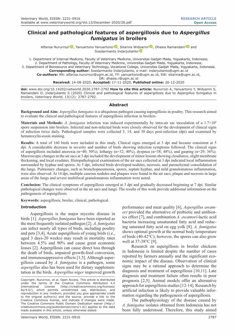
Clinical signs were observed in a varying num-ber of birds from 3 dpi to 8 dpi. The number of birds showing clinical signs, as well as the severity of symptoms, began to decrease at 8 dpi and then com-pletely disappeared at 12 dpi. The clinical signs of aspergillosis included anorexia (n=40; 50%), leth-argy (n=32; 40%), dyspnea (n=38; 48%), and gasping (n=29; 36%) (Figure-1). Some sporadic birds showed symptoms of other clinical signs, including torticollis, conjunctivitis, ascites, and stunting.
According to Okwara [16], the symptoms of asper-gillosis primarily consist of increased heavy breathing and a scratchy sound heard chiefly during expiration.
The clinical signs of dyspnea gradually become more severe, and snoring develops. The bird then loses its appetite, tends to ruffle its feathers, and becomes sleepy.
The infected group did not show clinical symp-toms of aspergillosis until 2 dpi. At 4 dpi, 37 of the 80 infected chickens began to show clinical signs of aspergillosis. Control group chickens injected with physiological NaCl showed no clinical symptoms on the same day. At 5 dpi, 41 sick chickens were recorded. At 6 dpi, the number of chickens showing clinical signs of aspergillosis gradually decreased (Figure-2).Macroscopic lesions
Necropsy was carried out on all dead chickens for macroscopic and microscopic pathological obser-vation. The data of pathological changes recorded at different time points are shown in Tables-3 and 4.Discussion
The clinical signs of early-onset aspergillosis observed in infected birds were breathing difficulties and increased frequency of inhalation; birds infected with aspergillosis often breathed by craning their neck or opening their beak. Respiratory problems appeared to be more common at night than during the day, possibly because of a limited oxygen supply. Anorexia levels in infected birds ranged from mild to severe. These findings are consistent with the previ-ous research [13,17,18], which reports that the clin-ical symptoms of acute aspergillosis manifest from between few days to 2 weeks of early maintenance. Aspergillosis has been clinically discovered in young chickens aged <13 days [19].
Dyspnea and gasping caused by hyphae growth lead to necrosis and inflammation in the air sacs and lungs [20]. Necrotic cells in the respiratory tract cause hypoxia, and the birds compensate for the oxy-gen demand of tissues by increasing their respiratory
40
32
38
29
37
051015202530354045
anorexia lethargy dyspnea gasping tortikolis other
num
ber o
f bird
Clinical signs
Figure-1: Clinical observations of Aspergillus fumigatus infection.
frequency. Exudates and inflammation in the lung cause disrupted air circulation [21]. The presence of plaques or necrotic areas in the respiratory tract due to aspergillosis could inhibit the exchange of oxygen in the lungs [2].
In this study, intra-air sac infection with 1.7×10⁸ spores caused acute aspergillosis with a morbid-ity rate of 71.25%, thus confirming the data of ear-lier reports [11,16,20]. Mortality occurred primarily within the first 5 days after infection.
Macroscopic changes in the lungs of infected birds at 3 dpi included minor lesions (Table-5, Figure-3). A few birds revealed caseous nodules, necrosis in over half of the lung area, and parenchy-mal consolidation. The 3 dpi has not been found in the peripheral edema and pulmonary parenchymal con-solidation. The results of this study support the pre-vious publication by Cheng et al. [15], which states early in the pulmonary lesions occur on the edges of
pulmonary edema, progressive consolidation, and formed small white nodules.
At 5 dpi, the infected birds showed severe lung lesions (Table-5, Figure-3); indeed, nearly all of the birds in the infected group showed macroscopic lesions. Yellowish-white caseous nodules diffused throughout the lung to form plaque aggregates with a diameter of 5-8 mm spread evenly over the tissue. Large necrotic areas and parenchymal consolidation were found in the lungs at 5 dpi.
Examination of air sacs at 3 dpi revealed local inflammation surrounded with hyphae and spores (Table-6, Figure-4a). No granulomas were observed at 3 dpi. Several researchers [2,10,13-15] have found that microscopic changes in the air sacs of infected birds develop rapidly and are accompanied by thick-ening, vascularity, and turbidity. Granulomas mea-suring 1-5 mm appeared and tended to merge to form plaques. Caseous plaques in the air sacs formed from the merging of fungal colonies, eventually covering and blocking the entire lamina membrane [2,10,15].
Microscopic examination of organs of the control group at 5 dpi revealed the absence of lesions (Table-6). The air sacs of this group appeared normal; no accu-mulation of inflammatory cells and/or changes in the epithelium was noted. Microscopic changes in the air sacs of chickens in the infected group at 5 dpi indicated granulomatous inflammation surrounded by spores and hyphae, submucosal edema, infiltration of inflam-matory cells, and epithelial hypertrophy of the air sacs (Table-6, Figure-4b). Hypertrophy of epithelial cells in air sacs caused macroscopic changes leading to mem-brane thickening. Infiltration of mononuclear inflam-matory cells dominated by heterophils was observed all
Figure-3: Macroscopic lesions in the infected group. (a) Thickening and turbidity of the walls of the thoracic air sac (3 dpi). (b) Multiple caseous nodule or plaques and severe thickening of the air sacs (14 dpi). (c) Nodules and necrosis in the lungs (5 dpi). (d) Caseous plaques in wide areas of the lung (14 dpi).
a
c d
b
Veterinary World, EISSN: 2231-0916 2791
Available at www.veterinaryworld.org/Vol.13/December-2020/26.pdf
Table-5: Most prominent macroscopic changes observed in broilers infected with aspergillosis.
Days post-infection
Macroscopic lesion
Air sac Lung
3 Cloudy, slightly thickened, local exudate
No macroscopic changes were found
5 Yellow caseous exudate covering the air sac
Nodules and necrosis in over half of the lung area, parenchymal consolidation
14 Multiple caseous nodules or plaques, severely thickened air sac
Nodules/plaques and necrosis in wide areas of the lung, massive congestion
Table-6: Most prominent microscopic changes observed in broilers infected with aspergillosis.
Days post-infection
Microscopic lesion
Air sac Lung
3 Inflammation, hyphae, and spores found
Local inflammation
5 Granulomatous inflammation
Hyphae and spores found
14 Granulomatous inflammation (>2 in each field of view)Giant and severe necrotic cells
Granulomatous inflammation
over the air sacs. Spores were surrounded by inflamma-tory cells and cell debris. The absence of a submucosal layer, which indicates the occurrence of edema, was also noted.
The emergence of conidia and hyphae is closely related to the virulence factors of aspergillosis. A. fumi-gatus conidia are fairly small and have a diameter of only 2-3 μm. Thus, these conidia can pass through physical barriers and invade all tissues of the respira-tory system [21,22]. A. fumigatus spores have intracel-lular germination capability, which is associated with the degeneration and necrosis of macrophages.
Microscopic lesions in the lungs of chickens in the infected group at 5 dpi revealed bronchopneumo-nia and infiltration of inflammatory cells dominated by heterophils in the bronchi and around vesicles.
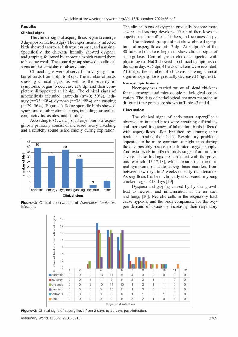
Pathognomonic microscopic findings in pulmonary were bronchopneumonia, spores, septate hyphae, and mild granulomatous inflammation (Table-6, Figure-4c).
Cacciutolo et al. [22] stated that the initial stage of aspergillosis infection is characterized by focal lym-phocytes and macrophages. Caseous necrosis with the
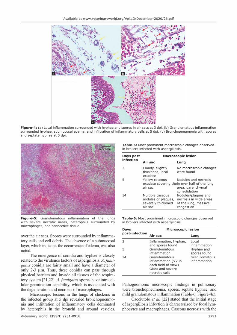
Figure-5: Granulomatous inflammation of the lungs with severe necrotic areas, heterophils surrounded by macrophages, and connective tissue.
Figure-4: (a) Local inflammation surrounded with hyphae and spores in air sacs at 3 dpi. (b) Granulomatous inflammation surrounded hyphae, submucosal edema, and infiltration of inflammatory cells at 5 dpi. (c) Bronchopneumonia with spores and septate hyphae at 5 dpi.
b
c
a
Veterinary World, EISSN: 2231-0916 2792
Available at www.veterinaryworld.org/Vol.13/December-2020/26.pdf
proliferation of connective tissue was observed at 4 dpi. Accumulation of caseous exudates in the bronchial lumen and air vesicles caused respiratory symptoms such as dyspnea, gasping, panting, and coughing.
Histopathological observations at 14 dpi showed no different results compared with 5 dpi (Table-6, Figure-5). Hyphae surrounded by severe multifocal granulomatous inflammations (i.e., >2 in a single field of view) were also found. Observation of all groups at 14 dpi revealed no spores (Figure-5). Lesions devel-oped into granulomas consisting of severe necrotic areas and heterophils surrounded by macrophages, lymphocytes; connective tissues were also found [23].Conclusion
The clinical symptoms of aspergillosis decreased by 7 dpi. Pathological features indicated permanent organ damage. Birds affected by aspergillosis should be culled because of irreversible pathological damage.Authors’ Contributions
SI conceptualized, managed, and supervised the study. AN and DR collected, recorded, and analyzed the samples. YY and SW identified and analyzed data. All authors drafted, revised and approved the final manuscript.Acknowledgments
The authors are highly thankful to the Department of Internal Medicine, Faculty of Veterinary Medicine, Universitas Gadjah Mada, Indonesia, for provid-ing funds and facilities (Grant no. 1539/ J01.1.22/HK4/2015).Competing Interests
The authors declare that they have no competing interests.Publisher’s Note
Veterinary World remains neutral with regard to jurisdictional claims in published institutional affiliation.References1. Beernaert, L.A., Pasmans, F., Van Waeyenberghe, L. and
Haesebrouck, F. (2010) Aspergillus infections in birds: A review. Avian Pathol. 39(5): 325-331.
2. Arné, P., Thierry, S., Wang, D., Deville, M., Le Loc’h, G., Desoutter, A., Féménia, F., Nieguitsila, A., Huang, W., Chermette, R. and Guillot, J. (2011) Aspergillus fumigatus in poultry. Int. J. Microbiol., special issue vol. 2011: 1-14
3. Martin, M.P., Bouck, K.P., Helm, J. and Dykstra, M.J. (2007) Disseminated Aspergillus flavus infection in broiler breeder pullets. Avian Dis., 51(2): 626-631.
4. Zafra, R., Perez, J. and Perez, R.A. (2008) Concurrent aspergillosis and ascites with high mortality in a farm of growing broiler chickens. Avian Dis., 52(4): 711-713.
5. Saif, Y.M. (2008) Diseases of Poultry. 11th ed. Blackwell
Publishing Company. United States. p883-894.6. Saleh, A.A., Eid, Y.Z., Ebeid, T.A., Kamizono, T.,
Ohtsuka, A. and Hayashi, K. (2011) Effects of feeding Aspergillus awamori and Aspergillus niger on growth per-formance and meat quality in broiler chickens. J. Poult. Sci., 48(1): 201-206.
7. Saleh, A.A., Hayashi, K., Ijiri, D. and Ohtsuka, A. (2014) Beneficial effects of Aspergillus awamori in broiler nutri-tion. Worlds Poult. Sci. J., 70(4): 857-864.
8. Saleh, A.A., Gálik, B., Arpášová, H., Capcarová, M., Kalafová, A., Šimko, M., Juráček, M., Rolinec, M., Bíro, D. and Abudabos, A.M. (2017) Synergistic effect of feeding Aspergillus awamori and lactic acid bacteria on perfor-mance, egg traits, egg yolk cholesterol and fatty acid profile in laying hens. Ital. J. Anim. Sci., 16(1): 132-139.
9. Munir, M.T., Rehman, Z.U., Shah, M.A. and Umar, S. (2019) Interaction of Aspergillus fumigatus with the respi-ratory system in poultry. Poult. Sci. J., 73(2): 321-336.
10. Femenia, F., Fontaine, J.J. and Lair-Fulleringer, S. (2007) Clinical, mycological and pathological findings in turkeys experimentally infected by Aspergillus fumigatus. Avian Pathol., 36(3): 213-219.
11. Sultana, S., Harun-Ur-Rashid, S.M., Islam, M.N. and Ali, M.Z. (2014) Pathological investigation of avian asper-gillosis in commercial broiler chicken at Chittagong district. Int. J. Adv. Res. Biol. Sci., 1(8): 74-85.
12. Clemons, K.V. and Stevens, D.A. (2005) The contribution of animal models of aspergillosis to understanding patho-genesis, therapy and virulence. Med. Mycol. J., 43(1): 101-110.
13. Beernaert, L.A., Pasmans, F., Haesebrouck, F. and Martel, A. (2008) Modelling Aspergillus fumigatus infections in racing pigeons (Columba livia domestica). Avian Pathol., 37(5): 545-549.
14. Desoubeaux, G. and Cray, C. (2018). Animal models of aspergillosis. Comp. Med., 68(2): 109-123.
15. Cheng, Z., Li, M., Wang, Y., Chai, T., Cai, Y. and Li, N. (2020) Pathogenicity and immune responses of Aspergillus fumigatus infection in chicken. Front. Vet. Sci., 7: 143
16. Okwara, N. (2016) Aspergillosis in Turkeys: A review. J. Agric. Vet. Sci., 9(6): 40-41.
17. Singh, S., Borah, M.K. and Sharma, D.K. (2009) Aspergillosis in Turkey poults. Indian J. Vet Pathol., 33(2): 220-221.
18. Melo, A.M., Silva-Filho, R.P., Poester, V.R., Von Groll, A., Fernandes, C.G., Stevens, D.A., Sabino, R. and Xavier, M.O. (2020) Aspergillosis in free-ranging aquatic birds. Med. Mycol. Case Rep., 28(1): 36-38.
19. Beernaert, L.A., Pasmans, F. and Baert, K. (2009) Designing a treatment protocol with voriconazole to elimi-nate Aspergillus fumigatus from experimentally inoculated pigeons. Vet. Microbiol., 139(3): 393-397.
20. Ahamad, D.B., Ranganathan, V., Punniyamurthy, N., Sivaseelan, S. and Puvarajan, B. (2018) Pathomorfology of aspergillosis in a Japanese quail. Indian Vet. J., 5(4): 36-42.
21. Chu, J., Zhang, Q., Zuo, Z.H., Elashram, S., Guo, Y.X. and Zhao, P. (2017) Co-infection of Chlamydia psittaci with H9N2, ORT and Aspergillus fumigatus contributes to severe pneumonia and high mortality in SPF chickens. Sci. Rep., 7(1): 13997.
22. Cacciutolo, E., Rossi, G., Nardoni, S., Legrottaglie, R. and Mani, P. (2009) Anatomopathological aspect of avian asper-gillosis. Vet. Res. Commun., 33(1): 521-527.
23. Pazhanivel, N., Saahithya, R., Thangapandiyan, M., Venkata, G., Rao, S. and Sridhar, K. (2018) Pulmonary aspergillosis in a seventeen-day old ostrich chick (Struthio camelus). J. Entomol. Zool. Stud., 6(5): 553-555.


![Aspergillosis - Youngstown State Universitypeople.ysu.edu/~crcooper01/Aspergillosis[1]- Katie Jacquie Qazi.pdf•People with Aspergillosis are in three distinct groups •Healthy immune](https://static.documents.pub/doc/80x56/5e3883b0e2f2970b7b1c24ad/aspergillosis-youngstown-state-crcooper01aspergillosis1-katie-jacquie-qazipdf.jpg)

