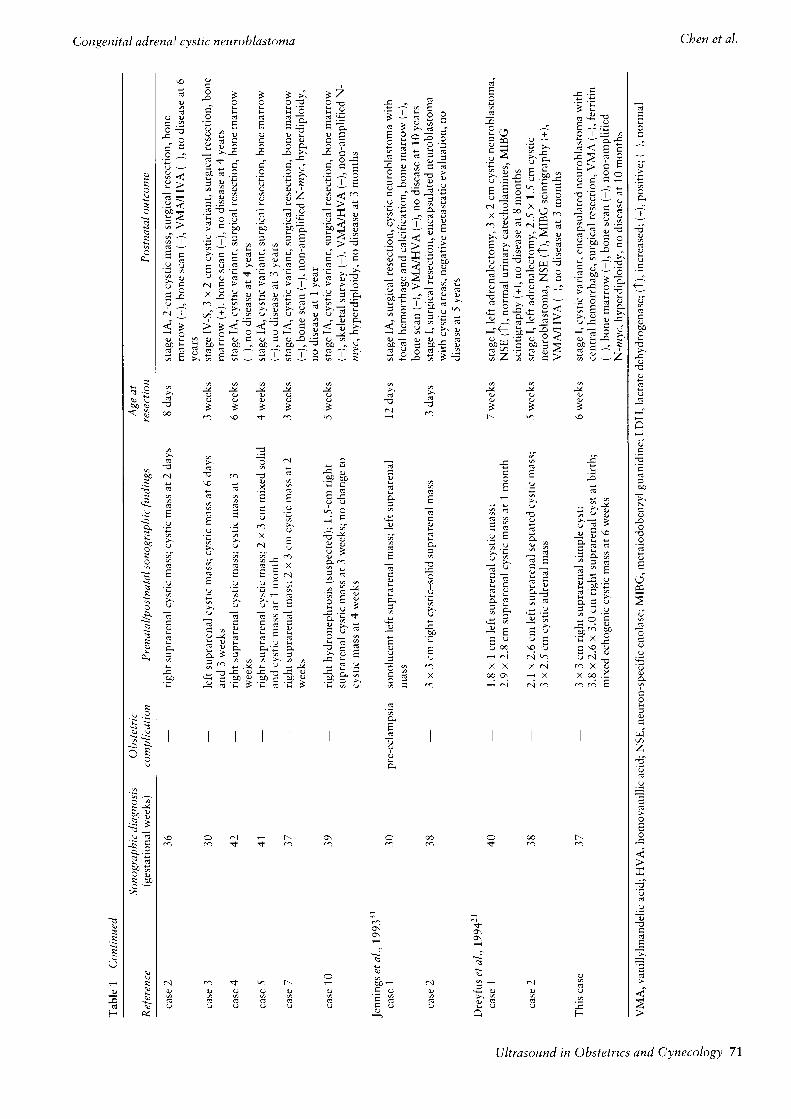
Ultrasound Obstet. Gynecol. 10 (1997) 68-73
Clinical and perinatal sonographic features of congenital adrenal cystic neuroblastoma: a case report with review of the literature
C.-P. Chen, S.-H. Chen”, C.-Y. Chuaq$, H.-C. Lee”, Y.-M. Hwu, P.-Y. Chang*, M.-L. Ched and
Department of 0 bstetrics and Gynecology; “Department *Department of Pediatric Surgery; ” *Department of Path01 Republic of China
of Pediatrics; +Department of Medical Research;
ogy, Mackay Memorial Hospital, Taipei, Taiwan,
Keywords: ADRENAL GLAND,HEMORRHAGE,NEUROBLASTOMA,CYST,RESOLUTION
ABSTRACT
Cystic formation in association with adrenal neuroblas-
toma may be related to hemorrhage and necrosis of the
tumor. We present an unusual case of congenital cystic
fetal neuroblastoma of the right adrenal gland detected at
37 weeks’ gestation which evolved into a complex echo-
genie mass 6 weeks after birth. Surgical exploration re-
vealed a 3.5 x 3 x 3 cm right complex adrenal tumor which
was resected. The infant did well 10 weeks after tumor
resection. Typically adrenal hemorrhage may appear sono-
graphically to be entirely ecbogenic, of mixed ecbogenicity,
or anecboic when first imaged. Gradually, the texture of
the bematoma will evolve and become more cystic and
ecbolucent on f(Alow-up ultrasound examinations. In con-
trast, our case of congenital adrenal cystic neuroblastoma
became more complex after resolution of the hemorrhagic
cyst. This case suggests that adrenal hemorrhage and
adrenal cystic neuroblastoma with a hemorrhagic cyst have
different sonograpbic appearances. We suggest that addi-
tional imaging and surgical intervention should be con-
sidered whenever a cystic suprarenal mass becomes more
complex after resolution and demonstrates no significant
decrease in size in postnatal examinations.
INTRODUCTION
Neuroblastoma is a tumor of the postganglionic sympa- thetic neurons and is the most common extracranial solid tumor found in children’. In half of the cases, the tumor arises in the adrenal gland’. Neuroblastoma occurs in about 1 per 10 000 to 1 per 30 000 live birthslm3. Purely
cystic lesions have been reported in fetal neuroblastoma,
but are rare in infancy4. The cystic formation in association
with neuroblastoma may be related to hemorrhage and
necrosis of a tumor. Purely cystic neuroblastoma may rep-
resent neuroblastoma in situ and may indicate a favorable
postnatal prognosis 4-8 The incidence of adrenal hemor- . rhage based on an extensive necropsy series has been
estimated at approximately 1.7 per 1000 births’. The eti-
ologies of adrenal hemorrhage include birth trauma, peri-
natal hypoxia, septicemia, shock, thrombocytopenia,
congenital syphilis and disseminated intravascular coagula-
tion9-13. Most hemorrhages are reported to occur at birth
or during the early neonatal period”. The differential diag-
nosis between adrenal cystic neuroblastoma and a purely
adrenal hemorrhage is difficult, because the blood flow on
ultrasound examination may be absent in both casesI and1
the urine vanillylmandelic acid levels have be’en reported toI
be normal in fetal or neonatal cystic neuroblastoma4T14V
We present our observation of an anechoic simple cyst
in the fetal adrenal gland which evolved to a complex
echogenic mass by 6 weeks postnatally and prompted
surgery for resection of an adrenal neuroblastoma. The
postnatal evolution on ultrasonography manifested an un-,
usual natural history of fetal adrenal cystic neuroblastoma.
We review the literature for other cases of congenital
adrenal cystic neuroblastoma detected by perinatal sono-
graph Y *
CASE REPORT
A D-year-old woman, gravida 3 para 2, was referred fo1
sonographic evaluation at 37 weeks’ gestation because of a
fetal intra-abdominal mass. The mother had two healthy
children. She had none of the following conlditions during
the course of her pregnancy: maternal exposure tcl
hydantoin, phenobarbital, alcohol, teratogenic agents 01
Correspondence: Dr C.-P. Chen, Department of Obstetrics and Gynecology, and Department of Medical Research, Mackay Memorial Hospital.,
92, Section 2, Chung-Shan North Road, Taipei, Taiwan, Republic of China
CASE REPORT AND REVIEW 68 Received 15-l O-96 Revised 8-l-97
Accepted 18-1-97
Congenital adrenar’ cystic neurohlastoma Chen et al.
Figure 1 Fetal sonogram at 37 weeks’ gestation, showing
anechoic simple cyst (arrowheads) superior to the right kidney
an
Figure 3 At age 6 weeks, the neuroblastoma appea
echogenicity (arrowheads) on ultrasound scanning
rs with mixed
Figure 2 Neonatal sonogram at age 3 days, demonstrating a
suprarenal cystic mass (M) and the right kidney (RK)
radiation, familial neuroblastoma, rnaternal diabetes and
infectious diseases. The father’s age was 34 years. The
Chinese couple involved in this pregnancy were unrelated
and there was no family history of malformations. Ultra-
sound examination of the 37-week fetus demonstrated a
3 x 3 cm anechoic simple cyst (Figure 1) superior to the
right kidney without the finding of blood flow by color
Doppler and flow imaging. Both kidneys were normal. At
Figure 4 Neonatal CT scan with contrast enhancement at age 6
weeks, demonstrating a well-defined hypodense nodular lesion
(arrowheads) over the upper pole of the right kidney
39 weeks’ gestation, a 3118-g healthy female baby was
delivered vaginally. Labor and delivery proceeded without
complication. At birth, no abdominal mass was palpable
on physical examination.
Repeat sonography demonstrated an anechoic cystic
mass (Figure 2) measuring 3.8 x 2.6 x 3.0 cm over the
upper pole of the right kidney. Examination of large blood
vessels within the renal pelvis showed no evidence of
Ultrasound in Obstetrics and Gynecology 69
Ref
eren
ce
Sono
grap
hic
diag
nosi
s O
bste
tric
(g
esta
tiona
l w
eeks
) co
mpl
icat
ion
Pre
nata
l/po
stna
tal
sono
grap
hic
find
ings
A
ge
at
rese
ctio
n P
ostn
atal
ou
tcom
e
Jane
tsch
ek
et a
l., 1?842
New
ton
et a
l.,
1985
19
34
35
- 4-
cm
cyst
ic
righ
t ad
rena
l m
ass
at
34
wee
ks;
6-cm
cyst
ic
and
solid
m
ass
at
36
wee
ks;
solid
an
d
lago
on-s
hape
d cy
stic
m
ass
a fe
w
hour
s af
ter
birt
h
1 da
y st
age
I, r
ight
ad
rena
lect
omy,
cy
stic
ne
urob
last
oma
with
cent
ral
hem
orrh
agic
ne
cros
is,
norm
al
urin
ary
cate
chol
amin
es,
no
dise
ase
at
6 m
onth
s
pre-
ecla
mps
ia
4cm
ri
ght
rena
l cy
stic
m
ass
at
35
wee
ks;
5 x 7 c
m
feta
l hy
drop
s,
neon
atal
de
ath,
bi
late
ral
adre
nal
cyst
ic
righ
t fl
ank
mas
s,
asci
tes,
ri
ght
neur
obla
stom
a,
diff
use
met
asta
ses
to
liver
, lu
ng,
brai
n,
hydr
onep
hros
is
(sus
pect
ed)
at
36
wee
ks
pitu
itary
, sp
inal
co
rd
and
plac
enta
Atk
inso
n et
al.,
19
864
case
1
case
2
Giu
lian
et a
l.,
1986
ls
Saka
ta
et a
l.,
1986
30
Pley
et
al.,
1
9892
0 36
Form
an
et a
l.,
1990
””
case
1
Hos
oda
et a
l.,
1992
22
Ho
et a
l.,
1993
’
case
1
34
32
38
39
4 da
ys
befo
re
birt
h
3.5
30
- co
mpl
ex
righ
t re
nal
mas
s;
pers
iste
nt
com
plex
supr
aren
al
mas
s
- co
mpl
ex
left
re
nal
mas
s,
left
hy
dron
ephr
osis
(sus
pect
ed);
a
mul
tiloc
ulat
ed
com
plex
, ec
hoge
nic
mas
s su
peri
or
and
med
ial
to
the
left
ki
dney
- 3.
5-cm
m
ixed
cy
stic
an
d so
lid
righ
t su
prar
enal
mas
s;
righ
t ad
rena
l co
mpl
ex
mas
s
- 3.
4 x
4.0
cm
intr
a-ab
dom
inal
cy
st;
mix
ed
cyst
ic
and
solid
ri
ght
adre
nal
mas
s
mat
erna
l 4c
m
left
up
per
abdo
men
so
notr
ansl
ucen
t co
bweb
-
hype
rten
sion
lik
e m
ass;
le
ft
hydr
onep
hros
is
(sus
pect
ed)
- ri
ght
adre
nal
cyst
ic
mas
s;
no
chan
ge
to
cyst
ic
mas
s
at
6 w
eeks
- 3.
5 x 3.
1 cm
m
ixed
cy
stic
le
ft
supr
aren
al
mas
s;
3.2
x 3.
4 cm
m
ixed
cy
stic
le
ft
adre
nal
mas
s at
4
days
toxe
mia
le
ft
supr
aren
al
mas
s at
30
w
eeks
; si
ze
incr
ease
d at
34
wee
ks;
no
chan
ge
to
cyst
ic
mas
s at
4
days
not
repo
rted
not
repo
rted
3 da
ys
19
days
not
repo
rted
not
repo
rted
16
days
13
days
stag
e I,
sur
gica
l re
sect
ion,
3.
5 X 3.
5 X 2
.5
cm
cyst
ic
mas
s,
neur
obla
stom
a in
sit
u w
ith
hem
orrh
agic
cy
st,
no
dise
ase
at
2 ye
ars
stag
e I,
sur
gica
l re
sect
ion,
5 x 4 x 3
cm
mul
tiloc
ulat
ed
mas
s w
ith
solid
an
d cy
stic
co
mpo
nent
s,
hem
orrh
agic
cy
sts
and
sero
us
cyst
s,
VM
A/H
VA
(-
),
bone
m
arro
w
(-),
bo
ne
scan
(-
),
no
dise
ase
at
1 ye
ar
stag
e I,
sur
gica
l re
sect
ion,
he
mor
rhag
ic
cong
enita
l
neur
obla
stom
a,
no
dise
ase
at
1 ye
ar
stag
e I,
5 x
4.5
x
3 cm
cy
stic
m
ass
with
bl
ood
clot
an
d
sero
sang
uine
ous
flui
d,
surg
ical
re
sect
ion,
V
MA
/HV
A
(-),
L
DH
(?
),
ferr
itin
(T),
b
one
mar
row
(-
),
bone
sc
an
(-),
no
dise
ase
at
9 m
onth
s
stag
e I,
sur
gica
l re
sect
ion,
en
caps
ulat
ed
adre
nal
neur
obla
stom
a w
ith
hem
orrh
age
and
necr
osis
, V
MA
/HV
A
(-),
no
di
seas
e at
1
year
stag
e II
, su
rgic
al
rese
ctio
n,
cyst
ic
prim
ary
tum
or,
no
dise
ase
at
2.8
year
s
stag
e I,
3.5
x 2 x 2
cm
hem
orrh
agic
tu
mor
, su
rgic
al
rese
ctio
n,
VM
A/I
-IV
A
(-),
N
SE
(-
),
non-
ampl
ifie
d N
-myc
,
hype
rdip
loid
y,
no
dise
ase
at
16
mon
ths
stag
e IA
, 5 x 4
cm
cy
stic
m
ass,
su
rgic
al
rese
ctio
n,
bone
mar
row
(-
),
bone
sc
an
(-),
V
MA
/HV
A
(-),
no
di
seas
e at
9
year
s
Tab
le
1 C
onti
nued
Ref
eren
ce
Sono
grap
hic
diag
nosi
s 0
bste
tric
(g
esta
tiona
l w
eeks
) co
mpl
icat
ion
Pre
nata
l/po
stna
tal
sono
grap
hic
find
ings
A
ge a
t re
sect
ion
Pos
tnat
al
outc
ome
case
2
36
- ri
ght
supr
aren
al
cyst
ic
mas
s;
cyst
ic
mas
s at
2
days
8
days
case
3
case
4
case
5
case
7
case
10
Jenn
ings
et
al.,
199
3 ‘I
ca
se
1
case
2
Dre
yfus
et
al.,
199
421
case
1
case
2
38
Thi
s ca
se
37
30
42
41
37
39
30
38
40
- le
ft
supr
aren
al
cyst
ic
mas
s;
cyst
ic
mas
s at
6
days
and
3 w
eeks
-
righ
t su
prar
enal
cy
stic
m
ass;
cy
stic
m
ass
at
3
wee
ks
- -
righ
t su
prar
enal
cy
stic
m
ass;
2
x 3
cm
m
ixed
so
lid
and
cyst
ic
mas
s at
i
mori
ih
righ
t su
prar
enal
m
ass;
2
x 3
cm
cy
stic
m
ass
at
2 w
eeks
- ri
ght
hydr
onep
hros
is
(sus
pect
ed);
1.
.5-c
m
righ
t
supr
aren
al
cyst
ic
mas
s at
3
wee
ks;
no
chan
ge
to
cyst
ic
mas
s at
4
wee
ks
pre-
ecla
mps
ia
- 3
x 3
cm
righ
t cy
stic
-sol
id
supr
aren
al
mas
s 3
days
sono
luce
nt
left
mas
s
supr
aren
al
mas
s;
left
su
prar
enal
12
da
ys
- 1.
8 x
1 cm
le
ft
supr
aren
al
cyst
ic
mas
s;
2.9
x 2.
8 cm
su
prar
enal
cy
stic
m
ass
at
1 m
onth
- 2.
1 x
2 .6
cm
le
ft
supr
aren
al
sept
ated
3 x
2.5
cm
cyst
ic
adre
nal
mas
s
cyst
ic
mas
s;
5 w
eeks
- 3
x 3
cm
righ
t su
prar
enal
si
mpl
e cy
st;
3.8
x 2.
6 x 3
.0 c
m
righ
t su
prar
enal
cy
st
at
birt
h;
mix
ed
echo
geni
c cy
stic
m
ass
at
6 w
eeks
3 w
eeks
6 w
eeks
4 w
eeks
3 w
eeks
5 w
eeks
7 w
eeks
6 w
eeks
stag
e IA
, 2-
cm
cyst
ic
mas
s,
surg
ical
re
sect
ion,
bo
ne
mar
row
(-
),
bone
sc
an
(-),
V
MA
/HV
A
(-),
no
di
seas
e at
6
year
s
stag
e IV
-S,
3 x 2
cm
cy
stic
va
rian
t, su
rgic
al
rese
ctio
n,
bone
mar
row
(+
).
bone
sc
an
(-),
no
di
seas
e at
4
year
s
stag
e IA
, cy
stic
va
rian
t, su
rgic
al
rese
ctio
n,
bone
m
arro
w
(-),
no
di
seas
e at
4
year
s
stag
e IA
, cy
stic
va
rian
t, su
rgic
al
rese
ctio
n,
bone
m
arro
w
stag
e IA
, cy
stic
va
rian
t, su
rgic
al
rese
ctio
n,
bone
m
arro
w
(-),
bo
ne
scan
(-
),
non-
ampl
ifie
d N
-myc
, hy
perd
iplo
idy,
no
dise
ase
at
I ye
ar
stag
e IA
, cy
stic
va
rian
t, su
rgic
al
rese
ctio
n,
bone
m
arro
w
(-),
sk
elet
al
surv
ey
(-),
V
MA
/HV
A
(-),
no
n-am
plif
ied
N-
myc
, hy
perd
iplo
idy,
no
di
seas
e at
3
mon
ths
stag
e IA
, su
rgic
al
rese
ctio
n,
cyst
ic
neur
obla
stom
a w
ith
foca
l he
mor
rhag
e an
d ca
lcif
icat
ion,
bo
ne
mar
row
(-
),
bone
sc
an
(-),
V
MA
/HV
A
(-),
no
di
seas
e at
10
yea
rs
stag
e I,
sur
gica
l re
sect
ion,
en
caps
ulat
ed
neur
obla
stom
a
with
cy
stic
ar
eas,
ne
gativ
e m
etas
tatic
ev
alua
tion,
no
dise
ase
at
5 ye
ars
stag
e I,
lef
t ad
rena
lect
omy,
3
x 2
cm
cyst
ic
neur
obla
stom
a,
NS
E
(T),
nor
mal
ur
inar
y ca
tech
olam
ines
, M
IBG
sc
intig
raph
y (+
),
no
dise
ase
at
8 m
onth
s
stag
e I,
lef
t ad
rena
lect
omy,
2.
5 x
1.5
cm c
ysti
c ne
urob
last
oma,
N
SE
(?
), M
IBG
sc
intig
raph
y (+
),
VM
A/H
VA
(-
), n
o di
seas
e at
3
mon
ths
stag
e I,
cys
tic
vari
ant,
enca
psul
ated
ne
urob
last
oma
with
cent
ral
hem
orrh
age,
su
rgic
al
rese
ctio
n,
VM
A
(-),
fer
ritin
(-),
bo
ne
mar
row
(-
),
bone
sc
an
(-),
no
n-am
plif
ied
N-m
yc,
hype
rdip
loid
y,
no
dise
ase
at
10 m
onth
s
VM
A,
vani
llylm
ande
lic
acid
; H
VA
, ho
mov
anill
ic
acid
; N
SE,
neur
on-s
peci
fic
enol
ase;
M
IBG
, m
etai
odob
enzy
l gu
anid
ine;
L
DH
, la
ctat
e de
hydr
ogen
ase;
(T
), in
crea
sed;
(+
), po
sitiv
e;
(-),
no
rmal
Congenital adrenal cystic neuroblastoma
thrombosis. A 24-h urinalysis for vanillylmandelic acid was
normal and serum lactate dehydrogenase, ferritin and
a-fetoprotein levels were within normal limits.
At the age of 6 weeks, subsequent sonographic examina-
tion showed that the adrenal cyst had spontaneously
evolved into a complex echogenic mass (Figure 3). A com-
puterized tomography (CT) scan revealed a well-defined
hypodense nodular lesion over the upper pole of the right
kidney (Figure 4). Surgical exploration at the age of 6
weeks revealed a 3.5 x 3 x 3 cm right complex adrenal
tumor and the primary tumor was resected. On micro-
scopic examination, the tumor tissues of the neuroblas-
toma were surrounded by a thick fibrous capsule and
central hemorrhage. The tumor was considered a stage I
neuroblastoma. It showed hyperdiploid cellular DNA con-
tent and absence of amplification of the N-myc oncogene.
The mixed echogenic mass contained bloody fibrous
exudate. The tumor tissues demonstrated the typical
appearance of darkly staining clusters of neuroblasts. A
radionuclear bone scan, bone marrclw aspiration and serial
liver sonography were all normal. No further therapy was
given. The infant did well 10 months after tumor resection.
Peripheral blood karyotyping revealed a normal 46,Xx
complement.
DISCUSSION
The purely adrenal hemorrhage may be sonographically
entirely echogenic, mixed echogenic or anechoic when first
imaged, but the texture of the hematoma will gradually
evolve and become more cystic and echolucent on follow-
up ultrasound examinations 16-18. In contrast, our case of
concomitant adrenal hemorrhage and cystic neuroblastoma
became more complex after resolution. The mass shrinkage
in this case at follow-up represented absorption of intra-
cystic fluid rather than an absence of the tumor. Our
presentation implies that purely adrenal hemorrhage and
neuroblastoma with a concomitant adrenal hemorrhagic
cyst have different natures and sonographic appearances.
Twenty cases of congenital cystic neuroblastoma located
in the adrenal gland have been reported to date (Table 1).
These 20 cases and the present case were all detected in the
third trimester of pregnancy. Among these 21 cases of
congenital adrenal cystic neuroblastoma, 15 were purely
cystic and six were mixed solid and cystic when first im-
aged, 12 were right-sided, eight were left-sided and one was
bilateral. The sonotranslucency of fetal cystic neuroblas-
toma was prenatally diagnosed as hydronephrosis in four cases497J9,20 . Maternal complications such as pre-eclampsia,
toxemia or hypertension occurred in four cases. In most
cases, the sonotranslucent areas represented hemorrhage
and liquified necrosis. The urinary vanillylmandelic acid
and homovanillic acid were negative in all (nine of nine)
patients who were evaluated preoperatively. Two of three
cases had high levels of neuron-specific enolase21722. Meta-
static involvement at the time of the initial postnatal exami-
nation was found in the bone marrow of one patient7.
Clinical staging was favorable. Of the 21 patients, 18 had
stage I disease, one had stage II disease23, one had diffuse
72 Ultrasound in Obstetrics and G:ynecology
Chen et al.
metastases and died neonatally” and one was categorized
as stage IV-S7. Twenty patients underwent surgical resec-
tion of the primary tumor at ages ranging from 1 day to 7
weeks and all these patients remained free of disease on
follow-up, ranging from 3 months to 10 ‘years. Diploid
cellular DNA content and amplification of the N-myc
oncogene have been indicated as carrying a poor prognosis,
whereas hyperdiploid cellular DNA content and absence of
amplification of the N-myc oncogene appear to carry a
favorable prognosis24-2h. Such biological characteristics of
neuroblastoma cells were assessed in the case reported by
Hosada and co-workers’“, the two cases reported by Ho
and colleagues’ and the present case. All four tumors had
favorable features of hyperdiploidy and the absence of
amplification of the N-myc oncogene.
Another common cause of an adrenal cyst in the new-
born is adrenal hemorrhage. The fetal adrenal glands are
susceptible to hemorrhage because of their relatively large
size and vascularity in utero2’. Adrenal hemorrhage of the
newborn affects the right side three to four times more
commonly than the left side, possibly because of the pro-
pensity of the right adrenal gland for compression between
the liver and spine and for change in venous pressure27y2K.
Adrenal hemorrhage is usually followed by necrosis and
later resolution and regression of the mass with fibrosis and
calcification27y29. In the present case, the uneventful label
and delivery made the diagnosis of spontaneous adrenal
hemorrhage unlikely. Other possible suprarenal cystic
masses or lesions at the upper pole of the ki’dney - includ-
ing renal cysts, cystic Wilm’s tumor, obstructed upper pole
duplication anomalies with ectopic ureteral implantation,
hepatic cysts, choledochal cysts, ovarian cysts or enteric
cysts - should also be considered prenatally and be con-
firmed postnatally”.
Prenatal interventions such as aspiration and biopsy of
congenital adrenal cysts remain controversial. We conside]
that in utero interventions for these fetuses, with our cur-
rent knowledge, are unwarranted, because such procedures
may not be adequate for diagnosis of a congenital neo-
plasm. Furthermore, if follow-up of any infant for whom
conservative treatment is proposed cannot be assured, sur-
gical removal of the mass may be prudent. We suggest that
additional imaging and surgical intervention should be
considered whenever a cystic suprarenal mass becomes
more complex after resolution of the cyst and demonstrates
no significant decrease in size in postnatal examinations,,
even with a normal urinary vanillylmandelic acid level.
REFERENCES
. Brodeur, G. M. (1991). Neuroblastoma and other peripheral
neuroectodermal tumors. In Fernbach, D. J. and Vietti, T. J,
(eds.) Clinical Pediatric Oncology, 4th edn., pp. 437-64. (St Louis: Mosby Year Book)
Janetschek, G., Weitzel, D., Stein, W., Miintefering, H. and
Alken, P. (1984). Prenatal diagnosis of neuroblastoma b,
sonography. Urology, 24,397-402
Fowlie, F., Giacomantonio, M., McKenzie, E., Baird, D. and
Covert, A. (1986). Antenatal sonographic diagnosis of adrenal
neuroblastoma.]. Can. Assoc. Radiol., 37, 50-l
Congenital adrenal cystic neuroblastoma Chen et al.
4. Atkinson, G. 0. Jr, Zaatari, G. S., Lorenzo, R. L., Gay, B. B.
Jr and Garvin, A. J. (1986). Cystic neuroblastoma in infants:
radiographic and pathologic features. Am. J. Roentgevzol., 146,113-17
5. Garmel, S. H., Crombleholme, T. M., Semple, J. P. and Bhan,
I. (1994). Prenatal diagnosis and management of fetal tumors.
Semin. Perinatol., 18, 350-65
6. Tubergen, D. G. and Heyn, R. M. (1970). In situ neuroblas-
toma associated with an adrenal cyst. 1. Pediatr., 76,451-3 7. Ho, P. T. C., Estroff, J. A., Kozakewich, H., Shamberger,
R. C., Lillehei, C. W., Grier, H. E. anId Diller, L. (1993). Pre-
natal detection of neuroblastoma: a ten-year experience from
the Dana-Farber cancer institute and children’s hospital. Pedi- atrics, 92, 358-64
8. Crombleholme, T. M., Murray, T. A. and Harris, B. H.
(1994). Diagnosis and management (of fetal neuroblastoma.
Curr. Opin. Obstet. Gynecol., 6, 199--202 9. DeSa, D. J. and Nicholls, S. (1972). Haemorrhagic necrosis of
the adrenal gland in perinatal infants: a clinico-pathological
study.J. Pathol., 106, 133-49
10. Sones, P. J. Jr (1983). Sonography case of the day. Am. J. Roentgenol., 140,1024-9
11. Cohen, E. K., Daneman, A., Stringer, D. A., Soto, G. and
Thorner, P. (1986). Focal adrenal hemorrhage: a new US
appearance. Radiology, 161,631-3 12. Mineau, D. E. and Koehler, P. R. (1979). Ultrasound diagno-
sis of neonatal adrenal hemorrhage. Porn. J. Roentgenol., 132, 443-4
13. Alanen, A. and K.ormano, M. (1984). Real time sonographic
diagnosis of adrenal haemorrhage. Eur. J. Radiol., 4,210-12 14. Goldstein, I., Gomez, K. and Copel, J. A. (1994). The real-
time and color Doppler appearance of adrenal neuroblastoma
in a third-trimester fetus. Obstet. Gynecol., 83, 854-6 15. Eklof, O., Mortensson, W. and Sandstedt, B. (1986). Supra-
renal hematoma versus neuroblastoma complicated by hemor- rhage: a diagnostic dilemma in newborn. Acta Radiol. Diagn., 27,3-10
16. Mittelstaedt, C. A., Volberg, F. M., Merten, D. F. and Brill,
P. W. (1979). The sonographic diagnosis of neonatal adrenal
hemorrhage. Radiology, 131,453-7 17. Strouse, P. J., Bowerman, R. A. and Schlesinger, A. E. (1995).
Antenatal sonographic findings of fetal adrenal hemorrhage. J.
Clin. Ultrasound, 23,442-6 18. Giulian, B. B., Chang, C. C. N. and Yoss, B. S. (1986). Pre-
natal ultrasonographic diagnosis of fetal adrenal neuro-
blastoma. J. Clin. Ultrasound, 14,225-7 19. Newton, E. R., Louis, F., Dalton, M. E. and Feingold, M.
(1985). Fetal neuroblastoma and catecholamine-induced
maternal hypertension. Obstet. Gynecol., 65 (Suppl.),
49S-52s
20. Pley, E. A. P., Wouters, E. J. M., de Jong, I’. A., Tielens, A. G. W. M. and <zlermonts, G. J. F. (1989). The sonographic
imaging of fetal neuroblastoma: a case report. Eur. J. Obstet. Gynecol. Reprod. Biol., 31, 95-9
21. Dreyfus, M., Neuhart, D., Baldauf, J. J., Casanova, R., Bec-
meur, F. and Ritter, J. (1994). Prenatal diagnosis of cystic
neuroblastoma. Fetal Diagn. T’her., 9,269-72 22. Hosoda, Y., Miyano, T., Kimura, K., Oya, T. Ishimoto, K.,
Tanno, M. and Takeuchi, H. (1992). Characteristics and man-
agement of patients with fetal neuroblastoma. J. Pediatr. Surg., 27, 623-5
23. Forman, H. P., Leonidas, J. C., Berdon, W. E.!, Slovis, T. L., Wood, B. P. and Samudrala, R. (1990). Congenital neuroblas-
toma: evaluation with multimodality imaging. Radiology, 175,365-8
24. Look, A. T., Hayes, F. A., Nitschke, R., McWilliams, N. B.
and Green, A. A. (1984). Cellular DNA content as predictor
of response to chemotherapy in infants with unresectable
neuroblastoma. N. Engl. J. Med., 311,231-5 25. Look, A. T., Hayes, F. A., Shuster, J. J., Douglass, E. C.,
Castleberry, R. P., Bowman, L. C., Smith, E. I. and Brodeur,
G. M. (1991). Cl inical relevance of tumor cell ploidy and
N-myc gene amplification in childhood neuroblastoma: a pe-
diatric oncology group study. J. Clin. Oncol., 9: 581-91 26. Seeger, R. C., Brodeur, G. M., Sather, H., Dalton, A., Siegel,
S. E., Wong, K. Y. and Hammond, D. (1985). Association of
multiple copies of the N-myc oncogene with rapid progression
of neuroblastomas. N. Engl. J. Med., 313,1111-16
27. Smith, J. A. Jr and Middleton, R. G. (1979). Nelonatal adrenal
hemorrhage. J. Ural., 122,674-7 28. Khuri, F. J., Alton, D. J., Hardy, B. E., Cook, G. T. and Chur-
chill, B. M. (1980). Adrenal hemorrhage in neonates: report of
5 cases and review of the literature. J. Ural., 124, 684-7
29. Gotoh, T., Adachi, Y., Nounaka, O., Mori, T. and Koyanagi,
T. (1989). Ad renal hemorrhage in the newborn with evidence
of bleeding in utero. J. Ural., 141,1145-7 30. Sakata, Y., Kakuya, F., Kaeriyama, M., Oka, T., Yoshioka,
H., Nishikawa, Y., Sato, T., Ikeda, H., Kawamura, M. and
Asakawa, T. (1986). Cystic congenital neuroblastoma of the
adrenal gland detected by prenatal ultrasound examination. A
case report.]. Pediatr. Pratt., 3,459-62 3 1. Jennings, R. W., LaQuaglia, M. P., Leong, K., Hendren,
W. H. and Adzick, N. S. (1993). Fetal neuroblastoma: pre-
natal diagnosis and natural history. J. Pediatr. Surg., 28, 1168-74
Ultrasound in Obstetrics and Gynecology 73