Clinical application of a 3D ultrasound-guided prostate biopsy system
Shyam Natarajan, M.S.a,*, Leonard S. Marks, M.D.b, Daniel J.A. Margolis, M.D.c,Jiaoti Huang, M.D., Ph.D.d, Maria Luz Macairan, M.D.b, Patricia Lieub,
Aaron Fenster, Ph.D.e
a Biomedical Engineering IDP, University of California, Los Angeles, CA 90095, USAb Department of Urology, David Geffen School of Medicine, University of California, Los Angeles, CA 90095, USA
c Department of Radiology, David Geffen School of Medicine, University of California, Los Angeles, CA 90095, USAd Department of Pathology, David Geffen School of Medicine, University of California, Los Angeles, CA 90095, USA
e Robarts Research Institute, University of Western Ontario, London, Ontario, Canada
Received 24 December 2010; received in revised form 16 February 2011; accepted 17 February 2011
Abstract
Objectives: Prostate biopsy (Bx) has for 3 decades been performed in a systematic, but blind fashion using 2D ultrasound (US). Hereinis described the initial clinical evaluation of a 3D Bx tracking and targeting device (Artemis; Eigen, Grass Valley, CA). Our main objectivewas to test accuracy of the new 3D method in men undergoing first and follow-up Bx to rule out prostate cancer (CaP).
Materials and methods: Patients in the study were men ages 35–87 years (66.1 � 9.9), scheduled for Bx to rule out CaP, who enteredinto an IRB-approved protocol. A total of 218 subjects underwent conventional trans-rectal US (TRUS); the tracking system was thenattached to the US probe; the prostate was scanned and a 3D reconstruction was created. All Bx sites were visualized in 3D and trackedelectronically. In 11 men, a pilot study was conducted to test ability of the device to return a Bx to an original site. In 47 men,multi-parametric 3 Tesla MRI, incorporating T2-weighted images, dynamic contrast enhancement, and diffusion-weighted imaging, wasperformed in advance of the TRUS, allowing the stored MRI images to be fused with real-time US during biopsy. Lesions on MRI weredelineated by a radiologist, assigned a grade of CaP suspicion, and fused into TRUS for biopsy targeting.
Results: 3D Bx tracking was completed successfully in 180/218 patients, with a success rate approaching 95% among the last 50 men.Average time for Bx with the Artemis device was 15 minutes with an additional 5 minutes for MRI fusion and Bx targeting. In the trackingstudy, an ability to return to prior Bx sites (n � 32) within 1.2 � 1.1 mm SD was demonstrated and was independent of prostate volumer location of Bx site. In the MRI fusion study, when suspicious lesions were targeted, a 33% Bx-positivity rate was found compared with7% positivity rate for systematic, nontargeted Bx (19/57 cores vs. 9/124 cores, P � 0.03).Conclusion: Use of 3D tracking and image fusion has the potential to transform MRI into a clinical tool to aid biopsy and improve
“The discovery that would have the greatest impact onour field would be the development of accurate imaging oftumor within the prostate.” —Patrick C. Walsh [1].
Imaging prostate cancer (CaP), while in a curable state,has proven elusive, despite a half-century of interest andeffort. Virtually all major cancers can be easily imaged
within the organ of origin, but not CaP. Thus, diagnosis ofCaP is often fortuitous, materializing only when systematicbiopsy, which is usually driven by an elevated PSA level, ispositive [2]. However, recent developments in magneticresonance imaging (MRI) technologies—3 Tesla magnetsand a multi-parametric approach—have led to a promisingadvance in prostate cancer imaging. Moreover, fusion ofultrasound and MRI by a new technology appears capableof bringing those images to the patient for biopsy guidance.
Challenges to imaging cancer within the prostate include(1) histologic similarity of cancer and benign tissue in many
cases, (2) heterogeneity of prostate tissue in aging men, (3)
335S. Natarajan et al. / Urologic Oncology: Seminars and Original Investigations 29 (2011) 334–342
decreasing volumes of CaP found today as a result of earlybiopsy stimulated by PSA levels, and (4) limited resolvingpower of available imaging devices. Systematic biopsy of-ten detects insignificant cancers [3], which cannot reliablybe distinguished by available biomarkers [4], and treatmentdecisions based on biopsy alone may be problematic. Over-treatment of localized CaP has been increasingly recognized[5], and active surveillance is gaining traction as a firstchoice for many men judged to have ‘low-risk’ CaP [6,7]. Intwo groups especially—men undergoing active surveillanceand those with elevated PSA levels but negative biopsies—theability to image CaP within the prostate (or exclude it) couldhelp clarify characteristics of the underlying pathology.
Recent advances in magnetic resonance imaging maysoon alter the landscape of CaP diagnosis. As detailedbelow, MRI has evolved to yield images within the prostatethat are approaching a considerable degree of diagnosticaccuracy [8–11]. The increased accuracy is attributable tomachines that employ powerful 3 Tesla magnets, diffusionweighted imaging, and dynamic contrast enhancement.However, direct prostate biopsy within MRI machines islargely restricted to research institutions [8]. We tested anew device (Artemis; Eigen, Grass Valley, CA), whichallows biopsy site tracking in ultrasound and fusion ofreal-time ultrasound with MRI. FDA approval [510(k)] wasgranted to the manufacturer in May 2008, but testing to datehas been entirely on phantoms. We became early adoptersof this technology, hoping to increase accuracy of prostatetissue sampling by recording biopsy sites and incorporatingmulti-parametric MRI detail into the site selection process.Development of the new technology at UCLA has involvedan integrated collaboration between urology, radiology, pa-thology, and biomedical engineering. The program goals areto improve accuracy of prostate biopsy, to develop a methodfor visual follow-up and tissue sampling of ‘low risk’ le-sions and, potentially, to aid in focal therapy. Herein wepresent an initial experience with the device, based onstudies in the first 218 men who underwent 3D systematicbiopsy in 2009–2010, 47 of whom underwent MRI/TRUSfusion biopsy.
2. Magnetic resonance imaging of prostate cancer
Magnetic resonance imaging has been used to evaluatethe prostate and surrounding structures for nearly a quartercentury [12]. Initially, investigators utilized the increasedsignal-to-noise ratio from the use of endorectal coils tostudy T1- and T2-weighted imaging (T2WI) and spectro-scopic imaging for local staging [13–16]. Standard T2-weighted imaging provides excellent resolution, but doesnot discriminate cancer from other processes with accept-able accuracy [17,18].
Diffusion-weighted imaging (DWI) and dynamic con-trast imaging (DCE), products of the past decade, appear
likely to increase accuracy of prostate cancer detection.
When added to T2-weighted imaging, these techniques con-stitute a form of “multi-parametric” MRI. The use of mul-tiple MR sequences in the detection of localized CaP hasshown to improve sensitivity over any single parameter[19–23]. Furthermore, the use of multiparametric imagingmay also enhance overall accuracy in cancer diagnosis[24,25]. The use of multiple parameters also appears to im-prove biopsy yield, both MR- and US-guided [11,26–29].Spectroscopy has also been evaluated in this context, but hasnot been shown to improve diagnostic accuracy when added toother imaging parameters [30–33]. Spectroscopy via endorec-tal coil is used for preoperative staging, but appears to add littlein the diagnosis of intracapsular lesions [8,34,35].
Dynamic contrast enhanced (DCE) MRI allows for thevisualization of blood perfusion, via a bolus injection ofgadolinium contrast during rapidly repeated scanning withhigh temporal resolution. The use of DCE MRI for thedetection of prostate cancer has been validated for over adecade [15,16]. DCE, modeled using pharmacokinetic pa-rameters, is thought to be able to accurately image vascularpathophysiology, such as angiogenesis [20,36]. Further-more, prior studies have suggested a correlation of suchparameters with the histologic grade of disease [37,38].Both simple and complex models of DCE have been shownuseful for the detection of prostate cancer [17,21,24,39,40].
Diffusion weighted imaging (DWI) involves the quanti-fication of free water motion, also known as “Brownian”motion, such that a lower apparent diffusion coefficient(ADC) corresponds to greater restriction in free water mo-tion. Prostate cancer tissues restrict free water motion, likelyon the basis of increased cellularity compared with normalprostate tissue [41–43]. The addition of diffusion-weightedimaging (DWI) to prostate MRI improves sensitivity andspecificity for both peripheral and central gland disease[44–49] and has been shown useful for localization ofbiopsy targets in high risk patients who are initially biopsy-negative [26]. The degree of diffusion restriction also ap-pears to correlate with Gleason score, perhaps reflectingcellular density [48,50]. Low ADC values are reported tocorrelate with unfavorable histology on repeat biopsy inmen on active surveillance [51].
3. MR technique and interpretation
In our current work, we utilize multiparametric MRI(T2WI, DWI, and DCE) to prospectively assess likelihoodof prostate cancer, and to improve CaP detection throughbiopsy. A transabdominal coil is used (1) to minimize pa-tient discomfort and (2) because with multiparametric tech-niques, the endorectal approach does not appear necessaryfor detection and grade stratification [8,52]. Imaging isperformed on a Siemens TrioTim Somatom 3T (SiemensMedical Solutions, Malvern, PA) magnet with high-perfor-
mance gradients using a multi-channel external phased-
Mbi
1
TD
n.
336 S. Natarajan et al. / Urologic Oncology: Seminars and Original Investigations 29 (2011) 334–342
array coil. The protocol used in this investigation is pro-vided in Table 1.
Each of the 3 MRI parameters utilized are interpreted bya radiologist (D.M.), and suspicious areas, or regions ofinterest (ROI), are identified on a DICOM viewer (CADstream;
erge Healthcare, Chicago, IL). MR interpretation islinded with respect to the patient history and priormaging (Fig. 1).
ROIs seen on each MR parameter are assigned imagegrades on a 1–5 scale, with “1” being unsuspicious and “5”as very suspicious of CaP. The overall level of suspicion is
*Dynamic contrast enhancement protocol involved 42 acquisitions eveadministered at 2 mL/s after the second acquisition for baseline calculatio
Fig. 1. Localized prostate cancer, visualized by MRI (image grade 5) on (enhancement. (D) Whole-mount study of radical prostatectomy specimen c
was correctly identified by targeted biopsy employing MRI fusion with real-time
a composite score determined primarily by the apparentdiffusion coefficient (ADC) and secondarily by the T2 andDCE appearance. The degree of suspicion for T2 is based onthe degree of signal darkening as well as the presence ofmass effect or surrounding distortion. ADC suspicion isgraded based on numerical values —1.2 mm2/s and above is, 1.0–1.2 mm2/s is 2, 0.8–1.0 mm2/s is 3, 0.6–0.8 mm2/s
is 4, and below 0.6 mm2/s is 5. These numbers are lowerthan those reported in the literature, because only high bvalues were used to reduce the perfusion component of theADC. The perfusion grading system is based on 3 components:
Matrix/FOV cm Parameters
256 � 205/14 � 14 ETL 13256 � 154/35 � 26 b � 400/800/1000320 � 225/28 � 30 See below
seconds, with 0.1 mg/kg gadopentetate dimeglumine (Magnevist, Bayer)
weighted image, (B) diffusion weighted image, and (C) dynamic contrastd MRI findings. Tumor was adenocarcinoma, Gleason score 3�3 � 6, and
/gap
mm5 mmmm
ry 6.1
A) T2-onfirme
ultrasound. (Color version of figure is available online.)
wsfbeIReaaadd
CtUc
btP6
2345 � 10
337S. Natarajan et al. / Urologic Oncology: Seminars and Original Investigations 29 (2011) 334–342
rapid wash-in (or early enhancement), intensity of enhance-ment, and washout. When rapid wash-in is intense, this alsoincreases the perfusion grade. DCE suspicion is given a pointfor rapid wash-in, intense enhancement, and washout, with 1more point for intense early enhancement (Table 2).
4. Clinical evaluation of targeted biopsy
The Artemis device is a 3D ultrasound-guided prostatebiopsy system [53] that provides tracking of biopsy sites
ithin the prostate [54]. The device software also allowstored MRI images to be electronically transferred andused with real-time ultrasound, allowing biopsy needles toe guided into targets. The current version of this devicevolved from a prototype built at the Robarts Researchnstitute in London, Ontario, Canada (Fig. 2) [53]. Atobarts Research, an affiliate of the University of West-rn Ontario, a team of several hundred scientists has beenssembled under the direction of Aaron Fenster, Ph.D., todvance medical imaging. Interest in 3D ultrasonic im-ging, which is the core utility of the Artemis device,ates from pioneering efforts at Robarts over the past 2ecades [55].
An Artemis unit was installed in the Clark Urologicalenter at UCLA in March 2009, and clinical work with
he device began shortly thereafter. Approval from theCLA Institutional Review Board was obtained. Our
urrent experience includes 218 patients with systematic
Table 2Image grading system for regions of interest found on multi-parametric M
Image grade T2-weighted imaging (T2WI) Apparen
1 Normal �1.2Faint decreased signal 1.0–1.2Moderately dark nodule 0.8–1.0Intensely dark nodule 0.6–0.8Dark nodule with mass effect �0.6
Fig. 2. 3D biopsy tracking system. (A) Prototype built at Robarts Research
by Eigen. The main components are a tracking arm, monitor, and digital video p
iopsy under 3D guidance, 47 of whom had an MRI prioro biopsy and underwent targeted biopsy with MR fusion.atients in the study were men with an average age of6.1 � 9.9 years (range, 35– 87). For men suspected of
CaP, initial biopsy sets are tracked with the device;pre-biopsy MRI and MRI/TRUS fusion was reserved formen in active surveillance or in men with prior negativebiopsies and persistently elevated PSA levels. We concurwith the rationale for selective use of pre-biopsy MRI,recently espoused by others [56,57]. Systematic biopsyunder 3D guidance was completed successfully in 180/218 patients, with a success rate approaching 95% amongthe last 50 men. Reasons for failure of 3D guidanceincluded difficulties in positioning the tracking arm, soft-ware issues early on, and poor patient compliance. Inthese instances, procedures were converted to freehandbiopsy.
5. Delineation of suspicious areas
Suspicious areas, or regions of interest (ROI), were lo-cated on each MR parameter during interpretation, and asuspicion index (image grade) was assigned to each (Table2). The ROI was then delineated in multiple 1–3 mm sliceson the axial T2-weighted images using a contour tool in aDICOM reader (OsiriX [58]). A smooth 3D model of theROI was then formed, and spatial coordinates of the modelwere output to a text file. This process was repeated for each
ion coefficient (ADC) Dynamic contrast enhancement (DCE)
te, London, Ontario, Canada, and (B) current version (Artemis) developed
RI
t diffus
� 10�3
� 10�3
� 10�3
� 10�3
�3
Institu
rocessor. (Color version of figure is available online.)
tFRmtrtmbatrtRdttpEa
ailable
338 S. Natarajan et al. / Urologic Oncology: Seminars and Original Investigations 29 (2011) 334–342
suspicious area, resulting in a 3D model for each ROI.These files were then imported into 3D ultrasound systemvia CD and were used for biopsy targeting. The axial T2-weighted MR DICOM images were also imported with thesame CD in order to visualize the MR and TRUS imagesside-by-side.
6. Biopsy technique
Biopsy was performed using a conventional spring-loaded gun and 18 ga needles. A preliminary cleansingenema and prophylactic quinolone antibiotic were used.Procedures began with the patient in left lateral decubitusposition, using a conventional ultrasound probe and ma-chine (Hitachi Hi-Vision 5500 [Hitachi Medical SystemsAmerica, Twinsburg, OH], 7.5 MHz end-fire) to image theprostate transrectally in transverse and longitudinal views.After a preliminary scan, the prostate was anesthetized witha peri-prostatic block, and a geometric volume determina-tion (L*W*H/2) was obtained. Next, the tracking arm of the3D biopsy system was attached to the ultrasound probe,keeping the arm in a horizontal orientation. The prostatewas scanned, as the probe was rotated 200° across thesurface of the organ. Ultrasonic images of the prostate werecaptured by the Artemis device and assembled into a 3Dvolume onscreen. Segmentation of the prostate (delineation
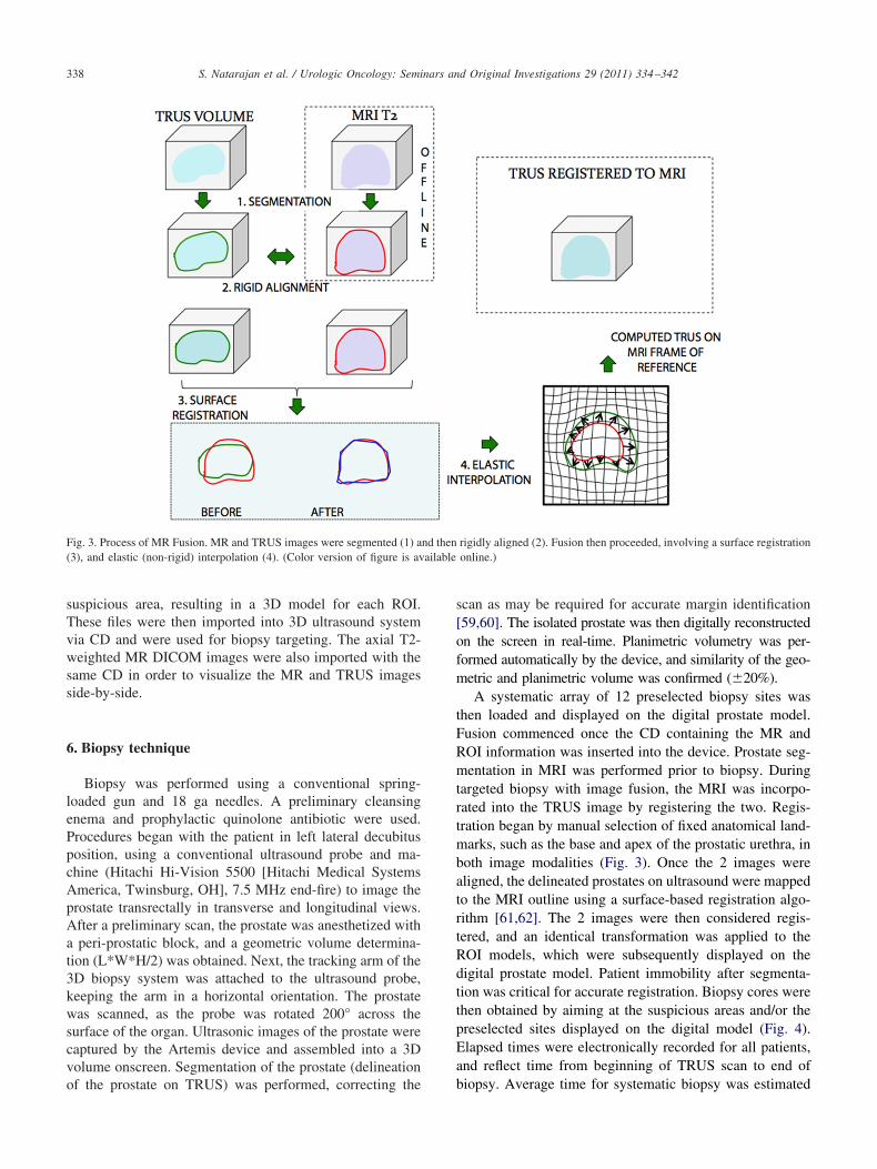
Fig. 3. Process of MR Fusion. MR and TRUS images were segmented (1) a(3), and elastic (non-rigid) interpolation (4). (Color version of figure is av
of the prostate on TRUS) was performed, correcting the b
scan as may be required for accurate margin identification[59,60]. The isolated prostate was then digitally reconstructedon the screen in real-time. Planimetric volumetry was per-formed automatically by the device, and similarity of the geo-metric and planimetric volume was confirmed (�20%).
A systematic array of 12 preselected biopsy sites washen loaded and displayed on the digital prostate model.usion commenced once the CD containing the MR andOI information was inserted into the device. Prostate seg-entation in MRI was performed prior to biopsy. During
argeted biopsy with image fusion, the MRI was incorpo-ated into the TRUS image by registering the two. Regis-ration began by manual selection of fixed anatomical land-arks, such as the base and apex of the prostatic urethra, in
oth image modalities (Fig. 3). Once the 2 images wereligned, the delineated prostates on ultrasound were mappedo the MRI outline using a surface-based registration algo-ithm [61,62]. The 2 images were then considered regis-ered, and an identical transformation was applied to theOI models, which were subsequently displayed on theigital prostate model. Patient immobility after segmenta-ion was critical for accurate registration. Biopsy cores werehen obtained by aiming at the suspicious areas and/or thereselected sites displayed on the digital model (Fig. 4).lapsed times were electronically recorded for all patients,nd reflect time from beginning of TRUS scan to end of
rigidly aligned (2). Fusion then proceeded, involving a surface registrationonline.)
nd then
iopsy. Average time for systematic biopsy was estimated
a
339S. Natarajan et al. / Urologic Oncology: Seminars and Original Investigations 29 (2011) 334–342
to be 15 minutes, with an additional 5 minutes for MRIfusion and biopsy targeting.
7. Biopsy tracking accuracy
Clinical accuracy of 3D biopsy tracking was tested in 11consecutive men, undergoing TRUS/Bx to rule out prostatecancer [54]. Locations of each biopsy site were recorded bythe device and displayed on the digital model, as describedabove. The ultrasound probe was then removed, detached,and cleaned, while patients remained on the procedure table.The probe was then reinserted, and a new 3D scan of theprostate was performed, and prior biopsy sites were re-called. The subsequent scan was used for guidance, andprior biopsy sites as targets. Three randomly selected sitesin each patient were re-biopsied. The distance betweenoriginal and re-biopsy sites was determined in a blindedfashion using geometric analysis at Robarts Research. Re-sults were stratified by prostate volume and site location(Table 3) [54]. Mean error (mm, target to center of re-biopsy core) for all 32 Bx was 1.2 � 1.1 (range, 0.2–5.1)
Fig. 4. Targeted biopsy using MR Fusion. (A) A lesion was identified onaxial T2WI and area of interest was fused with real-time ultrasound imagbiopsy-targeting of the lesion was established (parallel lines overlying blueultrasound guidance. (C) Sites of systematic and targeted biopsies werebiopsies penetrated region of interest. Biopsy results showed a Gleason scoonline.)
Table 3Clinical accuracy of repeat biopsy targeting (tracking)
and was independent of prostate volume or biopsy location.Reliability of the Artemis device to re-biopsy specific sitesin the prostate was supported by results of this pilot, ex-tending prior studies on phantoms (Fig. 5) [53].
mage grade 4) and delineated on T2WI by radiologist (blue ellipse). TheLesion was identified in sagittal and axial planes (blue enclosures), and
res). Both targeted and systematic biopsies were performed under real-timed within 3D reconstruction of prostate, confirming that several targeted� 6 cancer in only the targeted area. (Color version of figure is available
Fig. 5. 3D reconstruction of prostate showing proximity of repeat biopsysites (blue dots) to initial biopsy sites used as targets (green dots). In a pilotstudy, the average distance from repeat biopsy to target was 1.2 � 1.1 mmnd was independent of both volume and location within the prostate
MRI (ies. (B)
enclosurecordere 3�3
Table 3). (Color version of figure is available online.)
[
[
TI
340 S. Natarajan et al. / Urologic Oncology: Seminars and Original Investigations 29 (2011) 334–342
8. Targeted biopsy with MR fusion
Accuracy of biopsy targeting was tested by comparinghistologic results of targeted vs. systematic biopsy. Biopsy datawere obtained on 47 men, in which 65 suspicious areas, ortargets, were identified and biopsied. These men also under-went 12-core systematic biopsy. Of these men, 30 were foundto have CaP. The high rate of positivity was likely due to theinclusion of men on active surveillance (18 men). Targetedbiopsies were found to be more likely to reveal cancer thannon-targeted biopsies and high-grade areas were more likely tobe cancer than low-grade areas (Table 4). When high-gradeROIs were targeted, a 33% biopsy-positivity rate was found vs.a 7% positivity rate for systematic, nontargeted biopsies (19/57cores vs. 9/124 cores, P � 0.03).
Of the 30 men found to have CaP, 9 were diagnosedusing systematic biopsy only, 5 with MR fusion-guidedbiopsy only, and 16 with both protocols. Of the patients forwhom only systematic cores were positive, 5 patients (55%)had cancer in the same sextant as the target, indicatingmistargeting of the lesion. After a proper targeting tech-nique was established, 12 of the last 22 patients with targetswere positive for CaP upon biopsy. Three patients werediagnosed with CaP using systematic biopsy alone, 4 withtargeted biopsy alone, and 5 using both protocols.
An additional benefit of 3D biopsy tracking may be animproved systematic biopsy, i.e., more evenly distributedsites than with conventional methods. We have observedthrough 3D tracking that unguided biopsy locations are notalways symmetrically distributed and tend to be clustered,despite attempts at symmetric placement. Thus, in the abovecomparison, results with systematic biopsies may be fa-vored by wider sampling from the built-in sites.
9. Comment
Technologies to improve accuracy of prostate biopsy arerapidly emerging. In selecting and following men for activesurveillance, the new technologies are particularly compel-ling. In the future, men considering focal therapy may alsobenefit from improved biopsy accuracy. We have describedan initial clinical experience with a new 3D ultrasound
able 4mage grade of targets and probability of cancer
* Patients with more than 1 target were counted in the table multipletimes; 1 to 3 biopsy cores are taken of the ROI, depending on size of lesion.
device, which allows biopsy-site tracking for future recall
and fusion of MRI targets with real-time ultrasound. Whilepromising, these early experiences have not yet conclu-sively shown the benefit of tracking and targeted biopsywith MR fusion. Challenges exist in terms of perfectingregistration of ultrasound and MR images. The actual ana-tomic locations of tumor tissue are the final determinant ofbiopsy (and MR) accuracy. In this regard, whole-mountprostatectomy studies from men who have undergone both3D targeted and systematic biopsy will be critical. Furtherrefinements of the fusion technology are necessary beforewidespread clinical adoption is possible. These will likelyinclude multiple areas of hardware miniaturization, whichcould improve ease of operation. Biopsy tracking and targetingwith image fusion may become important tools to improvediagnosis and management of men with prostate cancer.
Acknowledgments
The authors thank Elizabeth Hamilton, LVN for herclinical assistance. This work was supported in part bygrants from the Steven C. Gordon Family Foundation, theBeckman Coulter Foundation, and the Jean Perkins Foun-dation. Co-author JH is supported by UCLA SPORE inProstate Cancer, Prostate Cancer Foundation ChallengeAward and Creativity Award, and Department of DefenseProstate Cancer Research Program.
References
[1] Walsh PC. 2008 Whitmore Lecture: Radical prostatectomy–where wewere and where we are going. Urol Oncol 2009;27:246–50.
[2] Stamey TA, Caldwell M, McNeal JE, et al. The prostate specificantigen era in the United States is over for prostate cancer: Whathappened in the last 20 years? J Urol 2004;172:1297–301.
[3] Epstein JI, Walsh PC, Carmichael M, et al. Pathologic and clinicalfindings to predict tumor extent of nonpalpable (stage T1c) prostatecancer. JAMA 1994;271:368–74.
[4] Sutcliffe P, Hummel S, Simpson E, et al. Use of classical and novelbiomarkers as prognostic risk factors for localized prostate cancer: Asystematic review. Health Technol Assess 2009;13:1–219.
[5] Cooperberg MR, Broering JM, Kantoff PW, et al. Contemporarytrends in low risk prostate cancer: Risk assessment and treatment.J Urol 2007;178:S14–9.
[6] Carter HB, Sauvageot J, Walsh PC, et al. Prospective evaluation ofmen with stage T1c adenocarcinoma of the prostate. J Urol 1997;157:2206–9.
[7] Klotz L, Zhang L, Lam A, et al. Clinical results of long-term fol-low-up of a large, active surveillance cohort with localized prostatecancer. J Clin Oncol 2010;28:126–31.
[8] Hambrock T, Somford DM, Hoeks C, et al. Magnetic resonanceimaging guided prostate biopsy in men with repeat negative biopsiesand increased prostate specific antigen. J Urol 2010;183:520–8.
10] Kim CK, Park BK, Kim B. Diffusion-weighted MRI at 3 T for theevaluation of prostate cancer. Am J Roentgenol 2010;194:1461–9.
11] Lawrentschuk N, Fleshner N. The role of magnetic resonance imag-
ing in targeting prostate cancer in patients with previous negative
[
[
[
[
[
[
[
[
[
[
[
[
[
[
[
[
[
[
[
[
[
[
[
[
[
[
[
[
[
[
[
[
[
[
[
[
[
[
[
[
[
341S. Natarajan et al. / Urologic Oncology: Seminars and Original Investigations 29 (2011) 334–342
biopsies and elevated prostate-specific antigen levels. BJUinternational 2009;103:730–3.
12] Dooms GC, Hricak H. Magnetic resonance imaging of the pelvis:Prostate and urinary bladder. Urol Radiol 1986;8:156–65.
13] Narayan P, Vigneron DB, Jajodia P, et al. Transrectal probe for 1HMRI and 31P MR spectroscopy of the prostate gland. Magn ResonMed 1989;11:209–20.
14] Outwater EK, Petersen RO, Siegelman ES, et al. Prostate carcinoma:Assessment of diagnostic criteria for capsular penetration on endo-rectal coil MR images. Radiology 1994;193:333–9.
15] Brown G, Macvicar D, Ayton V, et al. The role of intravenouscontrast enhancement in magnetic resonance imaging of prostaticcarcinoma. Clin Radiol 1995;50:601–6.
16] Jager G, Ruijter E, Van de Kaa C, et al. Dynamic TurboFLASH sub-traction technique for contrast-enhanced MR imaging of the prostate:Correlation with histopathologic results. Radiology 1997;203:645–52.
17] Engelbrecht MR, Huisman HJ, Laheij RJF, et al. Discrimination ofprostate cancer from normal peripheral zone and central gland tissue byusing dynamic contrast-enhanced MR imaging. Radiology 2003;229:248.
18] Quint L, Van Erp J, Bland P, et al. Prostate cancer: Correlation of MRimages with tissue optical density at pathologic examination. Radi-ology 1991;179:837–42.
19] Kozlowski P, Chang SD, Jones EC, et al. Combined diffusion-weighted and dynamic contrast-enhanced MRI for prostate cancerdiagnosis—correlation with biopsy and histopathology. J MagnReson 2006;24:108–13.
20] Ocak I, Bernardo M, Metzger G, et al. Dynamic contrast-enhancedMRI of prostate cancer at 3 T: A study of pharmacokinetic parame-ters. Am J Roentgenol 2007;189:W192–201.
21] Tanimoto A, Nakashima J, Kohno H, et al. Prostate cancer screening:The clinical value of diffusion-weighted imaging and dynamic MRimaging in combination with T2-weighted imaging. J Magn Reson2007;25:146–52.
22] Oto A, Kayhan A, Jiang Y, et al. Prostate cancer: Differentiation ofcentral gland cancer from benign prostatic hyperplasia by usingdiffusion-weighted and dynamic contrast-enhanced MR imaging. Ra-diology 2010;257:715–23.
23] Turkbey B, Xu S, Kruecker J, et al. Documenting the location ofprostate biopsies with image fusion. BJU Intl 2010;107:53–7.
24] Kitajima K, Kaji Y, Fukabori Y, et al. Prostate cancer detection with3 T MRI: Comparison of diffusion-weighted imaging and dynamiccontrast-enhanced MRI in combination with T2-weighted imaging. JMagn Reson 2010;31:625–31.
25] Langer DL, van der Kwast TH, Evans AJ, et al. Prostate tissuecomposition and MR measurements: Investigating the relationshipsbetween ADC, T2, Ktrans, ve, and corresponding histologic features.Radiology 2010;255:485–94.
26] Park BK, Lee HM, Kim CK, et al. Lesion localization in patients witha previous negative transrectal ultrasound biopsy and persistentlyelevated prostate specific antigen level using diffusion-weighted im-aging at 3 Tesla before re-biopsy. Invest Radiol 2008;43:789–93.
27] Xu S, Kruecker J, Turkbey B, et al. Real-time MRI-TRUS fusion forguidance of targeted prostate biopsies. Comp Aid Surg 2008;13:255–64.
28] Turkbey B, Pinto PA, Mani H, et al. Prostate Cancer: Value ofmultiparametric MR imaging at 3 T for detection—histopathologiccorrelation. Radiology 2010;255:89–99.
29] Rastinehad AR, Baccala AA, Chung PH, et al. D’Amico risk stratifica-tion correlates with degree of suspicion of prostate cancer on multipara-metric magnetic resonance imaging. J Urol 2011;185:815–20.
30] Fütterer JJ, Heijmink SW, Scheenen TWJ, et al. Prostate cancerlocalization with dynamic contrast-enhanced MR imaging and protonMR spectroscopic imaging. Radiology 2006;241:449–58.
31] Chen M, Dang HD, Wang JY, et al. Prostate cancer detection:Comparison of T2-weighted imaging, diffusion-weighted imaging,proton magnetic resonance spectroscopic imaging, and the 3 tech-
niques combined. Acta Radiologica 2008;49:602–10.
32] Riches SF, Payne GS, Morgan VA, et al. MRI in the detection ofprostate cancer: Combined apparent diffusion coefficient, metaboliteratio, and vascular parameters. Am J Roentgenol 2009;193:1583–91.
33] Weinreb JC, Blume JD, Coakley FV, et al. Prostate cancer: Sextantlocalization at MR imaging and MR spectroscopic imaging beforeprostatectomy—results of ACRIN prospective multi-institutionalclinicopathologic study. Radiology 2009;251:122–33.
34] Heijmink SW, Fütterer JJ, Hambrock T, et al. Prostate cancer: Body-array vs. endorectal coil MR imaging at 3 T—comparison of image quality,localization, and staging performance. Radiology 2007;244:184–95.
35] Park BK, Kim B, Kim CK, et al. Comparison of phased-array 3.0-Tand endorectal 1.5-T magnetic resonance imaging in the evaluation oflocal staging accuracy for prostate cancer. J Comput Assist Tomogr2007;31:534–8.
36] Alonzi R, Padhani AR, Allen C. Dynamic contrast enhanced MRI inprostate cancer. Eur J Radiol 2007;63:335–50.
37] van Dorsten FA, van der Graaf M, Engelbrecht MRW, et al. Com-bined quantitative dynamic contrast-enhanced MR imaging and 1HMR spectroscopic imaging of human prostate cancer. J Magn ResonImaging 2004;20:279–87.
38] Padhani AR, Gapinski CJ, Macvicar DA, et al. Dynamic contrast en-hanced MRI of prostate cancer: Correlation with morphology and tumorstage, histological grade, and PSA. Clinical Radiol 2000;55:99–109.
39] Fütterer JJ, Engelbrecht MR, Huisman HJ, et al. Staging prostatecancer with dynamic contrast-enhanced endorectal MR imaging priorto radical prostatectomy: Experienced vs. less experienced readers.Radiology 2005;237:541–9.
40] Girouin N, Mege-Lechevallier F, Tonina Senes A, et al. Prostatedynamic contrast-enhanced MRI with simple visual diagnostic crite-ria: Is it reasonable? Eur Radiol 2007;17:1498–509.
41] Sinha S, Sinha U. In vivo diffusion tensor imaging of the humanprostate. Magn Reson Med 2004;52:530–7.
42] Pickles MD, Gibbs P, Sreenivas M, et al. Diffusion-weighted imagingof normal and malignant prostate tissue at 3.0 T. J Magn Reson2006;23:130–4.
43] Gibbs P, Liney GP, Pickles MD, et al. Correlation of ADC and T2measurements with cell density in prostate cancer at 3.0 Tesla. In-vestig Radiol 2009;44:572–6.
44] Haider MA, van der Kwast TH, Tanguay J, et al. Combined T2-weighted and diffusion-weighted MRI for localization of prostatecancer. Am J Roentgenol 2007;189:323–8.
45] van As N, Charles-Edwards E, Jackson A, et al. Correlation ofdiffusion-weighted MRI with whole mount radical prostatectomyspecimens. Br J Radiol 2008;81:456–62.
46] Yoshimitsu K, Kiyoshima K, Irie H, et al. Usefulness of apparentdiffusion coefficient map in diagnosing prostate carcinoma: Correla-tion with stepwise histopathology. J Magn Reson 2008;27:132–9.
47] Mazaheri Y, Hricak H, Fine SW, et al. Prostate tumor volume mea-surement with combined T2-weighted imaging and diffusion-weighted MR: Correlation with pathologic tumor volume. Radiology2009;252:449–57.
48] Woodfield CA, Tung GA, Grand DJ, et al. Diffusion-weighted MRIof peripheral zone prostate cancer: Comparison of tumor apparentdiffusion coefficient with Gleason score and percentage of tumor oncore biopsy. Am J Roentgenol 2010;194:W316–22.
49] de Souza N, Reinsberg S, Scurr E, et al. Magnetic resonance imagingin prostate cancer: The value of apparent diffusion coefficients foridentifying malignant nodules. Br J Radiol 2007;80:90–5.
50] de Souza N, Riches S, Vanas N, et al. Diffusion-weighted magneticresonance imaging: A potential non-invasive marker of tumor aggres-siveness in localized prostate cancer. Clin Radiol 2008;63:774–82.
51] van As NJ, de Souza NM, Riches SF, et al. A study of diffusion-weighted magnetic resonance imaging in men with untreated local-ized prostate cancer on active surveillance. Eur Urol 2009;56:981–8.
52] Lee SH, Park KK, Choi KH, et al. Is endorectal coil necessary for thestaging of clinically localized prostate cancer? Comparison of non-
endorectal vs. endorectal MR imaging. World J Urol 2010;28:667–72.
[
[
[
[
[
[
342 S. Natarajan et al. / Urologic Oncology: Seminars and Original Investigations 29 (2011) 334–342
[53] Bax J, Cool D, Gardi L, et al. Mechanically assisted 3D ultrasoundguided prostate biopsy system. Med Phys 2008;35:5397–410.
[54] Marks L, Ward A, Gardi L, et al. Tracking of prostate biopsy sitesusing a 3D ultrasound device (Artemis). J Urol 2010;183:e832.
[55] Rankin R, Fenster A, Downey D, et al. Three-dimensional sono-graphic reconstruction: Techniques and diagnostic applications. Am JRoentgenol 1993;161:695–702.
[56] Ahmed HU, Kirkham A, Arya M, et al. Is it time to consider a role forMRI before prostate biopsy? Nat Rev Clin Oncol 2009;6:197–206.
57] Raz O, Haider M, Trachtenberg J, et al. MRI for men undergoingactive surveillance or with rising PSA and negative biopsies. Nat Rev
Urol 2010;7:543–51.
58] Rosset A, Spadola L, Ratib O. OsiriX: An open-source software for navi-gating in multidimensional DICOM images. J Digit Imag 2004;17:205–16.
59] Ladak H, Mao F, Wang Y, et al. Prostate boundary segmentationfrom 2D ultrasound images. Med Phys 2000;27:1777–88.
60] Wang Y, Cardinal N, Downey D, et al. Semi-automatic 3D segmentationof the prostate using 2D ultrasound images. Med Phys 2003;30:88–97.
61] Narayanan R, Kurhanewicz J, Shinohara K, et al. MRI-ultrasoundregistration for targeted prostate biopsy. IEEE Intl Symp BiomedImaging 2009;991–4.
62] Karnik V, Fenster A, Bax J, et al. Assessment of image registrationaccuracy in 3-dimensional transrectal ultrasound guided prostate bi-