+ All Categories
Home > Documents > CLINICAL CASE REPORT - Springer · PDF fileCLINICAL CASE REPORT Detailed analysis of family...

CLINICAL CASE REPORT - Springer · PDF fileCLINICAL CASE REPORT Detailed analysis of family...

Date post: 31-Mar-2018
Category:
Upload: vandang
View: 213 times
Download: 1 times
Share this document with a friend
11
CLINICAL CASE REPORT Detailed analysis of family with autosomal recessive bestrophinopathy associated with new BEST1 mutation Daiki Kubota . Kiyoko Gocho . Keiichiro Akeo . Sachiko Kikuchi . Michitaka Sugahara . Celso Soiti Matsumoto . Kei Shinoda . Atsushi Mizota . Kunihiko Yamaki . Hiroshi Takahashi . Shuhei Kameya Received: 6 February 2016 / Accepted: 7 April 2016 / Published online: 12 April 2016 Ó The Author(s) 2016. This article is published with open access at Springerlink.com Abstract Purpose To describe the clinical and genetic find- ings in a patient with autosomal recessive bestrophinopathy (ARB) and his healthy parents. Methods The patient and his healthy non-consan- guineous parents underwent detailed ophthalmic eval- uations including electro-oculography (EOG), spec- tral-domain optical coherence tomography (SD-OCT), and fundus autofluorescence (FAF) imaging. Mutation analysis of the BEST1 gene was performed by Sanger sequencing. Results The FAF images showed multiple spots of increased autofluorescence, and the sites of these spots corresponded to the yellowish deposits detected by ophthalmoscopy. SD-OCT showed cystoid macular changes and a shallow serous macular detachment. The Arden ratio of the EOG was markedly reduced to 1.1 in both eyes. Genetic analysis of the proband detected two sequence variants of the BEST1 gene in the heterozygous state: a novel variant c.717delG, p.V239VfsX2 and an already described c.763C [ T, p.R255W variant associated with Best vitelliform macular dystrophy and ARB. The proband’s father carried the c.717delG, p.V239VfsX2 variant in the heterozygous state, and the mother carried the c.763C [ T, p.R255W variant in the heterozygous state. The parents who were heterozygous for the BEST1 variants had normal visual acuity, EOG, SD- OCT, and FAF images. Conclusions In a truncating BEST1 mutation, the phenotype associated with ARB is most likely due to a marked decrease in the expression of BEST1 promoted by the nonsense-mediated decay surveillance mecha- nism, and it may depend on the position of the premature termination of the codon created. Keywords Autosomal recessive bestrophinopathy Á BEST1 Á Fundus autofluorescence Á Electro- oculography (EOG) D. Kubota Á K. Gocho Á K. Akeo Á S. Kikuchi Á K. Yamaki Á S. Kameya (&) Department of Ophthalmology, Nippon Medical School Chiba Hokusoh Hospital, 1715 Kamagari, Inzai, Chiba 270-1694, Japan e-mail: [email protected] M. Sugahara Inoue Eye Clinic, 4-3 Surugadai, Kanda, Chiyoda-ku, Tokyo 101-0062, Japan M. Sugahara Sugahara Eye Clinic, 1-13-3, Minami-senju, Arakawa-ku, Tokyo 116-0003, Japan C. S. Matsumoto Á K. Shinoda Á A. Mizota Department of Ophthalmology, Teikyo University School of Medicine, 2-11-1 Kaga, Itabashi-ku, Tokyo 173-8605, Japan H. Takahashi Department of Ophthalmology, Nippon Medical School, 1-1-5 Sendagi, Bunkyo-ku, Tokyo 113-8602, Japan 123 Doc Ophthalmol (2016) 132:233–243 DOI 10.1007/s10633-016-9540-3
Transcript

CLINICAL CASE REPORT

Detailed analysis of family with autosomal recessivebestrophinopathy associated with new BEST1 mutation

Daiki Kubota . Kiyoko Gocho . Keiichiro Akeo . Sachiko Kikuchi .

Michitaka Sugahara . Celso Soiti Matsumoto . Kei Shinoda .

Atsushi Mizota . Kunihiko Yamaki . Hiroshi Takahashi . Shuhei Kameya

Received: 6 February 2016 / Accepted: 7 April 2016 / Published online: 12 April 2016

� The Author(s) 2016. This article is published with open access at Springerlink.com

Abstract

Purpose To describe the clinical and genetic find-

ings in a patient with autosomal recessive

bestrophinopathy (ARB) and his healthy parents.

Methods The patient and his healthy non-consan-

guineous parents underwent detailed ophthalmic eval-

uations including electro-oculography (EOG), spec-

tral-domain optical coherence tomography (SD-OCT),

and fundus autofluorescence (FAF) imaging. Mutation

analysis of the BEST1 gene was performed by Sanger

sequencing.

Results The FAF images showed multiple spots of

increased autofluorescence, and the sites of these spots

corresponded to the yellowish deposits detected by

ophthalmoscopy. SD-OCT showed cystoid macular

changes and a shallow serous macular detachment.

The Arden ratio of the EOG was markedly reduced to

1.1 in both eyes. Genetic analysis of the proband

detected two sequence variants of the BEST1 gene in

the heterozygous state: a novel variant c.717delG,

p.V239VfsX2 and an already described c.763C[T,

p.R255W variant associated with Best vitelliform

macular dystrophy and ARB. The proband’s father

carried the c.717delG, p.V239VfsX2 variant in the

heterozygous state, and the mother carried the

c.763C[T, p.R255W variant in the heterozygous

state. The parents who were heterozygous for the

BEST1 variants had normal visual acuity, EOG, SD-

OCT, and FAF images.

Conclusions In a truncating BEST1 mutation, the

phenotype associated with ARB is most likely due to a

marked decrease in the expression of BEST1 promoted

by the nonsense-mediated decay surveillance mecha-

nism, and it may depend on the position of the

premature termination of the codon created.

Keywords Autosomal recessive bestrophinopathy �BEST1 � Fundus autofluorescence � Electro-oculography (EOG)

D. Kubota � K. Gocho � K. Akeo � S. Kikuchi �K. Yamaki � S. Kameya (&)

Department of Ophthalmology, Nippon Medical School

Chiba Hokusoh Hospital, 1715 Kamagari, Inzai,

Chiba 270-1694, Japan

e-mail: [email protected]

M. Sugahara

Inoue Eye Clinic, 4-3 Surugadai, Kanda, Chiyoda-ku,

Tokyo 101-0062, Japan

M. Sugahara

Sugahara Eye Clinic, 1-13-3, Minami-senju, Arakawa-ku,

Tokyo 116-0003, Japan

C. S. Matsumoto � K. Shinoda � A. Mizota

Department of Ophthalmology, Teikyo University School

of Medicine, 2-11-1 Kaga, Itabashi-ku, Tokyo 173-8605,

Japan

H. Takahashi

Department of Ophthalmology, Nippon Medical School,

1-1-5 Sendagi, Bunkyo-ku, Tokyo 113-8602, Japan

123

Doc Ophthalmol (2016) 132:233–243

DOI 10.1007/s10633-016-9540-3

Introduction

BEST1 (VMD2) is a gene located on chromosome 11

(11q12.3) that encodes for the 585 amino acid

transmembrane protein bestrophin 1 which is located

on the basolateral aspect of retinal pigment epithelial

(RPE) cells [1, 2]. Although the functional role of

bestrophin-1 within the RPE has not been determined

definitively, it has been postulated to function as a

Ca2?-activated Cl- channel [3], a regulator of voltage-

gated Ca2? channels [4], and a HCO3- channel [5].

Mutations in BEST1 therefore affect the RPE meta-

bolism and consequently the outer retinal function

with which the RPE is intimately associated.

Mutations of the BEST1 gene have been associated

with different ocular phenotypes [6]. The first disease

shown to be caused by BEST1 sequence variants was

Best vitelliform macular dystrophy (BVMD) [1], a

retinal disease characterized bybilateral yellowish yolk-

colored lesion in the macula. BEST1 mutations are also

associated with several other eye diseases including

adult-onset vitelliform macular dystrophy (AOVMD)

[7], autosomal dominant vitreo-retinochoroidopathy

(ADVIRC) [8], retinitis pigmentosa [9], and the micro-

cornea, retinal dystrophy, cataract, and posterior staphy-

loma (MRCS) syndrome [10].

Autosomal recessive bestrophinopathy (ARB), first

described in detail in 2008, is another member of the

phenotypic spectrum associated with mutations in

the BEST1 gene [11]. The characteristics of this

disorder include a progressive reduction in central

vision, absence of the electro-oculographic (EOG)

light rise, and reduced full-field electroretinograms

(ERGs). None of the patients have the vitelliform

lesions typical of Best disease, but have a diffuse

irregularity of the reflex from the RPE including

dispersed punctate flecks [11]. All of the patients have

an accumulation of fluid within and/or beneath the

neurosensory retina in the macular area [11].

ARB has been reported to be due to either

compound heterozygous or homozygous BEST1 gene

mutations [6, 11]. A recent manuscript described an

ocular phenotype similar to ARB associated with a

single heterozygous mutation of the BEST1 gene [12].

Several mutations associated with ARB have been

reported to be involved in causing dominant Best

disease when they were present in the heterozygous

state [6, 7, 13–15]. The clinical phenotype of some

patients with recessive bestrophinopathy is distinct

from that seen in Best disease, while in others it is

similar to the typical phenotype observed in autosomal

dominant vitelliform dystrophy [16, 17]. Identification

of additional families with recessive bestrophinopathy

and detailed characterization of the clinical phenotypes

of homozygous and heterozygous individuals will

assist in establishing the phenotype–genotype correla-

tions in patients with BEST1-associated diseases.

Methods

The protocol of this study conformed to the tenets of

the Declaration of Helsinki and was approved by the

Institutional Review Board of the Nippon Medical

School. A signed written informed consent was

obtained from the patient and his parents after the

nature and possible consequences of the study were

explained.

Blood samples were collected from the patient and

his parents, and genomic DNA was isolated from the

peripheral white blood cells using a blood DNA

isolation kit (NucleoSpin Blood XL; Macherey–

Nagel, Germany). The DNA was used as the template

to amplify the BEST1 gene. The coding regions and

flanking introns of the BEST1 gene were amplified by

polymerase chain reaction (PCR) using primers syn-

thesized by Greiner Bio-One (Tokyo, Japan). The PCR

products were purified (ExoSAP-IT; USB Corp.,

USA) and were used as the template for sequencing.

Both strands were sequenced on an automated

sequencer (Bio Matrix Research; Chiba, Japan).

The ophthalmological examinations included mea-

surements of the best-corrected visual acuity (BCVA),

refractive error and axial length, slit-lamp biomi-

croscopy, ophthalmoscopy, fundus photography, fun-

dus autofluorescence (FAF) imaging, fluorescein

angiography (FA), SD-OCT, full-field electroretinog-

raphy (ERG), multifocal ERGs (mfERGs), and elec-

tro-oculography (EOG). The EOGs and ERGs were

recorded using an extended testing protocol conform-

ing to the International Society for Clinical Electro-

physiology of Vision standards. The ERGs were

elicited and recorded with a LED built-in electrode

(LE2000, Tomey, Japan). The mfERGs were recorded

using a commercial mfERG system (VERIS Science;

Electro-Diagnostic Imaging, Inc. Redwood City, CA,

USA). The FAF images were acquired with the TRC-

NW8Fplus retinal camera (TOPCON, Tokyo, Japan),

234 Doc Ophthalmol (2016) 132:233–243

123

Fig. 1 Fundus photographs, autofluorescence images, fluores-

cein angiograms, and SD-OCT images from patient with

autosomal recessive bestrophinopathy (ARB) (proband, II-1).

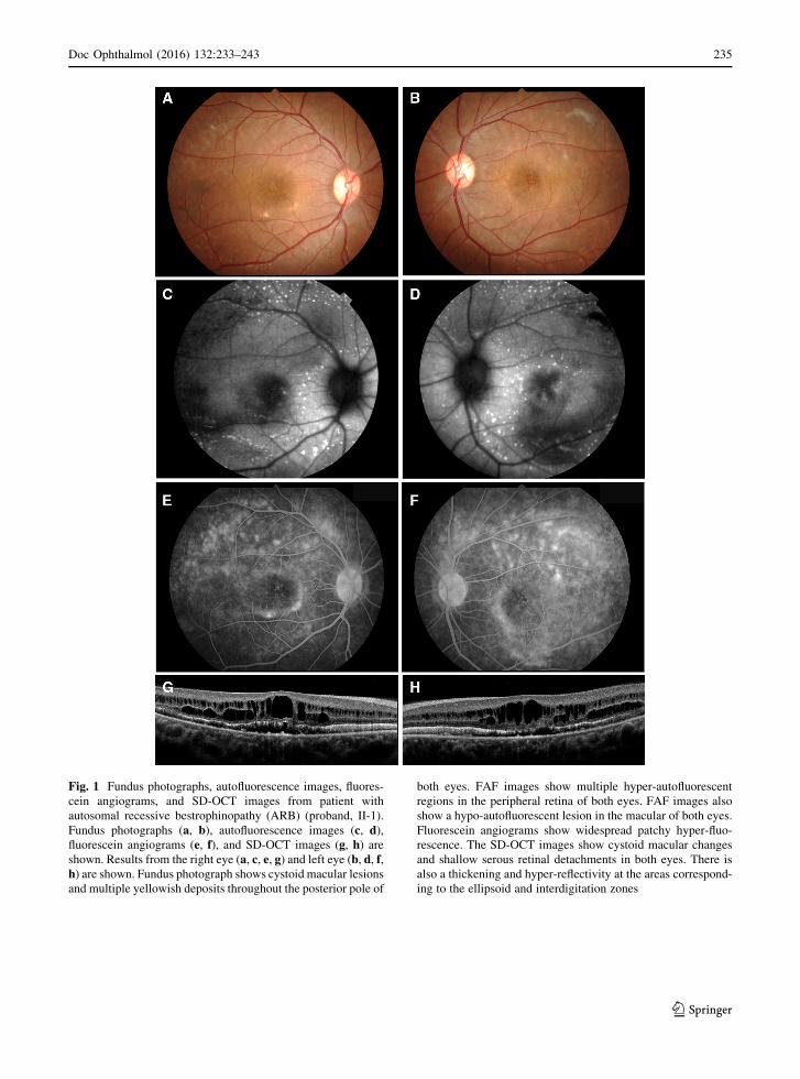
Fundus photographs (a, b), autofluorescence images (c, d),fluorescein angiograms (e, f), and SD-OCT images (g, h) areshown. Results from the right eye (a, c, e, g) and left eye (b, d, f,h) are shown. Fundus photograph shows cystoid macular lesions

and multiple yellowish deposits throughout the posterior pole of

both eyes. FAF images show multiple hyper-autofluorescent

regions in the peripheral retina of both eyes. FAF images also

show a hypo-autofluorescent lesion in the macular of both eyes.

Fluorescein angiograms show widespread patchy hyper-fluo-

rescence. The SD-OCT images show cystoid macular changes

and shallow serous retinal detachments in both eyes. There is

also a thickening and hyper-reflectivity at the areas correspond-

ing to the ellipsoid and interdigitation zones

Doc Ophthalmol (2016) 132:233–243 235

123

and the SD-OCT images were acquired with a Cirrus

HD-OCT (Carl Zeiss Meditec).

Results

The patient was a 25-year-old man whose decimal

best-corrected visual acuity (BCVA) was 0.9 in the

right eye and 0.3 in the left eye. His refraction was

S ? 0.5 C-1.25 at 180 in the right eye and S ? 0.5

C-2.0 at 175 in the left eye. Axial length was

23.71 mm in the right eye and 23.85 mm in the left

eye. The intraocular pressure and anterior ocular

segments were within normal limits in both eyes.

Fundus examinations revealed a cystoid macular

lesion and multiple yellowish deposits throughout

Fig. 2 Full-field electroretinograms (ERGs). Full-field ERGs

recorded from the right eye (top) and left eye (middle) of the

proband (II-1) are shown. The ERGs recorded from a normal

control are also shown (bottom). The dark-adapted 0.01, dark-

adapted 3.0, light-adapted 3.0, and light-adapted 3.0 flicker

ERGs are shown. The results of all the responses show a slight

reduction of the b-wave amplitudes in both eyes

Fig. 3 Multifocal ERGs. The mfERGs, topographic map, and average densities of the rings of the multifocal ERGs of right eye (a) andleft eye (b) of the proband are shown. The amplitudes of the mfERGs in the foveal area are severely reduced in both eyes

236 Doc Ophthalmol (2016) 132:233–243

123

the posterior pole of both eyes (Fig. 1). The vitelliform

lesions that are typical of Best disease were not

observed (Fig. 1). FAF imaging showed multiple

hyper-autofluorescent spots in the peripheral retina

of both eyes, and the site of the spots corresponded

with the yellowish deposits observed by ophthal-

moscopy (Fig. 1). FAF imaging also detected a hypo-

autofluorescent lesion in the macula of both eyes

(Fig. 1). FA showed widespread patchy hyper-fluo-

rescence (Fig. 1). The SD-OCT images showed cys-

toid changes in the macula and shallow serous retinal

detachments in both eyes. There was a thickening and

hyper-reflectivity at the areas corresponding to ellip-

soid and interdigitation zones of the photoreceptors in

the SD-OCT images (Fig. 1).

The amplitudes of both the cone and rod full-field

ERGs were reduced, and the waveforms were similar

in both eyes (Fig. 2). The amplitudes of the mfERGs

were reduced in the central and peripheral sectors of

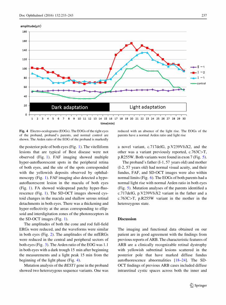
both eyes (Fig. 3). The Arden ratio of the EOGwas 1.1

in both eyes with a dark trough 15 min after beginning

the measurements and a light peak 15 min from the

beginning of the light phase (Fig. 4).

Mutation analysis of the BEST1 gene in the proband

showed two heterozygous sequence variants. One was

a novel variant, c.717delG, p.V239VfsX2, and the

other was a variant previously reported, c.763C[T,

p.R255W. Both variants were found in exon 7 (Fig. 5).

The proband’s father (I-1, 57 years old) and mother

(I-2, 57 years old) had normal visual acuity, and their

fundus, FAF, and SD-OCT images were also within

normal limits (Fig. 6). The EOGs of both parents had a

normal light rise with normal Arden ratio in both eyes

(Fig. 5). Mutation analyses of the parents identified a

c.717delG, p.V239VfsX2 variant in the father and a

c.763C[T, p.R255W variant in the mother in the

heterozygous state.

Discussion

The imaging and functional data obtained on our

patient are in good agreement with the findings from

previous reports of ARB. The characteristic features of

ARB are a clinically recognizable retinal dystrophy

with yellowish subretinal lesions scattered in the

posterior pole that have marked diffuse fundus

autofluorescence abnormalities [18–24]. The SD-

OCT findings of previous ARB cases included diffuse

intraretinal cystic spaces across both the inner and

Fig. 4 Electro-oculograms (EOGs). The EOGs of the right eyes

of the proband, proband’s parents, and normal control are

shown. The Arden ratio of the EOG of the proband is markedly

reduced with an absence of the light rise. The EOGs of the

parents have a normal Arden ratio and light rise

Doc Ophthalmol (2016) 132:233–243 237

123

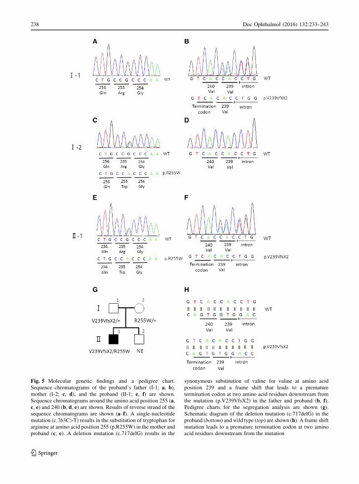
Fig. 5 Molecular genetic findings and a pedigree chart.

Sequence chromatograms of the proband’s father (I-1; a, b),mother (I-2; c, d), and the proband (II-1; e, f) are shown.

Sequence chromatograms around the amino acid position 255 (a,c, e) and 240 (b, d, e) are shown. Results of reverse strand of the

sequence chromatograms are shown (a–f). A single-nucleotide

mutation (c.763C[T) results in the substitution of tryptophan for

arginine at amino acid position 255 (p.R255W) in the mother and

proband (c, e). A deletion mutation (c.717delG) results in the

synonymous substitution of valine for valine at amino acid

position 239 and a frame shift that leads to a premature

termination codon at two amino acid residues downstream from

the mutation (p.V239VfsX2) in the father and proband (b, f).Pedigree charts for the segregation analysis are shown (g).Schematic diagram of the deletion mutation (c.717delG) in the

proband (bottom) and wild type (top) are shown (h). A frame shift

mutation leads to a premature termination codon at two amino

acid residues downstream from the mutation

238 Doc Ophthalmol (2016) 132:233–243

123

Fig. 6 Fundus

photographs, fundus

autofluorescence image, and

SD-OCT images from the

parents of the proband.

Fundus photographs (a, b, g,h), autofluorescence (c, d, i,j), and SD-OCT images (e, f,k, l) are shown. Results fromthe father (a–f) and mother

(g–l) are shown. Fundusappearance, FAF, and SD-

OCT of the proband’s

parents are normal

Doc Ophthalmol (2016) 132:233–243 239

123

Table

1Summaryofpreviouslyreported

biallelic

BEST1mutationswithpremature

term

inationcodon

Patient

number

Allele1

Allele2

Nucleotide

Aminoacid

Termination

position

Nucleotide

Aminoacid

Termination

position

Fundus

appearance

References

1c.15C[A

p.Y5X

5c.430A[G

p.S144G

ARB

Lacassagneet

al.[30]

2c.87C[G

p.Y29X

29

c.422G[A

p.R141H

BVMD

Schatzet

al.[27]

3c.102C[T

p.E35WfsX11

45

c.1470_1471delCA

p.H490QfsX24

513

ARB

Davidsonet

al.[18]

4c.172_173dupCA

p.Q58HfsX4

61

c.584C[T

p.A195V

ARB

Borm

anet

al.[21]

5c.175_176dupCA

p.Q59HfsX3

61

c.175_176dupCA

p.Q59HfsX3

61

ARB

Boonet

al.[22]

6c.475C[T

p.Q159X

159

c.422G[A

p.R141H

BVMD

Borm

anet

al.[21]

7c.519delA

p.K173NfsX2

174

c.860G[A

p.W

287X

287

ARB

Tianet

al.[29]

8c.598C[T

p.R200X

200

c.598C[T

p.R200X

200

ARB

Burgesset

al.[11]

9c.263_279del17

p.L88LfsX138

225

c.584C[T

p.A195V

ARB

Gerth

etal.[25]

10

c.521_522delTG

p.L174EfsX57

230

c.521_522delTG

p.L174EfsX57

230

ARB

Pomares

etal.[26]

11

c.762delG

p.R255GfsX4

258

c.74G[A

p.R25Q

ARB

Boonet

al.[22]

12

c.1100?

1G[A

p.V317PfsX33

349

c.1100?

1G[A

p.V317PfsX33

349

ARB?

BVMD

Pomares

etal.[26]

13

c.1066C[T

p.R356X

356

c.550C[T

p.P184S

ARB

Borm

anet

al.[21]

14

c.1038duC

p.Y347LfsX54

400

c.553A[C

p.H178P

ARB

Borm

anet

al.[21]

15

c.1212delC

p.P404PfsX78

481

c.637G[A

p.E213K

ARB

Silvaet

al.[ 31]

16

c.1415delT

p.L472PfsX10

481

c.1415delT

p.L472PfsX10

481

BVMD

Bitner

etal.[16]

17

c.1470_1471delCA

p.H490QfsX24

513

c.584C[T

p.A195V

BVMD

Kinnicket

al.[32]

18

c.1669delG

p.E557NfsX52

608

c.934G[A

p.D312N

BVMD

Sodiet

al.[33]

240 Doc Ophthalmol (2016) 132:233–243

123

outer plexiform layers, subretinal fluid with shallow

serous retinal detachment, and thickening and hyper-

reflectivity of the ellipsoid and interdigitation zones

which may represent an elongation of the photorecep-

tors [21, 24, 25]. The Arden ratio of the EOGs of

patients with ARB is reported to be low with an

absence of the light rise [11, 19, 23]. The imaging and

functional findings in our patient are typical of ARB.

The BEST1 mutation, c.717delG, p.V239VfsX2,

has not been reported and not included in the SNP

database. The allele frequency of the variant was

estimated from two databases; the Human Genetic

Variation Database (HGVD; http://www.genome.

med.kyoto-u.ac.jp/SnpDB/about.htm) which is speci-

fic for the Japanese population, and the ExACBrowser

(Beta)(http://exac.broadinstitute.org) database. Both

of these databases did not contain the allele frequency

of the variant, which indicates that this variant is very

rare.

Although most mutations associated with BVMD

are missense mutations that do not compromise

protein synthesis, the few ARB-causing mutations

reported to date are premature truncations or non-

sense substitutions that lead to early transcript

degradation or non-functional proteins. These are

associated with a null phenotype (Table 1). In

truncating BEST1 mutations, the null phenotype

associated with ARB is attributed to a severe

decrease in BEST1 expression promoted by the

nonsense-mediated decay (NMD) surveillance mech-

anism [26]. Recent evidence supports the idea that

NMD degradation depends on the position of the

premature translation termination codons. Pomares

et al. [26] reported that the BEST1 transcripts in a

patient who carried the premature stop codon at

position 230 are preserved in only 13 % of the case,

while the BEST1 transcripts of a patient who carry a

premature stop codon in position 349 are preserved in

22 % of the case. Patients who carry the premature

stop codon in position 230 have a characteristic ARB

phenotype, while patients who carry a premature stop

codon in position 349 have ophthalmological fea-

tures resembling both ARB and BVMD [26]. Thus,

the residual amount of aberrant protein can promote a

negative effect causing a mixed phenotype of both

ARB and BVMD traits. This hypothesis was sup-

ported by previous reports of biallelic BEST1 muta-

tions with at least a premature termination codon

(Table 1). Although patients 2 and 6 of Table 1 had

the BVMD phenotype which is not consistent with

the hypothesis, the same second allele mutation

(R141H) may be associated with the BVMD pheno-

type [21, 27]. Our patient with a premature termina-

tion codon at position 240 is consistent with the

hypothesis that the patient should have an ARB

phenotype.

The other mutation found in this study (R255W)

was reported to be present in both a BVMD family in

the heterozygous state and two ARB families in the

compound heterozygous state [28, 29]. In the BVMD

family with the R255W mutation, the parents of the

proband were not genetically examined [28]. In the

ARB families with the R255W mutation, each parent

of the proband was heterozygous carriers of the

R255Wmutation and they were healthy [29]. Our data

do not explain why the mother of our patient who

carried the heterozygous R255W mutation did not

have BVMD. One possibility is that the mutation

exhibits reduced penetrance for the phenotype. The

other possibility is that the previously described

BVMD patient who had heterozygous c.763C[T,

p.R255W mutation may have had an undiscovered

second allele mutation such as a large deletion.

In some cases, it is difficult to differentiate ARB

from BVMD and to speculate on the prognosis of the

disease. Identifying the genetic defect of BEST1 gene

and position of the premature termination codon may

help in differentiating the ARB from BVMD and

predict the prognosis of the disease.

Acknowledgments We thank Professor Emeritus Duco

Hamsaki of the Bascom Palmer Eye Institute, University of

Miami School of Medicine, Miami, FL, for discussions and

editing our manuscript.

Compliance with ethical standards

Conflict of interest All authors certify that they have no

affiliations with or involvement in any organization or entity

with any financial interest, or non-financial interest in the sub-

ject matter or materials discussed in this manuscript.

Statements of human rights All procedures performed in

studies involving human participants were in accordance with

the ethical standards of the institutional research committee and

with the 1964 Declaration of Helsinki and its later amendments

or comparable ethical standards.

Informed consent Informed consent was obtained from all

individual participants included in the study.

Doc Ophthalmol (2016) 132:233–243 241

123

Statement of welfare of animals No animals were involved

in the study.

Open Access This article is distributed under the terms of the

Creative Commons Attribution 4.0 International License (http://

creativecommons.org/licenses/by/4.0/), which permits unre-

stricted use, distribution, and reproduction in any medium,

provided you give appropriate credit to the original

author(s) and the source, provide a link to the Creative Com-

mons license, and indicate if changes were made.

References

1. Petrukhin K, Koisti MJ, Bakall B, Li W, Xie G, Marknell T,

Sandgren O, Forsman K, Holmgren G, Andreasson S, Vujic

M, Bergen AA, McGarty-Dugan V, Figueroa D, Austin CP,

Metzker ML, Caskey CT, Wadelius C (1998) Identification

of the gene responsible for Best macular dystrophy. Nat

Genet 19:241–247

2. Marmorstein AD, Marmorstein LY, Rayborn M, Wang X,

Hollyfield JG, Petrukhin K (2000) Bestrophin, the product

of the Best vitelliform macular dystrophy gene (VMD2),

localizes to the basolateral plasma membrane of the retinal

pigment epithelium. Proc Natl Acad Sci USA

97:12758–12763

3. Sun H, Tsunenari T, Yau KW, Nathans J (2002) The

vitelliform macular dystrophy protein defines a new family

of chloride channels. Proc Natl Acad Sci USA

99:4008–4013

4. Rosenthal R, Bakall B, Kinnick T, Peachey N, Wimmers S,

Wadelius C, Marmorstein A, Strauss O (2006) Expression

of bestrophin-1, the product of the VMD2 gene, modulates

voltage-dependent Ca2? channels in retinal pigment

epithelial cells. FASEB J 20:178–180

5. Qu Z, Hartzell HC (2008) Bestrophin Cl-channels are highly

permeable to HCO3. Am J Physiol Cell Physiol 294:C1371–

C1377

6. Boon CJ, Klevering BJ, Leroy BP, Hoyng CB, Keunen JE,

den Hollander AI (2009) The spectrum of ocular phenotypes

caused bymutations in the BEST1 gene. Prog Retin Eye Res

28:187–205

7. Kramer F, White K, Pauleikhoff D, Gehrig A, Passmore L,

Rivera A, Rudolph G, Kellner U, Andrassi M, Lorenz B,

Rohrschneider K, Blankenagel A, Jurklies B, Schilling H,

Schutt F, Holz FG, Weber BH (2000) Mutations in the

VMD2 gene are associated with juvenile-onset vitelliform

macular dystrophy (Best disease) and adult vitelliform

macular dystrophy but not age-related macular degenera-

tion. Eur J Hum Genet 8:286–292

8. Yardley J, Leroy BP, Hart-Holden N, Lafaut BA, Loeys B,

Messiaen LM, Perveen R, Reddy MA, Bhattacharya SS,

Traboulsi E, Baralle D, De Laey JJ, Puech B, Kestelyn P,

Moore AT, Manson FD, Black GC (2004) Mutations of

VMD2 splicing regulators cause nanophthalmos and auto-

somal dominant vitreo-retinochoroidopathy (ADVIRC).

Invest Ophthalmol Vis Sci 45:3683–3689

9. Davidson AE, Millar ID, Urquhart JE, Burgess-Mullan R,

Shweikh Y, Parry N, O’Sullivan J, Maher GJ, McKibbin M,

Downes SM, Lotery AJ, Jacobson SG, Brown PD, Black

GC, Manson FD (2009) Missense mutations in a retinal

pigment epithelium protein, bestrophin-1, cause retinitis

pigmentosa. Am J Hum Genet 85:581–592

10. Michaelides M, Urquhart J, Holder GE, Restori M, Kayali

N, Manson FD, Black GC (2006) Evidence of genetic

heterogeneity in MRCS (microcornea, rod-cone dystrophy,

cataract, and posterior staphyloma) syndrome. Am J Oph-

thalmol 141:418–420

11. Burgess R, Millar ID, Leroy BP, Urquhart JE, Fearon IM,

De Baere E, Brown PD, Robson AG, Wright GA, Kestelyn

P, Holder GE, Webster AR, Manson FD, Black GC (2008)

Biallelic mutation of BEST1 causes a distinct retinopathy in

humans. Am J Hum Genet 82:19–31

12. Toto L, Boon CJ, Di Antonio L, Battaglia Parodi M, Mas-

tropasqua R, Antonucci I, Stuppia L, Mastropasqua L

(2015) BESTROPHINOPATHY: a spectrum of ocular

abnormalities caused by the c.614T[C mutation in the

BEST1 gene. Retina. doi:10.1097/IAE.0000000000000950

13. Kramer F, Mohr N, Kellner U, Rudolph G, Weber BH

(2003) Ten novel mutations in VMD2 associated with Best

macular dystrophy (BMD). Hum Mutat 22:418

14. Lotery AJ, Munier FL, Fishman GA,Weleber RG, Jacobson

SG, Affatigato LM, Nichols BE, Schorderet DF, Sheffield

VC, Stone EM (2000) Allelic variation in the VMD2 gene in

best disease and age-related macular degeneration. Invest

Ophthalmol Vis Sci 41:1291–1296

15. Boon CJ, Klevering BJ, den Hollander AI, Zonneveld MN,

Theelen T, Cremers FP, Hoyng CB (2007) Clinical and

genetic heterogeneity in multifocal vitelliform dystrophy.

Arch Ophthalmol 125:1100–1106

16. Bitner H, Mizrahi-Meissonnier L, Griefner G, Erdinest I,

Sharon D, Banin E (2011) A homozygous frameshift

mutation in BEST1 causes the classical form of Best disease

in an autosomal recessive mode. Invest Ophthalmol Vis Sci

52:5332–5338

17. Burgess R, MacLaren RE, Davidson AE, Urquhart JE,

Holder GE, Robson AG, Moore AT, Keefe RO, Black GC,

Manson FD (2009) ADVIRC is caused by distinct mutations

in BEST1 that alter pre-mRNA splicing. J Med Genet

46:620–625

18. Davidson AE, Sergouniotis PI, Burgess-Mullan R, Hart-

Holden N, Low S, Foster PJ, Manson FD, Black GC,

Webster AR (2010) A synonymous codon variant in two

patients with autosomal recessive bestrophinopathy alters

in vitro splicing of BEST1. Mol Vis 16:2916–2922

19. Guerriero S, Preising MN, Ciccolella N, Causio F, Lorenz

B, Fischetto R (2011) Autosomal recessive bestrophinopa-

thy: new observations on the retinal phenotype—clinical

and molecular report of an Italian family. Ophthalmologica

225:228–235

20. Pineiro-Gallego T, Alvarez M, Pereiro I, Campos S, Sharon

D, Schatz P, Valverde D (2011) Clinical evaluation of two

consanguineous families with homozygous mutations in

BEST1. Mol Vis 17:1607–1617

21. Borman AD, Davidson AE, O’Sullivan J, Thompson DA,

Robson AG, De Baere E, Black GC, Webster AR, Holder

GE, Leroy BP, Manson FD, Moore AT (2011) Childhood-

onset autosomal recessive bestrophinopathy. Arch Oph-

thalmol 129:1088–1093

22. Boon CJ, van den Born LI, Visser L, Keunen JE, Bergen

AA, Booij JC, Riemslag FC, Florijn RJ, van Schooneveld

242 Doc Ophthalmol (2016) 132:233–243

123

MJ (2013) Autosomal recessive bestrophinopathy: differ-

ential diagnosis and treatment options. Ophthalmology

120:809–820

23. Sharon D, Al-Hamdani S, Engelsberg K, Mizrahi-Meis-

sonnier L, Obolensky A, Banin E, Sander B, Jensen H,

Larsen M, Schatz P (2014) Ocular phenotype analysis of a

family with biallelic mutations in the BEST1 gene. Am J

Ophthalmol 157:697–709

24. Fung AT, Yzer S, Goldberg N, Wang H, Nissen M, Gio-

vannini A,Merriam JE, Bukanova EN, Cai C, Yannuzzi LA,

Tsang SH, Allikmets R (2015) New best1 mutations in

autosomal recessive bestrophinopathy. Retina 35:773–782

25. Gerth C, Zawadzki RJ, Werner JS, Heon E (2009) Detailed

analysis of retinal function and morphology in a patient with

autosomal recessive bestrophinopathy (ARB). Doc Oph-

thalmol 118:239–246

26. Pomares E, Bures-Jelstrup A, Ruiz-Nogales S, Corcostegui

B, Gonzalez-Duarte R, Navarro R (2012) Nonsense-medi-

ated decay as the molecular cause for autosomal recessive

bestrophinopathy in two unrelated families. Invest Oph-

thalmol Vis Sci 53:532–537

27. Schatz P, Klar J, Andreasson S, Ponjavic V, Dahl N (2006)

Variant phenotype of Best vitelliform macular dystrophy

associated with compound heterozygous mutations in

VMD2. Ophthalmic Genet 27:51–56

28. Wong RL, Hou P, Choy KW, Chiang SW, Tam PO, Li H,

Chan WM, Lam DS, Pang CP, Lai TY (2010) Novel and

homozygous BEST1 mutations in Chinese patients with

Best vitelliform macular dystrophy. Retina 30:820–827

29. Tian R, Yang G, Wang J, Chen Y (2014) BEST1 gene

mutations in Chinese patients with bestrophinopathy. Mol

Vis 20:1594–1604

30. Lacassagne E, Dhuez A, Rigaudiere F, Dansault A, Vetu C,

Bigot K, Vieira V, Puech B, Defoort-Dhellemmes S, Abit-

bol M (2011) Phenotypic variability in a French family with

a novel mutation in the BEST1 gene causing multifocal best

vitelliform macular dystrophy. Mol Vis 17:309–322

31. Silva RA, Berrocal AM, Lam BL, Albini TA (2013) Novel

mutation in BEST1 associated with retinoschisis. JAMA

Ophthalmol 131:794–798. Erratum in (2013): JAMA Oph-

thalmol 131:1249

32. Kinnick TR, Mullins RF, Dev S, Leys M, Mackey DA, Kay

CN, Lam BL, Fishman GA, Traboulsi E, Iezzi R, Stone EM

(2011) Autosomal recessive vitelliform macular dystrophy

in a large cohort of vitelliform macular dystrophy patients.

Retina 31:581–595

33. Sodi A, Menchini F, Manitto MP, Passerini I, Murro V,

Torricelli F, Menchini U (2011) Ocular phenotypes asso-

ciated with biallelic mutations in BEST1 in Italian patients.

Mol Vis 17:3078–3087

Doc Ophthalmol (2016) 132:233–243 243

123


Recommended