Clinical Considerations With Glaucoma Ralph E. Hamor DVM, MS, DACVO Glaucoma is a leading cause of blindness in the middle-aged dog. Glaucoma should be considered as one of the "rule outs" in any case of "red eye" or "watery eye," especially in predisposed breeds (see lists below). Glaucoma is characterized by an elevation of intraocular pressure (IOP) that results in progressive loss of retinal ganglion cells and their axons. Aqueous, produced by the ciliary body, flows from the posterior chamber through the pupil into the anterior chamber and drains out of the eye via the trabecular meshwork in the iridocorneal angle and into the intrascleral venous plexus (conventional outflow). Uveoscleral (unconventional) outflow also occurs in most species: aqueous flows into the vitreous, across the uveal tract, along the supraciliary-suprachoroidal space into the adjacent sclera. Uveoscleral outflow accounts for 3% of aqueous outflow in the cat, 13% in rabbits, 15% in dogs and may be up to 50% in the horse. Increased IOP results from obstruction to outflow, not an increase in aqueous production. Whatever the cause, the most important consideration is whether the glaucoma is primary or secondary. PRIMARY GLAUCOMA occurs when there is no recognizable cause of the glaucoma. These patients are born with either an abnormal drainage angle, a drainage angle that becomes abnormal, or a drainage angle that appears normal but ceases to function normally. In some cases, gonioscopy (evaluation of the external drainage angle) demonstrates that a patient has an abnormal appearing drainage angle. This finding is only a risk factor that the patient will develop clinical signs of glaucoma. Patients with abnormal drainage angles (pectinate ligament dysplasia or mesodermal dysgenesis or goniodysgenesis) may develop clinical glaucoma as can patients with normal appearing drainage angles. High-frequency ultrasound (HFUS) can also be utilized to assist in the determination if your patient may have primary glaucoma. As with gonioscopy, HFUS helps to provide supportive evidence of primary glaucoma, but does not confirm a diagnosis of an abnormally functioning drainage angle. Patients with abnormal drainage angles (pectinate ligament dysplasia or mesodermal dysgenesis or goniodysgenesis) may develop clinical glaucoma as can patients with normal appearing drainage angles. Breeds that are predisposed to primary glaucoma (this is NOT a completely inclusive or exclusive list but bolded breeds are most commonly seen): Akita, Alaskan Malamute, Basset Hound, Beagle, Border Collie, Boston Terrier, Bouvier des Flandres, Brittany Spaniel, Cairn Terrier, Cardigan Welsh Corgi, Chihuahua, Chow chow, American Cocker Spaniel, Dachshund, Dalmatian, Dandie Dinmont Terrier, English Cocker Spaniel, English Springer Spaniel, Flat Coated Retriever, German Shepherd, Giant Schnauzer, Great Dane, Golden Retriever, Greyhound, Irish Setter, Italian Greyhound, Jack Russell Terrier, Keeshound, Lakeland Terrier, Maltese, Miniature Pinscher, Miniature Schnauzer, Norfolk Terrier, Norwegian Elkhound, Norwich Terrier, Poodle (Toy and Miniature), Samoyed, Scottish Terrier, Sealyham Terrier, Shar Pei, Shih Tzu, Siberian Husky, Skye Terrier, Smooth Fox Terrier, Tibetan Terrier, Welsh Springer Spaniel, Welsh Terrier, West Highland White Terrier, Wire Fox Terrier. Open angle glaucoma: Beagle, Great Dane, Keeshound, Norwegian Elkhound, Poodle (Toy and Miniature), Samoyed, Siberian Husky. Closed angle glaucoma: Akita, American Cocker Spaniel, Basset Hound, English Cocker Spaniel, English Springer Spaniel, Flat Coated Retriever, Golden Retriever, Poodle (Toy and Miniature), Samoyed, Shar Pei, Welsh Springer Spaniel. Primary lens luxation and glaucoma: The lens zonules are abnormal and break down resulting in lens subluxation or lens luxation. Sometimes it can be difficult to determine whether the lens luxation is primary or secondary. If it occurs in a predisposed breed without any other cause, then you should assume that it is a primary problem. Breeds where lens luxation is likely to be inherited (bolded breeds are most commonly seen): Border Collie, Cairn Terrier, Jack Russell Terrier, Lakeland Terrier, Manchester Terrier, Miniature Bull Terrier, Norfolk Terrier, Norwich Terrier, Scottish Terrier, Sky Terrier, Smooth Fox Terrier, Tibetan Terrier, West Highland White Terrier, Wire Fox Terrier. Predisposed Breeds: Australian Collie,
Transcript
Clinical Considerations With Glaucoma
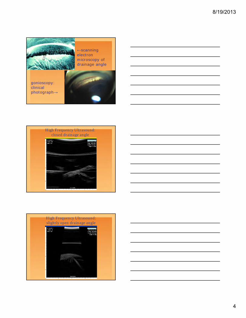
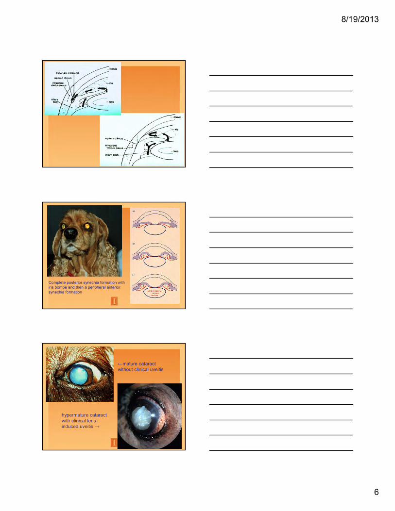
Ralph E. Hamor DVM, MS, DACVO Glaucoma is a leading cause of blindness in the middle-aged dog. Glaucoma should be considered as one of the "rule outs" in any case of "red eye" or "watery eye," especially in predisposed breeds (see lists below). Glaucoma is characterized by an elevation of intraocular pressure (IOP) that results in progressive loss of retinal ganglion cells and their axons. Aqueous, produced by the ciliary body, flows from the posterior chamber through the pupil into the anterior chamber and drains out of the eye via the trabecular meshwork in the iridocorneal angle and into the intrascleral venous plexus (conventional outflow). Uveoscleral (unconventional) outflow also occurs in most species: aqueous flows into the vitreous, across the uveal tract, along the supraciliary-suprachoroidal space into the adjacent sclera. Uveoscleral outflow accounts for 3% of aqueous outflow in the cat, 13% in rabbits, 15% in dogs and may be up to 50% in the horse. Increased IOP results from obstruction to outflow, not an increase in aqueous production. Whatever the cause, the most important consideration is whether the glaucoma is primary or secondary. PRIMARY GLAUCOMA occurs when there is no recognizable cause of the glaucoma. These patients are born with either an abnormal drainage angle, a drainage angle that becomes abnormal, or a drainage angle that appears normal but ceases to function normally. In some cases, gonioscopy (evaluation of the external drainage angle) demonstrates that a patient has an abnormal appearing drainage angle. This finding is only a risk factor that the patient will develop clinical signs of glaucoma. Patients with abnormal drainage angles (pectinate ligament dysplasia or mesodermal dysgenesis or goniodysgenesis) may develop clinical glaucoma as can patients with normal appearing drainage angles. High-frequency ultrasound (HFUS) can also be utilized to assist in the determination if your patient may have primary glaucoma. As with gonioscopy, HFUS helps to provide supportive evidence of primary glaucoma, but does not confirm a diagnosis of an abnormally functioning drainage angle. Patients with abnormal drainage angles (pectinate ligament dysplasia or mesodermal dysgenesis or goniodysgenesis) may develop clinical glaucoma as can patients with normal appearing drainage angles. Breeds that are predisposed to primary glaucoma (this is NOT a completely inclusive or exclusive list but bolded breeds are most commonly seen): Akita, Alaskan Malamute, Basset Hound, Beagle, Border Collie, Boston Terrier, Bouvier des Flandres, Brittany Spaniel, Cairn Terrier, Cardigan Welsh Corgi, Chihuahua, Chow chow, American Cocker Spaniel, Dachshund, Dalmatian, Dandie Dinmont Terrier, English Cocker Spaniel, English Springer Spaniel, Flat Coated Retriever, German Shepherd, Giant Schnauzer, Great Dane, Golden Retriever, Greyhound, Irish Setter, Italian Greyhound, Jack Russell Terrier, Keeshound, Lakeland Terrier, Maltese, Miniature Pinscher, Miniature Schnauzer, Norfolk Terrier, Norwegian Elkhound, Norwich Terrier, Poodle (Toy and Miniature), Samoyed, Scottish Terrier, Sealyham Terrier, Shar Pei, Shih Tzu, Siberian Husky, Skye Terrier, Smooth Fox Terrier, Tibetan Terrier, Welsh Springer Spaniel, Welsh Terrier, West Highland White Terrier, Wire Fox Terrier. Open angle glaucoma: Beagle, Great Dane, Keeshound, Norwegian Elkhound, Poodle (Toy and Miniature), Samoyed, Siberian Husky. Closed angle glaucoma: Akita, American Cocker Spaniel, Basset Hound, English Cocker Spaniel, English Springer Spaniel, Flat Coated Retriever, Golden Retriever, Poodle (Toy and Miniature), Samoyed, Shar Pei, Welsh Springer Spaniel. Primary lens luxation and glaucoma: The lens zonules are abnormal and break down resulting in lens subluxation or lens luxation. Sometimes it can be difficult to determine whether the lens luxation is primary or secondary. If it occurs in a predisposed breed without any other cause, then you should assume that it is a primary problem. Breeds where lens luxation is likely to be inherited (bolded breeds are most commonly seen): Border Collie, Cairn Terrier, Jack Russell Terrier, Lakeland Terrier, Manchester Terrier, Miniature Bull Terrier, Norfolk Terrier, Norwich Terrier, Scottish Terrier, Sky Terrier, Smooth Fox Terrier, Tibetan Terrier, West Highland White Terrier, Wire Fox Terrier. Predisposed Breeds: Australian Collie,
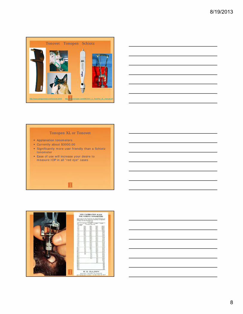
Basset Hound, Beagle, Chihuahua, German Shepherd, Greyhound, Miniature Poodle, Miniature Schnauzer, Norwegian Elkhound, Spaniel Breeds, Pembroke Welsh Corgi, Welsh Terrier, Toy Poodle and Toy Terrier. SECONDARY GLAUCOMA occurs secondary to some other primary cause. Uveitis: any uveitis can cause secondary glaucoma by blocking the drainage of aqueous from the eye. This can occur in many ways: preiridal fibrovascular membrane (PIFM), posterior synechia +/- iris bombé, and/or peripheral anterior synechia. A common cause of this is lens-induced (also called phacolytic glaucoma). Lens material leaks through the capsule and causes chronic inflammation. This chronic inflammation (and any other causes of chronic inflammation) leads to production of angiogenic factors in the aqueous humor and then the production of a PIFM which covers the surface of the iris and the drainage angle. The primary reason for any secondary glaucoma from chronic uveitis is the formation of a PIFM. Any uveitis can also cause thickening of the iris which can lead to formation of a peripheral anterior synechia and angle closure. It is unusual for hypopyon or hyphema to completely block the drainage angle by itself. Lens displacement (luxation or subluxation) is seen in Terriers and occasionally in other breeds. One must evaluate whether the lens luxation is part of the cause of the glaucoma (abnormal lens zonules) or if the luxation is secondary to chronic eye enlargement and breakdown of normal lens zonules. Tumors: Either primary (melanoma, ciliary body adenoma/adenocarcinoma, etc.) or secondary (lymphosarcoma, hemangiosarcoma, etc.) tumors. Pigmentary glaucoma of Cairn terriers—the iris and sclera progressively pigment causing secondary glaucoma and blindness. Currently, no effective long-term therapy. May also be seen in Boxers and Labrador retrievers. Swollen lens: lens intumescence (swelling) associated with acute cataract formation. This occurs primarily in dogs predisposed to glaucoma or in rapidly developing cataracts such as in diabetes. Malignant glaucoma: subluxated or luxated lens or vitreous blocks the pupil then aqueous flows back behind the vitreous pushing it forward causing rapid rise in IOP. Glaucoma in cats is almost always secondary to chronic intraocular inflammation leading to the formation of a PIFM membrane and glaucoma. Aqueous humor misdirection is a syndrome in cats and may be treated medically and/or with surgery to remove the lens. TONOMETRY—you cannot treat or diagnose glaucoma without a tonometer. The standard instrument for measuring intraocular pressure in the past was the Schiotz tonometer. This is an indentation tonometer which estimates IOP by indentation of the cornea. The foot plate must contact the cornea fully with the cornea positioned parallel to the ground. Three readings within one scale unit should be taken and the readings then converted to pressure in mmHg. Other types of tonometer include applanation or rebound tonometers. These tonometers are much easier to use but are more expensive. However, more and more private practices own them and actually use them. I would strongly recommend that any private practice consider purchasing one of these tonometers. Commonly used brands are the Tonopen® or Tonovet® which cost about $3000.00. The normal range for most domestic species is 12 to 24 mmHg. Digital tonometry (palpating the eye with fingers to estimate "normal" versus "high" pressure) is rarely accurate enough for diagnosis and never accurate for monitoring therapy. Gonioscopy – can be used by an ophthalmologist to characterize the appearance of the external portion of the drainage angle. High-frequency ultrasound – can be used by an ophthalmologist to visualize the inner aspect of the drainage angle. This equipment is not common even in specialty ophthalmology practices.
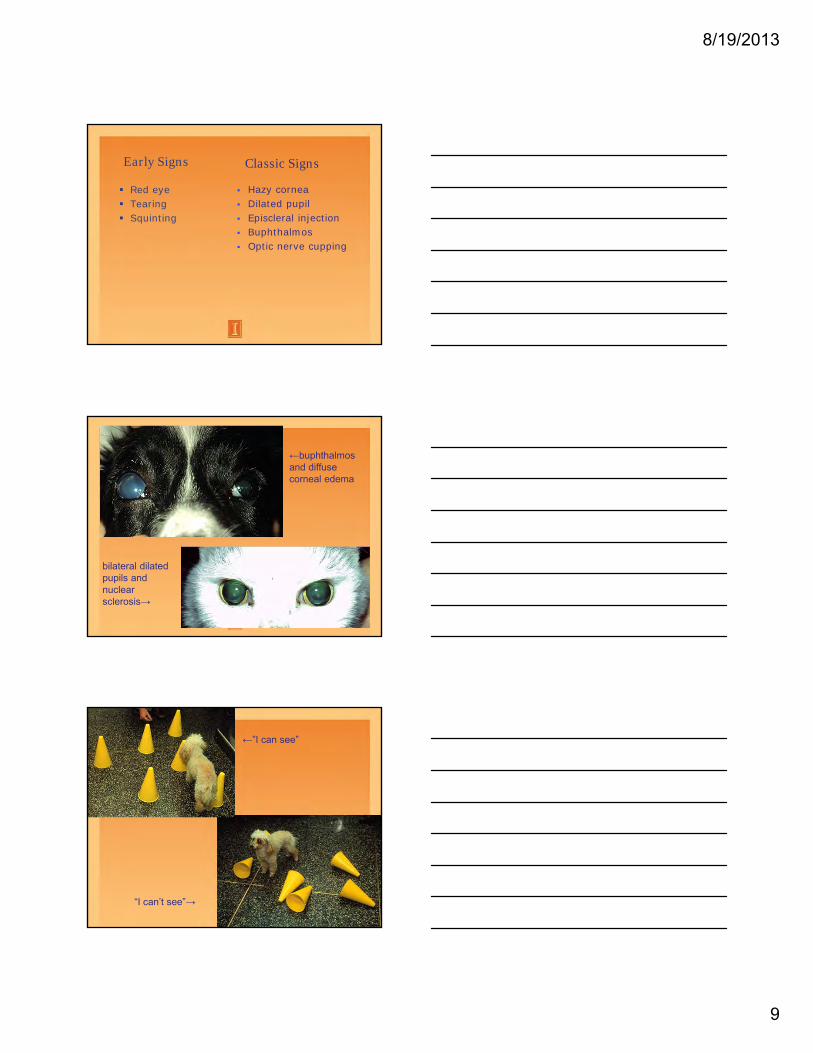
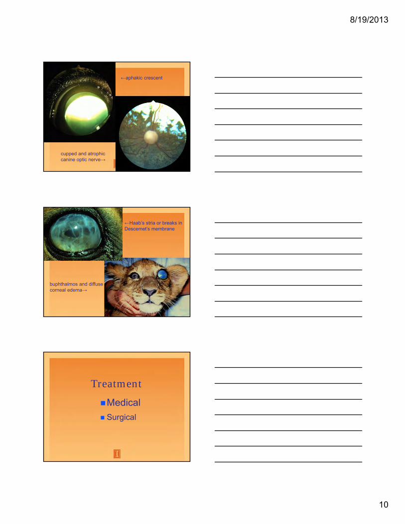
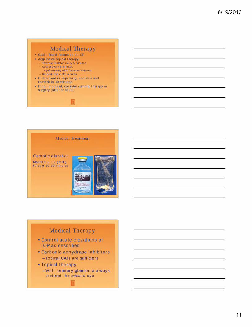
Signs of Glaucoma: Corneal edema: the cloudy appearance is caused by interference with the pumping mechanisms of the corneal endothelial cells. The density of the edema worsens as the IOP increases. Dilated pupil: high pressure causes the pupil to dilate and be less responsive to light, while lesser pressure increases may leave the pupil unaffected. The dilation is thought to be due to neurologic or vascular damage to the ciliary body and iris as well as impaired retinal and optic nerve function. Not every case will have a dilated pupil. The pupil usually is not dilated and unresponsive until the pressure is about 60 mm or higher. Episcleral and conjunctival blood vessel congestion: the pressure elevation causes blockage of normal venous drainage and engorgement of anterior ciliary veins. Glaucoma should be a "rule out" in any "red eye." Other signs include pain, blindness, enlargement of the globe, corneal insensitivity, shallow anterior chamber, cupping of the optic disc, and thinning of the retina causing hyperreflectivity especially around the optic disc. By the time eye enlargement, cupping of the disc, or hyperreflectivity of the retina are obvious, there is essentially no chance for return of sight. The goal is to make the diagnosis when more subtle signs are present. Corneal striae--(Haab's striae)--cracks in Descemet's membrane from stretching of the globe. Their presence indicates previous glaucoma regardless of the current IOP. Treatment Goals: Treatment of acute glaucoma by the primary care veterinarian should be directed toward one goal: rapid reduction of intraocular pressure before permanent blindness occurs. Medical therapy must be started immediately after the diagnosis is made in order to have the opportunity to preserve vision. Medical treatment is directed at opening the drainage angle, decreasing ciliary body aqueous production and increasing aqueous outflow. If a clinician is skilled in ophthalmic examination, gonioscopy and HFUS, a more precise diagnosis and therapy can be attempted. With the exception of glaucoma secondary to uveitis, miotics, carbonic anhydrase inhibitors and beta-blockers are the drugs of choice. If the intraocular pressure is over 40 mmHg on presentation, aggressive therapy should be instituted. Many topical medications are not efficacious until the pressure is <40 mm Hg. An exception to this rule is Xalatan or Travatan. This drug can decrease IOP from 70-80 mm Hg into the normal range. I usually start with using Xalatan/Travatan and Cosopt every 5 minutes for 30 minutes and recheck the IOP. If this does not work, then mannitol can be given to dehydrate the vitreous and reduce the pressure within an hour. Alternatively, it may be necessary to take the dog to surgery to perform a laser ciliary body ablation and/or implantation of an aqueous shunt. Treat the primary cause if the glaucoma is secondary to another disease process. For example, a dog with uveodermatologic syndrome and secondary glaucoma requires anti-inflammatory therapy aimed at the uveodermatologic syndrome as well as glaucoma therapy. Only secondary glaucoma has the potential for a cure without surgical intervention. Glaucoma is a multi-factorial disease that is not strictly defined as an increase in IOP. No one knows for sure what is a "safe IOP" for the dog and cat. Treatment is aimed at prevention of pressure-induced pathologic alterations of the retina and the optic nerve. The earlier therapy is begun, the better the response. Once pressure-induced damage to the optic nerve has occurred, pressure in the "normal range" may cause further damage. Medical therapy is recommended in those eyes where there is vision to salvage or as prophylactic therapy of the fellow eye in cases of primary glaucoma. Prophylactic topical therapy has been shown to increase the time period between the onset of clinical signs in the first and second eye.
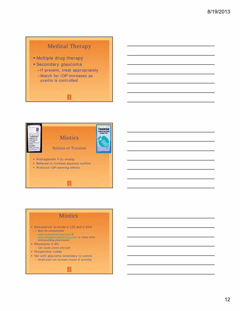
Surgical therapy is recommended for some visual eyes. Other surgical therapies are used for all blind and painful eyes. It is of no value to the patient to medically treat a blind eye. RECOMMENDED MEDICAL TREATMENT: Osmotic Diuretic --dehydrate to vitreous—used in emergency therapy only; crucial that the patient is know to be capable of producing urine prior to using osmotic diuretics. Mannitol IV: 1-2 g/kg IV in 15-20 minutes, water should be withheld for 2 to 3 hours after administration then given in limited quantities until water can be offered free choice without the dog drinking a large quantity. Can be given a second time 4 to 6 hours after the initial administration if considered necessary. Glycerol (50%): 1-2 ml/kg; given orally. Lasix is not an effective choice for glaucoma therapy. Miotics: increase outflow of aqueous typically by opening the drainage angle. Latanoprost 0.005% (Xalatan®) - 1 drop daily to BID. Prostaglandin F 2 analogue. We are unsure exactly how this drop works but it does result in changes to the extracellular matrix resulting in increased uveoscleral or unconventional outflow. Travoprost 0.004% (Travatan®) - 1 drop daily to BID. An isopropyl ester of a single enantiomer of the selective FP prostaglandin receptor agonist fluprostenol. Appears to have increased IOP lowering effects than latanoprost. Demecarium bromide 0.25% or 0.125%, - 1 drop daily to BID. Is a cholinesterase inhibitor. Currently not available except at a compounding pharmacy but is much cheaper than latanoprost or travoprost for use as a miotic alone in patients with a lens subluxation or posterior lens luxation. Pilocarpine 2% or 4%: 1 drop every 4 to 6 hours—not recommended in animals that concurrently have uveitis. Pilocarpine also causes uveitis and can be quite painful especially if used for prophylactic therapy. Carbonic Anhydrase Inhibitors: decrease production of aqueous. With the development of topical CAIs, the use of oral CAIs is of limited clinical value. Two research articles have demonstrated that there was no increased IOP lowering effects with the addition of oral CAIs if topical CAIs were already in use in glaucomatous canine patients. Neptazane (Methazolamide): 2.5 to 5 mg/kg, PO, BID-TID. Diamox (Acetazolamide): not recommended in small animals. Trusopt (2% dorzolamide hydrochloride): topical CAI, used BID to QID, decreases systemic side effects of oral CAIs. Cosopt: Combination of 2% dorzolamide hydrochloride (topical CAI) and timolol maleate (beta-blocker). May provide increased efficacy through increased owner compliance. Now in a generic form.
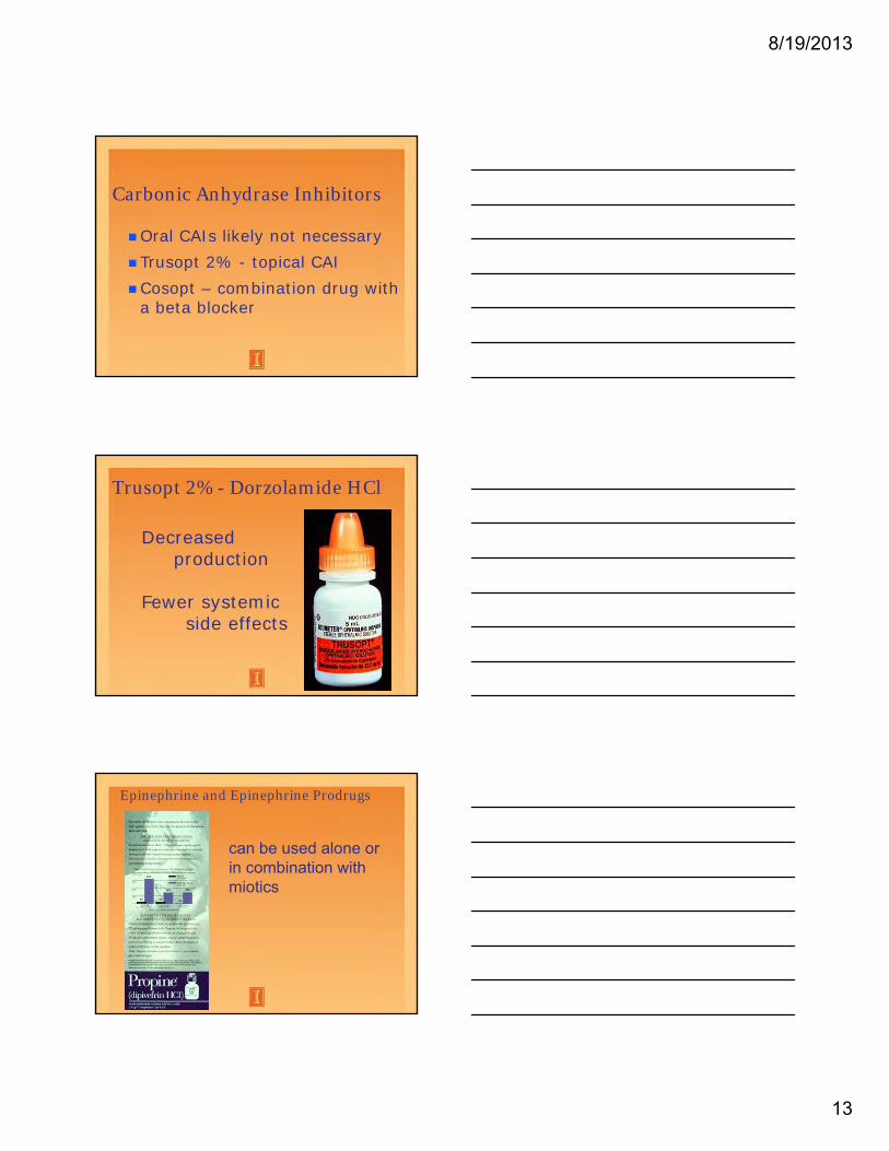
Propine (Dipivefrin hydrochloride) or topical epinephrine: 1 drop BID-TID. Sympatholytic--beta-blocker (increases aqueous outflow; reduces aqueous humor production). Timoptic (Timolol maleate): 1 drop BID-TID, non-selective. Also can use Betagan (levobunolol) – is noncardioselective or Betopic (betaxolol HCl) – is cardioselective (beta-1 adrenergic). Glaucoma Secondary to Uveitis: Treat the primary cause of the uveitis. Carbonic anhydrase inhibitor—topical CAIs are best (decrease aqueous production). In cases with a miotic pupil, topical tropicamide or atropine (mydriatic) may be used to dilate the pupil but should be used cautiously to limit IOP increases. Topical steroidal and/or nonsteroidal agents—if their use will not complicate the primary cause of uveitis. These drugs are synergistic when used together. Topical and/or systemic antibiotics are usually not needed as intraocular bacterial infections are a rare cause of secondary glaucoma. Subconjunctival corticosteroid. Don't start until intraocular infection is ruled out. If blastomycosis is suspected, rule it out before giving subconjunctival steroids. Prognosis: Since the majority of primary glaucomas will not be controlled permanently with medication, the owners should be advised that a surgical procedure may soon be indicated. Once diagnosis has been made and treatment started (the first 24 hours), it is advisable to refer the case to a veterinary ophthalmologist or a colleague who has had ophthalmology training and experience. In almost every situation, primary glaucoma is a potentially blinding disease that will need aggressive medical and/or surgical intervention to have an opportunity to save vision.
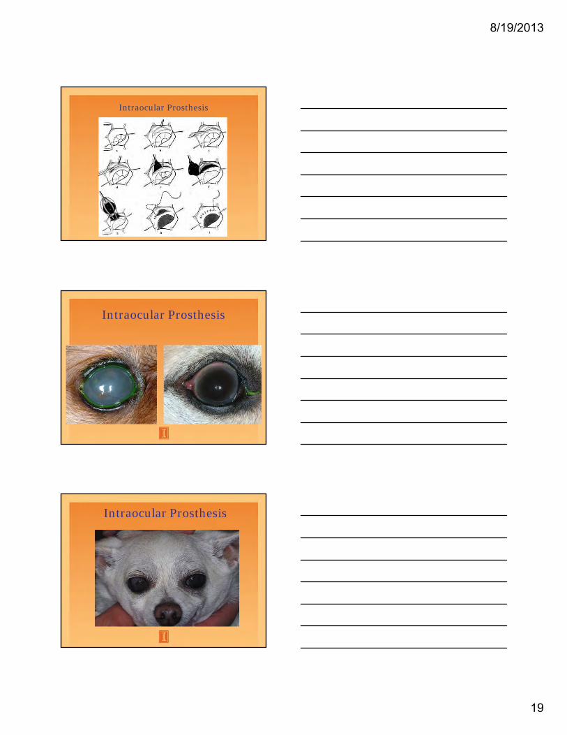
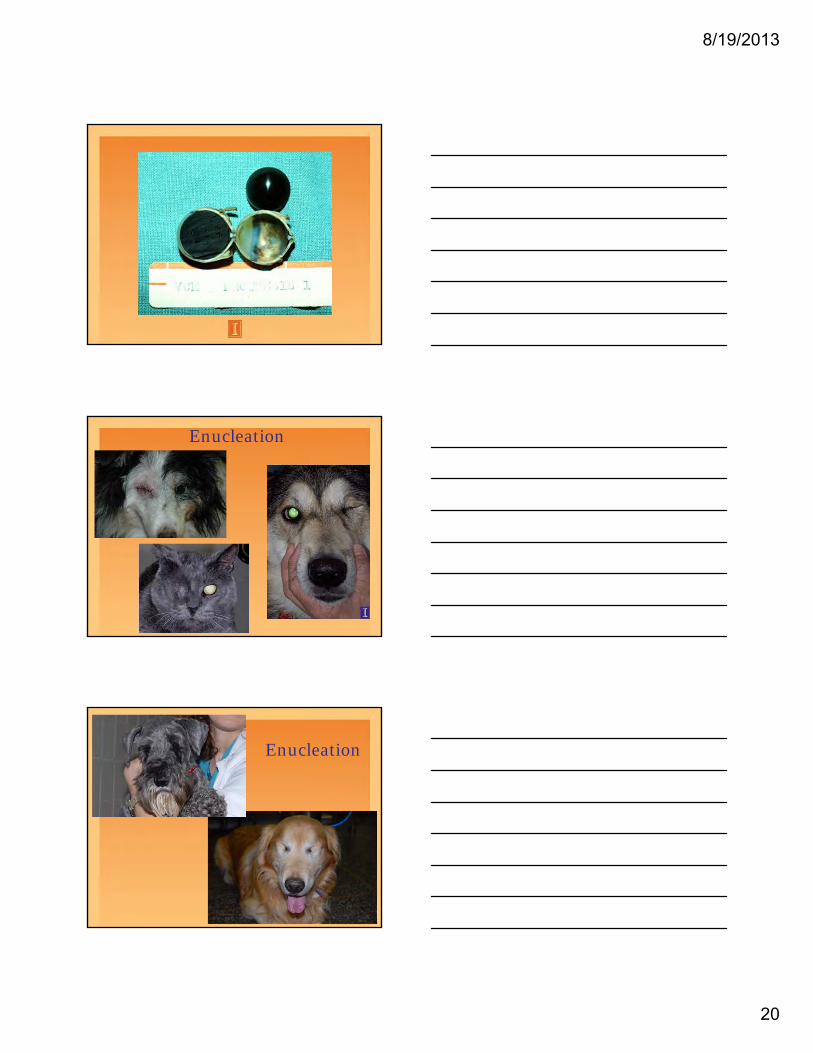
RECOMMENDED SURGICAL TREATMENTS: Laser Ciliary Body Ablation: Results indicate an encouraging mode of therapy for glaucoma by reducing aqueous humor production through noninvasive destruction of the ciliary body with a laser seems to have fewer secondary side effects associated with inflammation than is seen with cyclocryosurgery. Energy from the laser tip is directed through the sclera (transscleral) and is used to destroy part of the ciliary body and reduce aqueous production. Because of how the laser energy is directed and focused, you can kill a portion of the secretory cells of the ciliary processes without so much damage to the overlying conjunctiva and sclera as with cyclocryosurgery. The main side effects are uveitis and hyphema. Equipment is becoming much more available now and is the mainstay of surgical therapy today. Used most commonly in a visual globe after it has experienced a pressure spike and the IOP has been controlled with oral and topical medications. The most recent advance in laser therapy is an endolaser. It is used to perform endocylcophotocoagulation where the ciliary processes are directly visualized and directly treated with a laser. This equipment is expensive and not commonly available. However, this method of laser ciliary body ablation shows the most promise to provide effective IOP control. The most common side effect is development of a cataract so the procedure is often combined with phacoemulsification and intraocular lens implantation. This method may also be more effective in patients with secondary glaucoma that is not responsive to medical therapy. Some type of laser surgery is currently the treatment of choice in a visual globe to attempt to permanently decrease aqueous production and preserve vision. Filtration Procedures: Filtration procedures are designed to increase aqueous outflow when normal outflow channels are blocked. A variety of commercially manufactured devices are available, but most have been far more effective in human beings than in dogs. Shunts are available that divert aqueous into the subconjunctival space or the frontal sinus. I have had more success using frontal sinus shunts that subconjunctival shunts. Shunts appear to be most effective in cases of primary glaucoma rather than secondary glaucoma. In patients with secondary glaucoma, the shunts are more prone to blockage with fibrin. Enucleation: indicated for blind, painful eyes. Intraocular/Intrascleral Prostheses: indicated for blind, painful eyes. Case selection is crucial for success. Should not be placed in globes that are infected, neoplastic or have moderate to severe corneal disease. Chemical Ciliary Body Ablation / Intravitreal Gentocin Injection: indicated for blind, painful eyes. The most commonly described procedure involves an intravitreal injection of gentocin, which is aimed at pharmacologic ablation of the ciliary body. This in turn causes decreased production of aqueous. This treatment is designed only for selected cases of glaucoma in irreversibly blind and painful eyes and in which the primary disease has been absolutely determined and a tumor or endophthalmitis is not present. This procedure often results in a phthisical globe. May be the best option in aged patients or patients with debilitating disease associated with, or in conjunction with, the glaucoma. Inject 25 mg of gentocin +/- 1 mg of dexamethasone directly into the vitreous if < 0.25 mls are used (I use 25 mg of gentocin only). This can be done with sedation and topical anesthesia. If larger volumes are needed, inject 25 mg of gentocin after removal of at least a similar volume of vitreous. This requires a short-term general anesthetic episode. Be careful to reduce the amount of gentocin if used in very small patients…you do not want to exceed the systemic toxic dose of gentocin (4.4 mg/kg/day). POTENTIAL CASE SCENARIOS AND MANAGEMENT SCHEMES: Blind eyes: A comfortable, blind eye can be left alone. This, however, is a relatively rare occurrence. Any globe that is blind and painful needs some type of a surgery. Whether that is an enucleation, intrascleral prosthesis or intravitreal injection is up to the individual veterinarian and the owner. Any eye with an IOP > 40 mm Hg should be considered painful. Most dogs with chronic IOP increases in this range, however, appear relatively comfortable. It is routinely our experience that when any surgery is done, the owner returns and comments that the dog is, indeed, more comfortable after surgery than before.
"Normal" eye in a dog that has clinical primary glaucoma in the other eye: Whenever primary glaucoma is diagnosed or suspected, the second or "normal" eye is at extreme risk of developing glaucoma. First of all, the owner must be informed of this so that they are prepared early for the potential of having a blind dog in the future. It is crucial that the second or "normal" eye be prophylactically treated with anti-glaucoma medications. It has been demonstrated that this prophylactic treatment will increase the time frame until the second or "normal" eye will develop clinical signs of glaucoma. Along with the prophylactic therapy, it is important to set up a schedule to monitor the IOP and evaluate for any clinical signs of elevations in IOP. This schedule varies from dog to dog but we usually start with every two week evaluations at first. If the eye is stable for a few months, then monthly evaluations should be fine. If the IOP is stable for 6 - 9 months, we may begin to stretch that time frame out to 2 or 3 month intervals. Early IOP elevations or acute glaucoma in the second eye: If IOP elevations can be documented early (especially prior to overt clinical signs develop), we have a much better chance to extend the patient's visual lifespan. This can be done medically, surgically or in combination. In an acute glaucomatous crisis, it is crucial to decrease the IOP rapidly (Xalatan/Travatan and Cosopt OR mannitol) and to keep the IOP well within the normal range (<15 mm Hg). Once a globe has clinical signs of glaucoma, that eye is more sensitive to even high normal IOP. Once the IOP is decreased, therapy is again continued with medical, surgical or combination therapy. I most commonly use a combination of medical and surgical therapy. The bottom line is that primary glaucoma is a surgical disease IF the eye is going to have any chance of extended vision. Even with our best efforts, almost all dogs with primary glaucoma will eventually be blind in both eyes. "We win some battles, but we lose the war." As far as what medications are best, that question is a debate among ophthalmologists. Every ophthalmologist that I have ever worked with treats glaucoma a little differently. This basically tells me that none of us know exactly what we are doing. I generally recommend that you become familiar with the medications and protocols that the person you refer to uses. A general working knowledge of what the drugs are and how they work is important. There usually is no reason to use 2 miotics or 2 beta-blockers. We do, however, use multiple drug therapy and use many drugs more frequently than the label dose suggests. Ophthalmology Contact Information: Email for questions and pictures: [email protected] This goes to the entire Ophthalmology Service. Questions on appointments, costs, scheduling or need to reach a member of the service, call Shari or Lorri: Shari Poruba: 217-333-5374 Office (for clients or to leave a message) 217-649-4971 Direct Work Cell (for RDVMs needing immediate assistance) Lorri Zoch: 217-265-0008 Office (for clients or to leave a message) 217-552-0470 Direct Work Cell (for RDVMs needing immediate assistance) Ralph Hamor: 217-333-7451 [email protected] Amber Labelle: 217-244-3364 [email protected] Sean Collins (resident): 217-300-6323 [email protected] Dan Dorbandt (resident): 217-300-4823 [email protected]
8/19/2013
1
Medical and Surgical Therapy of Glaucoma -Can I Really Do Anything To Help?
Ralph E. Hamor DVM, MS, DACVOClinical Associate Professor
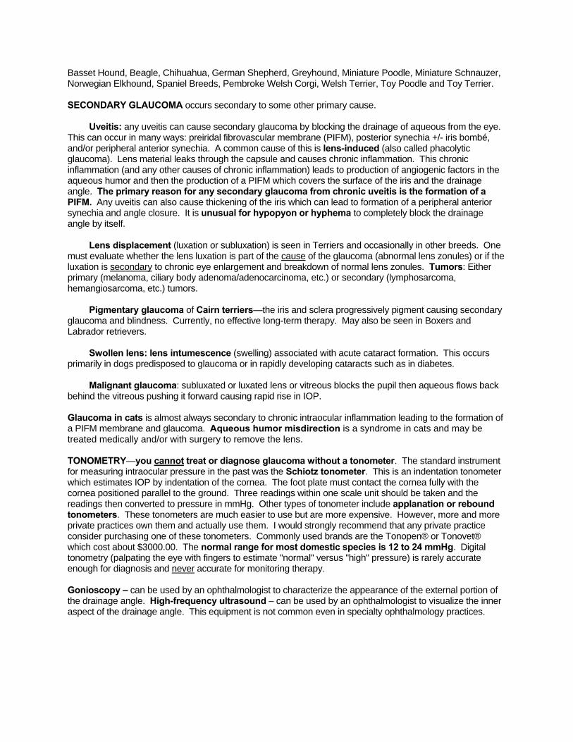
Glaucoma
Leading cause of blindness in middle aged dogs
Rule out in any “red” and/or “painful” eye Characterized by an elevation in intraocular
pressure (IOP)
8/19/2013
2
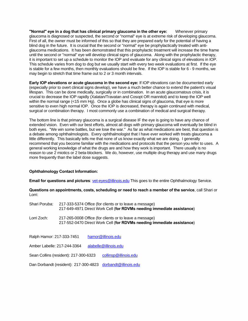
Aqueous Outflow
Conventional– Through drainage angle
Unconventional (uveoscleral)– Across uveal tract to supraciliary/suprachoroidal
space– Cats – 3%, rabbits – 13%, dogs – 15%,
horses – 50 %
Glaucoma
Main clinical consideration is to determine whether the cause of the glaucoma is primary or secondary.
Increased cost but likely has increased efficacy and increased owner compliance
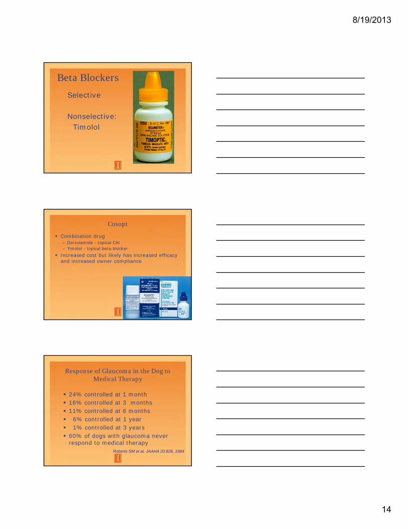
Response of Glaucoma in the Dog to Medical Therapy
24% controlled at 1 month 16% controlled at 3 months 11% controlled at 6 months 6% controlled at 1 year 1% controlled at 3 years 60% of dogs with glaucoma never
respond to medical therapyRoberts SM et al. JAAHA 20:828, 1984
8/19/2013
15
Treatment
Medical
Surgical
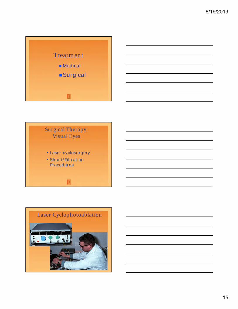
Surgical Therapy:Visual Eyes
Laser cyclosurgery Shunt/Filtration
Procedures
Laser Cyclophotoablation
8/19/2013
16
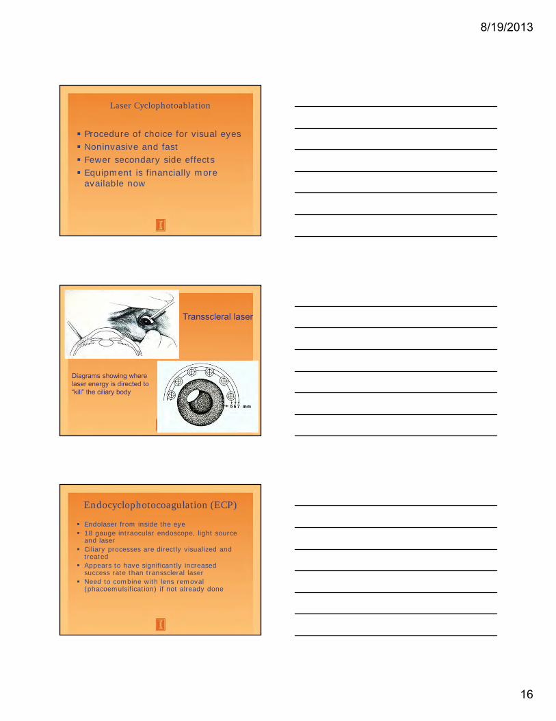
Laser Cyclophotoablation
Procedure of choice for visual eyes Noninvasive and fast Fewer secondary side effects Equipment is financially more
available now
Diagrams showing where laser energy is directed to “kill” the ciliary body
Transscleral laser
Endocyclophotocoagulation (ECP)
Endolaser from inside the eye 18 gauge intraocular endoscope, light source
and laser Ciliary processes are directly visualized and
treated Appears to have significantly increased
success rate than transscleral laser Need to combine with lens removal
(phacoemulsification) if not already done
8/19/2013
17
ECP
Highest success rate of any surgical procedure available thus far.
Filtration Procedures
Frontal sinus shunts Temporary shunt after laser Subconjunctival shunts
Frontal sinus shunt
8/19/2013
18
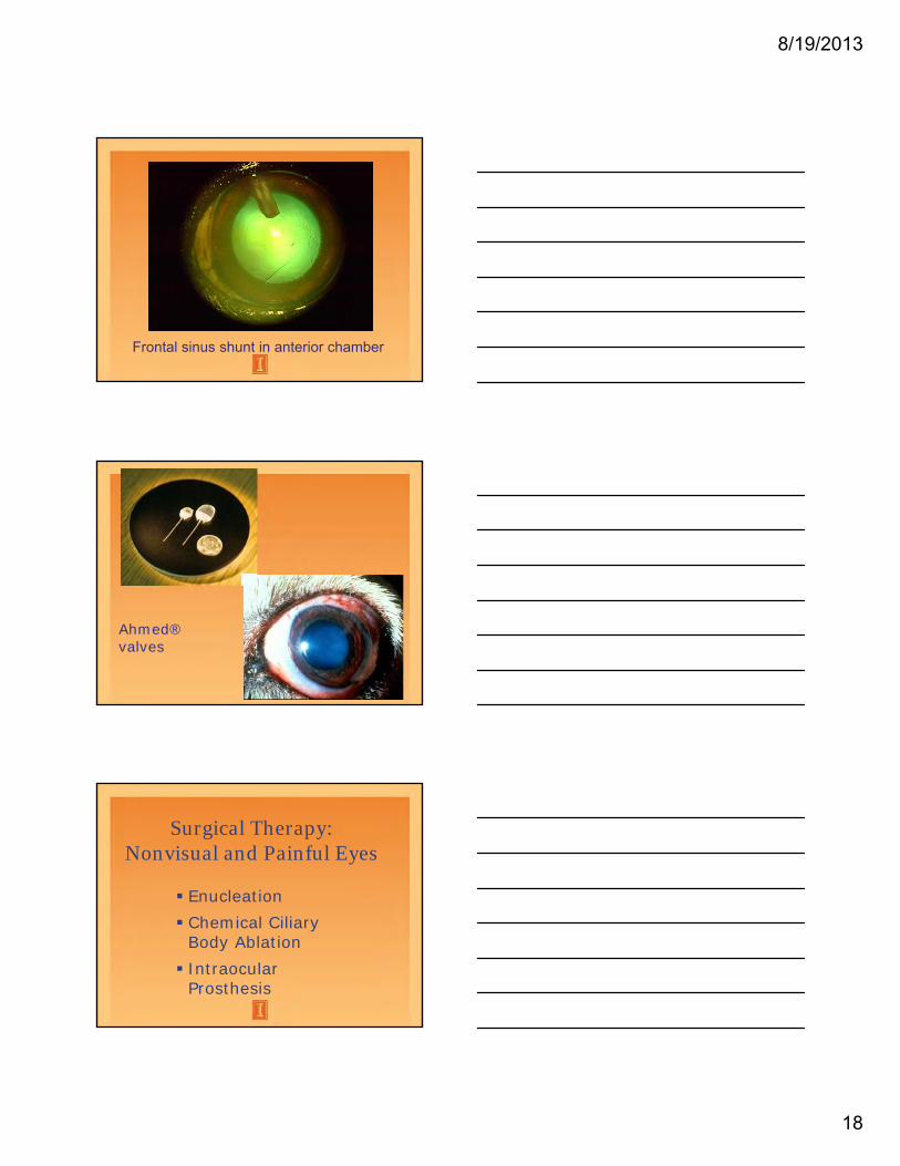
Frontal sinus shunt in anterior chamber
Ahmed® valves
Surgical Therapy:Nonvisual and Painful Eyes
Enucleation Chemical Ciliary
Body Ablation Intraocular
Prosthesis
8/19/2013
19
Intraocular Prosthesis
Intraocular Prosthesis
Intraocular Prosthesis
8/19/2013
20
Enucleation
Enucleation
8/19/2013
21
Blind Eyes
If comfortable, leave alone If painful, needs a surgery
Needs to be treated prophylactically once fellow eye is diagnosed with primary glaucoma
Treatment will increase the time frame between until the “normal” eye develops glaucoma
Monitor IOP monthly
Early IOP increase or acute glaucoma in second eye
Medical therapy to decrease IOP– try to keep IOP < 15 mmHg
Increase medical therapy as needed– medications alone will eventually fail
Surgical therapy once IOP and intraocular disease are stabilized
8/19/2013
22
Ophthalmology Contact Information: Email for questions and pictures: [email protected] This goes to the entire Ophthalmology Service. Questions on appointments, costs, scheduling or need to reach a member of the service, call Shari or Lorri: Shari Poruba: 217-333-5374 Office (for clients or to leave a message) 217-649-4971 Direct Work Cell (for RDVMs needing immediate assistance) Lorri Zoch: 217-265-0008 Office (for clients or to leave a message) 217-552-0470 Direct Work Cell (for RDVMs needing immediate assistance) Ralph Hamor: 217-333-7451 [email protected] Amber Labelle: 217-244-3364 [email protected] Sean Collins (resident): 217-300-6323 [email protected] Dan Dorbandt (resident): 217-300-4823 [email protected]