FOCUS ON MOLECULAR IMAGING Clinical Feasibility of Molecular Imaging of Plaque Inflammation in Atherosclerosis Nobuhiro Tahara 1,2 , Tsutomu Imaizumi 2 , Renu Virmani 3 , and Jagat Narula 1 1 University of California, Irvine, California; 2 Kurume University School of Medicine, Kurume, Japan; and 3 Cardiovascular Pathology Institute, Gaithersburg, Maryland Despite substantial advances in the diagnosis and manage- ment of coronary artery disease, acute coronary events con- tinue to occur in many patients. It has been increasingly realized that the lesions responsible for acute events may not necessarily be critically obstructive and hence not be associ- ated with inducible ischemia. Various morphologic features of plaque vulnerability have been described by CT angiography, in- travascular ultrasound, and optical coherence tomography. The culprit plaques often demonstrate large plaque and necrotic core volumes, positive vascular remodeling, and attenuation of fibrous plaque caps. The remaining obligatory component of plaque vulnerability is fibrous cap inflammation; molecular imag- ing is best suited for identification of monocyte–macrophage infiltration. Whereas multiple candidate targets have been evalu- ated in preclinical molecular imaging studies, only 18 F-FDG and 99m Tc-annexin-A5 have been recently used in the settings of acute vascular events. These 2 imaging strategies have demonstrated the clinical feasibility of imaging for detection of inflammation. Key Words: atherosclerosis; vulnerable plaque; molecular im- aging; inflammation; 18 F-labeled FDG PET; apoptosis; 99m Tc- labeled annexin-A5 J Nucl Med 2009; 50:331–334 DOI: 10.2967/jnumed.108.060376 Molecular imaging of various components of atherosclerotic plaques has been proposed, and proof of principle has been demonstrated in experimental models of disease (1). These preclinical studies have predominantly targeted plaque inflamma- tion with the premise that the extent of inflammation would determine the vulnerability of the plaque to rupture. Plaque inflammation has been detected by targeting alterations in monocytes that facilitate their migration to the neointima, ensure efficient scavenging of insudated lipid, oversee their transforma- tion to foam cells, or mediate cell death (1). Molecular targets have also included the events that are associated with or consequent to inflammation, such as production of cytokines and metalloproteinases. Although these experimental molecular imaging studies have offered significant promise, translational data in the clinical setting has just started to emerge. Clinical studies of molecular targeting are the major focus of the following review. We have referred to some of the early molecular imaging attempts that labeled white blood cells to follow their localization and labeled lipoproteins to trace their destination in the inflam- matory cells in plaques (1). Even though the incorporation of radiolabeled components in the plaque may not have been adequate, these studies created a sound foundation for the development of imaging strategies of the future. PATHOLOGIC BASIS OF INFLAMMATION IMAGING Ruptured Plaques Are Substantially Inflamed Vulnerable plaques have typically large necrotic cores that are covered by thin fibrous caps (2). Many foam cells are seen around the necrotic cores and within the fibrous caps (Fig. 1). Pathologic examination of culprit plaques in the victims of acute coronary events reveals extensive inflammation with macrophages; the more the macro- phages, the thinner the cap. Migration of monocytes to the subintimal layers of the plaque is mediated by development of receptors for chemoattractant factors such as monocyte chemotactic protein-1 (MCP-1) and those for adhesion molecules such as intercellular adhesion molecule-1 and vascular cell adhesion molecule-1 (VCAM-1) (1). After subintimal localization, the monocytes express scavenger receptors including SRAI/II, CD68, and FcRIII. In experimental models, these receptors have been targeted by radiolabeled MCP-1, VCAM-1, Fc-IgG, and lipoproteins. Inflammation Is Accompanied by Cytokine Release Foam cells in the neointima release numerous cytokines, such as interleukin-1, tumor necrosis factor-a, and MCP-1, that attract other monocytes and activate endothelial cells and smooth muscle cells (3). Activated macrophages also release metalloproteinases and other proteolytic enzymes such as cathepsins, which lead to degradation of the matrix, thinning of the fibrous cap, and positive outward remodeling of the vessel wall. Activated lymphocytes produce proinflammatory cytokines such as interferon-g, which is able to amplify the inflamma- tory response. Lymphokines also facilitate adventitial vasa vasorum proliferation and plaque neoangiogenesis, which contributes to red blood cell extravasation and necrotic core enlargement. Unstable Plaques Demonstrate Significant Cell Death Cell death is commonly observed in the vulnerable plaque; macrophage death leads to expansion of the necrotic core and perpetuates plaque instability (4). More than 40% of macrophages at the rupture site are in the process of cell death by apoptosis; Received Aug. 13, 2008; revision accepted Dec. 3, 2008. For correspondence or reprints contact: Jagat Narula, University of California, UCI Medical Center, 101 The City Dr., Bldg. 53, Rt. 81, Orange, CA. E-mail: [email protected]COPYRIGHT ª 2009 by the Society of Nuclear Medicine, Inc. MOLECULAR IMAGING OF PLAQUE INFLAMMATION • Tahara et al. 331 by on April 11, 2019. For personal use only. jnm.snmjournals.org Downloaded from
Transcript
F O C U S O N M O L E C U L A R I M A G I N G
Clinical Feasibility of Molecular Imaging ofPlaque Inflammation in Atherosclerosis
Nobuhiro Tahara1,2, Tsutomu Imaizumi2, Renu Virmani3, and Jagat Narula1
1University of California, Irvine, California; 2Kurume University School of Medicine, Kurume, Japan; and 3CardiovascularPathology Institute, Gaithersburg, Maryland
Despite substantial advances in the diagnosis and manage-ment of coronary artery disease, acute coronary events con-tinue to occur in many patients. It has been increasinglyrealized that the lesions responsible for acute events may notnecessarily be critically obstructive and hence not be associ-ated with inducible ischemia. Various morphologic features ofplaque vulnerability have been described by CT angiography, in-travascular ultrasound, and optical coherence tomography. Theculprit plaques often demonstrate large plaque and necroticcore volumes, positive vascular remodeling, and attenuationof fibrous plaque caps. The remaining obligatory component ofplaque vulnerability is fibrous cap inflammation; molecular imag-ing is best suited for identification of monocyte–macrophageinfiltration. Whereas multiple candidate targets have been evalu-ated in preclinical molecular imaging studies, only 18F-FDG and99mTc-annexin-A5 have been recently used in the settings of acutevascular events. These 2 imaging strategies have demonstratedthe clinical feasibility of imaging for detection of inflammation.
J Nucl Med 2009; 50:331–334DOI: 10.2967/jnumed.108.060376
Molecular imaging of various components of atheroscleroticplaques has been proposed, and proof of principle has beendemonstrated in experimental models of disease (1). Thesepreclinical studies have predominantly targeted plaque inflamma-tion with the premise that the extent of inflammation woulddetermine the vulnerability of the plaque to rupture. Plaqueinflammation has been detected by targeting alterations inmonocytes that facilitate their migration to the neointima, ensureefficient scavenging of insudated lipid, oversee their transforma-tion to foam cells, or mediate cell death (1). Molecular targetshave also included the events that are associated with orconsequent to inflammation, such as production of cytokinesand metalloproteinases. Although these experimental molecular
imaging studies have offered significant promise, translationaldata in the clinical setting has just started to emerge. Clinicalstudies of molecular targeting are the major focus of the followingreview. We have referred to some of the early molecular imagingattempts that labeled white blood cells to follow their localizationand labeled lipoproteins to trace their destination in the inflam-matory cells in plaques (1). Even though the incorporation ofradiolabeled components in the plaque may not have beenadequate, these studies created a sound foundation for thedevelopment of imaging strategies of the future.
PATHOLOGIC BASIS OF INFLAMMATION IMAGING
Ruptured Plaques Are Substantially InflamedVulnerable plaques have typically large necrotic cores that are
covered by thin fibrous caps (2). Many foam cells are seen around thenecrotic cores and within the fibrous caps (Fig. 1). Pathologic
examination of culprit plaques in the victims of acute coronary eventsreveals extensive inflammation with macrophages; the more the macro-
phages, the thinner the cap. Migration of monocytes to the subintimallayers of the plaque is mediated by development of receptors for
chemoattractant factors such as monocyte chemotactic protein-1(MCP-1) and those for adhesion molecules such as intercellular adhesion
molecule-1 and vascular cell adhesion molecule-1 (VCAM-1) (1). Aftersubintimal localization, the monocytes express scavenger receptors
including SRAI/II, CD68, and FcRIII. In experimental models, thesereceptors have been targeted by radiolabeled MCP-1, VCAM-1, Fc-IgG,
and lipoproteins.
Inflammation Is Accompanied by Cytokine ReleaseFoam cells in the neointima release numerous cytokines, such as
interleukin-1, tumor necrosis factor-a, and MCP-1, that attract othermonocytes and activate endothelial cells and smooth muscle cells (3).
Activated macrophages also release metalloproteinases and otherproteolytic enzymes such as cathepsins, which lead to degradation of
the matrix, thinning of the fibrous cap, and positive outward remodeling
of the vessel wall. Activated lymphocytes produce proinflammatorycytokines such as interferon-g, which is able to amplify the inflamma-
tory response. Lymphokines also facilitate adventitial vasa vasorumproliferation and plaque neoangiogenesis, which contributes to red
blood cell extravasation and necrotic core enlargement.
Unstable Plaques Demonstrate Significant Cell DeathCell death is commonly observed in the vulnerable plaque;
macrophage death leads to expansion of the necrotic core andperpetuates plaque instability (4). More than 40% of macrophages at
the rupture site are in the process of cell death by apoptosis;
Received Aug. 13, 2008; revision accepted Dec. 3, 2008.For correspondence or reprints contact: Jagat Narula, University of
California, UCI Medical Center, 101 The City Dr., Bldg. 53, Rt. 81, Orange, CA.E-mail: [email protected] ª 2009 by the Society of Nuclear Medicine, Inc.
MOLECULAR IMAGING OF PLAQUE INFLAMMATION • Tahara et al. 331
by on April 11, 2019. For personal use only. jnm.snmjournals.org Downloaded from
macrophages remote from the site of rupture do not show much
apoptosis. It has been reported that dying smooth muscle cells mayrelease large quantities of proinflammation cytokines such as MCP-1
and interleukin-8 (5), and dying macrophages may produce tissuefactor (6) and metalloproteinases.
MOLECULAR IMAGING OF PLAQUE INFLAMMATION
Numerous characteristic alterations evolve as monocytes traverse theintimal layer and prepare to ingest concurrently infiltrating lipids. These
unique features, which vary based on different phases of plaquedevelopment, have been targeted successfully by radiolabeled autol-
ogous leukocytes (Fig. 2) (7), low-density lipoprotein (LDL) (8), and Fcfragments of immunoglobulin (9) for targeting of the scavenger
function. More recent experimental studies have used radiolabeled
ligands of cytokine and adhesion molecule receptors, including MCP-1and VCAM-1, or cytokines released by infiltrating macrophages such
as metalloproteinases (1). It has not been entirely clear if such acharacterization would be of clinical significance or which candidate
molecule would be most informative. However, few recent correlativestudies have demonstrated that the presence of macrophages with high
respiratory burst or those with activation of cell death pathways areassociated with culprit lesions underlying an acute coronary event. The
metabolically active macrophages have been clinically recognized by18F-labeled FDG imaging (10), and dying macrophages have been
successfully targeted by using annexin-A5 (AA5) (Fig. 2) (4,11).Detection of active inflammation should allow identification of
vulnerable plaques if information is obtained before an acute eventhas occurred.
18F-FDG IMAGING FOR PLAQUE INFLAMMATION
18F-FDG Uptake in Vessels Is Commonly Seen inPatients Undergoing Evaluation of Malignant Tumors
PET imaging studies for localization of malignant tumors havereported incidental 18F-FDG uptake in the carotid, coronary, iliac, and
femoral arteries and thoracic and abdominal aorta; 18F-FDG uptake inlarge arteries was observed in up to 50% of patients evaluated for
oncologic reasons. In a prospective 18F-FDG PET study performed in
a large cohort of consecutive patients who had undergone carotidartery ultrasound imaging for screening, 18F-FDG uptake was seen in
30% of patients with evidence of carotid atherosclerosis (12). Glucoseuptake in atherosclerotic plaques has been hypothesized to represent
inflammatory activity on the basis of cell culture studies of prominent18F-FDG uptake by cytokine- or lipopolysaccharide-activated macro-
phages in parallel to the extent of cellular respiratory burst.
18F-FDG Uptake in Vessels Is Related to MacrophageInfiltration
A direct correlation between carotid 18F-FDG uptake (expressed as
the target-to-background ratio of standardized uptake value) and
macrophage density (mean percentage staining of CD68-positivecells) in the carotid endarterectomy specimens has been prospectively
demonstrated (r 5 0.85, P , 0.0001) (13). 18F-FDG uptake did notcorrelate with plaque area, thickness, or smooth muscle cell density.
18F-FDG Uptake Studies Allow Serial Assessment ofPlaque Inflammation
Recently, serial measurements of coronary neointimal thickeninghas gained significant popularity for demonstration of efficacy of
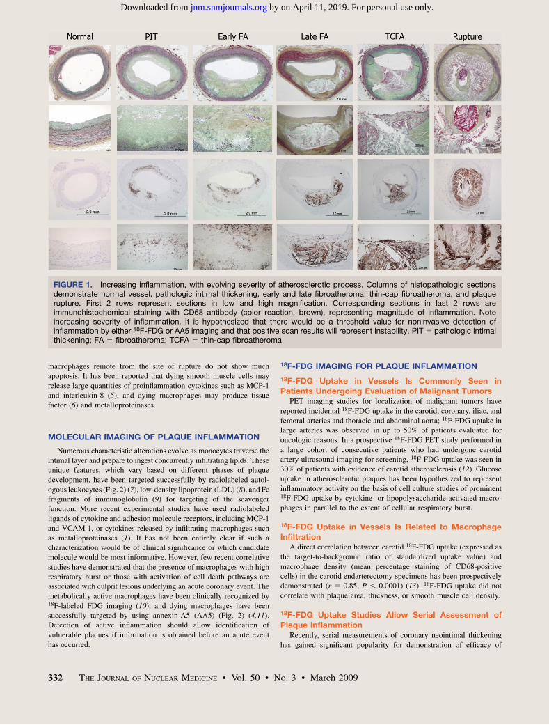
FIGURE 1. Increasing inflammation, with evolving severity of atherosclerotic process. Columns of histopathologic sectionsdemonstrate normal vessel, pathologic intimal thickening, early and late fibroatheroma, thin-cap fibroatheroma, and plaquerupture. First 2 rows represent sections in low and high magnification. Corresponding sections in last 2 rows areimmunohistochemical staining with CD68 antibody (color reaction, brown), representing magnitude of inflammation. Noteincreasing severity of inflammation. It is hypothesized that there would be a threshold value for noninvasive detection ofinflammation by either 18F-FDG or AA5 imaging and that positive scan results will represent instability. PIT 5 pathologic intimalthickening; FA 5 fibroatheroma; TCFA 5 thin-cap fibroatheroma.
332 THE JOURNAL OF NUCLEAR MEDICINE • Vol. 50 • No. 3 • March 2009
by on April 11, 2019. For personal use only. jnm.snmjournals.org Downloaded from
statin therapy and other agents likely to favorably affect atheroscle-
rosis (14,15). As such, if PET could also be used to monitor changesin plaque inflammation over time, a noninvasive tool would become
available as a surrogate marker of plaque instability and the efficacy ofpharmacologic interventions. Serial prospective 18F-FDG PET studies
have reported an excellent interobserver, intraobserver, and interscanreproducibility (14). The effect of statin intervention on 18F-FDG
uptake has been reported in many consecutive patients with carotidatherosclerosis (15). The follow-up PET scans revealed significant
reduction in 18F-FDG accumulation after statin therapy. In this study, onlydietary restrictions did not show resolution of vascular inflammation.
18F-FDG Imaging of Coronary Inflammation Is FeasibleAlthough various case reports and retrospective studies (16) have
demonstrated anecdotal 18F-FDG uptake in coronary arteries in
oncologic patients, a recent prospective 18F-FDG PET study withmultislice CT demonstrated the feasibility of precise 18F-FDG
localization in coronary arteries (Fig. 3) (17). In this elegant studydesign, myocardial 18F-FDG uptake was almost entirely suppressed by
a high-fat diet and restriction of carbohydrate meals for 1 d before thestudy and administration of b-blockers on the day of study. The
suppression of the myocardial background facilitated better targetdemarcation. The study also took advantage of CT angiography and
enrolled patients who had undergone coronary stent implantation foracute coronary syndrome or chronic stable angina. CT angiography
and stent location allowed precise coregistration of 18F-FDG uptake atthe plaque site. Culprit lesions demonstrated significantly higher 18F-
FDG uptake (Fig. 3) than did target lesions in chronic disease. 18F-FDG uptake was also prominently seen in some nonstented coronary
segments and also in the aortic root. Although it will be necessary todevelop measures to contain radiation burden imposed by combined
PET/CT studies, this study holds a promise of radical strategic shift incoronary artery disease management.
ANNEXIN IMAGING OF INFLAMED PLAQUES
The Principle and Basis of Cell Death ImagingBecause apoptotic cells express phosphatidylserine on their cell
surface and AA5 has a high affinity for binding to phosphatidylserine,imaging with 99mTc-labeled AA5 has been used to evaluate the
feasibility of the detection of unstable plaques. AA5 has beenextensively used previously for noninvasive imaging of experimental
atherosclerotic lesions (4), and its accumulation was predominantly
observed in American Heart Association–type IV lesions. There was adirect correlation of AA5 uptake with macrophage burden and the
magnitude of histologically verified apoptosis. It was subsequentlyindicated that pharmacologic intervention using stains and caspase
inhibitors could reduce the extent of apoptosis in experimentalatherosclerosis models (18,19). Studies of porcine atherosclerosis
have demonstrated the feasibility of coronary imaging with radiola-beled AA5 (20).
Annexin Uptake Is Correlated to the Extent of Cell Deathin Carotid Endarterectomy Specimens
99mTc-AA5 has been used in a small pilot study for imaging ofcarotid atherosclerosis in patients with recent or remote cerebrovas-
cular accidents (11); AA5 uptake was reported only after recentcerebrovascular accidents and not seen in patients being treated with
statins. AA5 binding was histologically localized to apoptoticmacrophages and also to the red blood cell membranes embedded
in necrotic cores. Radiolabeling of AA5 with PET-compatibleradiotracers such as 124I and 18F is under way and may provide
better avenues for coronary vascular imaging.
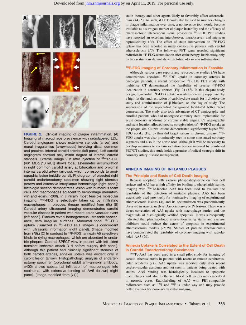
FIGURE 2. Clinical imaging of plaque inflammation. (A)Imaging of macrophage prevalence with radiolabeled LDL.Carotid angiogram shows extensive stenosis (arrow) andmural irregularities (arrowheads) involving distal commonand proximal internal carotid arteries (left panel). Left carotidangiogram showed only minor degree of internal carotidstenosis. External image 9 h after injection of 99mTc-LDL(481 MBq [13 mCi]) shows focal, asymmetric accumulationin right common carotid artery at bifurcation and proximalinternal carotid artery (arrows), which corresponds to angi-ographic lesion (middle panel). Photograph of bisected rightcarotid endarterectomy specimen showing tight stenosis(arrow) and extensive intraplaque hemorrhage (right panel);histologic section demonstrates lesion with numerous foamcells and macrophages adjacent to hemorrhage (hematox-ylin and eosin, ·200). In clinically most feasible molecularimaging, 18F-FDG is selectively taken up by infiltratingmacrophages in plaques. (Image modified from (8).) (B)Carotid artery ultrasound imaging demonstrates carotidvascular disease in patient with recent acute vascular event(left panel). Plaques reveal homogeneous ultrasonic appear-ance, with irregular surfaces. Abnormal focal 18F-FDGuptake visualized in 18F-FDG PET images is concordantwith ultrasonic information (right panel). (Image modifiedfrom (15).) (C) In contrast to 18F-FDG, annexin A5 selectivelybinds to dying macrophages, which are abundant in unsta-ble plaques. Coronal SPECT view in patient with left-sidedtransient ischemic attack 3 d before surgery (left panel).Although this patient had clinically significant stenosis ofboth carotid arteries, annexin uptake was evident only inculprit lesion (arrow). Histopathologic analysis of endarter-ectomy specimen (polyclonal rabbit anti–annexin antibody,·400) shows substantial infiltration of macrophages intoneointima, with extensive binding of AA5 (brown) (rightpanel). (Image modified from (11).)
MOLECULAR IMAGING OF PLAQUE INFLAMMATION • Tahara et al. 333
by on April 11, 2019. For personal use only. jnm.snmjournals.org Downloaded from
The likelihood that atherosclerotic plaques will result in acutevascular events is intimately associated with the morphologic traits of
the plaque and the extent of inflammation. A noninvasive strategydesigned to monitor the extent of plaque inflammation may allow
identification of unstable plaques, and serial interrogation may deter-
mine the efficacy of intervention. 18F-FDG uptake, which has beencommonly used in oncologic practice, offers information about plaque
inflammation and allows serial investigation. The feasibility of coronaryimaging with 18F-FDG has evoked tremendous enthusiasm in the
imaging community. Successful 18F-FDG imaging of coronary arterieshas also encouraged investigation with other promising molecules, such
as annexin. It is conceivable that the high-risk patients identified byclinical tools, including genetic information and biomarkers, will in the
future be more accurately risk-stratified by imaging targeted atmorphologic and functional characterization of high-risk plaques.
ACKNOWLEDGMENTS
This work was supported in part by National Institutes of Healthgrants RO1 HL68657 and RO1 HL078681 and research grants from
Mitsui Life Social Welfare Foundation, Japan Heart FoundationResearch, FUKUOKA clinical medicine of research prize, Interna-tional Research Fund for Subsidy of Kyusyu University School ofMedicine Alumni, and the Banyu Fellowship Program sponsored byBanyu Life Science Foundation International.
tomography imaging of atherosclerotic plaque inflammation is highly reproduc-
ible: implications for atherosclerosis therapy trials. J Am Coll Cardiol. 2007;
50:892–896.
15. Tahara N, Kai H, Ishibashi M, et al. Simvastatin attenuates plaque inflammation:
evaluation by fluorodeoxyglucose positron emission tomography. J Am Coll
Cardiol. 2006;48:1825–1831.
16. Alexanderson E, Slomka P, Cheng V, et al. Fusion of positron emission
tomography and coronary computed tomographic angiography identifies fluorine
18 fluorodeoxyglucose uptake in the left main coronary artery soft plaque. J Nucl
Cardiol. 2008;15:841–843.
17. Rogers IS, Figueroa AL, Nasir K, et al. Assessment of coronary segment
inflammation with combined 18-fluorodeoxyglucose positron emission tomog-
raphy and 64-slice multidetector computed tomography [abstract]. Circulation.
2007;116(suppl II):410.
18. Hartung D, Sarai M, Petrov A, et al. Resolution of apoptosis in atherosclerotic plaque
by dietary modification and statin therapy. J Nucl Med. 2005;46:2051–2056.
19. Sarai M, Hartung D, Petrov A, et al. Broad and specific caspase inhibitor-
induced acute repression of apoptosis in atherosclerotic lesions evaluated by
radiolabeled annexin A5 imaging. J Am Coll Cardiol. 2007;50:2305–2312.
20. Johnson LL, Schofield L, Donahay T, Narula N, Narula J. 99mTc-annexin V
imaging for in vivo detection of atherosclerotic lesions in porcine coronary
arteries. J Nucl Med. 2005;46:1186–1193.
FIGURE 3. 18F-FDG imaging of coronary inflammation. (A)Incidental 18F-FDG uptake is seen in left main coronaryartery region in 71-y-old patient undergoing PET for evalu-ation of recurrence of colon malignancy and metastaticdisease (modified from (16)) (left panel). This patient hadmultiple coronary risk factor; hence, CT angiography wasperformed that showed noncalcified plaque in left maincoronary and proximal left anterior descending artery (arrow)(middle panel). Corresponding image after fusion with 18F-FDG PET/CT localized inflammatory PET signal with max-imal standardized uptake value of 2.1 (arrow) (right panel).(B) On the other hand, prospective study has recentlydemonstrated potential feasibility of detecting inflammationin culprit plaque in patients presenting with acute coronarysyndrome. In 1 such patient who had undergone primarycoronary intervention, 18F-FDG imaging was performed.Radiotracer uptake is clearly visible (left) at site of coronarystent placement (right), suggesting that culprit lesion wasinflamed. 18F-FDG uptake in myocardium was suppressedby high-fat, low-carbohydrate diet and b-blocker adminis-tration. Stent sites in patients with chronic stable anginadid not show 18F-FDG uptake. MI 5 myocardial infarction.Figure 3B was provided by Ahmed Tawakol, MassachusettsGeneral Hospital, Boston, Massachusetts.
334 THE JOURNAL OF NUCLEAR MEDICINE • Vol. 50 • No. 3 • March 2009
by on April 11, 2019. For personal use only. jnm.snmjournals.org Downloaded from
Information about subscriptions to JNM can be found at:
http://jnm.snmjournals.org/site/misc/permission.xhtmlInformation about reproducing figures, tables, or other portions of this article can be found online at:
(Print ISSN: 0161-5505, Online ISSN: 2159-662X)1850 Samuel Morse Drive, Reston, VA 20190.SNMMI | Society of Nuclear Medicine and Molecular Imaging
is published monthly.The Journal of Nuclear Medicine